User login
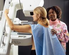
Screening Mammograms Overdiagnosed More Than 1 Million Women
Over the past 3 decades, screening mammography may have overdiagnosed more than a million clinically insignificant breast tumors, while having virtually no impact on the incidence of metastatic disease.
Compared with the premammography era, routine screening now picks up 122 additional cases of early cancer per 100,000 women – just eight of which would likely have progressed to distant disease, Dr. Archie Bleyer and Dr. Gilbert Welch wrote in the Nov. 22 issue of the New England Journal of Medicine.
Looking at the results in light of a corresponding 28% decrease in breast cancer mortality puts screening mammography in perspective, the authors said: "Our data show that the true contribution of mammography to decreasing mortality must be at the low end of this range. They suggest that mammography has largely not met the first prerequisite for screening to reduce cancer-specific mortality – a reduction in the number of women who present with late-stage cancer.
Population cancer screening is a doubled-edged sword, wrote Dr. Bleyer of Oregon Health and Science University in Portland, and Dr. Welch of Geisel School of Medicine at Dartmouth in Hanover, N.H. While it’s impossible to say which screen-detected cancers would have caused serious disease or death, "there is certainty about what happens to [these women]. They undergo surgery, radiation therapy, hormonal therapy for 5 years or more, chemotherapy, or (usually) a combination of these treatments for abnormalities that otherwise would not have caused illness."
The authors used the Surveillance , Epidemiology, and End Results (SEER) database to examine screening mammography and breast cancer incidence data from 1976-2008. They considered the incidence baseline to be the number of cancers reported from 1976-1978, and compared it with incidence in 2006-2008. All of the models in the study controlled for an upswing of breast cancer from 1990 to 2005, which was associated with hormone-replacement therapy.
Screening mammography increased from about 30% of women aged 40 or older in the mid-80s to almost 70% by 2008. This was mirrored by an increase in the diagnosis of early-stage breast cancers diagnosed, from 112/100,000 to 234/100,000 per year – representing an absolute increase of 122/100,000 (N. Engl. J. Med. 2012;367:1998-2005 [doi:10.1056/NEJMoa1206809]).

"[This] reflects both the detection of more cases of localized disease and the advent of the detection of [ductal carcinoma in situ] (which was virtually not detected before mammography was available)," the authors said.
There was a much smaller concomitant decrease in late-stage cancers, which fell from 102/100,000 to 94 /100,000 women. This was almost entirely driven by a drop in regional disease, from about 85/100,000 in 1976 to 78/100,000 in 2008. The incidence of distant disease was almost entirely unchanged, hovering around 17/100,000 for the entire study period.
"If a constant underlying disease burden is assumed, only 8 of the 122 additional early diagnoses were destined to progress to advanced disease, implying a detection of 114 excess cases per 100,000 women" – a total of more than 1.5 million over the study period.
The incidence of overdiagnosis held when the authors used other models designed to favor mammography’s impact.
In a "best-guess estimate," they assumed that breast cancer incidence increased by 0.25% over the study period. "This approach suggests that the excess detection attributable to mammography ... involved more than 1.3 million women in the past 30 years."
The "extreme" model assumed that incidence increased by 0.5% each year, an estimate that minimized surplus diagnoses of early-stage disease and maximized the deficit of late-stage cancer. This model found that screening mammography detected an excess of 1.2 million cancers over the study period.
The "very extreme assumption" model assumed that incidence increased by 0.5% each year, and that the baseline incidence of late-stage disease was the highest ever observed (113 cases per 100,000 women, in 1985). Even with this model – the most favorable toward mammography – the authors estimated overdiagnosis of slightly more than 1 million women.
"Our analysis suggests that whatever the mortality benefit, breast-cancer screening involved substantial harm of excess detection of additional early-stage cancers that was not matched by a reduction in late-stage cancers. This imbalance indicates a considerable amount of overdiagnosis involving more than 1 million women in the past 3 decades – and, according to our best-guess estimate, more than 70,000 women in 2008."
SEER breast cancer mortality data help put the findings into perspective, the authors said. There has been a substantial reduction in mortality, which fell from 71/100,000 to 51/100,000 over the study period. "This reduction in mortality is probably due to some combination of the effects of screening mammography and better treatment," they said. But because the absolute reduction in deaths (20/100,000) was larger than the absolute reduction in late-stage cancer (8/100,000), screening mammography can’t be the main driver of change.
"Furthermore ... the small reduction in cases of late-stage cancer that has occurred has been confined to regional (largely node-positive) disease – a stage that can now often be treated successfully, with an expected 5-year survival rate of 85% among women 40 years of age or older. Unfortunately, however, the number of women in the United States who present with distant disease, only 25% of whom survive for 5 years, appears not to have been affected by screening."
Better therapy may even have altered the impact of screening, they added.
"Ironically, improvements in treatment tend to deteriorate the benefit of screening. As treatment of [disease detected by methods other than screening] improves, the benefit of screening diminishes. For example, since pneumonia can be treated successfully, no one would suggest that we screen for pneumonia."
The findings show that there are not absolutes for women considering whether or not to get a mammogram.
"Proponents of screening should provide women with data from a randomized screening trial that reflects improvements in current therapy and includes strategies to mitigate overdiagnosis in the intervention group. Women should recognize that our study does not answer the question, ‘Should I be screened for breast cancer?’ However, they can rest assured that the question has more than one right answer."
Dr. Bleyer disclosed that he is a consultant and speaker for Sigma-Tau Pharmaceuticals. Dr. Welch disclosed that he speaks on the topic of overdiagnosis with universities and medical schools, as well as to the Pharmacy Benefit Management Institute; all honoraria were donated to charitable organizations.
This paper is going to create discussion and concern, and get people thinking again about mammography. But from my point of view it isn’t the final answer.
Dr. J. Leonard Lichtenfeld |

There’s been a substantial amount of research and commentary on the role of mammography – discussion that has split people into two political camps: one saying it’s the major reason for the reduced mortality and one saying it has no value whatsoever.
I think the truth lies somewhere in between.
Something has clearly affected breast cancer mortality in the past few decades. Before the 1990s, the rate of breast cancer death in this country was a flat line that had not changed for decades. Then suddenly it began to drop, and it has continued to do so. We are clearly doing something right. The question is: What is it? Probably a combination of mammography, improvements in adjuvant chemotherapy, and a general increase in breast awareness.
In the 1970s, when I was beginning practice – and before that, as a family member – breast cancer was not something anyone spoke about. It was never mentioned publicly. Now we are much more aware. Women are tuned in to the topic and aware of the need to do self-exams. It’s a national discussion.
Both our surgeries and our chemotherapy are much improved. But even now, if a woman presents with a palpable breast lesion, the odds are that it’s going to be fairly sizable and have lymph node involvement. Can we assume that we are able to effectively treat every woman in this group? I’m not sure we can.
This leads us to mammography. There is no question that it identifies subclinical lesions. But we have to recognize the problem of overdiagnosis and overtreatment.
Mammography and other new imaging modalities are driving the point of detection to much earlier in the history of a woman’s breast cancer. We have recognized from autopsy studies that certain cancers can exist in the body for long periods of time without ever causing any problems. We know that most women with breast cancers don’t have any readily identifiable risk factors. And we don’t have a test that allows us to tell most women whether or not their particular cancer is likely to be aggressive.
The current study doesn’t really help us with these questions. There are severe limitations on these data. How often did the women actually have a mammogram? What we consider "regional disease" today is not the same as it was in 1975. Advanced disease today isn’t the same as it was in 1975. There are cultural and insurance barriers that affect access to care and, thus, affect mortality. All of these issues must be weighed into the equation.
Right now, we at the American Cancer Society are still confident in our recommendation for women older than 40 to have an annual screening mammogram and clinical breast exam. There are other recommendations from other groups, which may be a better fit for other women. The important thing is for a woman and her physician to pick a program and stay with it.
Everyone wants clear-cut answers in a world that is not clear-cut and is unlikely to become so. So women and doctors are left in the difficult position of weighing what is best for them.
I think we would all be reluctant to completely give this up. A breast cancer diagnosis in the 40s can kill a woman in her 50s. I’m concerned that we will begin missing breast cancers now that will kill in 10-15 years, and that we will lose the gains we’ve seen in mortality.
Dr. J. Leonard Lichtenfeld is the deputy chief medical officer for the national office of the American Cancer Society.
This paper is going to create discussion and concern, and get people thinking again about mammography. But from my point of view it isn’t the final answer.
Dr. J. Leonard Lichtenfeld |
There’s been a substantial amount of research and commentary on the role of mammography – discussion that has split people into two political camps: one saying it’s the major reason for the reduced mortality and one saying it has no value whatsoever.
I think the truth lies somewhere in between.
Something has clearly affected breast cancer mortality in the past few decades. Before the 1990s, the rate of breast cancer death in this country was a flat line that had not changed for decades. Then suddenly it began to drop, and it has continued to do so. We are clearly doing something right. The question is: What is it? Probably a combination of mammography, improvements in adjuvant chemotherapy, and a general increase in breast awareness.
In the 1970s, when I was beginning practice – and before that, as a family member – breast cancer was not something anyone spoke about. It was never mentioned publicly. Now we are much more aware. Women are tuned in to the topic and aware of the need to do self-exams. It’s a national discussion.
Both our surgeries and our chemotherapy are much improved. But even now, if a woman presents with a palpable breast lesion, the odds are that it’s going to be fairly sizable and have lymph node involvement. Can we assume that we are able to effectively treat every woman in this group? I’m not sure we can.
This leads us to mammography. There is no question that it identifies subclinical lesions. But we have to recognize the problem of overdiagnosis and overtreatment.
Mammography and other new imaging modalities are driving the point of detection to much earlier in the history of a woman’s breast cancer. We have recognized from autopsy studies that certain cancers can exist in the body for long periods of time without ever causing any problems. We know that most women with breast cancers don’t have any readily identifiable risk factors. And we don’t have a test that allows us to tell most women whether or not their particular cancer is likely to be aggressive.
The current study doesn’t really help us with these questions. There are severe limitations on these data. How often did the women actually have a mammogram? What we consider "regional disease" today is not the same as it was in 1975. Advanced disease today isn’t the same as it was in 1975. There are cultural and insurance barriers that affect access to care and, thus, affect mortality. All of these issues must be weighed into the equation.
Right now, we at the American Cancer Society are still confident in our recommendation for women older than 40 to have an annual screening mammogram and clinical breast exam. There are other recommendations from other groups, which may be a better fit for other women. The important thing is for a woman and her physician to pick a program and stay with it.
Everyone wants clear-cut answers in a world that is not clear-cut and is unlikely to become so. So women and doctors are left in the difficult position of weighing what is best for them.
I think we would all be reluctant to completely give this up. A breast cancer diagnosis in the 40s can kill a woman in her 50s. I’m concerned that we will begin missing breast cancers now that will kill in 10-15 years, and that we will lose the gains we’ve seen in mortality.
Dr. J. Leonard Lichtenfeld is the deputy chief medical officer for the national office of the American Cancer Society.
Over the past 3 decades, screening mammography may have overdiagnosed more than a million clinically insignificant breast tumors, while having virtually no impact on the incidence of metastatic disease.
Compared with the premammography era, routine screening now picks up 122 additional cases of early cancer per 100,000 women – just eight of which would likely have progressed to distant disease, Dr. Archie Bleyer and Dr. Gilbert Welch wrote in the Nov. 22 issue of the New England Journal of Medicine.
Looking at the results in light of a corresponding 28% decrease in breast cancer mortality puts screening mammography in perspective, the authors said: "Our data show that the true contribution of mammography to decreasing mortality must be at the low end of this range. They suggest that mammography has largely not met the first prerequisite for screening to reduce cancer-specific mortality – a reduction in the number of women who present with late-stage cancer.
Population cancer screening is a doubled-edged sword, wrote Dr. Bleyer of Oregon Health and Science University in Portland, and Dr. Welch of Geisel School of Medicine at Dartmouth in Hanover, N.H. While it’s impossible to say which screen-detected cancers would have caused serious disease or death, "there is certainty about what happens to [these women]. They undergo surgery, radiation therapy, hormonal therapy for 5 years or more, chemotherapy, or (usually) a combination of these treatments for abnormalities that otherwise would not have caused illness."
The authors used the Surveillance , Epidemiology, and End Results (SEER) database to examine screening mammography and breast cancer incidence data from 1976-2008. They considered the incidence baseline to be the number of cancers reported from 1976-1978, and compared it with incidence in 2006-2008. All of the models in the study controlled for an upswing of breast cancer from 1990 to 2005, which was associated with hormone-replacement therapy.
Screening mammography increased from about 30% of women aged 40 or older in the mid-80s to almost 70% by 2008. This was mirrored by an increase in the diagnosis of early-stage breast cancers diagnosed, from 112/100,000 to 234/100,000 per year – representing an absolute increase of 122/100,000 (N. Engl. J. Med. 2012;367:1998-2005 [doi:10.1056/NEJMoa1206809]).
"[This] reflects both the detection of more cases of localized disease and the advent of the detection of [ductal carcinoma in situ] (which was virtually not detected before mammography was available)," the authors said.
There was a much smaller concomitant decrease in late-stage cancers, which fell from 102/100,000 to 94 /100,000 women. This was almost entirely driven by a drop in regional disease, from about 85/100,000 in 1976 to 78/100,000 in 2008. The incidence of distant disease was almost entirely unchanged, hovering around 17/100,000 for the entire study period.
"If a constant underlying disease burden is assumed, only 8 of the 122 additional early diagnoses were destined to progress to advanced disease, implying a detection of 114 excess cases per 100,000 women" – a total of more than 1.5 million over the study period.
The incidence of overdiagnosis held when the authors used other models designed to favor mammography’s impact.
In a "best-guess estimate," they assumed that breast cancer incidence increased by 0.25% over the study period. "This approach suggests that the excess detection attributable to mammography ... involved more than 1.3 million women in the past 30 years."
The "extreme" model assumed that incidence increased by 0.5% each year, an estimate that minimized surplus diagnoses of early-stage disease and maximized the deficit of late-stage cancer. This model found that screening mammography detected an excess of 1.2 million cancers over the study period.
The "very extreme assumption" model assumed that incidence increased by 0.5% each year, and that the baseline incidence of late-stage disease was the highest ever observed (113 cases per 100,000 women, in 1985). Even with this model – the most favorable toward mammography – the authors estimated overdiagnosis of slightly more than 1 million women.
"Our analysis suggests that whatever the mortality benefit, breast-cancer screening involved substantial harm of excess detection of additional early-stage cancers that was not matched by a reduction in late-stage cancers. This imbalance indicates a considerable amount of overdiagnosis involving more than 1 million women in the past 3 decades – and, according to our best-guess estimate, more than 70,000 women in 2008."
SEER breast cancer mortality data help put the findings into perspective, the authors said. There has been a substantial reduction in mortality, which fell from 71/100,000 to 51/100,000 over the study period. "This reduction in mortality is probably due to some combination of the effects of screening mammography and better treatment," they said. But because the absolute reduction in deaths (20/100,000) was larger than the absolute reduction in late-stage cancer (8/100,000), screening mammography can’t be the main driver of change.
"Furthermore ... the small reduction in cases of late-stage cancer that has occurred has been confined to regional (largely node-positive) disease – a stage that can now often be treated successfully, with an expected 5-year survival rate of 85% among women 40 years of age or older. Unfortunately, however, the number of women in the United States who present with distant disease, only 25% of whom survive for 5 years, appears not to have been affected by screening."
Better therapy may even have altered the impact of screening, they added.
"Ironically, improvements in treatment tend to deteriorate the benefit of screening. As treatment of [disease detected by methods other than screening] improves, the benefit of screening diminishes. For example, since pneumonia can be treated successfully, no one would suggest that we screen for pneumonia."
The findings show that there are not absolutes for women considering whether or not to get a mammogram.
"Proponents of screening should provide women with data from a randomized screening trial that reflects improvements in current therapy and includes strategies to mitigate overdiagnosis in the intervention group. Women should recognize that our study does not answer the question, ‘Should I be screened for breast cancer?’ However, they can rest assured that the question has more than one right answer."
Dr. Bleyer disclosed that he is a consultant and speaker for Sigma-Tau Pharmaceuticals. Dr. Welch disclosed that he speaks on the topic of overdiagnosis with universities and medical schools, as well as to the Pharmacy Benefit Management Institute; all honoraria were donated to charitable organizations.
FROM THE NEW ENGLAND JOURNAL OF MEDICINE
Major Finding: Compared with the premammography era, routine screening now picks up 122 additional cases of early cancer per 100,000 women – just eight of which would likely have progressed to distant disease.
Data Source: The authors extracted data from the Surveillance, Epidemiology, and End Results (SEER) database.
Disclosures: Dr. Bleyer disclosed that he is a consultant and speaker for Sigma-Tau Pharmaceuticals. Dr. Welch disclosed that he speaks on the topic of overdiagnosis with universities and medical schools, as well as to the Pharmacy Benefit.

Cardiac Toxicity Not Seen 25 Years after Breast Radiation
BOSTON – Here’s heartening news that physicians can convey to breast cancer survivors: Modern breast irradiation did not appear to cause late-term cardiac toxicity in a study that examined women a quarter of a century after they were treated.
Investigators found no significant differences in Framingham Heart Study risk scores, hemodynamic parameters, pericardial thickening, or heart failure among 50 women who had been randomized in the 1970s and 1980s to either mastectomy or breast-conserving surgery (BCS) and radiation, Dr. Charles B. Simone II reported at the annual meeting of the American Society for Radiation Oncology.

Although the survival rate was slightly lower among patients treated with breast-conserving therapy, the difference does not appear to be related to radiation dose to the heart, said Dr. Simone of the Hospital of the University of Pennsylvania in Philadelphia. There were no differences in survival among women treated with BCS and radiation to left- or right-sided tumors.
"Based on this study, in the era of 3D planning, patients with early-stage breast cancer treated with radiotherapy do not have a higher risk of long-term cardiac morbidity compared with patients having mastectomy," he said.
The patients studied included 50 of 102 survivors from an original cohort of 237 women who had participated in the National Cancer Institute’s Breast Conservation Trial (79-C-0111), with randomization from 1979 to 1986. In that trial, women with stage I or II breast cancer received modified radical mastectomy with axillary node dissection or they underwent lumpectomy plus node dissection and a radiation dose of 45-50.4 Gy to the whole breast; the latter came with or without treatment of regional nodes, followed by a boost of 15-20 Gy with either iridium 192 brachytherapy or electrons.
All node-positive patients underwent 6-11 cycles of chemotherapy with doxorubicin and cyclophosphamide, and beginning in 1985 postmenopausal women with positive nodes were given tamoxifen.
The trial was unique at the time in that it used CT simulations for treatment planning and dose inhomogeneity corrections, Dr. Simone noted.
Diverging Survival Curves
At a median of 25.7 years after randomization, 43.8% of mastectomy patients were still alive, compared with 37.9% of BCS patients. Although the difference was not significant, it appeared to represent a divergence of survival curves that had been virtually identical for the first 25 years.
The trend could not be accounted for by secondary malignancies, changes in distant metastasis, or any other breast cancer–related causes, leading the investigators to question whether it might be due to radiation toxicity to the heart, as some studies have suggested.
In all, 26 patients who had had BCS and 24 who underwent mastectomy agreed to come back for the cardiac toxicity study.
The investigators took a detailed cardiac history, and subjected the women to exams, cardiac labs, cardiac MRI with a 3 Tesla magnet to look for anatomic and functional abnormalities, and CT angiogram to look for stenotic coronary disease and determine coronary arterial calcium score (CAC) of atherosclerotic burden.
They also looked at radiation technique parameters such as central lung distance, field size, dose, and boost dose.
On cardiac MRI, they only saw two significant between-group differences. Time to peak filling rate was shorter for BCS patients (487 milliseconds vs. 647 ms for mastectomy patients; P = .02), but there was no difference in the peak filling rate itself. Left ventricular mass was smaller for BCS patients (mean 90.5 gm vs. 111 gm for mastectomy patients), but this difference was no longer significant after adjustment for systolic blood pressure, Dr. Simone noted.
"Interestingly, we didn’t see any evidence of myocardial fibrosis in any patient assessed, and only one patient in each arm had any degree of pericardial thickening," he said.
Reassuring Data
Additionally, investigators saw no significant differences on CT angiography in the presence of visible plaque or significant or severe vascular stenosis. There were also no differences in plaque or stenosis in the left anterior descending arteries of women treated with radiation for tumors on the left or the right side of the body.
"For each and every vessel we looked at, there was no difference in the degree of stenosis," Dr. Simone said.

Median CAC scores were low and in the normal range, but patients who had received chemotherapy had a trend toward increased atherosclerosis and plaque formation, Dr. Simone noted.
The study "gives some reassurance to our patients that, after 25 years of follow-up, using modern radiation techniques the delivery of radiation to the left does not cause cardiac toxicity," Dr. Bruce Haffty, chair of radiation oncology at the Cancer Institute of New Jersey, New Brunswick, said at a briefing.
The study was supported by the National Cancer Institute. Dr. Simone and Dr. Haffty reported no relevant disclosures.
BOSTON – Here’s heartening news that physicians can convey to breast cancer survivors: Modern breast irradiation did not appear to cause late-term cardiac toxicity in a study that examined women a quarter of a century after they were treated.
Investigators found no significant differences in Framingham Heart Study risk scores, hemodynamic parameters, pericardial thickening, or heart failure among 50 women who had been randomized in the 1970s and 1980s to either mastectomy or breast-conserving surgery (BCS) and radiation, Dr. Charles B. Simone II reported at the annual meeting of the American Society for Radiation Oncology.
Although the survival rate was slightly lower among patients treated with breast-conserving therapy, the difference does not appear to be related to radiation dose to the heart, said Dr. Simone of the Hospital of the University of Pennsylvania in Philadelphia. There were no differences in survival among women treated with BCS and radiation to left- or right-sided tumors.
"Based on this study, in the era of 3D planning, patients with early-stage breast cancer treated with radiotherapy do not have a higher risk of long-term cardiac morbidity compared with patients having mastectomy," he said.
The patients studied included 50 of 102 survivors from an original cohort of 237 women who had participated in the National Cancer Institute’s Breast Conservation Trial (79-C-0111), with randomization from 1979 to 1986. In that trial, women with stage I or II breast cancer received modified radical mastectomy with axillary node dissection or they underwent lumpectomy plus node dissection and a radiation dose of 45-50.4 Gy to the whole breast; the latter came with or without treatment of regional nodes, followed by a boost of 15-20 Gy with either iridium 192 brachytherapy or electrons.
All node-positive patients underwent 6-11 cycles of chemotherapy with doxorubicin and cyclophosphamide, and beginning in 1985 postmenopausal women with positive nodes were given tamoxifen.
The trial was unique at the time in that it used CT simulations for treatment planning and dose inhomogeneity corrections, Dr. Simone noted.
Diverging Survival Curves
At a median of 25.7 years after randomization, 43.8% of mastectomy patients were still alive, compared with 37.9% of BCS patients. Although the difference was not significant, it appeared to represent a divergence of survival curves that had been virtually identical for the first 25 years.
The trend could not be accounted for by secondary malignancies, changes in distant metastasis, or any other breast cancer–related causes, leading the investigators to question whether it might be due to radiation toxicity to the heart, as some studies have suggested.
In all, 26 patients who had had BCS and 24 who underwent mastectomy agreed to come back for the cardiac toxicity study.
The investigators took a detailed cardiac history, and subjected the women to exams, cardiac labs, cardiac MRI with a 3 Tesla magnet to look for anatomic and functional abnormalities, and CT angiogram to look for stenotic coronary disease and determine coronary arterial calcium score (CAC) of atherosclerotic burden.
They also looked at radiation technique parameters such as central lung distance, field size, dose, and boost dose.
On cardiac MRI, they only saw two significant between-group differences. Time to peak filling rate was shorter for BCS patients (487 milliseconds vs. 647 ms for mastectomy patients; P = .02), but there was no difference in the peak filling rate itself. Left ventricular mass was smaller for BCS patients (mean 90.5 gm vs. 111 gm for mastectomy patients), but this difference was no longer significant after adjustment for systolic blood pressure, Dr. Simone noted.
"Interestingly, we didn’t see any evidence of myocardial fibrosis in any patient assessed, and only one patient in each arm had any degree of pericardial thickening," he said.
Reassuring Data
Additionally, investigators saw no significant differences on CT angiography in the presence of visible plaque or significant or severe vascular stenosis. There were also no differences in plaque or stenosis in the left anterior descending arteries of women treated with radiation for tumors on the left or the right side of the body.
"For each and every vessel we looked at, there was no difference in the degree of stenosis," Dr. Simone said.
Median CAC scores were low and in the normal range, but patients who had received chemotherapy had a trend toward increased atherosclerosis and plaque formation, Dr. Simone noted.
The study "gives some reassurance to our patients that, after 25 years of follow-up, using modern radiation techniques the delivery of radiation to the left does not cause cardiac toxicity," Dr. Bruce Haffty, chair of radiation oncology at the Cancer Institute of New Jersey, New Brunswick, said at a briefing.
The study was supported by the National Cancer Institute. Dr. Simone and Dr. Haffty reported no relevant disclosures.
AT THE ANNUAL MEETING OF THE AMERICAN SOCIETY FOR RADIATION ONCOLOGY
Major Finding: There were no significant differences in major cardiac function parameters between women treated with modified radical mastectomy or breast-conserving surgery with radiation after a median 25.7 years of follow-up
Data Source: Investigators examined 50 women who had been randomized in the 1970s and 1980s to mastectomy or breast-conserving surgery and radiation.
Disclosures: The study was supported by the National Cancer Institute. Dr. Simone and Dr. Haffty reported no relevant disclosures.

Management of Zollinger-Ellison May Depend on Presence of Tumors
Patients with Zollinger-Ellison syndrome who also have multiple endocrine neoplasia type 1 usually follow an indolent clinical course that rarely results in disease-related death, unlike those who have the sporadic form of the syndrome, Dr. Maneesh H. Singh and his colleagues reported in the November issue of Clinical Gastroenterology and Hepatology.
This finding, from a retrospective study of 49 patients treated at a single tertiary care center since 1994, argues against surgery for the estimated 25%-50% of Zollinger-Ellison syndrome (ZES) patients who have multiple endocrine neoplasia type 1 (MEN-1). "Given these results, we support a conservative approach to disease management ... focusing on symptom control with pharmacologic agents," the investigators wrote (Clin. Gastroenterol. Hepatol. 2012 Aug. 20 [doi:10.1016/j.cgh.2012.08.014]).
Early and aggressive surgery is recommended for sporadic ZES because it improves survival and can be curative. But surgery’s role in those with MEN-1 has been contentious because it doesn’t appear to improve survival in these patients. Only the most radical surgery, which carries a 40% complication rate, appears to be curative in those with MEN-1,reported Dr. Singh and his colleagues at the Hospital of the University of Pennsylvania, Philadelphia.
Because ZES is such a rare disorder, this follow-up study of 34 ZES patients who underwent surgery and 15 who did not "represents one of the largest long-term studies of surgical outcomes from a tertiary care hospital" conducted to date, they wrote.
The study subjects’ mean age at diagnosis was 47 years. The mean duration of follow-up from the time of diagnosis was 7 years, ranging from 0 to 5 years for 19 patients, from 5 to 10 years for 10, from 10 to 20 years for 15, and for more than 20 years for 5.
Of the 15 patients who did not undergo surgery, 5 declined after a discussion of the risks and benefits of the procedure, 2 because they had no lesions greater than 2 cm in diameter in imaging studies, 3 because of extensive liver involvement that was deemed unresectable, and 5 because they had unrelated comorbidities that made them poor surgical candidates.
A total of 33 subjects had sporadic ZES, while the other 16 had associated MEN-1. In the latter group, there was no significant difference between the median survival for the nine who underwent surgery (22.4 years) and for the seven who did not (25.5 years).
Standard gastrinoma resection with duodenotomy did not achieve a cure in any of the ZES patients with accompanying MEN-1.
In contrast, surgery improved both disease-related and all-cause mortality in the sporadic form of the disorder, and surgery was deemed curative in 6 (32%) of the patients with sporadic ZES.
None of the patients with MEN-1died of progressive ZES, compared with 28 (85%) of the patients with sporadic ZES. Thus, the form of the disease associated with MEN-1 appears to have a much more benign course than the sporadic form, Dr. Singh and his associates reported.
ZES associated with MEN-1 also tends to have an earlier symptom onset than does sporadic ZES, but because it is more indolent, the mean age at death was nearly identical between the two groups.
In this study, ZES patients with MEN-1had rates of liver involvement at diagnosis and rates of later liver metastases that were similar to those of patients with sporadic ZES. But again, this did not reduce their survival as it did with sporadic ZES.
This finding suggests that there may be a fundamental difference in the basic tumor biology between the two forms of the disease. It supports the theory that sporadic ZES is "a rapidly progressive, malignant form of gastrinoma that defies prediction and advocates for swift surgical intervention," the researchers wrote.
They reported no industry support for this study and no other financial conflicts.
FROM CLINICAL GASTROENTEROLOGY AND HEPATOLOGY
CCR7 Predicts Cervical Metastasis in Oral Cancer
WASHINGTON – Chemokine receptor CCR7 expression is a significant predictor of cervical metastases in patients with squamous cell carcinoma in the oral cavity, based on data from 60 adults.
Metastatic spread of squamous cell carcinoma (SCC) is common, but the mechanisms behind the spread remain unclear, said Dr. Levi G. Ledgerwood of the University of California, Davis. "There has been a great deal of work that has looked at lymphocyte entry into lymphatics," he said at the annual meeting of the American Academy of Otolaryngology-Head and Neck Surgery Foundation.

Recent research has focused on the chemokine receptor CCR7, a cell-surface molecule that is required for T-cell entry from the bloodstream and peripheral tissues into lymphatics, he noted. Data from previous studies suggest that CCR7 might play a role in various cancers in the metastases of the lymph nodes.
Dr. Ledgerwood and his colleagues reviewed tissue samples from primary tumors in 60 oral SCC patients who underwent surgical resection at a single center between 2006 and 2011. The study included 30 samples from patients with metastases and 30 samples from patients without metastases. There were no significant demographic differences between the groups, although each group had more male than female patients, Dr. Ledgerwood noted. A total of 30 patients were node positive, and 30 were node negative.
Overall, patients with cervical metastases showed significantly higher CCR7 expression than those without cervical metastases (P less than .001). A total of 97% of node-positive patients were positive for CCR7 expression, but only 43% of patients without cervical metastases were positive for CCR7.

When the lymph nodes of the samples from metastatic cancer patients were examined, all 30 node-positive patients showed expression of CCR7, Dr. Ledgerwood added.
Although the study was limited by its small size, the results suggest a possible role for CCR7 in T-cell access to lymphatics, said Dr. Ledgerwood.
"This is a preliminary study, but we feel that this receptor could provide a very interesting target for future drug therapies and could also help in predicting the behavior of tumors," he said.
Dr. Ledgerwood had no financial conflicts to disclose.
WASHINGTON – Chemokine receptor CCR7 expression is a significant predictor of cervical metastases in patients with squamous cell carcinoma in the oral cavity, based on data from 60 adults.
Metastatic spread of squamous cell carcinoma (SCC) is common, but the mechanisms behind the spread remain unclear, said Dr. Levi G. Ledgerwood of the University of California, Davis. "There has been a great deal of work that has looked at lymphocyte entry into lymphatics," he said at the annual meeting of the American Academy of Otolaryngology-Head and Neck Surgery Foundation.
Recent research has focused on the chemokine receptor CCR7, a cell-surface molecule that is required for T-cell entry from the bloodstream and peripheral tissues into lymphatics, he noted. Data from previous studies suggest that CCR7 might play a role in various cancers in the metastases of the lymph nodes.
Dr. Ledgerwood and his colleagues reviewed tissue samples from primary tumors in 60 oral SCC patients who underwent surgical resection at a single center between 2006 and 2011. The study included 30 samples from patients with metastases and 30 samples from patients without metastases. There were no significant demographic differences between the groups, although each group had more male than female patients, Dr. Ledgerwood noted. A total of 30 patients were node positive, and 30 were node negative.
Overall, patients with cervical metastases showed significantly higher CCR7 expression than those without cervical metastases (P less than .001). A total of 97% of node-positive patients were positive for CCR7 expression, but only 43% of patients without cervical metastases were positive for CCR7.
When the lymph nodes of the samples from metastatic cancer patients were examined, all 30 node-positive patients showed expression of CCR7, Dr. Ledgerwood added.
Although the study was limited by its small size, the results suggest a possible role for CCR7 in T-cell access to lymphatics, said Dr. Ledgerwood.
"This is a preliminary study, but we feel that this receptor could provide a very interesting target for future drug therapies and could also help in predicting the behavior of tumors," he said.
Dr. Ledgerwood had no financial conflicts to disclose.
AT THE ANNUAL MEETING OF THE AMERICAN ACADEMY OF OTOLARYNGOLOGY-HEAD AND NECK SURGERY FOUNDATION
DNA Alone Inadequate to Identify HPV-Related Cancers
Testing for the presence of human papillomavirus DNA alone, especially using polymerase chain reaction methods, is not adequate to identify which head and neck squamous cell carcinomas are caused by the virus, according to two studies published online Sept. 18 in Cancer Research.
Identifying HPV-driven malignancies is important because they respond better to treatment and have better outcomes than those unrelated to HPV infection. Indeed, treatment of head and neck squamous cell carcinoma (HNSCC) may soon be guided by the tumor’s HPV status, since trials are now underway to determine whether de-escalation of chemo- and radiotherapy is safe and effective in such patients.
At present, however, the biomarkers that are best suited to making this identification are unclear.
Case Series Assesses Biomarkers
In the first study, researchers assessed the usefulness of four biomarkers in determining which HNSCCs in a case series were driven by HPV. They began by examining fresh-frozen tumor biopsy samples from 199 German adults diagnosed as having oropharyngeal squamous cell cancer between 1990 and 2008.
The four biomarkers were HPV-16 viral load, viral oncogene RNA (E6 and E7), p16INK4a, and RNA patterns similar to those characteristic of cervical carcinomas (CxCa RNA), said Dr. Dana Holzinger of the German Cancer Research Center at Heidelberg (Germany) University and her associates.
The simple presence of HPV DNA in a tumor sample was found to be a poor indicator of prognosis, likely because it often signaled past HPV infections or recent oral exposure, rather than active HPV infection that progressed to malignancy, the investigators said (Cancer Res. 2012 Sept. 18 [doi: 10.1158/0008-5472.CAN-11-3934]).
Instead, "we showed that high viral load and a cancer-specific pattern of viral gene expression are most suited to identify patients with HPV-driven tumors among patients with oropharyngeal cancer. Viral expression pattern is a completely new marker in this field, and viral load has hardly been analyzed before," Dr. Holzinger said in a press statement accompanying the publication of these findings.
"Once standardized assays for these markers, applicable in routine clinical laboratories, are established, they will allow precise identification" of cancers that are or are not HPV-driven, which will in turn influence prognosis and treatment, she added.
Results Back Combination Approach
In the second study, Dr. Caihua Liang of Brown University, Providence, R.I., and her associates examined 488 HNSCC samples as well as serum samples collected in a population-based study in the Boston area during 1999-2003.
As in the first study, these investigators found that the mere presence of HPV-16 DNA in these tumors, particularly when detected by PCR analysis, did not accurately predict overall survival or progression-free survival.
Instead, "our study strongly suggests that the combination of detection of HPV-16 DNA in HNSCC tumors [plus] p16 immunostaining with E6/E7 antibodies represents the most clinically valuable surrogate marker for the identification of patients . . . who have a better prognosis," they said (Cancer Res. 2012 Sept. 28 [doi: 10.1158/0008-5472.CAN-11-3277]).
"Assessment of HPV DNA using polymerase chain reaction methods as a biomarker in individual head and neck cancers is a poor predictor of outcome, and is also poorly associated with antibody response indicative of exposure and/or infection by HPV," senior author Dr. Karl T. Kelsey added in the press statement.
"We may not be diagnosing these tumors as accurately and precisely as we need to for adjusting treatments," said Dr. Kelsey, a professor in the department of epidemiology and the department of pathology and laboratory medicine at Brown University.
Dr. Holzinger’s study was funded in part by the European Commission, BMBG/HGAF-Canceropole Grand-Est, and the German Research Foundation. Her associates reported ties to Qiagen and Roche. Dr. Liang’s study was supported by the National Institutes of Health and the Flight Attendant Medical Research Institute, and one associate reported ties to Bristol-Myers Squibb.
Both of these studies demonstrate that the HPV DNA status of head and neck squamous cell carcinomas should be interpreted with caution, said Dr. Eduardo Mendez.
"Further testing to confirm HPV active infection may be warranted, particularly in consideration of de-escalation regimens," he said. In addition, other prognostic factors should be taken into account, such as tumor classification and lymph node status.
Dr. Mendez is at the University of Washington/Fred Hutchinson Cancer Research Center, Seattle. He reported ties to Intuitive Surgical. These remarks were taken from his commentary accompanying Dr. Holzinger’s and Dr. Liang’s reports (Cancer Res. 2012 Sept. 18 [doi: 10.1158/0008-5472.CAN-12-3285]).
Testing for the presence of human papillomavirus DNA alone, especially using polymerase chain reaction methods, is not adequate to identify which head and neck squamous cell carcinomas are caused by the virus, according to two studies published online Sept. 18 in Cancer Research.
Identifying HPV-driven malignancies is important because they respond better to treatment and have better outcomes than those unrelated to HPV infection. Indeed, treatment of head and neck squamous cell carcinoma (HNSCC) may soon be guided by the tumor’s HPV status, since trials are now underway to determine whether de-escalation of chemo- and radiotherapy is safe and effective in such patients.
At present, however, the biomarkers that are best suited to making this identification are unclear.
Case Series Assesses Biomarkers
In the first study, researchers assessed the usefulness of four biomarkers in determining which HNSCCs in a case series were driven by HPV. They began by examining fresh-frozen tumor biopsy samples from 199 German adults diagnosed as having oropharyngeal squamous cell cancer between 1990 and 2008.
The four biomarkers were HPV-16 viral load, viral oncogene RNA (E6 and E7), p16INK4a, and RNA patterns similar to those characteristic of cervical carcinomas (CxCa RNA), said Dr. Dana Holzinger of the German Cancer Research Center at Heidelberg (Germany) University and her associates.
The simple presence of HPV DNA in a tumor sample was found to be a poor indicator of prognosis, likely because it often signaled past HPV infections or recent oral exposure, rather than active HPV infection that progressed to malignancy, the investigators said (Cancer Res. 2012 Sept. 18 [doi: 10.1158/0008-5472.CAN-11-3934]).
Instead, "we showed that high viral load and a cancer-specific pattern of viral gene expression are most suited to identify patients with HPV-driven tumors among patients with oropharyngeal cancer. Viral expression pattern is a completely new marker in this field, and viral load has hardly been analyzed before," Dr. Holzinger said in a press statement accompanying the publication of these findings.
"Once standardized assays for these markers, applicable in routine clinical laboratories, are established, they will allow precise identification" of cancers that are or are not HPV-driven, which will in turn influence prognosis and treatment, she added.
Results Back Combination Approach
In the second study, Dr. Caihua Liang of Brown University, Providence, R.I., and her associates examined 488 HNSCC samples as well as serum samples collected in a population-based study in the Boston area during 1999-2003.
As in the first study, these investigators found that the mere presence of HPV-16 DNA in these tumors, particularly when detected by PCR analysis, did not accurately predict overall survival or progression-free survival.
Instead, "our study strongly suggests that the combination of detection of HPV-16 DNA in HNSCC tumors [plus] p16 immunostaining with E6/E7 antibodies represents the most clinically valuable surrogate marker for the identification of patients . . . who have a better prognosis," they said (Cancer Res. 2012 Sept. 28 [doi: 10.1158/0008-5472.CAN-11-3277]).
"Assessment of HPV DNA using polymerase chain reaction methods as a biomarker in individual head and neck cancers is a poor predictor of outcome, and is also poorly associated with antibody response indicative of exposure and/or infection by HPV," senior author Dr. Karl T. Kelsey added in the press statement.
"We may not be diagnosing these tumors as accurately and precisely as we need to for adjusting treatments," said Dr. Kelsey, a professor in the department of epidemiology and the department of pathology and laboratory medicine at Brown University.
Dr. Holzinger’s study was funded in part by the European Commission, BMBG/HGAF-Canceropole Grand-Est, and the German Research Foundation. Her associates reported ties to Qiagen and Roche. Dr. Liang’s study was supported by the National Institutes of Health and the Flight Attendant Medical Research Institute, and one associate reported ties to Bristol-Myers Squibb.
FROM CANCER RESEARCH
Major Finding: The simple presence of HPV DNA in tumor samples did not accurately identify which cancers were driven by active HPV infection, and thus did not predict which would show the greatest response to treatment.
Data Source: Two analyses were used to assess the prognostic accuracy of various biomarkers in tumor samples from adults with head and neck squamous cell carcinoma.
Disclosures: Dr. Holzinger’s study was funded in part by the European Commission, BMBG/HGAF-Canceropole Grand-Est, and the German Research Foundation. Her associates reported ties to Qiagen and Roche. Dr. Liang’s study was supported by the National Institutes of Health and the Flight Attendant Medical Research Institute, and one associate reported ties to Bristol-Myers Squibb.
Ablation Cost Effective for High-Grade Dysplasia in Barrett's Esophagus
Radiofrequency ablation for high-grade dysplasia in the setting of Barrett’s esophagus is more cost effective than endoscopic surveillance until progression to cancer, reported Dr. Chin Hur and his colleagues in the September issue of Gastroenterology.
Moreover, the use of radiofrequency ablation (RFA) for stable, confirmed low-grade dysplasia can also be cost effective, said the investigators.
Dr. Hur of Harvard University and Massachusetts General Hospital, both in Boston, and his colleagues conducted several analyses comparing three treatment strategies for each of three disease states: Barrett’s esophagus with high-grade dysplasia, Barrett’s esophagus with low-grade dysplasia, and Barrett’s esophagus with no dysplasia.
The treatment strategies consisted of surveillance followed by esophagectomy upon disease progression to cancer, RFA upon disease progression to a higher-grade dysplasia or cancer, or initial RFA before disease progression (Gastroenterology 2012 [doi: 10.1053/j.gastro.2012.05.010]).
Cost calculations were based on Medicare reimbursement rates for 2011. The cost estimate for RFA, for example, was $6,400, and the cost of esophagectomy was $25,882. Based on these values, the authors then conducted a base-case analysis.
They found that in high-grade dysplasia, the strategy of initial RFA resulted in 0.704 more quality-adjusted life-years (QALYs) and cost $25,609 less than the strategy of surveillance without RFA followed by esophagectomy upon disease progression to cancer, assuming a 1% disease progression rate.
Similarly, in low-grade dysplasia, RFA upon disease progression to high-grade dysplasia also bested the strategy of surveillance until cancer and esophagectomy, resulting in 0.17 more QALYs and costing $7,446 less, assuming a 0.5% progression rate.
However, in low-grade dysplasia, when comparing initial RFA versus surveillance until progression to high-grade dysplasia and then RFA, the authors found that the latter approach cost only $1,969 less than the former, and that the former was associated with a 0.108 gain in QALYs.
That amounted to an incremental cost-effectiveness ratio of $18,231 per QALY – "below our willingness-to-pay threshold of $100,000/QALY, making it the most plausible strategy in terms of cost-effectiveness."
Finally, in scenarios involving Barrett’s esophagus but no dysplasia, RFA upon progression of disease to high-grade dysplasia was still the most cost-effective strategy: It was associated with a savings of $7,709 as well as 0.194 additional QALYs, compared with esophagectomy upon disease progression to cancer, assuming a 0.33% progression rate.
The authors noted that their model relies on a stringent definition of low-grade dysplasia that assumes "review and agreement between more than one expert pathologist" as well as a consistent level of dysplasia found on more than one endoscopy spaced at least 6 months apart. In addition, the progression rates used in this analysis were based on the current literature, but were still estimates involving some uncertainty.
"We believe that a multicenter, randomized, controlled trial for initial RF ablation versus surveillance in patients with BE without dysplasia is needed to confirm our model results and to inform clinical decision making," they added.
Long-term follow-up data from such a study could "provide much needed data regarding cancer progression and the need for surveillance, which significantly impacts the cost-effectiveness and patients’ preferences for RFA."
The authors stated that they had no disclosures relevant to this study. The study was supported by grants from the National Institutes of Health.
FROM GASTROENTEROLOGY
Robotic Device for Gastric Neoplasia Found Safe in Five Patients
A novel robotic system designed to perform endoscopic submucosal dissection of early gastric neoplasia was safe and effective in a five-patient trial, reported Soo Jay Phee, Ph.D., and colleagues in Clinical Gastroenterology and Hepatology.
Indeed, the first human study of the robotic device showed that it achieved clear margins, no cases of major bleeding or perforation, and discharge within hours for several of the patients.
Dr. Phee of the School of Mechanical and Aerospace Engineering at Nanyang Technological University in Singapore and first author Dr. D. Nageshwar Reddy of the Asian Institute of Gastroenterology in Somajiguda, India, enrolled five patients from two centers in India and one in Hong Kong.
Only patients with gastric neoplasia limited to the mucosa, confirmed by biopsy and histopathology, were included.
All patients underwent endoscopic submucosal dissection with the assistance of a novel device called the Master and Slave Transluminal Endoscopic Robot (MASTER). The MASTER device has been described previously (Gastrointest. Endoscopy 2010;72:593-9).
According to the authors, the device is controlled by two operators, "one responsible for the steering of the endoscope while the other would be performing the submucosal dissection with the two robotic arms."
The investigators added, "The open edge of the mucosa with the tumor was grasped by one of the robotic arms to retract the mucosa and enhance exposure of the submucosa, while submucosal dissection was completed with the other L hook arm" (Clin. Gastroenterol. Hepatol. 2012 October [doi:10.1016/j.cgh.2012.05.019]).
All procedures were performed under general anesthesia and with ventilation by naso- or orotracheal intubation. All operators trained on porcine models prior to the study.
In the case of the first patient, a 41-year-old man with a 2-cm adenocarcinoma in the body of the stomach, "the submucosal dissection with the robotic system was successfully done in 19 minutes," reported the authors.
There was no bleeding for perforation, and histopathology of the specimen after retrieval showed intramucosal well-differentiated adenocarcinoma with clear resection margins. The patient was discharged after 12 hours.
The second case, a 60-year-old man, was found to have a 1.5-cm mucosal adenocarcinoma in the gastric antrum. "The submucosal dissection was completed in only 5 minutes," wrote the authors, with no complications, and clear margins on histopathology. The patient was discharged in 6 hours.
Similarly, the third patient (a 39-year-old man) had a 2-cm sessile lesion in the gastric antrum. Submucosal dissection with the MASTER was completed in 3 minutes, with no complications, clear margins, and discharge from the hospital 4 hours later.
The longest procedure was done in a 51-year-old woman with a 3-cm early gastric cancer; this procedure took 50 minutes. She had no major bleeding or perforation, had clear margins on histopathology, and had a 3-day hospital stay.
Finally, a 50-year-old man with a 2.5-cm sessile polypoid in the inferior wall of the prepyloric canal underwent a 16-minute dissection. "Severe bleeding was encountered during the procedure and required exchange of the MASTER-mounted endoscope for a waterjet endoscope to stop the bleeding with coagulation grapser," wrote the authors.
"The pathology showed hyperplastic polyp with clear margins. He was discharged from hospital on day 3 after the procedure."
At 30 days’ follow-up, no complications were reported by any of the patients, and the follow-up endoscopy showed "complete healing" of the resection site. At 6 months, the three patients with data available were "doing well," according to the authors.
The study was supported by a MedTech Seeding Fund from the National University of Singapore. The authors reported having no other relevant financial disclosures.
FROM CLINICAL GASTROENTEROLOGY AND HEPATOLOGY
Major Finding: Five patients who underwent endoscopic submucosal dissection of early-stage gastric neoplasia with the use of a novel robotic system had good 30-day outcomes and clear resection margins.
Data Source: The findings came from five case studies in India and Hong Kong.
Disclosures: The study was supported by a MedTech Seeding Fund from the National University of Singapore. The authors reported having no other relevant financial disclosures.
Transfusions Worsen Outcomes After Colorectal Cancer Surgery
The risks of death, postoperative infection, and other adverse clinical outcomes were significantly increased among patients undergoing colorectal cancer surgery who received perioperative allogeneic blood transfusions, according to a meta-analysis of 55 studies published in the August issue of Annals of Surgery.
Dr. Austin G. Acheson of the Nottingham (England) Digestive Disease Center at Queen’s Medical Centre and his associates reviewed 12 prospective studies and 43 retrospective cohort studies published between December 2004 and October 2010. The studies included 20,795 patients who were followed for a mean of about 5 years after undergoing surgery for colorectal cancer. Almost 60% (12,242) of these patients received a mean of three units of allogeneic red blood cells (Ann. Surg. 2012;256:235-44).
Patients who were transfused tended to be older, and the transfusion rate was significantly higher in women, those undergoing rectal surgery (compared with those undergoing right or left colon surgery), those with greater surgical blood loss, and those with worsening Duke's stage.
All associations between ABT and the adverse clinical outcomes described below were statistically significant.
The rates of all-cause mortality in the 29 studies that looked at this outcome were 45% among the patients who received blood transfusions vs. 35% of those who did not, a significant difference that represented a 72% increased risk. After adjusting for the duration of the observation period, the investigators found that the annual incidence of all-cause mortality was almost 9% among the transfused patients vs. 6.5% among those who were not transfused.
The rate of cancer-related mortality was 31% vs. 24% in the 17 studies that measured this outcome, a 71% increased risk. After adjusting for the length of observation, the investigators said the annual incidence of cancer-related mortality was 5.4% of those transfused and 4% of those who were not.
The rate of the combined end point of death resulting from recurrence/metastasis was 43% of those who were transfused vs. 33% of those who were not transfused – a 66% increase in risk – in the 19 studies that measured this outcome.
The risk of postoperative infections in the 12 studies that measured this outcome was 29% among those who were transfused vs. 11% of those who were not transfused, which was more than a threefold increased risk. In the two studies that measured the need for surgical reintervention, the risk was increased fourfold among those who were transfused.
In the four studies that measured the length of hospitalization, the hospital stay was a mean of almost 18 days among those who received transfusions, compared with 14 days among those who did not.
Based on evidence in this meta-analysis and other studies showing that preoperative anemia is an independent risk factor for a worse prognosis after colon surgery, and based on the association between ABTs and poorer clinical outcomes in this meta-analysis, the authors wrote that "appropriate blood management measures should, therefore, be given an important place in the care of patients with CRC [colorectal cancer] undergoing elective surgery."
The use of ABT has dropped over the past 25 years because of improvements in patient care, and the authors stated that they believe efforts should be made to further minimize ABT use. Well-designed studies are needed "to determine whether and in which patients preoperative corrective measures of anemia, other than ABTs, will contribute not only in the field of colorectal surgery but also to improve clinical outcomes," they added.
The study was supported by an unrestricted research grant from Vifor Pharma AG, a specialty pharmaceuticals company focused on the treatment of iron deficiency, according to its website. The authors disclosed receiving honoraria and/or travel support for consulting or lecturing for companies that include Vifor, Ethicon Endosurgery, Johnson & Johnson, and AstraZeneca AG; their research departments received grant support from companies that included Vifor.
The meta-analysis by Dr. Acheson and his colleagues and another recent publication using the American College of Surgeons National Surgical Quality Improvement Program (Surgery 2012;152:344-54) highlight concerns about the increased mortality associated with perioperative blood transfusions. Intraoperative transfusion is associated with higher rates of 30-day operative mortality, major complications, total number of complications, and prolonged length of stay.
In general, patients requiring transfusions tend to be older, more
anemic and debilitated, have more extensive disease, and often need
larger surgical procedures with attendant greater blood loss. Patients
with these characteristics should be relatively evenly distributed among hospitals performing colorectal cancer surgery, and one would expect the frequency of transfusion to be similar across institutions.
However, Dr. Acheson and his associates found that the transfusion rate varied from 17.4% to 82.2%, suggesting that the indications for transfusion were quite
variable. Furthermore, the National Surgical Quality Improvement Program noted that more than 60% of patients received only one to two units of blood. This
raises concerns about whether blood transfusions are being given appropriately.
Some of the steps can be taken to decrease the use of perioperative blood transfusions and prevent transfusion-related morbidity are:
• Correcting anemia prior to
surgery by giving iron with or with
out erythropoietin;
• Using blood conservation methods when extensive surgery is
planned;
• Educating anesthesiologists and
surgeons about the indications for
transfusion (there is no rigid “trans
fusion trigger”; each patient must be
assessed for the need to augment the
oxygen-carrying capacity of the
blood);
• Avoiding prospective donors alloimmunized to white cell antigens
and leuko-reduce blood products
(Blood 2009;113:3406-17);
• Avoiding the use of blood that has
undergone prolonged storage (new
blood is better than old) (N. Engl. J.
Med. 2008;20:358:1229-39; Blood
2011;118:6675-82); and
• Recognizing that acute illness after perioperative transfusions might be due to transfusion-transmitted infections or transfusion-associated lung injury (TRALI) (Blood 2012;119:1757-67).
Perioperative allogeneic blood transfusions can be life-saving, but must be used judiciously and with the recognition that they occasionally increase morbidity and mortality.
DAVID GREEN, M.D., PH.D., is in the
division of hematology/oncology in
the department of medicine at
Northwestern University Feinberg
School of Medicine, Chicago. He has
no relevant disclosures.
The risks of death, postoperative infection, and other adverse clinical outcomes were significantly increased among patients undergoing colorectal cancer surgery who received perioperative allogeneic blood transfusions, according to a meta-analysis of 55 studies published in the August issue of Annals of Surgery.
Dr. Austin G. Acheson of the Nottingham (England) Digestive Disease Center at Queen’s Medical Centre and his associates reviewed 12 prospective studies and 43 retrospective cohort studies published between December 2004 and October 2010. The studies included 20,795 patients who were followed for a mean of about 5 years after undergoing surgery for colorectal cancer. Almost 60% (12,242) of these patients received a mean of three units of allogeneic red blood cells (Ann. Surg. 2012;256:235-44).
Patients who were transfused tended to be older, and the transfusion rate was significantly higher in women, those undergoing rectal surgery (compared with those undergoing right or left colon surgery), those with greater surgical blood loss, and those with worsening Duke's stage.
All associations between ABT and the adverse clinical outcomes described below were statistically significant.
The rates of all-cause mortality in the 29 studies that looked at this outcome were 45% among the patients who received blood transfusions vs. 35% of those who did not, a significant difference that represented a 72% increased risk. After adjusting for the duration of the observation period, the investigators found that the annual incidence of all-cause mortality was almost 9% among the transfused patients vs. 6.5% among those who were not transfused.
The rate of cancer-related mortality was 31% vs. 24% in the 17 studies that measured this outcome, a 71% increased risk. After adjusting for the length of observation, the investigators said the annual incidence of cancer-related mortality was 5.4% of those transfused and 4% of those who were not.
The rate of the combined end point of death resulting from recurrence/metastasis was 43% of those who were transfused vs. 33% of those who were not transfused – a 66% increase in risk – in the 19 studies that measured this outcome.
The risk of postoperative infections in the 12 studies that measured this outcome was 29% among those who were transfused vs. 11% of those who were not transfused, which was more than a threefold increased risk. In the two studies that measured the need for surgical reintervention, the risk was increased fourfold among those who were transfused.
In the four studies that measured the length of hospitalization, the hospital stay was a mean of almost 18 days among those who received transfusions, compared with 14 days among those who did not.
Based on evidence in this meta-analysis and other studies showing that preoperative anemia is an independent risk factor for a worse prognosis after colon surgery, and based on the association between ABTs and poorer clinical outcomes in this meta-analysis, the authors wrote that "appropriate blood management measures should, therefore, be given an important place in the care of patients with CRC [colorectal cancer] undergoing elective surgery."
The use of ABT has dropped over the past 25 years because of improvements in patient care, and the authors stated that they believe efforts should be made to further minimize ABT use. Well-designed studies are needed "to determine whether and in which patients preoperative corrective measures of anemia, other than ABTs, will contribute not only in the field of colorectal surgery but also to improve clinical outcomes," they added.
The study was supported by an unrestricted research grant from Vifor Pharma AG, a specialty pharmaceuticals company focused on the treatment of iron deficiency, according to its website. The authors disclosed receiving honoraria and/or travel support for consulting or lecturing for companies that include Vifor, Ethicon Endosurgery, Johnson & Johnson, and AstraZeneca AG; their research departments received grant support from companies that included Vifor.
FROM THE ANNALS OF SURGERY
Major Finding: Adverse clinical outcomes including mortality were significantly increased among patients who had an allogeneic blood transfusion around the time of undergoing colorectal cancer surgery, compared with those who were not transfused.
Data Source: A meta-analysis of 55 mostly retrospective cohort studies evaluated mortality and other clinical outcomes associated with the use of allogeneic red blood cell transfusions in almost 21,000 patients undergoing colorectal cancer surgery, 60% of whom were transfused.
Disclosures: The study was supported by an unrestricted research grant from Vifor Pharma AG. The authors disclosed receiving honoraria and/or travel support for consulting or lecturing from companies that include Vifor, Ethicon Endosurgery, Johnson & Johnson, and AstraZeneca AG; their research departments received grant support from companies including Vifor.
Labs Find Evidence of Cancer Stem Cells
In an era of targeted cancer therapies, laboratory scientists working with mice may have found the ultimate target – a reservoir of stem cells that drive cancers to grow and metastasize.
Separate reports in the journals Science and Nature document the presence of cancer stem cells in intestinal adenomas (Science 2012 Aug. 1 [doi:10.1126/science.1224676]), squamous skin cancer, (Nature 2012 Aug. 1 [doi:10.1038/nature11344]), and glioblastoma multiforme (Nature 2012 Aug. 1 [doi:10.1038/nature11287]).
In the last study, mice with these highly lethal brain tumors were given temozolomide (Temodar), an approved treatment in humans, along with ganciclovir, an antiviral. Despite a transient therapeutic response to chemotherapy, the cancers continued to grow, driven by "a relatively quiescent subset of endogenous glioma cells, with properties similar to those proposed for cancer stem cells," the authors wrote.
Whether these reports will resolve controversy over the existence of stem cells or lead to clinically meaningful treatments remains to be seen. There is no doubt, however, that they will lead to further investigation.
Renal Cell Carcinoma Research Ongoing
Many renal cancers can now be managed in a minimally invasive fashion with either laparoscopic or robotic-assisted surgery. Potentially of greatest importance is the elucidation of the biological basis of sporadic as well as hereditary forms of renal carcinomas.
Most renal tumors arise from loss of function of the von Hippel-Lindau (VHL) gene and activation of the hypoxic response, including upregulation of hypoxia inducible factor leading to vascular endothelial growth factor induction and ultimately angiogenesis. In addition to the VHL syndrome, other hereditary forms include hereditary papillary renal carcinoma, Birt-Hogg-Dubé, and hereditary leiomyomatosis-renal cell cancer.
Surgical resection of localized renal cell carcinoma can be curative for lower-stage disease, but patients with advanced or metastatic disease are rarely cured by surgery alone. Traditional chemotherapy has had poor response rates and systemic options have been focused on immunotherapy with cytokines, such as interleukin-2 and interferon alfa. Hence, new targeted agents being tested in the neoadjuvant and adjuvant settings have created some excitement.
Seven novel targeted agents approved by the Food and Drug Administration are available for patients with metastatic disease: the tyrosine kinase inhibitors sorafenib, sunitinib, pazopanib, and axitinib; mTOR inhibitors temsirolimus and everolimus; and VEGF-inhibiting monoclonal antibody bevacizumab. The role of these therapies in either the neoadjuvant or adjuvant setting is being investigated.
The Adjuvant Sorafenib or Sunitinib in Unfavorable Renal Cell Carcinoma (ASSURE; ECOG 2805) trial has enrolled 1,865 patients randomized to one year of sunitinib, sorafenib, or placebo therapy after surgical excision of the primary tumor. The current standard of care, even for patients with high-risk pathologic features, is surveillance after surgical procedures when no evidence of residual disease can be found. Thus, the study is designed to determine whether adjuvant targeted therapy improves cancer-specific survival and to demonstrate a 25 percent reduction in the hazard rate of disease-free survival events.
The EVErolimus for Renal Cancer Ensuing Surgical Therapy (EVEREST) study (SWOG 0931), similar in design to ASSURE, examines the benefit of adjuvant systemic therapy after surgical procedures in patients with intermediate high-risk or very high-risk disease. Although sorafenib, sunitinib, and everolimus are all used clinically, sorafenib and sunitinib are tyrosine kinase inhibitors, whereas everolimus is an mTOR inhibitor. An estimated 1,218 patients will be randomized to either everolimus or placebo, stratified by pathologic stage, histologic subtype, and performance status.
Similar ongoing adjuvant trials include the Pfizer-sponsored S-TRAC trial (n=720) comparing sunitinib with placebo and the Medical Research Council SORCE trial (n=1,656) comparing sorafenib with placebo. Other agents are also being tested in the adjuvant setting, including pazopanib (PROTECT), as well as those that exploit the purported immunogenicity of renal cell carcinoma.
An accrued phase III trial has tested the antibody girentuximab, which binds specifically to carbonic anhydrase IX (G250 antigen) that is expressed on the cell surface of clear renal cell carcinomas. Future trials may extend the potential role of cell-based immunotherapy from patients with metastatic disease to the adjuvant setting.
Dr. Meng is associate professor of urology, department of urology, University of California, San Francisco, and director of the fellowship in urologic oncology.
Dr. Nelson is Fred C. Andersen Professor of Surgery and chair, division of surgery research, Mayo Clinic College of Medicine, Rochester, MN, and Program Director of the Alliance/American College of Surgeons Clinical Research Program.