User login
Do not miss cannabis use in gastroparesis patients
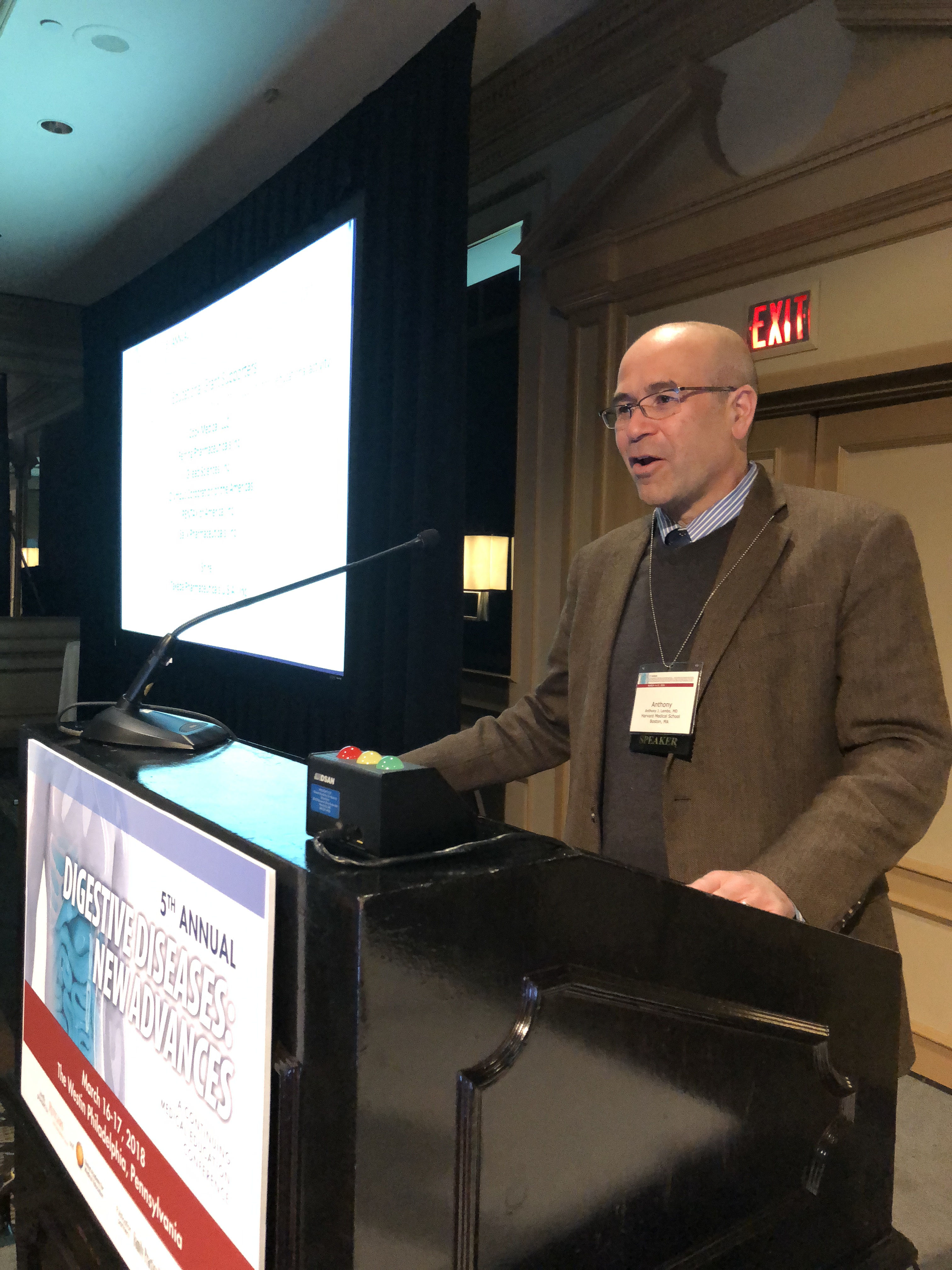
PHILADELPHIA – When evaluating potential causes of gastroparesis, cannabis use is a “do not miss” diagnosis that is easy to overlook and likely on the rise, according to Anthony J. Lembo, MD.
“This is not an infrequent problem, and I’ve even missed it a couple of times,” said Dr. Lembo, director of the GI Motility Laboratory at Beth Israel Deaconess Medical Center, Boston.
The rate of U.S. emergency department visits for vomiting with cannabis use disorder rose from 2.3 to 13.3 per 100,000 visits from 2006 to 2013, according to an analysis recently published by Dr. Lembo and colleagues (J Clin Gastroenterol. 2017 Oct 31. doi: 10.1097/MCG.0000000000000944).
The study showed that men between 20 and 29 years were the most common group presenting for vomiting with cannabis use disorder.
“Remember, 90% of people with chronic gastroparesis are women, so a young male is a red flag for cannabinoid use, whether or not you’ve got the right history,” Dr. Lembo told attendees at the meeting, jointly provided by Rutgers and Global Academy for Medical Education.
Dr. Lembo recounted an example from his own practice where a young male patient with recurrent nausea and vomiting denied cannabis use in the presence of family members.
“It was only after we managed to hospitalize him because he was losing so much weight that he came out and talked to one of the residents that he was an actually a daily pot smoker,” Dr. Lembo said. “Once we stopped it, the symptoms went away.”
Clinicians in states where cannabis use is increasing might need to be particularly alert for cannabis-related issues. According to the study by Dr. Lembo, the Midwest and West regions registered higher rates of vomiting with cannabis use disorder, compared with the Northeast and South.
Whether cannabinoids also can be a treatment for nausea or vomiting is a frequently asked question, Dr. Lembo said.
While there are no data for smoked marijuana, Dr. Lembo said, .
Dronabinol is indicated for adults for the treatment of chemotherapy-associated nausea and vomiting in patients who don’t respond adequately to conventional antiemetics, according to the agent’s prescribing information.
The cannabinoid medication is an isomer of tetrahydrocannabinol (THC), one of the active compounds in marijuana, according to Dr. Lembo.
“If you smoke marijuana, the levels go up high very quickly,” Dr. Lembo said. “If you take dronabinol, it takes 45 minutes to an hour. It’s a slower rise of it, so people are less likely to abuse dronabinol.”
In his talk, Dr. Lembo reported disclosures related to Allergan, Ironwood Pharmaceuticals, Salix Pharmaceuticals, and Takeda Pharmaceuticals.
Global Academy for Medical Education and this news organization are owned by the same company.
REPORTING FROM DIGESTIVE DISEASES: NEW ADVANCES
Dose-escalated radiation therapy brings mixed results in prostate cancer
Dose-escalated radiation therapy reduced the need for subsequent therapy in patients with intermediate-risk prostate cancer but did not improve overall survival, according to results of a large, randomized clinical trial.
The absence of a survival benefit compared with standard radiation therapy was seen despite a reduction in rates of both biochemical failure and distant metastases, Jeff M. Michalski, MD, and his associates reported in JAMA Oncology.
Negative overall survival results may be attributable to the growing availability of systemic salvage therapies that have prolonged the natural history of this disease, said Dr. Michalski, MD, of the department of radiation oncology at Washington University in St. Louis.
“Patients experiencing a biochemical or clinical failure may go on to receive several life-prolonging systemic agents, which may negate any clinical advantage from a more effective primary local therapy,” Dr. Michalski and associates wrote.
Results of the study support that hypothesis. Patients who received the standard dose radiotherapy were significantly more likely to go on to salvage therapy, compared with patients in the dose-escalation arm.
Dr. Michalski and his associated reported on the randomized NRG Oncology/RTOG 0126 clinical trial, which included 1,532 patients with intermediate-risk cancer enrolled between March 2002 and August 2008 at one of 104 North American sites.
In the trial, patients with intermediate-risk prostate cancer were randomized to receive three-dimensional conformal radiation therapy or intensity-modulated radiation therapy to 79.2 Gy in 44 fractions or 70.2 Gy in 39 fractions, the study said.
With a median follow-up of 8.4 years for 1,499 patients, there was no difference in overall survival between arms, study results show. The 8-year rates of overall survival were 76% for dose-escalated radiotherapy and 75% for standard radiotherapy (hazard ratio, 1.00; 95% confidence interval, 0.83-1.20; P = .98).
Otherwise, patients in the dose-escalated radiotherapy arm had significantly lower rates of distant metastases (4% vs. 6%; P = 0.05), and lower rates of biochemical failure rates at both 5 and 8 years, investigators said.
Patients in the high-dose arm less often went on to salvage therapy; however, investigators noted that they also had more toxic effects. compared with patients in the standard radiotherapy arm.
“,” Dr. Michalski and his associates wrote.
Dr. Michalski reported no conflicts of interest. The co-authors reported several conflicts of interest, including ties to ViewRay, Augmenix, Sanofi, AstraZeneca, AbbVie, and other companies.
SOURCE: Michalsky Jeff M et al. JAMA Oncol. 2018 Mar 15. doi: 10.1001/jamaoncol.2018.0039.
FROM JAMA ONCOLOGY
Key clinical point: Despite reducing the need for subsequent therapy, dose-escalated radiation therapy for intermediate-risk prostate cancer did not improve overall survival.
Major finding: Eight-year overall survival rates were 76% for dose-escalated radiation therapy, compared with 75% for standard radiation therapy (P = 0.98).
Study details: The randomized NRG Oncology/RTOG 0126 clinical trial including 1,532 patients enrolled between March 2002 and August 2008 at one of 104 North American sites.
Disclosures: Dr. Michalsky reported no conflicts of interest. The study authors disclosed conflicts tied to several companies, including ViewRay, Augmenix, Sanofi, AstraZeneca, and AbbVie.
Source: Michalsky Jeff M et al. JAMA Oncol. 2018 Mar 15. doi: 10.1001/jamaoncol.2018.0039.
Axitinib/avelumab combo shows preliminary efficacy in RCC
The combination of axitinib and avelumab had manageable toxicity and demonstrated preliminary efficacy as a front-line treatment of advanced renal cell carcinoma (RCC), according to results of a recent study.
More than half of patients had a response on the combination of axitinib (Inlyta), a receptor inhibitor of vascular endothelial growth factor (VEGF), and avelumab (Bavencio), an anti-PD-L1 monoclonal antibody, investigators reported in The Lancet Oncology.
The most common treatment-related adverse event was hypertension, wrote lead author Toni K. Choueiri, MD, of the Lank Center for Genitourinary Oncology at the Dana-Farber/Brigham and Women’s Cancer Center in Boston, and his colleagues.
“The combination of avelumab and axitinib in treatment-naive patients with advanced RCC had a manageable safety profile consistent with the profiles of the individual agents when administered as monotherapy, and antitumor activity was encouraging,” said Dr. Choueiri, and his colleagues.
The study, known as JAVELIN Renal 100, was a phase 1b investigation that included 55 patients with advanced clear-cell RCC. A total of 6 patients were enrolled in a smaller dose-finding cohort, and 49 were enrolled in a dose-expansion cohort.
In the dose-finding phase, patients received oral axitinib 5 mg twice a day for 7 days, at which point they started intravenous avelumab 10 mg/kg every 2 weeks, according to the study description. In the dose-expansion phase, patients were directly started on the combination.
Out of 55 patients enrolled, 26 (53%) had a response on the axitinib-avelumab combination, including 3 (6%) who had complete responses, investigators reported.
Grade 3 or greater treatment-related adverse events were reported in 32 patients (58%). The most common of those was hypertension, occurring in 16 patients (29%), followed by ALT increase and palmar-plantar erythrodysesthesia syndrome in 4 patients each (7%), Dr. Choueiri and colleagues wrote.
In the dose-finding phase, investigators reported one dose-limiting toxicity, which was grade 3 proteinuria due to axitinib.
. In that randomized trial, the axitinib-avelumab combination is being compared to sunitinib as a first-line approach in patients with advanced RCC.
“The combination of an antibody that inhibits PD-L1 and PD-1 interactions with a targeted antiangiogenic agent might take advantage of complementary mechanisms of action to provide clinical benefit in patients with advanced renal-cell carcinoma that exceeds the effects of the respective drugs alone without increasing associated toxicity,” Dr. Choueiri and colleagues wrote.
Pfizer and Merck funded the study. Dr. Choueiri declared interests related to several companies, including AstraZeneca, Bristol-Myers Squibb, Eisai, Exelixis, GlaxoSmithKline, Merck, Novartis, Pfizer, Peloton, and Roche/Genentech.
SOURCE: Choueiri TK et al. Lancet Oncol. 2018 Mar 9. doi: 10.1016/S1470-2045(18)30107-4.
FROM THE LANCET ONCOLOGY
Key clinical point: The targeted-immune combination of axitinib and avelumab had manageable toxicity and encouraging preliminary efficacy as a front-line treatment of advanced renal cell carcinoma (RCC).
Major finding: Out of 55 patients enrolled, 26 (53%) had a response, including 3 (6%) who had complete responses.
Study details: A dose-expansion and dose-finding phase 1b study including 55 patients with advanced clear-cell RCC.
Disclosures: Funding for the study came from Pfizer and Merck. Study authors declared interests related to several companies, including Pfizer, Merck, Bristol-Myers Squibb, Novartis, Roche/Genentech.
Source: Choueiri TK et al. Lancet Oncol. 2018 Mar 9. doi: 10.1016/S1470-2045(18)30107-4.
views
The antitumor activity of axitinib and avelumab in this study indicate the potential clinical benefit of targeted-immune combinations for advanced renal cell carcinoma (RCC), according to Viktor Grünwald, MD, PhD.
“Further studies are warranted to explore whether a first-line targeted-immune combination might overcome the standard of sequential targeted and immune therapies,” Dr. Grünwald said in an editorial.
The current absence of long-term safety data for the combination approach is limiting in terms of making definitive conclusions about the toxicity of axitinib and avelumab as a first line combination therapy for advanced clear-cell RCC, Dr. Grünwald wrote.
Even so, axitinib has a “low” incidence of hepatic toxicity, making it a “preferable” agent to evaluate in targeted-immune combination trials such as JAVELIN Renal 100, he added.
Objective responses in JAVELIN Renal 100 were seen in 32 out of 55 patients (58%), of which 3 patients (6%) were complete responses, according to reported data.
Similar findings previously reported for the immune-immune combination of ipilimumab and nivolumab versus sunitinib. In that study, patients receiving the immune-immune combination had an overall response rate of 42% and complete response rate of 9%, while the group patients receiving sunitinib had overall and complete response rates of 27% and 1%, respectively, he said.
“With the dawn of immune-immune and targeted-immune combinations, for the first time in a decade, major progress towards improving the median overall survival and possibly delivering cure to some of our patients with metastatic renal-cell carcinoma seems to have been made,” Dr. Grünwald said.
Viktor Grünwald, MD, PhD, is with the department of hematology, haemostasis, oncology, and stem cell transplantation at Hannover Medical School, Germany. These comments are based on an editorial accompanying the report (Lancet Oncol. 2018 Mar 9. doi: 10.1016/S1470-2045[18]30126-8). Dr. Grünwald disclosed ties to Bristol-Myers Squibb, Merck Sharp and Dohme, Ipsen, Novartis, Roche, AstraZeneca, Pfizer, Cerulean, Eisai, and EUSA Pharma.
In myeloma, third ASCT is a viable option
A third autologous stem cell transplantation (ASCT) is feasible and provides clinical benefit to patients with relapsed multiple myeloma, according to findings from a retrospective study.
The benefits appear to be most pronounced in patients who had a long duration of response to the previous ASCT, the researchers wrote in Biology of Blood and Marrow Transplantation.
“A salvage third ASCT is of value for patients with relapsed multiple myeloma,” Laurent Garderet, MD, of the department of hematology, Hôpital Saint Antoine, Paris, and coauthors wrote in the report.
A third transplantation is most commonly used in patients who relapse following tandem ASCT. Less often, it is done in patients who receive upfront ASCT, relapse, undergo a second ASCT, and relapse again.
“The first scenario gives much better results, due in part to a better remission status at the third ASCT with no signs of increased [second primary malignancy],” the researchers wrote.
In that group, median overall survival was greater than 5 years if the relapse occurred 3 years or more after the initial tandem ASCT, study results show.
The retrospective analysis, based on European Society for Blood and Marrow Transplantation data, included 570 patients who had undergone a third ASCT between 1997 and 2010. Of that group, 482 patients (81%) received the third transplantation after tandem ASCT and subsequent relapse, and 88 (15%) received it after second relapse.
After third ASCT, overall survival was 33 months in the larger tandem transplant group with 61 months of follow-up, and 15 months in the smaller group of patients who received two salvage ASCTs after 48 months of follow-up.
Median progression-free survival was 13 and 8 months for the tandem ASCT and two-salvage–ASCT groups, respectively, while 100-day nonrelapse mortality was 4% and 7%, respectively.
For both groups, better outcomes were associated with longer duration of remission after the second ASCT, the researchers reported.
Moreover, the time from second ASCT to relapse was the only favorable prognostic factor associated with survival after third ASCT in a multivariate analysis of the patients who relapsed following tandem transplant. The hazard ratio for relapse occurring between 18 and 36 months vs. within 18 months was 0.62 (95% confidence interval, 0.47-0.82; P = .01); for relapse after 36 months, the HR was 0.35 (95% CI, 0.25-0.49; P less than .001).
The researchers acknowledged that, beyond transplant, treatment of myeloma has changed substantially in recent years and could change the clinical picture for patients undergoing a third ASCT.
“The availability of novel agents may further improve the response to a third ASCT, rather than impairing its usefulness in the salvage setting, by enhancing the depth of response before ASCT, which could result in improved durability of the outcome,” they wrote.
The researchers reported having no financial disclosures related to this study.
SOURCE: Garderet L et al. Biol Blood Marrow Transplant. 2018 Feb 3. doi: 10.1016/j.bbmt.2018.01.035.
FROM BIOLOGY OF BLOOD AND MARROW TRANSPLANTATION
Key clinical point:
Major finding: Relapse-free interval was a favorable prognostic factor and significantly correlated with overall survival (P less than .001) in patients who underwent a third ASCT.
Study details: A retrospective analysis of European Society for Blood and Marrow Transplantation data including 570 patients who had undergone a third ASCT between 1997 and 2010.
Disclosures: The study authors reported having no financial disclosures related to the study.
Source: Garderet L et al. Biol Blood Marrow Transplant. 2018 Feb 3. doi: 10.1016/j.bbmt.2018.01.035.
Excessive daytime sleepiness linked to increase in Alzheimer’s biomarker
, results of a prospective, longitudinal cohort study suggest.
In the study, excessive daytime sleepiness (EDS) was associated with increased accumulation of beta-amyloid, an important biomarker of AD that manifests in early preclinical stages, wrote first author Diego Z. Carvalho, MD, and his colleagues at the Mayo Clinic, Rochester, Minn. The report was published online March 12 in JAMA Neurology.
“It remains unclear whether EDS is a result of greater sleep instability, synaptic or network overload, or neurodegeneration of wakefulness-promoting centers,” Dr. Carvalho and colleagues wrote in their report. “However, participants with EDS were more vulnerable to AD pathologic processes.”
The prospective, longitudinal cohort analysis from Dr. Carvalho and his colleagues included 283 participants age 70 or older without a diagnosis of dementia who filled out a sleepiness assessment survey and underwent baseline and follow-up imaging studies as part of the Mayo Clinic Study of Aging.
All participants included in the analysis underwent at least two consecutive carbon 11–labeled Pittsburgh compound B PET (PiB-PET) scans. EDS, defined as a score of at least 10 on the Epworth Sleepiness Scale, was seen in 63 participants (22.3%), the researchers found.
At baseline, EDS was significantly associated with increased beta-amyloid accumulation in the anterior cingulate (P = .04), posterior cingulate-precuneus (P = .02), and parietal (P = .04) regions. The association between EDS and longitudinal beta-amyloid accumulation was most pronounced in participants who had global PiB positivity at baseline in anterior cingulate and cingulate-precuneus regions (P = .02 for both), they reported.
Findings of the study are consistent with a previous investigation of middle-aged participants without dementia, Dr. Carvalho and coauthors said in a discussion of the results. In that study, increased daytime somnolence was associated with increased beta-amyloid burden in regions including the precuneus and anterior cingulate. Daytime sleepiness in that study was measured using a different measure, the Sleep Scale, which was originally developed as part of the Medical Outcomes Study.
Further investigation of the link between EDS and beta-amyloid accumulation is needed. In particular, the researchers suggested that future studies might evaluate whether amyloid accumulation can be avoided through early recognition of EDS and subsequent treatment of the underlying sleep disorder.
The study was funded by grants from the National Institutes of Health and several foundations. Dr. Carvalho reported no disclosures related to the study. Many of his coauthors reported relationships with a variety of pharmaceutical companies developing therapies for Alzheimer’s.
SOURCE: Carvalho D et al., JAMA Neurol. 2018 Mar 12. doi: 10.1001/jamaneurol.2018.0049
The study by Dr. Carvalho and his colleagues advances the understanding of sleep disturbance as a risk factor for Alzheimer’s disease.
Findings of the study suggest that poor sleep quality may be an early warning sign of AD-related processes, but we advise caution in interpreting the results of this prospective cohort study given that daytime sleepiness was assessed subjectively using the Epworth Sleepiness Scale (ESS), which may reflect declining sleep quality, but is not necessarily a warning sign for impending amyloidosis because of the low specificity of subjective sleepiness and the many underlying causes that could contribute to ESS scores.
Nevertheless, the results hint at a time in the future when sleep dysfunction might be managed with sleep-based interventions that are deployed at the most optimal time to intervene in the beta-amyloid cascade.
Future studies would ideally include other markers of AD progression, such as cortical atrophy, tau deposition, or cardiovascular changes, and better explain a physiologic link between subjective daytime sleepiness and longitudinal change in beta-amyloid.
Joseph R. Winer is with the department of psychology at the University of California, Berkeley, and Bryce A. Mander, PhD, is with the University of California, Irvine. The text above is derived from their editorial appearing in JAMA Neurology (2018 Mar 12. doi: 10.1001/jamaneurol.2018.0005). The authors reported no conflict of interest disclosures related to their editorial contribution.
, results of a prospective, longitudinal cohort study suggest.
In the study, excessive daytime sleepiness (EDS) was associated with increased accumulation of beta-amyloid, an important biomarker of AD that manifests in early preclinical stages, wrote first author Diego Z. Carvalho, MD, and his colleagues at the Mayo Clinic, Rochester, Minn. The report was published online March 12 in JAMA Neurology.
“It remains unclear whether EDS is a result of greater sleep instability, synaptic or network overload, or neurodegeneration of wakefulness-promoting centers,” Dr. Carvalho and colleagues wrote in their report. “However, participants with EDS were more vulnerable to AD pathologic processes.”
The prospective, longitudinal cohort analysis from Dr. Carvalho and his colleagues included 283 participants age 70 or older without a diagnosis of dementia who filled out a sleepiness assessment survey and underwent baseline and follow-up imaging studies as part of the Mayo Clinic Study of Aging.
All participants included in the analysis underwent at least two consecutive carbon 11–labeled Pittsburgh compound B PET (PiB-PET) scans. EDS, defined as a score of at least 10 on the Epworth Sleepiness Scale, was seen in 63 participants (22.3%), the researchers found.
At baseline, EDS was significantly associated with increased beta-amyloid accumulation in the anterior cingulate (P = .04), posterior cingulate-precuneus (P = .02), and parietal (P = .04) regions. The association between EDS and longitudinal beta-amyloid accumulation was most pronounced in participants who had global PiB positivity at baseline in anterior cingulate and cingulate-precuneus regions (P = .02 for both), they reported.
Findings of the study are consistent with a previous investigation of middle-aged participants without dementia, Dr. Carvalho and coauthors said in a discussion of the results. In that study, increased daytime somnolence was associated with increased beta-amyloid burden in regions including the precuneus and anterior cingulate. Daytime sleepiness in that study was measured using a different measure, the Sleep Scale, which was originally developed as part of the Medical Outcomes Study.
Further investigation of the link between EDS and beta-amyloid accumulation is needed. In particular, the researchers suggested that future studies might evaluate whether amyloid accumulation can be avoided through early recognition of EDS and subsequent treatment of the underlying sleep disorder.
The study was funded by grants from the National Institutes of Health and several foundations. Dr. Carvalho reported no disclosures related to the study. Many of his coauthors reported relationships with a variety of pharmaceutical companies developing therapies for Alzheimer’s.
SOURCE: Carvalho D et al., JAMA Neurol. 2018 Mar 12. doi: 10.1001/jamaneurol.2018.0049
FROM JAMA NEUROLOGY
Key clinical point: Elderly individuals who have excessive daytime sleepiness might be more susceptible to accumulation of beta-amyloid, an important biomarker of Alzheimer’s disease that manifests in early preclinical stages.
Major finding: Excessive daytime sleepiness was significantly associated with increased accumulation of beta-amyloid in the cingulate gyrus and precuneus regions (P = .02 for both brain regions).
Study details: A prospective, longitudinal, population-based study including 283 elderly participants without a diagnosis of dementia who filled out a sleepiness assessment survey and underwent imaging studies.
Disclosures: The study was funded by grants from the National Institutes of Health and several foundations. Many authors reported relationships with a variety of pharmaceutical companies developing therapies for Alzheimer’s.
Source: Carvalho D et al., JAMA Neurol. 2018 Mar 12. doi: 10.1001/jamaneurol.2018.0049.
Cancer groups offer guidance on immune-related adverse events
The American Society of Clinical Oncology (ASCO) and the National Comprehensive Cancer Network (NCCN) have released new guidelines designed to help clinicians manage the unique and sometimes severe side effects associated with cancer immunotherapy agents.
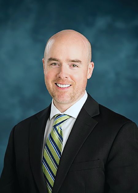
These guidelines meet a growing need to help practicing clinicians identify and best manage immune-related adverse events, according to Bryan J. Schneider, MD, of the University of Michigan Comprehensive Cancer Center, and vice chair of the NCCN Panel on Management of Immunotherapy-Related Toxicities.
“The mechanism of action of these anticancer therapies is so much different from anything that we’re used to,” Dr. Schneider said in an interview.
Critical need for guidance
The ASCO and NCCN guidelines are “critically important” to ensure uniform management of common immune-related adverse events, according to Stephen M. Ansell, MD, PhD, professor of medicine and chair of the Lymphoma Group at Mayo Clinic, Rochester, Minn.
“I think it also specifically highlights a few side effects that many people may not necessarily think about, from eye toxicities to thyroid effects, or the type of things that the average oncologist who is now using this in their practice quite regularly may not necessarily think about,” Dr. Ansell said. “Those kind of effects are now clearly outlined with clear guidance about what should be done, and I think that allows oncologists a resource to go and look at this carefully so that they do the right thing.”

The spectrum of adverse effects associated with checkpoint inhibitors is markedly different from what is seen with cytotoxic chemotherapy, the guidelines note. Most often, the side effects are seen in the skin, GI tract, and lungs, as well as the endocrine, adrenal, nervous, thyroid, pituitary, musculoskeletal, cardiovascular, ocular, and hematologic systems.
Stepwise approach
Side effects of checkpoint inhibitors are typically mild, but they can be severe and sometimes life-threatening, according to ASCO and NCCN.
If immune-related adverse events are mild (i.e., grade 1), treatment can continue with close monitoring, according to the guidelines. By contrast, moderate to severe immune-related adverse events can lead to severe declines in organ function and quality of life, or even fatal outcomes, so early detection and proper management are needed.
Grade 2 toxicities warrant suspending immune checkpoint inhibitor treatment, and resuming it once symptoms subside to grade 1 or less, according to the guidelines. Grade 3 toxicities should also prompt suspension of treatment, plus initiation of high-dose corticosteroids tapered over at least 4-6 weeks.
For most toxicities that reach grade 4, permanent discontinuation of checkpoint inhibitors is recommended.
A thoughtful discussion of potential risks and benefits is needed before using immune checkpoint inhibitors in patients who have autoimmune disease or prior organ transplant, according to the guidelines.
Vigilance required
Checkpoint inhibitors have been approved by the Food and Drug Administration to treat a variety of cancers, including melanoma, lung cancer, and Hodgkin lymphoma, as well as lung, liver, kidney, and bladder cancers.
Clinicians managing patients on checkpoint inhibitors should always be vigilant because immune-related adverse event symptoms can be subtle, according to Julie Brahmer, MD, of The Sidney Kimmel Comprehensive Cancer Center at Johns Hopkins in Baltimore.
“Everyone has to work as a team, which includes being educated on possible side effects to immunotherapy prior to prescribing it,” said Dr. Brahmer, chair of the ASCO panel and vice chair of the NCCN panel that developed the guidelines.
The guidelines were published Feb. 14 in two documents that are similar in content, but different in format. The ASCO guideline was published in the Journal of Clinical Oncology (doi: 10.1200/JCO.2017.77.6385) and the NCCN Clinical Practice Guidelines in Oncology were posted on the NCCN website.
While the first edition of the guidelines focuses specifically on immune checkpoint inhibitors, an update anticipated for 2019 will include guidance on chimeric antigen receptor (CAR) T-cell therapy, which is associated with several important side effects, notably cytokine release syndrome.
The American Society of Clinical Oncology (ASCO) and the National Comprehensive Cancer Network (NCCN) have released new guidelines designed to help clinicians manage the unique and sometimes severe side effects associated with cancer immunotherapy agents.
These guidelines meet a growing need to help practicing clinicians identify and best manage immune-related adverse events, according to Bryan J. Schneider, MD, of the University of Michigan Comprehensive Cancer Center, and vice chair of the NCCN Panel on Management of Immunotherapy-Related Toxicities.
“The mechanism of action of these anticancer therapies is so much different from anything that we’re used to,” Dr. Schneider said in an interview.
Critical need for guidance
The ASCO and NCCN guidelines are “critically important” to ensure uniform management of common immune-related adverse events, according to Stephen M. Ansell, MD, PhD, professor of medicine and chair of the Lymphoma Group at Mayo Clinic, Rochester, Minn.
“I think it also specifically highlights a few side effects that many people may not necessarily think about, from eye toxicities to thyroid effects, or the type of things that the average oncologist who is now using this in their practice quite regularly may not necessarily think about,” Dr. Ansell said. “Those kind of effects are now clearly outlined with clear guidance about what should be done, and I think that allows oncologists a resource to go and look at this carefully so that they do the right thing.”
The spectrum of adverse effects associated with checkpoint inhibitors is markedly different from what is seen with cytotoxic chemotherapy, the guidelines note. Most often, the side effects are seen in the skin, GI tract, and lungs, as well as the endocrine, adrenal, nervous, thyroid, pituitary, musculoskeletal, cardiovascular, ocular, and hematologic systems.
Stepwise approach
Side effects of checkpoint inhibitors are typically mild, but they can be severe and sometimes life-threatening, according to ASCO and NCCN.
If immune-related adverse events are mild (i.e., grade 1), treatment can continue with close monitoring, according to the guidelines. By contrast, moderate to severe immune-related adverse events can lead to severe declines in organ function and quality of life, or even fatal outcomes, so early detection and proper management are needed.
Grade 2 toxicities warrant suspending immune checkpoint inhibitor treatment, and resuming it once symptoms subside to grade 1 or less, according to the guidelines. Grade 3 toxicities should also prompt suspension of treatment, plus initiation of high-dose corticosteroids tapered over at least 4-6 weeks.
For most toxicities that reach grade 4, permanent discontinuation of checkpoint inhibitors is recommended.
A thoughtful discussion of potential risks and benefits is needed before using immune checkpoint inhibitors in patients who have autoimmune disease or prior organ transplant, according to the guidelines.
Vigilance required
Checkpoint inhibitors have been approved by the Food and Drug Administration to treat a variety of cancers, including melanoma, lung cancer, and Hodgkin lymphoma, as well as lung, liver, kidney, and bladder cancers.
Clinicians managing patients on checkpoint inhibitors should always be vigilant because immune-related adverse event symptoms can be subtle, according to Julie Brahmer, MD, of The Sidney Kimmel Comprehensive Cancer Center at Johns Hopkins in Baltimore.
“Everyone has to work as a team, which includes being educated on possible side effects to immunotherapy prior to prescribing it,” said Dr. Brahmer, chair of the ASCO panel and vice chair of the NCCN panel that developed the guidelines.
The guidelines were published Feb. 14 in two documents that are similar in content, but different in format. The ASCO guideline was published in the Journal of Clinical Oncology (doi: 10.1200/JCO.2017.77.6385) and the NCCN Clinical Practice Guidelines in Oncology were posted on the NCCN website.
While the first edition of the guidelines focuses specifically on immune checkpoint inhibitors, an update anticipated for 2019 will include guidance on chimeric antigen receptor (CAR) T-cell therapy, which is associated with several important side effects, notably cytokine release syndrome.
Dr. Paul E. Marik proclaims end to corticosteroid monotherapy for sepsis
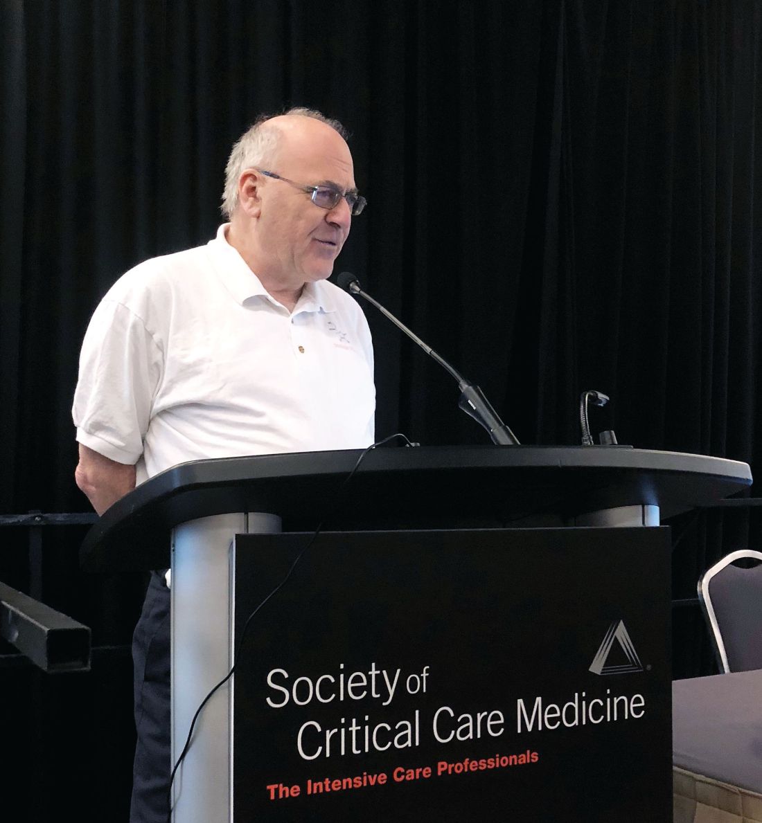
SAN ANTONIO – While critical care specialists await more data on a so-called sepsis cocktail with varying degrees of hope and skepticism, Paul E. Marik, MD, FCCP, has proclaimed the dawning of a new era.
Dr. Marik became a celebrity in the critical care medicine community after he and his colleagues reported the results of his retrospective study evaluating the combination of hydrocortisone, vitamin C, and thiamine for treatment of severe sepsis and septic shock (Chest. 2017 June. doi: 10.1016/j.chest.2016.11.036).
Since this study, several physicians have already been putting Dr. Marik’s method to practice, the investigator and audience members noted during a session at the Critical Care Congress sponsored by the Society of Critical Care Medicine.
in his presentation at the meeting.
These comments echoed Dr. Marik’s May 2017 editorial in Critical Care Medicine, in which he suggested that critically ill and injured patients may benefit from combination therapy with hydrocortisone and vitamin C (Crit Care Med 2017 May;45[5]910-1).
That editorial was quickly followed by the report on Dr. Marik and colleagues’ before-after study, in which hospital mortality was 8.5%, versus 0.4% in the treatment and control groups, respectively (P less than .001). This finding led the investigators to suggest that intravenous vitamin C administered along with corticosteroids and thiamine is “effective” in reducing mortality, in their paper published in CHEST®.
During Dr. Marik’s presentation at the meeting, he noted that he had been “misquoted” with regard to the finality of his study’s results. The final line of the CHEST® paper reads, “Additional studies are required to confirm these preliminary findings,” he emphasized.
Nevertheless, Dr. Marik alluded to a “big paradigm shift” in the treatment of sepsis.
“Our experience has been echoed by now hundreds, if not thousands, of clinicians across the world,” said Dr. Marik, chief of the division of pulmonary and critical care medicine, Eastern Virginia Medical School, Norfolk.
He recounted an anecdotal case submitted by “Josh from Ohio” describing an elderly man who was “started on cocktail and within a day his pressor requirements melted away and he was extubated.” Quoting “Josh from Ohio,” Dr. Mark continued, “Tomorrow he will probably leave the ICU with no residual organ dysfunction, no volume overload, (and) no ICU complications.”
Eddy Gutierrez, MD, of Jacksonville, Fla., noted in a question-and-answer period that he has had “positive results” with a similar approach.
“When we first learned about the vitamin C and the ‘Marik protocol,’ so to speak, I was in fellowship and I got laughed at,” Dr. Gutierrez said. “Nobody would let me try it.”
Others are taking a wait-and-see approach.
Greg S. Martin, MD, secretary of the Society of Critical Care Medicine, said in an interview that there are “at least two schools of thought” among critical care specialists regarding the use of hydrocortisone, vitamin C, and thiamine for treatment of sepsis and septic shock.
“One school of thought is that this is incredibly important if this is even fractionally as effective as what [Dr. Marik] showed, because we have not found an effective therapy for sepsis,” said Dr. Martin, associate professor of medicine at Grady Memorial Hospital, Atlanta.
“The contrarian approach is to say, ‘yes, but this seems remarkably unlikely to be as effective as what he has shown,’ ” Dr. Martin added. “Particularly in sepsis, people are very skeptical of whether a drug or a drug combination is going to be as effective when you really get down to a high-quality randomized controlled trial that would be the definitive level of evidence.”
The wait may not be long for at least some data. Multiple clinical trials are recruiting or planned, according to Dr. Marik. These included a 140-patient U.S. randomized, double-blind trial of vitamin C, hydrocortisone, and thiamine vs. placebo that started in February 2018 and is expected to be completed by February 2019, according to the study’s ClinicalTrials.gov listing.
“The good news is some people think this is of value,” Dr. Marik said.
As part of his presentation, Dr. Marik reported a disclosure related to Baxter (advisory board).
REPORTING FROM CCC47
Radiation offers no survival benefit in early lung cancer with positive margins
For patients with early-stage non–small-cell lung cancer (NSCLC) who had positive margins following lobectomy, additional radiation therapy was not associated with a long-term survival benefit in a recent retrospective study.
Positive margins were associated with significantly worse 5-year survival in the study of patients with stage I-II NSCLC, which was published in the Journal of Surgical Research (2018 Mar. doi: 10.1016/j.jss.2017.10.025).
The study is one of the latest to suggest radiation may not be of benefit for treatment of positive margins after surgical resection of lung cancer, according to Brian C. Gulack, MD, MHS, department of surgery, Duke University, Durham, N.C., and his coauthors.
“Our analysis adds important findings to the literature as it focuses specifically on patients undergoing a lobectomy for stage I or II disease,” they wrote.
Adjuvant radiation therapy and re-resection are considered to be two potential treatment options for patients with NSCLC who undergo lobectomy and have positive margins, wrote Dr. Gulack and his coauthors.
However, guidelines from the National Comprehensive Cancer Network recommend re-resection when possible, they added.
The retrospective analysis by Dr. Gulack and his colleagues was based on information in the National Cancer Data Base, which collects data from more than 1,500 hospitals.
The investigators identified 49,563 patients with stage I-II NSCLC who received lobectomy with no induction therapy between 1998 and 2006. Of those patients, 1,934 (3.9%) had positive margins, the authors reported.
Five-year survival was 34.5% for patients with reportedly positive margins, versus 57.2% for patients without positive margins, according to results of an unadjusted analysis.
To evaluate the effects of radiotherapy specifically for treatment of positive margins, the investigators excluded patients who had upstaged disease or who had received palliative radiotherapy. Out of the remaining 1,579 patients, 579 (38.2%) received radiation therapy, according to the report.
In that group, there was no significant difference in likelihood of death associated with radiation treatment (hazard ratio, 1.10, 95% confidence interval, 0.90-1.35).
“Despite the importance of these findings, there is still potential for substantial bias in this retrospective study, and therefore prospective investigation is necessary in order to validate our results,” Dr. Gulack and his colleagues concluded.
Study coauthor Thomas A. D’Amico, MD, reported serving as a consultant for Scanlan, and the rest of the authors had no disclosures.
SOURCE: Gulack et al. J Surg Res 2018 March doi: 10.1016/j.jss.2017.10.025.
FROM THE JOURNAL OF SURGICAL RESEARCH
Key clinical point: Additional radiation therapy was not associated with a long-term survival benefit in patients with early-stage non–small-cell lung cancer who had positive margins following lobectomy.
Major finding: There was no significant difference in likelihood of death associated with radiation treatment (hazard ratio, 1.10, 95% CI, 0.90-1.35).
Study details: A retrospective analysis of data in the National Cancer Data Base including 49,563 patients with stage I-II NSCLC who received lobectomy with no induction therapy between 1998 and 2006.
Disclosures: One study author reported serving as a consultant for Scanlan, and the rest had no disclosures.
Source: Gulack et al. J Surg Res. 2018 Mar. doi: 10.1016/j.jss.2017.10.025.
Critical illness-related corticosteroid insufficiency guidelines explained
SAN ANTONIO – When corticosteroids are used for septic shock, the dose should be low to moderate, the timing should be early, and the duration should be at least 3 days, said a speaker at the Critical Care Congress sponsored by the Society for Critical Care.
Dosing, timing, and duration are “three critical questions” critical care specialists face that are answered by the new critical illness–related corticosteroid insufficiency (CIRCI) guidelines, continued Stephen M. Pastores, MD, a cochair of the task force that developed guidelines for the diagnosis and management of CIRCI in critically ill patients.
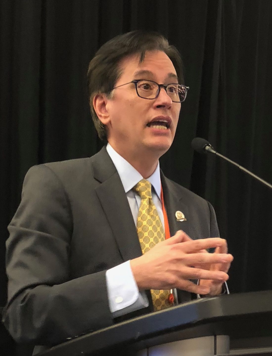
During his presentation, Dr. Pastores limited his remarks to discussion of sepsis and septic shock with corticosteroids. He cautioned that, despite careful deliberations by the panel, the level of evidence behind some of the recommendations was “low to moderate and never high” and that not all task force members agreed with all recommendations.
“There were a lot of back and forth disagreements behind these recommendations,” said Dr. Pastores, who is the director of the critical care medicine fellowship training and research programs at Memorial Sloan Kettering Cancer Center, New York. “We only required 80% of the panelists to agree that these were the recommendations and statements that we were going to go by.”
The guidelines recommend against the use of corticosteroids in adult patients who have sepsis without shock, Dr. Pastores noted.
In contrast,
In an analysis of available data from randomized clinical trials including patients with septic shock, corticosteroids significantly reduced 28-day mortality when compared with placebo, Dr. Pastores said.
That survival benefit seems to be dependent on several factors: dose of the corticosteroids (hydrocortisone less than 400 mg/day), longer duration (at least 3 or more days), and severity of sepsis. “The more severe the sepsis, the more septic shock the patient was in, the more likely the corticosteroids were likely to help those patients,” Dr. Pastores explained.
Accordingly, the guidelines further suggest using long-course, low-dose corticosteroid treatment, namely intravenous hydrocortisone at no more than 400 mg/day for at least 3 days.
The expert panel specifically recommended hydrocortisone as the corticosteroid of choice in this setting, according to Dr. Pastores. That recommendation was based in part on a recent systematic review and meta-analysis showing that hydrocortisone, given as a bolus or an infusion, was more likely than placebo or methylprednisolone to result in shock reversal.
Dr. Pastores reported disclosures related to Theravance Biopharma, Bayer HealthCare Pharmaceuticals, Spectral Diagnostics, and Asahi-Kasei.
SAN ANTONIO – When corticosteroids are used for septic shock, the dose should be low to moderate, the timing should be early, and the duration should be at least 3 days, said a speaker at the Critical Care Congress sponsored by the Society for Critical Care.
Dosing, timing, and duration are “three critical questions” critical care specialists face that are answered by the new critical illness–related corticosteroid insufficiency (CIRCI) guidelines, continued Stephen M. Pastores, MD, a cochair of the task force that developed guidelines for the diagnosis and management of CIRCI in critically ill patients.
During his presentation, Dr. Pastores limited his remarks to discussion of sepsis and septic shock with corticosteroids. He cautioned that, despite careful deliberations by the panel, the level of evidence behind some of the recommendations was “low to moderate and never high” and that not all task force members agreed with all recommendations.
“There were a lot of back and forth disagreements behind these recommendations,” said Dr. Pastores, who is the director of the critical care medicine fellowship training and research programs at Memorial Sloan Kettering Cancer Center, New York. “We only required 80% of the panelists to agree that these were the recommendations and statements that we were going to go by.”
The guidelines recommend against the use of corticosteroids in adult patients who have sepsis without shock, Dr. Pastores noted.
In contrast,
In an analysis of available data from randomized clinical trials including patients with septic shock, corticosteroids significantly reduced 28-day mortality when compared with placebo, Dr. Pastores said.
That survival benefit seems to be dependent on several factors: dose of the corticosteroids (hydrocortisone less than 400 mg/day), longer duration (at least 3 or more days), and severity of sepsis. “The more severe the sepsis, the more septic shock the patient was in, the more likely the corticosteroids were likely to help those patients,” Dr. Pastores explained.
Accordingly, the guidelines further suggest using long-course, low-dose corticosteroid treatment, namely intravenous hydrocortisone at no more than 400 mg/day for at least 3 days.
The expert panel specifically recommended hydrocortisone as the corticosteroid of choice in this setting, according to Dr. Pastores. That recommendation was based in part on a recent systematic review and meta-analysis showing that hydrocortisone, given as a bolus or an infusion, was more likely than placebo or methylprednisolone to result in shock reversal.
Dr. Pastores reported disclosures related to Theravance Biopharma, Bayer HealthCare Pharmaceuticals, Spectral Diagnostics, and Asahi-Kasei.
EXPERT ANALYSIS FROM THE CCC47
Death rate steady with pediatric early warning system
SAN ANTONIO – , but did not reduce the rate of all-cause hospital mortality, according to results of a large, multicenter trial.
Taken together, the findings of the trial do not support the use of the Bedside Pediatric Early Warning System (BedsidePEWS) to reduce hospital mortality, noted investigator Christopher S. Parshuram, MBChB, DPhil, during a presentation at the Critical Care Congress sponsored by the Society of Critical Care Medicine.
The multicenter randomized cluster study, called the EPOCH trial, included 21 hospitals in seven countries that provided inpatient pediatric care. Ten of the hospitals delivered the BedsidePEWS intervention, while the remaining 11 provided usual care. The study data included 144,539 patient discharges comprising 559,443 patient days. Enrollment began Feb. 28, 2011, and ended on June 21, 2015.
For the BedsidePEWS group, all-cause hospital mortality was 1.93 per 1,000 patient discharges, versus 1.56 per 1,000 patient discharges for usual care (adjusted odds ratio, 1.01; 95% confidence interval, 0.61-1.69; P = .96), according to a report on this study that was published in JAMA.
However, the BedsidePEWS group had a significant improvement in the secondary outcome of significant clinical deterioration events, a composite outcome reflecting late ICU admissions.
In the BedsidePEWS group, the rate of significant clinical deterioration events was 0.50 per 1,000 patient-days, compared with 0.84 per 1,000 patient-days at hospitals with usual care (adjusted rate ratio, 0.77; 95% CI, 0.61-0.97; P = .03), the investigators wrote.
The goal of the EPOCH trial was to determine whether BedsidePEWS could reduce rates of all-cause hospital mortality and significant clinical deterioration among hospitalized children, according to the researchers.
“The BedsidePEWS versus usual care did improve processes of care and early detection of critical illness, aligned with the notion of providing the right care, right now,” Dr. Parshuram, associate professor of critical care medicine and pediatrics at the University of Toronto, said during his presentation at the meeting. “Certainly more vital signs were documented, and anecdotally there were reports of culture change.
“However, when we looked further, there was no difference in hospital mortality, nor hospital resource utilization,” Dr. Parshuram added.
The Canadian Institutes of Health Research funded the study. Dr. Parshuram is an inventor of BedsidePEWS and owns shares in a company that is commercializing it.
SOURCE: Parshuram et al. JAMA. 2018 Feb 27. doi: 10.1001/jama.2018.0948.
REPORTING FROM CCC47
Key clinical point: Use of a pediatric early warning system (BedsidePEWS) did not reduce rates of all-cause hospital mortality among hospitalized children, compared with usual care, but did reduce rates of significant clinical deterioration events.
Major finding: For hospitals implementing BedsidePEWS, all-cause hospital mortality was 1.93 per 1,000 patient discharges, versus 1.56 per 1,000 at hospitals with usual care (adjusted odds ratio, 1.01; 95% confidence interval, 0.61-1.69; P = .96).
Study details: A multicenter cluster randomized trial of 144,539 patient discharges from 21 hospitals in seven countries providing pediatric care.
Disclosures: The Canadian Institutes of Health Research funded the study. Dr. Parshuram is an inventor of BedsidePEWS and owns shares in a company that is commercializing it.
Source: Parshuram et al. JAMA. 2018 Feb 27. doi: 10.1001/jama.2018.0948.