User login
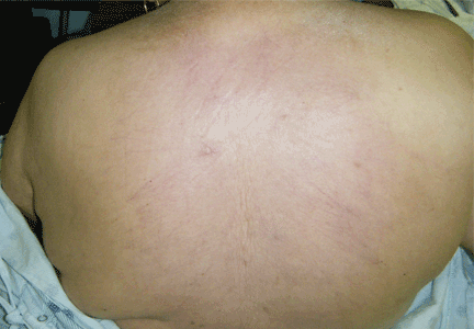
Thick skin on the back
Q: Which is the correct diagnosis?
- Scleroderma (systemic sclerosis)
- Scleredema diabeticorum
- Amyloidosis
- Cutaneous sarcoidosis
- Porphyria cutanea tarda
A: The correct answer is scleredema diabeticorum, a common, underdiagnosed skin manifestation of uncontrolled diabetes mellitus seen in 2.5% to 14% of diabetic patients.1,2 It most often presents with the insidious onset of painless induration and nonpitting thickening of the skin, predominantly on the upper back and neck. Biopsy of the skin usually reveals thickening of the dermis with deposition of collagen and hyaluronic acid without an inflammatory infiltrate.3
Of note, patients may present with similar skin changes acutely in conditions such as postinfectious scleredema (scleredema of Buschke) and paraproteinemias.
Treatment of scleredema is usually difficult, but options include radiotherapy, ultraviolet light therapy, low-dose methotrexate, psoralen, and extracorporeal photopheresis.4–7
- Cole GW, Headley J, Skowsky R. Scleredema diabeticorum: a common and distinct cutaneous manifestation of diabetes mellitus. Diabetes Care 1983; 6:189–192.
- Sattar MA, Diab S, Sugathan TN, Sivanandasingham P, Fenech FF. Scleroedema diabeticorum: a minor but often unrecognized complication of diabetes mellitus. Diabet Med 1988; 5:465–468.
- Varga J, Gotta S, Li L, Sollberg S, Di Leonardo M. Scleredema adultorum: case report and demonstration of abnormal expression of extracellular matrix genes in skin fibroblasts in vivo and in vitro. Br J Dermatol 1995; 132:992–999.
- Seyger MM, van den Hoogen FH, de Mare S, van Haelst U, de Jong EM. A patient with a severe scleroedema diabeticorum, partially responding to low-dose methotrexate. Dermatology 1999; 198:177–179.
- Lee MW, Choi JH, Sung KJ, Moon KC, Koh JK. Electron beam therapy in patients with scleredema. Acta Derm Venereol 2000; 80:307–308.
- Bowen AR, Smith L, Zone JJ. Scleredema adultorum of Buschke treated with radiation. Arch Dermatol 2003; 139:780–784.
- Beers WH, Ince A, Moore TL. Scleredema adultorum of Buschke: a case report and review of the literature. Semin Arthritis Rheum 2006; 35:355–359.
Q: Which is the correct diagnosis?
- Scleroderma (systemic sclerosis)
- Scleredema diabeticorum
- Amyloidosis
- Cutaneous sarcoidosis
- Porphyria cutanea tarda
A: The correct answer is scleredema diabeticorum, a common, underdiagnosed skin manifestation of uncontrolled diabetes mellitus seen in 2.5% to 14% of diabetic patients.1,2 It most often presents with the insidious onset of painless induration and nonpitting thickening of the skin, predominantly on the upper back and neck. Biopsy of the skin usually reveals thickening of the dermis with deposition of collagen and hyaluronic acid without an inflammatory infiltrate.3
Of note, patients may present with similar skin changes acutely in conditions such as postinfectious scleredema (scleredema of Buschke) and paraproteinemias.
Treatment of scleredema is usually difficult, but options include radiotherapy, ultraviolet light therapy, low-dose methotrexate, psoralen, and extracorporeal photopheresis.4–7
- Cole GW, Headley J, Skowsky R. Scleredema diabeticorum: a common and distinct cutaneous manifestation of diabetes mellitus. Diabetes Care 1983; 6:189–192.
- Sattar MA, Diab S, Sugathan TN, Sivanandasingham P, Fenech FF. Scleroedema diabeticorum: a minor but often unrecognized complication of diabetes mellitus. Diabet Med 1988; 5:465–468.
- Varga J, Gotta S, Li L, Sollberg S, Di Leonardo M. Scleredema adultorum: case report and demonstration of abnormal expression of extracellular matrix genes in skin fibroblasts in vivo and in vitro. Br J Dermatol 1995; 132:992–999.
- Seyger MM, van den Hoogen FH, de Mare S, van Haelst U, de Jong EM. A patient with a severe scleroedema diabeticorum, partially responding to low-dose methotrexate. Dermatology 1999; 198:177–179.
- Lee MW, Choi JH, Sung KJ, Moon KC, Koh JK. Electron beam therapy in patients with scleredema. Acta Derm Venereol 2000; 80:307–308.
- Bowen AR, Smith L, Zone JJ. Scleredema adultorum of Buschke treated with radiation. Arch Dermatol 2003; 139:780–784.
- Beers WH, Ince A, Moore TL. Scleredema adultorum of Buschke: a case report and review of the literature. Semin Arthritis Rheum 2006; 35:355–359.

