User login
M. Alexander Otto began his reporting career early in 1999 covering the pharmaceutical industry for a national pharmacists' magazine and freelancing for the Washington Post and other newspapers. He then joined BNA, now part of Bloomberg News, covering health law and the protection of people and animals in medical research. Alex next worked for the McClatchy Company. Based on his work, Alex won a year-long Knight Science Journalism Fellowship to MIT in 2008-2009. He joined the company shortly thereafter. Alex has a newspaper journalism degree from Syracuse (N.Y.) University and a master's degree in medical science -- a physician assistant degree -- from George Washington University. Alex is based in Seattle.
Maternal DTaP Vaccine Protected Newborns From Pertussis
VANCOUVER, B.C. – Infants born to women who receive diphtheria-tetanus-acellular pertussis vaccine during pregnancy have higher pertussis antibody levels during their first few months of life than infants born to unvaccinated women, Dr. Abbey Hardy-Fairbanks reported.
The levels are sufficient to protect infants against pertussis prior to their first diphtheria-tetanus-acellular pertussis (DTaP) shot at around 2 months, a period of “significant pertussis morbidity and mortality,” said Dr. Hardy-Fairbanks, an ob.gyn.at the University of Iowa, Iowa City. “This is the first evidence to document that pertussis immunization during pregnancy is likely to be beneficial to infants when they are most vulnerable to pertussis disease. [Physicians] should consider vaccination of women during pregnancy with DTaP,” she said at the meeting.
In the prospective cohort study, 16 (23%) of 70 pregnant women received DTaP vaccine; 54 (77%) pregnant women selected as controls did not and had not been vaccinated for at least 2 years.
Four of the women (25%) in the DTaP group were vaccinated in the first trimester, eight (50%) in the second, and four (25%) in the third. Vaccination did not cause any adverse pregnancy outcomes.
Maternal blood and cord blood were collected at delivery. Blood was also collected from children before and after their primary DTaP series and toddler booster doses at 12-18 months.
Blood samples were measured for pertussis antigens, including pertussis toxoid, filamentous hemagglutinin, pertactin, and fimbriae, by enzyme-linked immunosorbent assay.
Newborns in the DTaP group had higher pertussis antibody concentrations than their mothers, “showing efficient placental transfer of antibodies to the infant,” Dr. Hardy-Fairbanks said.
They also had substantially higher concentrations than infants in the control group prior to the start of the primary DTaP series, and the differences were statistically significant.
However, at month 7, following completion of the DTaP series, infants born to vaccinated mothers had slightly lower antibody levels than infants in the control group. The differences were not statistically significant, but “may represent some blunting of the infant immune response to the [vaccine],” Dr. Hardy-Fairbanks said. By the time they got their toddler booster doses, however, antibody levels “were essentially equivalent” in the two groups, she said.
Dr. Hardy-Fairbanks said she had no conflicts of interest. The study was funded by Sanofi-Pasteur, maker of Daptacel DTaP vaccine.
View on the News
Possible Blunted Immune Response?
Dr. Sarah Long thanked the study authors for their work. “Your findings are so very helpful. We don't have this kind of information.”
She was concerned, however, that infants born to vaccinated mothers mounted only a blunted immune response to their primary DTaP vaccine series, and wondered if responses would be blunted to other vaccines. The study's presenter said the question is currently being investigated, but so far that does not appear to be the case.
DR. LONG is the chief of the section of infectious diseases at St. Christopher's Hospital for Children in Philadelphia. She said she had no conflicts of interest.
Family Physicians Face Reform's Regulatory Challenges
DENVER – The federal Affordable Care Act was a win for family physicians, but there’s room for improvement.
That was the main message from officers at the annual Congress of Delegates of the American Academy of Family Physicians. They also pledged to continue advocating for family physicians as regulations to enact the law are drafted.
“We are into the regulatory phase, folks. The devil is in the details. We have restructured our D.C. office to focus on this phase,” Dr. Lori Heim, chair of the AAFP board of directors, told delegates.
Dr. Glen Stream, who was chosen as the academy’s president-elect at the meeting, said although the AAFP and other medical groups succeeded in getting provisions favorable to family physicians included in the law, he was “not na?ve enough to think the future is assured. We must continue our legislative advocacy.”
The law recognizes a central role for primary care in the nation’s health, and includes, among other measures, the following:
– A 10% Medicare bonus payment starting in 2011 for certain primary care services, enacted to attract medical students to the field;
– A 2-year experiment starting in 2013 that guarantees that Medicaid pays at least as much as Medicare for primary care services, including immunizations;
– Continuation of federal funding for family medicine residency programs; and
– Recognition and advancement of the patient-centered medical home (PCMH) concept.
Overall, the law “advances our agenda by leaps and bounds,” said academy president, Dr. Roland Goertz, but “it’s not perfect.”
The 10% bonus, for instance, doesn’t apply to enough family physicians, the officers said. At least 60% of Medicare billings must be for primary care services to qualify. The academy “will continue to ask legislators” to lower the threshold to 50% and expand qualifying services, according to a report released at the meeting.
In addition, the Affordable Care Act did not amend or replace the problematic sustainable growth rate formula that is used by the federal government to calculate Medicare reimbursements, and which has been a longstanding legislative priority for the AAFP and other physician groups.
It’s too early to tell how other, potentially problematic measures in the law will be resolved, including the provision for accountable care organizations (ACOs). Those regional organizations will be given lump sum payments to coordinate and streamline care of enrolled patients, with providers sharing in savings to the Medicare program.
The law allows providers to form such organizations and specifies adequate participation by primary care physicians. However, there is concern that antitrust laws may block family physician practices from organizing into ACOs.
Academy officers told delegates it is also possible that the hospital industry could dominate ACOs, which would exclude family physicians from positions of leadership and control.
Are ACOs “going to be based on primary care practitioners, or large hospitals and large hospital systems?” asked Dr. Heim. It’s “a debate at the state and national level. Larger systems are already poised to move into this arena and take it over.”
The academy said in the report that it will work to prevent that. However, as the Centers for Medicare and Medicaid Services work out the regulatory definitions of ACOs, it is difficult for the academy to give members guidance on how to proceed, AAFP officials noted.
The federal health reform act also encourages CMS to test patient-centered medical homes for “high-need” beneficiaries and allows pilot projects to expand if they improve care, save money, or both. The objective is to have one medical practice coordinate a patient’s care and offer health counseling, and other services that go beyond traditional problem-oriented visits.
Given the legislative encouragement, positive results from PCMH demonstration projects, and other factors, PCMHs are “an unavoidable future” for family physicians, Dr. Stream told delegates.
Even with federal money available to make the switch to electronic medical records, ramping up to offer PCMH services is prohibitively expensive for small or solo practices, some delegates cautioned – especially at a time when most payers aren’t covering the extra costs.
Several AAFP officers said they understood the concerns. With continued advocacy, however, they expect those costs eventually will be covered to some extent – and even before that happens, a move toward the PCMH model promises better patient care.
“Sometimes, you have to [act on] faith,” Dr. David Ellington, a board member, told concerned delegates.
Resolutions Reveal AAFP Delegates' Wide Range of Concerns
DENVER – Although health reform’s impact on family physicians dominated speeches at this year’s Congress of Delegates of the American Academy of Family Physicians, the more than 60 resolutions introduced by state chapters indicated that delegates had much more on their minds.

The AAFP will continue its corporate alliance with the Coca-Cola Co., following failure of resolutions from Washington, Oregon, California, and Illinois delegates calling for its end. The resolutions’ backers said that the partnership betrays the AAFP’s health promotion principles and has cost the group members. The pointed language used by alliance opponents – at one point, a delegate likened it to partnering with cigarette maker R.J. Reynolds on a COPD campaign – indicated that some delegates had lost trust in the board, AAFP board member Dr. David Ellington said during a question-and-answer session.
He and other officers said that they regret approving the program without vetting it with members. “It was not meant to be this way. We are sorry about that. We have learned our lesson,” said Dr. Ellington, promising, along with other officers, better communication and greater transparency on future board actions.
Nonetheless, the Congress of Delegates voted against the measures to end the alliance. AAFP leaders said that without the roughly $550,000 that the alliance earns the academy annually, membership dues might need to be significantly increased or programs cut because revenue from other sources has declined. In addition, the academy has years of experience managing tricky relationships, AAFP officials said, including those with the pharmaceutical industry. Relationships with the pharmaceutical industry brought AAFP $22 million in revenue in fiscal year 2009-2010 – down 35% from 2004-2005, according to an AAFP report.
In response to several resolutions seeking to curb competition from nurse practitioners, the AAFP will promote studies comparing them with family physicians. During testimony, the assumption was clear that such studies would prove family physicians to have better outcomes and cost effectiveness. The academy also will continue public relations efforts to highlight the more rigorous training of physicians. A call for the AAFP to come out against nurse practitioners using “doctor” in their titles was rejected, however, as a too-negative media message.
The AAFP will not issue a statement to U.S. congressional leaders asking for the repeal of “don’t ask, don’t tell,” following failure of a resolution calling for it to do so. Supporters said the law prevents military personnel from disclosing sexual histories to military physicians. Uniformed services delegates, however, testified that soldiers do, in fact, disclose their sexual practices, and that military policy prevents information revealed to physicians from being used for discharges.
In response to many resolutions about payment and insurance issues, the AAFP will provide guidance on surviving recovery audits, petition federal regulatory bodies or lawmakers to reduce or eliminate minimum hospital-stay requirements for Medicare coverage of postdischarge treatments, and develop a policy statement in support of physicians’ rights to bargain collectively with insurance companies.
Following adoption of a resolution on the matter, the academy cannot sell members’ e-mail addresses without their permission.
To ensure that a patient’s desire to be cremated is honored, the AAFP will advocate for laws that grant a patient’s chosen representative or executor authority over the body’s disposal. Families sometimes ignore the wish, according to the adopted Michigan resolution.
A Kansas resolution noted, “As the for-profit hospice industry has grown over the years, the pressure on the attending physician to certify the patient for the Medicare hospice benefit has intensified.” The AAFP will work with hospice and other organizations to ensure certification is justified, following adoption of part of the resolution. However, the AAFP will not investigate the “financial relationships between hospice agencies and nursing homes,” which also was requested by the measure. The resolution committee concluded that such financial data are unavailable.
The academy will not support a ban on “spice,” synthetic marijuana sold as incense under the names K2, Pep, and Kind but usually smoked. The focus of the Indiana resolution was too specific, said the resolution committee. Other products quickly replaced spice when it was outlawed in some states, noted the committee, which welcomed a broader substance abuse resolution next year.
The AAFP will offer guidance and strategies on handling pressure from patients and durable goods manufacturers to certify patients for new equipment, following a call for it do so from Tennessee. AAFP will also investigate ways to make it easier for members to report fraud, but the delegates declined an additional request for the academy itself to report abuses to the Medicare program. It would be inappropriate to require the academy to report “anecdotal information,” the resolution committee said.
In response to a request from the Illinois chapter, the academy’s board will consider providing state chapters with a template for a proposed law to grant out-of-state physicians permission to work in emergency relief centers and free clinics. Opponents were concerned that carving out such a niche could create opportunities for fraudulent practitioners.
DENVER – Although health reform’s impact on family physicians dominated speeches at this year’s Congress of Delegates of the American Academy of Family Physicians, the more than 60 resolutions introduced by state chapters indicated that delegates had much more on their minds.
The AAFP will continue its corporate alliance with the Coca-Cola Co., following failure of resolutions from Washington, Oregon, California, and Illinois delegates calling for its end. The resolutions’ backers said that the partnership betrays the AAFP’s health promotion principles and has cost the group members. The pointed language used by alliance opponents – at one point, a delegate likened it to partnering with cigarette maker R.J. Reynolds on a COPD campaign – indicated that some delegates had lost trust in the board, AAFP board member Dr. David Ellington said during a question-and-answer session.
He and other officers said that they regret approving the program without vetting it with members. “It was not meant to be this way. We are sorry about that. We have learned our lesson,” said Dr. Ellington, promising, along with other officers, better communication and greater transparency on future board actions.
Nonetheless, the Congress of Delegates voted against the measures to end the alliance. AAFP leaders said that without the roughly $550,000 that the alliance earns the academy annually, membership dues might need to be significantly increased or programs cut because revenue from other sources has declined. In addition, the academy has years of experience managing tricky relationships, AAFP officials said, including those with the pharmaceutical industry. Relationships with the pharmaceutical industry brought AAFP $22 million in revenue in fiscal year 2009-2010 – down 35% from 2004-2005, according to an AAFP report.
In response to several resolutions seeking to curb competition from nurse practitioners, the AAFP will promote studies comparing them with family physicians. During testimony, the assumption was clear that such studies would prove family physicians to have better outcomes and cost effectiveness. The academy also will continue public relations efforts to highlight the more rigorous training of physicians. A call for the AAFP to come out against nurse practitioners using “doctor” in their titles was rejected, however, as a too-negative media message.
The AAFP will not issue a statement to U.S. congressional leaders asking for the repeal of “don’t ask, don’t tell,” following failure of a resolution calling for it to do so. Supporters said the law prevents military personnel from disclosing sexual histories to military physicians. Uniformed services delegates, however, testified that soldiers do, in fact, disclose their sexual practices, and that military policy prevents information revealed to physicians from being used for discharges.
In response to many resolutions about payment and insurance issues, the AAFP will provide guidance on surviving recovery audits, petition federal regulatory bodies or lawmakers to reduce or eliminate minimum hospital-stay requirements for Medicare coverage of postdischarge treatments, and develop a policy statement in support of physicians’ rights to bargain collectively with insurance companies.
Following adoption of a resolution on the matter, the academy cannot sell members’ e-mail addresses without their permission.
To ensure that a patient’s desire to be cremated is honored, the AAFP will advocate for laws that grant a patient’s chosen representative or executor authority over the body’s disposal. Families sometimes ignore the wish, according to the adopted Michigan resolution.
A Kansas resolution noted, “As the for-profit hospice industry has grown over the years, the pressure on the attending physician to certify the patient for the Medicare hospice benefit has intensified.” The AAFP will work with hospice and other organizations to ensure certification is justified, following adoption of part of the resolution. However, the AAFP will not investigate the “financial relationships between hospice agencies and nursing homes,” which also was requested by the measure. The resolution committee concluded that such financial data are unavailable.
The academy will not support a ban on “spice,” synthetic marijuana sold as incense under the names K2, Pep, and Kind but usually smoked. The focus of the Indiana resolution was too specific, said the resolution committee. Other products quickly replaced spice when it was outlawed in some states, noted the committee, which welcomed a broader substance abuse resolution next year.
The AAFP will offer guidance and strategies on handling pressure from patients and durable goods manufacturers to certify patients for new equipment, following a call for it do so from Tennessee. AAFP will also investigate ways to make it easier for members to report fraud, but the delegates declined an additional request for the academy itself to report abuses to the Medicare program. It would be inappropriate to require the academy to report “anecdotal information,” the resolution committee said.
In response to a request from the Illinois chapter, the academy’s board will consider providing state chapters with a template for a proposed law to grant out-of-state physicians permission to work in emergency relief centers and free clinics. Opponents were concerned that carving out such a niche could create opportunities for fraudulent practitioners.
Capillary Density Marks Active Dermatomyositis
Portland, Ore. - Children with dermatomyositis should not be taken off treatment when their distal fingernail beds have fewer than six capillaries per millimeter, even if they appear to be in remission, advised Dr. Brian Feldman.
Capillary vasculopathy is the primary pathogenic finding in dermatomyositis, which makes capillary density of the nail bed a sensitive marker of disease activity, reported Dr. Feldman, a professor of pediatrics at the University of Toronto and a pediatric rheumatologist at the Hospital for Sick Children in that city. He is head of the hospital’s dermatomyositis clinic.
Normal density is 7-11 nail bed capillaries per millimeter. That number is reduced in active dermatomyositis, often to three nail bed capillaries per millimeter, and those capillaries are usually tortuous, dilated, and bushy. Cuticle overgrowth and periungual erythema are also common.
Lower densities have proved to be “such a good indication of what’s going on inside the body that I won’t take children [who appear to be in remission] off treatment if the capillary density is abnormal [because] they will often flair within weeks,” Dr. Feldman said.
He uses a Nikon stereomicroscope to make the assessment.
He and his colleagues at other institutions have discovered that systemic disease is less common in children with dermatomyositis than in adults, and fever is less common at presentation. Heart involvement is also less common, and dermatomyositis in children is “not associated with malignancy,” he said.
At his clinic, MRI has generally replaced muscle biopsy because it is less invasive. To allow for immediate home treatment, Dr. Feldman usually initiates therapy with oral rather than intravenous formulations of prednisone and methotrexate.
The standard initial dose of oral prednisone is 2 mg/kg per day; more aggressive corticosteroid dosing has not been shown to improve 3-year outcomes, Dr. Feldman said (Arthritis Rheum. 2008;59:989-95).
He and his colleagues have found that adding methotrexate 15 mg/m2 per week to the corticosteroid dose allows for a more rapid taper off steroids, typically within a year.
Compared with children treated for 2 years or longer with steroids alone, children who were treated with both agents and tapered off steroids more quickly have greater height velocity during the first year of treatment and smaller increases in body mass index over the first 2 years (Arthritis Rheum. 2005;52:3570-8).
Intravenous immunoglobulin is a useful adjunct to therapy, particularly when methotrexate alone fails to heal Gottron’s papules or other dermatomyositis skin manifestations. “Instead of adding back or increasing prednisone, IV immunoglobulin seems to work a charm,” he said.
Cyclosporine may help, as well, to restore muscle strength in children with dermatomyositis, Dr. Feldman said.
In a recent study, he and his colleagues found that the median time to remission among 84 patients with pediatric dermatomyositis was 4.67 years following initiation of treatment.
They found that the presence of Gottron’s papules at 3 months, despite treatment, was the earliest predictor of a longer time to remission.
At 6 months, the presence of nail fold abnormalities and rash also predicted a longer time to remission (Arthritis Rheum. 2008;58:3585-92).
“If the rash is gone in 3 months, children are twice as likely to go into remission” at some point, Dr. Feldman said at the meeting. Otherwise, more than half of patients will still have active disease after 10 years.
Still, with modern treatments, “functional outcome is good, even with chronic disease,” he said.
Disclosures: Dr. Feldman reported having no financial conflicts.
Body text goes here
Doctor’s Bio
Body text goes here
Doctor’s Bio
Body text goes here
Doctor’s Bio
Portland, Ore. - Children with dermatomyositis should not be taken off treatment when their distal fingernail beds have fewer than six capillaries per millimeter, even if they appear to be in remission, advised Dr. Brian Feldman.
Capillary vasculopathy is the primary pathogenic finding in dermatomyositis, which makes capillary density of the nail bed a sensitive marker of disease activity, reported Dr. Feldman, a professor of pediatrics at the University of Toronto and a pediatric rheumatologist at the Hospital for Sick Children in that city. He is head of the hospital’s dermatomyositis clinic.
Normal density is 7-11 nail bed capillaries per millimeter. That number is reduced in active dermatomyositis, often to three nail bed capillaries per millimeter, and those capillaries are usually tortuous, dilated, and bushy. Cuticle overgrowth and periungual erythema are also common.
Lower densities have proved to be “such a good indication of what’s going on inside the body that I won’t take children [who appear to be in remission] off treatment if the capillary density is abnormal [because] they will often flair within weeks,” Dr. Feldman said.
He uses a Nikon stereomicroscope to make the assessment.
He and his colleagues at other institutions have discovered that systemic disease is less common in children with dermatomyositis than in adults, and fever is less common at presentation. Heart involvement is also less common, and dermatomyositis in children is “not associated with malignancy,” he said.
At his clinic, MRI has generally replaced muscle biopsy because it is less invasive. To allow for immediate home treatment, Dr. Feldman usually initiates therapy with oral rather than intravenous formulations of prednisone and methotrexate.
The standard initial dose of oral prednisone is 2 mg/kg per day; more aggressive corticosteroid dosing has not been shown to improve 3-year outcomes, Dr. Feldman said (Arthritis Rheum. 2008;59:989-95).
He and his colleagues have found that adding methotrexate 15 mg/m2 per week to the corticosteroid dose allows for a more rapid taper off steroids, typically within a year.
Compared with children treated for 2 years or longer with steroids alone, children who were treated with both agents and tapered off steroids more quickly have greater height velocity during the first year of treatment and smaller increases in body mass index over the first 2 years (Arthritis Rheum. 2005;52:3570-8).
Intravenous immunoglobulin is a useful adjunct to therapy, particularly when methotrexate alone fails to heal Gottron’s papules or other dermatomyositis skin manifestations. “Instead of adding back or increasing prednisone, IV immunoglobulin seems to work a charm,” he said.
Cyclosporine may help, as well, to restore muscle strength in children with dermatomyositis, Dr. Feldman said.
In a recent study, he and his colleagues found that the median time to remission among 84 patients with pediatric dermatomyositis was 4.67 years following initiation of treatment.
They found that the presence of Gottron’s papules at 3 months, despite treatment, was the earliest predictor of a longer time to remission.
At 6 months, the presence of nail fold abnormalities and rash also predicted a longer time to remission (Arthritis Rheum. 2008;58:3585-92).
“If the rash is gone in 3 months, children are twice as likely to go into remission” at some point, Dr. Feldman said at the meeting. Otherwise, more than half of patients will still have active disease after 10 years.
Still, with modern treatments, “functional outcome is good, even with chronic disease,” he said.
Disclosures: Dr. Feldman reported having no financial conflicts.
Expert analysis from the annual meeting of the Society for Pediatric Dermatology
Epidermolysis Bullosa Wounds Heal Quickly After Fibroblast Injections
PORTLAND, Ore. - Wounds from recessive dystrophic epidermolysis bullosa healed more rapidly following injection of their margins with donor allogeneic fibroblasts, according to early study results summarized by Dr. John A. McGrath.
In some cases, wounds caused by recessive dystrophic epidermolysis bullosa (RDEB) healed almost completely within 2 weeks, said Dr. McGrath, professor of molecular dermatology at King’s College London, who is studying the technique.
The procedure is in its earliest stages of clinical investigation, tried so far in just a handful of patients.
And it is not a cure; the healing effect may taper off after several months; reformed wounds would likely need additional injections.
Even so, initial results suggest fibroblast injections could be an “effective approach to therapy for RDEB,” said Dr. Dedee Murrell, professor and chair of the dermatology department at St. George Hospital and the University of New South Wales in Sydney. She is also studying the technique.
Patients with RDEB, which results from a genetic mutation, produce low or no amounts of type VII collagen at the dermal-epidermal junction (DEJ). As a result of the weak or absent fibrils in RDEB,
the slightest skin trauma causes the epidermal layer to blister off, resulting in recurrent wounds, pain, infection, scarring, deformity, digit loss, and failure to thrive. The condition also predisposes patients to squamous cell carcinoma.
Supportive care is the only treatment option at present; the new technique offers the hope of a truly effective intervention.
Though the injected fibroblasts disappear within 2 weeks, they appear to encourage native keratinocytes – and perhaps resident fibroblasts – to increase production of the patient’s own mutant, but partially functional, collagen VII, increasing anchoring fibrils at the junction and leading to better skin adhesion and wound repair.
Those who have a higher baseline expression collagen VII appear to benefit most from the intervention, Dr. McGrath said in an interview following the meeting.
So far, “we have injected [fibroblasts into] wounds in 12 people with RDEB and have observed more rapid skin closure in most subjects. We can show a single injection of fibroblasts increases the patient’s type VII collagen gene expression for 3-6 months, and [collagen VII] expression for 9-12 months,” he said.
Dr. McGrath’s team uses foreskin fibroblast preparations made by Intercytex Ltd. There have been no serious side-effects; antibodies to the donor fibroblasts have not been detected. Patients were 18-35 years old.
They will soon start a phase II RDEB wound-healing trial. “Thereafter, we hope to launch multicenter phase III clinical trials in Europe,” he said.
Dr. McGrath also believes his team has identified a growth factor that triggers collagen VII production at the DEJ and has filed a patent on it through his institution.
The hope is that “we don’t actually need the cells,” Dr. McGrath said.
In Australia, Dr. Murrell’s team has injected allogeneic fibroblasts into the wounds of 5 RDEB patients aged 20-25 years, she said in an interview.
Compared with untreated wounds, those injected rapidly improved; ulcer sizes were at least halved 2 weeks after injection, and continued to heal – or at least did not worsen – after 6 months.
In a surprising twist, Dr. Murrell’s group found that wounds healed equally as well when injected with the fibroblast transport solution, without the cells. Her team used the inert mineral solution Plasma-Lyte with 2% albumen as the transport medium.
“We do not know if it was the needling itself or the placebo solution, or both, that had this beneficial effect,” she said, adding that needling is known to increase skin collagen remodeling. Her team is studying the finding.
Dr. McGrath’s group also found that injecting saline, without cells, increases collagen VII in RDEB skin, probably because of minor inflammation, he said. The investigators also found that the degree and duration of response was considerably less than following fibroblast injection.
But because of Dr. Murrell’s findings, “our future clinical trials will include use of the transport medium – minus the cells – as a control,” he said.
Dr. McGrath noted that other forms of cell therapy also are being studied for RDEB, including intradermal injections of allogeneic, unmatched bone marrow cells and, most recently, whole bone marrow transplantation from matched, related donors (Cytotherapy 2010;12:429-31; N. Engl. J. Med. 2010;363:629-39).
“We should also remember that cell therapy is just one approach; a combination of gene, protein, and drug [therapies] may well be necessary for optimal patient management in the future,” he said.
Major Finding: Recessive dystrophic epidermolysis bullosa (RDEB) wounds injected with allogeneic fibroblasts healed rapidly, with ulcer sizes at least halved 2 weeks after injection.
Data Source: Early clinical investigations in a small number of patients.
Disclosures: Dr. Murrell said she has no conflicts of interest. Her work on RDEB wound healing is funded primarily by the Australian government through Research Infrastructure Support Services Ltd. Dr. McGrath is the principal investigator on a growth factor patent for RDEB wound healing. He said he has no other conflicts of interest. His RDEB wound-healing investigations are funded by the Dystrophic Epidermolysis Bullosa Research Association and the Technology Strategy Board, a British government agency.
Epidermolysis Bullosa Wounds Heal Rapidly Following Fibroblast Injections
PORTLAND, Ore. – Wounds from recessive dystrophic epidermolysis bullosa healed more rapidly following injection of their margins with donor allogeneic fibroblasts, according to early study results summarized by Dr. John A. McGrath.
In some cases, wounds caused by recessive dystrophic epidermolysis bullosa (RDEB) healed almost completely within 2 weeks, said Dr. McGrath, professor of molecular dermatology at King’s College London, who is studying the technique.
The procedure is in its earliest stages of clinical investigation, tried so far in just a handful of patients.
And it is not a cure; the healing effect may taper off after several months; reformed wounds would likely need additional injections.
Even so, initial results suggest fibroblast injections could be an “effective approach to therapy for RDEB,” said Dr. Dedee Murrell, professor and chair of the dermatology department at St. George Hospital and the University of New South Wales in Sydney. She is also studying the technique.
Patients with RDEB, which results from a genetic mutation, produce low or no amounts of type VII collagen at the dermal-epidermal junction (DEJ). As a result of the weak or absent fibrils in RDEB, the slightest skin trauma causes the epidermal layer to blister off, resulting in recurrent wounds, pain, infection, scarring, deformity, digit loss, and failure to thrive. The condition also predisposes patients to squamous cell carcinoma.
Supportive care is the only treatment option at present; the new technique offers the hope of a truly effective intervention.
Though the injected fibroblasts disappear within 2 weeks, they appear to encourage native keratinocytes – and perhaps resident fibroblasts – to increase production of the patient’s own mutant, but partially functional, collagen VII, increasing anchoring fibrils at the junction and leading to better skin adhesion and wound repair.
Those who have a higher baseline expression collagen VII appear to benefit most from the intervention, Dr. McGrath said in an interview following the meeting.
So far, “we have injected [fibroblasts into] wounds in 12 people with RDEB and have observed more rapid skin closure in most subjects. We can show a single injection of fibroblasts increases the patient’s type VII collagen gene expression for 3-6 months, and [collagen VII] expression for 9-12 months,” he said.
Dr. McGrath’s team uses foreskin fibroblast preparations made by Intercytex Ltd. There have been no serious side-effects; antibodies to the donor fibroblasts have not been detected. Patients were 18-35 years old.
They will soon start a phase II RDEB wound-healing trial. “Thereafter, we hope to launch multicenter phase III clinical trials in Europe,” he said.
Dr. McGrath also believes his team has identified a growth factor that triggers collagen VII production at the DEJ and has filed a patent on it through his institution.
The hope is that “we don’t actually need the cells,” Dr. McGrath said.
In Australia, Dr. Murrell’s team has injected allogeneic fibroblasts into the wounds of 5 RDEB patients aged 20-25 years, she said in an interview.
Compared with untreated wounds, those injected rapidly improved; ulcer sizes were at least halved 2 weeks after injection, and continued to heal – or at least did not worsen – after 6 months.
In a surprising twist, Dr. Murrell’s group found that wounds healed equally as well when injected with the fibroblast transport solution, without the cells. Her team used the inert mineral solution Plasma-Lyte with 2% albumen as the transport medium.
“We do not know if it was the needling itself or the placebo solution, or both, that had this beneficial effect,” she said, adding that needling is known to increase skin collagen remodeling. Her team is studying the finding.
Dr. McGrath’s group also found that injecting saline, without cells, increases collagen VII in RDEB skin, probably because of minor inflammation, he said. The investigators also found that the degree and duration of response was considerably less than following fibroblast injection.
But because of Dr. Murrell’s findings, “our future clinical trials will include use of the transport medium – minus the cells – as a control,” he said.
Dr. McGrath noted that other forms of cell therapy also are being studied for RDEB, including intradermal injections of allogeneic, unmatched bone marrow cells and, most recently, whole bone marrow transplantation from matched, related donors (Cytotherapy 2010;12:429-31; N. Engl. J. Med. 2010;363:629-39).
“We should also remember that cell therapy is just one approach; a combination of gene, protein, and drug [therapies] may well be necessary for optimal patient management in the future,” he said.
Disclosures: Dr. Murrell said she has no conflicts of interest. Her RDEB work is funded primarily by the Australian government through Research Infrastructure Support Services Ltd. Dr. McGrath is the principal investigator on the growth factor patent. He said he has no other financial disclosures and no financial connection to Intercytex or its related companies. His RDEB research is funded by the Dystrophic Epidermolysis Bullosa Research Association and the Technology Strategy Board, a British government agency.
From the annual meeting of the Society for Pediatric Dermatology
Major Finding: Recessive dystrophic epidermolysis bullosa (RDEB) wounds injected with allogeneic fibroblasts healed rapidly, with ulcer sizes at least halved 2 weeks after injection.
Data Source: Early clinical investigations in a small number of patients.
Disclosures: Dr. Dedee Murrell said she has no conflicts of interest. Her work on RDEB wound healing is funded primarily by the Australian government through Research Infrastructure Support Services Ltd. Dr. John McGrath is the principal investigator on a growth factor patent for RDEB wound healing. He said he has no other conflicts of interest. His RDEB wound-healing investigations are funded by the Dystrophic Epidermolysis Bullosa Research Association and the Technology Strategy Board, a British government agency.
Cyclosporine Often Best Option for Pediatric Pustular Psoriasis Flare
PORTLAND, Ore. - Cyclosporine is often the best rescue therapy for acute generalized pustular psoriasis in children, according to Dr. Kelly M. Cordoro.
“When speed is important, my first choice is cyclosporine,” said Dr. Cordoro of the department of dermatology at the University of California, San Francisco. When adequately dosed, it can end pustulation within days.
Methotrexate and acitretin are also options, but can take several weeks to have maximum benefit, which is too slow for acute generalized pustular psoriasis (GPP) patients. GPP can cause fatal secondary infections, calcium imbalances, and cardiorespiratory collapse, although these outcomes are rare in children. She also noted methotrexate’s hepatotoxic potential, a concern in the treatment of GPP because the disease itself can cause acute liver dysfunction.
Acute GPP patients, apart from the pathognomonic skin findings, can present with fever, anorexia, and chills. Patients “are very sick,” she said.
Severe cases warrant hospital admission for treatment and supportive care, including rehydration, electrolyte imbalance correction, and bed rest. Compresses followed by bland emollients, such as petrolatum jelly, are essential to help prevent secondary infections and sepsis.
Rescue therapy is only the first of three stages by which Dr. Cordoro conceptualizes GPP management.
The second, transition therapy, usually includes tapering the cyclosporine and initiating other treatments for long-term control, such as acitretin, phototherapy, or both.
The maintenance stage comes after the patient has been stable or clear for months. Phototherapy and topical vitamin D analogues, such as Dovonex, are “great choices for maintenance,” Dr. Cordoro said. Low-dose acitretin, topical corticosteroids, and topical calcineurin inhibitors are other options.

Dr. Cordoro also tests the pharynx and perianal area for group A streptococcus colonization, and treats to eliminate it once her GPP patients are well enough for antibiotics. “It’s not known if it helps in the acute setting, but strep antigen may trigger psoriasis again at some point,” she said.
There are no consensus treatment guidelines for GPP in children; clinical decisions are based on patient characteristics, clinical experience and setting, and hunches about the best way to proceed. “There’s no cookbook approach, no one-size-fits-all treatment,” Dr. Cordoro said.
One of her patients, an 8-year-old boy with severe, relapsing/remitting GPP, illustrates the point.
He had been doing fairly well on low-dose acitretin, but then flared. His mother took him to an urgent care center, and the doctor there, mistaking GPP for an infection, started the boy on a cephalosporin.
His condition worsened and he was toxic by the time Dr. Cordoro arrived.
She admitted the patient to the hospital and initiated rescue doses of cyclosporine 5 mg/kg per day, along with supportive care. His condition deteriorated over the next few days, and his liver enzymes became elevated. Since he was failing on cyclosporine, she wanted to use infliximab for rescue therapy.
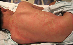
The elevated enzymes, though, caused a dilemma: Was it safe to start a potentially hepatotoxic tumor necrosis factor inhibitor in a patient with elevated liver enzymes? And were the enzymes elevated because of the GPP flare itself, or one of the concomitant medications?
Because cephalosporins can cause transient liver enzyme elevations, Dr. Cordoro discontinued the cephalosporin; the boy’s enzymes dropped within a day, which cleared the way for infliximab.
He turned the corner in 2 days; pustulation ceased and his skin began to heal. Given the severity of his flare, he is on adalimumab for maintenance therapy, Dr. Cordoro said.
“It gets complicated. You have to take everything into account,” she said, but “children with GPP tend to have an excellent prognosis as long as secondary complications are prevented or managed.”
Dr. Cordoro noted an educational Web site for children where they can learn about psoriasis and interact with other children with the condition.
Disclosures: Dr. Cordoro said she had no conflicts of interest.
PORTLAND, Ore. - Cyclosporine is often the best rescue therapy for acute generalized pustular psoriasis in children, according to Dr. Kelly M. Cordoro.
“When speed is important, my first choice is cyclosporine,” said Dr. Cordoro of the department of dermatology at the University of California, San Francisco. When adequately dosed, it can end pustulation within days.
Methotrexate and acitretin are also options, but can take several weeks to have maximum benefit, which is too slow for acute generalized pustular psoriasis (GPP) patients. GPP can cause fatal secondary infections, calcium imbalances, and cardiorespiratory collapse, although these outcomes are rare in children. She also noted methotrexate’s hepatotoxic potential, a concern in the treatment of GPP because the disease itself can cause acute liver dysfunction.
Acute GPP patients, apart from the pathognomonic skin findings, can present with fever, anorexia, and chills. Patients “are very sick,” she said.
Severe cases warrant hospital admission for treatment and supportive care, including rehydration, electrolyte imbalance correction, and bed rest. Compresses followed by bland emollients, such as petrolatum jelly, are essential to help prevent secondary infections and sepsis.
Rescue therapy is only the first of three stages by which Dr. Cordoro conceptualizes GPP management.
The second, transition therapy, usually includes tapering the cyclosporine and initiating other treatments for long-term control, such as acitretin, phototherapy, or both.
The maintenance stage comes after the patient has been stable or clear for months. Phototherapy and topical vitamin D analogues, such as Dovonex, are “great choices for maintenance,” Dr. Cordoro said. Low-dose acitretin, topical corticosteroids, and topical calcineurin inhibitors are other options.
Dr. Cordoro also tests the pharynx and perianal area for group A streptococcus colonization, and treats to eliminate it once her GPP patients are well enough for antibiotics. “It’s not known if it helps in the acute setting, but strep antigen may trigger psoriasis again at some point,” she said.
There are no consensus treatment guidelines for GPP in children; clinical decisions are based on patient characteristics, clinical experience and setting, and hunches about the best way to proceed. “There’s no cookbook approach, no one-size-fits-all treatment,” Dr. Cordoro said.
One of her patients, an 8-year-old boy with severe, relapsing/remitting GPP, illustrates the point.
He had been doing fairly well on low-dose acitretin, but then flared. His mother took him to an urgent care center, and the doctor there, mistaking GPP for an infection, started the boy on a cephalosporin.
His condition worsened and he was toxic by the time Dr. Cordoro arrived.
She admitted the patient to the hospital and initiated rescue doses of cyclosporine 5 mg/kg per day, along with supportive care. His condition deteriorated over the next few days, and his liver enzymes became elevated. Since he was failing on cyclosporine, she wanted to use infliximab for rescue therapy.
The elevated enzymes, though, caused a dilemma: Was it safe to start a potentially hepatotoxic tumor necrosis factor inhibitor in a patient with elevated liver enzymes? And were the enzymes elevated because of the GPP flare itself, or one of the concomitant medications?
Because cephalosporins can cause transient liver enzyme elevations, Dr. Cordoro discontinued the cephalosporin; the boy’s enzymes dropped within a day, which cleared the way for infliximab.
He turned the corner in 2 days; pustulation ceased and his skin began to heal. Given the severity of his flare, he is on adalimumab for maintenance therapy, Dr. Cordoro said.
“It gets complicated. You have to take everything into account,” she said, but “children with GPP tend to have an excellent prognosis as long as secondary complications are prevented or managed.”
Dr. Cordoro noted an educational Web site for children where they can learn about psoriasis and interact with other children with the condition.
Disclosures: Dr. Cordoro said she had no conflicts of interest.
Prompt Vertebroplasty Eased Acute Fracture Pain
Major Finding: 101 patients randomized to vertebroplasty for acute osteoporotic compression fractures had a mean reduction of 5.2 points on a 10-point pain scale a month after the procedure; 101 randomized to conservative treatment had a mean reduction of 2.7 points.
Data Source: Randomized, open-label trial.
Disclosures: The authors reported no conflicts of interest. The study was funded by a ZonMw, a Dutch health care research organization, and Cook Medical, which makes the bone cement used in the trial. The commentators reported receiving consulting fees and travel and accommodation expenses from Medtronic Spinal and Biologics Europa BVBA for their role in the FREE balloon kyphoplasty trial.
Vertebroplasty provided quicker, stronger, and more durable pain relief from acute, osteoporotic vertebral compression fractures than did conservative pain management, judging from the findings of a randomized, open-label trial.
“In a subgroup of patients with acute [fractures] and persistent pain, percutaneous vertebroplasty is effective and safe,” concluded Dr. Caroline Klazen, a radiologist at St. Elisabeth Hospital in Tilburg, the Netherlands, and her colleagues (Lancet 2010 Aug. 10 [doi:10.1016/S0140-6736(10)60954-3
Two previous studies found no benefit for vertebroplasty compared with bed rest, analgesics, and other conservative measures, but both trials included patients with fractures that were up to a year old (N. Engl. J. Med. 2009;361:557-68; N. Engl. J. Med. 2009;361:569-79).
The Lancet study pitted vertebroplasty against conservative treatment within a mean of 5.6 weeks of fracture symptom onset; vertebroplasty patients experienced greater pain relief initially and throughout the trial's year-long follow-up.
“Apparently,” vertebroplasty shortly after a fracture “is more effective for pain relief” than vertebroplasty performed months afterward, Dr. Klazen and her colleagues wrote.
Recruited from the radiology departments of six hospitals in the Netherlands and Belgium, 101 patients were randomized to vertebroplasty and 101 to conservative measures. Patients were at least 50 years old, and 69% were female.
All of the patients had radiologically confirmed compression fractures at or below thoracic vertebrae 5 with bone edema on magnetic resonance imaging and a minimum height loss of 15%, the authors noted.
They also had tenderness at the fracture level; bone density T scores at or below – 1; back pain for 6 weeks or less; and a pain score of at least 5 on a 10-point visual analog scale (VAS), with 10 being the worst pain.
In the vertebroplasty group, fractures were injected with a mean volume of 4.1 mL of polymethylmethacrylate bone cement under fluoroscopic guidance.
At 1 month, those injected with the bone cement had a mean reduction of 5.2 VAS points from baseline (95% CI, 5.88-4.72), compared with a mean reduction of 2.7 points (95% CI, 3.22-1.98) in those who were treated conservatively.
At 1 year, vertebroplasty subjects had a mean reduction of 5.7 VAS points from baseline (95% CI, 6.22-4.98); conservatively treated patients had a mean reduction of 3.7 points (95% CI, 4.35-3.05).
During the study, vertebroplasty patients used significantly less pain-relieving medication at day 1, week 1, and month 1, but the difference in drug use was not significant at later stages of follow-up.
The authors noted that the intervention was not blinded, and that “knowledge of the treatment assignment might have affected patient responses to questions or radiologist assessments.”
Computed tomographic scanning found that polymethylmethacrylate bone cement leaked out of 97 of the 134 vertebral bodies injected in the 101 vertebroplasty subjects. “Most leaks were discal or into segmental veins; none were into the spinal canal,” the authors noted.
Cement did deposit into a segmental pulmonary artery in one patient, however all cement leaks remained asymptomatic.
In a commentary, orthopedic surgeons Dr. Douglas Wardlaw, of Woodend Hospital in Aberdeen, Scotland, and Dr. Jan Van Meirhaeghe, of St. Jan General Hospital in Brugge, Belgium, noted that the study “lends support to the large body of medical opinion that vertebroplasty has a part to play in the management of the pain of vertebral compression fractures” (Lancet 2010 Aug. 10 [doi:10.1016/S0140-6736(10)61162-2
But they noted “an unexplained significant difference at baseline” in quality of life and disability measurements between the two groups that suggests “the control group might have been generally healthier than the vertebroplasty group.”
Dr. Klazen and her colleagues attributed the differences to chance.
Dr. Wardlaw and Dr. Van Meirhaeghe also noted that in the two previous studies that found no benefit for vertebroplasty, the comparators were sham treatments, not conservative pain management.
In one of the trials, the sham treatment included injecting bupivacaine, a long-acting local anesthetic, directly into fractures, which in itself might have brought relief, the commentators wrote.
By using conservative pain management as a comparator, Dr. Klazen and her colleagues noted, vertebroplasty was tested against “the reference treatment and thus provides the clinician with directly applicable information about how to best treat the patient.”
Dr. Klazen and her colleagues noted that ZonMw and Cook Medical, the sponsors of the study “had no role in study design, data collection, data analysis, data interpretation, writing of the report, or the decision to submit for publication.”
From The Lancet
Femur Fractures May Be Tied to Bisphosphonates
MARINA DEL REY, CALIF. — As the debate unfolds over both whether bisphosphonates cause femur fractures and the degree to which the benefits of drugs still outweigh the risks, a phenomenon has emerged.
Women who have taken bisphosphonates for years are being seen in doctors' offices with thigh pain that is easy to mistake for hip or knee arthritis. They have a unique constellation of radiologic findings on imaging, and they either have permanent titanium rods placed in their thigh bones or go on to full femur fractures—sometimes bilaterally—and permanent disability.
No one can yet say for sure whether they would have had those fractures regardless of bisphosphonate use, nor can anyone say if femur fractures are limited to alendronate (Fosamax) users or are a bisphosphonate class effect.
But a trend is emerging, and with it a treatment protocol.
“The thinking [among colleagues] is that this is novel and specific to bisphosphonate treatment, but only time will tell,” Dr. Benjamin C. Bengs, an orthopedic surgeon at the University of California, Los Angeles, said in an interview.
The possibility must be added to the hip pain differential in women with long-standing use of the drugs, according to Dr. Bengs and others who were interviewed for this story.
Unusual Radiology Findings
For many of the doctors who were interviewed, the question isn't so much whether bisphosphonates cause femur fractures, but rather how to care for women with a long history of using the drugs, and how to recognize signs and symptoms of impending trouble.
Any woman with pain over the thigh and those x-ray findings is definitely a candidate for prophylactic rodding, said Dr. Bengs.
The unique findings on x-ray are cortical thickening that is most pronounced on the lateral side of the femur, accompanied by a beaking lesion, also on the lateral side of the femur. Intramedullary edema is often present.
The beaking lesion is the start of a horizontal or oblique stress fracture. In time, there generally develops a “little, lucent, dark line extending from the beak to the middle of the bone,” Dr. Joseph Robinson, a radiology fellow at Cedars-Sinai Medical Center in Los Angeles, said in an interview.
“Lateral stress fractures are unusual. In our area, they are all related to bisphosphonates,” Dr. Kambiz Motamedi, a diagnostic radiologist at UCLA Medical Center.
There is a strong belief “from our sports medicine folks and rheumatologists” that there is a relationship with bisphosphonates, he said.
Both Legs Must Be Examined
Standard hip x-rays don't go far enough down to detect the lesion, which is closer to the knee than the typical femur fracture would be, Dr. Robinson said. When they do a hip series, Cedars-Sinai radiologists are careful to image lower down so they don't miss it, he said, noting that they are also putting markers on skin to identify the source of pain.
If the lesion is found, it is imperative to image the other femur as well, said Dr. Stuart L. Silverman, a rheumatologist in private practice in Beverly Hills, Calif. The other femur can go on to fracture, often within 18 months, he said in an interview.
Dr. Bengs noted that in most case, rodding is the usual treatment. It takes only a matter of weeks to recover from hip-to-knee rod placement, he said. It takes months to recover from a fracture, however, and older patients usually lose 10%-15% of their strength and ambulation during their convalescence, he added.
“The fractures are devastating,” said UCLA and Cedars-Sinai rheumatologist Dr. Solomon N. Forouzesh, who is the medical director of the rehabilitation department at Brotman Medical Center in Culver City, Calif. He said he's seen two cases in his practice.
Active, Osteopenic Women at Risk
Active women who have osteopenia—not osteoporosis—appear to be most at risk, Dr. Nancy Lane, a University of California, Davis, rheumatologist, said in an interview. “What I think is going on” is that bisphosphonates, by reducing bone turnover, lead to the overmineralization of cortical bone, she explained. “Over time, the bones become brittle [and] fail from too much mineralization. They cannot dissipate the load.”
It's “probably best not [to use bisphosphonates] in people with low risk of fractures who are very active,” she said. In the past, she added, use of the drugs might have been “too aggressive.”
A Drug Holiday Is Advised
As the story unfolds, doctors are using bisphosphonates for shorter lengths of time than in the past, followed by a drug holiday and ongoing bone-density monitoring.
The risk of fracture seems to “start at about 3 years and peaks between 5 and 6 years,” Dr. Forouzesh said. To be ahead of the game, he advises not waiting until the risk peaks. “Back off ahead of time. I do a drug holiday in 3 or 4 years,” he said.
It's not clear at this point if the phenomenon—if it is truly real—is limited to alendronate or is a bisphosphonate class effect.
Alendronate has been on the market the longest and has been the most widely used agent, Dr. Lane noted. “I am sure over time” it will emerge with other members of the class, as well, she said.
Meanwhile, there are efforts to raise awareness of the issue. The American College of Rheumatology issued a bulletin on the matter in March (“Atypical Femoral Fractures With Long-Term Bisphosphonate Use,” which can be accessed online at
www.rheumatology.org/publications/hotline/2010_03_22_bisphosphonate.asp
At Cedars-Sinai, “we are trying to have [a conference] to make rheumatologists and others aware of how to deal with the problem,” Dr. Robinson said.
A New Finding Unraveled, Slowly
Doctors first became aware of the issue a few years ago.
“First, there were a lot of reports in Singapore of unusual hip fractures before the lesser trochanter, [with] unusual x-ray changes,” Dr. Silverman said.
Reports started to be seen in the United States, and physicians across the country began sharing their stories. Only by reexamining medical records did doctors realize that the patients shared a common history of bisphosphonate use, Dr. Bengs said.
At the present, it's “a slow-rolling snowball that might turn out to be a very big deal given the number of women on bisphosphonates. Further vigilance and analysis are required,” he said.
Many of the physicians who were interviewed for this story said they are concerned that women will go off the drugs because of press reports.
They point out that in a recent bisphosphonate manufacturer–sponsored study, investigators couldn't disprove a connection, but did conclude that the fractures are rare, and that the benefit of taking the drugs still outweighs the risks (N. Engl. J. Med. 2010;362:1761-71).
Another manufacturer-sponsored study showed that subtrochanteric femur fractures have occurred in people who have not used bisphosphonates (Osteoporos. Int. 2010;21[suppl. 1]:S7-24).
“We have a new radiologic finding, but no data that the incidence of the fractures has gone up,” Dr. Silverman said.
The Food and Drug Administration said that it is staying on top of the issue. The agency continues “to evaluate the issue of the use of bisphosphonates and atypical femur fracture,” a press person wrote in response to an e-mail inquiry.
The agency has no plans at present for an advisory committee meeting regarding the issue.
Concerned Women
As the issue continues to be examined, doctors are fielding questions from concerned women.
“My answer [to them] is that it's an unknown and unconfirmed [phenomenon], and that the data are not out there to answer the questions,” said Dr. Eric M. Ruderman of the division of rheumatology at Northwestern University in Chicago.
At present, Dr. Ruderman doesn't stop bisphosphonate use because of fracture concerns, but he does have his patients take a drug holiday after 7 years because evidence is lacking for benefit after that point, he said. He continues monitoring bone density thereafter to assess the need for further treatment.
Disclosures: Dr. Motamedi, Dr. Robinson, and Dr. Forouzesh said that they had no conflicts to disclose. Dr. Bengs disclosed he is a paid consultant for Amgen Inc. Dr. Lane disclosed research grants, royalties, consulting fees from and positions of influence and ownership interests in Amgen, Eli Lilly & Co., and Pfizer Inc. Dr. Ruderman disclosed that he is a consultant for Amgen and Pfizer. Dr. Silverman disclosed he has served as a speaker, member of a speakers bureau, advisor for Eli Lilly, Novartis Pharmaceuticals Corp., Procter & Gamble, and Roche Inc., and that he has received research support from Eli Lilly, Procter & Gamble, Roche, and Novartis.
This femur fractured 2 days after the image revealed cortical thickening and a beaklike stress fracture, perhaps from bisphosphonates.
Source Courtesy UCLA
Women with thigh pain may have a bisphosphonate-induced fracture, said Dr. Benjamin C. Bengs.
Source Courtesy Dr. Benjamin C. Bengs
Expert analysis from a Rheumatology seminar sponsored by UCLA
Prompt Vertebroplasty Eased Acute Fracture Pain
Major Finding: 101 patients randomized to vertebroplasty for acute osteoporotic compression fractures had a mean reduction of 5.2 points on a 10-point pain scale a month after the procedure; 101 randomized to conservative treatment had a mean reduction of 2.7 points.
Data Source: Randomized, open-label trial.
Disclosures: The authors reported no conflicts of interest. The study was funded by a ZonMw, a Dutch health care research organization, and Cook Medical, which makes the bone cement used in the trial. The commentators reported receiving consulting fees and travel and accommodation expenses from Medtronic Spinal and Biologics Europa BVBA for their role in the FREE balloon kyphoplasty trial.
Vertebroplasty provided quicker, stronger, and more durable pain relief from acute, osteoporotic vertebral compression fractures than did conservative pain management, according to the findings of a randomized, open-label trial.
“In a subgroup of patients with acute [fractures] and persistent pain, percutaneous vertebroplasty is effective and safe,” concluded Dr. Caroline Klazen, a radiologist at St. Elisabeth Hospital in Tilburg, the Netherlands, and her colleagues (Lancet 2010 Aug. 10 [doi:10.1016/S0140-6736(10)60954-3]).
Two previous studies found no benefit for vertebroplasty compared with bed rest, analgesics, and other conservative measures.
But both trials included patients with fractures of up to a year old (N. Engl. J. Med. 2009;361:557-68; N. Engl. J. Med. 2009;361:569-79).
The current study pitted vertebroplasty against conservative treatment within a mean of 5.6 weeks of fracture symptom onset; vertebroplasty patients experienced greater pain relief initially and throughout the trial's year-long follow-up.
“Apparently,” vertebroplasty shortly after a fracture “is more effective for pain relief” than vertebroplasty performed months afterward, Dr. Klazen and her colleagues wrote.
Recruited from the radiology departments of six hospitals in the Netherlands and Belgium, 101 patients were randomized to vertebroplasty and 101 to conservative measures.
Patients were at least 50 years old, and 69% were female.
All of the patients had radiologically confirmed compression fractures at or below thoracic vertebrae 5 with bone edema on magnetic resonance imaging and a minimum height loss of 15%.
They also had tenderness at the fracture level; bone density T scores at or below −1; back pain for 6 weeks or less; and a pain score of at least 5 on a 10-point visual analog scale (VAS), with 10 being the worst pain.
In the vertebroplasty group, fractures were injected with a mean volume of 4.1 mL polymethylmetacrylate bone cement under fluoroscopic guidance.
At 1 month, those injected with the bone cement had a mean reduction of 5.2 VAS points from baseline (95% CI, 5.88-4.72), compared with a mean reduction of 2.7 points (95% CI, 3.22-1.98) in those treated conservatively. At 1 year, vertebroplasty subjects had a mean reduction of 5.7 VAS points from baseline (95% CI, 6.22-4.98).
Conservatively treated patients had a mean reduction of 3.7 points (95% CI, 4.35-3.05).
Vertebroplasty patients used significantly less pain-relieving medication at day 1, week 1, and month 1, but the difference in drug use was not significant at later stages of follow-up.
The authors noted that the intervention was not blinded, and that “knowledge of the treatment assignment might have affected patient responses to questions or radiologist assessments.”
Computed tomographic scanning found that cement leaked out of 97 of the 134 vertebral bodies injected in the 101 vertebroplasty subjects.
“Most leaks were discal or into segmental veins; none were into the spinal canal,” the authors noted.
Cement deposited in a segmental pulmonary artery in one patient, but all cement leaks remained asymptomatic.
In a related commentary, orthopedic surgeons Douglas Wardlaw, of Woodend Hospital in Aberdeen, Scotland, and Jan Van Meirhaeghe, of St. Jan General Hospital in Brugge, Belgium, noted that the study “lends support to the large body of medical opinion that vertebroplasty has a part to play in the management of the pain of vertebral compression fractures” (Lancet 2010 Aug. 10 [doi:10.1016/S0140-6736(10)61162-2]).
But they noted “an unexplained significant difference at baseline” in quality of life and disability measurements between the two groups.
That baseline disparity between treatment groups suggests “the control group might have been generally healthier than the vertebroplasty group.”
Dr. Klazen and her colleagues attributed the differences to chance.
Dr. Wardlaw and Dr. Van Meirhaeghe also noted that in the two previous studies that found no benefit for vertebroplasty, the comparators were sham treatments, not conservative pain management.
In one of the trials, the sham included injecting bupivacaine, a long-acting local anesthetic, into fractures, which itself might have brought relief, they wrote.
By using conservative pain management with activity restriction and pain medications as a comparator, Dr. Klazen and her colleagues noted, vertebroplasty was tested against “the reference treatment and thus provides the clinician with directly applicable information about how to best treat the patient.”
Dr. Klazen and her colleagues reported no conflicts of interest. The study was funded by ZonMw, a Dutch organization for health care research and innovation, and Cook Medical, makers of the bone cement used in the trial.
“The sponsors of the study had no role in study design, data collection, data analysis, data interpretation, writing of the report, or the decision to submit for publication,” Dr. Klazen and her colleagues wrote.