User login
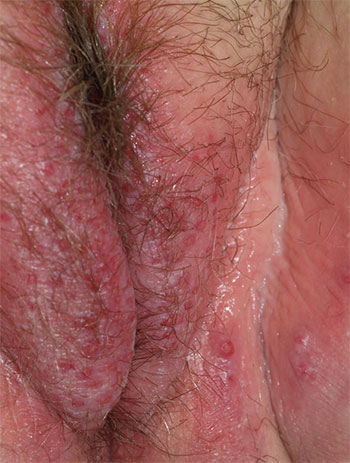
1. A 25-year-old woman reports anogenital itching, burning, and redness, present for 3 months. She says she developed a yeast infection after antibiotic therapy for a dental infection. The yeast infection was treated with terconazole, which resulted in immediate severe burning, redness, and swelling. Clobetasol cream used twice daily also caused burning, so she discontinued it. Her symptoms improved when she tried cool soaks and applied topical benzocaine gel as a local anesthetic.

Diagnosis: Irritant contact dermatitis (as opposed to allergic contact dermatitis) associated with the use of terconazole and clobetasol. This was followed by allergic contact dermatitis in association with benzocaine. Treatment consists of withdrawal of benzocaine, reinitiation of cool soaks, and a switch to clobetasol ointment rather than cream. Nighttime sedation enables the patient to sleep through the itching and gradually allows her skin to heal.
For more information on this case, see “Chronic vulvar irritation, itching, and pain. What is the diagnosis?” OBG Manag. 2014;26(6):30-37.
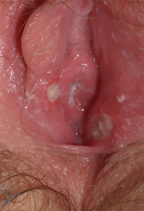
2. This 13-year-old presents with sudden-onset vulvar pain and sores. The child developed a sore throat and low-grade fever 3 days earlier, with vulvar pain and vulvar dysuria the next day. Oral acyclovir was prescribed for herpes simplex virus infection, but the girl’s condition has not improved. She claims sexual abstinence, and her mother believes her.

Diagnosis: Vulvar aphthae, believed to be of hyperimmune origin, are often precipitated by a viral syndrome. They are most common in girls aged 9 to 18 years.
Aphthae are uncommon and under-recognized on the vulva. Genital aphthae are usually much larger than oral aphthae. Most patients are mistakenly evaluated and treated for sexually transmitted infection, but the large, well-demarcated, painful, nonindurated, deep nature of the ulcer is pathognomonic for an aphthous ulcer.
Recommended treatment is prednisone 40 mg/day plus hydrocodone in usual doses of 5/325, one or two tablets every 4 to 6 hours, as needed; topical petroleum jelly (especially before urination); and sitz baths. When the patient returns one week later, she is much improved.
For more information on this case, see “Chronic vulvar irritation, itching, and pain. What is the diagnosis?” OBG Manag. 2014;26(6):30-37.
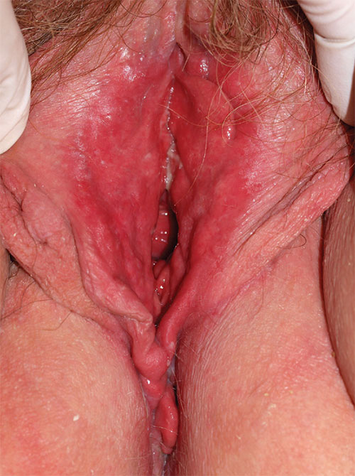
3. A 36-year-old woman reports introital itching, vulvar dysuria, and superficial dyspareunia that have lasted 6 months. Apparent on physical examination are redness of the vestibule, medial labia minora, and vaginal walls, with edema of the surrounding skin and yellowish, copious vaginal secretions at the introitus. Lab tests for chlamydia, trichomonas, and gonorrhea are returned as normal.

Diagnosis: Desquamative inflammatory vaginitis (DIV) is described as noninfectious inflammatory vaginitis in a setting of normal estrogen and absence of skin disease of the mucous membranes of the vagina. The condition is characterized by an increase in white blood cells and parabasal cells and absent lactobacilli, with relatively high vaginal pH. DIV is thought to represent an inflammatory dermatosis of the vaginal epithelium. Although some clinicians believe that DIV is actually lichen planus, the latter exhibits erosions as well as redness, nearly always affects the mouth and the vulva, and produces remarkable scarring. DIV does not erode, affect any other skin surfaces, or scar.
Treatment for DIV consists of clindamycin vaginal cream, 1/2 to 1 full applicator nightly, with a weekly oral fluconazole tablet (200 mg is more easily covered by insurance) to prevent secondary candidiasis. Schedule a follow-up visit in one month.
For more information on this case, see “Chronic vulvar irritation, itching, and pain. What is the diagnosis?” OBG Manag. 2014;26(6):30-37.
4. A 43-year-old woman reports a “recalcitrant yeast infection” of the vulva, with itching and irritation. She is overweight and diabetic, with mild stress incontinence. Physical examination reveals a fairly well-demarcated red, rough plaque on the vulva and labiocrural folds, with satellite red papules and peripheral peeling. Similar plaques occur in the gluteal cleft, umbilicus, and axillae as well as under the breasts. A fungal preparation of the vagina and skin is negative.

Diagnosis: Of the several morphologic types of psoriasis, anogenital psoriasis is most often of the inverse pattern. Inverse psoriasis preferentially affects skin folds and is frequently mistaken for (and often initially superinfected with) candidiasis. Scale is thin and unapparent, and there often is a shiny, glazed appearance to the skin. Tiny satellite lesions are often visible as well. A skin biopsy of inverse psoriasis often is not diagnostic, showing only nonspecific psoriasiform dermatitis; this does not disprove psoriasis.
Psoriasis is a systemic condition and is associated with metabolic syndrome, carrying an increased risk for overweight, hypertension, diabetes, and cardiovascular disease. Management of these conditions is very important in the overall treatment of the patient.
The recommended treatment is clobetasol ointment applied to the skin folds, along with continuation of the topical miconazole cream. A week later, the patient’s condition is remarkably improved, and her biopsy shows psoriasiform dermatitis. The potency of her corticosteroid was reduced by switching to desonide cream, sparingly applied daily.
For more information on this case, see “Chronic vulvar irritation, itching, and pain. What is the diagnosis?” OBG Manag. 2014;26(6):30-37.
1. A 25-year-old woman reports anogenital itching, burning, and redness, present for 3 months. She says she developed a yeast infection after antibiotic therapy for a dental infection. The yeast infection was treated with terconazole, which resulted in immediate severe burning, redness, and swelling. Clobetasol cream used twice daily also caused burning, so she discontinued it. Her symptoms improved when she tried cool soaks and applied topical benzocaine gel as a local anesthetic.

Diagnosis: Irritant contact dermatitis (as opposed to allergic contact dermatitis) associated with the use of terconazole and clobetasol. This was followed by allergic contact dermatitis in association with benzocaine. Treatment consists of withdrawal of benzocaine, reinitiation of cool soaks, and a switch to clobetasol ointment rather than cream. Nighttime sedation enables the patient to sleep through the itching and gradually allows her skin to heal.
For more information on this case, see “Chronic vulvar irritation, itching, and pain. What is the diagnosis?” OBG Manag. 2014;26(6):30-37.
2. This 13-year-old presents with sudden-onset vulvar pain and sores. The child developed a sore throat and low-grade fever 3 days earlier, with vulvar pain and vulvar dysuria the next day. Oral acyclovir was prescribed for herpes simplex virus infection, but the girl’s condition has not improved. She claims sexual abstinence, and her mother believes her.

Diagnosis: Vulvar aphthae, believed to be of hyperimmune origin, are often precipitated by a viral syndrome. They are most common in girls aged 9 to 18 years.
Aphthae are uncommon and under-recognized on the vulva. Genital aphthae are usually much larger than oral aphthae. Most patients are mistakenly evaluated and treated for sexually transmitted infection, but the large, well-demarcated, painful, nonindurated, deep nature of the ulcer is pathognomonic for an aphthous ulcer.
Recommended treatment is prednisone 40 mg/day plus hydrocodone in usual doses of 5/325, one or two tablets every 4 to 6 hours, as needed; topical petroleum jelly (especially before urination); and sitz baths. When the patient returns one week later, she is much improved.
For more information on this case, see “Chronic vulvar irritation, itching, and pain. What is the diagnosis?” OBG Manag. 2014;26(6):30-37.
3. A 36-year-old woman reports introital itching, vulvar dysuria, and superficial dyspareunia that have lasted 6 months. Apparent on physical examination are redness of the vestibule, medial labia minora, and vaginal walls, with edema of the surrounding skin and yellowish, copious vaginal secretions at the introitus. Lab tests for chlamydia, trichomonas, and gonorrhea are returned as normal.

Diagnosis: Desquamative inflammatory vaginitis (DIV) is described as noninfectious inflammatory vaginitis in a setting of normal estrogen and absence of skin disease of the mucous membranes of the vagina. The condition is characterized by an increase in white blood cells and parabasal cells and absent lactobacilli, with relatively high vaginal pH. DIV is thought to represent an inflammatory dermatosis of the vaginal epithelium. Although some clinicians believe that DIV is actually lichen planus, the latter exhibits erosions as well as redness, nearly always affects the mouth and the vulva, and produces remarkable scarring. DIV does not erode, affect any other skin surfaces, or scar.
Treatment for DIV consists of clindamycin vaginal cream, 1/2 to 1 full applicator nightly, with a weekly oral fluconazole tablet (200 mg is more easily covered by insurance) to prevent secondary candidiasis. Schedule a follow-up visit in one month.
For more information on this case, see “Chronic vulvar irritation, itching, and pain. What is the diagnosis?” OBG Manag. 2014;26(6):30-37.
4. A 43-year-old woman reports a “recalcitrant yeast infection” of the vulva, with itching and irritation. She is overweight and diabetic, with mild stress incontinence. Physical examination reveals a fairly well-demarcated red, rough plaque on the vulva and labiocrural folds, with satellite red papules and peripheral peeling. Similar plaques occur in the gluteal cleft, umbilicus, and axillae as well as under the breasts. A fungal preparation of the vagina and skin is negative.

Diagnosis: Of the several morphologic types of psoriasis, anogenital psoriasis is most often of the inverse pattern. Inverse psoriasis preferentially affects skin folds and is frequently mistaken for (and often initially superinfected with) candidiasis. Scale is thin and unapparent, and there often is a shiny, glazed appearance to the skin. Tiny satellite lesions are often visible as well. A skin biopsy of inverse psoriasis often is not diagnostic, showing only nonspecific psoriasiform dermatitis; this does not disprove psoriasis.
Psoriasis is a systemic condition and is associated with metabolic syndrome, carrying an increased risk for overweight, hypertension, diabetes, and cardiovascular disease. Management of these conditions is very important in the overall treatment of the patient.
The recommended treatment is clobetasol ointment applied to the skin folds, along with continuation of the topical miconazole cream. A week later, the patient’s condition is remarkably improved, and her biopsy shows psoriasiform dermatitis. The potency of her corticosteroid was reduced by switching to desonide cream, sparingly applied daily.
For more information on this case, see “Chronic vulvar irritation, itching, and pain. What is the diagnosis?” OBG Manag. 2014;26(6):30-37.
1. A 25-year-old woman reports anogenital itching, burning, and redness, present for 3 months. She says she developed a yeast infection after antibiotic therapy for a dental infection. The yeast infection was treated with terconazole, which resulted in immediate severe burning, redness, and swelling. Clobetasol cream used twice daily also caused burning, so she discontinued it. Her symptoms improved when she tried cool soaks and applied topical benzocaine gel as a local anesthetic.

Diagnosis: Irritant contact dermatitis (as opposed to allergic contact dermatitis) associated with the use of terconazole and clobetasol. This was followed by allergic contact dermatitis in association with benzocaine. Treatment consists of withdrawal of benzocaine, reinitiation of cool soaks, and a switch to clobetasol ointment rather than cream. Nighttime sedation enables the patient to sleep through the itching and gradually allows her skin to heal.
For more information on this case, see “Chronic vulvar irritation, itching, and pain. What is the diagnosis?” OBG Manag. 2014;26(6):30-37.
2. This 13-year-old presents with sudden-onset vulvar pain and sores. The child developed a sore throat and low-grade fever 3 days earlier, with vulvar pain and vulvar dysuria the next day. Oral acyclovir was prescribed for herpes simplex virus infection, but the girl’s condition has not improved. She claims sexual abstinence, and her mother believes her.

Diagnosis: Vulvar aphthae, believed to be of hyperimmune origin, are often precipitated by a viral syndrome. They are most common in girls aged 9 to 18 years.
Aphthae are uncommon and under-recognized on the vulva. Genital aphthae are usually much larger than oral aphthae. Most patients are mistakenly evaluated and treated for sexually transmitted infection, but the large, well-demarcated, painful, nonindurated, deep nature of the ulcer is pathognomonic for an aphthous ulcer.
Recommended treatment is prednisone 40 mg/day plus hydrocodone in usual doses of 5/325, one or two tablets every 4 to 6 hours, as needed; topical petroleum jelly (especially before urination); and sitz baths. When the patient returns one week later, she is much improved.
For more information on this case, see “Chronic vulvar irritation, itching, and pain. What is the diagnosis?” OBG Manag. 2014;26(6):30-37.
3. A 36-year-old woman reports introital itching, vulvar dysuria, and superficial dyspareunia that have lasted 6 months. Apparent on physical examination are redness of the vestibule, medial labia minora, and vaginal walls, with edema of the surrounding skin and yellowish, copious vaginal secretions at the introitus. Lab tests for chlamydia, trichomonas, and gonorrhea are returned as normal.

Diagnosis: Desquamative inflammatory vaginitis (DIV) is described as noninfectious inflammatory vaginitis in a setting of normal estrogen and absence of skin disease of the mucous membranes of the vagina. The condition is characterized by an increase in white blood cells and parabasal cells and absent lactobacilli, with relatively high vaginal pH. DIV is thought to represent an inflammatory dermatosis of the vaginal epithelium. Although some clinicians believe that DIV is actually lichen planus, the latter exhibits erosions as well as redness, nearly always affects the mouth and the vulva, and produces remarkable scarring. DIV does not erode, affect any other skin surfaces, or scar.
Treatment for DIV consists of clindamycin vaginal cream, 1/2 to 1 full applicator nightly, with a weekly oral fluconazole tablet (200 mg is more easily covered by insurance) to prevent secondary candidiasis. Schedule a follow-up visit in one month.
For more information on this case, see “Chronic vulvar irritation, itching, and pain. What is the diagnosis?” OBG Manag. 2014;26(6):30-37.
4. A 43-year-old woman reports a “recalcitrant yeast infection” of the vulva, with itching and irritation. She is overweight and diabetic, with mild stress incontinence. Physical examination reveals a fairly well-demarcated red, rough plaque on the vulva and labiocrural folds, with satellite red papules and peripheral peeling. Similar plaques occur in the gluteal cleft, umbilicus, and axillae as well as under the breasts. A fungal preparation of the vagina and skin is negative.

Diagnosis: Of the several morphologic types of psoriasis, anogenital psoriasis is most often of the inverse pattern. Inverse psoriasis preferentially affects skin folds and is frequently mistaken for (and often initially superinfected with) candidiasis. Scale is thin and unapparent, and there often is a shiny, glazed appearance to the skin. Tiny satellite lesions are often visible as well. A skin biopsy of inverse psoriasis often is not diagnostic, showing only nonspecific psoriasiform dermatitis; this does not disprove psoriasis.
Psoriasis is a systemic condition and is associated with metabolic syndrome, carrying an increased risk for overweight, hypertension, diabetes, and cardiovascular disease. Management of these conditions is very important in the overall treatment of the patient.
The recommended treatment is clobetasol ointment applied to the skin folds, along with continuation of the topical miconazole cream. A week later, the patient’s condition is remarkably improved, and her biopsy shows psoriasiform dermatitis. The potency of her corticosteroid was reduced by switching to desonide cream, sparingly applied daily.
For more information on this case, see “Chronic vulvar irritation, itching, and pain. What is the diagnosis?” OBG Manag. 2014;26(6):30-37.