User login
Bringing you the latest news, research and reviews, exclusive interviews, podcasts, quizzes, and more.
Maintenance lenalidomide prolongs progression-free survival in DLBCL
Maintenance therapy with lenalidomide significantly prolongs progression-free survival (PFS) in elderly patients with diffuse large B-cell lymphoma (DLBCL), according to findings from a phase III trial.
None of the previous trials assessing a maintenance drug in this patient population – including bevacizumab, rituximab, enzastaurin, or everolimus – have reported such a benefit, Catherine Thieblemont, MD, PhD, of the department of hematology-oncology, Hôpital Saint-Louis, Paris, and her associates, reported.
The standard regimen for newly diagnosed DLBCL is R-CHOP (rituximab, cyclophosphamide, doxorubicin, vincristine, and prednisone), but 30%-40% of patients have disease progression or relapse, usually during the first 2 years after diagnosis. Lenalidomide is an immunomodulator with antineoplastic activity and has shown activity in relapsed DLBCL. The investigators compared 24 months of lenalidomide maintenance therapy against a matching placebo in an international, randomized double-blind phase III trial involving 650 patients aged 60-80 years who showed either partial or complete responses to first-line R-CHOP.
After a median follow-up of 39 months (range, 0-74 months), median PFS was not yet reached in the lenalidomide group and was estimated to be 58.9 months in the placebo group, for a hazard ratio of 0.708 favoring lenalidomide. This represents a statistically significant and clinically meaningful improvement in PFS, Dr. Thieblemont and her associates noted (J Clin Oncol. 2017 Apr 20. doi: 10.1200/JCO.2017.72.6984).
This survival benefit was consistent across all subgroups of patients. Most important, PFS was significantly prolonged regardless of whether patients had shown only a partial response or a complete response to R-CHOP, the investigators said.
The maintenance therapy did not appear to prolong overall survival, which was 87% for lenalidomide and 89% for placebo.
“We do not yet fully understand the basis for lack of an [overall survival] benefit despite the positive PFS data. Other than that, this is not due to excessive toxicity in the experimental arm,” they said. “We speculate the reason may be differences in the outcomes after progression or some other unrecognized reason.”
The study was sponsored by the Lymphoma Academic Research Organisation of France and Celgene. Dr. Thieblemont reported ties to Celgene, Bayer, AbbVie, Janssen, and Roche. Her associates reported ties to numerous industry sources.
Key clinical point:
Major finding: Median progression-free survival was not yet reached in the lenalidomide group and was estimated to be 58.9 months in the placebo group, for a hazard ratio of 0.708 favoring lenalidomide.
Data source: A 5-year international, randomized double-blind placebo-controlled phase III trial involving 650 patients aged 60-80 years.
Disclosures: The study was sponsored by the Lymphoma Academic Research Organisation of France and Celgene. Dr. Thieblemont reported ties to Celgene, Bayer, AbbVie, Janssen, and Roche. Her associates reported ties to numerous industry sources.
In mantle cell lymphoma, triple therapy proves too toxic
Combined idelalisib, lenalidomide, and rituximab proved excessively toxic for the treatment of relapsed and refractory mantle cell and follicular lymphoma in two phase I trials conducted by the Alliance for Clinical Trials in Oncology.
The unexpected outcome, which led to early study termination, underscores the need for caution as new treatment combinations are proposed, Sonali M. Smith, MD, of the University of Chicago and her colleagues said in The Lancet Haematology.
In four of the first eight patients enrolled in the mantle cell lymphoma (A051201) and follicular lymphoma (A051202) phase I trials between July 9, 2013, and Sept. 30, 2014, unexpected dose-limiting toxicities occurred, including grade 4 sepsis syndrome, grade 4 hypotension with grade 3 rash and fevers, grade 4 aspartate aminotransferase (AST) or alanine aminotransferase (ALT) elevation with fevers, and grade 3 pulmonary infection with grade 3 maculopapular rash.
The adverse events occurred between 9 and 20 days after treatment initiation and coincided with rituximab infusions, the researchers said. No treatment-related deaths occurred (Lancet Haematol. 2017 Apr;4:e176-82).
Overall, 8 of 11 patients were removed from treatment because of an adverse event, and 3 of those required intensive care unit level of care.
Although rituximab was removed in both trials, two of the remaining three patients in the studies, including three with mantle cell lymphoma and eight with follicular lymphoma, experienced grade 3 rashes, and one had grade 3 AST elevations. In those with mantle cell lymphoma, the most common grade 3-4 adverse events were ALT elevations and rash. In those with follicular lymphoma, the most common grade 3-4 adverse events were neutropenia and rash.
“Given the inability to deliver treatment due to toxicity, both studies were permanently closed,” the researchers wrote, noting that the primary endpoint of safety and tolerability was not met.
The trials had the overall goal of developing targeted regimens to replace cytotoxic therapy.
“Both ... trials were designed to capitalize on the clinical synergy of lenalidomide and rituximab observed in previous trials by adding the highly specific PI3K delta inhibitor, idelalisib, for patients with relapsed mantle cell lymphoma and follicular lymphoma,” they said.
Previously available data implied that lenalidomide plus rituximab would be a safe backbone for therapy, and there was clinical rationale for adding idelalisib to that combination, they explained.
“Overall, our brief experience underscores the limited knowledge regarding drug interactions and off-target effects and serves as a cautionary note in developing biological agents in combination and against ad-hoc combinations outside of carefully monitored clinical trials,” they said.
The researchers noted that the nature of the toxicities observed in these trials supports an immune-activated state characterized by excessive inflammation.
“A more detailed assessment of effect on cytokines, T-cell subsets, natural killer cells, and clinical features predictive of toxicity and response should be included in any further testing of these classes of agents, and they should never be combined outside of a carefully designed and diligently monitored clinical trial setting,” they concluded.
The study was funded by the National Cancer Institute. Dr. Smith received research funding and consulting fees from Gilead and Celgene.
The findings by Dr. Smith and her colleagues add to several other reported studies that involved unexpected toxicities with various combinations of targeted agents in lymphoid malignancies.
Combinations of B-cell receptor signaling inhibitors can lead to immune dysregulation, which can be acute and severe when combined with immunomodulatory agents.
While the study of rational targeted combinations continues to hold immense potential in both untreated and relapsed/refractory disease, the combination must be thoroughly studied in the context of carefully and conservatively designed clinical trials.
Given the unpredictable nature of adverse events, the use of novel combinations outside of a clinical trial should be strongly discouraged.
Patrick M. Reagan, MD , and Paul M. Barr, MD , are with the James P. Wilmot Cancer Institute, University of Rochester, New York. Dr. Reagan reported having no disclosures. Dr. Barr has consulted for Gilead, Pharmacyclics, AbbVie, and Celgene. They made their remarks in an editorial that accompanied the article.
Combined idelalisib, lenalidomide, and rituximab proved excessively toxic for the treatment of relapsed and refractory mantle cell and follicular lymphoma in two phase I trials conducted by the Alliance for Clinical Trials in Oncology.
The unexpected outcome, which led to early study termination, underscores the need for caution as new treatment combinations are proposed, Sonali M. Smith, MD, of the University of Chicago and her colleagues said in The Lancet Haematology.
In four of the first eight patients enrolled in the mantle cell lymphoma (A051201) and follicular lymphoma (A051202) phase I trials between July 9, 2013, and Sept. 30, 2014, unexpected dose-limiting toxicities occurred, including grade 4 sepsis syndrome, grade 4 hypotension with grade 3 rash and fevers, grade 4 aspartate aminotransferase (AST) or alanine aminotransferase (ALT) elevation with fevers, and grade 3 pulmonary infection with grade 3 maculopapular rash.
The adverse events occurred between 9 and 20 days after treatment initiation and coincided with rituximab infusions, the researchers said. No treatment-related deaths occurred (Lancet Haematol. 2017 Apr;4:e176-82).
Overall, 8 of 11 patients were removed from treatment because of an adverse event, and 3 of those required intensive care unit level of care.
Although rituximab was removed in both trials, two of the remaining three patients in the studies, including three with mantle cell lymphoma and eight with follicular lymphoma, experienced grade 3 rashes, and one had grade 3 AST elevations. In those with mantle cell lymphoma, the most common grade 3-4 adverse events were ALT elevations and rash. In those with follicular lymphoma, the most common grade 3-4 adverse events were neutropenia and rash.
“Given the inability to deliver treatment due to toxicity, both studies were permanently closed,” the researchers wrote, noting that the primary endpoint of safety and tolerability was not met.
The trials had the overall goal of developing targeted regimens to replace cytotoxic therapy.
“Both ... trials were designed to capitalize on the clinical synergy of lenalidomide and rituximab observed in previous trials by adding the highly specific PI3K delta inhibitor, idelalisib, for patients with relapsed mantle cell lymphoma and follicular lymphoma,” they said.
Previously available data implied that lenalidomide plus rituximab would be a safe backbone for therapy, and there was clinical rationale for adding idelalisib to that combination, they explained.
“Overall, our brief experience underscores the limited knowledge regarding drug interactions and off-target effects and serves as a cautionary note in developing biological agents in combination and against ad-hoc combinations outside of carefully monitored clinical trials,” they said.
The researchers noted that the nature of the toxicities observed in these trials supports an immune-activated state characterized by excessive inflammation.
“A more detailed assessment of effect on cytokines, T-cell subsets, natural killer cells, and clinical features predictive of toxicity and response should be included in any further testing of these classes of agents, and they should never be combined outside of a carefully designed and diligently monitored clinical trial setting,” they concluded.
The study was funded by the National Cancer Institute. Dr. Smith received research funding and consulting fees from Gilead and Celgene.
Key clinical point:
Major finding: Of 11 patients, 8 were removed from treatment because of an adverse event, and 3 of those required intensive care unit–level care.
Data source: Two phase I trials involving 11 patients.
Disclosures: The study was funded by the National Cancer Institute. Dr. Smith received research funding and consulting fees from Gilead and Celgene.
Prognostic tool may allow tailored therapy for Hodgkin lymphoma
A newly developed “robust and inexpensive” prognostic tool, the Childhood Hodgkin International Prognostic Score (CHIPS), may allow better tailoring of therapy at the time of diagnosis for children and adolescents who have intermediate-risk Hodgkin lymphoma.
Researchers in the Children’s Oncology Group first assessed 562 patients receiving uniform standard treatment to identify which risk factors present at diagnosis best predicted response to therapy, and used them to develop the prognostic score. They considered such factors as patient age, the number of involved sites, hemoglobin level, albumin level, erythrocyte sedimentation rate, the presence or absence of a large mediastinal mass, nodal involvement, the total bulk of disease, and the presence or absence of B symptoms (fever, weight loss, and/or night sweats). The final CHIPS prognostic tool included four predictors of poor event-free survival that are easily ascertained at diagnosis: stage IV disease, a large mediastinal mass (one with a tumor to thoracic diameter ratio over 0.33), the presence of fever, and hypoalbuminemia (a level of less than 3.5 g/dL).
The investigators then confirmed the accuracy of that score in a validation cohort of 541 patients from the United States, Canada, Switzerland, Australia, New Zealand, the Netherlands, and Israel. All the study participants received four cycles of doxorubicin, bleomycin, vincristine, and etoposide (ABVE) with prednisone and cyclophosphamide (PC), followed by involved-field radiation therapy, said Cindy L. Schwartz, MD, of the division of pediatrics, University of Texas MD Anderson Cancer Center, Houston, and her associates.
Patients who had low a CHIPS of 0 or 1 had excellent 4-year event-free survival (93% and 89%, respectively), while patients with a high CHIPS of 2 or 3 had poorer 4-year event-free survival (78% and 69%, respectively). These findings remained consistent across all subgroups of patients, regardless of whether the tumors had nodular sclerosis histology or mixed cellular histology (Pediatr Blood Cancer. 2017 Apr;64[4]).
The study results suggest that patients with CHIPS 2 or 3 could be considered for high-risk Hodgkin lymphoma treatment such as higher-dose cyclophosphamide or the addition of brentuximab vedotin, while those with a CHIPS 0 or 1 could be considered for less aggressive treatment such as foregoing or reducing radiation therapy, Dr. Schwartz and her associates said.
This study was supported by a grant from the National Cancer Institute to the Children’s Oncology Group. Dr. Schwartz and her associates reported having no relevant financial disclosures.
Key clinical point: A newly developed “robust and inexpensive” prognostic tool, the Childhood Hodgkin International Prognostic Score, or CHIPS, may allow more tailored therapy for children and adolescents who have intermediate-risk Hodgkin lymphoma.
Major finding: Patients who had a low CHIPS of 0 or 1 had excellent 4-year event-free survival (93.1% and 88.5%, respectively), while patients with a high CHIPS of 2 or 3 had poorer 4-year event-free survival (77.6% and 69.2%).
Data source: A cohort study to develop (in 562 patients) and validate (in 541 patients) a score for predicting the response to standard treatment in pediatric Hodgkin lymphoma.
Disclosures: This study was supported by a grant from the National Cancer Institute to the Children’s Oncology Group. Dr. Schwartz and her associates reported having no relevant financial disclosures.
Thousands expected to get CAR T-cells by 2018
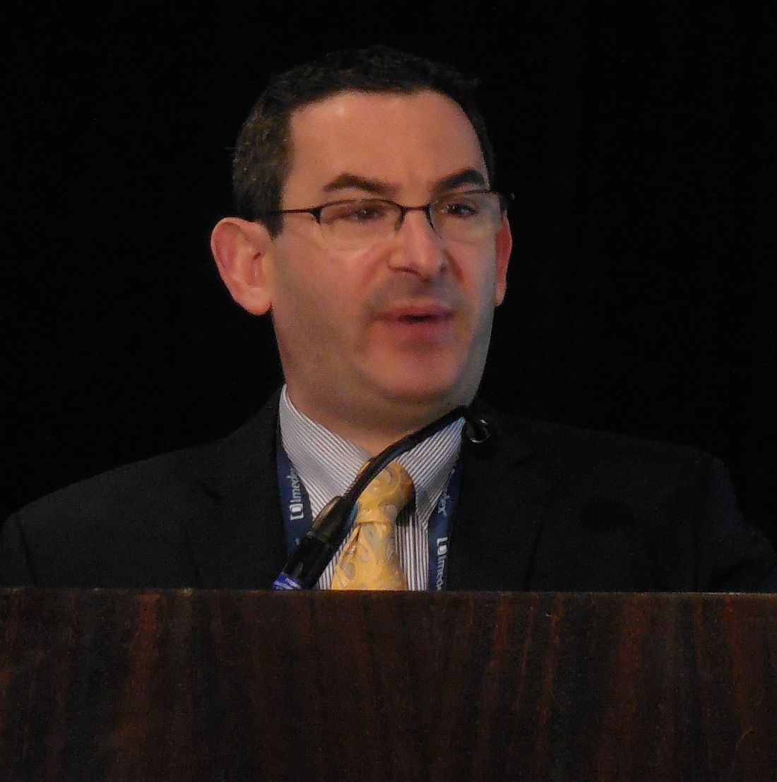
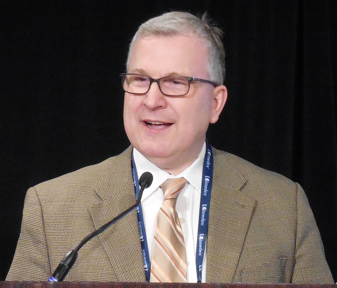
NEW YORK – Should the first chimeric antigen receptor (CAR) T-cell treatment receive Food and Drug Administration approval for refractory, aggressive non-Hodgkin lymphoma, possibly before the end of 2017, several thousand U.S. patients will be potential candidates for the treatment, Jeremy S. Abramson, MD, predicted at a conference held by Imedex.
Dr. Abramson has led studies using a CAR T cell that differs from the one furthest along in development. He based his predicted timetable for an approved biologics license of the process, with which he can engineer patient-specific T cells that are under development by Kite Pharma, on the “remarkable” level of complete responses the intervention produced in a pivotal phase II study.
“Seeing a complete response rate of close to 40% that is sustained for more than 3 months and a complete response rate of close to a third at 6 months is light years beyond what is now available for patients” with these chemotherapy refractory B-cell lymphomas, said Dr. Abramson, clinical director of the Center for Lymphoma at Massachusetts General Hospital in Boston. “This is the first treatment to show a significant impact on large B-cell lymphoma, and that makes me optimistic” that the FDA will grant license approval later in 2017, he said in an interview.
Dr. Abramson acknowledged that some patients developed grade 3 or 4 cytokine-release syndrome and neurologic events, but the events were reversible and manageable if treated with the immunosuppressant tocilizumab (Actemra) or a corticosteroid.
At the end of March, Kite reported the completion of its FDA application, and, on April 2, the ZUMA-1 investigators presented an updated report on their results, with a 36% complete response rate across all enrolled patients at 6 month follow-up and a 39% complete response rate in all patients out to a median follow-up of 8.7 months.
Annually, in U.S. practice, perhaps 3,000 new patients with diffuse large B-cell lymphoma would meet the enrollment criteria for ZUMA-1, Dr. Abramson estimated. Once axicabtagene ciloleucel becomes commercially available in the United States, several thousand U.S. patients might initially seek the treatment.
Dr. Abramson has been a consultant to Kite Pharma and to AbbVie, Genentech, Gilead, and Seattle Genetics.
[email protected]
On Twitter @mitchelzoler
EXPERT ANALYSIS FROM A MEETING ON HEMATOLOGIC MALIGNANCIES
Waldenström macroglobulinemia panel advises on IgM paraproteinemic neuropathies
With optimal approaches still evolving for the diagnosis and management of peripheral neuropathies associated with Waldenström macroglobulinemia and other IgM paraproteinemias, new consensus recommendations from a multidisciplinary panel were recently published in the British Journal of Haematology.
Up to half of patients with IgM monoclonal gammopathies develop peripheral neuropathy, according to the 11-member panel (Br J Haematol. 2017 Mar;176[5]:728-42). The panel began deliberations at the eighth International Workshop on Waldenström Macroglobulinemia in London and was led by Shirley D’Sa, MD, of the Waldenström Clinic, Cancer Division, University College London Hospitals NHS Foundation Trust.
• Diagnostic evaluation: “The indications for invasive investigations such as cerebrospinal fluid analysis, nerve conduction tests, and sensory nerve biopsies are unclear,” according to the panelists.
When clinical examination identifies a neuropathy, neurophysiologic testing can ascertain its nature and inform additional work-up. Cerebrospinal fluid examination is not mandatory in cases of demyelinating neuropathy, but it is indicated when clinical evaluation is inconclusive and malignancy or CNS invasion is suspected.
Nerve biopsy carries substantial risk and is rarely indicated. It may be warranted when a comprehensive systemic work-up has not identified a cause and clinicians still suspect amyloid, vasculitis, or direct cellular invasion; in atypical cases not responding to treatment; or when the neuropathy is progressive and debilitating.
When it comes to imaging, “MRI of the neuraxis should be performed prior to lumbar puncture to avoid false positive meningeal enhancement,” they advised. “Prior discussion of likely sites of involvement with an experienced neuroradiologist will ensure that the correct sequences of the correct anatomical area are performed with appropriate gadolinium enhancement.”
• Clinical phenotypes and their treatment: IgM-associated neuropathies vary with respect to specific antibodies present and the likelihood that they are causally associated with the neuropathy, Dr. D’Sa and her colleagues noted. They provided a decision tree to help guide the work-up to determine the specific etiology.
“The presence of a neuropathy alone is not a justification for treatment, but steady progression with accumulating disability should prompt action,” they maintained.
Patients with antibody-negative peripheral neuropathy associated with IgM monoclonal gammopathies of undetermined significance who have mild disease and no hematologic reason for treatment can be managed with surveillance, according to the panelists.
However, immunosuppressive or immunomodulatory treatment should be considered when there is substantial or progressive disability associated with demyelination.
Patients with anti-MAG (myelin-associated glycoprotein) demyelinating neuropathy may benefit from rituximab (Rituxan). In those with more advanced disease, clinicians should consider immunosuppressive or immunomodulatory treatment instead.
Surveillance is also an option for Waldenström macroglobulinemia–associated peripheral neuropathy that is progressing slowly. When used, treatment should be tailored to severity of both systemic and neurologic disease.
• Treatment response assessment: “The optimum way to measure clinical response to treatment unknown,” Dr. D’Sa and her fellow panelists noted. A variety of measures of muscle strength, sensory function, and disability are used.
“The I-RODS [Inflammatory Rasch-Built Overall Disability Scale] more often captures clinically meaningful changes over time, with a greater magnitude of change, compared with the INCAT-ONLS [Inflammatory Neuropathy Cause and Treatment–Overall Neuropathy Limitation Scale] disability scale and its use is therefore suggested in future trials involving patients with inflammatory neuropathies,” they wrote.
• Model of care: Management of patients with IgM-associated neuropathies requires multidisciplinary care with good collaboration to optimize patient outcomes, the consensus panel said.
“A suggested model of care is a combined neurological and hematological clinic, in which patients are seen jointly by a specialist neurologist and hematologist and a decision can be made about the sequence of investigations, interventions, and the formulation of a treatment plan,” they proposed. “Appropriate and timely referral to physical, occupational, and orthotic professionals is recommended in order to maximize safety and function.”
• Future perspectives: “There is much to be done to improve outcomes for patients with IgM and [Waldenström macroglobulinemia]-associated peripheral neuropathies,” the panelists concluded.
Key areas of focus are “early recognition of the problem, appropriate causal attribution achieved through sensitive diagnostics that are not overly invasive, timely therapeutic intervention with effective and nonneurotoxic therapies, achievement of an appropriate degree of clonal reduction for optimum clinical outcomes, and the use of reproducible and readily applicable tools to measure outcomes.”
Dr. D’Sa disclosed that she receives honoraria from Janssen.
FROM THE BRITISH JOURNAL OF HAEMATOLOGY
Key clinical point:
Major finding: The indications for invasive testing and definitive answers about when and how to treat peripheral neuropathies due to Waldenström macroglobulinemia and other IgM paraproteinemias are unclear.
Data source: Recommendations from the eighth International Workshop on Waldenström Macroglobulinemia (IWWM-8) consensus panel.
Disclosures: Dr. D’Sa disclosed that she receives honoraria from Janssen.
Obinutuzumab vs. rituximab weighed as follicular lymphoma therapy
NEW YORK – Does obinutuzumab have a leg up over rituximab for treating follicular lymphoma?
A strict reading of the efficacy records of the two anti-CD20 antibodies when they went head-to-head suggests that obinutuzumab (Gazyva) edged out rituximab (Rituxan), but a broader view leaves the door open for rituximab as a still viable option depending on a patient’s status and priorities, experts said at the conference held by Imedex.
While conceding that quality of life correlates with progression-free survival (PFS), he stressed that it also correlates with treatment toxicities, treatment duration, and disease-related side effects.
Trial results have indicated that patients with newly diagnosed follicular lymphoma are reasonably treated with rituximab alone, or with rituximab plus bendamustine, without need for maintenance therapy, Dr. Leonard said.
In contrast, GALLIUM, a phase III trial that compared rituximab against obinutuzumab, used a maintenance phase of monotherapy with each of these two drugs following an induction phase when each of the drugs was combined with chemotherapy.
“If you use this approach [tested in GALLIUM] you need to use maintenance therapy,” and it was in GALLIUM that the most dramatic efficacy advantage for obinutuzumab over rituximab appeared, in the form of longer PFS although, so far, without demonstrated advantage in overall survival. The GALLIUM results, reported in December 2016 at the American Society of Hematology meeting, showed a 3-year PFS rate of 80% among patients treated with obinutuzumab and 73% among those treated with rituximab, a hazard ratio of 0.66 in favor of obinutuzumab that was statistically significant (P = .001) for the study’s primary endpoint (Blood. 2016 Dec 4;abstract 6).
“If you follow this study, you commit the patient to maintenance. We need to talk with patients about the pros and cons of maintenance, the pros and cons of chemotherapy, and the pros and cons of single agent therapy” with one of these anti-CD20 antibodies, Dr. Leonard said. “Right now, I think it’s unclear which antibody is best,” he concluded
To further buttress the case for obinutuzumab, he also cited the higher response rate among relapsed patients when single-agent obinutuzumab went against single-agent rituximab (J Clin Oncol. 2015 Oct 20:33[30]:3467-74), and the overall survival advantage that obinutuzumab gave patients when combined with bendamustine in patients refractory to rituximab (Blood. 2016 Dec 5; abstract 615).
Agreeing that the design of GALLIUM focused on combining an anti-CD20 antibody with chemotherapy, Dr. Friedberg acknowledged that, as initial therapy, “using rituximab monotherapy is very reasonable for many patients. I divide [follicular lymphoma] patients into those who are very symptomatic” (for example, those with hydronephrosis) “and need chemotherapy, and those who are not that symptomatic for whom single-agent rituximab is very reasonable,” he said in an interview.
Tumor aggressiveness is another way to identify patients who need chemotherapy plus an antibody, he added. “If the patient is not symptomatic, I generally first observe them, and if the growth is slow, you can sometimes intervene with rituximab alone, but, if the growth is fast, you also need chemotherapy,” Dr. Friedberg said.
Cost may soon become another consideration now that the U.S. patent on rituximab has expired leading to the ongoing development of several biosimilar versions of the antibody. If biosimilar formulations of rituximab soon appear on the U.S. market and if they result in a significant drop in drug price, it would introduce yet another significant variable. “Presuming biosimilar rituximab lowers the cost, that would be another important treatment decision,” he said.
Dr. Friedberg has been a consultant to Bayer. Dr. Leonard has been a consultant to 13 drug companies. Neither disclosed a relationship with the companies that market obinutuzumab or rituximab.
[email protected]
On Twitter @mitchelzoler
EXPERT ANALYSIS FROM A MEETING ON HEMATOLOGIC MALIGNANCIES
FDA grants Hodgkin lymphoma indication for pembrolizumab
Pembrolizumab is now approved for the treatment of adults and children who have refractory classical Hodgkin lymphoma, or who have relapsed after three or more prior lines of therapy, the Food and Drug Administration announced on March 14.
Pembrolizumab (Keytruda) is a humanized monoclonal antibody administered intravenously. According to a press release from Merck, the manufacturer of Keytruda, the approval is based on data from 210 patients aged 18 years and older in the KEYNOTE-087 trial, which found an overall response rate of 69% among patients who received 200 mg of the drug every 3 weeks. Among responders, the median duration of response was 11.1 months.
“For the patients with classical Hodgkin lymphoma who are not cured with existing treatments, there are limited options, and treating their disease becomes more challenging,” Craig H. Moskowitz, MD, clinical director of the division of hematologic oncology at Memorial Sloan Kettering Cancer Center, New York, said in the press release. “This approval is an important step forward in treating these patients, who are generally young and have a particularly poor prognosis.”
According to Merck, continued approval for this indication may be contingent upon verification and description of clinical benefit in the confirmatory trials. The revised drug label information can be found here.
[email protected]
Connective tissue diseases reported in patients receiving immune checkpoint inhibitors
For the first time, new-onset connective tissue disease has been reported in patients who were treated with anti-PD1/PDL-1 agents, according to findings published in the Annals of the Rheumatic Diseases.
In a cohort of 447 cancer patients who received therapy with immune checkpoint inhibitors (ICIs), Sébastien Le Burel, MD, of the Bicêtre Hospital in Le Kremlin-Bicêtre, France, and his colleagues described four patients who developed a connective tissue disease (CTD). There were two cases of Sjögren’s syndrome in patients taking an anti–programmed cell death 1 (anti-PD1) drug, one case of cryoglobulinemic vasculitis as a complication of suspected Sjögren’s syndrome in a patient taking an anti–programmed cell death ligand 1 (PDL-1) agent, and a case of a patient with antinuclear antibody positive myositis who was taking an anti-PDL-1 drug (Ann Rheum Dis. 2017 Feb 27. doi: 10.1136/annrheumdis-2016-210820).
“While the onset of systemic autoimmune disease after ICI treatment remains uncommon, greater awareness of these conditions should enable physicians to provide more effective patient care,” the investigators wrote. “This underlines the need for close collaboration within a network of oncologists and other specialist physicians in the new era of immunotherapy.”
The investigators discovered the cases by screening the French prospective, multicenter, academic REISAMIC registry for reports of CTD among patients being treated with anti-PD1 or anti-PDL-1 agents.
All four of the patients who developed a CTD had metastatic cancer, and their mean age was 62 years. Two patients had been treated with anti-PD1 agents and two with anti-PDL-1 agents. None of the four patients had presented with symptoms of CTD before they began treatment.
The mean time interval between the first treatment dose and the first symptom of CTD was 60 days (range, 24-72), and the mean time interval between the first symptom and subsequent diagnosis of CTD was 40 days (range, 10-74).
Three patients discontinued the ICI agent, and two patients were treated with steroids (1 mg/kg/day).
The estimated prevalence of CTD was 0.7% in the REISAMIC registry, and the authors emphasize that the high proportion of cases of Sjögren’s syndrome is noteworthy, with two of the patients fulfilling the recent American College of Rheumatology/European League Against Rheumatism criteria for Sjögren’s syndrome.
A limitation of the study is that some patients presenting with milder symptoms might not have been investigated by their oncologist.
The findings raise the question of whether asymptomatic patients taking ICIs who are at risk for immune-related adverse events should be screened and monitored closely, the authors explained.
One of the study authors received research funding from Novartis and Pfizer for the current paper. Several authors report relationships with industry.
Key clinical point:
Major finding: In a cohort of 447 patients, 4 with metastatic cancer developed connective tissue disease following anti-PD-1/PDL-1 treatment.
Data source: A prospective, multicenter, academic registry was screened for reports of CTD among patients being treated with anti-PD1/PDL-1 agents.
Disclosures: One of the study authors received research funding from Novartis and Pfizer for the current paper. Several authors report relationships with industry.
Adding rituximab to reduced intensity conditioning boosts PFS
ORLANDO – Rituximab conferred a significant progression-free survival benefit in reduced intensity conditioning regimens for patients with B-cell non-Hodgkin lymphoma who underwent allogeneic hematopoietic cell transplantation, based on data from the Center for International Blood & Marrow Transplant Research.
Further, higher cumulative rituximab doses appeared to confer a benefit in overall survival.
Rituximab is frequently a component of reduced intensity conditioning (RIC) regimens in allogeneic hematopoietic cell transplantation (HCT), but there has been a “paucity of comparative data” for rituximab-containing (R-RIC) versus non–R-RIC conditioning regimens for allogeneic transplant patients, Narendranath Epperla, MD, of the Medical College of Wisconsin, Milwaukee, said during the combined annual meetings of the Center for International Blood & Marrow Transplant Research and the American Society of Blood and Marrow Transplantation.
Using data from the Center for International Blood & Marrow Transplant Research, Dr. Epperla and his colleagues identified 1,022 patients who received rituximab and 379 patients who did not with diffuse large B-cell lymphoma, follicular lymphoma, mantle cell lymphoma, and marginal zone lymphoma. The patients received their first RIC or non-myeloablative allogeneic HCT between 2008 and 2014. The donors were matched, and either related or 8x8 allele-matched unrelated; the graft source could be bone marrow or peripheral blood. Graft versus host disease (GVHD) suppression was calcineurin inhibitor based.
Patients who had received myeloablative conditioning, or who had received radioimmunotherapy or alemtuzumab were excluded, as were those who received alternative donor allografts.
Dr. Epperla and his colleagues factored in patient and disease characteristics, as well as differences in transplant regimen, in determining the adjusted cumulative incidence of relapse or progression, as well as the incidence of nonrelapse mortality.
In the multivariable analysis, overall survival did not differ between the R-RIC and the non–R-RIC cohorts (relative risk [RR] of all-cause mortality, R-RIC = 0.83, 95% CI 0.67-1.03, P = .09).
Based on the cumulative dose of rituximab that patients had received, though, “we noted that patients who got higher doses of rituximab had lower risk of nonrelapse mortality,” Dr. Epperla said. “Higher cumulative doses of rituximab seem to confer overall survival benefit.” This was true even though the higher rituximab doses had no significant effect on the risk of therapy failure, nonrelapse mortality, or the risk of progression/relapse.
When the cumulative rituximab dose was 2,000 to 3,375 mg/m2, the hazard ratio for all-cause mortality fell to 0.43 compared to a cumulative rituximab dose of less than 1,000 mg/m2 (95% confidence interval [CI] 0.21-0.90, P = .02).
Among the R-RIC group, there was a nonsignificant trend toward reduced risk of progression or relapse (relative risk of progression/relapse, R-RIC = 0.79, 95% CI 0.63-1.01, P = .055). However, the R-RIC group fared significantly better in terms of progression-free survival (RR of PFS, R-RIC = 0.76, 95% CI 0.62-0.92, P = .006).
After transplant, patients in the R-RIC group were no more likely than those in the non–R-RIC group to experience chronic GVHD (RR of GVHD, R-RIC = 1.15, 95% CI 0.96-1.39, P = .13). There was no difference in the adjusted curves of nonrelapse mortality between the groups (RR of nonrelapse mortality, R-RIC = 0.90, 95% CI 0.67-1.22, P = .51).
Also, there were no fatal cytopenias in the R-RIC arm, although the literature warrants some concern for increased risk of infection with rituximab, Dr. Epperla said.
At baseline, there were no significant differences in demographic characteristics between the nonrituximab and rituximab arms of the study population. More than 90% of patients were white, and 65% were male; the median age was 57 years (range, 18-74).
Patients had been diagnosed about 3 years before receiving HCT; about 60% of patients had a baseline Karnofsky performance score greater than 90, and the HCT comorbidity index was 2. About 86% of patients were chemosensitive, and patients in both study arms had received a median of three prior lines of therapy.
There were some differences in conditioning regimens between the two groups. “There were a significantly higher number of patients in the nonrituximab group who received fludarabine/busulfan, while there were a significantly high number in the rituximab group who received a fludarabine/cyclophosphamide-based conditioning regimen,” Dr. Epperla said. Follicular lymphomas were more common in the R-RIC arm, while diffuse large B-cell lymphomas were seen more in the non–R-RIC arm.
Given the survival benefit and similar rates of chronic GVHD seen in the retrospective analysis, a prospective, randomized head-to-head trial of R-RIC versus non–R-RIC is warranted, Dr. Epperla concluded.
During the postpresentation discussion, Dr. Epperla acknowledged the variability of the lymphomas in the study, but that there was no significant statistical effect of specific histologies on the findings in a subgroup analysis. Dr. Epperla added that the chemosensitivity status at transplant was checked to account for patient exposure to rituximab before RIC, and that there was no effect of prior rituximab exposure on the outcomes examined.
Dr. Epperla reported no conflicts of interest.
[email protected]
On Twitter @karioakes
Key clinical point:
Major finding: Patients with rituximab-containing RIC regimens had better progression-free survival (PFS; relative risk of PFS, non–R-RIC=1, R-RIC=076, 95% CI 0.62-092, P = .006).
Data source: Retrospective review of 1,022 allogeneic HCT B-cell non-Hodgkin lymphoma patients who received rituximab and 379 who did not.
Disclosures: The data were obtained from the Center for International Blood & Marrow Transplant Research. Dr. Epperla reported no disclosures.
Phase III trial: VZV protects auto-HCT patients
ORLANDO – An inactivated varicella zoster virus vaccine currently in development for adult patients undergoing autologous hematopoietic stem cell transplantation is efficacious and well tolerated, according to findings from a randomized, placebo-controlled, phase III trial.
During the course of the 2 1/2-year pivotal multicenter trial, confirmed herpes zoster infections occurred in 42 of 560 patients who were randomized to receive inactivated varicella zoster virus vaccine (ZVIN) consistency lot (overall incidence of 32.8 cases/1,000 patient-years), compared with 113 of 564 patients who received placebo (overall incidence of 91.8/1,000 patient-years). The estimated vaccine efficacy was 63.8% after adjusting for age and duration of antiviral prophylaxis, Drew J. Winston, MD, reported at the combined annual meetings of the Center for International Blood & Marrow Transplant Research and the American Society for Blood and Marrow Transplantation.
The vaccine also was effective for reducing moderate and severe herpes zoster pain (estimated vaccine efficacy, 69.5%), for preventing postherpetic neuralgia (estimated vaccine efficacy, 83.7%), and for prevention of herpes zoster–related complications (estimated vaccine efficacy, 73.5%), he noted.
Study subjects were adults aged 18 years or older who were undergoing autologous hematopoietic stem cell transplantation (auto-HCT) for a malignancy or other indication. The most common underlying diseases were lymphoma and multiple myeloma. All patients had a history of varicella infection or were seropositive for varicella zoster virus (VZV) antibody, and had no history of VZV vaccine or herpes zoster infection within the prior year.
They were randomized to receive a four-dose regimen of either ZVIN consistency lot, ZVIN high-antigen lot, or placebo. A group of 106 patients who received the ZVIN high-antigen lot were included in the safety analysis only. The first ZVIN dose was administered about a month before transplantation, and doses two through four were administered about 30, 60, and 90 days after transplantation. About 90% in each group received antiviral agents after transplantation, and the duration of the use of antivirals also was similar in the groups. All patients were followed for the duration of the study, and those who developed herpes zoster were followed for 6 months after onset.
Herpes zoster cases were confirmed by polymerase chain reaction or by blinded endpoint committee adjudication.
Serious adverse events and vaccine-related serious adverse events occurred in a similar proportion of patients in the treatment and placebo groups (32.9% and 32.7%, and 0.8% and 0.9%, respectively). Vaccine-related events were primarily injection-site reactions. Systemic adverse events that occurred up to 28 days after vaccination were mainly gastrointestinal side effects, such as diarrhea, nausea, and vomiting. Pyrexia, oral mucositis, thrombocytopenia, and febrile neutropenia also were reported.
The most common serious adverse events were infectious complications, such as febrile neutropenia and relapse of underlying disease.
The findings are notable, as patients undergoing auto-HCT have an increased risk of developing herpes zoster infection and its complications, including postherpetic neuralgia, secondary bacterial infections, and disseminated VZV infection, as well as an increased risk of hospitalization and mortality, Dr. Winston explained.
Herpes zoster infections are associated primarily with cell-mediated immunity, and in older studies done prior to the routine use of antiviral prophylaxis, the reported incidence in auto-HCT patients was between 16% and 25%. Because of this high risk, current guidelines call for antiviral prophylaxis during auto-HCT, but even in this current era of acyclovir or valacyclovir prophylaxis, infections occur at relatively high rates after auto-HCT, he noted.
“Now another approach to prevention of herpes zoster infection is vaccination,” he said.
The live attenuated vaccine currently on the market is generally contraindicated in immunocompromised patients – at least in early period after transplantation, but ZVIN showed promise with respect to safety in earlier studies, which led to the current trial.
“This study demonstrated that the inactivated varicella vaccine is very effective for preventing herpes zoster after autologous stem cell transplantation,” Dr. Winston said, noting that efficacy was observed both in those younger than age 50 years and in those aged 50 and older, and also in those who received prophylaxis for less than 3 months and for 3-6 months.
“Finally!” said one audience member, who noted during a discussion of the findings that there has long been a need for a vaccine to prevent herpes zoster in auto-HCT patients.
Dr. Winston reported receiving research funding from Oxford, and serving as a consultant to Merck and Chimerix.
AT THE 2017 BMT TANDEM MEETINGS
Key clinical point:
Major finding: Overall incidence of herpes zoster was 32.8 cases/1,000 patient-years vs. 91.8/1,000 patient-years in patients in the vaccine and placebo groups, respectively.
Data source: A randomized, placebo-controlled phase III trial involving 1,230 patients.
Disclosures: Dr. Winston reported receiving research funding from Oxford, and serving as a consultant to Merck and Chimerix.
