User login
Transcatheter pulmonary valve shows 5-year durability in postapproval study
LAS VEGAS – that followed 65 patients, a majority of whom were children or teenagers.
After 5 years, 69% of the replacement valve recipients had no valvular hemodynamic dysfunction, compared with a 67% rate among patients enrolled in the original Investigational Device Exemption (IDE) study that led to Food and Drug Administration marketing approval for the Melody valve in 2010 under a humanitarian device exemption. (Full approval followed in 2017.)
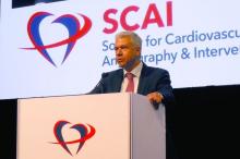
The 5-year rate of any reintervention, including explants, was 78% in the postapproval study, again similar to the 76% rate reported in the IDE study after a median 4.5 year follow-up (Circulation. 2015 Jun 2;131[22]:1960-70), Aimee K. Armstrong, MD, said at the Society for Cardiovascular Angiography & Interventions annual scientific sessions.
The new 5-year postapproval study findings “confirm that the hemodynamic effectiveness achieved by real-world providers is equivalent to the historical control established in the IDE study,” concluded Dr. Armstrong, professor of pediatrics at the Ohio State University and director of cardiac catheterization and interventional therapies at Nationwide Children’s Hospital, both in Columbus.
The postapproval study ran at 10 U.S. centers, none of which were among the five U.S. centers that ran the IDE study. Today, the Melody transcatheter pulmonary valve “is very commonly used” at many additional U.S. sites, Dr. Armstrong said in an interview. And the outcomes achieved using the valve likely surpass those seen in the IDE and postapproval studies because of innovations in technique, such as more routine use of “prestenting,” placing a stent in the vascular site where the pulmonary valve conduit will sit to address stenosis at this location and prevent subsequent conduit fracture (JACC Cardiovasc Interv. 2017 Sep;10[17]:1760-2).
“In 2010 [when the postapproval study began], we didn’t understand the importance of prestenting the way we do now. In 2010, I did not prestent every patient; now I do,” she said. The results reported by Dr. Armstrong included a 5% cumulative rate of major stent fractures in the Melody devices.
The postapproval study results also documented a concerning 4.5% annualized incidence of endocarditis among pulmonary valve recipients, with a nearly 300% increased rate of endocarditis among patients aged 12 years or younger, compared with older patients. Dr. Armstrong cautioned that this age association may be confounded by other factors, such as a residual pressure gradient in the right ventricular outflow tract of 15 mm Hg or greater. “We are discovering that we need to reduce the pressure gradient as much as we can, to perhaps less than 15 mm Hg, to reduce endocarditis, and that is something we did not know even a year ago. Practice is still evolving.”
The Melody Transcatheter Pulmonary Valve Postapproval Study performed cardiac catheterization for valve placement in 121 patients, and successfully implanted the valve for at least 24 hours in 99 of these patients. Patient age ranged from 5 to 45 years, with a median of 17 years; two-thirds were boys or men. The median age of the patients in the postapproval study was about 2 years younger than in the IDE study. Dr. Armstrong and her associates had previously published the 1-year outcomes from the postapproval study (JACC Cardiovasc Interv. 2014 Nov;7[11]:1254-62).
The enrolled patients usually needed a new right ventricular outflow tract because of a congenital heart defect, such as tetralogy of Fallot with pulmonary atresia and truncus arteriosus. Patients also included those who underwent a Ross operation. These patients often receive surgical placement of a right ventricular-to-pulmonary artery conduit, which can over time develop stenosis, insufficiency, or both because of calcification, intimal proliferation, and graft degeneration.
Multiple conduit reoperations to restore right ventricular outflow tract function are usually needed over a patient’s lifetime because of conduit degeneration. This makes a transcatheter procedure in a child or adolescent an attractive option because the prosthetic conduit will need replacement relatively quickly, and the transcatheter approach avoids an episode of open-heart surgery.
The Melody system is not the only transcatheter option for treating a leak or stenosis in a right ventricular outflow tract. The Sapien XT Transcatheter Heart Valve, marketed by Edwards, has FDA labeling for replacement of a dysfunctional right ventricular outflow tract.
Because the Sapien XT system was designed for replacing an aortic valve it’s challenging to place the conduit in the pulmonary valve position, Dr. Armstrong said. Operators find the Sapien 3 valve, a more modern design of the XT model that’s also primarily intended for aortic valve replacement, easier to position than the XT for pulmonary valve replacement, but Sapien 3 does not have FDA labeling for the right ventricular outflow tract indication. The Sapien valves are attractive because they don’t fracture, but Melody is easier to place and operators can reduce the fracture risk by prestenting, she noted.
Overall, the 5-year results from the postapproval study represented success, because 78% of patients who received the Melody device avoided any further interventions during follow-up. “That’s a big deal to a 12, 15, or 18 year old,” said Dr. Armstrong. “A surgically placed valve won’t last long in a teen, so it’s nice to do something noninvasively. It’s great if you can delay surgery for a few years” and avoid having the patient grow out of a surgically placed conduit or developing lots of calcification in the conduit during a growth spurt.
The postapproval study was funded by Medtronic, the company that sells the Melody valve. Dr. Armstrong has received research funding from Medtronic as well as Abbott, Edwards, and Siemens, and she has been a consultant to Abbott.
REPORTING FROM SCAI 2019
FFR changes coronary management in one-third of patients
LAS VEGAS – Interventional cardiologists who used fractional flow reserve to assess coronary lesions with an uncertain hemodynamic impact by angiography alone changed their initial therapeutic decision based on angiography for 35% of patients, and for 30% of all lesions examined in a real-world registry with more than 2,200 patients enrolled at 70 worldwide centers.
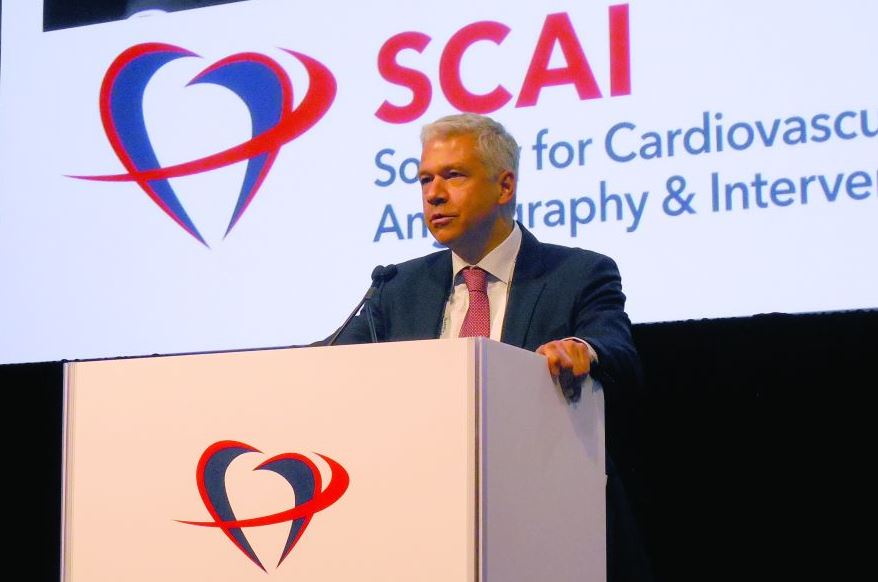
“Use of fractional flow reserve in contemporary, real-world, global clinical practice changed treatment plans for more than one-third of all comers,” including both patients with stable coronary artery disease and those with acute coronary syndrome, Erick Schampaert, MD, said at the Society for Cardiovascular Angiography & Interventions annual scientific sessions.
The impact of fractional flow reserve (FFR) was greatest when operators used it to assess nonculprit lesions among the 31% of the 2,217 total patients enrolled who presented with acute coronary syndrome. In this subgroup, FFR changed the treatment plan for nonculprit lesions that had been based on angiography and clinical status for 36% of these lesions. The changes included an increase in lesions identified to receive medical management, rising from 53% of the nonculprit lesions before FFR to 65% after, while treatment with percutaneous coronary intervention (PCI) fell from 37% of nonculprit lesions before FFR to 28% after, with the remaining lesions designated for coronary artery bypass grafting. Among patients with stable coronary disease the angiography-based treatment decision changed for 28% of nonculprit lesions after FFR.
“These results may provide support to increase use of FFR,” said Dr. Schampaert, an interventional cardiologist and head of cardiology at Hôpital du Sacré-Cœur in Montreal. The analysis “was an attempt to see the current impact of FFR at places where its use is established,” when it’s routinely used to assess the need to treat nonculprit lesions with an uncertain impact on blood flow through a coronary artery. Dr. Schampaert estimated that about one-quarter of patients who present for angiography have nonculprit lesions that leave operators uncertain about their hemodynamic significance after angiography and are candidates for FFR assessment.
The findings “are a call to do more FFR,” agreed M. Chadi Alraies, MD, an interventional cardiologist at the Detroit Medical Center Heart Hospital. “We are underusing FFR and overstenting people, and that worsens outcomes. We don’t do enough FFR,” Dr. Alraies commented.
“We’ve known for some time that angiography alone can lead to overtreatment,” commented Philippe Généreux, MD, an interventional cardiologist at Morristown (N.J.) Medical Center. “With FFR, physiology is the key to optimizing outcomes.”
The PRESSUREwire study included 2,217 consecutive patients who underwent FFR assessment at 70 centers in 15 countries during October 2016–February 2018. The only exclusions were patients with extremely tortuous or calcified arteries or patients with a bypass graft to the target vessel. Enrolled patients averaged 65 years of age, and three-quarters were men; 63% had stable coronary disease, 31% had acute coronary syndrome, and the remainder had silent ischemia documented by noninvasive testing. A stenosis of 50%-69% occluded 54% of the tested coronaries; 24% had a 70%-90% occlusion; 20% had an occlusion of less than 50%; and the remaining patients had an occlusion of more than 90%.
While the overall percentage of patients whose treatment plan changed following FFR assessment shifted moderately, the changes within each treatment category were more striking. For example, among the 62% of all patients initially designated for medical management based on angiography, the FFR findings changed the management plan to PCI in 19% of this subgroup. Conversely, among the 33% of all patients initially designated for PCI based on angiography, 52% instead received medical management based only on their FFR results. Because shifts in treatment strategy following FFR had some patients go from medical management to PCI, and others went from PCI to medical, overall the percentage of patients who received medical management without immediate revascularization had just a modest up-tick, from 62% before FFR to 67% after, Dr. Schampaert said.
PRESSUREwire was funded by Abbott Vascular, a company that markets an FFR device. Dr. Schampaert has been a consultant to Abbott Vascular as well as AstraZeneca, Bayer, Medtronic, Volcano-Philips, Sanofi, and Servier. Dr. Alaries had no disclosures. Dr. Généreux has been a consultant to Abbott Vascular and to several other companies, and he has an equity interest in Saranas.
SOURCE: Schampaert E et al. SCAI 2019, Abstract.
REPORTING FROM SCAI 2019
ProGlide outperformed Prostar for femoral closure after TAVR
LAS VEGAS – Comparison of two of the most commonly used vascular-access closure devices following transfemoral aortic valve replacement showed that Abbott’s ProGlide device led to significantly fewer vascular complications, especially minor complications; significantly less acute kidney injury; and may have also cut the average procedure duration and length of hospital stay, compared with the Prostar device sold by the same company, based on post hoc analysis of data collected from 746 patients.
The analysis also revealed that overall access-site vascular complications occurred in 24% of these patients, who were treated from October 2012 to May 2015 at any of 31 sites in seven countries, David A. Power, MBBCh, said at the Society for Cardiovascular Angiography & Interventions annual scientific sessions.
Although the results came from a nonrandomized (but multivariate-adjusted) comparison of data collected from a trial designed to address a completely different question, the findings appeared to support where the field has moved in recent years, toward greater reliance on the ProGlide device over Prostar, said Dr. Power, a researcher at the Icahn School of Medicine at Mount Sinai, New York.
“The preponderance of data is moving away from using Prostar. ProGlide appears to be coming out on top,” he said during a press briefing. Many operators familiar with both devices seem more comfortable using the ProGlide. But Dr. Power also noted that newer methods have become available for closing a femoral artery puncture following a transvascular procedure with a large-bore catheter, such as new types of plugs and patches.
The nearly 25% rate of vascular complications seen in the study “shows we have a way to go” in limiting and dealing with these adverse events, commented Timothy D. Henry, MD, an interventional cardiologist at the Christ Hospital in Cincinnati. The number of vascular complications from large-bore catheters will likely increase now that lower-risk patients will start to routinely undergo transcatheter aortic valve replacement. Methods for optimizing femoral artery closure after catheter puncture “have not received as much attention as they should, so this is a nice and important study,” Dr. Henry said.
Dr. Power and associates used data collected from patients in the BRAVO-3 (Effect of Bivalirudin on Aortic Valve Intervention Outcomes) study, which was designed to compare two different anticoagulants during transcatheter aortic valve replacement procedures. The trial found no statistically significant difference in patient outcomes regardless of the anticoagulant used (J Am Coll Cardiol. 2015 Dec 27;66[25]:2860-8). Review of the patient data showed that 352 of the 802 patients enrolled in BRAVO-3 had their femoral-artery puncture closed with a ProGlide device and 394 had their wound closed with Prostar. These 746 total patients accounted for 93% of all BRAVO-3 patients, highlighting the reliance that the operators on these cases had for these two closure devices in recent practice. The choice of vascular-access closure device in each BRAVO-3 case was at the discretion of the operator for that case.
A multivariate-adjusted analysis that took into account baseline differences between patients treated with ProGlide and Prostar showed that the ProGlide-treated patients had a significant 46% reduced rate of major or minor vascular complications, driven primarily by a reduction in minor complications, Dr. Power reported. The ProGlide-treated patients also showed a statistically significant 39% relative reduction in acute kidney injury, compared with the Prostar patients, a cut Dr. Powers attributed to a reduced need for contrast to check for residual bleeding. The results also showed that the ProGlide-treated patients had an average hospital length of stay about 20% shorter than the Prostar patients, and the average procedure time for the ProGlide-treated patients was about 30% shorter than with Prostar closure.
Concurrently with his report at the meeting, the results also appeared in an article published online (Catheter Cardiovasc Interv. 2019 May 22. doi: 10.1002/ccd.28295).
BRAVO-3 received funding from the Medicines Company. Dr. Power had no disclosures.
SOURCE: Power DA et al. SCAI 2019, Abstract 5743.
REPORTING FROM SCAI 2019
Key clinical point:
Major finding: ProGlide closure produced 46% fewer vascular complications than Prostar in a multivariate-adjusted analysis.
Study details: Post hoc analysis of data collected in the BRAVO-3 trial, with data from 746 of the 802 patients enrolled in BRAVO-3.
Disclosures: BRAVO-3 received funding from the Medicines Company. Dr. Power had no disclosures.
Source: Power DA et al. SCAI 2019, Abstract 5743.
Novel vascular sheath detects periprocedural bleeds
LAS VEGAS – A set of electrodes arrayed in a standard, vascular-access catheter sheath accurately alerted operators to access-site bleeding in a first-in-human study with 60 patients treated at any of five U.S. centers.
“The Early Bird bleed-monitoring system was safe, easily incorporated in standard flow of work, and demonstrated the capacity to detect bleeding before progression to a more severe or symptomatic phase,” Philippe Généreux, MD, said at the Society for Cardiovascular Angiography & Interventions annual scientific sessions. The study in 60 patients undergoing standard intravascular procedures via femoral-artery access – most often transcatheter aortic valve replacement – showed that an alert for access-site bleeding occurred with a Cohen’s kappa of 0.84, compared with CT imaging for bleeding, a score that shows “almost perfect” concordance between the two methods, noted Dr. Généreux, an interventional cardiologist and director of the structural heart program at Morristown (N.J.) Medical Center.
The study protocol called for keeping the sheath in place for up to 12 hours post procedure, and in practice the sheath remained in place for an average of about 160 minutes post procedure; 31% of the bleeds occurred during the procedure, with the remaining 69% occurring later. Another notable finding from the CT imaging at the time of sheath removal was that only 4 of the 60 patients had absolutely no bleeding, while 34 patients (57%) had blood infiltration at the access site and 22 patients (37%) had an access-site hematoma. No patients had retroperitoneal bleeding, but the system is designed to also detect bleeding within that space.
The Early Bird system received de novo classification as a new device from the Food and Drug Administration in March 2019; based on this, Saranas – the company developing the device – will likely start U.S. marketing before the end of 2019, Dr. Généreux said during a press briefing. The company is planning a registry of cases that use the device to collect data on patient outcomes to try to eventually document the clinical impact and cost-effectiveness of the system. Until now, standard of care has been to identify vascular-access associated bleeds once they become overt or symptomatic. If bleeds are identified at an earlier stage they could potentially be resolved before symptoms develop or become severe and hence provide a potential opportunity for cost savings.
Although the current study did not target specific types of patients, it makes sense in routine practice to target the device to patients at high risk for either developing a bleed or complications secondary to a bleed, such as patients undergoing transcatheter aortic valve replacement, patients receiving a mechanical circulatory assist device, or patients scheduled for complex procedures that will use multiple sheaths, he said.
The Early Bird sheath is 30 cm long, and is placed through the left or right femoral vein to the bifurcation of the iliac artery. (In the current study, more than 80% of the 60 treated patients had the sheath placed in their right femoral vein.) The access sheath for the catheters involved in the procedures themselves were most often placed in the left or right femoral artery. Electrodes within the Early Bird sheath detect leaked blood by its impact on bioimpedance of the tissue surrounding the sheath, with the system able to roughly gauge the volume of released blood based on the local level of bioimpedance change.
A couple of years ago, Dr. Généreux and his associates documented an 18% incidence of bleeding complications among 17,672 U.S. patients who underwent a transcatheter procedure with a large-bore catheter during 2013-2014 using data collected by the National Inpatient Sample (JAMA Cardiol. 2017 Jul;2[7]:798-802). Their analysis also documented that the patients with bleeding-related complications had in-hospital costs that averaged more than 50% higher than the costs for patients without bleeding complications, findings that raised the possibility that earlier identification of a bleed, before severe complications ensured, could be both cost effective and beneficial to patients, Dr. Généreux said.
Dr. Généreux has been a consultant to Saranas and to several other companies. He is also chief medical officer for Saranas and has an equity interest in the company.
SOURCE: Généreux P. SCAI 2019, Abstract 5713.
REPORTING FROM SCAI 2019
Novel cardiogenic shock, PCI protocol nets 72% acute survival
LAS VEGAS – A novel protocol for acute management of patients in cardiogenic shock secondary to an acute MI that started hemodynamic support prior to coronary revascularization produced an unprecedented in-hospital survival rate of 72% in 171 patients treated at any of 35 U.S. centers.
The 72% acute survival compares with historical rates of roughly 50% starting with the landmark SHOCK trial from 1999 (N Engl J Med. 1999 Aug 26;341[9]:625-34) and continuing in much more recent reports (J Am Coll Cardiol. 2017 Jan 24;69[3]:278-87)
“This is a first step toward reducing the futility in treating a disease where management has not changed for more than 20 years,” Mir B. Basir, D.O., said at the annual scientific sessions of the Society for Cardiovascular Angiography and Interventions. While Dr. Basir acknowledged that the new protocol needs further testing, as well as further improvement, a need exists to immediately implement changes in the routine management of cardiogenic shock caused by an acute MI because “50% in-hospital survival is no longer acceptable,” said Dr. Basir, an interventional cardiologist at the Henry Ford Health System in Detroit.
The National Cardiogenic Shock Initiative operates as a single-arm study with no control group. The novel management protocol used by the Initiative in the current study included the following five key best-practice steps, Dr. Basir said in a video interview:
1. Begin hemodynamic support before increasing dosages of vasopressors or inotropes.
2. Use right-heart catheterization to monitor the patient’s hemodynamics, which shows the efficacy of the hemodynamic support and guides tapering down of vasopressor and inotrope drugs.
3. Apply hemodynamic support before starting percutaneous coronary intervention (PCI).
4. Act fast, with a goal of less than 90 minutes from door to hemodynamic support. In the 171 patients that Dr. Basir reviewed, the average door-to-support time was 85 minutes.
5. Mitigate complications from the devices and vascular access.
This protocol started at four hospitals in the Detroit region, and then expanded to the National Cardiogenic Shock Initiative that now includes 68 U.S. sites and more than 200 patients treated, with another 23 U.S. hospitals about to join. The 68 active sites include 26 academic centers and 42 community hospitals. The initiative has enrolled patients who match the enrollment criteria of the SHOCK trial so that historical comparisons are possible. The initiative’s patients would be classified as class C, D, or E patients based on the society’s newly published cardiogenic shock classification scheme (Catheter Cardiovasc Interv. 2019 May 19. doi: 10.1002/ccd.28329).
“The numbers that Dr. Basir has reported are very encouraging and provocative,” commented Chandanreddy M. Devireddy, MD, an interventional cardiologist at Emory Healthcare in Atlanta. “In light of the fact that we have had few solutions for these patients, this will accelerate the discussion.”
Several barriers exist for widespread adoption of the initiative’s protocol, Dr. Basir said. The protocol requires a lot of resources and the ability to deliver this care 24/7. Currently, hemodynamic support is “greatly underused,” and right-heart catheterization is not standard of care for these patients at many U.S. centers, he noted.
A few weeks before Dr. Basir’s report at the meeting, his data from the National Cardiogenic Shock Initiative appeared in an article published online (Catheter Cardiovasc Interv. 2019 Apr 25. doi: 10.1002/ccd.28307).
REPORTING FROM SCAI 2019
SCAI releases first definition of cardiogenic shock
LAS VEGAS – The Society for Cardiovascular Angiography & Interventions released on May 19 the first-ever classification scheme for cardiogenic shock, dividing the condition into five severity levels.

The expert consensus panel that devised the new definition and classification model hopes it will spearhead a reset of research into the management of cardiogenic shock so that clinicians can assess interventions and introduce them into practice in a more precise, reproducible, and systematic way, Srihari S. Naidu, MD, said while presenting the proposal at the society’s annual scientific sessions.
The writing panel’s hope is that the new definition will “drive earlier recognition of shock and at a more precise stage to guide appropriate and timely escalation of care” and to “better define prospectively the value of mechanical circulatory support, extracorporeal membrane oxygenation, and other therapies,” said Dr. Naidu, chair of the writing group, as well as professor of medicine at New York Medical College and director of the cardiac catheterization laboratory at Westchester Medical Center, both in Valhalla, N.Y.
At the core of the classification scheme are the definitions for five strata of disease, which start at stage A, the “at-risk” patients before shock onset, and progress through stage B, “beginning”; stage C, “classic”; stage D, “deteriorating”; and stage E, “extremis,” which defines a patient with circulatory collapse (Catheter Cardiovasc Interv. 2019 May 19; doi: 10.1002/ccd.28329). Another key element of the classification model is the cardiac arrest “modifier,” designated by a subscripted letter A, which identifies patients who have had a cardiac arrest, regardless of duration. So a patient could be a stage BA, which identifies a patient with clinical evidence of relative hypotension or tachycardia without hypoperfusion and with a history of cardiac arrest.
The statement also itemizes several biomarkers and hemodynamic measurements that need regular, serial monitoring, such as blood lactate and right arterial pressure. Although the document leaves specific, defining values for some of these measures vaguely defined – the intent is that future research will fill in these gaps – the overall message is that clinicians caring for cardiogenic shock patients “need to be aggressive and look for these things,” Dr. Naidu said in a video interview.
“Until we agree on a definition of cardiogenic shock, we can’t go anywhere,” commented Larry S. Dean, MD, professor of medicine at and director of the Regional Heart Center of the University of Washington in Seattle. “There are a lot of conflicting data out there, and until we have a shared definition, we can’t advance our practice. We need to start looking at shock patients in a more precise way.”
“Without a clear definition of cardiogenic shock we will never improve patient outcomes. Every shock trial must define shock. If investigators just say ‘patients were in shock,’ I don’t know what that means,” noted Navin K. Kapur, MD, director of the Interventional Research Laboratories at Tufts Medical Center in Boston and a member of the writing panel.
Dr. Naidu and others on the panel highlighted the need to now validate the classification scheme’s ability to consistently categorize patients and predict their disease trajectories. They have begun the validation process with a 10,000-patient database of “all-comers” with cardiogenic shock maintained by the Mayo Clinic. Full results from this analysis will be out soon, but Dr. Naidu revealed in passing that it successfully provided validation of the proposed scheme.
The new definition received endorsements from the American College of Cardiology, the American Heart Association, the Society of Critical Care Medicine, and the Society of Thoracic Surgeons.
Dr. Naidu had no disclosures.
LAS VEGAS – The Society for Cardiovascular Angiography & Interventions released on May 19 the first-ever classification scheme for cardiogenic shock, dividing the condition into five severity levels.

The expert consensus panel that devised the new definition and classification model hopes it will spearhead a reset of research into the management of cardiogenic shock so that clinicians can assess interventions and introduce them into practice in a more precise, reproducible, and systematic way, Srihari S. Naidu, MD, said while presenting the proposal at the society’s annual scientific sessions.
The writing panel’s hope is that the new definition will “drive earlier recognition of shock and at a more precise stage to guide appropriate and timely escalation of care” and to “better define prospectively the value of mechanical circulatory support, extracorporeal membrane oxygenation, and other therapies,” said Dr. Naidu, chair of the writing group, as well as professor of medicine at New York Medical College and director of the cardiac catheterization laboratory at Westchester Medical Center, both in Valhalla, N.Y.
At the core of the classification scheme are the definitions for five strata of disease, which start at stage A, the “at-risk” patients before shock onset, and progress through stage B, “beginning”; stage C, “classic”; stage D, “deteriorating”; and stage E, “extremis,” which defines a patient with circulatory collapse (Catheter Cardiovasc Interv. 2019 May 19; doi: 10.1002/ccd.28329). Another key element of the classification model is the cardiac arrest “modifier,” designated by a subscripted letter A, which identifies patients who have had a cardiac arrest, regardless of duration. So a patient could be a stage BA, which identifies a patient with clinical evidence of relative hypotension or tachycardia without hypoperfusion and with a history of cardiac arrest.
The statement also itemizes several biomarkers and hemodynamic measurements that need regular, serial monitoring, such as blood lactate and right arterial pressure. Although the document leaves specific, defining values for some of these measures vaguely defined – the intent is that future research will fill in these gaps – the overall message is that clinicians caring for cardiogenic shock patients “need to be aggressive and look for these things,” Dr. Naidu said in a video interview.
“Until we agree on a definition of cardiogenic shock, we can’t go anywhere,” commented Larry S. Dean, MD, professor of medicine at and director of the Regional Heart Center of the University of Washington in Seattle. “There are a lot of conflicting data out there, and until we have a shared definition, we can’t advance our practice. We need to start looking at shock patients in a more precise way.”
“Without a clear definition of cardiogenic shock we will never improve patient outcomes. Every shock trial must define shock. If investigators just say ‘patients were in shock,’ I don’t know what that means,” noted Navin K. Kapur, MD, director of the Interventional Research Laboratories at Tufts Medical Center in Boston and a member of the writing panel.
Dr. Naidu and others on the panel highlighted the need to now validate the classification scheme’s ability to consistently categorize patients and predict their disease trajectories. They have begun the validation process with a 10,000-patient database of “all-comers” with cardiogenic shock maintained by the Mayo Clinic. Full results from this analysis will be out soon, but Dr. Naidu revealed in passing that it successfully provided validation of the proposed scheme.
The new definition received endorsements from the American College of Cardiology, the American Heart Association, the Society of Critical Care Medicine, and the Society of Thoracic Surgeons.
Dr. Naidu had no disclosures.
LAS VEGAS – The Society for Cardiovascular Angiography & Interventions released on May 19 the first-ever classification scheme for cardiogenic shock, dividing the condition into five severity levels.

The expert consensus panel that devised the new definition and classification model hopes it will spearhead a reset of research into the management of cardiogenic shock so that clinicians can assess interventions and introduce them into practice in a more precise, reproducible, and systematic way, Srihari S. Naidu, MD, said while presenting the proposal at the society’s annual scientific sessions.
The writing panel’s hope is that the new definition will “drive earlier recognition of shock and at a more precise stage to guide appropriate and timely escalation of care” and to “better define prospectively the value of mechanical circulatory support, extracorporeal membrane oxygenation, and other therapies,” said Dr. Naidu, chair of the writing group, as well as professor of medicine at New York Medical College and director of the cardiac catheterization laboratory at Westchester Medical Center, both in Valhalla, N.Y.
At the core of the classification scheme are the definitions for five strata of disease, which start at stage A, the “at-risk” patients before shock onset, and progress through stage B, “beginning”; stage C, “classic”; stage D, “deteriorating”; and stage E, “extremis,” which defines a patient with circulatory collapse (Catheter Cardiovasc Interv. 2019 May 19; doi: 10.1002/ccd.28329). Another key element of the classification model is the cardiac arrest “modifier,” designated by a subscripted letter A, which identifies patients who have had a cardiac arrest, regardless of duration. So a patient could be a stage BA, which identifies a patient with clinical evidence of relative hypotension or tachycardia without hypoperfusion and with a history of cardiac arrest.
The statement also itemizes several biomarkers and hemodynamic measurements that need regular, serial monitoring, such as blood lactate and right arterial pressure. Although the document leaves specific, defining values for some of these measures vaguely defined – the intent is that future research will fill in these gaps – the overall message is that clinicians caring for cardiogenic shock patients “need to be aggressive and look for these things,” Dr. Naidu said in a video interview.
“Until we agree on a definition of cardiogenic shock, we can’t go anywhere,” commented Larry S. Dean, MD, professor of medicine at and director of the Regional Heart Center of the University of Washington in Seattle. “There are a lot of conflicting data out there, and until we have a shared definition, we can’t advance our practice. We need to start looking at shock patients in a more precise way.”
“Without a clear definition of cardiogenic shock we will never improve patient outcomes. Every shock trial must define shock. If investigators just say ‘patients were in shock,’ I don’t know what that means,” noted Navin K. Kapur, MD, director of the Interventional Research Laboratories at Tufts Medical Center in Boston and a member of the writing panel.
Dr. Naidu and others on the panel highlighted the need to now validate the classification scheme’s ability to consistently categorize patients and predict their disease trajectories. They have begun the validation process with a 10,000-patient database of “all-comers” with cardiogenic shock maintained by the Mayo Clinic. Full results from this analysis will be out soon, but Dr. Naidu revealed in passing that it successfully provided validation of the proposed scheme.
The new definition received endorsements from the American College of Cardiology, the American Heart Association, the Society of Critical Care Medicine, and the Society of Thoracic Surgeons.
Dr. Naidu had no disclosures.
REPORTING FROM SCAI 2019


