User login
Fatigue linked to increased risk of ACL injury
SAN DIEGO – Fatigue increases anterior cruciate ligament injury risk in adolescent athletes, results from a field-based drop-jump study demonstrate.
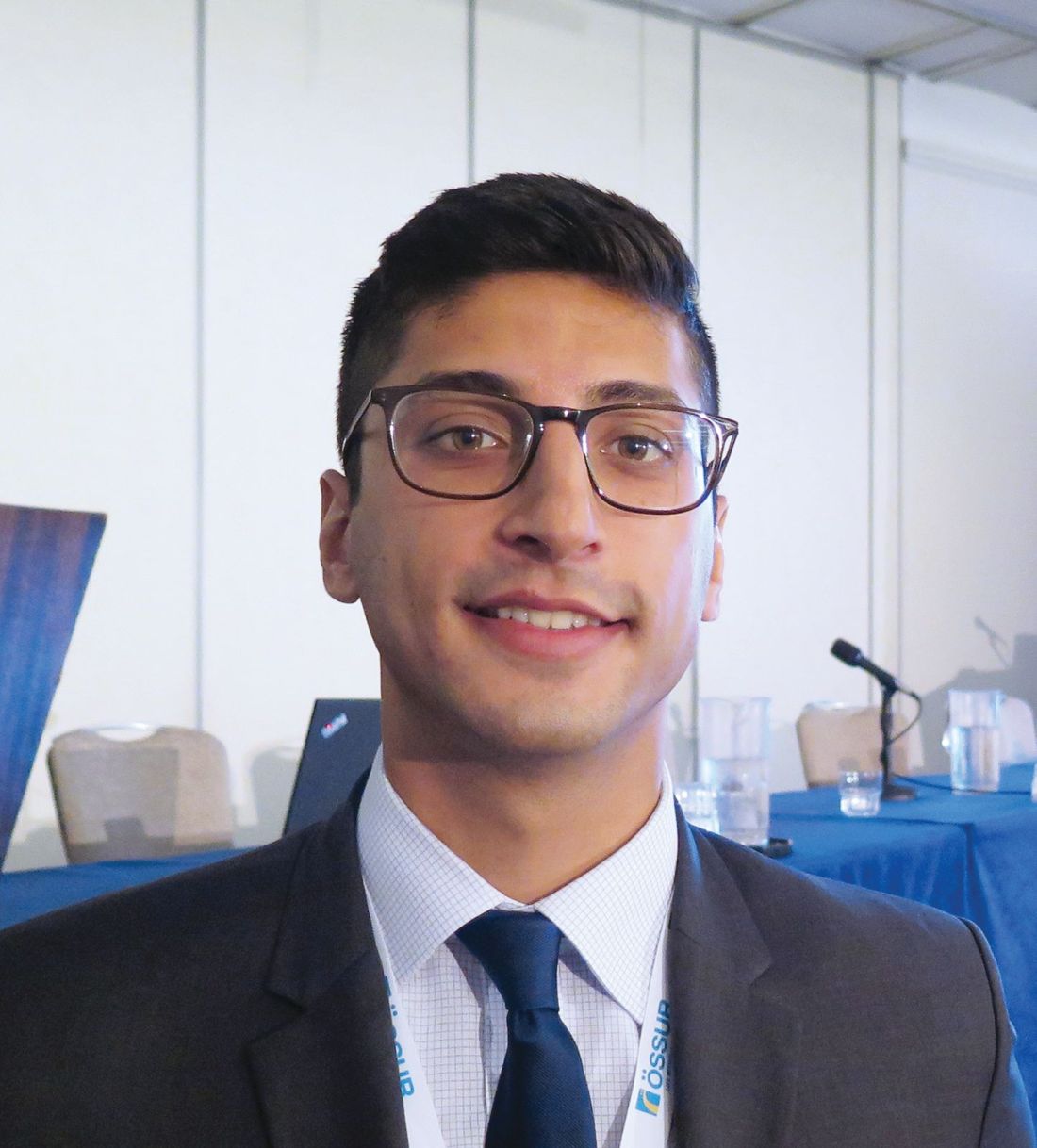
“The number of ACL reconstructions that occur annually are on the rise, particularly in high school and adolescent aged athletes,” lead study author Mohsin S. Fidai, MD, said at the annual meeting of the American Orthopaedic Society for Sports Medicine. “About 70% of these are accounted for by noncontact injuries, the majority of which occur during jump landing. A number of risk factors that have previously been implicated in ACL injury include genetics and anatomy, but a modifiable risk factor is landing biomechanics.”
In 2005, researchers led by Timothy E. Hewett, PhD, determined biomechanical measures of neuromuscular control that might pose certain athletes to be at risk for ACL injury, particularly knee abduction and dynamic knee valgus during a drop-jump test (Am J Sports Med. 2005;33[4]:492-501). “Historically, these studies have required the use of sophisticated computer technology, which can be cumbersome from a time and cost perspective,” said Dr. Fidai, a third-year orthopedic surgery resident at Henry Ford Health System, Detroit.
In a more recent analysis, researchers validated a field-based drop vertical jump screening test for ACL injury (Phys Sportmed. 2016;44[1]:46-52). The sensitivity was 95%, the specificity was 46%, and it had a strong inter-rater reliability (k = 0.92; P less than .05).
The purpose of the current study was to evaluate the effect of fatigue on ACL injury risk using a field-based drop-jump test. “We hypothesized that fatigue would lead to greater dynamic knee valgus during a drop-jump test,” Dr. Fidai said. “We also wanted to identify individual characteristics which may place athletes at increased risk for ACL injury.”
The researchers recruited 85 athletes who competed in track and field, basketball, volleyball, and soccer. More than half (55%) were female, and the mean age was 15.4 years. They excluded athletes with any previous or current lower extremity injuries or neuromuscular deficits. Each athlete performed a maximum vertical jump, followed by a drop-jump test.
“We then fatigued all of our athletes with a standardized high-intensity fatigue protocol, and had each athlete perform another maximum vertical jump and drop-jump test,” Dr. Fidai said. “All drop-jumps were video recorded and sent to a number of orthopedic surgery residents, athletic trainers, and physical therapists for review.”
Of the 85 athletes, nearly half (45%) showed an increased risk for ACL injury after high-intensity aerobic activity. In addition, 68% of study participants were identified as having a medium or high risk for injury following the aerobic activity, compared with 44% at baseline. Dr. Fidai noted. “In the group of athletes with higher levels of fatigue, there is a significantly increased risk, compared with their counterparts with lower levels of fatigue.”
Specifically, 14 of the 22 athletes who demonstrated over 20% fatigue showed an increased ACL injury risk. Subgroup analysis revealed that female athletes and those older than age 15 were more likely to demonstrate an increased injury risk.
“The findings of this study advocate for changes to current neuromuscular training programs to incorporate fatigue resistance, as well as to raise awareness amongst physical therapists, athletic trainers, coaches, and athletes about the effect of fatigue on ACL injury risk,” Dr. Fidai concluded. “We can target vulnerable athletes, particularly female athletes, in an effort to negate some of those effects.”
The study’s principal investigator was Eric C. Makhni, MD. Dr. Makhni, an orthopedic surgeon in West Bloomfield, Mich., disclosed that he is a paid consultant for Smith & Nephew and that he receives publishing royalties from Springer. Dr. Fidai reported having no financial disclosures.
REPORTING FROM AOSSM 2018
Key clinical point: Athletes who experience fatigue as tested by a standardized assessment demonstrated increased risk of ACL injury.
Major finding: Nearly half of athletes (45%) showed an increased injury risk after high-intensity aerobic activity.
Study details: A field-based study of 85 athletes that used vertical and drop-jump assessments of each athlete, which were captured on video and reviewed by professional health observers.
Disclosures: Dr. Makhni disclosed that he is a paid consultant for Smith & Nephew and that he receives publishing royalties from Springer. Dr. Fidai reported having no financial disclosures.
Steroid injection prior to rotator cuff surgery elevates risk of revision repair
SAN DIEGO – Patients who received a corticosteroid injection within 6 months prior to rotator cuff repair were more likely to undergo a revision rotator cuff surgery within the following 3 years, results from a large database study show.
“Corticosteroid injections are frequently utilized in the nonoperative management of rotator cuff tears,” researchers led by Sophia A. Traven, MD, wrote in an abstract presented during a poster session at the annual meeting of the American Orthopaedic Society for Sports Medicine. “However, recent literature suggests that injections may reduce biomechanical strengths of tendons and ligaments in animal models.”
In an effort to examine the effect of preoperative shoulder injections on the rate of revision cuff repair following arthroscopic rotator cuff repair, the researchers retrospectively reviewed MarketScan claims data between 2010 and 2014 to identify 4,959 patients with an ICD-9 diagnosis of a rotator cuff tear with subsequent arthroscopic rotator cuff repair (CPT 29827).
They used multivariable logistic regression to compare the odds of reoperation between groups, while controlling for certain demographic and comorbid variables, including age and gender, tobacco use, diabetes, and the Charlson comorbidity index score.
Dr. Traven, an orthopedic surgeon at the Medical University of South Carolina, Charleston, and her associates reported that 392 of the 4,959 patients required rotator cuff repair revision within the following 3 years. Compared with those who did not require revision, those who did were older (a mean age of 53 vs. 49 years, respectively), more likely to be smokers (7% vs. 4%), and more likely to receive any injection prior to rotator cuff repair (36% vs 25%; P less than .0001 for all associations).
(odds ratio, 1.822), followed by those who received an injection 0-3 months before the primary repair (OR, 1.375), and those who received an injection 6-12 months before the primary repair (OR, 1.237).
“The risk of revision rotator cuff repair remains elevated for 6 months following a shoulder injection,” the researchers concluded in their poster. “Consideration should therefore be given to minimizing preoperative injections in patients who may require rotator cuff repair.”
They reported having no financial disclosures.
REPORTING FROM AOSSM 2018
Key clinical point: Consideration should be given to minimizing preoperative injections in patients who may require rotator cuff repair.
Major finding: The risk for revision rotator cuff repair was highest for patients who received an injection 3-6 months before the primary rotator cuff repair (odds ratio, 1.822).
Study details: A retrospective analysis of 4,959 patients with an ICD-9 diagnosis of a rotator cuff tear with subsequent arthroscopic rotator cuff repair.
Disclosures: The researchers reported having no financial disclosures.
Various soft tissue recovery methods get different results
SAN DIEGO – When it comes to soft tissue recovery modalities for elite athletes beyond rest, recovery, and retaining movement efficiency, not all options are created equal.
In fact, the science for most supplemental recovery modalities stems from cohort studies examining physiologic response – not high-level randomized clinical trials, Chuck Thigpen, PhD, said at the annual meeting of the American Orthopaedic Society for Sports Medicine.
“We should be very careful when we discuss overtraining and overload,” said Dr. Thigpen, senior director of practice innovation and analytics for ATI Physical Therapy, Greenville, S.C. “In fact, we need training load to create an anabolic response, so then the question is, how do we manage that load? I would suggest that it’s not overtraining, but underrecovery after a load that results in increasing fatigue, decreased performance, and potential increased injury risk.”
One option for soft tissue recovery is whole body vibration, for which the athlete stands, sits, or lies on a machine with a vibrating platform, while he or she performs static or isotonic exercise. “With this modality, you get a rapid co-contraction of muscle, which increases muscle preactivation,” said Dr. Thigpen, who is also directs the program in observational clinical research in orthopedics at the Greenville, S.C.–based Center for Effectiveness Research in Orthopaedics. “It has demonstrated increased blood flow as well as increased motor neuron excitability. There seems to be some physiologic benefit coming potentially from muscle waste removal (lactate) and nutrient delivery, as well as decreasing subsequent inhibition.”
In terms of parameters, benefits have been observed when athletes perform one or two sets of a static stretch or contact massage on a body vibration machine for a minute or so at a frequency of 30-50 Hz. “The application is what becomes challenging,” Dr. Thigpen said. “Where are you going to work this in? Is it a pre or post activity? Recent evidence implicates use during halftime may maintain strength and power. However, most of the work that has been done with vibration has been as an adjunct to exercise and not really in terms of recovery.”
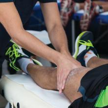
Massage is another popular recovery tool, and most elite sports team have a masseuse on staff. Soft tissue manipulation creates release of oxytocin and other neurotransmitters, some central nervous system response, and increased blood flow to the treated area, but it also influences the athlete’s general disposition.
“There’s something about laying hands on somebody that seems to affect a person’s mood state,” Dr. Thigpen said. “Some studies have reported better perceived recovery status, even though the physiologic markers are about the same. Therefore, I would classify body vibration and massage in the same bucket. They seem to work; they seem to have some perceived benefit.”
Another soft tissue recovery option, compression therapy, has been shown to increase the local pressure gradient of the impacted area, thereby increasing progressive venous return and creating some muscle splinting (or protective muscle spasms). “,” Dr. Thigpen said. “A couple of studies have looked at the ultrastructure of the muscle concurrently after using compression garments. The nice thing is that you can put them on right after the activity. They should be worn for 24 hours.”
Another way to get compression therapy is to use compression devices; it is recommended that they are worn for 15-minute intervals for up to 4 hours after intense physical activity, depending on the device. “You see some of the same benefits that you see with compression garments,” he said.
Dr. Thigpen went on to discuss cryotherapy such as cold-water immersion in a tub, which has a long history of use in muscle recovery. In fact, many basic science studies have demonstrated a reduction of inflammatory markers and other immunologic responses after its use. “Cryotherapy is thought to create an acute decrease in blood flow and a concurrent increase in blood flow after you remove it, which creates the release of these neurotransmitters and immunosuppressants that seem to be helpful in the healing process,” he explained.
“The thought is, because of the decreased pain reduction, the waste removal, and the change in oxidative stress, this would be beneficial.” For example, cold water immersion in a tub four times over a 72-hour period has been found to decrease soreness and increase athletic performance on the backside. “That seems to be helpful in recovery, as an adjunct to heavy resistance training or eccentric and plyometric training,” he said.
Neuromuscular electrical stimulation has been shown to provide some analgesic effect to sore muscles via afferent stimulation, but the primary mechanism is contractile via the motor unit. Typically, neuromuscular electrical stimulation consists of about a 20-minute application to affected muscles, “and you can do multiple applications per day interspersed with periods of high-intensity training to restore the neuromuscular profile during the recovery period,” Dr. Thigpen said.
He reported having no financial disclosures.
REPORTING FROM AOSSM 2018


