User login
Alcohol use, abuse rise after bariatric surgery
ORLANDO –
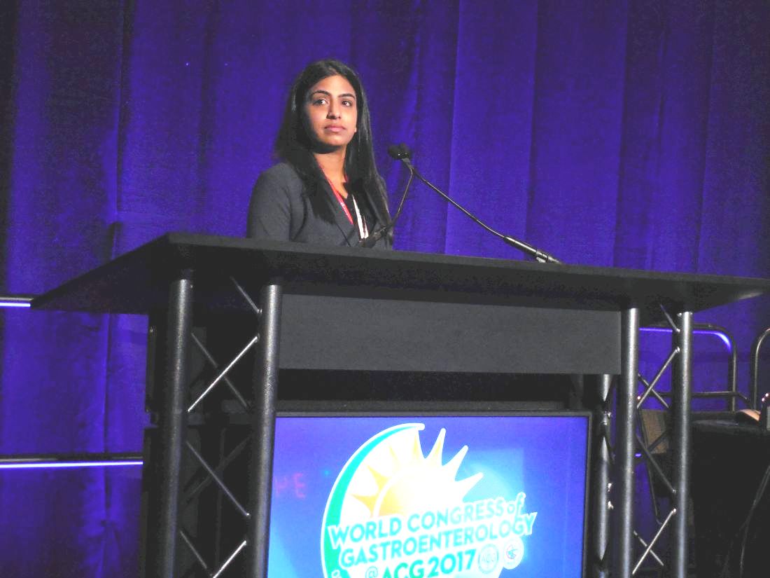
Following any of several methods of bariatric surgery, patients showed a statistically significant 8% higher rate of new onset alcohol abuse, and a relative 50% increased rate of significant alcohol use, compared with rates before surgery, Prandeet Wander, MD, said at the World Congress of Gastroenterology at ACG 2017.
Her meta-analysis identified prospective, retrospective, and cross-sectional studies of alcohol use that included more than 100 bariatric surgery patients and that had follow-up beyond 1 year. Patients could have undergone Roux-en-Y gastric bypass, sleeve gastrectomy, or laparoscopic adjustable gastric banding. Comparator populations had to be either the surgery patients prior to the procedure or the controls matched by age and body mass index.
The 28 included studies enrolled 15,714 patients who averaged 43 years old, with more than three quarters women. Follow-up averaged 2.6 years. The most common surgery was Roux-en-Y, used in 23 studies, followed by banding in 12 studies, and sleeves in 8 studies (some studies used more than one type of surgery).
Nineteen of the studies examined the prevalence of “significant alcohol abuse” following surgery in a total of 4,552 patients, with 23% of patients overall showing this behavior. Five studies, involving 2,698 patients, documented the rate of new-onset alcohol abuse after surgery, with an overall rate of 8% that was statistically significant. All five studies individually showed increased incidence of alcohol abuse, with rates that ranged from 4% to 8%.
The analysis that showed a relative 50% higher rate of “significant” alcohol use after surgery, compared with the same patients before their surgery used data from 11 studies with 3,370 patients. Five of these 11 studies individually showed a statistically significant increase in alcohol use, 1 showed a significant, 34% relative decrease, and the remaining 5 studies did not show statistically significant changes, with 3 studies trending toward an increased rate and two trending toward a decreased rate after surgery.
None of the 28 included studies had a randomized control arm, and the studies collectively ran in six countries, including the United States, and hence involved different societal norms of alcohol use. Changes in alcohol absorption and metabolism following bariatric surgery may play roles in the observed effects, as might undiagnosed depression or substance use by patients who undergo this surgery, Dr. Wander suggested.
SOURCE: Wander P et al. World Congress of Gastroenterology, abstract 10.
REPORTING FROM WORLD CONGRESS OF GASTROENTEROLOGY
Key clinical point: Following bariatric surgery patients have increased alcohol use and abuse.
Major finding: Alcohol abuse rose by 8%; significant alcohol use rose by a relative 50%.
Study details: Meta-analysis of 28 reports with 15,714 patients
Disclosures: Dr. Wander had no disclosures.
Source: Wander P et al. World Congress of Gastroenterology, abstract 10.
Meta-analysis confirms probiotics’ pediatric safety and efficacy
ORLANDO – Probiotics are safe and effective for treating functional abdominal pain in children, based on findings from a meta-analysis of 11 randomized studies with a total of 790 patients.
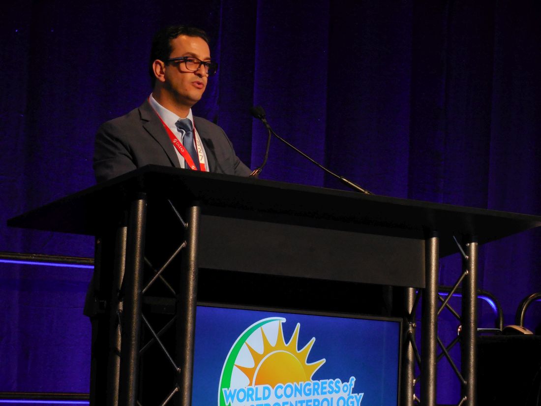
“We think there is pretty strong evidence” for the efficacy of probiotics, and “by any analysis you can throw at them probiotics are safe,” Gordon Morris, MD, said at the World Congress of Gastroenterology at ACG 2017. “The evidence is of moderate and high quality,” added Dr. Morris, a pediatric gastroenterologist at the University of Central Lancashire in Preston, England.
The most widely studied probiotic in the analysis was Lactobacillus reuteri, used in six of the studies with a total of 405 randomized patients. The next most commonly studied agent was Lactobacillus rhamnosus GG, the focus of four studies and tested in a total of 270 randomized patients. Both microbes showed statistically significant and clinically meaningful levels of pain reduction when compared with placebo in subgroup analyses, said Dr. Morris, who performed the meta-analysis as a Cochrane Review Groups systematic review.
“The pain score reductions we saw [with these two strains] could certainly have an impact. I think it matters clinically,” he explained. “Severity of pain is most important to patients.”
Both L. reuteri and L. rhamnosus GG have received “generally regarded as safe” designations from the Food and Drug Administration.
Based on these findings, “I don’t think we can justify, especially with these two main strains, any further basic efficacy studies,” Dr. Morris said. The primary focus for future clinical assessments of these probiotics should be long-term efficacy and safety and whether patients have rebound pain on withdrawal from probiotic use, he added.
The meta-analysis used studies that compared probiotics against placebo in children aged 4-18 years who received treatment for 4-16 weeks. The full analysis showed an average 0.57-unit reduction in pain scores across all 11 studies included, with an average 0.61-unit reduction using L. reuteri and an average 0.75-unit reduction using L. rhamnosus GG. All three between-group differences were statistically significant. Safety data came from eight of the included studies, and they collectively showed absolutely no safety difference between actively treated and control patients.
Dr. Morris noted that the mechanism by which probiotic bacilli relieve abdominal pain remains unclear, but suggested that both prokinetic and anti-inflammatory effects might be involved.
[email protected]
On Twitter @mitchelzoler
AT THE WORLD CONGRESS OF GASTROENTEROLOGY
Key clinical point:
Major finding: Probiotic treatment led to an average 0.57-unit reduction in pain intensity compared with placebo controls.
Data source: A Cochrane Group meta-analysis of 11 studies with 790 patients.
Disclosures: Dr. Morris had no disclosures.
VIDEO: Back off on screening colonoscopy after nonadvanced adenomas
ORLANDO – Evidence supports “backing off” from screening colonoscopies every 5 years for patients who had one or two nonadvanced adenomas removed during a prior colonoscopy, Thomas F. Imperiale, MD, AGAF, said at the World Congress of Gastroenterology at ACG 2017.
He reported findings from more than 66,000 U.S. veterans followed at any one of 13 Veterans Affairs medical centers for an average of more than 7 years. The 10,220 patients who underwent a second screening colonoscopy after an index colonoscopy that led to removal of one or two nonadvanced adenomas had 0.16% colorectal cancer mortality, compared with 0.13% among 8,718 patients with a similar history who did not receive follow-up colonoscopy. The rate of colorectal cancer death was 0.12% among 47,629 control veterans who had no adenomas removed during their index colonoscopy.
In current U.S. practice, many gastroenterologists perform follow-up colonoscopy about 5 years after removing one or two nonadvanced adenomas during a screening colonoscopy, Dr. Imperiale said during a video interview. Deferring follow-up colonoscopy in the absence of any clinical indication seems advisable, he said, especially for older patients with two or more comorbidities who had a high-quality index colonoscopy with good preparation and good colonic visibility.
“We just can’t do colonoscopy for surveillance on this subgroup continuously; it doesn’t make sense,” he said.
No randomized trial results have documented the need for stepped up colonoscopies in patients with a history of one or two nonadvanced adenomas, and these new observational findings are consistent with prior observational reports.
“These data need to be integrated with common sense,” he said. An extended interval before repeat surveillance seems particularly appropriate for patients with a higher risk for adverse effects from the colonoscopy preparation and for patients more likely to die from a cause other than colorectal cancer.
Backing off on repeat colonoscopy “minimizes the harm from surveillance. As patients get older they don’t tolerate the prep as well. It grows more onerous, and the returns diminish,” Dr. Imperiale said.
The patients included in the review had their index colonoscopy performed during 2002-2009, when they averaged about 61 years old, and about 95% were men. Their average Charlson comorbidity index was about 1.3. The incidence of colorectal cancer during follow-up after the index colonoscopy was 0.18% in patients with one or two nonadvanced adenomas in their index examination and no follow-up colonoscopy, 0.71% in those with nonadvanced adenomas who had one or more subsequent colonoscopies, and 0.31% in the people with no adenomas removed during the index procedure.
The rates of all-cause death during follow-up of the three subgroups were notably different: 34% in those with nonadvanced adenomas and no repeat colonoscopy, 13% in patients with nonadvanced adenomas and repeat colonoscopy, and 21% in those without nonadvanced adenomas. Dr. Imperiale discounted the significance of comparing rates of all-cause mortality, stressing that the most relevant primary endpoint is colorectal cancer mortality.
Dr. Imperiale reported having no disclosures.
[email protected]
On Twitter @mitchelzoler
The video associated with this article is no longer available on this site. Please view all of our videos on the MDedge YouTube channel
AT THE WORLD CONGRESS OF GASTROENTEROLOGY
Key clinical point:
Major finding: Colorectal cancer mortality was 0.13% in patients without follow-up colonoscopy and 0.16% in patients who had a second colonoscopy.
Data source: Review of 66,567 people at 13 U.S. VA Medical Centers.
Disclosures: Dr. Imperiale reported having no disclosures.
VIDEO: Burnout affects half of U.S. gastroenterologists
ORLANDO – Nearly half of U.S. gastroenterologists who responded to a recent survey had symptoms of burnout that seemed largely driven by work-life balance issues.
Burnout appeared to disproportionately affect younger gastroenterologists, those who spend more time on chores at home including caring for young children, physicians who were neutral toward or dissatisfied with a spouse or partner, and clinicians planning to soon leave their practice, Carol A. Burke, MD, said at the World Congress of Gastroenterology at ACG 2017.
Factors not linked with burnout included their type of practice, whether the gastroenterologists worked full or part time, their location, and their compensation, said Dr. Burke, director of the Center for Colon Polyp and Cancer Prevention at the Cleveland Clinic.
The life issues that appeared most strongly linked to burnout “speak to a problem for physicians to balance” their professional and personal lives, Dr. Burke said in a video interview. Several interventions exist that can potentially mitigate burnout, and the American College of Gastroenterology, which ran the survey, is taking steps to make information on these interventions available to members, noted Dr. Burke, the organization’s president.
Dr. Burke and her associates sent a 60-item survey to all 11,080 College members during 2014 and 2015 and received 1,021 replies including 754 fully completed responses. Their prespecified definition of burnout was a high score for emotional exhaustion or for depersonalization, or both, on the Maslach Burnout Inventory. The results showed that 45% of respondents had a high score for emotional exhaustion, 21% scored high on depersonalization, and overall 49% met the burnout criteria set by the investigators. The Inventory answers also showed that 18% had a low sense of personal accomplishment.
A multivariate analysis showed that significant links with burnout were younger age, more time spent on domestic chores, having a neutral or dissatisfying relationship with a spouse or partner, and plans for imminent retirement from gastroenterology practice, Dr. Burke reported.
The main reasons for planning imminent retirement were reimbursement, cited by 32% of this subgroup, regulations, cited by 21%, recertification, cited by 16%, and electronic medical records, cited by 10% as the main reason for leaving practice.
Strategies and resources aimed at better dealing with burnout were requested by 60% of all survey respondents, and the College is in the process of making these tools available, Dr. Burke said.
A recent study conducted by the AGA Institute Education and Training Committee and reported in AGA Perspectives supports these findings. Read more here and join the discussion on the AGA Community.
The video associated with this article is no longer available on this site. Please view all of our videos on the MDedge YouTube channel
[email protected]
On Twitter @mitchelzoler
AT THE 13TH WORLD CONGRESS OF GASTROENTEROLOGY
Key clinical point:
Major finding: Forty-nine percent of surveyed U.S. gastroenterologists showed a high level of emotional exhaustion, depersonalization, or both.
Data source: Survey results from 754 members of the American College of Gastroenterology.
Disclosures: The American College of Gastroenterology funded the survey. Dr. Burke had no relevant disclosures.
eConsult gastroenterology model could improve access
ORLANDO – A review of gastroenterology electronic consultations, or eConsults, at a tertiary care academic medical center suggests that such referrals could improve timely access to specialist care, while cutting costs.
The findings underscore the need for careful study of this burgeoning care delivery model, which is a form of telemedicine, Jennifer Wang, MD, said at the World Congress of Gastroenterology at ACG 2017.
The review of 130 eConsults conducted between Jan. 1, 2015, and May 8, 2017, looked at questions asked, gastroenterology content, eConsult response time, change in referral plans, and indirect cost savings through avoided referrals and travel, according to Dr. Wang of the University of Virginia, Charlottesville, which is one of five centers that are part of an eConsult model project.
Of the 130 eConsults, 68 (52%) were resolved without face-to-face consultation with a gastroenterologist; the patients followed up with a primary care physician. The remaining 62 cases led to a face-to-face visit in the GI clinic.
The mean response time to eConsult was 54 hours, compared with a greater than 30-day wait time for an initial consultation in the ambulatory GI clinic, she said.
The most frequently queried subjects were etiology of chronic diarrhea (14%), colon cancer screening modality (12%), and chronic abdominal pain management (9%). The most common type of question asked pertained to diagnosis (70%).
The total mileage saved between patients’ homes and the GI clinic was estimated to be 1,583 miles. “You can also imagine the cost saved by not having to miss a day of work,” Dr. Wang said.
The model is not only cost effective, but can potentially be life saving, she added.
In one case, a 40-year-old woman with a 6-month history of abdominal pain was diagnosed with lymphoma during an eConsult and underwent biopsy and chemotherapy immediately, whereas the 30-day wait for a face-to-face visit would have delayed her diagnosis, Dr. Wang explained.
The eConsult model is being tested as a means of providing primary care physicians with direct, efficient, and timely access to specialist expertise in the management of their patients and potentially avoiding the need for face-to-face referrals, Dr. Wang said.
Increased demand for eConsult is anticipated, and therefore its financial and medical-legal implications should be further studied, she said. One question is how specialists can be incentivized to provide eConsults.
“I think the key would be to come up with a sustainable payment model and reimbursement strategy, and to have protected time for specialists to review eConsults,” she said.
Dr. Wang reported having no financial disclosures.
AT THE WORLD CONGRESS OF GASTROENTEROLOGY
Key clinical point:
Major finding: The mean response time to eConsult was 54 hours, compared with a greater than 30-day wait time for an initial consultation in the ambulatory GI clinic.
Data source: A review of 130 eConsults.
Disclosures: Dr. Wang reported having no financial disclosures.
Endoscopic therapy effective for early cancer in Barrett’s esophagus
ORLANDO – Endoscopic therapy is as effective in Barrett’s esophagus patients with early cancer as in those with high-grade dysplasia, according to findings from an international multicenter consortium.
The findings suggest that invasive surgery may be avoidable in many Barrett’s esophagus patients with early cancer, Rajesh Krishnamoorthi, MD, of Virginia Mason Medical Center, Seattle, reported at the World Congress of Gastroenterology at ACG 2017.
Further, after adjustment for age, sex, and Barrett’s esophagus length, there was no statistical difference in the CE-IM rate (hazard ratio, 1.15) or CE-D rate (HR, 1.21) between the two groups.
The rates of recurrent intestinal metaplasia (Re-IM) in the groups were also statistically similar at 43.9% and 34.7%, respectively, said Dr. Krishnamoorthi, whose work received a 2017 Esophagus Category Award at the meeting.
Endoscopic therapy is the treatment of choice for Barrett’s esophagus patients with high-grade dysplasia, and is also used in some cases as a noninvasive alternative to surgery in Barrett’s esophagus patients with intramucosal cancer. However, data comparing outcomes of endoscopic therapy for these two conditions are lacking.
For the current study, all subjects from the EET database of patients from 10 centers in the United States, Europe, and Australia with either intramucosal cancer or high-grade dysplasia who underwent endoscopic therapy since April 2012 were reviewed. The patients were treated with endoscopic mucosal resection if visible lesions were noted, and/or with mucosal ablation for the flat Barrett’s esophagus. Those who underwent at least four esophagogastroduodenoscopies with endoscopic therapy were included.
The median age of the patients was 66 years, 84% were men, and median Barrett’s esophagus segment length was 6 cm. Baseline characteristics did not differ between the groups, Dr. Krishnamoorthi noted.
Although limited by the relatively small number of patients in each study group, by the exclusion of patients who were lost to follow-up, and by the observational nature of the study, the findings could have implications for treatment selection in some patients with Barrett’s esophagus and early cancer.
“In this large well-defined cohort of Barrett’s patients, effectiveness of endoscopic therapy in intramucosal cancer is comparable to that of high-grade dysplasia. Consideration of endoscopic therapy in Barrett’s patients with early cancer could reduce the need for invasive surgery,” he concluded.
During a discussion period, however, it was pointed out that the centers involved in this study are “centers with a lot of expertise in this,” and that the generalizability of the findings to gastroenterology practices is something that should be looked at, especially considering that the diagnosis of intramucosal cancer “may not be uniformly accurate across the spectrum of gastroenterology practices.”
“I completely agree with that,” Dr. Krishnamoorthi said, adding that the diagnosis must be confirmed by a pathologist, and that the procedure should be performed by an endoscopist with extensive experience.
Dr. Krishnamoorthi reported having no disclosures.
AT THE 13th WORLD CONGRESS OF GASTROENTEROLOGY
Key clinical point:
Major finding: Outcomes did not differ significantly between Barrett’s esophagus patients with early cancer and those with high-grade dysplasia (hazard ratios for CE-IM and CE-D, respectively: 1.15, and 1.21).
Data source: Study of 276 patients from a prospective database.
Disclosures: Dr. Krishnamoorthi reported having no disclosures.
ADMIRE CD trial: Stem cells promote long-term fistula remission
ORLANDO – A single treatment with a suspension of allogeneic expanded adipose-derived mesenchymal stem cells, or Cx601, promotes long-term combined remission of complex perianal fistulas in patients with Crohn’s disease, according to 52-week results from the phase 3 ADMIRE CD trial.
Combined remission – a stringent endpoint consisting of closure of all treated external openings that were draining at baseline and of an absence of collections more than 2 cm of treated perianal fistulas as confirmed by blinded central MRI – was achieved in 56.3% of 103 treated patients, compared with 38.6% of 101 patients who received placebo (P = .010), Daniel C. Baumgart, MD, reported at the World Congress of Gastroenterology at ACG 2017.
This parallel-group, double-blind, multicenter study included patients who had draining, treatment-refractory, complex perianal fistulas and inactive or mildly active luminal Crohn’s disease (Crohn’s Disease Activity Index scores of 220 or less) at baseline. Those randomized to the Cx601 group received a single intralesional injection of 120 million expanded adipose-derived stem cells and standard of care. Those in the control arm received a placebo injection plus standard of care, said Dr. Baumgart of Charité Medical School, which is affiliated with both Humboldt University in Berlin and the Free University of Berlin.
Prior to receiving treatment or placebo, the patients underwent fistula curettage and, if indicated, seton placement and subsequent removal, he noted, adding that baseline concomitant medications, including immunosuppressants and anti–tumor necrosis factors, were continued without dose or regimen modification and that antibiotics were allowed for up to 4 weeks.
The 52-week findings were evaluated in the modified intention-to-treat population of patients who were randomized, were treated, and had at least one postbaseline efficacy assessment (61.8% of the study population). These findings showed that Cx601 is associated with even better outcomes at 1 year than those reported at 24 weeks; those prior results, published in The Lancet in July 2016, showed combined remission rates in the modified intention-to-treat population of 51% vs. 36% for placebo.
Furthermore, 75% of treated patients who achieved combined remission at 24 weeks maintained that remission at 52 weeks, compared with 55.9% of those in the placebo group (P = .052), Dr. Baumgart said.
Sensitivity analyses in this long-term assessment supported the long-term effectiveness of Cx601 over that of the control treatment, which provided evidence on the robustness of its advantage, he said, noting that safety results were also encouraging.
“The safety profile was similar to week 24; there were no new safety signals there at all,” he said.
The findings are of note because existing therapies for complex perianal fistulas in Crohn’s disease are often ineffective, he said, adding that fistulas occur in up to 50% of Crohn’s disease patients and that 70%-80% are complex and difficult to treat. Most are refractory to conventional anti–tumor necrosis factor therapies, and 60%-70% of patients relapse, he explained.
“If we’re honest, very few medications have been properly studied,” he said, adding that fistula patients often are excluded from industry trials.
“So [the ADMIRE CD trial] is new, design-wise, and has addressed a true medical need,” he said.
He attributed the good placebo response in this trial to the ongoing standard of care treatment in both groups, as well as to the team approach to care used in the trial. He also noted that a number of questions regarding the use of Cx601 remain to be answered, including the when the ideal retreatment time point should be, whether the treatment can be used for rectovaginal fistulas, and which patients should not receive treatment.
“So there is a lot to learn still, but I think it’s a revolutionary step forward, compared to what we have today, due to the trial design, which I think is robust, and also the encouraging outcomes,” he said.
The ADMIRE CD trial was sponsored by TiGenix SAU. Dr. Baumgart has received consulting fees and nonfinancial support from AbbVie, Biogen, and BMS.
AT THE WORLD CONGRESS OF GASTROENTEROLOGY
Key clinical point:
Major finding: Combined remission was achieved in 56.3% of treated patients, compared with 38.6% of controls.
Data source: The phase 3 ADMIRE CD trial of 204 patients.
Disclosures: The ADMIRE CD trial was sponsored by TiGenix SAU. Dr. Baumgart has received consulting fees and nonfinancial support from AbbVie, Biogen, and Bristol-Myers Squibb.
Autoimmune endocrinopathies spike after celiac disease diagnosis
ORLANDO – Patients diagnosed with celiac disease subsequently showed a high incidence of autoimmune endocrinopathies in a review of 249 patients in a longitudinal, population-based database.
The finding that celiac disease patients developed autoimmune endocrinopathies (AE) at a rate of 9.14 cases per person-year of follow-up suggests that “screening for AEs is recommended in treated celiac disease patients,” Imad Absah, MD, said at the World Congress of Gastroenterology at ACG 2017.
Autoimmune thyroid disorders are the screening focus, and Dr. Asbah recommended a screening interval of every 2 years. Among the 14 patients in the review who developed an AE following a diagnosis of celiac disease, the two most common conditions were Hashimoto’s thyroiditis (4 patients) and hypothyroidism (4 patients), said Dr. Absah, a pediatric gastroenterologist at the Mayo Clinic in Rochester, Minn. One additional patient developed Graves disease.
He also suggested screening for type 1 diabetes in patients who show symptoms of diabetes. In the review, one patient developed type 1 diabetes following an index diagnosis of celiac disease.
His study used data collected in the Rochester Epidemiology Project on residents of Olmsted County, Minn. during 1997-2015. The database included 90 children and 159 adults less than 80 years old diagnosed with celiac disease after they entered the study. The children averaged 9 years old, and the adults averaged 32 years old; about two-thirds were girls or women.
Fifty-four of these people (22%) had been diagnosed with an AE prior to developing celiac disease, and then an additional 20 people (8%) had an incident AE during an average 5.7 years of follow-up for the children and an average 8.5 years of follow-up among the adults. Six of these 20 patients also had a different AE prior to their celiac disease diagnosis. Dr. Absah censored out these six patients and focused his analysis on the 14 patients with no AE prior to developing celiac disease. The incidence rate in both subgroups was 7%, which worked out to an overall incidence rate of 9.14 cases of AE for every person-year of follow-up in newly diagnosed patients with celiac disease.
Finding similar incidence rates among both children and adults suggests that “the length of gluten exposure prior to celiac disease did not affect the risk for an AE,” Dr. Absah said.
Dr. Absah had no relevant disclosures.
This article was updated 10/30/17.
[email protected]
On Twitter @mitchelzoler
AT THE WORLD CONGRESS OF GASTROENTEROLOGY
Key clinical point:
Major finding: Following celiac disease diagnosis, the annual incidence of autoimmune endocrinopathies was 0.9%.
Data source: Review of 249 patients diagnosed with celiac disease in the Rochester Epidemiology Project database.
Disclosures: Dr. Absah had no disclosures.
Barrett’s esophagus length predicts disease progression
ORLANDO – Barrett’s esophagus length is a readily accessible endoscopic marker for disease progression, and it could aid in risk stratification and decision making about patient management, according to a review of records at a tertiary care center.
Of 301 patients who were diagnosed with Barrett’s esophagus and who underwent radiofrequency ablation (RFA) between March 2006 and 2016, 106 met a standardized definition of Barrett’s esophagus and were included in the study on the basis of the remaining criteria, including having nondysplastic Barrett’s esophagus and at least 1 year of follow-up from the time of initial diagnosis.
Of those 106 patients, 53 progressed to high-grade dysplasia/esophageal adenocarcinoma (HGD/EAC). The overall annual risk of EAC and combined HGD/EAC for the entire cohort was 1.23%/year and 5.94%/year, respectively. Those who progressed had significantly longer Barrett’s esophagus length, compared with 53 nonprogressors (6.37 cm vs. 4.3 cm).
In fact, of all characteristics assessed, including Barrett’s esophagus length, age, sex, race, mean body mass index, family history of esophageal cancer, proton pump inhibitor use, and total duration of follow-up, only the first was a significant predictor of progression.
“For every 1-cm increase in length of BE [Barrett’s esophagus], the risk of progression to EAC increases by 16%,” Dr. Spataro said.
Although this work, which was awarded a “Presidential Poster” ribbon, is limited by the retrospective design, lack of standardization of surveillance intervals and biopsy protocols, and by the possibility of elevated progression rates due to the nature of the center (a referral center with ablative therapy options), the study included a “decent sample and follow-up,” and has important implications for patient care, he noted, explaining that the incidence of EAC has increased faster than any other malignancy in the Western world.
Despite therapeutic advances, the prognosis for patients with EAC remains poor; the annual risk of progression from Barrett’s esophagus to HGD is 0.38%, he added.
Currently, the most commonly used risk-stratification tool for determining surveillance intervals and management of patients with Barrett’s esophagus is the degree of dysplasia. Prior studies have evaluated Barrett’s esophagus length as a predictor of progression to HGD/EAC, but findings have been conflicting, he said.
The current findings suggest that until molecular biomarkers are identified and validated as adjunctive tools for risk stratification, Barrett’s esophagus length could be used to identify patients with nondysplastic Barrett’s esophagus at risk for disease progression.
This could facilitate more rational tailoring of endoscopic surveillance, explained lead author Christina Tofani, MD.
Currently, Barrett’s esophagus patients at the center who have dysplasia generally undergo ablation, while those without dysplasia generally undergo surveillance. Barrett’s esophagus length could be used to adjust surveillance intervals, or to lower the bar for ablation in some cases, she said.
The authors reported having no disclosures.
AT THE 13TH WORLD CONGRESS OF GASTROENTEROLOGY
Key clinical point:
Major finding: Barrett’s esophagus length was found to be a significant independent predictor of progression to adenocarcinoma (odds ratio, 1.16).
Data source: A retrospective review of 106 cases.
Disclosures: The authors reported having no disclosures.
VIDEO: Burnout affects half of U.S. gastroenterologists
ORLANDO – Nearly half of U.S. gastroenterologists who responded to a recent survey had symptoms of burnout that seemed largely driven by work-life balance issues.
Burnout appeared to disproportionately affect younger gastroenterologists, those who spend more time on chores at home including caring for young children, physicians who were neutral toward or dissatisfied with a spouse or partner, and clinicians planning to soon leave their practice, Carol A. Burke, MD, said at the World Congress of Gastroenterology at ACG 2017.
Factors not linked with burnout included their type of practice, whether the gastroenterologists worked full or part time, their location, and their compensation, said Dr. Burke, director of the Center for Colon Polyp and Cancer Prevention at the Cleveland Clinic.
The life issues that appeared most strongly linked to burnout “speak to a problem for physicians to balance” their professional and personal lives, Dr. Burke said in a video interview. Several interventions exist that can potentially mitigate burnout, and the American College of Gastroenterology, which ran the survey, is taking steps to make information on these interventions available to members, noted Dr. Burke, the organization’s president.
Dr. Burke and her associates sent a 60-item survey to all 11,080 College members during 2014 and 2015 and received 1,021 replies including 754 fully completed responses. Their prespecified definition of burnout was a high score for emotional exhaustion or for depersonalization, or both, on the Maslach Burnout Inventory. The results showed that 45% of respondents had a high score for emotional exhaustion, 21% scored high on depersonalization, and overall 49% met the burnout criteria set by the investigators. The Inventory answers also showed that 18% had a low sense of personal accomplishment.
A multivariate analysis showed that significant links with burnout were younger age, more time spent on domestic chores, having a neutral or dissatisfying relationship with a spouse or partner, and plans for imminent retirement from gastroenterology practice, Dr. Burke reported.
The main reasons for planning imminent retirement were reimbursement, cited by 32% of this subgroup, regulations, cited by 21%, recertification, cited by 16%, and electronic medical records, cited by 10% as the main reason for leaving practice.
Strategies and resources aimed at better dealing with burnout were requested by 60% of all survey respondents, and the College is in the process of making these tools available, Dr. Burke said.
The video associated with this article is no longer available on this site. Please view all of our videos on the MDedge YouTube channel
[email protected]
On Twitter @mitchelzoler
AT THE 13TH WORLD CONGRESS OF GASTROENTEROLOGY
Key clinical point:
Major finding: Forty-nine percent of surveyed U.S. gastroenterologists showed a high level of emotional exhaustion, depersonalization, or both.
Data source: Survey results from 754 members of the American College of Gastroenterology.
Disclosures: The American College of Gastroenterology funded the survey. Dr. Burke had no relevant disclosures.

