User login
VIDEO: Celiac disease runs ninefold higher in eosinophilic esophagitis
ORLANDO – Patients with eosinophilic esophagitis had a ninefold increased prevalence of celiac disease, compared with the general public, in a review of more than 35 million U.S. residents.
This finding, which corresponded to a 2% overall prevalence rate of celiac disease in patients diagnosed with eosinophilic esophagitis, suggests that routine screening for celiac disease in eosinophilic esophagitis patients is warranted, Emad Mansoor, MD, said at the World Congress of Gastroenterology at ACG 2017.
This high prevalence level “has great implications for how we screen, treat, and manage” patients with either disorder, Dr. Mansoor said in a video interview. He hypothesized that celiac disease and eosinophilic esophagitis could share genetic etiologies or environmental or autoimmune triggers that produce the high level of overlap that the results showed.
The same analysis also found high rates of celiac disease in patients with either eosinophilic gastroenteritis or colitis, but because these are both much less prevalent than eosiniphillic esophagitis the absolute number of patients with either of these eosinophilic disorders who also had celiac disease was much lower.
It’s very possible that the prevalence of eosinophilic esophagitis among patients with celiac disease is also significantly elevated, compared with the general population, but he and his associates have not run this analysis.
Their study included diagnostic records for 35,795,250 people in the Explorys database during May 2012 to May 2017, with entries from 317,000 providers at 360 U.S. hospitals. The review identified 84,040 patients with a diagnosis of celiac disease, 15,360 with eosinophilic esophagitis, 1,440 with eosinophilic gastritis, and 800 with eosinophilic colitis. This worked out to a 5-year prevalence rate of 234.8 cases of celiac disease per 100,000 patients (0.235%), an eosinophilic esophagitis prevalence of 43.7 per 100,000, an eosinophilic gastroenteritis rate of 4.0 per 100,000, and an eosinophilic colitis rate of 2.2 per 100,000, said Dr. Mansoor, a gastroenterologist at University Hospitals Cleveland Medical Center.
The prevalence of celiac disease among patients with eosinophilic gastroenteritis or colitis was higher than in the eosinophilic esophagitis patients, with rates of 3.5% and 3.7%, respectively, that translated into odds ratios about 16-fold higher than the prevalence rates in the general population for both of these eosionophilic disorders.
The analyses reported by Dr. Mansoor also showed that the prevalence of celiac disease among patients with eosinophilic esophagitis was nearly twice as high in children (not more than 18 years old) as in adults and 50% higher in women than in men. These age and sex differences were both statistically significant.
Dr. Mansoor had no disclosures.
[email protected]
On Twitter @mitchelzoler
AT THE WORLD CONGRESS OF GASTROENTEROLOGY
Key clinical point:
Major finding: Among patients with eosinophilic esophagitis, the celiac disease prevalence was ninefold higher than in the general population.
Data source: Review of more than 35 million U.S. patients during 2012-2017.
Disclosures: Dr. Mansoor had no disclosures.
Home-based cognitive-behavioral therapy aids IBS
ORLANDO – A 10-week program of home-based cognitive-behavioral therapy led to significantly better improvements in irritable bowel syndrome than did a control education program in a prospective, randomized, single-center trial with 436 patients.
Study data also showed that the improvements produced by the home-based cognitive-behavioral therapy (CBT) program were durable, persisting in 63% of high responders out to 6 months after treatment, Jeffrey M. Lackner, PsyD, said at the World Congress of Gastroenterology at ACG 2017.

He suggested that the minimal contact, home-based approach actually enhanced the efficacy of the CBT training that patients received.
“Patients are given tasks to carry out. Responsibility is placed on them. It changes the dynamic between the clinician and patient,” Dr. Lackner said. Skills patients learned during the minimal contact sessions included self-monitoring, muscle relaxation, worry control, problem solving, and modification of core beliefs.
The study enrolled adults up to 70 years old with at least moderately severe IBS symptoms at least twice weekly who met the Rome III diagnostic criteria. When patients performed a self-assessment 2 weeks after the end of the 10-week intervention, 61% of those in the home-based CBT program group rated themselves as much or very much improved, compared with 55% of patients who received standard CBT and 44% of patients in the control group, who attended generic education sessions. The differences between each of the two CBT groups and the controls were statistically significant. Patient assessments performed by blinded gastroenterologists rated 56% of the home-based CBT patients as much or very much improved, compared with 51% of those who received standard CBT and 40% of the controls.
When reassessed 3 and 6 months later, the edge that home-based CBT patients showed over the control patients persisted. After 6 months off treatment, 57% of those who received home-based CBT continued to say they were much or very much improved over their baseline status, compared with 47% of the controls.
Dr. Lackner’s analysis also examined whether patients treated with CBT, either standard or home based, went into remission. He defined remission as having no or only mild symptoms during an assessment 2 weeks after the end of the intervention and then maintaining this response out to 6 months. No or only mild symptoms were reported by 35% of all CBT patients soon after treatment, compared with 23% of the controls. Six months later, 63% of the high-responding patients on CBT and 52% of the high responders with education maintained their high response.
“CBT appears to have an enduring effect that protects against subsequent relapse and recurrence in a sizable subsample of patients,” he concluded. The findings “suggest possible disease modification by CBT.”
Dr. Lackner had no relevant financial disclosures.
[email protected]
On Twitter @mitchelzoler
ORLANDO – A 10-week program of home-based cognitive-behavioral therapy led to significantly better improvements in irritable bowel syndrome than did a control education program in a prospective, randomized, single-center trial with 436 patients.
Study data also showed that the improvements produced by the home-based cognitive-behavioral therapy (CBT) program were durable, persisting in 63% of high responders out to 6 months after treatment, Jeffrey M. Lackner, PsyD, said at the World Congress of Gastroenterology at ACG 2017.

He suggested that the minimal contact, home-based approach actually enhanced the efficacy of the CBT training that patients received.
“Patients are given tasks to carry out. Responsibility is placed on them. It changes the dynamic between the clinician and patient,” Dr. Lackner said. Skills patients learned during the minimal contact sessions included self-monitoring, muscle relaxation, worry control, problem solving, and modification of core beliefs.
The study enrolled adults up to 70 years old with at least moderately severe IBS symptoms at least twice weekly who met the Rome III diagnostic criteria. When patients performed a self-assessment 2 weeks after the end of the 10-week intervention, 61% of those in the home-based CBT program group rated themselves as much or very much improved, compared with 55% of patients who received standard CBT and 44% of patients in the control group, who attended generic education sessions. The differences between each of the two CBT groups and the controls were statistically significant. Patient assessments performed by blinded gastroenterologists rated 56% of the home-based CBT patients as much or very much improved, compared with 51% of those who received standard CBT and 40% of the controls.
When reassessed 3 and 6 months later, the edge that home-based CBT patients showed over the control patients persisted. After 6 months off treatment, 57% of those who received home-based CBT continued to say they were much or very much improved over their baseline status, compared with 47% of the controls.
Dr. Lackner’s analysis also examined whether patients treated with CBT, either standard or home based, went into remission. He defined remission as having no or only mild symptoms during an assessment 2 weeks after the end of the intervention and then maintaining this response out to 6 months. No or only mild symptoms were reported by 35% of all CBT patients soon after treatment, compared with 23% of the controls. Six months later, 63% of the high-responding patients on CBT and 52% of the high responders with education maintained their high response.
“CBT appears to have an enduring effect that protects against subsequent relapse and recurrence in a sizable subsample of patients,” he concluded. The findings “suggest possible disease modification by CBT.”
Dr. Lackner had no relevant financial disclosures.
[email protected]
On Twitter @mitchelzoler
ORLANDO – A 10-week program of home-based cognitive-behavioral therapy led to significantly better improvements in irritable bowel syndrome than did a control education program in a prospective, randomized, single-center trial with 436 patients.
Study data also showed that the improvements produced by the home-based cognitive-behavioral therapy (CBT) program were durable, persisting in 63% of high responders out to 6 months after treatment, Jeffrey M. Lackner, PsyD, said at the World Congress of Gastroenterology at ACG 2017.

He suggested that the minimal contact, home-based approach actually enhanced the efficacy of the CBT training that patients received.
“Patients are given tasks to carry out. Responsibility is placed on them. It changes the dynamic between the clinician and patient,” Dr. Lackner said. Skills patients learned during the minimal contact sessions included self-monitoring, muscle relaxation, worry control, problem solving, and modification of core beliefs.
The study enrolled adults up to 70 years old with at least moderately severe IBS symptoms at least twice weekly who met the Rome III diagnostic criteria. When patients performed a self-assessment 2 weeks after the end of the 10-week intervention, 61% of those in the home-based CBT program group rated themselves as much or very much improved, compared with 55% of patients who received standard CBT and 44% of patients in the control group, who attended generic education sessions. The differences between each of the two CBT groups and the controls were statistically significant. Patient assessments performed by blinded gastroenterologists rated 56% of the home-based CBT patients as much or very much improved, compared with 51% of those who received standard CBT and 40% of the controls.
When reassessed 3 and 6 months later, the edge that home-based CBT patients showed over the control patients persisted. After 6 months off treatment, 57% of those who received home-based CBT continued to say they were much or very much improved over their baseline status, compared with 47% of the controls.
Dr. Lackner’s analysis also examined whether patients treated with CBT, either standard or home based, went into remission. He defined remission as having no or only mild symptoms during an assessment 2 weeks after the end of the intervention and then maintaining this response out to 6 months. No or only mild symptoms were reported by 35% of all CBT patients soon after treatment, compared with 23% of the controls. Six months later, 63% of the high-responding patients on CBT and 52% of the high responders with education maintained their high response.
“CBT appears to have an enduring effect that protects against subsequent relapse and recurrence in a sizable subsample of patients,” he concluded. The findings “suggest possible disease modification by CBT.”
Dr. Lackner had no relevant financial disclosures.
[email protected]
On Twitter @mitchelzoler
AT THE WORLD CONGRESS OF GASTROENTEROLOGY
Key clinical point:
Major finding: After completion of a 10-week treatment, 61% of CBT patients and 44% of controls showed either much or very much improvement.
Data source: A prospective, randomized, single-center study with 436 patients.
Disclosures: Dr. Lackner had no relevant financial disclosures.
Pilot study: Novel spray powder stops GI bleeding
ORLANDO – TC-325 (Hemospray), a proprietary mineral powder blend developed for endoscopic hemostasis, promoted immediate hemostasis and prevented rebleeding in patients with malignant gastrointestinal bleeding in a randomized pilot trial.
Nine of 10 patients randomized to receive treatment with TC-325 experienced immediate hemostasis, compared with 4 of 10 patients randomized to receive standard of care (usually argon plasma coagulation, sometimes with radiation therapy), Alan Barkun, MD, of McGill University, Montreal reported at the World Congress of Gastroenterology at ACG 2017.
Five of six patients in the standard of care group who did not achieve immediate hemostasis crossed over to TC-325. Hemostasis was then achieved at index endoscopy in 80% of these crossovers, said Dr. Barkun, whose work received the 2017 GI Bleeding Category Award at the congress.
“So a total of 15 patients were treated with Hemospray among both groups, and 100% of them achieved immediate hemostasis,” he said. “We also assessed feasibility of recruitment and randomization, and it was indeed demonstrated in the context of this feasibility trial.”
Secondary measures, including the use of additional hemostatic approaches, blood transfusions, length of stay, and mortality, among others, did not differ between the two groups.
“This pilot trial is the first to assess TC-325 in patients with malignant bleeding, allowing us to plan for adequate powering and demonstrating feasibility for a larger multicenter, randomized, controlled trial,” he said. “Although this trial was not powered to seek statistically significant differences, the observed results suggest that TC-325 may indeed be a promising hemostatic modality in managing patients with malignant bleeding in achieving both immediate hemostasis and in our minds, surprisingly, perhaps delayed rebleeding.”
Hemospray, which is approved in Canada for upper/lower gastrointestinal bleeding of any etiology, as well as in Mexico and in some countries in Europe, Asia, and South America, works by forming a mechanical barrier over the bleeding site. The powder absorbs water, then acts both cohesively and adhesively to form that barrier, according to information from Cook Medical, which developed the product. It is not currently approved for this indication in the United States.
“An adequately powered randomized, controlled trial is now needed to better determine any beneficial downstream effect on subsequent rebleeding and health care resource use when compared to existing standard of care,” he concluded.
Dr. Barkun is an advisory committee/board member and consultant for Cook Medical and has received grant/research support from the company.
AT THE WORLD CONGRESS OF GASTROENTEROLOGY
Key clinical point:
Major finding: All 15 patients treated with Hemospray achieved immediate hemostasis.
Data source: A randomized pilot study of 20 patients.
Disclosures: Dr. Barkun is an advisory committee/board member and consultant for Cook Medical and has received grant/research support from the company.
Vedolizumab improves social satisfaction among IBD patients
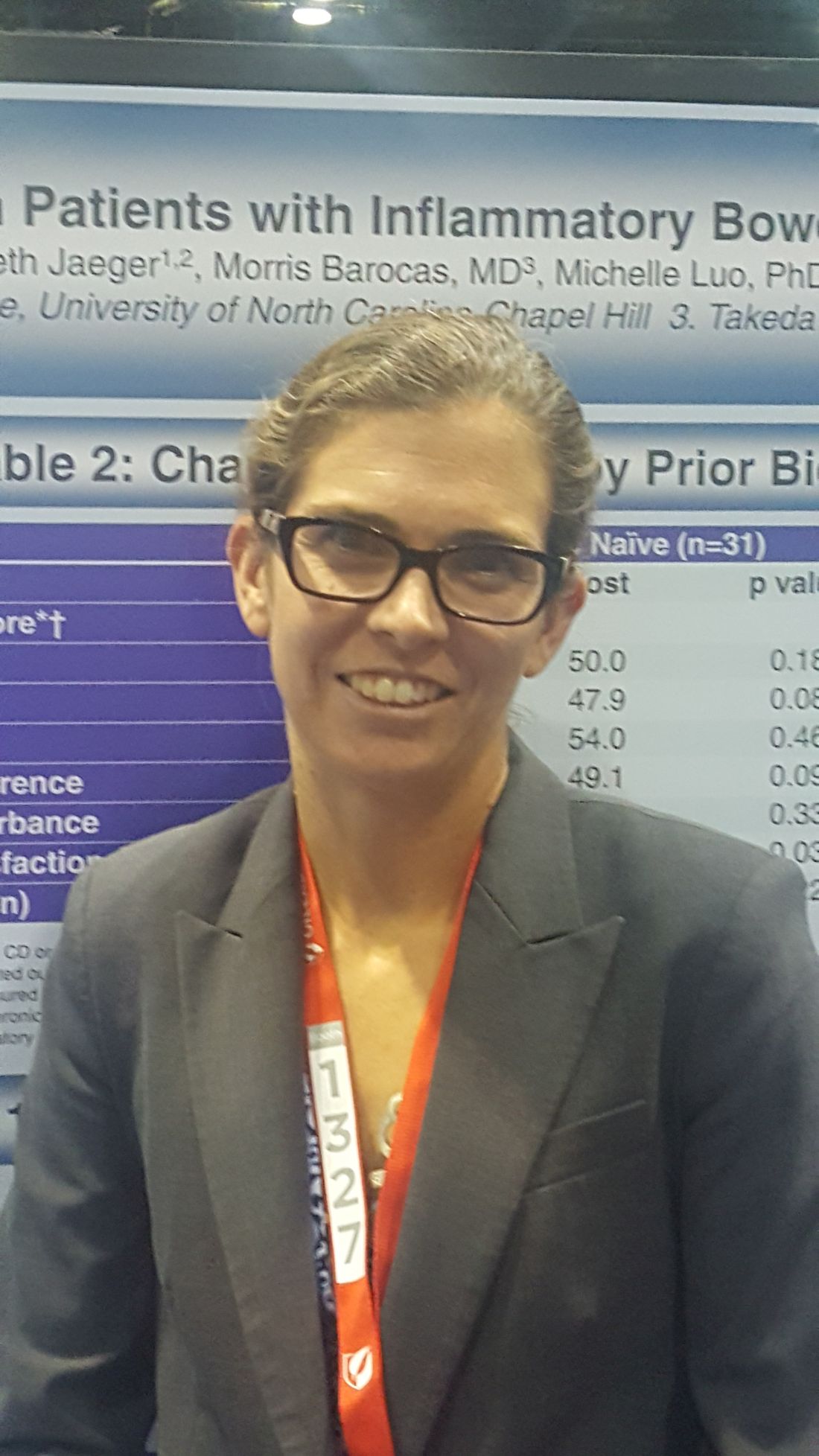
ORLANDO – Vedolizumab therapy was associated with significant improvements in social satisfaction scores and steroid-free remission rates in biologic-naive patients with inflammatory bowel diseases (IBD) in a large prospective cohort.
The Internet-based cohort – Crohn’s & Colitis Foundation of America (CCFA) Partners – includes more than 15,000 IBD patients. For the current study, researchers evaluated 348 participants with Crohn’s disease or ulcerative colitis who initiated vedolizumab therapy between 2014 and 2017 and who had at least 6 months’ follow-up.
The difference in social satisfaction T scores was also improved among biologic-exposed patients (45.8 vs. 47.2, respectively), but the difference did not reach statistical significance, said Dr. Long of the University of North Carolina, Chapel Hill.
Improvements were also seen for numerous other measures, including anxiety, depression, fatigue, pain interference, and sleep disturbance – for both biologic-naive and -exposed patients – but the differences were not significant.
“But these [patient-reported outcomes] are clearly improving,” she said, explaining that trends toward minimally clinically important differences were seen for multiple measures.
As for steroid-free remission, the rate improved from 20% to 45% from baseline to 6-12 months among biologic-naive patients, and from 24% to 30% among biologic-exposed patients, Dr. Long said.
Vedolizumab in this real-world cohort was predominantly used in patients with refractory disease and prior biologic exposure.
The CCFA cohort provides an important glimpse into the effects of vedolizumab on patient-reported outcomes in real-world settings, Dr. Long said, noting that while vedolizumab has demonstrated important quality of life improvements in IBD clinical trials, little has been known about the effects of vedolizumab on quality of life in real-world settings.
The finding with respect to social satisfaction is particularly important, she said.
“These are sick patients. [These scores show that] they’re able to leave the house, they’re able to do the things they want to do,” she said. “It has made a big impact to be able to address this.”
This study was funded by Takeda Pharmaceuticals USA. CCFA Partners is supported by the Crohn’s & Colitis Foundation and the Patient Centered Outcomes Research Institute.
AT THE WORLD CONGRESS OF GASTROENTEROLOGY
Key clinical point:
Major finding: T scores in biologic-naive patients improved significantly (46.1 before treatment vs. 51.0 after 6 months).
Data source: A prospective cohort study of 348 patients.
Disclosures: This study was funded by Takeda Pharmaceuticals USA. CCFA Partners is supported by the Crohn’s and Colitis Foundation and the Patient Centered Outcomes Research Institute.
VIDEO: Gastroenterologist survey shows opportunity to expand Lynch syndrome testing
ORLANDO – A large percentage of U.S. gastroenterologists said that they don’t routinely order genetic testing for Lynch syndrome for patients with early-onset colorectal cancer, often because the physicians believe that the test is too expensive, or because they are unfamiliar with interpreting or applying the results, according to survey replies from 442 gastroenterologists.
Another factor hindering broader screening for Lynch syndrome (also known as hereditary nonpolyposis colorectal cancer) is that many of the surveyed gastroenterologists did not see themselves as having primary responsibility for ordering Lynch syndrome testing in patients who develop colorectal cancer before reaching age 50 years, Jordan J. Karlitz, MD, and his associates reported in a poster at the World Congress of Gastroenterology at ACG 2017.
The survey results showed that only a third of the survey respondents believed it primarily was the attending gastroenterologist’s responsibility to order testing for Lynch syndrome using either a microsatellite DNA instability test or by immunohistochemistry. A larger percentage, 38%, said that ordering one of these tests was something that a pathologist should arrange, 15% said it was primarily the responsibility of the attending medical oncologist, and the remaining respondents cited a surgeon or genetic counselor as having primary responsibility for ordering the test.
This absence of a clear consensus on who orders the test shows a “diffusion of responsibility” that often means testing is never ordered, Dr. Karlitz said in a video interview. What’s needed instead is “reflex testing” that’s done automatically for appropriate patients, an approach that has become standard at several U.S. medical centers, he noted.
The survey Dr. Karlitz and his associates ran stemmed from a report they published in 2015 that focused on management of the 274 patients diagnosed with early-onset colorectal cancer in Louisiana during 2011, defined as cancers diagnosed in patients aged 50 years or younger. Data collected in the Louisiana Tumor Registry showed that Lynch syndrome testing occurred for only 23% of these patients, the researchers reported (Am J Gastroenterol. 2015 Jul;110[7]:948-55).
To better understand the underpinnings of this low testing rate they sent a survey about Lynch syndrome testing by email in March 2017 to nearly 12,000 physicians on the membership roster of the American College of Gastroenterology. They received 455 replies, with 442 (97%) of the responses from gastroenterologists. When asked why they might not order Lynch syndrome testing for patients with early-onset colorectal cancer, 22% said the cost of testing was prohibitive, 18% blamed their lack of familiarity with the Lynch syndrome tests and how to properly interpret their results, and 15% attributed their decision to a lack of easy access to genetic counseling for their patients, with additional reasons cited by fewer respondents.
Dr. Karlitz noted that current recommendations from the National Comprehensive Cancer Network call for Lynch syndrome testing for all patients who develop colorectal cancer regardless of their age at diagnosis.
The video associated with this article is no longer available on this site. Please view all of our videos on the MDedge YouTube channel
[email protected]
On Twitter @mitchelzoler
AT THE WORLD CONGRESS OF GASTROENTEROLOGY
Key clinical point:
Major finding: Among gastroenterologist survey respondents, one-third said they had primary responsibility for ordering Lynch syndrome testing.
Data source: Survey emailed to members of the American College of Gastroenterology and completed by 455 physicians and surgeons.
Disclosures: Dr. Karlitz has been a speaker on behalf of Myriad Genetics, a company that markets genetic tests for Lynch syndrome.
VIDEO: Endoscopy surpasses surgery for acute necrotizing pancreatitis
ORLANDO – An endoscopic approach to treatment of acute necrotizing pancreatitis was substantially safer than was minimally invasive surgical treatment in a randomized study of 66 patients.
Performing drainage and necrosectomy endoscopically in 34 patients with necrotizing pancreatitis that was symptomatic, infected, or both resulted in a 12% rate of major adverse events over the 3 months following intervention compared with a 38% rate among 32 similar patients who underwent laparoscopic drainage followed by either internal debridement or video-assisted retroperitoneal debridement, Ji Young Bang, MD, said at the World Congress of Gastroenterology at ACG 2017.
This statistically significant reduction in the study’s primary endpoint was driven primarily by a major reduction in the incidence of pancreaticocutaneous fistula, which occurred in none of the endoscopy patients and in eight (25%) of the surgery patients, and a smaller reduction in enterocutaneous fistula, which occurred in none of the endoscopy patients and in four (13%) of the patients treated surgically, said Dr. Bang, a gastroenterologist at the Center for Interventional Endoscopy at Florida Hospital, Orlando.
Based on these results, the endoscopic approach “is the treatment of the future,” Dr. Bang said in a video interview. Although the randomized study had a modest number of patients, it was adequately powered to address the hypothesis that endoscopy caused fewer major adverse events than did minimally invasive surgery, and hence the findings should have “an important clinical impact” on the choice of endoscopy or a minimally invasive surgical approach. But Dr. Bang also stressed that a successful endoscopic approach as obtained in this study requires treatment at a center that can offer multidisciplinary expertise from gastrointestinal endoscopists, surgeons, and radiologists, as well as infectious disease physicians, to minimize infections.
Prior to this study, results from the Pancreatitis, Endoscopic Transgastric vs Primary Necrosectomy in Patients With Infected Necrosis (PENGUIN) study run in the Netherlands had also shown significantly fewer adverse events with endoscopic treatment compared with laparoscopic surgery in 20 randomized patients (JAMA. 2012 Mar 14;307[10]:1053-61).
The study reported by Dr. Bang, the Minimally Invasive Surgery vs. Endoscopy Randomized (MISER) trial, enrolled patients with an average necrotic collection size of about 11 cm. The average age of the patients was 59 years. Nearly half of the patients had confirmed infected necrosis. More than 90% had American Society of Anesthesiologists class III or IV disease, and about half had systemic inflammatory response syndrome. All patients had disease that was amenable to both the endoscopic and minimally invasive surgical approaches.
The study’s primary endpoint included several other adverse events in addition to fistulas during 3-month follow-up: death, new-onset organ failure or multiple systemic dysfunction, visceral perforation, and intra-abdominal bleeding. The incidence of each of these outcomes was about the same in the two study arms.
The results also showed that endoscopy was significantly better than surgery for several other secondary outcomes, including new-onset systemic inflammatory response syndrome as well as the prevalence of this complication 3 days after intervention (21% compared with 66%), days in the ICU, average total procedure and hospitalization cost ($76,000 compared with $117,000), and physical quality of life 3 months after treatment. For all other measured outcomes the endoscopic approach and surgical approach produced similar outcomes, and no outcome measured showed that endoscopy was significantly inferior to surgery, Dr. Bang reported.
The video associated with this article is no longer available on this site. Please view all of our videos on the MDedge YouTube channel
[email protected]
On Twitter @mitchelzoler
AT THE WORLD CONGRESS OF GASTROENTEROLOGY
Key clinical point:
Major finding: Major adverse events occurred in 12% of patients treated endoscopically and in 38% of patients treated surgically.
Data source: MISER, a multicenter randomized study of 66 evaluable patients.
Disclosures: MISER received no commercial funding. Dr. Bang had no disclosures.
VIDEO: IBD epidemiology provides clues into disease underpinnings
ORLANDO – The incidence of Crohn’s disease and ulcerative colitis has stabilized in the Western world, but is rising rapidly in newly industrialized countries, according to a systematic review of population-based studies.
The findings could provide important new insights into the environmental, genetic, and microbiome-related factors and interactions that form the underpinnings of IBD, Gilaad Kaplan, MD, of the University of Calgary (Alta.) said at the World Congress of Gastroenterology at ACG 2017.
In turn, that information could lead to approaches to reduce IBD incidence, he said in a video interview.
It has been known that Crohn’s disease and ulcerative colitis are “modern diseases of modern times,” but few studies have addressed the epidemiology of IBD in newly industrialized countries in Asia, Africa, and South America, he said.
“We see a pattern that as newly industrialized countries transition toward a westernized society, IBD emerges and its incidence rises, and there are many different explanations for that,” he said, noting that in part, the increase is due to improved health care infrastructure and advances in adoption of medical technology that lead to better identification of new cases.
“But probably one of the most important factors is that there are environmental exposures linked to the westernization of society that are creating this pressure that’s driving incidence of IBD up in many of the countries of the world,” he said. “I think if we do a lot more research focused on how environment influences microbiome, we might start to see things we could do that could potentially stem the tide of IBD.”
Dr. Kaplan reported having no relevant disclosures.
The video associated with this article is no longer available on this site. Please view all of our videos on the MDedge YouTube channel
AT THE 13TH WORLD CONGRESS OF GASTROENTEROLOGY
VIDEO: Mechanical colonoscope enhancements improve adenoma detection
ORLANDO – Mechanical enhancements to existing colonoscopes may be better than optical enhancements for improving adenoma detection, according to findings from a meta-analysis of data from 240 studies.
“Even though colonoscopy is felt to be our best test compared to others … we also recognize that we do not see every square inch of the colon,” Seth Gross, MD, of New York University Langone Medical Center said in a video interview at the World Congress of Gastroenterology at ACG 2017.
There has been a “tremendous drive” to improve the ability to inspect blind spots in the colon, and also to better recognize subtle precancerous lesions in visible areas of the colon, but it has been unclear whether optical or mechanical enhancements will better achieve that goal, Dr. Gross said.
Based on the findings of his meta-analysis, it appears that mechanical enhancements, including integrated balloons and single-use caps with finger-like projections or discs that clip on to the colonoscope to engage the colon wall and flatten areas to allow access to areas behind folds, are most effective.
The preliminary data should lead to more clinical questions about what can be done to improve exams, he said.
In fact, one four-arm study looking at standard colonoscopy vs. colonoscopy with various mechanical enhancements was just completed, and others looking at “deep learning” and computer assistance are underway.
The latter technology is intriguing, as “not every polyp that we’re missing is behind a fold,” Dr. Gross noted.
Preliminary findings from a study out of China demonstrated the feasibility of such computer assistance, and the researchers are now working on a prospective study of real-time cases to see if that type of integrated learning with computer assistance can improve polyp detection.
“Sometimes it’s just these subtle mucosal changes that we have to train our eye to identify,” he said. “So imagine having another set of eyes … where there’s a computer sort of highlighting an area that we should focus on.”
Dr. Gross reported having no relevant financial disclosures.
The video associated with this article is no longer available on this site. Please view all of our videos on the MDedge YouTube channel
AT THE WORLD CONGRESS OF GASTROENTEROLOGY
More IBD remissions with higher induction vedolizumab levels
ORLANDO – Higher vedolizumab levels during induction were associated with better responses to therapy at 22 weeks in patients with inflammatory bowel diseases in a prospective cohort study.
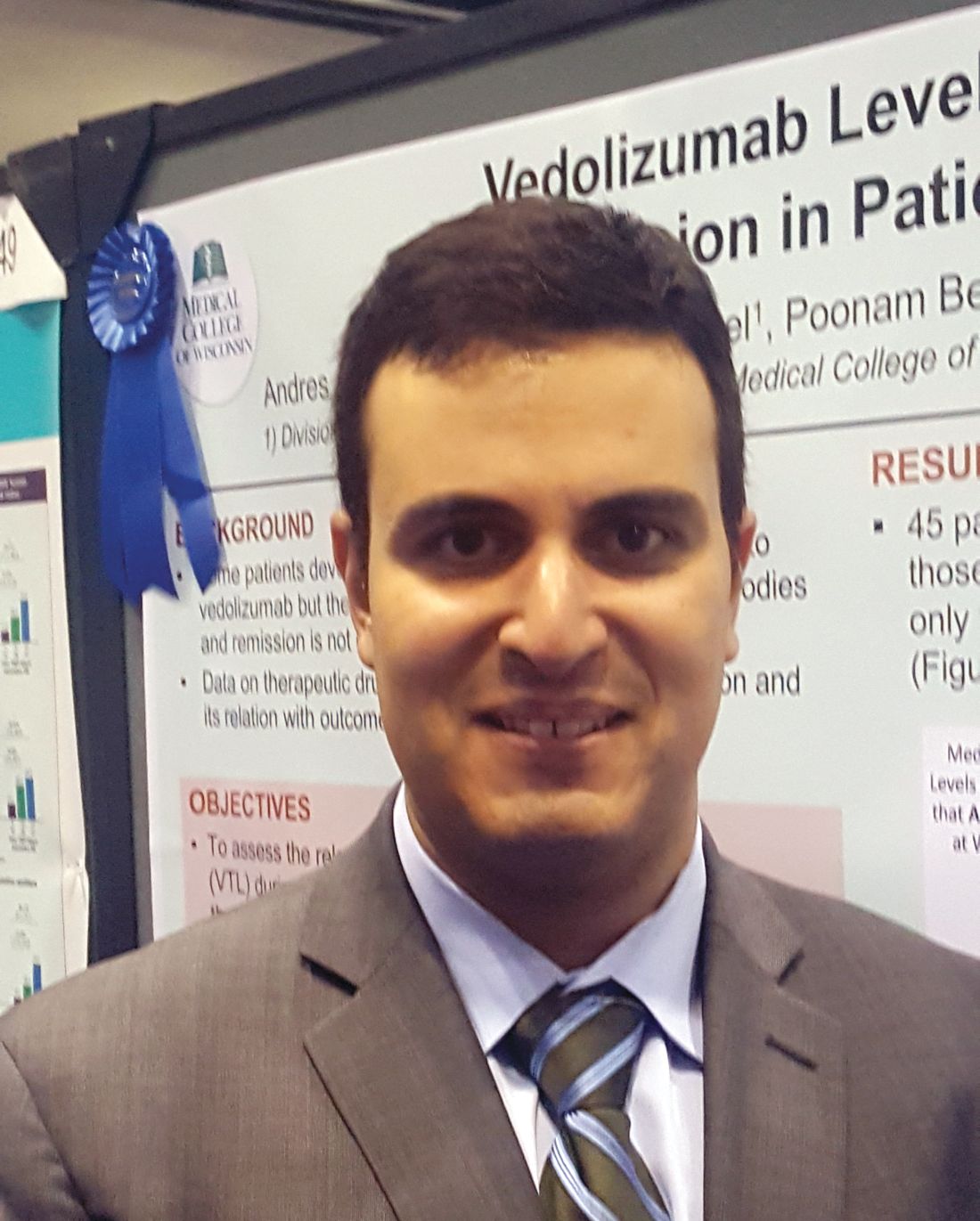
The findings suggest that therapeutic drug monitoring and early optimization could play an important role in improving outcomes in patients with Crohn’s disease or ulcerative colitis who are receiving treatment with the monoclonal antibody, Andres J. Yarur, MD, reported in a poster at the World Congress of Gastroenterology at ACG 2017.
Patients with a VTL of 24 mcg/mL or greater at week 2, and 10.6 mcg/mL or greater at week 6, were more likely to be in remission at week 22 (odds ratios, 5 and 13.5, respectively).
Of note, VTLs were numerically higher in patients receiving combination therapy, compared with those receiving vedolizumab monotherapy, but the difference was statistically significant only at week 2 (24.7 vs. 21.8 mcg/mL, respectively), he said.
Similar correlations between trough levels and response rates have been seen with other biologics, but data on such correlations has been lacking for vedolizumab. Since some patients develop primary or secondary nonresponse, Dr. Yarur and his colleagues assessed the relationship between serum VTLs during induction and disease remission after 22 weeks, he explained in an interview.
They also investigated the presence of antibodies to vedolizumab .
The primary outcome of deep remission at 22 weeks was defined as normal C-reactive protein levels and Simple Endoscopic Score for Crohn’s Disease of 2 or less in patients with Crohn’s disease, and Mayo Endoscopic score of 1 or less in patients with ulcerative colitis, plus clinical remission (Harvey-Bradshaw Index score of less than 5 in patients with Crohn’s disease and Mayo Clinical Score of less than 3 in ulcerative colitis).
Three patients developed antibodies to vedolizumab during induction, but the antibodies were undetectable by week 14 in all three, he said.
“The findings open the question of whether higher doses during induction will improve the rate of remission,” he said, noting that such early optimization is currently being evaluated in ongoing studies.
Dr. Yarur reported having no relevant disclosures.
AT THE WORLD CONGRESS OF GASTROENTEROLOGY
Key clinical point:
Major finding: Vedolizumab trough levels at weeks 2 and 6 were higher among those who achieved remission at week 22, compared with those who did not (25 vs. 21.8 mcg/mL and 26.1 vs. 12.7 mcg/mL, respectively).
Data source: A prospective cohort study of 45 patients.
Disclosures: Dr. Yarur reported having no relevant disclosures.
Deep learning can assist real-time polyp detection during colonoscopies
A deep-learning algorithm for automatic polyp detection during colonoscopies showed both high sensitivity and high specificity, according to a study presented at the World Congress of Gastroenterology at ACG 2017.
The algorithm was set up using a retrospective set of 5,545 images annotated by colonoscopists, while the validation set used for the study consisted of 27,461 colonoscopy images from 1,235 patients, according to Pu Wang, MD, of the Sichuan Academy of Medical Sciences & Sichuan Provincial People’s Hospital, Chengdu, China, and his associates.
At the high-sensitivity operating point, the algorithm had a sensitivity of 94.96% and a specificity of 92.01%, and at the low–false positive rate operating point, the algorithm had a sensitivity of 92.35% and a specificity of 97.05%. The area under the curve was 0.958 in a receiver operating characteristic curve analysis.
In subgroup analyses of flat polyps, polyps less than or equal to 0.5 cm, and isochromatic polyps, the algorithm had areas under the curve of 0.943, 0.957, and 0.957 respectively.
The algorithm reported results in 60-80 ms, offering real-time assistance with polyp detection during colonoscopies, Dr. Wang and his colleagues noted.
The study was not funded by industry grants, and no disclosures were reported.
FROM WORLD CONGRESS OF GASTROENTEROLOGY
Key clinical point:
Major finding: The deep-learning algorithm had a sensitivity of 95% and a specificity of 92% when adjusted to the high-sensitivity operating point.
Data source: A set of 27,461 colonoscopy images from 1,235 patients.
Disclosures: The study was not funded by industry grants, and no disclosures were reported.
