User login
The Official Newspaper of the American Association for Thoracic Surgery
Consider invasive mediastinal staging in higher risk NSCLC patients, despite guidelines
Endobronchial ultrasound transbronchial needle aspiration (EBUS-TBNA) appears to be cost effective for use in non–small cell lung cancer (NSCLC) staging if the prevalence of mediastinal lymph node metastasis (MLNM) is greater than or equal to 2.5%, according to the results of single institution modeling study. In addition, the study found that confirmatory mediastinoscopy should be performed in high-risk patients in cases of negative EBUS-TBNA.
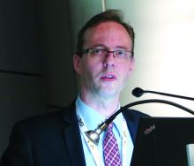
Katarzyna Czarnecka-Kujawa, MD, of the University of Toronto and Toronto General Hospital, and her colleagues performed a decision analysis to compare health outcomes and costs of four mediastinal staging strategies. They assessed the following: no invasive staging, endobronchial ultrasound-guided transbronchial need aspiration (EBUS-TBNA), mediastinoscopy, and EBUS-TBNA followed by mediastinoscopy if EBUS-TBNA results were negative. They determined incremental cost-effectiveness ratios (ICER) for all strategies and performed comprehensive sensitivity analyses using a willingness to pay threshold of $80,000 [Canadian]/quality adjusted life-year (QALY).
They used data obtained for staging, outcomes, and costs from the patients in the lung cancer program at the Toronto General Hospital from Jan. 1, 2005 to Dec. 31, 2014, as detailed in a report published in the June issue of the Journal of Thoracic and Cardiovascular Surgery (2017. doi: 10.1016/j.jtcvs.2016.12.048).
After exclusions, they utilized a final case count of 499 cases for developing their surgical and procedure cost analysis, and a total of 750 cases in their endoscopy database for endoscopy analysis. For the base-case analysis, they assumed a prevalence of mediastinal metastasis of 9%, and obtained the prevalence of a pathologic lymph nodal stage disease following EBUS-TBNA from their institutional data.
Their results showed that EBUS-TBNA followed by mediastinoscopy was the strategy that resulted in the highest QALYs, but that it had a prohibitive ICER of greater than $1.4 million/QALY. Accordingly, it may not be justifiable to use mediastinoscopy after negative EBUS-TBNA in all patients, the researchers noted. However, the researchers’ data suggest that invasive screening may be justified in a very-low-risk population (MLNM above 2.5%).
In addition, the researchers stated that “[the] benefit conveyed by detecting mediastinal metastatic disease becomes more apparent as the prevalence of MLNM increases, with confirmatory mediastinoscopy becoming cost effective in cases of negative EBUS-TBNA in patients with moderate to high probability of MLNM” (greater than 57%).
Our model points out that there is a well-defined role for the use of different modalities, including mediastinoscopy. This stresses the need for ongoing focus on maintenance of competency and skill acquisition in mediastinoscopy and EBUS-TBNA by currently practicing and future thoracic surgeons respectively,” the researchers concluded.
Dr. Czarnecka-Kujawa disclosed that she is a research consultant with Olympus America. The study was funded in part by agencies of the Austrian government.
The authors make a compelling argument for invasive mediastinal staging in patients with clinical stage I non–small cell lung cancer and acknowledge that this conflicts with current guidelines, according to Biniam Kidane, MD, of the University of Manitoba, Winnipeg, in his invited comments on the study in the Journal of Thoracic and Cardiovascular Surgery (2017 Mar 10. doi: 10.1016/j.jtcvs.2017.02.051).
Their single-payer system is likely to have a different willingness-to-pay threshold, compared with those in other countries, especially the United States, where the EBUS-TBNA strategy without invasive staging is likely to remain the cost-effective choice.
“Cost-economic analyses such as these provide a window into the factors necessary to bridge guidelines from the realm of the abstract to the realm of local reality. When interpreting these findings, clinicians should consider: 1) What EBUS resources are available? (2) What is your local EBUS sensitivity? 3) What is the prevalence of MLNM?” Dr. Kidane concluded, with the caveat that such studies are not infallible and models are based on assumptions and must be treated with care.
Dr. Kidane reported no disclosures with regard to commercial support.
Endobronchial ultrasound transbronchial needle aspiration (EBUS-TBNA) appears to be cost effective for use in non–small cell lung cancer (NSCLC) staging if the prevalence of mediastinal lymph node metastasis (MLNM) is greater than or equal to 2.5%, according to the results of single institution modeling study. In addition, the study found that confirmatory mediastinoscopy should be performed in high-risk patients in cases of negative EBUS-TBNA.
Katarzyna Czarnecka-Kujawa, MD, of the University of Toronto and Toronto General Hospital, and her colleagues performed a decision analysis to compare health outcomes and costs of four mediastinal staging strategies. They assessed the following: no invasive staging, endobronchial ultrasound-guided transbronchial need aspiration (EBUS-TBNA), mediastinoscopy, and EBUS-TBNA followed by mediastinoscopy if EBUS-TBNA results were negative. They determined incremental cost-effectiveness ratios (ICER) for all strategies and performed comprehensive sensitivity analyses using a willingness to pay threshold of $80,000 [Canadian]/quality adjusted life-year (QALY).
They used data obtained for staging, outcomes, and costs from the patients in the lung cancer program at the Toronto General Hospital from Jan. 1, 2005 to Dec. 31, 2014, as detailed in a report published in the June issue of the Journal of Thoracic and Cardiovascular Surgery (2017. doi: 10.1016/j.jtcvs.2016.12.048).
After exclusions, they utilized a final case count of 499 cases for developing their surgical and procedure cost analysis, and a total of 750 cases in their endoscopy database for endoscopy analysis. For the base-case analysis, they assumed a prevalence of mediastinal metastasis of 9%, and obtained the prevalence of a pathologic lymph nodal stage disease following EBUS-TBNA from their institutional data.
Their results showed that EBUS-TBNA followed by mediastinoscopy was the strategy that resulted in the highest QALYs, but that it had a prohibitive ICER of greater than $1.4 million/QALY. Accordingly, it may not be justifiable to use mediastinoscopy after negative EBUS-TBNA in all patients, the researchers noted. However, the researchers’ data suggest that invasive screening may be justified in a very-low-risk population (MLNM above 2.5%).
In addition, the researchers stated that “[the] benefit conveyed by detecting mediastinal metastatic disease becomes more apparent as the prevalence of MLNM increases, with confirmatory mediastinoscopy becoming cost effective in cases of negative EBUS-TBNA in patients with moderate to high probability of MLNM” (greater than 57%).
Our model points out that there is a well-defined role for the use of different modalities, including mediastinoscopy. This stresses the need for ongoing focus on maintenance of competency and skill acquisition in mediastinoscopy and EBUS-TBNA by currently practicing and future thoracic surgeons respectively,” the researchers concluded.
Dr. Czarnecka-Kujawa disclosed that she is a research consultant with Olympus America. The study was funded in part by agencies of the Austrian government.
FROM THE JOURNAL OF THORACIC AND CARDIOVASCULAR SURGERY
Key clinical point:
Major finding: Once the pathologic lymph nodal stage reaches 57%, EBUS-TBNA followed by mediastinoscopy is cost effective.
Data source: A model of health care outcomes and costs was developed from data obtained from patients treated over a 10-year period at a single institution.
Disclosures: Dr. Czarnecka-Kujawa disclosed that she is a research consultant with Olympus America. The study was funded in part by agencies of the Austrian government.
Embolism major cause of stroke after open arch surgery in patients with carotid/intracranial stenosis
Embolization was the major cause of permanent stroke in patients with moderate or severe carotid or intracranial atherosclerosis who underwent elective open aortic arch surgery at a single institution, according to the results of a retrospective study.
Preoperative craniocervical and aortic screening may aid in modifying the operative strategy to reduce the incidence of stroke in these patients, according to a report published in the May issue of the Journal of Thoracic and Cardiovascular Surgery.
Preventing stroke in this patient population is an important consideration, because perioperative stroke is approximately 4 times more common in open aortic arch surgery (OAAS) than in coronary artery bypass grafting or valve surgery, according to Ken-ichi Imasaka, MD, and his colleagues at the National Hospital Organization Kyushu Medical Center, Fukuoka, Japan.
The study population comprised 200 consecutive patients undergoing elective OAAS at the institution between October 2008 and October 2015, including 34% women and with a mean patient age of 71 years (J Thorac Cardiovasc Surg. 2017;153:1045-53).
After preoperative screening, 21% of patients were diagnosed with carotid or intracranial artery disease (CIAD). None of these patients were diagnosed with impaired cerebral perfusion reserve on brain SPECT (single-photon emission computed tomography). A total of 92% of patients underwent ascending aorta or aortic arch replacement through a median sternotomy, while the remaining 8% underwent extended aortic arch replacement via L-incision (15 patients) or combined median sternotomy and left posterior lateral thoracotomy (1 patient). Among the patients, 16% underwent ascending aorta replacement; 8% had partial arch replacement; and the remaining 76% had total arch replacement.
Shaggy aorta was present in 19% of the patients, with 51% of these showing CIAD (P less than .0001). A total of 30% of the patients with shaggy aorta had the total arch replacement through an L-incision or combined median sternotomy and left posterior lateral thoracotomy, a significant difference (P less than .0001).
The overall in-hospital mortality rate was 3.5%. The overall incidence of permanent stroke and paraplegia or paraparesis was 4% (8 patients) and 2% (4 patients), respectively. Three (37.5%) of the 8 permanent stroke patients died during the postoperative hospital stay, compared with 2.1% of the 192 patients without stroke.
Univariate analysis indicated that previous cerebrovascular accident (P = .0002), shaggy aorta (P less than .0001), cardiopulmonary bypass time (P = .003), selective antegrade cerebral perfusion time (P = .004), operation time (P = .02), and extended aortic repair through L-incision or combined median sternotomy and left posterior lateral thoracotomy (P = .0002) were significant risk factors for neurologic morbidity.
“Preoperative intensive screening of carotid and intracranial artery disease is a useful step to identify patients at higher risk of hemodynamic ischemic stroke. Advanced systemic atherosclerosis may be a crucial determinant of perioperative stroke due to atherothrombotic embolization. Antiembolic measures during surgery are essential to prevent perioperative stroke,” the researchers concluded.
The authors reported that they had no disclosures.
During aortic arch surgery, the lack of blood supply as a result of emboli, rather than atherosclerosis itself, kills the cerebral neurons, according to Ourania Preventza, MD, and Joseph S. Coselli, MD, of the Baylor College of Medicine, Houston, in their invited commentary (J Thorac Cardiovasc Surg. 2017;153:1054-5).
Patients with carotid and intracranial disease should indeed have more intensive screening before undergoing major aortic surgery, they agreed, but pointed out that in the absence of carotid disease, large or complex aortic atheromas can be seen in the arch, indicating that, even though atherosclerosis is a systemic disease, using different sites of prediction can be uncertain.
This requires a broader approach to prevent stroke, including careful selection of the cannulation site in patients with diffuse and heavy arch atherosclerosis or currently ulcerated plaque, they added.
“To minimize postoperative neurologic morbidities after aortic arch surgery, an individually tailored perioperative approach should be in the armamentarium of cardiac surgeons,” Dr. Preventza and Dr. Coselli concluded.
Dr. Preventza consults for Medtronic and W. L. Gore & Associates. Dr. Coselli participates in clinical research trials conducted by GlaxoSmithKline, Edwards Lifesciences, and Bolton Medical, and consults for various companies.
Embolization was the major cause of permanent stroke in patients with moderate or severe carotid or intracranial atherosclerosis who underwent elective open aortic arch surgery at a single institution, according to the results of a retrospective study.
Preoperative craniocervical and aortic screening may aid in modifying the operative strategy to reduce the incidence of stroke in these patients, according to a report published in the May issue of the Journal of Thoracic and Cardiovascular Surgery.
Preventing stroke in this patient population is an important consideration, because perioperative stroke is approximately 4 times more common in open aortic arch surgery (OAAS) than in coronary artery bypass grafting or valve surgery, according to Ken-ichi Imasaka, MD, and his colleagues at the National Hospital Organization Kyushu Medical Center, Fukuoka, Japan.
The study population comprised 200 consecutive patients undergoing elective OAAS at the institution between October 2008 and October 2015, including 34% women and with a mean patient age of 71 years (J Thorac Cardiovasc Surg. 2017;153:1045-53).
After preoperative screening, 21% of patients were diagnosed with carotid or intracranial artery disease (CIAD). None of these patients were diagnosed with impaired cerebral perfusion reserve on brain SPECT (single-photon emission computed tomography). A total of 92% of patients underwent ascending aorta or aortic arch replacement through a median sternotomy, while the remaining 8% underwent extended aortic arch replacement via L-incision (15 patients) or combined median sternotomy and left posterior lateral thoracotomy (1 patient). Among the patients, 16% underwent ascending aorta replacement; 8% had partial arch replacement; and the remaining 76% had total arch replacement.
Shaggy aorta was present in 19% of the patients, with 51% of these showing CIAD (P less than .0001). A total of 30% of the patients with shaggy aorta had the total arch replacement through an L-incision or combined median sternotomy and left posterior lateral thoracotomy, a significant difference (P less than .0001).
The overall in-hospital mortality rate was 3.5%. The overall incidence of permanent stroke and paraplegia or paraparesis was 4% (8 patients) and 2% (4 patients), respectively. Three (37.5%) of the 8 permanent stroke patients died during the postoperative hospital stay, compared with 2.1% of the 192 patients without stroke.
Univariate analysis indicated that previous cerebrovascular accident (P = .0002), shaggy aorta (P less than .0001), cardiopulmonary bypass time (P = .003), selective antegrade cerebral perfusion time (P = .004), operation time (P = .02), and extended aortic repair through L-incision or combined median sternotomy and left posterior lateral thoracotomy (P = .0002) were significant risk factors for neurologic morbidity.
“Preoperative intensive screening of carotid and intracranial artery disease is a useful step to identify patients at higher risk of hemodynamic ischemic stroke. Advanced systemic atherosclerosis may be a crucial determinant of perioperative stroke due to atherothrombotic embolization. Antiembolic measures during surgery are essential to prevent perioperative stroke,” the researchers concluded.
The authors reported that they had no disclosures.
Key clinical point:
Major finding: Previous cerebrovascular accident and shaggy aorta were significant determinants of neurologic morbidity.
Data source: Retrospective study of 200 consecutive patients undergoing elective aortic arch surgery at a single institution.
Disclosures: The authors reported having no conflicts of interest.
Salvage chemo for NSCLC more effective after PD-1/PD-L1 inhibitors
GENEVA – Patients with stage IV non–small cell lung cancer (NSCLC) who have disease progression following treatment with an immune checkpoint inhibitor have a 30% better chance of achieving at least a partial response with salvage chemotherapy compared with patients who had received prior chemotherapy but not immunotherapy, according to Swiss and U.S. investigators.
In a study of 82 patients with advanced NSCLC, 18 of 67 patients (27%) who had progressed on an inhibitor of programmed death-1 (PD-1) or programmed death ligand-1 (PD-L1) had a partial response to combination chemotherapy, compared with just 1 of 15 (7%) of patients who had not received a checkpoint inhibitor, reported Sacha Rothschild, MD, PhD, of University Hospital Basel, Switzerland, and colleagues.
“The odds of achieving a partial response to salvage chemotherapy were significantly higher in patients with prior exposure to PD-1/PD-L1 inhibitors. This observed difference, however, warrants confirmation in larger cohorts. Ongoing investigations include the duration of response as well as evaluation of toxicity,” the researchers wrote in a scientific poster presented at the European Lung Cancer Conference.
Immune checkpoint inhibitors have been shown to be active in patients with late-stage NSCLC who have experienced disease progression following chemotherapy. To see whether salvage chemotherapy could offer any additional benefit to patients who had disease progression on immunotherapy, the investigators conducted a retrospective case-control study. They reviewed 355 patient records, and identified 82 patients – 46 men and 36 women – who met the study criteria.
Of this group, 67 patients had received a PD-1/PD-L1 checkpoint inhibitor, either nivolumab (Opdivo, 56 patients), pembrolizumab (Keytruda, 7), or atezolizumab (Tecentriq, 4). These patients were designated as cases. The remaining 15 patients, who had received prior chemotherapy or chemoradiotherapy only, were designated as controls.
Of all 82 patients, 63 (77%) had adenocarcinomas, 18 (22%) had squamous cell carcinomas, and 1 (1%) had a large-cell carcinoma.
Cases had a mean of 2.37 chemotherapy regimens prior to salvage chemotherapy, and controls had a mean of 1.93. Drugs used in the salvage regimens were docetaxel in 62% of patients, pemetrexed in 20%, gemcitabine in 12%, and paclitaxel in 6%.
The odds ratio for achieving a partial response was 0.30 (95% confidence interval, 0.18-0.50, P less than .0001).
In a multiple logistic regression model, neither age, sex, number of prior chemotherapy regimens, tumor histology, smoking status, nor type of salvage chemotherapy regimen were significantly associated with the likelihood of achieving a partial response.
In a poster discussion session, Simon Ekman, MD, of Karolinska Institute in Stockholm, Sweden, the invited discussant, said that the data from the study are convincing, and raise the question of what to do next.
“The question is how do we combine immunotherapy with chemotherapy? Should we use the immune therapy first, like in this trial, or vice versa, or should we combine concurrently” he said.
The strategy of chemotherapy first is supported by research showing that neoantigens crucial for the immune response are released during chemotherapy and radiotherapy, enabling immunotherapeutic agents to work better, he said.
The study was internally funded. Dr. Rothschild and Dr. Ekman reported no conflicts relevant to the research.
FROM ELCC
Key clinical point: Salvage chemotherapy was more effective among patients who had disease progression following immunotherapy in the second line than among patients who received only chemotherapy.
Major finding: Among patients with disease progression while on a checkpoint inhibitor, 18 of 67 had a partial response to salvage chemotherapy, compared with just 1 of 15 controls.
Data source: Retrospective case-control study of 82 patients with advanced NSCLC.
Disclosures: The study was internally funded. Dr. Rothschild and Dr. Ekman reported no conflicts relevant to the research.
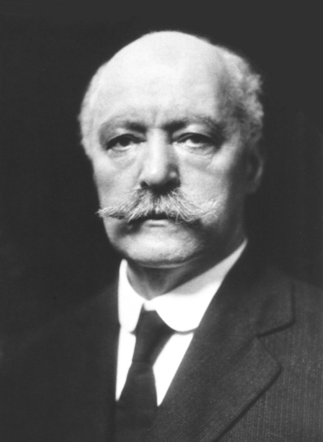
Confronting the open chest – Samuel J. Meltzer and the first AATS annual meeting
In retrospect, the founding of the American Association for Thoracic Surgery (AATS) in 1917 may seem surprisingly optimistic, given the status of cardiothoracic surgery as a discipline at that time. While important strides had been made in dealing with open chest wounds, to the modern eye, the field in the second decade of the 20th century seems more characterized by what was not yet possible rather than by what was.
One of the most critical issues holding back the development of cardiothoracic surgery in this early period was the problem of acute pneumothorax that occurred whenever the chest was opened.
As Willy Meyer (1858-1932), second president of the AATS, described the problem at the first AATS annual meeting in 1917: “What is it that happens when the thorax is opened, let us say [for example] by a stab wound in an intercostal space in an affray on the street? Immediately air rushes into the pleural cavity and this normal atmospheric pressure, being greater than the normal pressure within ... the lung contracts to a very small organ around its hilum. Air fills the space formerly occupied by the lung. This condition, with its immediate clinical pathologic consequences, is called ‘acute pneumothorax.’ It has been the stumbling block for almost a century to the proper development of the surgery of the chest. … Carbonic acid is retained in the blood … The accumulation of CO, with its deleterious effect increases, finally ending in the patient’s death.”
But in the first decade of the 20th century, two major and competing techniques evolved to solve the problem, each one represented by the first and second presidents of the fledgling AATS. For a short period of time a controversy seemed to separate the two men, but their views were expressly reconciled at the first annual meeting of the AATS.
The Meltzer/Auer technique was significantly improved upon by the addition of a carbon dioxide absorption method and the creation of a closed circuit apparatus by Dennis Jackson, MD (1878-1980) in 1915. “This fulfilled the criteria of oxygen supply, carbon dioxide absorption, and ether regulation with a hand bag-breather. With this apparatus, respiration could be maintained with the open thorax,” said pioneering thoracic surgeon Rudolf Nissen, MD, and Roger H.L. Wilson, MD, in their Pages in the History of Chest Surgery (Springfield, IL: Charles C. Thomas, 1960).
However, insufflation was not universally applauded when it was first introduced. It was considered a poor second by many who instead embraced the alternate method of preventing chest collapse – the differential pressure–maintaining Sauerbruch chamber. The Sauerbruch chamber was developed by Ernst Ferdinand Sauerbruch (1875-1951) and first reported in 1904 in his paper, “The pathology of open pneumothorax and the bases of my methods for its elimination.”
As described by Nissen and Wilson, “He transformed the operating room into a kind of extended or enlarged pleural cavity, lowering the atmospheric pressure by vacuum. The head of the patient was outside the operating room, tightly sealed at the neck. This ‘pneumatic chamber’ solved in an ideal way the problem of negative pleural pressure.”
Sauerbruch was aware of the Meltzer/Auer technique, but specifically rejected it, and his powerful influence in Germany helped to prevent it from being adopted there.
Among the earliest and most vocal advocates of using the Sauerbruch negative pressure chamber approach in the United States was Dr. Meyer. Both he and Dr. Meltzer addressed the issue and the controversy at the first meeting of the AATS in 1917.
“You probably remember the little battle between differential pressure and intratracheal insufflation. It occurred only 8 years ago; but it seems now like history. When I presented my paper on intratracheal insufflation at the New York Academy of Medicine, my views were opposed, in the interest of conservatism in surgery, by three able surgeons,” Dr. Meltzer said in his address.
“Now, these same surgeons are among the principal founders of the American Association for Thoracic Surgery, and my being the first presiding officer of the Association is due exclusively to their generous spirit and not to any merits of mine. This is my little story of how the introduction of a stomach tube carried a mere medical man into the presidential chair of a national surgical association.”
Dr. Meyer, one of the three surgeons mentioned by Dr. Meltzer, responded shortly thereafter in his own speech at the meeting: “Dr. Meltzer mentioned in his inaugural address today that, in the discussion following his presentation of the matter before the New York Academy of Medicine his views were opposed, in the interest of conservatism in surgery, by three surgeons.
“Inasmuch as I was one of the three, I would, in explanation, here state that ... at that very time it was reported to me that Dr. Meltzer had stated that in his opinion thoracic operations on human beings could be done in a much simpler way than by working in the negative chamber; that a catheter in the trachea and bellows was all that was needed. He, a physiologist who had always done scientific surgical work on animals, certainly found these paraphernalia sufficient. I personally had meanwhile seen and learned to admire the absolutely reliable working of the mechanism of the chamber, without the possibility of doing the slightest harm to the patient.
“In my remarks on that memorable evening at the New York Academy of Medicine, I therefore tried to impress upon my colleagues the great importance of absolute safety. I stated that no matter what apparatus we might use in thoracic surgery on the usually much run down human being, it must be so constructed that it could not possibly do harm to the patient. I further stated that I would be only too happy to personally use intratracheal insufflation as soon as it was sufficiently perfected to render it safe under all conditions. … I want to lay stress upon the statement that I for my part have never been in opposition, but rather in full accord with his splendid discovery. The fact is that I personally have been among the very first in New York to use intratracheal insufflation in thoracic operations upon the human subject,” said Dr. Meyer.
“But, please bear in mind … that only the use of the differential pressure method – no matter what the apparatus – enables the surgeon to work in the thorax with the same equanimity and tranquility as in the abdomen,” he summarized.
So by the early years of the founding of the AATS, no matter the barriers that remained, the fact that thoracic surgery had reached the same level of confidence in terms of attempting operations as had already existed for the abdomen permitted the fledgling association to move forward with a confidence and optimism that had not existed before, when opening the chest in the operating room was generally considered deadly.
Sources
Meltzer, S. J., 1917. First President’s Address. http://t.aats.org/annualmeeting/Program-Books/50th-Anniversary-Book/First-Presidents-Address.cgi
Meyer, W., 1917. Surgery Within the Past Fourteen Years. http://t.aats.org/annualmeeting/Program-Books/50th-Anniversary-Book/A-Review-of-the-Evolution-of-Thoracic-Surgery-Within-the-Pas.cgi
Nissen, R., Wilson, R.H.L. Pages in the History of Chest Surgery. Springfield, IL: Charles C. Thomas, 1960.
One-third of drug postmarket studies go unpublished
More than one-third of postmarket studies following drug approval that should be published are not, according to new research.
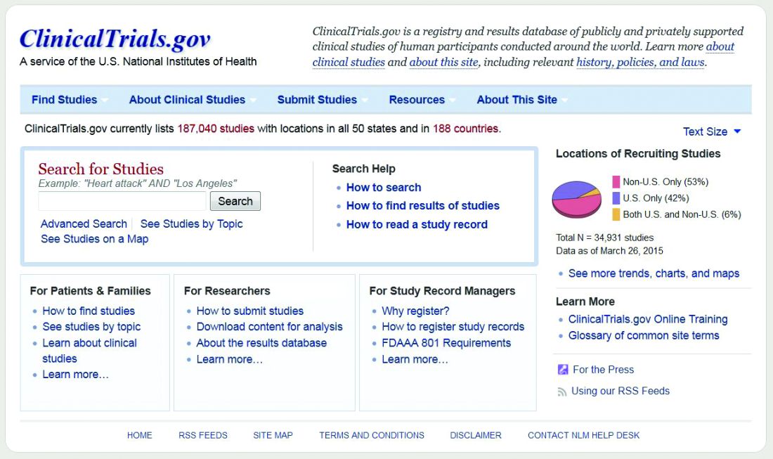
Investigators examined a Food and Drug Administration internal database to identify all postmarket drug studies between 2009 and 2013 identified by the agency as completed, with a follow-up search to find if/where the results of the studies were published.
“As of July 2016, 183 of the 288 postmarket studies (63.5%) meeting inclusion criteria were published in either the scientific literature or on the ClinicalTrials.gov website,” Marisa Cruz, MD, medical officer in the Food and Drug Administration’s Office of Public Health Strategy and Analysis, and her colleagues wrote in a researcher letter published online May 15 in JAMA Internal Medicine (doi: 10.1001/jamainternmed.2017.1313).
More studies were published in journals (175) than in the agency’s clinical trial registry (87), and the 183 interventional clinical trials had a higher overall publication rate (87.4%) than the other 105 studies combined (21.9%).
Of the 69 interventional clinical trials that were focused on efficacy, 86.2% were categorized as having results that were favorable to the trial sponsor. However, the 57 interventional clinical trials with positive results were no more likely to be published than the 12 trials with negative results, Dr. Cruz and colleagues noted.
The findings are consistent with previous research, the researchers noted, with the analysis demonstrating “that postmarket study results are not consistently disseminated, either through journal publication or trial registries.”
“Despite calls for data sharing and publication of all clinical trial results, publication rates for completed postmarket studies required by the FDA remain relatively low,” the researchers wrote.
While the FDA could publish the data itself, “this approach would likely require new regulations,” the authors noted. “Alternatively, increased sponsor commitment to submitting to journals and to publish all clinical trial results on trial registries, regardless of whether publication is legally required, may serve to promote dissemination of scientific knowledge.”
The researchers reported no conflicts of interest.
Mycobacteria subset plagues pulmonary patients
Nontuberculous mycobacteria accounts for an increasing percentage of pulmonary disease, and nonsurgical treatment alone has not shown effectiveness, according to data from a meta-analysis of 24 studies and 1,224 patients. The study results were published online in Chest.
Data on therapeutic successes in cases of nontuberculosis mycobacteria (NTM)–related pulmonary disease are limited, in particular for those species not related to the Mycobacterium avium complex (non-MAC), wrote Roland Diel, MD, of University Medical Hospital Schleswig-Holstein, Germany, and his colleagues.
In particular, non-MAC species Mycobacterium xenopi (MX), Mycobacterium abscessus, Mycobacterium malmoense, and Mycobacterium kansasii (MK) were addressed in the studies, which included 16 retrospective chart reviews, 5 randomized trials, and 3 prospective, nonrandomized studies (Chest 2017. doi: 10.1016/j.chest.2017.04.166).
Treatment success was measured by rates of sputum culture conversion (SCC).
Overall, the average proportion of SCC for patients with M. abscessus was 41% after subtraction for posttreatment relapses, but reached 70% for subspecies M. massiliense in macrolide-containing treatments. The average proportion of SCC was 80% for patients with M. kansasii, 32% for those with MX, and 54% for those with M. malmoense.
Treatment success ranged from 9% to 73% for M. xenopi patients, but all-cause mortality was 69%. Of note, a 100% success rate was noted in M. kansasii patients using a three-drug TB regimen of isoniazid, rifampicin, and ethambutol, or with a combination of ethambutol, rifampicin, and clarithromycin, the researchers noted.
The percentage of SCC in 55 patients with lung resection and either MX or M. abscessus was considered high at 76%.
The study findings were limited by the diverse definitions of treatment success and by the variety of treatments and “an optimal multidrug treatment cannot be derived from the few studies and has yet to be determined,” the researchers said. In the absence of optimal drug therapy, functional and quality of life elements deserve greater consideration when evaluating outcomes in patients with non-MAC NTM pulmonary disease, they added.
Dr. Diel reported receiving lecturing and/or consulting fees from Insmed and Riemser.
FROM CHEST
Key clinical point: An optimal multidrug treatment has not yet been found for patients with nontuberculosis mycobacteria (NTM)–related pulmonary disease.
Major finding: The average proportion of sputum culture conversion (SCC) for patients with M. abscessus was 42% after subtraction for posttreatment relapses, but reached 79% for subspecies M. massiliense in macrolide-containing treatments. The average proportion of SCC was 80% for patients with M. kansasii, 32% for those with M. xenopi, and 54% for those with M. malmoense.
Data source: A meta-analysis of 24 studies and 1,224 patients.
Disclosures: Dr. Roland Diel reported receiving lecturing and/or consulting fees from Insmed and Riemser.
Atezolizumab improves OS in NSCLC with brain metastases
GENEVA – Immune checkpoint inhibitors may improve survival in patients with non–small cell lung cancer (NSCLC) and brain metastases compared with chemotherapy, without adding unacceptable toxicities, pooled analyses of clinical trials suggest.
Among 1452 patients with NSCLC treated with the programmed death ligand-1 (PD-L1) inhibitor atezolizumab (Tecentriq), rates of treatment-related serious adverse events (SAEs) were similar between patients with brain metastases at baseline and those without brain metastases, reported Rimas V Lukas, MD, from the University of Chicago.
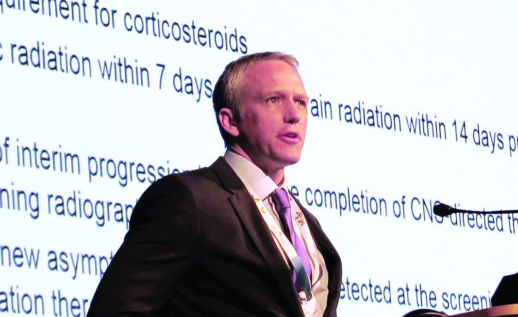
“Overall, I think that these results support the investigation of atezolizumab in non–small cell lung cancer patients with CNS metastases,” Dr. Lukas said at the European Lung Cancer Conference.
Lung cancer accounts for about 40%-50% of all cases of brain metastases, and approximately 20%-40% of patients with advanced NSCLC will develop metastases, which are associated with poor overall survival, according to Solange Peters, MD, from the Centre Hospitalier Universitaire Vaudois in Lausanne, Switzerland, the invited discussant.
To evaluate the safety and efficacy of the PD-L1 inhibitor in patients with brain metastases at baseline, Lukas et al. looked at pooled safety data on patients enrolled in one of five treatment studies with atezolizumab – PCD4989g; BIRCH, POPLAR, FIR, and OAK – and efficacy data from a subset of patients in OAK.
The pooled analysis included 1452 patients, 79 of whom (5%) had brain metastases at baseline. This analysis showed that, although there was a higher incidence of any neurological AE, treatment-related AE, or treatment–related neurologic AE among patients with brain metastases, treatment-related SAEs and treatment related neurological SAEs were similar between the two groups. The most common neurological AE was headache, reported by 8% of patients with metastases and 3% without.
The efficacy analysis, which included a cohort of the first 850 patients with or without brain metastases treated, showed a significant survival for atezolizumab in the OAK trial, with median overall survival of 20.1 months for patients with brain metastases assigned to the PD-L1 inhibitor, compared with 11.9 months for patients assigned to docetaxel. This translated into a hazard ratio for atezolizumab of 0.54 (P = .0279).
Among the 750 patients without baseline brain metastases in OAK, the respective OS rates were 13.0 months, vs. 9.4 months (HR, 0.75; P = .001).
Although it was not statistically significant, the risk for developing new central nervous system lesions also appeared to be lower with atezolizumab than with docetaxel, with a median time to new lesions not reached, vs. 9.5 months with docetaxel (HR, 0.42; 95% confidence interval, 0.15-1.18).
Among patients without baseline brain metastases, there was also a hint that atezolizumab could delay onset of CNS metastases, although the trend was not significant, the investigators found.
In her commentary on the study, Dr. Peters said that “checkpoint inhibitors demonstrate activity in the brain that remains to be quantified and prospectively compared to systemic activity in larger series, and these are very small series.”
“Limitations in duration and level of activity might exist, however, related to the blood brain barrier and the brain immune system characteristics,” she added.
The study was supported by F. Hoffmann-La Roche/Genentech, a member of the Roche Group. Dr. Lukas disclosed serving on advisory boards for AstraZeneca and Novocure and receiving honoraria from AbbVie. Two coauthors are employees of Genentech, and two are employed by Roche. The remaining author had no disclosures. Dr. Peters reported relationships with Bristol-Myers Squibb, F. Hoffmann-La Roche, Eli Lilly, AstraZeneca, Pfizer, Boehringer Ingelheim, Daiichi-Sankyo, Morphotek, Merrimack, Merck Sharp and Dohme, and Merck Serono.
Key clinical point: The PD-L1 inhibitor atezolizumab improved overall survival, vs. docetaxel, in patients with non–small cell lung cancer and brain metastases.
Major finding: The hazard ratio for overall survival was 0.54 for patients assigned to atezolizumab, vs. docetaxel, in the OAK trial.
Data source: Pooled safety analysis of 1452 patients and efficacy analysis of 850 patients with NSCLC with or without brain metastases.
Disclosures: The study was supported by F. Hoffmann-La Roche/Genentech, a member of the Roche Group. Dr. Lukas disclosed serving on advisory boards for AstraZeneca and Novocure and receiving honoraria from AbbVie. Two coauthors are employees of Genentech, and two are employed by Roche. The remaining author had no disclosures. Dr. Peters reported relationships with Bristol-Myers Squibb, F. Hoffmann-La Roche, Eli Lilly, AstraZeneca, Pfizer, Boehringer Ingelheim, Daiichi-Sankyo, Morphotek, Merrimack, Merck Sharp and Dohme, and Merck Serono.
FDA approves pembrolizumab for first-line advanced NSCLC
The Food and Drug Administration has granted accelerated approval to checkpoint inhibitor pembrolizumab in combination with pemetrexed and carboplatin for the treatment of patients with previously untreated metastatic nonsquamous non–small cell lung cancer (NSCLC).
The immunotherapy pembrolizumab was approved as a second-line treatment for metastatic NSCLC in 2015.
First-line approval was based on an improved overall response rate (ORR) and progression-free survival (PFS) in a cohort of 123 patients within an open-label, multicohort study (KEYNOTE-21). Enrollees in cohort G1 had locally advanced or metastatic NSCLC and no prior systemic treatment for metastatic disease. They were randomized to receive either pembrolizumab, in combination with pemetrexed and carboplatin (PC) for four cycles followed by pembrolizumab for a maximum of 24 months (n = 60) or PC alone (n = 63). Randomization was stratified by PD-L1 tumor expression (tumor proportion score [TPS] less than 1% vs. TPS greater than or equal to 1%).
The hazard ratio for PFS was 0.53 (95% CI: 0.31, 0.91, P = .0205). The median PFS was 13.0 months for the pembrolizumab plus PC arm and 8.9 months for the PC-alone arm. In the TPS less than 1% subgroup, the ORR was 57% and 13% in the pembrolizumab-plus-PC and in the PC-alone arms, respectively. In the TPS greater-than-or-equal-to-1% subgroup, the ORR was 54% in the pembrolizumab-plus-PC arm and 38% in the pembrolizumab-plus-PC arm, the FDA said.
There were serious adverse events in 41% of the patients in the pembrolizumab-plus-PC arm compared with 28% in the PC-alone arm. Pembrolizumab was discontinued for adverse reactions in 10% of patients, most commonly due to acute kidney injury. The most common grade 3-4 adverse reactions were fatigue, dyspnea, nausea, vomiting, diarrhea, and rash.
The FDA cautioned that immune-mediated adverse reactions can occur with pembrolizumab including pneumonitis, colitis, hepatitis, endocrinopathies, and nephritis. Based on the severity of the adverse reaction, pembrolizumab should be withheld or discontinued and corticosteroids administered when appropriate. The recommended dose and schedule for NSCLC is 200 mg as an intravenous infusion every 3 weeks until disease progression, unacceptable toxicity, or up to 24 months in patients without disease progression.
Pembrolizumab is marketed as Keytruda by Merck.
Don’t Miss the 2017 AATS International Cardiovascular Symposium
Make your plans to attend the 2017 AATS International Cardiovascular Symposium.
December 8-9, 2017
Renaissance São Paulo Hotel
São Paulo, Brazil
Program Directors
Joseph S. Coselli
Walter J. Gomes
Marc R. Moon
Thoralf M. Sundt, III
For more information, go to: aats.org/ics
Make your plans to attend the 2017 AATS International Cardiovascular Symposium.
December 8-9, 2017
Renaissance São Paulo Hotel
São Paulo, Brazil
Program Directors
Joseph S. Coselli
Walter J. Gomes
Marc R. Moon
Thoralf M. Sundt, III
For more information, go to: aats.org/ics
Make your plans to attend the 2017 AATS International Cardiovascular Symposium.
December 8-9, 2017
Renaissance São Paulo Hotel
São Paulo, Brazil
Program Directors
Joseph S. Coselli
Walter J. Gomes
Marc R. Moon
Thoralf M. Sundt, III
For more information, go to: aats.org/ics
Register for the AATS Surgical Treatment of Arrhythmias and Rhythm Disorders 2017
Don’t miss this two-day, interactive forum that supports the exchange of information between electrophysiologists and surgeons.
November 17-18, 2017
Nobu Eden Roc Hotel
Miami Beach FL USA
Program Overview
The AATS Surgical Treatment of Arrhythmias and Rhythm Disorders provides a comprehensive, unbiased review of the surgical treatment of cardiac arrhythmias and rhythm disorders. The meeting will cover basic underlying mechanisms, cardiac recording and mapping techniques, clinical electrophysiology, operative techniques, cardiac monitoring and appropriate follow-up. The esteemed faculty will cover: interventional approaches to atrial fibrillation, ventricular tachycardia, inappropriate sinus tachycardia, and transvenous and epicardial lead implantation and extraction techniques.
Program Directors
Ralph J. Damiano, Jr.
A. Marc Gillinov
Program Committee
Niv Ad
Vinay Badhwar
Manuel Castella
James L. Cox
Mark LaMeir
Patrick M. McCarthy
Takashi Nitta
Harold G. Roberts
Richard Schuessler
To register and reserve housing, go to: www.aats.org/stars


