User login
FDA gives nod to earlier use of Nplate in adults with ITP
Romiplostim (Nplate) earned a new indication from the Food and Drug Administration, allowing for earlier usage in adult patients with immune thrombocytopenia (ITP) who have had an insufficient response to corticosteroids, immunoglobulins, or splenectomy.
The new indication is for newly diagnosed and persistent adult patients.
Romiplostim is already approved for the treatment of pediatric patients aged 1 year and older who have had ITP for at least 6 months and had an insufficient response to other treatments, as well as for adults patients with chronic ITP who had insufficient response to other therapies.
The latest approval is based on positive results from a single-arm, phase 2 trial in adults with ITP who had an insufficient response to first-line treatment. The study met its primary endpoint – platelet response (50 x 109/L or greater). The median number of months with platelet response was 11 months during a 12-month treatment period. The median time to first platelet response was just over 2 weeks.
Adverse events of at least 5% incidence in patients taking romiplostim with an ITP duration up to 12 months included bronchitis, sinusitis, vomiting, arthralgia, myalgia, headache, dizziness, diarrhea, upper respiratory tract infection, cough, nausea, and oropharyngeal pain. There was a 2% incidence of thrombocytosis among adults with an ITP duration up to 12 months.
Global blood supply runs low
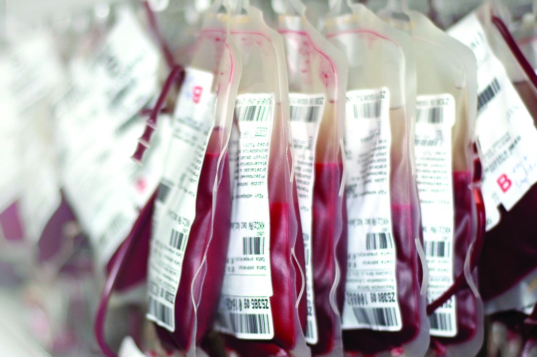
Nearly two-thirds of countries worldwide have an insufficient supply of blood for transfusion, according to findings from a recent modeling study including 195 countries and territories.
Low- and middle-income countries are most often affected, reported lead author Nicholas Roberts of the University of Washington in Seattle and colleagues. Among all countries in Oceania, south Asia, and eastern, central, and western sub-Saharan Africa, not one had enough blood to meet estimated needs, they noted.
Shortages are attributable to a variety of challenges, including resource constraints, insufficient component production, and a high prevalence of infectious diseases, according to investigators. And existing guidelines, which call for 10-20 donations per 1,000 people in the population, may need to be revised, investigators suggested.
“These estimates are important, as they can be used to guide further investments in blood transfusion services, for analysis of current transfusion practices, and to highlight need for alternatives to transfusions such as antifibrinolytics, blood saving devices, and implementation of blood management systems,” the investigators wrote in Lancet Haematology.
Blood availability was calculated using the 2016 WHO Global Status Report on Blood Safety and Availability, in which 92% of countries participated. Blood needs were calculated using multiple databases, including the (U.S.) National Inpatient Sample datasets from 2000-2014, the State Inpatient Databases from 2003-2007, and the Global Burden of Disease 2017 study.
The global blood need was almost 305 million blood product units, while the supply totaled approximately 272 million units. These shortages, however, were not distributed equally across the globe. Out of 195 countries, 119 (61%) had an insufficient supply of blood to meet anticipated demand. Within this group, the shortage equated to about 102 million blood product units.
Denmark had the greatest supply of blood products (red blood cell products, platelets, and plasma), at 14,704 units per 100,000 population, compared with South Sudan, the least prepared to meet transfusion needs, with just 46 blood product units available per 100,000 population. This pattern was echoed across the globe; high-income countries were usually better stocked than those with low or middle income.
To meet demands, some of the most affected countries would need to raise their collection goals from single-digit figures to as high as 40 donations per 1,000 population, the investigators advised.
“Many countries face critical undersupply of transfusions, which will become more pronounced as access to care improves,” the investigators wrote.
The study was funded by the National Institutes of Health. The investigators reported having no other financial disclosures.
SOURCE: Roberts N et al. Lancet Haematol. 2019 Oct 17. doi: 10.1016/ S2352-3026(19)30200-5.
FROM LANCET HAEMATOLOGY
Translucent particles found in Hemlibra
But toxicology and safety assessments conducted by the company found that the benefit-risk profile of the product remains unchanged.
Hemlibra is a bispecific factor IXa– and factor X–directed antibody that has been approved in the United States and other countries for routine prophylaxis in adult and pediatric patients with hemophilia A, with or without factor VIII inhibitors. It is administered as a subcutaneous injection.
The particles were first identified in March 2019 during a routine examination of drug product batches by Genentech. At the time, the company informed the U.S. Food and Drug Administration; the European Medicines Agency; Swissmedic; Health Canada; and the Ministry for Health, Labour, and Welfare in Japan. An initial company assessment found that the particles consisted of protein (Hemlibra drug substance) and silicone oil (polydimethylsiloxane), an organic polymer included in all parenteral medicines, according to Genentech.
Since the health authorities all agreed with the company’s initial conclusion that the product’s safety remained unchanged, no further action was taken.
The presence of the particles became more widely known in early October 2019, when Genentech notified the National Hemophilia Foundation, the NHF’s Medical and Scientific Advisory Council (MASAC), and the Hemophilia Federation of America. Genentech officials reached out to these groups after finishing a root cause investigation and issuing a final report to health authorities concluding that there was no change in the benefit-risk profile, according to a company spokesman.
MASAC issued its own statement recommending no change in prescribing or interruption in the use of Hemlibra. This is an “interim recommendation pending our assessment of the full review by Roche/Genentech of their manufacturing and quality control,” MASAC said. The council noted that it had been informed by representatives from Roche/Genentech that there have been no reports of adverse events linked to the particulate matter. Additionally, the problem had been present since the initial clinical trials of the product, but was only recently identified.
But at least one patient advocate is raising concerns about the timing of this notification. Jesse Clark, president and CEO of HemoAware, wants to know why patients were not informed of the particle issue for more than 6 months.
“The bleeding disorder community was never notified by either Genentech or any other agency,” Mr. Clark said in an interview. “The lack of transparency and communication is extremely concerning.”
A spokesman for Genentech said the company had not alerted patients earlier because there was no indication of an impact on the safety of the product. “We take inquiries from the community very seriously and are providing context and additional details in response to requests from patient organizations,” he said in an interview.
The company does not expect this issue to impact any patients in the United States and the availability of Hemlibra will not be affected, he added.
Monthly and twice monthly emicizumab dosing safe for children with severe hemophilia A
Administration of twice-monthly or monthly emicizumab appears safe and effective for children with severe hemophilia A without inhibitors, according to a small cohort study.
After 24 weeks of treatment, only one moderate-intensity injection site reaction was reported, but no thrombotic microangiopathy or thromboembolic complications were observed.
The researchers evaluated the efficacy, safety, and pharmacokinetics of emicizumab in Japanese pediatric patients aged less than 12 years with severe hemophilia A without factor VIII inhibitors, wrote Midori Shima, MD, PhD, of Nara Medical University, Kashihara, Japan, and colleagues. The results were published in Haemophilia.
The open-label, nonrandomized study included 13 children who initially received weekly loading doses (3 mg/kg) of subcutaneous emicizumab for 4 weeks. Subsequently, patients received maintenance doses of 3 mg/kg every 2 weeks or 6 mg/kg every 4 weeks until week 24.
At baseline, the median age of patients in the 2- and 4-week dosing cohorts were 6.6 and 4.1 years, respectively. All participants had received factor VIII prophylaxis prior to starting emicizumab, with the exception of one patient.
Among six patients in the twice-monthly dosing cohort, two had no treated bleeding episodes, with an annualized bleeding rate for treated bleeding episodes of 1.3 (95% confidence interval, 0.6-2.9).
Among seven patients in the monthly dosing cohort, five had no treated bleeding episodes, with an annualized bleeding rate for treated bleeding episodes of 0.7 (95% CI, 0.2-2.6).
Caregivers completed a preference survey after the first 16 weeks of treatment, and “all reported a preference for emicizumab prophylaxis over the patient’s previous haemophilia treatment.” They cited the lower frequency of treatment and easier route of administration for favoring emicizumab.
With respect to pharmacokinetics, mean steady-state trough levels were within acceptable limits based on previous studies. No patients tested positive for anti-emicizumab antibodies.
The small sample size and nonrandomized design were key limitations of the study.
The results “confirm the appropriateness” of applying the every 2-week and every 4-week regimens of emicizumab in pediatric patients with hemophilia A without inhibitors, the researchers wrote.
The authors reported having financial affiliations with Chugai Pharmaceutical Co., which funded the study, and other companies.
SOURCE: Shima M et al. Haemophilia. 2019 Sep 12. doi: 10.1111/hae.13848.
FROM HAEMOPHILIA
New consensus recommendations on bleeding in acquired hemophilia
New consensus statements, released by a group of 36 experts, provide specific recommendations related to monitoring bleeding and assessing efficacy of treatment in patients with acquired hemophilia.
A global survey was developed by a nine-member steering committee with expertise in the hemostatic management of patients with acquired hemophilia. The Delphi methodology was used to obtain consensus on a list of statements on the location-specific treatment of bleeding in acquired hemophilia.
“The initial survey was circulated via email for refinement and was formally corroborated at a face-to-face meeting,” wrote Andreas Tiede, MD, PhD, of Hannover (Germany) Medical School and fellow experts. The report is published in Haemophilia.
The key areas outlined include the initial management of bleeding, and management of location-specific bleeding, including urological, gastrointestinal, muscle, and pharyngeal bleeds, as well as intracranial and postpartum hemorrhage.
If an expert hematologist is not available, and the bleeding event is life‐threatening, the emergency physician should initiate treatment in accordance with local or national recommendations, according to the initial management guidelines.
With respect to urological bleeds, the best interval for evaluating successful achievement of hemostasis is every 6-12 hours. The experts also reported that, if first-line hemostatic therapy is not effective, more intensive treatment should be considered every 6-12 hours.
In the management of intracranial hemorrhage, the frequency of clinical evaluation is subject to the particular scenario, and it can vary from every 2 hours (for clinical assessment) to every 24 hours (for imaging studies), they wrote.
If initial hemostatic treatment is not effective, more intensive therapy should be considered every 6 hours, they recommended.
“The statement addressing optimal frequency for assessing hemostasis in intracranial bleeds was the subject of much deliberation among the steering committee regarding timing of assessment,” the experts acknowledged.
The geographic diversity and global representation of expert participants were major strengths of these recommendations. However, these statements did not consider socioeconomic parameters or geopolitical differences that could affect patient care. As a result, they may not be applicable to all patient populations.
The manuscript was funded by Novo Nordisk AG. The authors reported having financial affiliations with Novo Nordisk and several other companies.
SOURCE: Tiede A et al. Haemophilia. 2019 Sep 13. doi: 10.1111/hae.13844.
FROM HAEMOPHILIA
Hemophilia prevalence is nearly three times higher than previously reported
The number of people with hemophilia worldwide is higher than previously estimated, and patients still face a shortened life expectancy, according to an international meta-analysis of registry data.
Approximately 1.125 million people have hemophilia worldwide, compared with the previous estimate of 400,000, reported lead author Alfonso Iorio, MD, PhD, of McMaster University, Hamilton, Ont., and colleagues.
The previous estimate, from the early 2000s, was based on prevalence in the United States and the global population at the time, the investigators explained. Their report is in Annals of Internal Medicine.
They noted a lack of clarity in prior estimates concerning type and severity of hemophilia, and aimed to correct this knowledge gap with the present meta-analysis.
Prevalence was estimated using data from registries in Australia, Canada, Italy, France, the United Kingdom, and New Zealand, which are all high-income countries. Prevalence at birth was estimated using the Canadian, French, and British registries, as these are the most established databases, according to the investigators. The World Federation of Hemophilia Annual Global survey was used to estimate the total global number of patients with hemophilia, while national statistics databases were used to determine the number of males and live male births.
Of the 1.125 million cases of hemophilia worldwide, the investigators estimated that 418,000 are likely severe. Proportionally, 17.1 out of 100,000 males have hemophilia A, with 6.0 out of 100,000 males exhibiting severe hemophilia A. Hemophilia B is less common, occurring in 3.8 out of 100,000 males, with a 1.1 out of 100,000 classified as severe.
Turning to prevalence at birth, the investigators estimated that there are 24.6 cases of hemophilia A per 100,000 male births and 5.0 cases of hemophilia B per 100,000 male births.
The associated life expectancy disadvantage in high-income countries is highest for severe hemophilia A (37%), followed by all severities of hemophilia A (30%), severe hemophilia B (27%), and all severities of hemophilia B (24%).
“Having 1,125,000 persons with hemophilia worldwide, of whom about 418,000 have severe and mostly undiagnosed disease, constitutes a formidable challenge and burden for researchers and health care systems, especially because only 196,706 patients have been identified and reported globally,” the investigators wrote. “More efficient diagnostic approaches are needed in less wealthy countries to take advantage of current and future treatment modalities, including gene therapy. Increased demand for care should drive new policy planning and spur renewed effort toward the development and manufacture of new drugs.”
The updated prevalence figures will serve as a valuable roadmap for the future, according to J. Michael Soucie, PhD, of the Centers for Disease Control and Prevention, Atlanta.
“Although the magnitude of the global gaps in care for persons with hemophilia is daunting, country specific data generated by application of the prevalence estimates reported by Iorio and colleagues are an important step toward prioritizing efforts to address these gaps,” Dr. Soucie wrote in an accompanying editorial. “Having more accurate prevalence data might also allow identification of ways in which regional efforts to improve care access could generate considerable benefits for patients and cost savings for countries. Armed with these data for action, we can hope to make substantial progress toward the goal of improving the lives of persons with hemophilia wherever they live.”
The study received no financial support. The investigators reported relationships with Pfizer, Roche, Novo Nordisk, and others. Dr. Soucie reported having no conflicts of interest.
SOURCE: Iorio A et al. Ann Intern Med. 2019 Sept 10. doi: 10.7326/M19-1208.
FROM ANNALS OF INTERNAL MEDICINE
In older patients with immune-mediated TTP, atypical features may delay diagnosis
Older patients with immune thrombotic thrombocytopenic purpura (iTTP) more often have an atypical neurological presentation, which could result in a delayed diagnosis, according to authors of a recent retrospective analysis.
“Practitioners should be aware of this in order to shorten the time to treatment, which could improve the prognosis in older iTTP patients,” Paul Coppo, MD, PhD, of Hôpital Saint-Antoine, Paris, and coauthors wrote in Blood.
The older patients also had increased 1-month and 1-year mortality compared with younger patients, and had more than a threefold risk of long-term mortality compared with elderly patients without iTTP, according to the study report.
The analysis included 411 patients with iTTP entered into a national registry in France between 2000 and 2016. Seventy-one patients were 60 years of age or older.
Time from hospital admission to diagnosis was 3 days for those older patients, versus just 1 day for patients under 60 years of age (P = .0001), Dr. Coppo and colleagues reported.
Clinical records were available for 67 of the older iTTP patients, of whom 17 had no evidence of delayed diagnosis. The remainder had a “possible diagnostic delay,” according to the report; among those, the iTTP diagnosis was preceded by neurological manifestations in 26 cases, and transient ischemic stroke that usually led to focal deficiency or aphasia in 14 cases. Other features preceding the diagnosis included malaise, behavioral abnormalities, seizure, and dizziness.
Many of these findings are “not specific to a disease, and they are less alarming than in young patients,” the researchers wrote. “In this context, the presence of a thrombocytopenia with anemia should alert physicians to this possible rare diagnosis.”
Older patients also presented with less pronounced cytopenias compared with younger patients, which could have contributed to a late diagnosis, they added.
Older age is a known risk factor for mortality related to iTTP. In the present study, rates of 1-month mortality were 37% for patients aged 60 years and older, and 9% for those younger than age 60 (P less than .0001). The 1-year mortality rates were 49% and 11% for older and younger patients, respectively (P less than .0001).
Compared with older individuals without iTTP from a different study, older iTTP patients had a lower long-term survival rate. iTTP remained an independent risk factor for death even after adjustment for age, sex, and some comorbidities (hazard ratio, 3.44; 95% confidence interval, 2.02-5.87).
The study was partly funded by a grant from the French Ministry of Health. Dr. Coppo reported that he is a clinical advisory board member for Alexion, Ablynx (now part of Sanofi), Shire, and Octapharma. Two other co-authors reported participating in advisory boards for Ablynx.
SOURCE: Prevel R et al. Blood. 2019 Sep 17. doi: 10.1182/blood.2019000748.
FROM BLOOD
Eltrombopag elicits positive responses in secondary ITP
Eltrombopag showed good safety and promising clinical activity in patients with immune thrombocytopenia (ITP) secondary to chronic lymphoproliferative disorders, according to results from a phase 2 trial.
Carlo Visco, MD, of the University of Verona (Italy), and colleagues investigated the efficacy and safety of eltrombopag in increasing platelet counts in patients with ITP that was secondary to chronic lymphoproliferative disorders. The findings were published in Blood.
The single-arm, open-label study included 18 patients with ITP secondary to chronic lymphocytic leukemia (14), Waldenstrom macroglobulinemia (2), and classical Hodgkin lymphoma (2). The median age at baseline was 70 years (range, 43-83 years), and all patients were previously treated with ITP.
Study participants were recruited from seven Italian centers from September 2012 to November 2015. Eligible participants were enrolled into an extension phase if a response was observed.
Study patients received oral eltrombopag at 50 mg daily, up to a maximum of 150 mg daily. At weeks 4 and 24, the median dose was 50 mg (ranges, 25-100 mg and 25-150 mg, respectively), with a median total exposure time of 16 months.
At 4 weeks, the researchers found that the platelet response rate was 78%, with a complete response rate of 50%.
After 24 weeks of therapy, the platelet response rate was 59%, with a complete response rate of 30%.
With respect to safety, the therapy was well tolerated, with no adverse events higher than grade 2 reported.
Fifteen patients discontinued therapy: eight due to loss of response, six for disease progression or death, and one for inefficacy and protocol violation, they reported.
The researchers acknowledged two key limitations of the study: the small sample size and lack of a comparison group. “Further prospective studies comparing eltrombopag to standard of care are needed to confirm our findings on the efficacy of this treatment and also to expand our knowledge on its safety, including the potential increased risk of thrombosis,” they wrote.
The study was funded by the Hematology Project Foundation, Vicenza. The authors reported financial affiliations with Amgen, Argenx, and Novartis.
SOURCE: Visco C et al. Blood. 2019 Sep 30. doi: 10.1182/blood.2019001617.
FROM BLOOD
Early post-ACS bleeding may signal cancer
PARIS –, according to work presented at the annual congress of the European Society of Cardiology.
Of 3,644 patients discharged with dual-antiplatelet therapy after acute coronary syndrome (ACS), 1,215 (33%) had postdischarge bleeding. Taken together, patients who bled had a hazard ratio (HR) of 3.43 for a new cancer diagnosis (P less than .001).
Of the patients in the post-ACS cohort, 227 were newly diagnosed with cancer after discharge, making up 1% of the patients who did not bleed after discharge, and 3.9% of the patients who experienced postdischarge bleeding. Put another way, “[t]he positive predictive value for cancer diagnosis of post-discharge bleeding was 7.7%,” wrote Isabel Muñoz Pousa, MD, and her colleagues in the poster accompanying the presentation.
This elevated risk for cancer diagnosis was driven primarily by the 827 incidents of spontaneous bleeding; here, the HR was 4.38 (P less than .001). The 389 bleeds occuring after trauma, such as bladder catheterization or a fall, did not carry an increased risk for a new cancer diagnosis.
“Spontaneous post-discharge bleeding in ACS patients is strongly associated with subsequent cancer diagnosis within the first 6 months,” wrote Dr. Muñoz Pousa and her colleagues of the Hospital Universitario Alvaro Cunqueiro, Vigo, Spain. The investigators found a median time of 4.6 months from the bleeding episode to cancer diagnosis.
Of all anatomic locations, genitourinary bleeds were the most strongly associated with new cancer: 228 patients saw a HR of 8.63 for a new cancer diagnosis (P less than .001). Bronchopulmonary bleeds, sustained by 56 patients, carried a HR of 4.26 for new cancer diagnosis, and gastrointestinal bleeds a HR of 3.78 (P = .001 and P less than .001, respectively). Dr. Muñoz Pousa and her coinvestigators aggregated data from patients who had bleeding at other sites and saw no significant association with new cancers in this group of patients.
Though patients were initially discharged on dual-antiplatelet therapy, many patients stopped taking the medication over the mean 56.2 months of follow-up. The risk of bleeding did not differ significantly between those who were taking DAPT and those off DAPT, wrote Dr. Muñoz Pousa and her colleagues, adding: “We found a higher incidence of cancer in the first six months after discharge regardless of whether patients were taking dual-antiplatelet therapy or not.”
In their statistical analysis, Dr. Muñoz Pousa and colleagues adjusted for potential confounders, and looked at the effect of bleeding as a time-varying covariate on subsequent cancer diagnosis, using Cox regression models.
“Most of the bleeding episodes in the study were mild,” noted Dr. Munoz Pousa in a press statement. However, she said, “The bleeding events more strongly related with a new cancer diagnosis were severe hemorrhages of unknown cause requiring surgery – for example digestive bleeding needing endoscopic treatment.”
Breaking bleeding severity down by Bleeding Academic Research Consortium (BARC) criteria, the investigators found that most patients had relatively mild bleeding episodes categorized as BARC 1 or 2, with about half of all bleeding falling into the BARC 1 category.
Still, the 436 patients who had BARC 2 bleeding had a hazard ratio of 4.88 for cancer diagnosis, and the 71 BARC 3A patients saw the HR climb to 7.30. The risk for cancer subsequent to bleeding peaked at BARC 3B, with a HR of 12.29 for these 46 individuals (P less than .001 for all). Just 37 patients experienced BARC 3C bleeds, which were associated with a nonsignificant HR of 3.17 for new cancer diagnosis.
Although it’s not known why the post ACS–cancer bleeding association exists, Dr. Munoz Pousa put forward a plausible reason for the link. “A possible explanation is that there is a preexisting subclinical lesion in an organ that is triggered to become cancer by antiplatelet drugs or a stressful situation such as heart attack,” she said in the press release.
Antiplatelet therapy should be taken as prescribed post-ACS, and the physician threshold for further evaluation should be low when a significant spontaneous bleed is seen soon after ACS. “A prompt evaluation of bleeding could be useful for enabling an early detection of cancer in these patients,” said Dr. Munoz Pousa and her colleagues. “Our results suggest that patients should seek medical advice if they experience bleeding after discharge for a heart attack.”
The authors reported no conflicts of interest.
SOURCE: Munoz Pousa, I. et al. ESC Congress 2019, Abstract P677.
FROM ESC CONGRESS 2019
Extended half-life products cut infusions in Europe
Novel extended half-life factor VIII (FVIII) and factor IX (FIX) products appear to decrease the number of infusions and maintain higher trough levels, especially for patients with hemophilia B, according to recent survey findings.
Preliminary data from a European multinational survey suggest these benefits may help overcome current limitations with standard clotting factor products.
“We administered a survey to determine the efficacy of [extended half-life] products after they became available in several European countries,” wrote Flora Peyvandi, MD, PhD, of Fondazione IRCCS Ca’ Granda Ospedale Maggiore Policlinico in Milan and colleagues. These results were published in Haemophilia.
The questionnaire, designed by the European Association for Haemophilia and Allied Disorders (EAHAD), was distributed to 48 hemophilia treatment centers in January 2018. In total, 33 centers completed the survey.
The survey explored the real-life clinical experiences of patients with hemophilia A and B using extended half-life FVIII and FIX products. At the time of the survey, pegylated factor products were not available for use. In particular, the survey collected general information related to the efficacy of prophylaxis after transitioning patients to novel extended half-life products.
After analysis, the researchers found that among responding centers, extended half-life FVIII products decreased the number of infusions by 30% or greater among hemophilia A patients and achieved trough levels of 3%-5% in 66%-67% of centers.
With respect to FIX products, all responding centers were able to reduce infusions by more than 30% among hemophilia B patients, with 67% maintaining a FIX trough level of no less than 5%-10%.
The researchers acknowledged that the findings are preliminary and should be confirmed by conducting a repeat survey.
“Evaluating the safety of these new drugs is of the utmost importance and should be monitored through careful, long‐term observation,” they concluded.
No funding sources were reported. The authors reported financial affiliations with Alnylam, Grifols, Kedrion, Pfizer, Roche, Sanofi, Bayer, Shire, and several other companies.
SOURCE: Peyvandi F et al. Haemophilia. 2019 Aug 16. doi: 10.1111/hae.13834.
FROM HAEMOPHILIA


