User login
APHINITY 6-year data: Benefit ongoing in HER2+ early BC, no significant OS benefit
SAN ANTONIO – Adding pertuzumab to trastuzumab and chemotherapy after surgery for HER2-positive early breast cancer continued to show a slight, but statistically nonsignificant overall survival benefit, compared with placebo, at a preplanned 6-year interim analysis of the phase 3 APHINITY trial.
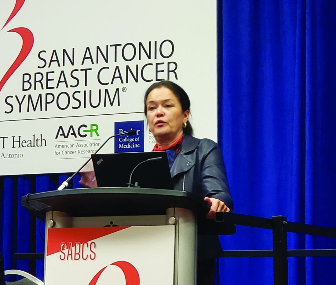
Invasive disease-free survival (IDFS) was significantly improved with pertuzumab at this second interim analysis, and node-positive patients continued to derive the greatest benefit, as was the case in the primary analysis reported in the New England Journal of Medicine in 2017, Martine Piccart, MD, PhD, reported at the San Antonio Breast Cancer Symposium.
At a median of 74.1 months of follow-up, overall survival (OS) was 94.8% in 2,400 patients in the pertuzumab arm, compared with 93.9% in 2,405 patients in the placebo arm (hazard ratio, 0.85), said Dr. Piccart of Institut Jules Bordet, Brussels.
She noted that a “very stringent” P value of.0012 was required for statistical significance in this interim OS analysis.
IDFS rates at follow-up were 90.6% vs. 87.8% in the intent-to-treat population, a difference caused mainly by a reduction in distant and loco-regional recurrence, she noted.
“[That translates] to a 2.8% absolute improvement with pertuzumab at 6 years,” she said, adding that the risk of both distant and loco-regional recurrences was reduced with pertuzumab. “The rate of [central nervous system] metastases, contralateral invasive breast cancers, and death without a prior event – not different between the two treatment groups.”
In the node-positive cohort, the 6-year IDFS rates were 87.9% vs. 83.4% with pertuzumab vs. placebo (4.5% absolute benefit; HR, 0.72), showing a clear benefit.
“In contrast, no treatment effect is detected in the node-negative population [95.0% and 94.9%, respectively; HR, 1.02],” she said.
Importantly, the clinical benefits were seen regardless of hormone receptor status (HRs, 0.73 and 0.83 for hormone receptor–positive and –negative disease, respectively), she said, noting that this finding differs from the 3-year analysis, which suggested an enhanced benefit only in the hormone receptor–negative cohort.
“These [hormone receptor–negative] patients still benefit from pertuzumab ... but interestingly, now the curves are diverging in the hormone receptor–positive population, and there is a benefit emerging,” she said.
An updated descriptive analysis of cardiac safety was also performed, and no new safety concerns emerged, Dr. Piccart said.
“What is important to remember is the rate of severe cardiac events is below 1% in both groups (0.8% and 0.3% with pertuzumab and placebo),” she said.
APHINITY is a randomized, multicenter, double-blind, placebo-controlled trial which previously demonstrated that pertuzumab added to standard chemotherapy plus 1 year of trastuzumab in operable HER2-positive breast cancer was associated with modest but statistically significant improvement in IDFS, compared with placebo and chemotherapy plus trastuzumab (HR, 0.81; P = .04).
The effect was more pronounced in node-negative patients (HR, 0.77) and hormone receptor–negative patients (HR, 0.76).
Patients with node-positive or high-risk node-negative, HER2-positive, operable early breast cancer were enrolled between November 2011 and August 2013, and the primary analysis was conducted at 45.4 months of follow-up. Based on those findings, pertuzumab in combination with trastuzumab was approved for high-risk early HER2-positive breast cancer patients.
The first interim OS analysis was conducted at that time, and no significant treatment effect was observed, Dr. Piccart said.
The 6-year findings demonstrate that the small OS benefit and the statistically significant IDFS benefit with pertuzumab in this setting is maintained, with the node-positive population deriving the greatest benefit.
“Further follow-up will be very important to determine whether there is a survival benefit associated with pertuzumab administration in early HER2-positive breast cancer,” she said, noting that a calendar-driven third interim OS analysis is planned in 2.5 years.
The APHINITY trial is funded by Roche. Dr. Piccart reported receiving consulting fees from Roche and research funding to her institution from Roche and several other companies. She also is a consultant for the advisory boards of AstraZeneca, Camel-IDS, Crescendo Biologics, Debiopharm, G1 Therapeutics, Huya Bioscience International, and Immunomedics.
SOURCE: Piccart M et al. SABCS 2019, Abstract GS1-04.
SAN ANTONIO – Adding pertuzumab to trastuzumab and chemotherapy after surgery for HER2-positive early breast cancer continued to show a slight, but statistically nonsignificant overall survival benefit, compared with placebo, at a preplanned 6-year interim analysis of the phase 3 APHINITY trial.
Invasive disease-free survival (IDFS) was significantly improved with pertuzumab at this second interim analysis, and node-positive patients continued to derive the greatest benefit, as was the case in the primary analysis reported in the New England Journal of Medicine in 2017, Martine Piccart, MD, PhD, reported at the San Antonio Breast Cancer Symposium.
At a median of 74.1 months of follow-up, overall survival (OS) was 94.8% in 2,400 patients in the pertuzumab arm, compared with 93.9% in 2,405 patients in the placebo arm (hazard ratio, 0.85), said Dr. Piccart of Institut Jules Bordet, Brussels.
She noted that a “very stringent” P value of.0012 was required for statistical significance in this interim OS analysis.
IDFS rates at follow-up were 90.6% vs. 87.8% in the intent-to-treat population, a difference caused mainly by a reduction in distant and loco-regional recurrence, she noted.
“[That translates] to a 2.8% absolute improvement with pertuzumab at 6 years,” she said, adding that the risk of both distant and loco-regional recurrences was reduced with pertuzumab. “The rate of [central nervous system] metastases, contralateral invasive breast cancers, and death without a prior event – not different between the two treatment groups.”
In the node-positive cohort, the 6-year IDFS rates were 87.9% vs. 83.4% with pertuzumab vs. placebo (4.5% absolute benefit; HR, 0.72), showing a clear benefit.
“In contrast, no treatment effect is detected in the node-negative population [95.0% and 94.9%, respectively; HR, 1.02],” she said.
Importantly, the clinical benefits were seen regardless of hormone receptor status (HRs, 0.73 and 0.83 for hormone receptor–positive and –negative disease, respectively), she said, noting that this finding differs from the 3-year analysis, which suggested an enhanced benefit only in the hormone receptor–negative cohort.
“These [hormone receptor–negative] patients still benefit from pertuzumab ... but interestingly, now the curves are diverging in the hormone receptor–positive population, and there is a benefit emerging,” she said.
An updated descriptive analysis of cardiac safety was also performed, and no new safety concerns emerged, Dr. Piccart said.
“What is important to remember is the rate of severe cardiac events is below 1% in both groups (0.8% and 0.3% with pertuzumab and placebo),” she said.
APHINITY is a randomized, multicenter, double-blind, placebo-controlled trial which previously demonstrated that pertuzumab added to standard chemotherapy plus 1 year of trastuzumab in operable HER2-positive breast cancer was associated with modest but statistically significant improvement in IDFS, compared with placebo and chemotherapy plus trastuzumab (HR, 0.81; P = .04).
The effect was more pronounced in node-negative patients (HR, 0.77) and hormone receptor–negative patients (HR, 0.76).
Patients with node-positive or high-risk node-negative, HER2-positive, operable early breast cancer were enrolled between November 2011 and August 2013, and the primary analysis was conducted at 45.4 months of follow-up. Based on those findings, pertuzumab in combination with trastuzumab was approved for high-risk early HER2-positive breast cancer patients.
The first interim OS analysis was conducted at that time, and no significant treatment effect was observed, Dr. Piccart said.
The 6-year findings demonstrate that the small OS benefit and the statistically significant IDFS benefit with pertuzumab in this setting is maintained, with the node-positive population deriving the greatest benefit.
“Further follow-up will be very important to determine whether there is a survival benefit associated with pertuzumab administration in early HER2-positive breast cancer,” she said, noting that a calendar-driven third interim OS analysis is planned in 2.5 years.
The APHINITY trial is funded by Roche. Dr. Piccart reported receiving consulting fees from Roche and research funding to her institution from Roche and several other companies. She also is a consultant for the advisory boards of AstraZeneca, Camel-IDS, Crescendo Biologics, Debiopharm, G1 Therapeutics, Huya Bioscience International, and Immunomedics.
SOURCE: Piccart M et al. SABCS 2019, Abstract GS1-04.
REPORTING FROM SABCS 2019
Ten-year results support partial breast irradiation
SAN ANTONIO – For patients with early, low-risk breast cancer, accelerated partial breast irradiation (APBI) may be considered a standard alternative to whole breast irradiation, according to investigators.

This conclusion was based on 10-year follow-up results from the APBI IMRT Florence phase III trial, which showed that APBI was associated with significantly fewer adverse events and better cosmetic results than whole breast irradiation without increasing risks of tumor recurrence or mortality, reported lead author Irco Meattini, MD, of the University of Florence, Italy, and colleagues.
“As we well know, recent developments in radiation oncology ... show a move toward a deescalation strategy for early breast cancer, including accelerated and nonaccelerated partial breast irradiation,” Dr. Meattini said during a presentation at the San Antonio Breast Cancer Symposium. “What we have learned from [previous] phase 3 trials [is that with adequate patient selection for] partial breast irradiation, safety profile and cosmetic outcome are strongly associated with technique – the approach, the dose, the number of fractions per day, and the total dose.”
The current phase 3 trial, which enrolled 520 patients with early breast cancer, aimed to determine long-term efficacy, safety, and cosmetic outcomes for partial versus whole breast irradiation. All patients enrolled were at least 40 years of age and had a maximum pathological tumor size of 25 mm. Patients were randomized in a 1:1 ratio to receive either whole breast irradiation (WBI) at a dose of 50 Gy in 25 fractions, followed by 10 Gy in five fractions delivered to the tumor bed; or APBI, which was delivered to the tumor bed at a dose of 30 Gy in five daily fractions.
The primary endpoint was ipsilateral breast tumor recurrence (IBTR). Secondary endpoints were overall survival, breast cancer–specific survival, distant metastasis-free survival, locoregional recurrences, and contralateral breast cancer. Adverse events and cosmesis also were evaluated.
Five-year results, previously reported, revealed no significant difference in survival rates or IBTR between treatment techniques, and results of the present 10-year analysis maintained these findings. Between groups, no significant differences were observed in any of the primary or secondary endpoints, suggesting that major efficacy outcomes were unaffected by type of irradiation delivered.
While major efficacy endpoints were comparable between groups, safety profiles and cosmetic results differed significantly.
Adverse events of all levels of severity were significantly more common with WBI than APBI. Grade 2 or higher acute adverse events occurred in 37.7% of patients treated with WBI, compared with just 2.0% of patients treated with APBI (P = .0001). The rate of grade 2 or higher adverse events was also significantly higher in the WBI group than in the APBI group in the late setting, albeit with a narrower margin than in the acute setting (2.7% vs 0%; P = .015). Skin toxicity rates followed a similar pattern, favoring APBI both in the acute phase (66.5% vs. 21.1%; P = .0001) and the late phase (30.0% vs. 4.5%; P = .0001).
In further support of APBI, cosmetic results, as measured by the Harvard Breast Cosmesis Scale, were significantly better in the APBI group than in the WBI group. Both physicians and patients were significantly more likely to report good or excellent cosmetic results with APBI than WBI.
“APBI might be considered a standard alternative to WBI in low risk and very low risk early breast cancer patients,” Dr. Meattini concluded.
The investigators reported no disclosures.
SOURCE: Meattini et al. SABCS. 2019 Dec 12. Abstract GS4-06.
SAN ANTONIO – For patients with early, low-risk breast cancer, accelerated partial breast irradiation (APBI) may be considered a standard alternative to whole breast irradiation, according to investigators.
This conclusion was based on 10-year follow-up results from the APBI IMRT Florence phase III trial, which showed that APBI was associated with significantly fewer adverse events and better cosmetic results than whole breast irradiation without increasing risks of tumor recurrence or mortality, reported lead author Irco Meattini, MD, of the University of Florence, Italy, and colleagues.
“As we well know, recent developments in radiation oncology ... show a move toward a deescalation strategy for early breast cancer, including accelerated and nonaccelerated partial breast irradiation,” Dr. Meattini said during a presentation at the San Antonio Breast Cancer Symposium. “What we have learned from [previous] phase 3 trials [is that with adequate patient selection for] partial breast irradiation, safety profile and cosmetic outcome are strongly associated with technique – the approach, the dose, the number of fractions per day, and the total dose.”
The current phase 3 trial, which enrolled 520 patients with early breast cancer, aimed to determine long-term efficacy, safety, and cosmetic outcomes for partial versus whole breast irradiation. All patients enrolled were at least 40 years of age and had a maximum pathological tumor size of 25 mm. Patients were randomized in a 1:1 ratio to receive either whole breast irradiation (WBI) at a dose of 50 Gy in 25 fractions, followed by 10 Gy in five fractions delivered to the tumor bed; or APBI, which was delivered to the tumor bed at a dose of 30 Gy in five daily fractions.
The primary endpoint was ipsilateral breast tumor recurrence (IBTR). Secondary endpoints were overall survival, breast cancer–specific survival, distant metastasis-free survival, locoregional recurrences, and contralateral breast cancer. Adverse events and cosmesis also were evaluated.
Five-year results, previously reported, revealed no significant difference in survival rates or IBTR between treatment techniques, and results of the present 10-year analysis maintained these findings. Between groups, no significant differences were observed in any of the primary or secondary endpoints, suggesting that major efficacy outcomes were unaffected by type of irradiation delivered.
While major efficacy endpoints were comparable between groups, safety profiles and cosmetic results differed significantly.
Adverse events of all levels of severity were significantly more common with WBI than APBI. Grade 2 or higher acute adverse events occurred in 37.7% of patients treated with WBI, compared with just 2.0% of patients treated with APBI (P = .0001). The rate of grade 2 or higher adverse events was also significantly higher in the WBI group than in the APBI group in the late setting, albeit with a narrower margin than in the acute setting (2.7% vs 0%; P = .015). Skin toxicity rates followed a similar pattern, favoring APBI both in the acute phase (66.5% vs. 21.1%; P = .0001) and the late phase (30.0% vs. 4.5%; P = .0001).
In further support of APBI, cosmetic results, as measured by the Harvard Breast Cosmesis Scale, were significantly better in the APBI group than in the WBI group. Both physicians and patients were significantly more likely to report good or excellent cosmetic results with APBI than WBI.
“APBI might be considered a standard alternative to WBI in low risk and very low risk early breast cancer patients,” Dr. Meattini concluded.
The investigators reported no disclosures.
SOURCE: Meattini et al. SABCS. 2019 Dec 12. Abstract GS4-06.
REPORTING FROM SABCS 2019
Capecitabine extends survival in triple-negative breast cancer
SAN ANTONIO – For patients with early-stage triple-negative breast cancer, adding capecitabine to systemic treatment may extend overall survival, according to a meta-analysis involving more than 15,000 patients.

Although a variety of trials have tested capecitabine therapy for early-stage breast cancer, this study is the first to evaluate individual patient data across trials, reported lead author Marion van Mackelenbergh, MD, of University Medical Center Schleswig-Holstein in Kiel, Germany, and colleagues.
According to Dr. Mackelenbergh, who presented findings at the San Antonio Breast Cancer Symposium, the two previous literature-based meta-analyses of capecitabine for patients with early-stage breast cancer reported conflicting results; the first study suggested that capecitabine had no benefit as a neoadjuvant therapy, whereas the second study concluded that capecitabine could improve disease-free survival (DFS).
To build upon these findings, the primary objective of the present meta-analysis was to determine how treatment with capecitabine impacts DFS, Dr. Mackelenbergh said. A variety of secondary objectives were also evaluated, including overall survival and a possible interaction between capecitabine-specific toxicities and treatment effects.
The analysis involved 15,457 patients from 12 randomized prospective clinical trials, of whom about half (n = 7,477) were treated in control arms. Slightly more than half (55.9%) of the patients had stage II tumors and about three-fourths (74.0%) presented with nodal involvement. About two-thirds of patients (66.0%) had estrogen receptor–positive disease, about half (56.9%) were progesterone receptor–positive, and 15.1% were human epidermal growth factor receptor 2–positive. Most of the patients (81.8%) were treated with chemotherapy in the adjuvant setting, whereas the remainder (18.2%) received neoadjuvant therapy.
Cox regression analysis involving all patients in the dataset showed that capecitabine was not associated with a significant improvement in DFS, nor was a significant improvement seen in trials that compared capecitabine against other treatment options. In contrast, adding capecitabine to systemic treatment supported a modest but significant improvement in DFS (hazard ratio, 0.888; P = .0005).
Across all patients, capecitabine was associated with an overall survival advantage, although this benefit was relatively small, with a hazard ratio of 0.892. The overall survival benefit became more pronounced when capecitabine was added to systemic treatment (HR, 0.837).
Of clinical importance, biological subtype analysis showed that only patients with triple-negative breast cancer were deriving survival benefit from capecitabine, particularly when capecitabine was added to systemic treatment. Among patients with triple-negative disease, a 17% overall survival benefit was associated with capecitabine (HR, 0.828). When capecitabine was added to systemic treatment, this survival advantage improved to 22% (HR, 0.778).
No relationship was found between capecitabine-specific toxicity (mucositis, hand-foot syndrome, diarrhea) and patient outcome.
“It can be concluded that the addition of capecitabine to other systemic treatment may be recommended for triple-negative breast cancer patients,” Dr. Mackelenbergh said.

Invited discussant Priyanka Sharma, MD, of the University of Kansas Medical Center in Kansas City agreed with this conclusion.
“Routine use of capecitabine as a component of neoadjuvant or adjuvant regimens in unselected patients cannot be endorsed,” Dr. Sharma said. “However, capecitabine should be considered in patients with triple-negative breast cancer, especially if response to neoadjuvant chemotherapy is utilized as a way to identify eligible patients so as to limit exposure and toxicity to those at the highest risk.”
In terms of the future, Dr. Sharma suggested that research is needed to determine the potential for adjuvant capecitabine among patients with triple-negative breast cancer who have received platinum-based chemotherapy and/or immune checkpoint inhibitor therapy as part of their neoadjuvant regimen. In addition, investigators should evaluate capecitabine efficacy in terms of residual disease and identify relevant predictive biomarkers, Dr. Sharma said.
The investigators reported relationships with Amgen, Lilly, Pfizer, and others.
SOURCE: van Mackelenbergh M et al. SABCS 2019, Abstract GS1-07.
SAN ANTONIO – For patients with early-stage triple-negative breast cancer, adding capecitabine to systemic treatment may extend overall survival, according to a meta-analysis involving more than 15,000 patients.
Although a variety of trials have tested capecitabine therapy for early-stage breast cancer, this study is the first to evaluate individual patient data across trials, reported lead author Marion van Mackelenbergh, MD, of University Medical Center Schleswig-Holstein in Kiel, Germany, and colleagues.
According to Dr. Mackelenbergh, who presented findings at the San Antonio Breast Cancer Symposium, the two previous literature-based meta-analyses of capecitabine for patients with early-stage breast cancer reported conflicting results; the first study suggested that capecitabine had no benefit as a neoadjuvant therapy, whereas the second study concluded that capecitabine could improve disease-free survival (DFS).
To build upon these findings, the primary objective of the present meta-analysis was to determine how treatment with capecitabine impacts DFS, Dr. Mackelenbergh said. A variety of secondary objectives were also evaluated, including overall survival and a possible interaction between capecitabine-specific toxicities and treatment effects.
The analysis involved 15,457 patients from 12 randomized prospective clinical trials, of whom about half (n = 7,477) were treated in control arms. Slightly more than half (55.9%) of the patients had stage II tumors and about three-fourths (74.0%) presented with nodal involvement. About two-thirds of patients (66.0%) had estrogen receptor–positive disease, about half (56.9%) were progesterone receptor–positive, and 15.1% were human epidermal growth factor receptor 2–positive. Most of the patients (81.8%) were treated with chemotherapy in the adjuvant setting, whereas the remainder (18.2%) received neoadjuvant therapy.
Cox regression analysis involving all patients in the dataset showed that capecitabine was not associated with a significant improvement in DFS, nor was a significant improvement seen in trials that compared capecitabine against other treatment options. In contrast, adding capecitabine to systemic treatment supported a modest but significant improvement in DFS (hazard ratio, 0.888; P = .0005).
Across all patients, capecitabine was associated with an overall survival advantage, although this benefit was relatively small, with a hazard ratio of 0.892. The overall survival benefit became more pronounced when capecitabine was added to systemic treatment (HR, 0.837).
Of clinical importance, biological subtype analysis showed that only patients with triple-negative breast cancer were deriving survival benefit from capecitabine, particularly when capecitabine was added to systemic treatment. Among patients with triple-negative disease, a 17% overall survival benefit was associated with capecitabine (HR, 0.828). When capecitabine was added to systemic treatment, this survival advantage improved to 22% (HR, 0.778).
No relationship was found between capecitabine-specific toxicity (mucositis, hand-foot syndrome, diarrhea) and patient outcome.
“It can be concluded that the addition of capecitabine to other systemic treatment may be recommended for triple-negative breast cancer patients,” Dr. Mackelenbergh said.
Invited discussant Priyanka Sharma, MD, of the University of Kansas Medical Center in Kansas City agreed with this conclusion.
“Routine use of capecitabine as a component of neoadjuvant or adjuvant regimens in unselected patients cannot be endorsed,” Dr. Sharma said. “However, capecitabine should be considered in patients with triple-negative breast cancer, especially if response to neoadjuvant chemotherapy is utilized as a way to identify eligible patients so as to limit exposure and toxicity to those at the highest risk.”
In terms of the future, Dr. Sharma suggested that research is needed to determine the potential for adjuvant capecitabine among patients with triple-negative breast cancer who have received platinum-based chemotherapy and/or immune checkpoint inhibitor therapy as part of their neoadjuvant regimen. In addition, investigators should evaluate capecitabine efficacy in terms of residual disease and identify relevant predictive biomarkers, Dr. Sharma said.
The investigators reported relationships with Amgen, Lilly, Pfizer, and others.
SOURCE: van Mackelenbergh M et al. SABCS 2019, Abstract GS1-07.
REPORTING FROM SABCS 2019
Tucatinib called game-changer in HER2-positive metastatic breast cancer
SAN ANTONIO – Tucatinib, an investigational oral inhibitor of human epidermal growth factor receptor 2 (HER2) tyrosine kinase, constitutes a major advance in the treatment of heavily pretreated HER2-positive metastatic breast cancer – including patients with brain metastases, Rashmi K. Murthy, MD, declared at the San Antonio Breast Cancer Symposium.

She presented the results of the pivotal HER2CLIMB trial, in which 612 women with heavily pretreated HER2-positive metastatic breast cancer were randomized two-to-one to oral tucatinib at 300 mg twice daily or placebo in combination with standard guideline-recommended treatment with trastuzumab and capecitabine. This landmark double-blind study, conducted at 155 sites in 15 countries, was the first-ever randomized trial to include HER2-positive metastatic breast cancer patients with baseline untreated or previously treated but progressing brain metastases; indeed, nearly half of participants had baseline brain metastases, 40% of which were untreated or treated and progressing.
Not only did tucatinib on top of background trastuzumab and capecitabine reduce the risk of death by 34% compared with placebo, it also more than doubled progression-free survival. And most encouragingly, it did so in patients with or without baseline brain metastases.
“I would like to take a minute here to highlight that this overall survival benefit was seen in patients who had already received trastuzumab, pertuzumab, and T-DM1 [trastuzumab emtansine] and included patients who had brain metastases. Tucatinib in combination with trastuzumab and capecitabine has the potential to become a new standard of care in this population with and without brain metastases,” said Dr. Murthy, a medical oncologist at the University of Texas MD Anderson Cancer Center, Houston.
Tucatinib, which crosses the blood-brain barrier, is highly selective for the kinase domain of HER2, with only miniscule inhibition of epidermal growth factor receptor. Its high selectivity is reflected in a favorable tolerability profile: Only 6% of patients discontinued tucatinib because of adverse events. This low discontinuation rate permitted longer treatment, even in this heavily pretreated population.
The most common adverse events in the tucatinib study arm were diarrhea, hand-foot syndrome, nausea, fatigue, and vomiting, with most cases being low grade. Diarrhea, the most common adverse event, occurred in 81% of tucatinib-treated patients and 53% of controls. However, only 13% of the tucatinib group experienced grade 3 or worse diarrhea. Notably, antidiarrheal prophylaxis wasn’t mandated in HER2CLIMB. Antidiarrheal medications were utilized in less than half of the treatment cycles where diarrhea was reported, and even then, for a median of only 3 days per cycle.
Median study follow-up was 14 months. The primary study endpoint – 1-year progression-free survival assessed by blinded independent central review – was 33% in the tucatinib group and 12% in controls. Thus, the risk of disease progression or death was reduced by 46% in the tucatinib group. The median duration of progression-free survival was 7.8 months in tucatinib-treated patients, compared with 5.6 months in controls.
Two-year estimated overall survival was 45% with tucatinib and 27% with placebo. The tucatinib group’s 21.9-month median overall survival was 4.5 months greater than in controls.
One-year estimated progression-free survival in patients with baseline brain metastases was 25% in the tucatinib group and zero in controls.
The confirmed objective response rate was 41% in the tucatinib group, nearly double the 23% figure in controls.
The overall and progression-free survival results were consistent across all prespecified subgroups based upon age, race, hormone receptor status, geographic location, and other factors.
The study met with an extremely favorable reception peppered with comments such as “tremendous results.”
“I think that the HER2CLIMB study is practice-changing,” Hope S. Rugo, MD, said in an interview.
“The overall survival benefit in all patients and in those who have brain metastases is clinically relevant for our patients who have HER2-positive metastatic breast cancer treated with trastuzumab, pertuzumab, and T-DMI. In addition, the toxicity profile is superior to prior oral TKIs, which is a huge issue for our patients with metastatic disease,” observed Dr. Rugo, professor of medicine and director of breast oncology and clinical trials education at the University of California, San Francisco.
The HER2CLIMB results raise an important question for future research: Namely, could tucatinib have a role in preventing brain metastases by giving the drug earlier in the course of treatment of patients who are at high risk for developing brain metastases, she added.
Simultaneously with Dr. Murthy’s presentation in San Antonio, the HER2CLIMB results were published online in the New England Journal of Medicine.
Dr. Murthy reported receiving institutional research support from and serving as a consultant to Seattle Genetics, sponsor of the HER2CLIMB trial, as well as from several other pharmaceutical companies.
SOURCE: Murthy RK. SABCS 2019 Abstract GS1-01.
SAN ANTONIO – Tucatinib, an investigational oral inhibitor of human epidermal growth factor receptor 2 (HER2) tyrosine kinase, constitutes a major advance in the treatment of heavily pretreated HER2-positive metastatic breast cancer – including patients with brain metastases, Rashmi K. Murthy, MD, declared at the San Antonio Breast Cancer Symposium.
She presented the results of the pivotal HER2CLIMB trial, in which 612 women with heavily pretreated HER2-positive metastatic breast cancer were randomized two-to-one to oral tucatinib at 300 mg twice daily or placebo in combination with standard guideline-recommended treatment with trastuzumab and capecitabine. This landmark double-blind study, conducted at 155 sites in 15 countries, was the first-ever randomized trial to include HER2-positive metastatic breast cancer patients with baseline untreated or previously treated but progressing brain metastases; indeed, nearly half of participants had baseline brain metastases, 40% of which were untreated or treated and progressing.
Not only did tucatinib on top of background trastuzumab and capecitabine reduce the risk of death by 34% compared with placebo, it also more than doubled progression-free survival. And most encouragingly, it did so in patients with or without baseline brain metastases.
“I would like to take a minute here to highlight that this overall survival benefit was seen in patients who had already received trastuzumab, pertuzumab, and T-DM1 [trastuzumab emtansine] and included patients who had brain metastases. Tucatinib in combination with trastuzumab and capecitabine has the potential to become a new standard of care in this population with and without brain metastases,” said Dr. Murthy, a medical oncologist at the University of Texas MD Anderson Cancer Center, Houston.
Tucatinib, which crosses the blood-brain barrier, is highly selective for the kinase domain of HER2, with only miniscule inhibition of epidermal growth factor receptor. Its high selectivity is reflected in a favorable tolerability profile: Only 6% of patients discontinued tucatinib because of adverse events. This low discontinuation rate permitted longer treatment, even in this heavily pretreated population.
The most common adverse events in the tucatinib study arm were diarrhea, hand-foot syndrome, nausea, fatigue, and vomiting, with most cases being low grade. Diarrhea, the most common adverse event, occurred in 81% of tucatinib-treated patients and 53% of controls. However, only 13% of the tucatinib group experienced grade 3 or worse diarrhea. Notably, antidiarrheal prophylaxis wasn’t mandated in HER2CLIMB. Antidiarrheal medications were utilized in less than half of the treatment cycles where diarrhea was reported, and even then, for a median of only 3 days per cycle.
Median study follow-up was 14 months. The primary study endpoint – 1-year progression-free survival assessed by blinded independent central review – was 33% in the tucatinib group and 12% in controls. Thus, the risk of disease progression or death was reduced by 46% in the tucatinib group. The median duration of progression-free survival was 7.8 months in tucatinib-treated patients, compared with 5.6 months in controls.
Two-year estimated overall survival was 45% with tucatinib and 27% with placebo. The tucatinib group’s 21.9-month median overall survival was 4.5 months greater than in controls.
One-year estimated progression-free survival in patients with baseline brain metastases was 25% in the tucatinib group and zero in controls.
The confirmed objective response rate was 41% in the tucatinib group, nearly double the 23% figure in controls.
The overall and progression-free survival results were consistent across all prespecified subgroups based upon age, race, hormone receptor status, geographic location, and other factors.
The study met with an extremely favorable reception peppered with comments such as “tremendous results.”
“I think that the HER2CLIMB study is practice-changing,” Hope S. Rugo, MD, said in an interview.
“The overall survival benefit in all patients and in those who have brain metastases is clinically relevant for our patients who have HER2-positive metastatic breast cancer treated with trastuzumab, pertuzumab, and T-DMI. In addition, the toxicity profile is superior to prior oral TKIs, which is a huge issue for our patients with metastatic disease,” observed Dr. Rugo, professor of medicine and director of breast oncology and clinical trials education at the University of California, San Francisco.
The HER2CLIMB results raise an important question for future research: Namely, could tucatinib have a role in preventing brain metastases by giving the drug earlier in the course of treatment of patients who are at high risk for developing brain metastases, she added.
Simultaneously with Dr. Murthy’s presentation in San Antonio, the HER2CLIMB results were published online in the New England Journal of Medicine.
Dr. Murthy reported receiving institutional research support from and serving as a consultant to Seattle Genetics, sponsor of the HER2CLIMB trial, as well as from several other pharmaceutical companies.
SOURCE: Murthy RK. SABCS 2019 Abstract GS1-01.
REPORTING FROM SABCS 2019
Trastuzumab deruxtecan has good activity in advanced HER2-positive breast cancer
SAN ANTONIO –A novel antibody-drug conjugate pairing trastuzumab (Herceptin) with a topoisomerase I inhibitor as the toxic payload was associated with a good overall response rate in patients with heavily pretreated HER2-positive metastatic breast cancer.

The confirmed overall response rate among 184 patients treated with trastuzumab deruxtecan and followed for a median of 11.1 months was 60.9%, consisting of 6% complete responses and 54.9% partial responses, reported Ian Krop, MD, PhD, from the Dana-Farber Cancer Institute in Boston.
“These data demonstrate the potential of trastuzumab deruxtecan to establish a new standard of care for patients with advanced HER2-positive breast cancer,” he said at a briefing prior to presentation of the data at the San Antonio Breast Cancer Symposium.
Results of the open-label phase 2 DESTINY-BREAST01 study were also published online in the New England Journal of Medicine.
Although the antibody-drug conjugate was associated with a good response rate among heavily pretreated patients (median of six prior lines of therapy), it was also associated with interstitial lung disease (ILD), including 4 fatalities among 184 patients who received the drug at the recommended study dose of 5.4 mg/kg.
“ILD is confirmed as an important risk of trastuzumab deruxtecan, it can be severe, and requires careful monitoring and prompt intervention,” Dr. Krop said.
Trastuzumab deruxtecan is composed of a humanized anti-HER2 IgG1 monoclonal antibody with the same amino acid sequence as trastuzumab, and exatecan derivative as the topoisomerase I inhibitor payload conjugated by a tetrapeptide-based cleavable linker.
Although topoisomerase I inhibitors are not commonly used in breast cancer because of limited activity in clinical trials and toxicities when used systemically, the rationale for using a topoisomerase I inhibitor in this setting is that the toxic effects can be largely confined to the local tumor environment.
In addition, “this is an agent that the cancers generally haven’t been exposed to, so there is the hope this will be non–cross-resistant,” Dr. Krop said at the briefing.
The product has a high drug molecule-to-antibody ratio or approximately 8:1, which is higher than that seen with other antibody-drug conjugates, and the topoisomerase inhibitor payload is membrane permeable, giving it extra potency.
Study details
DESTINY-BREAST01 is an open-label, multicenter, phase 2 study consisting of a dose-finding phase and a continuous phase. Dr. Krop reported on all 184 patients who have been treated at the recommend 5.4-mg/kg dose.
As noted, the patients had received a median of 6 prior lines of therapy, with at least one patient receiving a staggering 27 prior lines. All had previously received trastuzumab and a different antibody-drug conjugate, ado-trastuzumab emtansine (T-DMI; Kadcyla). Two-thirds of patients (65.8%) had received pertuzumab (Perjeta), 54.3% received other anti-HER2 therapies. 48.9% had received hormone therapy, and 99.5% had received other systemic therapies.
ORR by independent review, the primary endpoint, was as noted before. In addition to the 60.9% ORR, 36.4% of patients had stable disease, for a disease control rate of 97.3%.
The median time to response was 1.6 month, and the median duration of response was 14.8 months.
The median progression-free survival at follow-up was 16.4 months. The median overall survival had not been reached at the time of data cutoff in August 2019.
Interstitial lung disease
After a median treatment duration of 10 months, all but one patient had at least one drug-related adverse event. Grade 3 or greater drug-related adverse events occurred in 48.4% of patients, and serious drug-related events occurred in 12.5%.
A total of 25 patients had ILD of any grade, including 5 with grade 1, 15 with grade 2, 1 with grade 4, and the aforementioned 4 patients who died from the disease.
In the four patients with fatal ILD, onset ranged from 63-146, with the deaths occurring 9-60 days after ILD diagnosis. Three of the patients had received steroids as part of their ILD treatment.
The investigators recommend close monitoring of patients for signs and symptoms of lung disease, including fever, cough, or dyspnea. They also recommend that patients with suspected ILD be evaluated with high-resolution CT and testing for pulmonary function and oxygen saturation, ideally under consultation with a pulmonologist.
However, despite the known cardiotoxic effects of trastuzumab, there were no reported cases of heart failure with decline in left ventricular ejection fraction, and only three patients had a decrease in left ventricular ejection fraction, none of which grade 4.
“The response rate and overall efficacy observed with trastuzumab deruxtecan in this study appear to substantially exceed those of currently available HER2-directed regimens and new agents in development, although cross-trial comparisons must be interpreted with caution,” the investigators wrote in the New England Journal of Medicine.
The study was funded by Daiichi Sankyo and AstraZeneca. Dr. Krop disclosed consulting fees, honoraria from each company, and research support from Genentech/Roche and Pfizer.
SOURCE: Krop I et al. SABCS 2019, Abstract GS1-03.
SAN ANTONIO –A novel antibody-drug conjugate pairing trastuzumab (Herceptin) with a topoisomerase I inhibitor as the toxic payload was associated with a good overall response rate in patients with heavily pretreated HER2-positive metastatic breast cancer.
The confirmed overall response rate among 184 patients treated with trastuzumab deruxtecan and followed for a median of 11.1 months was 60.9%, consisting of 6% complete responses and 54.9% partial responses, reported Ian Krop, MD, PhD, from the Dana-Farber Cancer Institute in Boston.
“These data demonstrate the potential of trastuzumab deruxtecan to establish a new standard of care for patients with advanced HER2-positive breast cancer,” he said at a briefing prior to presentation of the data at the San Antonio Breast Cancer Symposium.
Results of the open-label phase 2 DESTINY-BREAST01 study were also published online in the New England Journal of Medicine.
Although the antibody-drug conjugate was associated with a good response rate among heavily pretreated patients (median of six prior lines of therapy), it was also associated with interstitial lung disease (ILD), including 4 fatalities among 184 patients who received the drug at the recommended study dose of 5.4 mg/kg.
“ILD is confirmed as an important risk of trastuzumab deruxtecan, it can be severe, and requires careful monitoring and prompt intervention,” Dr. Krop said.
Trastuzumab deruxtecan is composed of a humanized anti-HER2 IgG1 monoclonal antibody with the same amino acid sequence as trastuzumab, and exatecan derivative as the topoisomerase I inhibitor payload conjugated by a tetrapeptide-based cleavable linker.
Although topoisomerase I inhibitors are not commonly used in breast cancer because of limited activity in clinical trials and toxicities when used systemically, the rationale for using a topoisomerase I inhibitor in this setting is that the toxic effects can be largely confined to the local tumor environment.
In addition, “this is an agent that the cancers generally haven’t been exposed to, so there is the hope this will be non–cross-resistant,” Dr. Krop said at the briefing.
The product has a high drug molecule-to-antibody ratio or approximately 8:1, which is higher than that seen with other antibody-drug conjugates, and the topoisomerase inhibitor payload is membrane permeable, giving it extra potency.
Study details
DESTINY-BREAST01 is an open-label, multicenter, phase 2 study consisting of a dose-finding phase and a continuous phase. Dr. Krop reported on all 184 patients who have been treated at the recommend 5.4-mg/kg dose.
As noted, the patients had received a median of 6 prior lines of therapy, with at least one patient receiving a staggering 27 prior lines. All had previously received trastuzumab and a different antibody-drug conjugate, ado-trastuzumab emtansine (T-DMI; Kadcyla). Two-thirds of patients (65.8%) had received pertuzumab (Perjeta), 54.3% received other anti-HER2 therapies. 48.9% had received hormone therapy, and 99.5% had received other systemic therapies.
ORR by independent review, the primary endpoint, was as noted before. In addition to the 60.9% ORR, 36.4% of patients had stable disease, for a disease control rate of 97.3%.
The median time to response was 1.6 month, and the median duration of response was 14.8 months.
The median progression-free survival at follow-up was 16.4 months. The median overall survival had not been reached at the time of data cutoff in August 2019.
Interstitial lung disease
After a median treatment duration of 10 months, all but one patient had at least one drug-related adverse event. Grade 3 or greater drug-related adverse events occurred in 48.4% of patients, and serious drug-related events occurred in 12.5%.
A total of 25 patients had ILD of any grade, including 5 with grade 1, 15 with grade 2, 1 with grade 4, and the aforementioned 4 patients who died from the disease.
In the four patients with fatal ILD, onset ranged from 63-146, with the deaths occurring 9-60 days after ILD diagnosis. Three of the patients had received steroids as part of their ILD treatment.
The investigators recommend close monitoring of patients for signs and symptoms of lung disease, including fever, cough, or dyspnea. They also recommend that patients with suspected ILD be evaluated with high-resolution CT and testing for pulmonary function and oxygen saturation, ideally under consultation with a pulmonologist.
However, despite the known cardiotoxic effects of trastuzumab, there were no reported cases of heart failure with decline in left ventricular ejection fraction, and only three patients had a decrease in left ventricular ejection fraction, none of which grade 4.
“The response rate and overall efficacy observed with trastuzumab deruxtecan in this study appear to substantially exceed those of currently available HER2-directed regimens and new agents in development, although cross-trial comparisons must be interpreted with caution,” the investigators wrote in the New England Journal of Medicine.
The study was funded by Daiichi Sankyo and AstraZeneca. Dr. Krop disclosed consulting fees, honoraria from each company, and research support from Genentech/Roche and Pfizer.
SOURCE: Krop I et al. SABCS 2019, Abstract GS1-03.
REPORTING FROM SABCS 2019
Novel agent boosts adjuvant therapy for high-risk breast cancer
SAN ANTONIO – For postoperative breast cancer patients with a high risk of recurrence, the oral fluoropyrimidine-based drug S-1 could boost benefits of standard adjuvant treatment, based on results from the Japanese phase 3 POTENT trial.

Adding S-1 to endocrine therapy increased 5-year invasive disease-free survival (iDFS) by approximately 5% among patients with hormone receptor (HR)–positive, HER2-negative breast cancer, reported lead author Masakazu Toi, MD, PhD, of Kyoto University Hospital in Japan, and colleagues.
S-1 is a combination drug based on a biochemical modification of fluorouracil, with components aimed at potentiating activity and reducing gastrointestinal toxicity, Dr. Toi said at the San Antonio Breast Cancer Symposium.
Session moderator Carlos Arteaga, MD, of the University of Texas, Dallas, said that standard adjuvant treatment for breast cancer may evolve over the next few years, with S-1 representing one of several novel approaches currently under investigation.

“We’re all trying to optimize adjuvant endocrine therapy for patients that need it,” Dr. Arteaga said. “One approach is to add chemotherapy for those with a high-risk recurrence score. The other one is to … use CDK4/6 inhibitors in addition to endocrine therapy. … Clearly, endocrine therapy works in a majority of patients but in some it’s not sufficient, and we need to add a second intervention—that could be chemo in some cases, or it could be CDK4/6 inhibitors, if those trials pan out. This is another [strategy].”
The open-label POTENT trial was conducted at 139 centers in Japan, involving 1,932 patients with stage I-IIIB HR-positive, HER2-negative postoperative breast cancer who had intermediate to high risk of recurrence. Patients were enrolled within 1 year of surgery and 6 months of starting adjuvant therapy.
Patients were randomized at a 1:1 ratio to receive either standard endocrine therapy or endocrine therapy plus S-1, with S-1 given on a 2-weeks-on/1-week-off basis for 1 year. The primary endpoint was iDFS, defined as time from randomization to invasive disease recurrence, occurrence of second invasive cancer event, or death
After a median follow-up of 51.4 months, iDFS events were significantly more common in the control arm than the S-1 arm (15.9% vs. 10.6%; hazard ratio, 0.63; P = .0003). This translated to an estimated 5-year iDFS of 81.5% among patients who received endocrine therapy alone versus 86.9% among patients who also received S-1 (P less than .001).
While adding S-1 to endocrine therapy did increase the rate of adverse events, most instances were mild, leading the investigators to describe the novel regimen as “well tolerated and manageable.”
Among severe adverse events, grade 3-4 diarrhea and neutropenia were significantly more common in the S-1 arm than the control arm, with diarrhea occurring at a rate of 1.9% versus 0%, respectively, and neutropenia occurring at a rate of 7.5% versus 0.7%, respectively.
Based on these findings, Dr. Toi concluded that adding S-1 could be a viable option for improving outcomes in select patients.
“Our findings support the addition of S-1 to standard endocrine therapy in the postoperative adjuvant setting for patients with HR-positive/HER2-negative disease and an intermediate or higher risk of recurrence,” Dr. Toi said.
But according to invited discussant Priyanka Sharma, MD, of the University of Kansas, Kansas City, a place for S-1 in the clinic remains to be seen, partially because of potential differences in fluoropyrimidine drug metabolism based on ethnic background.
“The POTENT trial design does not allow us to discern in which setting and patient population addition of S-1 is most meaningful,” Dr. Sharma said. “This trial was done in Japan, so efficacy and toxicity in a non-Asian population is unclear.”
The study was funded by the Comprehensive Support Project for Oncology Research of the Public Health Research Foundation and Taiho Pharmaceutical. The investigators reported additional relationships with Bristol-Myers Squibb, Daiichi Sankyo, Genomic Health, and others.
SOURCE: Toi M et al. SABCS 2019, Abstract GS1-09.
SAN ANTONIO – For postoperative breast cancer patients with a high risk of recurrence, the oral fluoropyrimidine-based drug S-1 could boost benefits of standard adjuvant treatment, based on results from the Japanese phase 3 POTENT trial.
Adding S-1 to endocrine therapy increased 5-year invasive disease-free survival (iDFS) by approximately 5% among patients with hormone receptor (HR)–positive, HER2-negative breast cancer, reported lead author Masakazu Toi, MD, PhD, of Kyoto University Hospital in Japan, and colleagues.
S-1 is a combination drug based on a biochemical modification of fluorouracil, with components aimed at potentiating activity and reducing gastrointestinal toxicity, Dr. Toi said at the San Antonio Breast Cancer Symposium.
Session moderator Carlos Arteaga, MD, of the University of Texas, Dallas, said that standard adjuvant treatment for breast cancer may evolve over the next few years, with S-1 representing one of several novel approaches currently under investigation.
“We’re all trying to optimize adjuvant endocrine therapy for patients that need it,” Dr. Arteaga said. “One approach is to add chemotherapy for those with a high-risk recurrence score. The other one is to … use CDK4/6 inhibitors in addition to endocrine therapy. … Clearly, endocrine therapy works in a majority of patients but in some it’s not sufficient, and we need to add a second intervention—that could be chemo in some cases, or it could be CDK4/6 inhibitors, if those trials pan out. This is another [strategy].”
The open-label POTENT trial was conducted at 139 centers in Japan, involving 1,932 patients with stage I-IIIB HR-positive, HER2-negative postoperative breast cancer who had intermediate to high risk of recurrence. Patients were enrolled within 1 year of surgery and 6 months of starting adjuvant therapy.
Patients were randomized at a 1:1 ratio to receive either standard endocrine therapy or endocrine therapy plus S-1, with S-1 given on a 2-weeks-on/1-week-off basis for 1 year. The primary endpoint was iDFS, defined as time from randomization to invasive disease recurrence, occurrence of second invasive cancer event, or death
After a median follow-up of 51.4 months, iDFS events were significantly more common in the control arm than the S-1 arm (15.9% vs. 10.6%; hazard ratio, 0.63; P = .0003). This translated to an estimated 5-year iDFS of 81.5% among patients who received endocrine therapy alone versus 86.9% among patients who also received S-1 (P less than .001).
While adding S-1 to endocrine therapy did increase the rate of adverse events, most instances were mild, leading the investigators to describe the novel regimen as “well tolerated and manageable.”
Among severe adverse events, grade 3-4 diarrhea and neutropenia were significantly more common in the S-1 arm than the control arm, with diarrhea occurring at a rate of 1.9% versus 0%, respectively, and neutropenia occurring at a rate of 7.5% versus 0.7%, respectively.
Based on these findings, Dr. Toi concluded that adding S-1 could be a viable option for improving outcomes in select patients.
“Our findings support the addition of S-1 to standard endocrine therapy in the postoperative adjuvant setting for patients with HR-positive/HER2-negative disease and an intermediate or higher risk of recurrence,” Dr. Toi said.
But according to invited discussant Priyanka Sharma, MD, of the University of Kansas, Kansas City, a place for S-1 in the clinic remains to be seen, partially because of potential differences in fluoropyrimidine drug metabolism based on ethnic background.
“The POTENT trial design does not allow us to discern in which setting and patient population addition of S-1 is most meaningful,” Dr. Sharma said. “This trial was done in Japan, so efficacy and toxicity in a non-Asian population is unclear.”
The study was funded by the Comprehensive Support Project for Oncology Research of the Public Health Research Foundation and Taiho Pharmaceutical. The investigators reported additional relationships with Bristol-Myers Squibb, Daiichi Sankyo, Genomic Health, and others.
SOURCE: Toi M et al. SABCS 2019, Abstract GS1-09.
REPORTING FROM SABCS 2019
Failed ATEMPT: T-DM1 no safer in early HER2+ breast cancer
SAN ANTONIO – Nice try, ATEMPT investigators, but trastuzumab emtansine (T-DM1) does not have a disease-free survival or safety advantage over paclitaxel plus trastuzumab in the adjuvant setting for patients with stage 1 HER2-positive breast cancer.
For 497 patients randomized on a 3:1 basis to receive either T-DM1 every 3 weeks for 17 cycles or paclitaxel plus trastuzumab (TH) weekly for 12 cycles followed by trastuzumab every 3 weeks for an additional 13 cycles, there were no significant between-regimen differences in the co-primary endpoints of 3-year disease-free survival (DFS) or clinically relevant toxicities, reported Sara M. Tolaney, MD, MPH of Dana-Farber Cancer Institute, Boston.
“While there was no difference in the overall incidence of clinically relevant toxicities between the two arms, there were differences in the toxicity profiles that were seen between T-DM1 and TH. It’s also important to know that not all toxicities that are significant for our patients are captured in this clinically relevant toxicity endpoint, such as alopecia, and patient-reported outcomes should be considered when assessing the tolerability of therapy,” she said at the San Antonio Breast Cancer Symposium.
For patients with stage 1, HER2-positive breast cancer at high risk of recurrence, paclitaxel and trastuzumab is associated with a 93% disease survival rate.
T-DM1, an drug antibody conjugate of trastuzumab and DM1, a cytotoxic agent, is active against metastatic HER2-positive breast cancer and in patients with residual disease after neoadjuvant HER2-directed therapy.
“Importantly, T-DM1 has been associated with less toxicity when compared to chemotherapy with trastuzumab,” said Dr. Tolaney.
To see whether T-DM1 could be a less toxic treatment option for patients with stage 1 HER2-positive breast cancer at risk for recurrence, the investigators enrolled 512 patients who were within 90 days of surgery, had N0 or microscopic N1 disease, left ventricular ejection fraction (LVEF) of 50% or greater, and no prior invasive breast cancer.
The patients were stratified by age (younger than 55 or 55 and older), planned radiation (yes or no), and planned hormonal therapy (yes or no), and then randomly assigned on a 3:1 basis to receive either T-DM1 3.6 mg/kg intravenously every 3 weeks for 17 cycles, or TH, consisting of paclitaxel 80 mg/m2 plus trastuzumab 2 mg/kg intravenously weekly for 12 cycles, followed by trastuzumab 6 mg/kg every 3 weeks for an additional 13 cycles.
A total of 383 patients assigned to T-DM1 and 114 assigned to TH were included in the intention-to-treat analysis.
The study arms were well balanced by tumor size, histologic grade, hormone receptor status, and HER2 status by fluorescent in situ hybridization (1+, 2+, 3+ or not performed).
Three-year disease-free survival with was 97.7% with T-DM1 and 93.2% with TH, but the study was not powered to detect efficacy differences between the two adjuvant regimens, Dr. Tolaney noted.
In all, 46% of patients in each arm had clinically relevant toxicities. Grade 3 or greater nonhematologic toxicities were seen in 10% of those on T-DM1, vs. 11% of those on TH. Grade 2 or greater neurotoxicity was seen in 11% of patients and 23%, respectively.
Four patients on T-DM1 had grade 4 hematologic toxicity vs. none on TH. Febrile neutropenia was not seen among patients on T-DM1, but occurred in two patients on TH.
The incidence of toxicity requiring a dose delay was 28% and 26%, respectively, while nearly 3 times as many patients on T-DM1 had toxicities requiring early discontinuation (17% vs. 6%).
T-DM1 was also associated with higher incidences of grade 2 or greater thrombocytopenia (11% vs. 1%), alanine aminotransferase elevation (9% vs. 4%), and bilirubin increase (5% vs. 1%).
Three patients on T-DM1 and one on TH had symptomatic heart failure. Asymptomatic declines in LVEF were seen in five and seven patients, respectively.
“Given the low event rate seen in this trial, T-DM1 may be considered an alternative treatment approach to TH for select patients with stage 1 HER2-positive disease who are concerned about specific TH-related side effects and who understand the potential for T-DM1 toxicities. There may be some patients and physicians, however, who will want longer follow-up before adopting such an approach,” Dr. Tolaney said.
In the question and response session, an audience member said, “I would like to add one more toxicity that has not been considered, which is financial toxicity. There’s a huge difference in the price of both regimens, and the total cost of care.”
Dr. Tolaney replied that “certainly we did consider this and we had our pharmacist do some calculations looking at this financial toxicity, and it is true that a year of T-DM1 does cost a little more than two times as much as TH.”
She agreed that financial toxicity is a very important consideration when making treatment decisions, “but I think there are differences in toxicity profiles that do need to be considered when making individual decisions for our patients.”
Invited discussant A. Jo Chien, MD of the University of California, San Francisco noted that 75% of all patients enrolled in ATEMPT had hormone receptor positive disease “and therefore 3 years median follow-up is relatively short for this cohort.
“Due to the high rates of discontinuation in the T-DM1 arm, it is important to remember that duration of toxicity is a contributor to overall tolerability, which often is not well characterized by standard toxicity assessments, which often just report highest-grade toxicity at one point in time. High-grade toxicities that are short-lived may be acceptable, but low-grade toxicities for longer duration may not,” she said.
The ATEMPT trial was funded by Genentech. Dr. Tolaney has disclosed advisory board participation, institutional research funds, honoraria, and travel expense reimbursement from the company. Dr. Chien disclosed institutional research funding from Merck, Puma, Seattle Genetics, Astellas, and Amgen.
SOURCE: Tolaney SM et al. SABCS 2019, Abstract GS1-05.
REPORTING FROM SABCS 2019
Key clinical point: Trastuzumab emtansine did not have a lower incidence of toxicities compared with trastuzumab/paclitaxel.
Major finding: In each trial arm, 46% of patients had clinically relevant toxicities.
Study details: Randomized phase 2 trial in 497 patients with stage 1 HER2-positive breast cancer.
Disclosures: The ATEMPT trial was funded by Genentech. Dr. Tolaney has disclosed advisory board participation, institutional research funds, honoraria, and travel expense reimbursement from the company. Dr. Chien disclosed institutional research funding from Merck, Puma, Seattle Genetics, Astellas, and Amgen.
Source: Tolaney SM et al. SABCS 2019. Abstract GS1-05.
Adjuvant denosumab falls short in early-stage breast cancer
Adjuvant denosumab did not improve bone metastasis–free survival and related outcomes in women with early-stage breast cancer, according to a phase 3 trial.
“We hypothesised that denosumab would modify the clinical course of early breast cancer, delaying the development of clinical bone metastases with or without disease recurrence at other sites,” reported Robert Coleman, MBBS, MD, of the University of Sheffield, England, and colleagues. Their report is in The Lancet Oncology.
The randomized, placebo-controlled, phase 3 D-CARE study included 4,509 women with early-stage, high-risk disease. The effects of adding denosumab to standard-of-care adjuvant or neoadjuvant chemotherapy was studied in 389 institutions around the globe. In the initial treatment phase, study patients received denosumab or placebo every 3-4 weeks in combination with adjuvant or neoadjuvant chemotherapy for approximately 6 months.
After completion of chemotherapy, the dosing interval was extended to every 12 weeks (range, 14 days) for a total of 5 years. The median age of women who received denosumab was 50 years (range, 44-59 years), 65% of whom were hormone receptor positive, HER2-negative. In the study, patients were stratified by various factors, including type of therapy, age, lymph node status, geographical region, and others. The primary outcome was a composite endpoint of bone metastasis–free survival.
At 5-year follow-up, the researchers found no significant difference in bone metastasis–free survival between the denosumab and placebo treatment arms (median survival not reached in either arm; P = .70). With respect to safety, the most frequently seen grade 3 or higher treatment-emergent adverse events were neutropenia (15% vs. 15%), febrile neutropenia (5% vs. 6%), and leukopenia (3% vs. 3%). Positively adjudicated osteonecrosis of the jaw occurred in 122 (5%) of 2,241 patients treated with denosumab versus 4 (less than 1%) of 2,218 patients treated with placebo, Dr. Coleman and colleagues wrote.
The researchers acknowledged that a key limitation of the study was the smaller than anticipated number of events for efficacy outcomes. As a result, the study protocol was modified, which could have limited the statistical power of the study. “The results of this study do not support a role for denosumab as an antitumour agent in this setting,” they concluded.
Amgen funded the study. The authors reported financial affiliations with AbbVie, Amgen, Astellas, Bristol-Myers Squibb, Celgene, Covance, Lilly, Medivation, Merck Serono, Merck Sharp and Dohme, Novartis, Pfizer, and several others.
SOURCE: Coleman R et al. Lancet Oncol. 2019 Dec 2. doi: 10.1016/S1470-2045(19)30687-4.
FROM LANCET ONCOLOGY
Bilateral mastectomy reduces second breast cancer risk, but not deaths
Bilateral mastectomy significantly decreases the risk for a second contralateral breast cancer, but does not decrease the risk of death, compared with breast-conserving therapy, results of a large retrospective study indicate.
Among 245,418 patients followed for a median of 6.7 years, the risk of death from breast cancer was similar for those who had undergone either breast-conserving therapy or bilateral mastectomy (BLM) but was 20% higher among women who had undergone unilateral mastectomy (ULM) when compared with breast-conserving therapy, reported Allison W. Kurian, MD, MSc, from Stanford (Calif.) University, and colleagues.
“Second breast cancers are rare, and their reduction should be weighed against the harms associated with BLM,” they wrote in a study published online in Cancer.
The investigators extracted data from the Surveillance, Epidemiology, and End Results program on all women diagnosed with American Joint Committee on Cancer stage 0 to stage III unilateral breast cancer in California from 1998 to 2015 who were treated with either BLM versus breast-conserving therapy, including surgery and radiation or unilateral mastectomy.
They calculated the absolute excess risk of contralateral breast cancer as the observed minus expected number of breast cancers in the general population divided by 10,000 person-years at risk.
Of 421,643 women with a first diagnosis of primary breast cancer during the study period, 245,418 met the study criteria. Of this cohort, 7,784 (3.2%) developed a contralateral second breast cancer more than 6 months after diagnosis of the first, after a median 6.7 years of follow-up.
Slightly more than half of the cohort (52.1%) had undergone breast-conserving therapy, 37.5% underwent unilateral mastectomy, and 7.6% had bilateral mastectomy. An additional 2.9% of patients were women aged 70 years and older with stage I hormone receptor–positive, HER2-negative disease who underwent breast-conserving surgery without radiation (percentages exceed 100% because of rounding).
A multivariate-adjusted model showed that, as might be expected, patients who underwent bilateral mastectomy had a 90% reduction in risk of contralateral cancer (hazard ratio, 0.10; P less than .001), compared with breast-conserving therapy. In contrast, patients who underwent unilateral mastectomy had a slight but significant increase in risk for a second contralateral breast cancer (HR, 1.07; P = .008).
The absolute excess risk for second contralateral breast cancer was 5 per 10,000 person-years with breast-conserving therapy, 13.6 per 10,000 person-years with unilateral mastectomy, and –28.6 per 10,000 person-years with bilateral mastectomy.
When they looked at risk for death, however they found that, compared with breast-conserving therapy, breast-conserving surgery alone (HR, 1.36; P = .0001) and unilateral mastectomy (HR, 1.21; P less than .001), but not bilateral mastectomy (HR, 1.03; P = .35) were significantly associated with increased risk for breast cancer death.
The authors noted that their estimates of absolute risk of second contralateral breast cancer jibe with those of earlier studies, and can help clinicians frame the discussion of the benefits versus risks for individual patients.
“What one patient might consider to be a negligible benefit of BLM, weighed against its potential harms of greater pain, recovery time, and impact on body image and employment, might appear worthwhile to another,” they wrote.
The study was funded by the National Cancer Institute, National Institutes of Health, Department of Health & Human Services, Suzanne Pride Bryan Fund for Breast Cancer Research, Jan Weimer Faculty Chair for Breast Oncology, and the BRCA Foundation. Dr. Kurian disclosed institutional research funding from Myriad Genetics.
SOURCE: Kurin AW et al. Cancer. 2019 Nov 21. doi: 10.1002/cncr.32618.
FROM CANCER
Survival gains in HR+/HER2– MBC trials yet to be seen in real world
The introduction over the last decade of new systemic therapies for the treatment of hormone receptor positive, HER2-negative metastatic breast cancer has not translated into improved survival in a real-world setting, results of a retrospective study suggest.
Among 2,197 patients who received at least one line of systemic therapy for hormone receptor positive, HER2-negative metastatic breast cancer (HR+/HER2– MBC) from 2003 to 2013, there were no significant differences in median duration of hormonal therapy or median overall survival (OS) for patients treated in any of three time spans during that 10-year period, reported Dan Le, MD, MHA, of BC Cancer, Surrey, B.C., and colleagues.
“Despite the introduction of 9 new adjuvant therapies and 2 new metastatic treatments, survival in the metastatic setting for HR-positive, HER2-negative breast cancer did not improve between 2003 and 2013,” they wrote in a report published in Cancer.
Improvements in adjuvant therapy such as the introduction of cyclin-dependent kinase inhibitors (CDKI) may result in fewer relapses but may also affect the response of relapsed cancers to additional lines of therapy, the authors contended.
“Improved adjuvant therapy means that the cancers that do relapse may have more adverse biology, either intrinsically or because of selective pressure and clonal evolution from exposure to more and better drugs in the adjuvant setting. These factors could, in part, explain the lack of improved survival over time observed in this study,” they wrote.
To see whether significant increases in progression-free survival (PFS) in a clinical trial translated into improved outcomes – including OS – in population-based settings, the investigators identified 2,432 patients with HR+/HER2– MBC from data in the prospective Breast Cancer Outcomes Unit Database of BC Cancer. Of this group, 2,197 received at least one line of systemic therapy after an MBC diagnosis, and 1,752 received first and/or second-line hormonal therapy as well.
The patients were treated in one of three time cohorts: from 2003 through 2005, 2007 through 2009, or 2011 through 2013.
Nine new adjuvant systemic therapies with or without neoadjuvant therapy were approved by BC Cancer during the study period. For the entire decade of the study, the mean survival time was 3.1 years, and the median OS was 2.0 years.
The longest survival for patients diagnosed from 2003 through 2005 was 14.6 years, with 18.1% of these patients living at least 5 years after diagnosis. For patients diagnosed from 2007 through 2009, the longest survival was 10.6 years, with 17.7% of these patients living 5 years or longer post diagnosis. For patients in the most recent cohort (with patients diagnosed after August 2012 excluded), the longest survival was 6.6 years, with 17.3% living at least 5 years after diagnosis.
Overall, patients had a median of 9 months of first-line hormonal treatment, and 6.1 months of second-line hormonal therapy, with nearly identical duration across all three time cohorts.
“Ultimately, it seems likely that the greater the proportion of patients we cure with modern adjuvant therapy, the more challenging it will be to improve outcomes for patients with relapsed disease. This underscores the importance of 1) making continued progress in the adjuvant management of potentially curable breast cancer by first studying new therapeutic agents in the metastatic setting and 2) developing a better understanding of how selective pressure and clonal evolution may lead to more resistant biologic phenotypes in MBC,” the investigators wrote.
No specific study funding was disclosed. No authors disclosed potential conflicts of interest.
SOURCE: Le D et al. Cancer 2019 Nov 21. doi: 10.1002/cncr.32631.
FROM CANCER