User login
pCR may obviate need for adjuvant chemotherapy
SAN ANTONIO – Women with localized breast cancer who achieve a pathological complete response (pCR) after neoadjuvant chemotherapy may be able to safely skip adjuvant chemotherapy, suggest new data from a patient-level meta-analysis reported in a session and press conference at the San Antonio Breast Cancer Symposium.

“The focus of many breast cancer trials for several years has been on adding additional systemic therapies to reduce recurrence risk. However, adding therapies can result in additional toxicity and overtreatment for many patients,” noted lead investigator Laura M. Spring, MD, of Massachusetts General Hospital Cancer Center and Harvard Medical School, both in Boston. “The neoadjuvant chemotherapy model offers several additional clinical and research advantages over adjuvant chemotherapy, including the rapid evaluation of treatment response utilizing surrogate biomarkers, such as pathological complete response.”
The meta-analysis of 52 studies, which included a total of 27,895 women who were given neoadjuvant chemotherapy, confirmed a large positive prognostic effect of pCR on outcomes in the entire sample, with a 69% relative reduction in event-free survival (EFS) events and a 78% relative reduction in risk of death, a pattern that was consistent across clinical subtypes of breast cancer. More importantly, among those achieving a pCR, EFS did not differ significantly whether they went on to receive more chemotherapy after surgery or not.
“Achieving pCR following neoadjuvant chemotherapy is associated with significantly improved EFS and overall survival, particularly for triple-negative and HER2-positive breast cancer. The results are highly significant despite inclusion of a variety of neoadjuvant regimens, suggesting the path taken to attain a pCR may not be critical,” Dr. Spring proposed. “The similar outcomes with or without adjuvant chemotherapy in patients who attained pCR after neoadjuvant chemotherapy likely reflects tumor biology and suggests adjuvant chemotherapy could potentially be omitted in certain circumstances. Further research is needed to evaluate the clinical utility of escalation and de-escalation strategies in the adjuvant setting based on neoadjuvant response.”
“Basically, it appears that the impact of chemotherapy is going to be early, or if you use it early, you really don’t lose the impact [that it has] if you wait until after surgery,” commented SABCS codirector and press conference moderator Carlos Arteaga, MD, director of the Harold C. Simmons Comprehensive Cancer Center and associate dean of Oncology Programs at UT Southwestern Medical Center, Dallas, Texas. “So if it was me, I would have it done before the operation, frankly, because at a minimum, it’s not worse than delaying it until after surgery – at a minimum. And you get the benefit of potential breast conservation, a lesser surgery.”
Study details
For the meta-analysis, Dr. Spring and her coinvestigators searched for published studies of localized breast cancer that had 25 patients or more, featured neoadjuvant chemotherapy, and reported pCR using definitions allowed by the FDA (ypT0 ypN0 or ypT0/is ypN0), as well as recurrence and/or survival based on pathologic outcome. They excluded studies reporting only local recurrence and those using neoadjuvant endocrine therapy or neoadjuvant radiation.
Results showed that the pCR rate averaged 21.1% for the entire study population, according to Dr. Spring. But there was wide variation by tumor subtype, as expected, with a rate of less than 10% for hormone receptor–positive/HER2-negative breast cancer, to rates in the mid-30% range for triple-negative breast cancer and HER2-positive breast cancer.
Women who had pCR after neoadjuvant chemotherapy had a significantly lower risk of EFS events than peers who had residual disease (hazard ratio, 0.31; 95% probability interval, 0.24-0.39). The corresponding 5-year EFS rates were 88% and 67%.
Similarly, women who had pCR had a significantly lower risk of death (HR, 0.22; 95% probability interval, 0.15-0.30). The corresponding 5-year overall survival rates were 94% and 75%.
The EFS benefit of pCR versus residual disease was consistently seen across subgroups with triple-negative breast cancer (90% vs. 57%), HER2-positive breast cancer (86% vs. 63%), and hormone receptor–positive/HER2-negative breast cancer (97% vs. 88%). Findings were essentially the same for overall survival, according to Dr. Spring.
Among patients attaining pCR, the 5-year EFS rate was 86% for those who went on to receive adjuvant chemotherapy (HR, 0.36; 95% probability interval, 0.19-0.67) and 88% for those who did not (HR, 0.36; 95% probability interval, 0.27-0.54). The difference in hazard ratios between groups was not significant (P = .60).
Finally, the investigators conducted modeling to assess how the change in pCR rate corresponded with the change in EFS benefit. “This approach could be helpful in the design of neoadjuvant studies,” Dr. Spring explained.
“Assuming pCR is a valid surrogate endpoint, this is, it mediates all treatment effects, and that the average pCR is 50%, the magnitude of pCR change is predictive of treatment effects on EFS within a certain amount of uncertainty, based on the model,” she reported. For example, a change in pCR of 0.3 had a corresponding HR of 0.72, with a 95% probability interval of 0.68-0.77.
Dr. Spring disclosed that she has a consulting or advisory role with Novartis and that she receives institutional research funding from Tesaro. The study was supported by grants from the National Cancer Institute and Susan G. Komen.
SOURCE: Spring LM et al. SABCS 2018, Abstract GS2-03.
SAN ANTONIO – Women with localized breast cancer who achieve a pathological complete response (pCR) after neoadjuvant chemotherapy may be able to safely skip adjuvant chemotherapy, suggest new data from a patient-level meta-analysis reported in a session and press conference at the San Antonio Breast Cancer Symposium.
“The focus of many breast cancer trials for several years has been on adding additional systemic therapies to reduce recurrence risk. However, adding therapies can result in additional toxicity and overtreatment for many patients,” noted lead investigator Laura M. Spring, MD, of Massachusetts General Hospital Cancer Center and Harvard Medical School, both in Boston. “The neoadjuvant chemotherapy model offers several additional clinical and research advantages over adjuvant chemotherapy, including the rapid evaluation of treatment response utilizing surrogate biomarkers, such as pathological complete response.”
The meta-analysis of 52 studies, which included a total of 27,895 women who were given neoadjuvant chemotherapy, confirmed a large positive prognostic effect of pCR on outcomes in the entire sample, with a 69% relative reduction in event-free survival (EFS) events and a 78% relative reduction in risk of death, a pattern that was consistent across clinical subtypes of breast cancer. More importantly, among those achieving a pCR, EFS did not differ significantly whether they went on to receive more chemotherapy after surgery or not.
“Achieving pCR following neoadjuvant chemotherapy is associated with significantly improved EFS and overall survival, particularly for triple-negative and HER2-positive breast cancer. The results are highly significant despite inclusion of a variety of neoadjuvant regimens, suggesting the path taken to attain a pCR may not be critical,” Dr. Spring proposed. “The similar outcomes with or without adjuvant chemotherapy in patients who attained pCR after neoadjuvant chemotherapy likely reflects tumor biology and suggests adjuvant chemotherapy could potentially be omitted in certain circumstances. Further research is needed to evaluate the clinical utility of escalation and de-escalation strategies in the adjuvant setting based on neoadjuvant response.”
“Basically, it appears that the impact of chemotherapy is going to be early, or if you use it early, you really don’t lose the impact [that it has] if you wait until after surgery,” commented SABCS codirector and press conference moderator Carlos Arteaga, MD, director of the Harold C. Simmons Comprehensive Cancer Center and associate dean of Oncology Programs at UT Southwestern Medical Center, Dallas, Texas. “So if it was me, I would have it done before the operation, frankly, because at a minimum, it’s not worse than delaying it until after surgery – at a minimum. And you get the benefit of potential breast conservation, a lesser surgery.”
Study details
For the meta-analysis, Dr. Spring and her coinvestigators searched for published studies of localized breast cancer that had 25 patients or more, featured neoadjuvant chemotherapy, and reported pCR using definitions allowed by the FDA (ypT0 ypN0 or ypT0/is ypN0), as well as recurrence and/or survival based on pathologic outcome. They excluded studies reporting only local recurrence and those using neoadjuvant endocrine therapy or neoadjuvant radiation.
Results showed that the pCR rate averaged 21.1% for the entire study population, according to Dr. Spring. But there was wide variation by tumor subtype, as expected, with a rate of less than 10% for hormone receptor–positive/HER2-negative breast cancer, to rates in the mid-30% range for triple-negative breast cancer and HER2-positive breast cancer.
Women who had pCR after neoadjuvant chemotherapy had a significantly lower risk of EFS events than peers who had residual disease (hazard ratio, 0.31; 95% probability interval, 0.24-0.39). The corresponding 5-year EFS rates were 88% and 67%.
Similarly, women who had pCR had a significantly lower risk of death (HR, 0.22; 95% probability interval, 0.15-0.30). The corresponding 5-year overall survival rates were 94% and 75%.
The EFS benefit of pCR versus residual disease was consistently seen across subgroups with triple-negative breast cancer (90% vs. 57%), HER2-positive breast cancer (86% vs. 63%), and hormone receptor–positive/HER2-negative breast cancer (97% vs. 88%). Findings were essentially the same for overall survival, according to Dr. Spring.
Among patients attaining pCR, the 5-year EFS rate was 86% for those who went on to receive adjuvant chemotherapy (HR, 0.36; 95% probability interval, 0.19-0.67) and 88% for those who did not (HR, 0.36; 95% probability interval, 0.27-0.54). The difference in hazard ratios between groups was not significant (P = .60).
Finally, the investigators conducted modeling to assess how the change in pCR rate corresponded with the change in EFS benefit. “This approach could be helpful in the design of neoadjuvant studies,” Dr. Spring explained.
“Assuming pCR is a valid surrogate endpoint, this is, it mediates all treatment effects, and that the average pCR is 50%, the magnitude of pCR change is predictive of treatment effects on EFS within a certain amount of uncertainty, based on the model,” she reported. For example, a change in pCR of 0.3 had a corresponding HR of 0.72, with a 95% probability interval of 0.68-0.77.
Dr. Spring disclosed that she has a consulting or advisory role with Novartis and that she receives institutional research funding from Tesaro. The study was supported by grants from the National Cancer Institute and Susan G. Komen.
SOURCE: Spring LM et al. SABCS 2018, Abstract GS2-03.
REPORTING FROM SABCS 2018
Key clinical point: Adjuvant chemotherapy does not further improve outcome after a pathological complete response to neoadjuvant chemotherapy.
Major finding: Patients achieving pCR had a similar reduction in risk of EFS events whether they went on to receive adjuvant chemotherapy (hazard ratio, 0.36) or not (hazard ratio, 0.36; P = .60 for difference between groups).
Study details: Individual-level meta-analysis of 27,895 patients who received neoadjuvant chemotherapy for localized breast cancer.
Disclosures: Dr. Spring disclosed that she has a consulting or advisory role with Novartis and that she receives institutional research funding from Tesaro. The study was supported by grants from the National Cancer Institute and Susan G. Komen.
Source: Spring LM et al. SABCS 2018, Abstract GS2-03.
Extended anastrozole improves DFS, distant DFS
SAN ANTONIO – Extending treatment with adjuvant anastrozole (Arimidex) to 10 years led to significantly higher rates of disease-free and distant disease-free survival in postmenopausal women with hormone receptor–positive breast cancer in the prospective, randomized, open-label phase 3 Arimidex Extended Adjuvant Randomized Study (AERAS).
After a median of 4.9 years of follow-up, the primary endpoint of disease-free survival (DFS) was 91.9% in 840 women who were randomized to continue receiving anastrozole for an additional 5 years versus 84.4% in 843 who stopped after the initial 5 years (hazard ratio, 0.548; P = .0004), Shoichiro Ohtani, MD, reported at the San Antonio Breast Cancer Symposium.
The rate of 5-year distant DFS was 97.2% vs. 94.3% in the groups, respectively (HR, 0.514; P = .0077), said Dr. Ohtani, of Hiroshima City (Japan) Hiroshima Citizens Hospital.
“As we expected, there was no difference between the two groups in overall survival,” he said; overall survival was 99.5% and 99.6% in the groups, respectively (HR, 1.389; P = .665).
Study subjects were postmenopausal patients with stages I-III hormone receptor-positive breast cancer (HR+ BC) with a median age of 64 years who were disease-free after 5 years of either anastrozole alone or tamoxifen for 2-3 years followed by anastrozole 2-3 years. They were enrolled between November 2007 and November 2012.
Treatment with an aromatase inhibitor such as anastrozole for up to 5 years either as up-front monotherapy or after 2-3 years of tamoxifen therapy is the treatment of choice for HR+ BC in postmenopausal women, but it was thought that extending aromatase inhibitor therapy to 10 years might reduce the risk of breast cancer recurrence, Dr. Ohtani explained.
Indeed, while women randomized to extended anastrozole treatment in the current study experienced more bone-related adverse events, including arthralgia (19.2% vs. 11.7%), stiff joints (11.7% vs. 4.9%), bone fractures (2.8% vs. 1.1%), and new-onset osteoporosis (33% vs. 28%) than did those in the group that stopped anastrozole at 5 years, extended treatment significantly reduced recurrence rates.
The findings show that extended adjuvant anastrozole treatment for an additional 5 years after initial treatment is safe and provides important DFS and distant DFS benefits, he concluded.
Dr. Ohtani has received speaker fees from CHUGAI, Astra Zeneca, Novartis,and Ezai.
SOURCE: Ohtani S et al. SABCS 2018, Abstract GS3-04.
REPORTING FROM SABCS 2018
Key clinical point: Extending treatment with adjuvant anastrozole to 10 years improves disease free survival and distant DFS in HR+ breast cancer.
Major finding: DFS was 91.9% in patients who continued anastrozole versus 84.4% in those who stopped anastrozole after the initial 5 years (hazard ratio, 0.548; P = .0004).
Study details: A prospective, randomized, open-label, phase 3 study of 1,683 patients.
Disclosures: Dr. Ohtani has received speaker fees from CHUGAI, Astra Zeneca, Novartis, and Ezai.
Source: Ohtani S et al. SABCS 2018, Abstract GS3-04.
Tamoxifen at 5 mg halves recurrence of breast intraepithelial neoplasia
SAN ANTONIO – Good old tamoxifen, there’s life in the old girl yet: A 3-year course of low-dose tamoxifen – one-fourth of the standard dose – reduced the risk of breast intraepithelial neoplasia (IEN) recurrence by half, compared with placebo.
And although the patient numbers were relatively small, tamoxifen at 5 mg/day also reduced the risk of contralateral breast cancer by 75%, results of the TAM01 study showed.
Among 500 patients with either atypical ductal hyperplasia (ADH), ductal carcinoma in situ (DCIS), or lobular carcinoma in situ (LCIS) randomized either to tamoxifen 5 mg/day for 3 years or placebo, there were 28 cases of either invasive breast cancer or recurrent DCIS in patients after a median follow-up of 5.1 years for patients assigned to placebo, compared with 14 events for patients assigned to tamoxifen, translating into a hazard ratio of 0.48 (P = .024), reported Andrea De Censi, MD, from Ospidali Galliera in Genoa, Italy, at the San Antonio Breast Cancer Symposium.
“We think our results have external validity because of the pragmatic nature of the study and the easy accessibility of the drug, and so they are generalizable,” he said, adding that “our findings are applicable in clinical practice from tomorrow [on].”
Despite concerns about the known carcinogenic and cardiovascular side effects of tamoxifen, there were no significant differences in either the rate of endometrial cancer or of deep vein thrombosis (DVT)/pulmonary embolism between the groups, and there was only a borderline increase in hot flashes among patients randomized to tamoxifen.
IEN accounts for approximately 15%-25% of all breast neoplasms. Although tamoxifen has a long track record of efficacy in prevention of recurrence, its side effects, including increased risk of endometrial cancer and thrombotic events as well as menopausal-type symptoms, have dissuaded some clinicians from prescribing it and scared some patients away from taking it.
Dr. De Censi and his colleagues conducted a phase 3 trial comparing tamoxifen 5 mg daily with placebo in 500 women with hormone-sensitive breast IEN following surgery. Women with grade 3 disease, positive margins, or comedo/necrosis DCIS received radiotherapy.
The patients were followed every 6 months, and had annual mammograms for at least 5 years after randomization.
As noted before, after 5.1 years median follow-up the primary endpoint of invasive breast cancer or DCIS had occurred in 28 patients on placebo versus 14 on tamoxifen, for an HR of 0.48. The respective incidences of contralateral breast cancer were 12 and 3, translating into a HR for tamoxifen at 0.24 (P = .018).
There were no statistically significant differences in serious adverse events between groups, including endometrial cancer (one in the tamoxifen group vs. one in the placebo group), DVT/PE (one in each group), other neoplasms (four vs. six cases, respectively), coronary heart disease (two cases each), other nonfatal events (three vs. five), or deaths (one vs. two).
“If we compare these findings with the NSABP-P1 prevention trial with a dose of 20 mg per day we would expect 2.7 endometrial cancers on tamoxifen, and 2.4 DVT or pulmonary emboli,” Dr. De Censi said in a briefing prior to his presentation of the data in a general session.
Patient-reported daily hot flashes occurred slightly but significantly more often with tamoxifen (P = .05). But when the frequency of reported hot flashes was multiplied by the intensity, the difference between the arms was not significant.
There were no significant differences between the groups for the patient-reported outcomes of vaginal dryness or pain at intercourse, or of musculoskeletal pain/arthralgia. Treatment adherence over 3 years did not differ between the groups.
Dr. De Censi noted that because 5 mg tablets of tamoxifen are not currently available, investigators recommend either cutting 10 mg tablets in half or taking the 10-mg dose every other day.
Virginia Kaklamani, MD, leader of the breast cancer program at the University of Texas, San Antonio, who moderated the briefing, commented that the study provides valuable information about the dosing of this time-tested drug.
“When you look at drug development, in many cases we rush these drugs out and we don’t pay attention to the dose that’s needed – we pay attention to the dose that’s not very toxic, and so many of the drugs that we use we end up using them at higher doses than we need to use them,” she said.
“A drug doesn’t work if you don’t take it, and so if you can find ways to take the drug, like in giving it at lower doses, then these women are going to benefit,” Dr. Kaklamani added.
She said that based on these data she would “definitely” give patients with ADH and LCIS lower doses of tamoxifen, and while she wants to see more data on DCIS patients with further follow-up, “if I have a DCIS patient who’s not tolerating tamoxifen at the 20 mg dose, I’d be extremely happy lowering to 5 mg.”
Sandhya Pruthi, MD, from the Mayo Clinic in Rochester, Minnesota, who was not involved in the study, said in an interview that, while she was impressed by both the reduction in risk and the favorable side effect profile, the patient sample was too small to draw firm conclusions.
“Could I go back to the clinic and tell all my patients who are taking 20 mg of tamoxifen that you can now cut your dose in half to 10 mg or even to the 5-mg dose based on this trial? That would I think be a little premature,” she said.
“Where I would like to go with this is that we do a larger trial – a randomized, controlled trial,” Dr. Pruthi added.
The TAM01 study was supported by the Italian Ministry of Health, Italian Association for Cancer Research, and the Italian League Against Cancer. Dr. De Censi and his coauthors reported having no direct conflicts of interest. Dr. Kaklamani and Dr. Pruthi reported no relevant conflicts of interest.
SOURCE: De Censi A et al. SABCS 2018, Abstract GS3-01.
REPORTING FROM SABCS 2018
Key clinical point: Tamoxifen at one-fourth of the standard dose prevents invasive breast disease or ductal carcinoma in situ recurrence with toxicities, comparable with those of placebo.
Major finding: Tamoxifen at 5 mg/day reduced the risk of invasive disease or ductal carcinoma in situ by 52%, and the risk of contralateral breast cancer by 76%.
Study details: A randomized, phase 3 trial in 500 women with breast intraepithelial neoplasia.
Disclosures: The TAM01 study was supported by the Italian Ministry of Health, Italian Association for Cancer Research, and the Italian League Against Cancer. Dr. De Censi and his coauthors reported having no direct conflicts of interest. Dr. Kaklamani and Dr. Pruthi reported no relevant conflicts of interest.
Source: De Censi A et al. SABCS 2018, Abstract GS3-01.
TNBC survival appears better when adjuvant chemotherapy is delivered within 30 days
SAN ANTONIO – The longer the delay in initiating adjuvant chemotherapy, the worse the survival in patients with triple-negative breast cancer (TNBC), findings from a review of nearly 700 cases suggest.
Delays of more than 30 days between surgery and initiation of chemotherapy were associated with lower disease-free survival (DFS), distant recurrence–free survival (DRFS), and overall survival (OS), Zaida Morante, MD, reported at the San Antonio Breast Cancer Symposium.
In 687 women with clinical stage I, II, or III TNBC who were diagnosed at the Instituto Nacional de Enfermedades Neoplasicas in Lima, Peru, during 2000-2014 and followed for a median of 8.5 years, time to chemotherapy was less than 30 days in 189 patients (27.5%), 31-60 days in 329 patients (47.9%), 61-90 days in 115 patients (16.7%), and more than 91 days in 54 patients (7.9%), said Dr. Morante, a medical oncologist at the institute.
Overall survival at 10 years was 82% in those who received chemotherapy within 30 days of surgery, compared with 67.4%, 67.1%, and 65.1% in those treated at 31-60, 61-90, and more than 91 days after surgery, respectively, she said.
“The difference was consistent across the different periods of the evaluation,” she said during a press briefing at the symposium. “Additionally, the benefit of receiving chemotherapy within 30 days exists and is statistically significant for [nodal status] N0 and N1 (hazard ratios, 1.701 and 2.498).”
In those with N2 and N3 nodal status, there was a numerical difference, but it didn’t reach statistical significance.
DFS was also significantly worse if treated later than 30 days after surgery; those treated within 30 days had 10-year DFS of 81.4%, compared with 68.8%, 70.8%, and 68.1% in the other groups, respectively. The difference was even more pronounced for 10-year DRFS, which was 80.2%, 64.9%, 67.5%, and 58.6% in the groups, respectively.
Multivariate analyses confirmed that time to adjuvant chemotherapy was an independent prognostic factor for survival, she said, noting that compared with patients treated within 30 days of surgery, those treated at 31-60 days had 1.9-fold increased risk of death, and those treated at 61-90 days had a 2.4-fold increased risk of death.
“The difference in 10-year overall survival rates between receiving chemotherapy within 30 days after surgery and after 30 days was more than 10%,” she said. “These results represent a feasible opportunity for improving outcomes in triple-negative breast cancer patients.”
Although only 28% of patients in this review received adjuvant chemotherapy within 30 days, most patients in the United States “will fall within the 30 days and under” category, said press briefing moderator Carlos Arteaga, MD, professor and director of the Harold C. Simmons Comprehensive Cancer Center at UT Southwestern Medical Center in Dallas.
However, the findings might suggest a greater role for neoadjuvant chemotherapy in these patients.
“Because this is systemic therapy ... it’s treating the systemic disease. I wonder if this is arguing ... that we need to have an impetus to deliver the systemic therapy as soon as we can – early, even before the operation,” he said.
Indeed, while timing isn’t everything, Dr. Morante’s findings and others presented at the meeting “highlight the possibility that perhaps it is more important than we previously suspected,” discussant Joseph A. Sparano, MD, said at the meeting, adding that the findings raise questions about current paradigms for management of breast cancer.
“We now have substantial data suggesting that the timing of adjuvant chemotherapy matters in triple-negative breast cancer, and that 30 days may be optimal,” said Dr. Sparano, professor at the Albert Einstein College of Medicine, New York.
“This doesn’t mean that patients who may not be ready for the chemotherapy because of complications related to the surgery should be forced into a situation where they are at higher risk from receiving the chemotherapy, but nevertheless, the results are important,” he said.
Dr. Morante and Dr. Arteaga each reported having no relevant conflicts of interest to declare. Dr. Sparano has received consulting fees from Roche, Eli Lilly, Novartis, Celldex, AstraZeneca, Pfizer, and Adgero. He also has ownership interests with MetaStat.
SOURCE: Morante Z et al. SABCS 2018, Abstract GS2-05.
REPORTING FROM SABCS 2018
Key clinical point: Outcomes are improved with adjuvant chemotherapy within 30 days of surgery, compared with beyond 30 days, in triple-negative breast cancer.
Major finding: 10-year overall survival was 82% with chemotherapy within 30 days of surgery versus 67.4%, 67.1%, and 65.1% with chemotherapy at 31-60, 61-90, and more than 91 days after surgery, respectively.
Study details: A retrospective review of 687 cases of TNBC.
Disclosures: Dr. Morante and Dr. Arteaga each reported having no relevant conflicts of interest to declare. Dr. Sparano has received consulting fees from Roche, Eli Lilly, Novartis, Celldex, AstraZeneca, Pfizer, and Adgero. He also has ownership interests with MetaStat.
Source: Morante Z et al., SABCS 2018 Abstract GS2-05.
CTC matches MD judgment for mBC therapeutic choice
SAN ANTONIO – For patients with estrogen-receptor positive, HER2-negative metastatic breast cancer, the use of circulating tumor cell (CTC) counts can help clinicians decide with confidence between ordering first-line hormonal therapy or chemotherapy, investigators say.
In the phase 3 STIC CTC trial, patients were randomly assigned to receive therapy based on either the clinician’s judgment of the best course of therapy for each patient; or on the CTC count with a cutoff of less than 5 CT/7.5 mL, indicating hormonal therapy; and 5 CTC/7.5 mL or above, indicating higher-risk disease requiring chemotherapy. In the clinician’s choice arm, the CTC reading was recorded but not implemented, and in the CTC arm, the clinician’s choice was dismissed.
The trial met its primary noninferiority endpoint, indicating that, in the overall population, CTC counts can provide clinician’s with confidence in the therapeutic choice, said Francois-Clement Bidard, MD, PhD, from Institut Curie in Paris.
In a video interview, Dr. Bidard discussed the trial findings, including the provocative exploratory analysis suggesting that, in patients in whom there is discordance between CTC and clinician choice, chemotherapy may be a better therapeutic option.
The study was funded by the National Cancer Institute of France, Institut Curie, and Menarini Silicon Biosystems. Dr. Bidard disclosed research funding and travel grants from Menarini.
REPORTING FROM SABCS 2018
RT of lymph nodes as good as dissection for the long-term
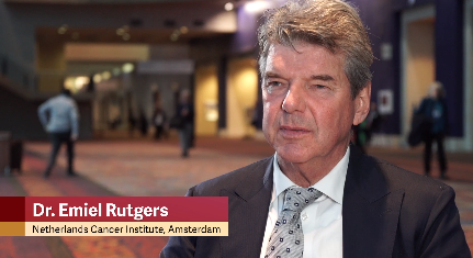
SAN ANTONIO – Both axillary radiation therapy and axillary lymph node dissection provide excellent, comparable locoregional control in patients with early-stage breast cancer who have a positive sentinel node, according to updated results of the European Organisation for Research and Treatment of Cancer’s AMAROS trial.
The 10-year cumulative incidence rate of axillary recurrence was 1.82% with radiation and 0.93% with lymph node dissection, a nonsignificant difference (hazard ratio, 1.71; P = .365). Distant metastasis–free survival and overall survival also were statistically on par. The findings reinforce the trial’s 5-year results, which additionally showed a markedly lower incidence of lymphedema with axillary radiation therapy. Lead investigator Emiel J. T. Rutgers, MD, PhD, reflected on hesitation in the uptake of axillary radiation therapy among oncologists and discussed the AMAROS results in the context of the ACOSOG Z11 trial. Dr. Rutgers, the principal investigator of the AMAROS trial and a surgical oncologist at the Netherlands Cancer Institute in Amsterdam, also described how the trial’s findings have altered practice at his institution.
Dr. Rutgers disclosed that he had no relevant conflicts of interest. The study was supported by the EORTC Charitable Trust.
REPORTING FROM SABCS 2018
Nothing to gain from chemo after pCR achieved
SAN ANTONIO – Women with localized breast cancer who achieve a pathological complete response (pCR) after neoadjuvant chemotherapy may have little to gain from subsequent adjuvant chemotherapy except toxicity, according to a patient-level meta-analysis of more than 27,000 women. The analysis, reported by lead investigator Laura M. Spring, MD, at the San Antonio Breast Cancer Symposium, confirmed that, compared with residual disease, pCR was associated with significantly reduced risks of event-free survival events (hazard ratio, 0.31) and death (HR, 0.22). Moreover, the EFS benefit of a pCR was similar whether women went on to receive adjuvant chemotherapy (HR, 0.36) or not (HR, 0.36) (P = .60 for difference). Dr. Spring discussed overall and subgroup findings, implications for use of neoadjuvant chemotherapy, and how these new data may inform escalation and de-escalation of adjuvant therapy.
Dr. Spring disclosed that she has a consulting or advisory role with Novartis and that she receives institutional research funding from Tesaro. The study was supported by grants from the National Cancer Institute and Susan G. Komen.
REPORTING FROM SABCS 2018

Small absolute difference between partial and whole breast irradiation
SAN ANTONIO – A phase 3 randomized NRG Oncology trial (NSABP B-39/RTOG 0413) was unable to rule out the possibility that, after lumpectomy, partial breast irradiation is inferior to whole breast irradiation when it comes to the ipsilateral breast tumor recurrences (invasive disease or ductal carcinoma in situ), reported Frank Vicini, MD, of MHP Radiation Oncology Institute, Pontiac, Mich.
The hazard ratio for this event with the former versus latter modality was 1.22, with the 90% confidence interval (0.94-1.58) falling just outside the predefined range to declare the two modalities equivalent (0.667-1.5). However, the absolute difference in the 10-year cumulative incidence of ipsilateral breast tumor recurrences was just 0.7% (4.6% vs. 3.9%). In a video interview, Dr. Vicini discussed whether this difference is clinically important, and the implications of the trial’s findings, taken together, for offering partial breast irradiation to patients.
Dr. Vicini disclosed that he is a research adviser for ImpediMed. The study was sponsored by the National Cancer Institute.
REPORTING FROM SABCS 2018
Low-dose tamoxifen halves recurrence of breast intraepithelial neoplasia
SAN ANTONIO – New life for old medicine: Women aged under 75 years with breast intraepithelial neoplasms (IEN) who took tamoxifen for 3 years at a dose of 5 mg per day – one-fourth the standard dose – had a 50% reduction in risk of IEN recurrence and an even more remarkable 75% reduction in the risk of contralateral breast cancer, compared with women who took placebos in the TAMO1 study.
Despite concerns about the known side effects of tamoxifen, there were no significant differences in either the rate of endometrial cancer or of deep vein thrombosis/pulmonary embolism between groups, and there was only a borderline increase in hot flashes among patients randomized to tamoxifen, reported Dr. Andrea De Censi, MD, from Ospedali Galliera in Genoa, Italy.
In a video interview, Dr. De Censi discusses how tamoxifen, a decades-old, inexpensive drug still offers real clinical benefit in day-to-day practice for patients with IEN.
The TAM01 study was supported by the Italian Ministry of Health, Italian Association for Cancer Research, and the Italian League Against Cancer. Dr. De Censi and his coauthors reported having no direct conflicts of interest.
REPORTING FROM SABCS 2018
Adjuvant capecitabine found disappointing in TNBC
SAN ANTONIO – Adjuvant capecitabine does not improve outcomes in women with early-stage triple-negative breast cancer (TNBC) who have undergone resection and received standard chemotherapy, finds a phase 3, randomized, controlled trial jointly conducted by the Spanish GEICAM group and the Central and South American CIBOMA group. But the story may not end there.

Findings reported in a session and press conference at the San Antonio Breast Cancer Symposium showed that, compared with observation, eight cycles of adjuvant capecitabine (Xeloda) reduced the 5-year risks of disease-free survival events and death by a nonsignificant relative 18% and 8%, respectively, among all 876 women randomized. However, the subgroup whose tumors had the nonbasal phenotype saw large significant benefits, with 47% and 58% relative reductions in these risks, respectively.
“The trial is formally negative,” commented lead investigator Miguel Martín, MD, PhD, head of the medical oncology service at Hospital Gregorio Marañón in Madrid. However, the observation arm fared better than expected. In addition, the trial had a very low–risk patient population, which may help explain why its findings differ somewhat from the more positive findings of the similar CREATE-X trial.
“Our data don’t speak against the CREATE-X study. My personal view is that capecitabine is useful for some TNBC patients,” Dr. Martin said. “Our study is not finished because we are going to look at the genomic characteristics of this group defined as non–basal-like because we want to know more about this subgroup. We are planning also to reproduce our subset in the CREATE-X trial to see if this is a real finding because we are in the era of personalized medicine.”
TNBC is a broad group defined only by negative findings for the main markers having available treatments, he elaborated. “So if we could define a subpopulation that actually benefited from capecitabine, this will be great for the patients.” Currently, conventional pathology does not routinely report on tumor basal phenotype, so all TNBC patients receive the same drugs. “This is a mistake. We should select the right drug for the right patient. Probably not all breast cancer patients are sensitive to the same drugs. But the fact is that we don’t have funding to run trials looking at that because this kind of trial is not interesting for pharma companies.”

“That’s an important message that triple-negative is really a big, very heterogeneous group,” agreed SABCS codirector and press conference moderator Carlos Arteaga, MD, director of the Harold C. Simmons Comprehensive Cancer Center and associate dean of oncology programs at the University of Texas, Dallas.
Patients and clinicians alike are largely unaware of the presence of TNBC basal and nonbasal subtypes and their potential importance, he said. “We all need some education on that, us included. It’s a very, very heterogeneous group, it is one that is very challenging. We need to start by educating all of us that there is a need to do research on that. ... We have a duty to define this phenotype better.”
Study details
“TNBC is sensitive to chemotherapy, but a significant proportion of patients will eventually relapse after conventional anthracycline and taxane combinations, so we need new approaches to this population,” Dr. Martín noted.
The trial, joint GEICAM/2003-11 and CIBOMA/2004-01, was designed in 2004. Although no information about capecitabine in breast cancer was available at the time, the investigators selected this drug because it is non–cross-resistant with anthracyclines and taxanes.
About 55% of the patients randomized had node-negative disease and roughly 80% received adjuvant chemotherapy alone because neoadjuvant chemotherapy was generally not used 14 years ago, Dr. Martín noted.
After a median follow-up of 7.34 years, the 5-year disease-free survival rate—the trial’s primary endpoint—was 79.6% with capecitabine and 76.8% with observation, a nonsignificant difference in both unadjusted analysis (hazard ratio, 0.82; P = .136) and adjusted analysis (HR, 0.79; P = .082). The 5-year overall survival rate was 86.2% with capecitabine and 85.9% with observation, another nonsignificant difference (HR, 0.92; P = .623).
However, in subgroup analyses among the 248 patients with nonbasal disease, defined as immunohistochemically negative for both EGFR and CK5/6, capecitabine conferred a significant disease-free survival advantage (HR, 0.53; P = .02) and overall survival advantage (HR, 0.420; P = .007) relative to observation.
Interaction of basal/nonbasal phenotype with treatment was marginal for disease-free survival (P = .0694) and significant for overall survival (P = .0052).
In the nonbasal subgroup, the disease-free survival benefit of capecitabine was mainly driven by a reduction in distant recurrences, particularly in the liver and the brain.
Adjuvant capecitabine had tolerability that was “exactly as expected,” according to Dr. Martín. The median dose intensity was 86.3%, and 75.2% of patients completed all of the planned eight cycles.
The trial was supported by Roche, which also provided capecitabine. Dr. Martín reported receiving speaker’s honoraria from Pfizer and Eli Lilly; honoraria for participation in advisory boards from AstraZeneca, Novartis, Roche-Genentech, Pfizer, GlaxoSmithKline, PharmaMar, Taiho Oncology, and Eli Lilly; and research grants from Novartis and Roche.
SOURCE: Martín M et al. SABCS 2018, Abstract GS2-04.
SAN ANTONIO – Adjuvant capecitabine does not improve outcomes in women with early-stage triple-negative breast cancer (TNBC) who have undergone resection and received standard chemotherapy, finds a phase 3, randomized, controlled trial jointly conducted by the Spanish GEICAM group and the Central and South American CIBOMA group. But the story may not end there.
Findings reported in a session and press conference at the San Antonio Breast Cancer Symposium showed that, compared with observation, eight cycles of adjuvant capecitabine (Xeloda) reduced the 5-year risks of disease-free survival events and death by a nonsignificant relative 18% and 8%, respectively, among all 876 women randomized. However, the subgroup whose tumors had the nonbasal phenotype saw large significant benefits, with 47% and 58% relative reductions in these risks, respectively.
“The trial is formally negative,” commented lead investigator Miguel Martín, MD, PhD, head of the medical oncology service at Hospital Gregorio Marañón in Madrid. However, the observation arm fared better than expected. In addition, the trial had a very low–risk patient population, which may help explain why its findings differ somewhat from the more positive findings of the similar CREATE-X trial.
“Our data don’t speak against the CREATE-X study. My personal view is that capecitabine is useful for some TNBC patients,” Dr. Martin said. “Our study is not finished because we are going to look at the genomic characteristics of this group defined as non–basal-like because we want to know more about this subgroup. We are planning also to reproduce our subset in the CREATE-X trial to see if this is a real finding because we are in the era of personalized medicine.”
TNBC is a broad group defined only by negative findings for the main markers having available treatments, he elaborated. “So if we could define a subpopulation that actually benefited from capecitabine, this will be great for the patients.” Currently, conventional pathology does not routinely report on tumor basal phenotype, so all TNBC patients receive the same drugs. “This is a mistake. We should select the right drug for the right patient. Probably not all breast cancer patients are sensitive to the same drugs. But the fact is that we don’t have funding to run trials looking at that because this kind of trial is not interesting for pharma companies.”
“That’s an important message that triple-negative is really a big, very heterogeneous group,” agreed SABCS codirector and press conference moderator Carlos Arteaga, MD, director of the Harold C. Simmons Comprehensive Cancer Center and associate dean of oncology programs at the University of Texas, Dallas.
Patients and clinicians alike are largely unaware of the presence of TNBC basal and nonbasal subtypes and their potential importance, he said. “We all need some education on that, us included. It’s a very, very heterogeneous group, it is one that is very challenging. We need to start by educating all of us that there is a need to do research on that. ... We have a duty to define this phenotype better.”
Study details
“TNBC is sensitive to chemotherapy, but a significant proportion of patients will eventually relapse after conventional anthracycline and taxane combinations, so we need new approaches to this population,” Dr. Martín noted.
The trial, joint GEICAM/2003-11 and CIBOMA/2004-01, was designed in 2004. Although no information about capecitabine in breast cancer was available at the time, the investigators selected this drug because it is non–cross-resistant with anthracyclines and taxanes.
About 55% of the patients randomized had node-negative disease and roughly 80% received adjuvant chemotherapy alone because neoadjuvant chemotherapy was generally not used 14 years ago, Dr. Martín noted.
After a median follow-up of 7.34 years, the 5-year disease-free survival rate—the trial’s primary endpoint—was 79.6% with capecitabine and 76.8% with observation, a nonsignificant difference in both unadjusted analysis (hazard ratio, 0.82; P = .136) and adjusted analysis (HR, 0.79; P = .082). The 5-year overall survival rate was 86.2% with capecitabine and 85.9% with observation, another nonsignificant difference (HR, 0.92; P = .623).
However, in subgroup analyses among the 248 patients with nonbasal disease, defined as immunohistochemically negative for both EGFR and CK5/6, capecitabine conferred a significant disease-free survival advantage (HR, 0.53; P = .02) and overall survival advantage (HR, 0.420; P = .007) relative to observation.
Interaction of basal/nonbasal phenotype with treatment was marginal for disease-free survival (P = .0694) and significant for overall survival (P = .0052).
In the nonbasal subgroup, the disease-free survival benefit of capecitabine was mainly driven by a reduction in distant recurrences, particularly in the liver and the brain.
Adjuvant capecitabine had tolerability that was “exactly as expected,” according to Dr. Martín. The median dose intensity was 86.3%, and 75.2% of patients completed all of the planned eight cycles.
The trial was supported by Roche, which also provided capecitabine. Dr. Martín reported receiving speaker’s honoraria from Pfizer and Eli Lilly; honoraria for participation in advisory boards from AstraZeneca, Novartis, Roche-Genentech, Pfizer, GlaxoSmithKline, PharmaMar, Taiho Oncology, and Eli Lilly; and research grants from Novartis and Roche.
SOURCE: Martín M et al. SABCS 2018, Abstract GS2-04.
REPORTING FROM SABCS 2018
Key clinical point: Adjuvant capecitabine fails to improve outcomes in early-stage triple-negative breast cancer treated with surgery and standard chemotherapy.
Major finding: The 5-year disease-free survival rate was 79.6% with eight cycles of adjuvant capecitabine versus 76.8% with observation (adjusted hazard ratio, 0.79; P = .082).
Study details: A phase 3, randomized, controlled trial among 876 women with early-stage triple-negative breast cancer who had undergone surgery and received standard chemotherapy (joint GEICAM/2003-11 and CIBOMA/2004-01 trial).
Disclosures: The trial was supported by Roche, which also provided capecitabine. Dr. Martín reported receiving speaker’s honoraria from Pfizer and Eli Lilly; honoraria for participation in advisory boards from AstraZeneca, Novartis, Roche-Genentech, Pfizer, GlaxoSmithKline, PharmaMar, Taiho Oncology, and Eli Lilly; and research grants from Novartis and Roche.
Source: Martín M et al. SABCS 2018, Abstract GS2-04.