User login
Neonates at highest risk for Ebstein's malformation treatment
LOS ANGELES – Ebstein’s malformation is a rare congenital cardiac anomaly. Surgery for Ebstein’s involves a range of procedures, and with low institutional volumes, the only available data on treatment are limited to individual reports demonstrating highly variable approaches. Neonates in particular are at risk for poor outcomes, showing significantly higher mortality than infants, children, and adults, according to a database study presented by Dr. Ryan R. Davies at the annual meeting of the Society of Thoracic Surgeons.
Dr. Davies of the Nemours/A.I. duPont Hospital for Children, Wilmington, Del., and his colleagues performed a retrospective study of procedures performed on patients with a primary diagnosis of Ebstein’s malformation (2002-2009) in the STS Congenital Heart Surgery Database.

A total of 595 operations on patients with Ebstein’s were included: 116 on neonates (19%), 122 on infants (21%), 264 on children (44%), and 93 on adults (16%). The authors found that average annual institutional case volumes were low (median, 1 case/year), and procedures varied according to age. Neonates had a high rate of palliative procedures: tricuspid valve (TV) closure (16%) and systemic-to-pulmonary shunts with or without TV closure (37%) and isolated TV closure (8.6%), with Ebstein’s repair or TV valvuloplasty performed in 32%.
Infants usually underwent superior cavopulmonary connections (52%).
Among older patients, procedures were mostly in three categories: TV surgery (children, 55%; adults, 69%), arrhythmia procedures (children, 9%; adults, 17%), and Fontan (children, 16%). In-hospital mortality was higher among neonatal patients (23%) than in infants (4%), children (0.8%), and adults (1.1%).
Among neonates, 36 subsequent procedures were performed during the same hospitalization in 27 patients (23%); including TV closure (11%); shunt (15%); Ebstein’s repair (17%) or TV replacement (15%); and heart transplantation (7.4%). Mortality was similar among neonates who had a second procedure and those who did not (27% vs. 23%, respectively). ECMO (extracorporeal membrane oxygenation) was used in 9% of neonates but in less than 2% of patients in other age groups.
"This study represents a broad overview of the diverse options for surgical treatment of Ebstein’s anomaly. It shows the challenges faced in caring for extremely ill neonatal patients. We have also shown that repair of Ebstein’s anomaly is performed infrequently at most centers, limiting the ability of individual series to define optimal management strategies," Dr. Davies said in an interview.
"Unfortunately, currently available databases do not contain information that may be important in defining such strategies (both surgical and nonsurgical), including anatomic and physiologic variables – whether they are neonates presenting in severe heart failure or older patients presenting for tricuspid valve repair or replacement," he added.
"We feel that in this setting, a prospective multi-institutional study would be of significant value. It should include operative and nonoperative patients, as well as precise diagnostic information and procedural details, to evaluate long-term outcomes including survival, reoperation and other reinterventions, as well as neurodevelopmental outcomes, functional health status, and quality of life," Dr. Davies concluded.
Dr. Davies and his colleagues reported having no relevant disclosures.
LOS ANGELES – Ebstein’s malformation is a rare congenital cardiac anomaly. Surgery for Ebstein’s involves a range of procedures, and with low institutional volumes, the only available data on treatment are limited to individual reports demonstrating highly variable approaches. Neonates in particular are at risk for poor outcomes, showing significantly higher mortality than infants, children, and adults, according to a database study presented by Dr. Ryan R. Davies at the annual meeting of the Society of Thoracic Surgeons.
Dr. Davies of the Nemours/A.I. duPont Hospital for Children, Wilmington, Del., and his colleagues performed a retrospective study of procedures performed on patients with a primary diagnosis of Ebstein’s malformation (2002-2009) in the STS Congenital Heart Surgery Database.
A total of 595 operations on patients with Ebstein’s were included: 116 on neonates (19%), 122 on infants (21%), 264 on children (44%), and 93 on adults (16%). The authors found that average annual institutional case volumes were low (median, 1 case/year), and procedures varied according to age. Neonates had a high rate of palliative procedures: tricuspid valve (TV) closure (16%) and systemic-to-pulmonary shunts with or without TV closure (37%) and isolated TV closure (8.6%), with Ebstein’s repair or TV valvuloplasty performed in 32%.
Infants usually underwent superior cavopulmonary connections (52%).
Among older patients, procedures were mostly in three categories: TV surgery (children, 55%; adults, 69%), arrhythmia procedures (children, 9%; adults, 17%), and Fontan (children, 16%). In-hospital mortality was higher among neonatal patients (23%) than in infants (4%), children (0.8%), and adults (1.1%).
Among neonates, 36 subsequent procedures were performed during the same hospitalization in 27 patients (23%); including TV closure (11%); shunt (15%); Ebstein’s repair (17%) or TV replacement (15%); and heart transplantation (7.4%). Mortality was similar among neonates who had a second procedure and those who did not (27% vs. 23%, respectively). ECMO (extracorporeal membrane oxygenation) was used in 9% of neonates but in less than 2% of patients in other age groups.
"This study represents a broad overview of the diverse options for surgical treatment of Ebstein’s anomaly. It shows the challenges faced in caring for extremely ill neonatal patients. We have also shown that repair of Ebstein’s anomaly is performed infrequently at most centers, limiting the ability of individual series to define optimal management strategies," Dr. Davies said in an interview.
"Unfortunately, currently available databases do not contain information that may be important in defining such strategies (both surgical and nonsurgical), including anatomic and physiologic variables – whether they are neonates presenting in severe heart failure or older patients presenting for tricuspid valve repair or replacement," he added.
"We feel that in this setting, a prospective multi-institutional study would be of significant value. It should include operative and nonoperative patients, as well as precise diagnostic information and procedural details, to evaluate long-term outcomes including survival, reoperation and other reinterventions, as well as neurodevelopmental outcomes, functional health status, and quality of life," Dr. Davies concluded.
Dr. Davies and his colleagues reported having no relevant disclosures.
AT THE STS ANNUAL MEETING
Major Finding: In-hospital mortality was higher among neonatal patients (23%) than in infants (4%), children (0.8%), and adults (1.1%).
Data Source: A retrospective database analysis of 595 operations on patients with Ebstein’s malformation.
Disclosures: Dr. Davies and his colleagues reported having no relevant disclosures.

Valve-sparing root replacement carries the day
SNOWMASS, COLO. – Valve-sparing root replacement has emerged as the procedure of choice in patients with isolated aortic root disease and a normally functioning aortic valve, according to Dr. Thoralf M. Sundt III.
"The valve-sparing root operations, in contrast to some of the other things we surgeons have come up with over the last decade or so, are increasing in popularity. They’re more and more commonly done, and that’s a good sign. I think the marketplace has spoken and this is clearly a good operation. It’s an operation that can be learned, and surgeons can do it with good results," he said at the Annual Cardiovascular Conference at Snowmass.
Valve-sparing root replacement (VSRR) spares a patient from the complications associated with lifelong anticoagulation for a mechanical valve, and the durability of VSRR appears to be superior to that of third-generation bioprostheses, the surgeon added.
"They’re holding up pretty well. The outcomes approach those with mechanical valves," said Dr. Sundt, chief of cardiac surgery at Massachusetts General Hospital and professor of surgery at Harvard Medical School, Boston.
Moreover, he continued, VSRR has another big advantage over bioprosthetic valves: "If you have to re-operate, it’s a whole lot more fun to do so on someone who’s had a VSRR and put a new biologic valve inside a native annulus than it is to try to take out that old bioprosthesis and put a new bioprosthesis in."
A meta-analysis of 11 studies comparing VSRR with total root replacement in patients with Marfan syndrome concluded that composite valve-related event rates for the two surgical strategies were not significantly different. The thromboembolic event rate was 0.3% per year in VSRR-treated patients, significantly lower than the still-quite-reasonable 0.7% per year rate after total root replacement (Heart 2011;97:955-8).
A recent study by surgeons at Stanford (Calif.) University gave VSRR a thumbs up regarding mid-term durability of outcomes through 6 years of follow-up, with a mean 2.9-year and maximum 6-year follow-up. The series included 75 patients with bicuspid aortic valve disease treated by VSRR.
Six-year actuarial survival was 99%, with 90% freedom from reoperation and no strokes. Thirty-one percent of patients had 2+ aortic regurgitation preoperatively; at echocardiographic follow-up a mean of 2.9 years post surgery, only a couple of patients had 2+ aortic regurgitation and no one was more severely affected. The Stanford investigators plan to update their results when follow-up reaches 10 years or more (J. Thorac. Cardiovasc. Surg. Dec. 20, 2012 [doi:10.1016/j.jtcvs.2012.11.043]).
The VSRR was developed by Dr. Tirone David of the University of Toronto. The procedure involves skeletonizing the root while preserving the leaflets and their attachments to the aortic wall. The aortic valve is then reimplanted inside a tubular Dacron graft, and then the coronary arteries are reimplanted.
"It’s probably the neatest development in terms of surgical options for the aortic valve in a long time," Dr. Sundt said.
He reported having no financial conflicts.
EXPERT ANALYSIS FROM THE CARDIOVASCULAR CONFERENCE AT SNOWMASS
TAVR trial to assess alternative access sites
Two heart societies, a device maker, and two federal agencies have collaborated to develop a trial for transcatheter aortic valve replacement, once again extending the concept of teamwork, which is the cornerstone of this technology, far beyond the operating room walls.
During the past year, the Society of Thoracic Surgeons and the American College of Cardiology worked with the Food and Drug Administration, the Centers for Medicare and Medicaid Services (CMS), and Edwards Lifesciences to develop a trial that assesses the safety and efficacy of nontransfemoral approaches for TAVR, using the already-approved Edwards SAPIEN valves.
"What makes it unusual is, to the best of our knowledge, this is the first investigational device exemption [IDE] granted by the FDA to medical societies who operate national clinical registries," said ACC President Dr. William Zoghbi.
The societies will run the trial with funding from Edwards, and Medicare will pay for the procedures.
"This allows physicians to get reimbursed for off-label use," said Dr. Michael J. Mack, past president of STS and chair of the STS/ACC TVT (Transcatheter Valvular Therapy) Registry Steering Committee. "And it also allows for controlled off-label use, in which the outcomes can be captured and the sites can be paid while this information is being captured. People should be excited about it."
The goal of the trial is to expand the field and to extend TAVR to a broader group of patients, the societies said.
"A similar mechanism has been used in the past for the implantation of ICDs [implantable cardioverter-defibrillators] for patients who met particular criteria and get the funding as the registry moves forward," said Dr. Sidney Goldstein, professor of medicine at Wayne State University in Detroit, who is not involved in the trial or registry.
But currently, "the ICD Registry is not conducting an IDE to evaluate and reimburse for other possible indications of ICDs," said Dr. Zoghbi.
In the United States, an estimated one in four inoperable patients with severe aortic stenosis is not eligible for TAVR through a transfemoral or transapical approach because of vessel size, vessel disease, or other anatomical restrictions, according to the societies. Alternative routes, such as the transaortic approach, could provide an option for them.
STS and ACC are also working to get FDA approval for two more studies.
"The collaboration and use of registries for research in this pilot can be a model for specialty societies, industry, and federal regulators," Dr. Zoghbi said in a statement. "We have aligned our efforts to ensure patient access to a new technology in a safe and cost-effective way."
The study also stands out in the list of TAVR clinical trials approved by CMS, which began covering TAVR in May 2012: The other five are sponsored by Edwards or Medtronic.
The earlier collaboration of the entities that have developed this trial resulted in the STS/ACC TVT Registry, which captures TAVR-related patient demographics, procedure details, and facility and physician information.
The observational study is conducted in the TVT Registry, and will follow 1,000 patients. Any of the nearly 180 sites using the TVT Registry can participate in the study.
The trial will gather 30-day safety endpoints for patients who undergo alternative access approaches such as transapical and transaortic routes. The lumped data will then be compared with the 30-day outcomes of the transapical approach reported in Cohort A of the PARTNER trial.
Because the trial is covered by Medicare, it has to adhere to conditions set by the agency. The requirements will ensure better patient care, Dr. Jeffrey B. Rich, the STS immediate past president, said in a statement, because the "preoperative evaluation, interoperative deployment of the valve, and postoperative care must be jointly shared by cardiologists and cardiothoracic surgeons, utilizing the heart team approach."

Alternative access approaches in the trial include the left ventricular apex (transapical), ascending aorta, subclavian and axillary arteries, and distal aorta, as well as retroperitoneal access to the iliac artery.
The approaches have several advantages, according to the societies. For one, the risks associated with inserting large-caliber catheters into small, diseased femoral arteries are reduced. Also, nonfemoral access sites can provide for better catheter control and safer closure of the access site.
But since some of the alternative approaches, such as the transaortic approach, have not been approved, the operator training lags behind the already-approved transfemoral and transapical approaches. To address this, the societies and Edwards will probably have to create a contract to train the surgeons and cardiologists for the specific purpose of this trial, said Dr. Mack.
The alternative approaches also require additional equipment, and operators may be exposed to greater amounts of radiation. They may also lead to longer recovery and more incisional pain for patients, according to the societies.
Dr. Mack said that the trial will likely be completed in 6 months or less from its start date.
"Using registries for this kind of study has the potential to make new technology available in a timely manner while protecting patient safety," ACC Immediate Past President Dr. David Holmes said in a statement.
None of the physicians had relevant disclosures.
On Twitter @NaseemSMiller
Two heart societies, a device maker, and two federal agencies have collaborated to develop a trial for transcatheter aortic valve replacement, once again extending the concept of teamwork, which is the cornerstone of this technology, far beyond the operating room walls.
During the past year, the Society of Thoracic Surgeons and the American College of Cardiology worked with the Food and Drug Administration, the Centers for Medicare and Medicaid Services (CMS), and Edwards Lifesciences to develop a trial that assesses the safety and efficacy of nontransfemoral approaches for TAVR, using the already-approved Edwards SAPIEN valves.
"What makes it unusual is, to the best of our knowledge, this is the first investigational device exemption [IDE] granted by the FDA to medical societies who operate national clinical registries," said ACC President Dr. William Zoghbi.
The societies will run the trial with funding from Edwards, and Medicare will pay for the procedures.
"This allows physicians to get reimbursed for off-label use," said Dr. Michael J. Mack, past president of STS and chair of the STS/ACC TVT (Transcatheter Valvular Therapy) Registry Steering Committee. "And it also allows for controlled off-label use, in which the outcomes can be captured and the sites can be paid while this information is being captured. People should be excited about it."
The goal of the trial is to expand the field and to extend TAVR to a broader group of patients, the societies said.
"A similar mechanism has been used in the past for the implantation of ICDs [implantable cardioverter-defibrillators] for patients who met particular criteria and get the funding as the registry moves forward," said Dr. Sidney Goldstein, professor of medicine at Wayne State University in Detroit, who is not involved in the trial or registry.
But currently, "the ICD Registry is not conducting an IDE to evaluate and reimburse for other possible indications of ICDs," said Dr. Zoghbi.
In the United States, an estimated one in four inoperable patients with severe aortic stenosis is not eligible for TAVR through a transfemoral or transapical approach because of vessel size, vessel disease, or other anatomical restrictions, according to the societies. Alternative routes, such as the transaortic approach, could provide an option for them.
STS and ACC are also working to get FDA approval for two more studies.
"The collaboration and use of registries for research in this pilot can be a model for specialty societies, industry, and federal regulators," Dr. Zoghbi said in a statement. "We have aligned our efforts to ensure patient access to a new technology in a safe and cost-effective way."
The study also stands out in the list of TAVR clinical trials approved by CMS, which began covering TAVR in May 2012: The other five are sponsored by Edwards or Medtronic.
The earlier collaboration of the entities that have developed this trial resulted in the STS/ACC TVT Registry, which captures TAVR-related patient demographics, procedure details, and facility and physician information.
The observational study is conducted in the TVT Registry, and will follow 1,000 patients. Any of the nearly 180 sites using the TVT Registry can participate in the study.
The trial will gather 30-day safety endpoints for patients who undergo alternative access approaches such as transapical and transaortic routes. The lumped data will then be compared with the 30-day outcomes of the transapical approach reported in Cohort A of the PARTNER trial.
Because the trial is covered by Medicare, it has to adhere to conditions set by the agency. The requirements will ensure better patient care, Dr. Jeffrey B. Rich, the STS immediate past president, said in a statement, because the "preoperative evaluation, interoperative deployment of the valve, and postoperative care must be jointly shared by cardiologists and cardiothoracic surgeons, utilizing the heart team approach."
Alternative access approaches in the trial include the left ventricular apex (transapical), ascending aorta, subclavian and axillary arteries, and distal aorta, as well as retroperitoneal access to the iliac artery.
The approaches have several advantages, according to the societies. For one, the risks associated with inserting large-caliber catheters into small, diseased femoral arteries are reduced. Also, nonfemoral access sites can provide for better catheter control and safer closure of the access site.
But since some of the alternative approaches, such as the transaortic approach, have not been approved, the operator training lags behind the already-approved transfemoral and transapical approaches. To address this, the societies and Edwards will probably have to create a contract to train the surgeons and cardiologists for the specific purpose of this trial, said Dr. Mack.
The alternative approaches also require additional equipment, and operators may be exposed to greater amounts of radiation. They may also lead to longer recovery and more incisional pain for patients, according to the societies.
Dr. Mack said that the trial will likely be completed in 6 months or less from its start date.
"Using registries for this kind of study has the potential to make new technology available in a timely manner while protecting patient safety," ACC Immediate Past President Dr. David Holmes said in a statement.
None of the physicians had relevant disclosures.
On Twitter @NaseemSMiller

No early cancer risk with donor lungs from heavy smokers
LOS ANGELES – Use of lungs from donors who smoked heavily does not worsen lung transplantation outcomes including risk for lung cancer death, at least in the medium term.
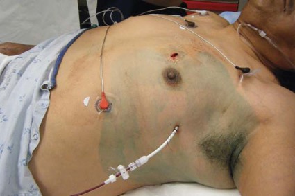
At a median follow-up of 2 years for 5,900 adults who had double-lung transplants, those who received lungs from heavy smokers had an actuarial median overall survival of roughly 5.5 years, and their lung function was essentially the same as that of patients who received lungs from other donors, Dr. Sharven Taghavi reported at the annual meeting of the Society of Thoracic Surgeons.

The study data came from the United Network for Organ Sharing (UNOS) database. A team led by Dr. Taghavi, of Temple University Hospital in Philadelphia, compared data for double-lung transplants from 2005-2011, comparing donors with a history of smoking exceeding 20 pack-years with other donors.
About 13% of the study patients received lungs from donors who had smoked heavily. Compared with other recipients, these recipients were more likely to have a primary diagnosis of chronic obstructive pulmonary disease and less likely to have a diagnosis of idiopathic pulmonary fibrosis. Otherwise, they were similar.
The rate of deaths due to cancer was based on case reports, as UNOS does not capture this outcome. Cancer deaths were 5.8% among recipients of lungs from heavy smokers and 3.6% among other recipients.
"There is a fairly low capture rate for this field, so it’s difficult to draw significant conclusions from it," cautioned Dr. Taghavi.
Patients who received lungs from heavy smokers had a 1-day longer length of stay in the hospital (18 days vs. 17 days), which "may not really be clinically relevant." Rates of acute rejection during hospitalization were comparable (10.7% vs. 8.8%), as was post-transplant airway dehiscence (1.8% vs. 1.8%).
Post-transplant peak forced expiratory volume in 1 second (FEV1) was the same (80% vs. 79%), as was decline in this measure over time. Median duration of freedom from bronchiolitis obliterans syndrome was 1,583 days vs. 1,827 days.
Risk-adjusted median all-cause survival – the study’s primary endpoint – did not differ significantly between the recipients given lungs from donors who smoked heavily and the other recipients (2,043 vs. 1,928 days).
The rate of cancer deaths did not differ significantly; however, the follow-up time is too short to address this concern in a meaningful way, Dr. Taghavi said.
"Currently, we recommend when evaluating a donor who has a heavy smoking history, that they undergo a thorough examination for lung tumors or evidence of cancer. This includes obtaining a chest x-ray, CT scans, and bronchoscopies. In addition, when the lungs are procured, they should undergo a very thorough visual inspection," he advised.
"Informed consent is very important. You have to discuss the donor’s smoking status with the recipient and explain the risks and the benefits," Dr. Taghavi said. Lung cancer risk, given the donor’s history, is about 1% to 2% annually, and that needs to be considered against the high likelihood of dying within 1 or 2 years without a transplant.
"One thing that is unquestionable is that survival will be better accepting these lungs than it will be sitting on a waiting list," he added. Only about half of the people listed for lung transplant in the United States each year actually undergo the surgery.
Recipients of lungs from heavy smokers do not need any extra follow-up or surveillance, as they are already diligently tested and monitored, according to Dr. Taghavi. The recipient’s immunosuppression does theoretically put one at additional risk for lung cancer.
Current guidelines of the International Society of Heart and Lung Transplantation advise against considering use of lungs from donors who have a smoking history of more than 20 pack-years, Dr. Taghavi noted. But he stopped short of saying that the study should prompt a formal revision of those guidelines.
"I think the findings start the conversation," he commented. "We should consider looking at these potential donors," especially when a recipient’s situation is dire.
Dr. Taghavi disclosed no conflicts of interest.
LOS ANGELES – Use of lungs from donors who smoked heavily does not worsen lung transplantation outcomes including risk for lung cancer death, at least in the medium term.
At a median follow-up of 2 years for 5,900 adults who had double-lung transplants, those who received lungs from heavy smokers had an actuarial median overall survival of roughly 5.5 years, and their lung function was essentially the same as that of patients who received lungs from other donors, Dr. Sharven Taghavi reported at the annual meeting of the Society of Thoracic Surgeons.
The study data came from the United Network for Organ Sharing (UNOS) database. A team led by Dr. Taghavi, of Temple University Hospital in Philadelphia, compared data for double-lung transplants from 2005-2011, comparing donors with a history of smoking exceeding 20 pack-years with other donors.
About 13% of the study patients received lungs from donors who had smoked heavily. Compared with other recipients, these recipients were more likely to have a primary diagnosis of chronic obstructive pulmonary disease and less likely to have a diagnosis of idiopathic pulmonary fibrosis. Otherwise, they were similar.
The rate of deaths due to cancer was based on case reports, as UNOS does not capture this outcome. Cancer deaths were 5.8% among recipients of lungs from heavy smokers and 3.6% among other recipients.
"There is a fairly low capture rate for this field, so it’s difficult to draw significant conclusions from it," cautioned Dr. Taghavi.
Patients who received lungs from heavy smokers had a 1-day longer length of stay in the hospital (18 days vs. 17 days), which "may not really be clinically relevant." Rates of acute rejection during hospitalization were comparable (10.7% vs. 8.8%), as was post-transplant airway dehiscence (1.8% vs. 1.8%).
Post-transplant peak forced expiratory volume in 1 second (FEV1) was the same (80% vs. 79%), as was decline in this measure over time. Median duration of freedom from bronchiolitis obliterans syndrome was 1,583 days vs. 1,827 days.
Risk-adjusted median all-cause survival – the study’s primary endpoint – did not differ significantly between the recipients given lungs from donors who smoked heavily and the other recipients (2,043 vs. 1,928 days).
The rate of cancer deaths did not differ significantly; however, the follow-up time is too short to address this concern in a meaningful way, Dr. Taghavi said.
"Currently, we recommend when evaluating a donor who has a heavy smoking history, that they undergo a thorough examination for lung tumors or evidence of cancer. This includes obtaining a chest x-ray, CT scans, and bronchoscopies. In addition, when the lungs are procured, they should undergo a very thorough visual inspection," he advised.
"Informed consent is very important. You have to discuss the donor’s smoking status with the recipient and explain the risks and the benefits," Dr. Taghavi said. Lung cancer risk, given the donor’s history, is about 1% to 2% annually, and that needs to be considered against the high likelihood of dying within 1 or 2 years without a transplant.
"One thing that is unquestionable is that survival will be better accepting these lungs than it will be sitting on a waiting list," he added. Only about half of the people listed for lung transplant in the United States each year actually undergo the surgery.
Recipients of lungs from heavy smokers do not need any extra follow-up or surveillance, as they are already diligently tested and monitored, according to Dr. Taghavi. The recipient’s immunosuppression does theoretically put one at additional risk for lung cancer.
Current guidelines of the International Society of Heart and Lung Transplantation advise against considering use of lungs from donors who have a smoking history of more than 20 pack-years, Dr. Taghavi noted. But he stopped short of saying that the study should prompt a formal revision of those guidelines.
"I think the findings start the conversation," he commented. "We should consider looking at these potential donors," especially when a recipient’s situation is dire.
Dr. Taghavi disclosed no conflicts of interest.
AT THE ANNUAL MEETING OF THE SOCIETY OF THROACIC SURGEONS
Major Finding: Risk-adjusted median all-cause survival did not differ significantly between patients given lungs from donors who smoked heavily and those receiving lungs from donors who did not smoke heavily (2,043 vs. 1,928 days).
Data Source: An observational cohort study of 5,900 adult primary double-lung transplant recipients in the UNOS database
Disclosures: Dr. Taghavi disclosed no relevant conflicts of interest.
Assay may target early lung cancers for adjuvant therapy
LOS ANGELES – A novel genetic assay helps identify patients with early, aggressive lung cancer who might benefit from adjuvant therapy.
The assay, marketed as Pervino Lung RS by Life Technologies, is the only lung cancer signature to undergo blinded validation in two large cohorts from different countries, one in the United States and one in China (Lancet 2012;379:823-32).
It assesses expression of 14 genes involved in lung cancer tumorigenesis, including ones on the EGFR and KRAS signaling pathways. The assay provides considerably more prognostic information than do conventional criteria proposed by the National Comprehensive Cancer Network (NCCN) as defining high-risk tumors warranting treatment, according to Dr. Johannes R. Kratz, who reported the data at the annual meeting of the Society of Thoracic Surgeons.
The assay results were used to stratify the 269 study patients who had undergone resection of T1a node-negative and nonmetastatic, nonsquamous, non–small cell lung cancer (NSCLC) into groups with distinctly different 5-year survival rates.
Compared with their counterparts in the low-risk group, those in the intermediate- and high-risk groups had a respective doubling and more than tripling of the risk of death, said Dr. Kratz, who was the study’s lead investigator.
As a result of recommendations for CT screening in patients at high risk for lung cancer, resections of small node-negative tumors that are in fact deadly are likely to increase, he observed. Nearly 30% of all patients with stage IA tumors – the lowest level in the current classification system – will nonetheless die in the subsequent 5 years.
"These tumors with highly aggressive tumor biology can now be identified reliably with a prognostic gene signature. The identification of these small but deadly tumors may allow for personalized patient prognosis and could allow us to maximize the benefit of the early detection of these small but deadly tumors via low-dose CT screening," he added.
The current postoperative standard of care for stage IA disease is simply observation, according to Dr. Kratz, a former surgical resident at the Massachusetts General Hospital in Boston, and now a postdoctoral fellow at the University of California, San Francisco.
However, "we should strongly consider changing the way we think about patients with high-risk T1a tumors," he recommended. To that end, a randomized controlled trial of assay-guided adjuvant chemotherapy for early lung cancer is underway in China among roughly 1,000 patients.
Dr. Kratz said that studies to date have not examined a potential prognostic role of the assay in EGFR (epidermal growth factor receptor) mutations. "We haven’t performed an additional mutation analysis on these patients’ EGFR. The original assay was designed to work on patients with resected paraffin-embedded specimens and not fresh-frozen tissue specimens. As a result, it is difficult for us to do extensive EGFR mutation analysis. But that’s definitely something to consider, and it would be nice to explore that association."
It remains to be seen whether the assay, in fact, predicts chemotherapy benefit, he acknowledged in a related press conference. But research has suggested that such prognostic signatures in lung cancer are also predictive (J. Clin. Oncol. 2010;28:4417-24). "That is what we hope to show in the China trial as well," he said.
In the reported study, patients with T1a tumors were drawn from the initial validation cohorts. Fully 40% were under age 60. "This is important, because ... we’d like to be more aggressive in younger patients, both because they can tolerate it and we are more likely to treat them more aggressively," he noted.
The patients’ actual 5-year mortality rate was 32% overall, showing that "these tumors are as deadly as advertised."
The main study results, reported at the meeting and also published (JAMA 2012;308:1629-31), showed that the 5-year actuarial overall survival was 83%, 69%, and 52% among patients in assay-defined low-, medium-, and high-risk groups, respectively (P less than .0001).
In multivariate analyses, relative to their counterparts in the low-risk group, patients in the intermediate-risk group had a 2.0-fold higher risk of death (P = .04) and patients in the high-risk group had a 3.3-fold higher risk (P = .00).
The assay also showed good risk discrimination in analyses restricted to the smallest of tumors, those measuring 1.5 cm or less (P = .001 for difference across groups) and even those measuring 1.0 cm or less (P = .008).
And when compared with tumor size alone, the combination of the assay and tumor size significantly improved on the identification of patients who died (c-statistic, 0.68 vs. 0.57; P less than .0001).
Although these T1a tumors can be ablated nonoperatively, their genetic makeup offers a rich source of information about their subsequent behavior, Dr. Kratz said.
"Despite the popularity and endorsement of our radiology colleagues for techniques such as stereotactic radiation for small T1aN0M0 tumors, we should remember that these techniques don’t provide us with potentially important lung tissue that can provide prognostic and predictive information," he commented.
Dr. Kratz disclosed that he has been a consultant for Pinpoint Genomics, the company that developed the assay, and is a consultant for Life Technologies, which has acquired Pinpoint Genomics.
AT THE ANNUAL MEETING OF THE SOCIETY OF THORACIC SURGEONS
Major Finding: The 5-year actuarial overall survival was 83%, 69%, and 52% among patients in assay-defined low-, medium-, and high-risk groups, respectively (P less than .0001).
Data Source: A cohort study of 269 patients who underwent resection of T1aN0M0 nonsquamous NSCLC.
Disclosures: Dr. Kratz disclosed that he has been a consultant for Pinpoint Genomics, the company that developed the assay, and is a consultant for Life Technologies, which has acquired Pinpoint Genomics.

Pregnancy and Marfan: New insight into risks
SNOWMASS, COLO–Pregnancy increases the long-term risk of aortic complications in women with Marfan syndrome, according to a recent prospective study causing a stir among adult congenital heart disease specialists.
"This is the first study that says, ‘Even if the aortic root size is okay before pregnancy, the aorta is going to get bigger during pregnancy and it’s not going to go back to baseline. And if your aorta is bigger at the outset, there is a risk for long-term adverse outcomes,’ " Dr. Carole A. Warnes explained at the annual cardiovascular conference at Snowmass sponsored by the American College of Cardiology (ACC).
This study on pregnancy’s impact on aortic growth rate and complications in patients with Marfan syndrome sheds much needed light on an area where there has been a paucity of data. The deficiency of data is reflected in discordant recommendations in the current U.S., European, and Canadian guidelines, said Dr. Warnes, professor of medicine at the Mayo Clinic, Rochester, Minn.
The U.S. guidelines put forth jointly by the ACC, American Heart Association, American Association for Thoracic Surgery, and other groups advocate that Marfan syndrome patients avoid pregnancy if their aortic root diameter exceeds 40 mm and recommend prophylactic aortic replacement in those interested in pregnancy (J. Am. Coll. Cardiol. 2010;55:e27-129).
In contrast, the European guidelines (Eur. Heart J. 2010;31:2915-57) consider an aortic root diameter of 45 mm or less to be generally safe, while strongly discouraging pregnancy in Marfan syndrome patients with a measurement above that threshold because of the associated increased dissection risk. The Canadian guidelines take a similar stance, albeit with a safety threshold of 44 mm rather than 45 mm (Can. J. Cardiol. 2010;26:e80-e97).

The Europeans qualify their position by noting that patients with a prepregnancy aortic root diameter of 40-45 mm who have a rapid aortic root growth rate or a family history of dissection ought to be considered high risk for pregnancy. The European and Canadian guidelines characterize dissection as a rare problem in patients with an aortic root diameter of less than 40 mm.
The recent Utah study included 98 women with Marfan syndrome, 69 of whom collectively had 199 pregnancies, with 170 live births, 26 spontaneous abortions, and 2 ectopic pregnancies.
Serial echocardiograms demonstrated that the aortic growth rate was significantly greater during pregnancy than beforehand, and after pregnancy it didn’t return to baseline. Obstetric complications occurred in 10% of pregnancies. Adverse fetal outcomes occurred in 13%.
Reassuringly, there were no catastrophic peripartum complications. No one required cardiac surgery or experienced aortic dissection during pregnancy. However, women with a prior pregnancy had a greater prevalence of both aortic dissection and elective aortic surgery during long-term follow-up, compared with matched childless women with Marfan syndrome. Thus, it’s important during prepregnancy counseling of women with Marfan syndrome to let them know they’ll need to have elective aortic root surgery at a younger age than if they remain childless, Dr. Warnes noted.
A larger initial root diameter and a faster increase in diameter were independent predictors of long-term adverse cardiovascular events in the Utah study.
Besides the recent Utah study, only two other prospective studies of pregnancy’s impact on aortic growth and complications have been done. Both were much smaller. In an editorial accompanying the Utah study, Dutch physicians combined the three studies to get a fuller picture. No type A dissections occurred during 145 pregnancies in 78 nonoperated women with Marfan syndrome. Of 25 women with an aortic root diameter of 40-51 mm during 29 pregnancies, one experienced a type B dissection, two had carotid artery dissections, and one developed accelerated aortic regurgitation, which went from mild to severe during pregnancy.
Five women underwent aortic root replacement (three electively), prior to six pregnancies. Two of them developed a type B dissection during pregnancy. Both women who underwent a valve-sparing elective aortic root replacement prior to pregnancy had pregnancies complicated by a worsening of aortic regurgitation, which went from trivial to moderate. These findings raise a red flag for Dr. Warnes.
"Even if they’ve had a successful root replacement, it doesn’t mean they’re out of the woods in terms of pregnancy. I think we have to question the role of prophylactic root replacement [as recommended in the U.S. guidelines] because these women will still have type B dissections, and trying to look for a type B dissection during pregnancy is a real difficult issue," the cardiologist observed.
The authors of the editorial concluded that Marfan syndrome patients without previous cardiac complications and who have a baseline aortic root diameter not in excess of 45 mm seem to tolerate pregnancy well as long as they receive good clinical care before, during, and after pregnancy. In contrast, pregnancy should be discouraged in patients with a history of aortic dissection because they are at elevated risk for aortic complications (J. Am. Coll. Cardiol. 2012;60:230-1).
Marfan syndrome is a genetic connective tissue disorder with an incidence of roughly 1 in 5,000 and autosomal dominant inheritance, so the fetus of an affected mom has a 50% chance of having the disorder. Dr. Warnes said that because the diagnostic criteria were overhauled in 2010, patients believed to have Marfan syndrome really ought to be referred to a specialized center in order to confirm or refute the diagnosis according to the contemporary Ghent criteria.
Dr. Warnes reported having no relevant financial interests.
SNOWMASS, COLO–Pregnancy increases the long-term risk of aortic complications in women with Marfan syndrome, according to a recent prospective study causing a stir among adult congenital heart disease specialists.
"This is the first study that says, ‘Even if the aortic root size is okay before pregnancy, the aorta is going to get bigger during pregnancy and it’s not going to go back to baseline. And if your aorta is bigger at the outset, there is a risk for long-term adverse outcomes,’ " Dr. Carole A. Warnes explained at the annual cardiovascular conference at Snowmass sponsored by the American College of Cardiology (ACC).
This study on pregnancy’s impact on aortic growth rate and complications in patients with Marfan syndrome sheds much needed light on an area where there has been a paucity of data. The deficiency of data is reflected in discordant recommendations in the current U.S., European, and Canadian guidelines, said Dr. Warnes, professor of medicine at the Mayo Clinic, Rochester, Minn.
The U.S. guidelines put forth jointly by the ACC, American Heart Association, American Association for Thoracic Surgery, and other groups advocate that Marfan syndrome patients avoid pregnancy if their aortic root diameter exceeds 40 mm and recommend prophylactic aortic replacement in those interested in pregnancy (J. Am. Coll. Cardiol. 2010;55:e27-129).
In contrast, the European guidelines (Eur. Heart J. 2010;31:2915-57) consider an aortic root diameter of 45 mm or less to be generally safe, while strongly discouraging pregnancy in Marfan syndrome patients with a measurement above that threshold because of the associated increased dissection risk. The Canadian guidelines take a similar stance, albeit with a safety threshold of 44 mm rather than 45 mm (Can. J. Cardiol. 2010;26:e80-e97).
The Europeans qualify their position by noting that patients with a prepregnancy aortic root diameter of 40-45 mm who have a rapid aortic root growth rate or a family history of dissection ought to be considered high risk for pregnancy. The European and Canadian guidelines characterize dissection as a rare problem in patients with an aortic root diameter of less than 40 mm.
The recent Utah study included 98 women with Marfan syndrome, 69 of whom collectively had 199 pregnancies, with 170 live births, 26 spontaneous abortions, and 2 ectopic pregnancies.
Serial echocardiograms demonstrated that the aortic growth rate was significantly greater during pregnancy than beforehand, and after pregnancy it didn’t return to baseline. Obstetric complications occurred in 10% of pregnancies. Adverse fetal outcomes occurred in 13%.
Reassuringly, there were no catastrophic peripartum complications. No one required cardiac surgery or experienced aortic dissection during pregnancy. However, women with a prior pregnancy had a greater prevalence of both aortic dissection and elective aortic surgery during long-term follow-up, compared with matched childless women with Marfan syndrome. Thus, it’s important during prepregnancy counseling of women with Marfan syndrome to let them know they’ll need to have elective aortic root surgery at a younger age than if they remain childless, Dr. Warnes noted.
A larger initial root diameter and a faster increase in diameter were independent predictors of long-term adverse cardiovascular events in the Utah study.
Besides the recent Utah study, only two other prospective studies of pregnancy’s impact on aortic growth and complications have been done. Both were much smaller. In an editorial accompanying the Utah study, Dutch physicians combined the three studies to get a fuller picture. No type A dissections occurred during 145 pregnancies in 78 nonoperated women with Marfan syndrome. Of 25 women with an aortic root diameter of 40-51 mm during 29 pregnancies, one experienced a type B dissection, two had carotid artery dissections, and one developed accelerated aortic regurgitation, which went from mild to severe during pregnancy.
Five women underwent aortic root replacement (three electively), prior to six pregnancies. Two of them developed a type B dissection during pregnancy. Both women who underwent a valve-sparing elective aortic root replacement prior to pregnancy had pregnancies complicated by a worsening of aortic regurgitation, which went from trivial to moderate. These findings raise a red flag for Dr. Warnes.
"Even if they’ve had a successful root replacement, it doesn’t mean they’re out of the woods in terms of pregnancy. I think we have to question the role of prophylactic root replacement [as recommended in the U.S. guidelines] because these women will still have type B dissections, and trying to look for a type B dissection during pregnancy is a real difficult issue," the cardiologist observed.
The authors of the editorial concluded that Marfan syndrome patients without previous cardiac complications and who have a baseline aortic root diameter not in excess of 45 mm seem to tolerate pregnancy well as long as they receive good clinical care before, during, and after pregnancy. In contrast, pregnancy should be discouraged in patients with a history of aortic dissection because they are at elevated risk for aortic complications (J. Am. Coll. Cardiol. 2012;60:230-1).
Marfan syndrome is a genetic connective tissue disorder with an incidence of roughly 1 in 5,000 and autosomal dominant inheritance, so the fetus of an affected mom has a 50% chance of having the disorder. Dr. Warnes said that because the diagnostic criteria were overhauled in 2010, patients believed to have Marfan syndrome really ought to be referred to a specialized center in order to confirm or refute the diagnosis according to the contemporary Ghent criteria.
Dr. Warnes reported having no relevant financial interests.
EXPERT ANALYSIS FROM THE ANNUAL CARDIOVASCULAR CONFERENCE AT SNOWMASS

Anticoagulant dabigatran ups the required dose of heparin
SAN JUAN, P.R. – The new oral anticoagulant dabigatran is the cardiologist’s darling but the intensivist’s headache and the trauma surgeon’s nightmare, suggested investigators here.
Dabigatran (Pradaxa) is a direct thrombin inhibitor approved in the United States for the reduction of risk from stroke and systemic embolism in patients with nonvalvular atrial fibrillation. Unlike Coumadin/warfarin, dabigatran’s effects are not reversible.

Pharmacists at the Scripps Mercy Hospital in San Diego evaluated prescribing patterns for dabigatran among patients in their hospital and found that 13% received it for off-label indications, a practice that has the potential for patient harm, they said at the annual meeting of the Society of Critical Care Medicine.
Of 38 patients prescribed dabigatran during their hospital stay, 33 received it for the Food and Drug Administration–approved indication but 5 (13%) received it for other, unspecified indications, reported Dr. Trevor Perry and Dr. Harminder Sikand, both clinical pharmacists at Scripps. "Prescribing was equally divided between house staff, hospitalists, and cardiologists," researchers reported (Crit. Care Med. 2012 [doi:10.1097/01.ccm.0000424500.73199.04]).
The incidence of gastrointestinal bleeding with the drug in their study was 10.5%, higher than the 6.1% rate for any gastrointestinal bleeding stated in the package insert, Dr. Perry said in an interview.
In addition, pharmacists needed to correct the dabigatran dose in 24% of patients, and dabigatran had noticeable effects on clotting parameters, with 74% of patients having an activated partial thromboplastin time (aPTT) above the upper limit of normal, and 64% of patients having an international normalized ratio (INR) above the upper limit.
This finding suggests that in these patients, the clotting assays "may be useful to determine medication adherence but not to determine the level of anticoagulation," the authors wrote in a poster presentation.
"Clinicians need to be aware of the appropriate indication for use and renal dosing of dabigatran to prevent patient harm, as only 87% of patients were prescribed dabigatran for the FDA-labeled indication," they noted.
Major heparin boost needed
In a separate study, Dr. Thomas Edrich from the department of anesthesiology, perioperative and pain medicine at Brigham & Women’s Hospital in Boston and his colleagues found that for patients scheduled for catheter-based atrial ablation procedures, those who were on dabigatran required an approximately 50% greater dose of heparin to achieve full anticoagulation for the procedure than did patients on warfarin (Crit. Care Med. 2012 [doi:10.1097/01.ccm.0000425177.10736.a4]).
Patients who had been on warfarin until 12 hours before the procedure required about 3,000-4,000 IU of heparin/hr to achieve an activated clotting time of 350 seconds, compared with about 6,500-9,000 IU/hr in patients on dabigatran, Dr. Edrich said. They studied retrospective data for 36 patients on dabigatran, 100 patients on warfarin (53 with an INR above 2.0), and 29 patients on no anticoagulation.
"The interesting finding here is that if you’ve been on dabigatran, you’re going to need twice as much heparin," he said in a poster discussion session.
Patients like it, surgeons don’t
Although patients like the convenience of oral dosing without the need for regular INR monitoring with the new anticoagulants, often they are not told that convenience may come at a very high price if there is no effective therapy to reverse the anticoagulation effect, commented Dr. Christine Toevs, a critical care surgeon at the West Penn Allegheny Health System in Pittsburgh.
"The problem is that patients aren\'t informed enough to make that choice. They understand that they’re not getting their labs drawn once a week or twice a week, and they understand that’s not a cost that they are paying. But if they fall and they have a head injury, that is a life lost, and we cannot stop it," she said during a debate on the costs of new medications.
Dr. Perry’s and Dr. Edrich’s studies were internally funded; they reported having no financial disclosures. Dr. Toevs reported having no financial disclosures.
SAN JUAN, P.R. – The new oral anticoagulant dabigatran is the cardiologist’s darling but the intensivist’s headache and the trauma surgeon’s nightmare, suggested investigators here.
Dabigatran (Pradaxa) is a direct thrombin inhibitor approved in the United States for the reduction of risk from stroke and systemic embolism in patients with nonvalvular atrial fibrillation. Unlike Coumadin/warfarin, dabigatran’s effects are not reversible.
Pharmacists at the Scripps Mercy Hospital in San Diego evaluated prescribing patterns for dabigatran among patients in their hospital and found that 13% received it for off-label indications, a practice that has the potential for patient harm, they said at the annual meeting of the Society of Critical Care Medicine.
Of 38 patients prescribed dabigatran during their hospital stay, 33 received it for the Food and Drug Administration–approved indication but 5 (13%) received it for other, unspecified indications, reported Dr. Trevor Perry and Dr. Harminder Sikand, both clinical pharmacists at Scripps. "Prescribing was equally divided between house staff, hospitalists, and cardiologists," researchers reported (Crit. Care Med. 2012 [doi:10.1097/01.ccm.0000424500.73199.04]).
The incidence of gastrointestinal bleeding with the drug in their study was 10.5%, higher than the 6.1% rate for any gastrointestinal bleeding stated in the package insert, Dr. Perry said in an interview.
In addition, pharmacists needed to correct the dabigatran dose in 24% of patients, and dabigatran had noticeable effects on clotting parameters, with 74% of patients having an activated partial thromboplastin time (aPTT) above the upper limit of normal, and 64% of patients having an international normalized ratio (INR) above the upper limit.
This finding suggests that in these patients, the clotting assays "may be useful to determine medication adherence but not to determine the level of anticoagulation," the authors wrote in a poster presentation.
"Clinicians need to be aware of the appropriate indication for use and renal dosing of dabigatran to prevent patient harm, as only 87% of patients were prescribed dabigatran for the FDA-labeled indication," they noted.
Major heparin boost needed
In a separate study, Dr. Thomas Edrich from the department of anesthesiology, perioperative and pain medicine at Brigham & Women’s Hospital in Boston and his colleagues found that for patients scheduled for catheter-based atrial ablation procedures, those who were on dabigatran required an approximately 50% greater dose of heparin to achieve full anticoagulation for the procedure than did patients on warfarin (Crit. Care Med. 2012 [doi:10.1097/01.ccm.0000425177.10736.a4]).
Patients who had been on warfarin until 12 hours before the procedure required about 3,000-4,000 IU of heparin/hr to achieve an activated clotting time of 350 seconds, compared with about 6,500-9,000 IU/hr in patients on dabigatran, Dr. Edrich said. They studied retrospective data for 36 patients on dabigatran, 100 patients on warfarin (53 with an INR above 2.0), and 29 patients on no anticoagulation.
"The interesting finding here is that if you’ve been on dabigatran, you’re going to need twice as much heparin," he said in a poster discussion session.
Patients like it, surgeons don’t
Although patients like the convenience of oral dosing without the need for regular INR monitoring with the new anticoagulants, often they are not told that convenience may come at a very high price if there is no effective therapy to reverse the anticoagulation effect, commented Dr. Christine Toevs, a critical care surgeon at the West Penn Allegheny Health System in Pittsburgh.
"The problem is that patients aren\'t informed enough to make that choice. They understand that they’re not getting their labs drawn once a week or twice a week, and they understand that’s not a cost that they are paying. But if they fall and they have a head injury, that is a life lost, and we cannot stop it," she said during a debate on the costs of new medications.
Dr. Perry’s and Dr. Edrich’s studies were internally funded; they reported having no financial disclosures. Dr. Toevs reported having no financial disclosures.
AT THE ANNUAL MEETING OF THE SOCIETY OF CRITICAL CARE MEDICINE
Major finding: Of 38 patients prescribed dabigatran during their hospital stay, 5 (13%) received it for unlabeled indications.
Data source: A prospective drug evaluation study of 38 patients; a retrospective study of 189 patients scheduled for catheter ablation of atrial fibrillation.
Disclosures: Dr. Perry's and Dr. Edrich's studies were internally funded; they reported having no financial disclosures. Dr. Toevs reported having no financial disclosures.
Opt for redo root surgery, not TAVI, in young patients
CHICAGO – Whether to perform transcatheter aortic valve implantation in a 57-year-old patient with a severely calcified aortic homograft was the subject of a case report presented at Heart Valve Summit 2012.
"In our center, even though we do a huge number of TAVIs [transcatheter aortic valve implantations], we still believe TAVI should be left for the older patients and those with a restricted life expectancy, because the long-term results are not known," said Dr. Michael Borger, assistant director of the Leipzig (Germany) Heart Center.
The patient in this case was a 57 year-old woman who presented with increased shortness of breath and was found to have NYHA II-III heart failure. At age 40, she had received a homograft aortic root replacement for destructive endocarditis.
Her ejection fraction and left ventricular dimensions were preserved, her AV gradient was 86/52 mm Hg, and her effective orifice area (EOA) was 0.6 cm2. She had no other risk factors and was taking no medications. A coronary angiogram showed a severely calcified homograft and isolated proximal stenosis of the right coronary artery.
Dr. Borger discussed two options: redo root replacement surgery with a bypass to the right coronary artery or a transcatheter aortic valve replacement (TAVR) with a PCI to the right coronary artery.
The decision was to perform conventional surgery. "The patient underwent a redo Bentall operation with a mechanical valve, as well as a RIMA (right internal mammary artery) to RCA (right coronary artery) bypass. She did very well postoperatively without any complications," he said.
Aortic root replacements with aortic homografts are not without risk, and the literature shows relatively high mortality for homograft reoperations. Reoperation after stentless AVR also has been performed, but it often requires replacement of the aortic root and is associated with increased mortality (Ann. Thorac. Surg. 2007;84:737-43).
"Most of those [stentless] operations are difficult because of the inability to get a good plane of cleavage between the stentless valve and the native aortic root," said Dr. Borger. "The large majority of those patients end up leaving the OR with a full root replacement."
Dr. Borger’s fellow panelist, Dr. Michael Reardon, agreed. "I was a real fan of the Toronto SPV valve, and ... I’ve taken a couple out," said Dr. Reardon, professor and vice chair of the Methodist DeBakey Heart and Vascular Center in Houston. "They all end up in root replacements. By the time you’re finished you end up having these shards of tissue ... and porcine tissue incites a huge dermoplastic reaction. ... I personally found it very difficult."
Dr. Borger noted, "In Leipzig we have put in a large number of Toronto roots and, knock on wood, have not reoperated on one of them yet. However, they have not yet reached the period whereby you expect to observe structural valve dysfunction."
A study of valve-related events after aortic root replacement with cryopreserved aortic homografts examined risk after placement of a homograft and found that a heavily calcified homograft at the time of reoperation was a significant predictor of death (Ann. Thorac. Surg. 2005;79:1491-5).
"I’m not saying that TAVI is not an option for all young patients," said Dr. Borger. "If you’ve got a young patient who is at very high risk for conventional surgery with lots of comorbidities, whose life expectancy is 5 years or less, then sure, do a TAVI. But for this particular patient, if she survives the operation, you’re going to put her back on an almost normal survival curve."
Dr. Borger disclosed consultant fees and honoraria from Medtronic, St. Jude Medical, and Edwards Lifesciences.
EXPERT ANALYSIS FROM HEART VALVE SUMMIT 2012
Minimally invasive surgery with sutureless valves shows benefits
CHICAGO – European data suggest that minimally invasive aortic valve surgery has several benefits and few downsides when compared to conventional aortic valve replacement.
Bleeding is decreased, as is ICU and hospital length of stay. Cosmesis is improved. Sutureless valves may facilitate the performance of minimally invasive aortic valve replacement (AVR), and are associated with decreased ischemic times and lower transvalvular gradients – an advantage when concomitant procedures are performed or patients are high risk, according to a presentation at the 2012 Heart Valve summit.
Sutureless aortic valves facilitate minimal basic AVR and minimize myocardial ischemic time, said Dr. Michael Borger of Leipzig (Germany) Heart Center.
He described the evidence to date, as well as his personal experience, at this educational program of the American Association for Thoracic Surgery (AATS) and the American College of Cardiology Foundation (ACCF).
Leipzig Heart Center is one of Europe’s largest cardiac facilities, with 4,000 cardiac operations per year.
The number of patients who undergo minimally invasive aortic valve procedures is still very low, said Dr. Borger, and he urged the audience to consider this option in the future, especially when transcatheter aortic valve implantation (TAVI) becomes as widely accepted in America as it is in Europe. In Germany alone, 31% of isolated AVRs are now done by TAVI.
"Decreased myocardial ischemic time doesn’t matter for someone who needs an isolated aortic valve, [such as a] 70-year-old patient with no risk factors. It doesn’t matter if you shorten their ischemic time by 20 minutes. That’s not the point. The point is that a sutureless valve facilitates minimally invasive surgery by making it easier to do; and a marker for that is decreased ischemic time."
One subgroup of patients who do benefit from decreased ischemic times, however, are those requiring complex multivalve procedures. Patients who have a poor ejection fraction also benefit.
"We are applying lessons that we’ve learned from TAVI to these sutureless valves," said Dr. Borger. "TAVI has been in Europe – transfemoral for 10 years, transapical for 6 years – and what we know from TAVI is that a stent is able to hold an aortic valve in the left ventricular outflow tract and prevent it from embolizing afterwards. In addition, we now know that you do not need to fully decalcify the annulus. As a matter of fact, you’re better off leaving some calcification in the annulus when implanting sutureless valves ... to avoid the formation of annular tears or defects."
There are two approaches: the upper hemisternotomy approach, which is favored in Leipzig, and the upper right midlateral thoracotomy approach.
Three sutureless valves are currently in use in Europe: the Medtronic (formerly ATS) Enable sutureless valve, the Sorin Perceval S sutureless valve, and the Edwards Intuity valve.
The Medtronic Enable valve is equine pericardium and was the first sutureless valve to get CE mark approval in Europe. The literature is limited, but a study from Switzerland (J. Thorac. Cardiovasc. Surg. 2010;140:313-6) of the 3f Enable valve in 28 patients showed that the valve could be implanted safely and quickly with favorable early hemodynamics.
"Again, I just want to stress, the goal is not to go from 60 minutes down to 40 minutes of ischemia; the goal is to facilitate a minimally invasive approach," said Dr. Borger. He noted that the hemodynamics were excellent for all three sutureless valves.
The second valve to receive CE mark approval in Europe was the Sorin Perceval S, at the beginning of 2012. It has a bovine pericardial leaflet, also attached to a Nitinol stent, and is crimped at the bedside. This results in an advantage for implantation.
"You can see exactly that the valve is in the position where you want it to be before deploying it," said Dr. Borger. The long-term results of crimping the leaflets are not yet known.
This particular model is currently available in sizes of 21, 23, and 25 mm. Even a 25-mm valve can only be put in a relatively small annulus, making the Perceval valve most appropriate for the small, elderly patient.
Sorin has reported results from 186 patients who received the Perceval valve, and noted short cross-clamp times (mean cross-clamp time for isolated AVR, 28.6 minutes) and 2.8% mortality.
The third and final sutureless aortic prosthesis is the Edwards Intuity valve, whose top is a traditional design based on the Magna Ease valve, and whose bottom is stent technology borrowed from Edwards’ TAVI program. A recent study of 152 patents revealed an average cross-clamp time of 41 minutes for isolated AVR, with a perioperative mortality rate of 2.1% and a paravalvular leak rate of 1.4% (J. Thorac. Cardiovasc. Surg. 2012;145:110-16). In addition, hemodynamics were excellent, with a mean transvalvular gradient of 9.8 mm Hg.
Dr. Borger disclosed receiving speaking honoraria from Edwards Lifesciences, Medtronic, and St. Jude Medical. He was a researcher in the Edwards Intuity and Sorin Perceval studies.
Leipzig Heart Center,
CHICAGO – European data suggest that minimally invasive aortic valve surgery has several benefits and few downsides when compared to conventional aortic valve replacement.
Bleeding is decreased, as is ICU and hospital length of stay. Cosmesis is improved. Sutureless valves may facilitate the performance of minimally invasive aortic valve replacement (AVR), and are associated with decreased ischemic times and lower transvalvular gradients – an advantage when concomitant procedures are performed or patients are high risk, according to a presentation at the 2012 Heart Valve summit.
Sutureless aortic valves facilitate minimal basic AVR and minimize myocardial ischemic time, said Dr. Michael Borger of Leipzig (Germany) Heart Center.
He described the evidence to date, as well as his personal experience, at this educational program of the American Association for Thoracic Surgery (AATS) and the American College of Cardiology Foundation (ACCF).
Leipzig Heart Center is one of Europe’s largest cardiac facilities, with 4,000 cardiac operations per year.
The number of patients who undergo minimally invasive aortic valve procedures is still very low, said Dr. Borger, and he urged the audience to consider this option in the future, especially when transcatheter aortic valve implantation (TAVI) becomes as widely accepted in America as it is in Europe. In Germany alone, 31% of isolated AVRs are now done by TAVI.
"Decreased myocardial ischemic time doesn’t matter for someone who needs an isolated aortic valve, [such as a] 70-year-old patient with no risk factors. It doesn’t matter if you shorten their ischemic time by 20 minutes. That’s not the point. The point is that a sutureless valve facilitates minimally invasive surgery by making it easier to do; and a marker for that is decreased ischemic time."
One subgroup of patients who do benefit from decreased ischemic times, however, are those requiring complex multivalve procedures. Patients who have a poor ejection fraction also benefit.
"We are applying lessons that we’ve learned from TAVI to these sutureless valves," said Dr. Borger. "TAVI has been in Europe – transfemoral for 10 years, transapical for 6 years – and what we know from TAVI is that a stent is able to hold an aortic valve in the left ventricular outflow tract and prevent it from embolizing afterwards. In addition, we now know that you do not need to fully decalcify the annulus. As a matter of fact, you’re better off leaving some calcification in the annulus when implanting sutureless valves ... to avoid the formation of annular tears or defects."
There are two approaches: the upper hemisternotomy approach, which is favored in Leipzig, and the upper right midlateral thoracotomy approach.
Three sutureless valves are currently in use in Europe: the Medtronic (formerly ATS) Enable sutureless valve, the Sorin Perceval S sutureless valve, and the Edwards Intuity valve.
The Medtronic Enable valve is equine pericardium and was the first sutureless valve to get CE mark approval in Europe. The literature is limited, but a study from Switzerland (J. Thorac. Cardiovasc. Surg. 2010;140:313-6) of the 3f Enable valve in 28 patients showed that the valve could be implanted safely and quickly with favorable early hemodynamics.
"Again, I just want to stress, the goal is not to go from 60 minutes down to 40 minutes of ischemia; the goal is to facilitate a minimally invasive approach," said Dr. Borger. He noted that the hemodynamics were excellent for all three sutureless valves.
The second valve to receive CE mark approval in Europe was the Sorin Perceval S, at the beginning of 2012. It has a bovine pericardial leaflet, also attached to a Nitinol stent, and is crimped at the bedside. This results in an advantage for implantation.
"You can see exactly that the valve is in the position where you want it to be before deploying it," said Dr. Borger. The long-term results of crimping the leaflets are not yet known.
This particular model is currently available in sizes of 21, 23, and 25 mm. Even a 25-mm valve can only be put in a relatively small annulus, making the Perceval valve most appropriate for the small, elderly patient.
Sorin has reported results from 186 patients who received the Perceval valve, and noted short cross-clamp times (mean cross-clamp time for isolated AVR, 28.6 minutes) and 2.8% mortality.
The third and final sutureless aortic prosthesis is the Edwards Intuity valve, whose top is a traditional design based on the Magna Ease valve, and whose bottom is stent technology borrowed from Edwards’ TAVI program. A recent study of 152 patents revealed an average cross-clamp time of 41 minutes for isolated AVR, with a perioperative mortality rate of 2.1% and a paravalvular leak rate of 1.4% (J. Thorac. Cardiovasc. Surg. 2012;145:110-16). In addition, hemodynamics were excellent, with a mean transvalvular gradient of 9.8 mm Hg.
Dr. Borger disclosed receiving speaking honoraria from Edwards Lifesciences, Medtronic, and St. Jude Medical. He was a researcher in the Edwards Intuity and Sorin Perceval studies.
Leipzig Heart Center,
Leipzig Heart Center,
EXPERT ANALYSIS FROM HEART VALVE SUMMIT 2012
Pigtail catheters less painful than chest tubes for traumatic pneumothorax
In patients with traumatic pneumothorax, smaller really is better, a study has shown.
The prospective, randomized study found that 14-French pigtail catheters function as well as do traditional, large-bore 28-French chest tubes, and result in significantly less tube-site pain.
The average numerical rating tube-site pain score was 3.2 with the pigtail catheter vs. 7.7 with the chest tube on the day of insertion, 1.9 vs. 6.2 on day 1 (both P values less than .001), and 2.1 vs. 5.5 on day 2 (P = .04).

Chest wall pain was similar between the two groups at all three time points (6.1 vs. 6.0; 5.5 vs. 5.9; and 4.2 vs. 5.9), as was intravenous pain medication use on day 1 (mean 10.3 U vs. 15.4 U) and day 2 (5.0 U vs. 8.6 U). One unit equaled 1 mg morphine, 25 mcg fentanyl, or 0.1 mg hydromorphone HCl (Dilaudid).
It stands to reason that the pigtail catheters are less painful because they’re soft, have a coiled tip, and are inserted with a less-invasive percutaneous technique, whereas traditional, large-bore chest tubes are straight and stiff, and are typically placed via a cutdown technique, Dr. Narong Kulvatunyou said at the annual meeting of the Eastern Association for the Surgery of Trauma.

A recent prospective observational study found no difference in tube-site pain with large (36- to 40-French) versus small (28- to 32-French) chest tubes inserted for chest trauma (J. Trauma Acute Care Surg. 2012;72:422-7), but the analysis still compared two large bores and both used the cutdown technique, he observed.
Based on their early experience in trauma patients, Dr. Kulvatunyou and his colleagues at the University of Arizona, Tucson, reported that 11% of pigtail catheters failed versus 4% of chest tubes (P = .06) (J. Trauma 2011;71:1104-7).
The 2-year review did not assess tube-site pain, prompting the current prospective study involving 40 patients with traumatic pneumothorax evenly randomized to a 14F pigtail catheter placed at the bedside with a modified Seldinger technique or a 28F chest tube placed via a cutdown technique. The tubes were left on suction for 24 hours.
Patients were excluded if they required emergency pigtail or chest tube placement or were unable to respond to the nurse-led pain assessment.
Demographics in the pigtail and chest tube groups were similar in average Injury Severity Score (14.5 vs. 12.2), abbreviated chest injury score (3 vs. 3), blunt trauma injury (85% vs. 80%), rib fractures (both 1.5), and pulmonary contusion (both 25%). Patients’ mean age was 46 years; 80% were male.
Contrary to expectations, tube-site pain was similar whether the pigtail catheter was placed anteriorly between the second and third rib (n = 9) or laterally (n = 11) between the fourth and fifth rib, Dr. Kulvatunyou said.
Failure rate, defined as unresolved or recurrent pneumothorax requiring a second tube, was 5% in the pigtail group and 10% in the chest tube group (P = .55). Secondary endpoints included insertion-related complications (10% for both groups), median number of tube days (2 for both), and median hospital stay (4 days for both).

Invited discussant Dr. David King, a trauma and acute care surgeon at Massachusetts General Hospital, Boston, questioned whether patients were receiving oral pain medications, as this could impact the results, and how the team mitigated a potential Hawthorne effect and observer bias.
"I know you said that the nurses who were getting the pain scores were blinded, but it’s pretty difficult to blind someone to the difference between a garden hose and a straw coming out of their chest wall," he said.
Dr. Kulvatunyou acknowledged that 10 out of 10 patients would prefer a smaller tube if asked, but that patients were asked only whether they would be willing to receive a "different tube that works pretty well," with no mention of tube size. As for the nurses, he said the tubes were typically under dressings and that some nurses may not have noticed the difference – a remark that was not well received based on comments after the session.
Data on oral medications were not collected because they were rarely used in the first 2 days and it was difficult to compare the oral medications, but Dr. Kulvatunyou said the point was well taken.
He noted that pigtail catheters could have a role in other traumatic indications such as hemothorax, hemopneumothorax, and emergency situations, but that additional studies are needed to show that it has equal efficacy to those chest tubes.
"I’m sure everyone puts central lines in pretty quickly, so just imagine if you could do pigtails just as quick as central lines," he said. "It would change everything."
Dr. Kulvatunyou, his coauthors, and Dr. King reported no relevant financial disclosures.
In patients with traumatic pneumothorax, smaller really is better, a study has shown.
The prospective, randomized study found that 14-French pigtail catheters function as well as do traditional, large-bore 28-French chest tubes, and result in significantly less tube-site pain.
The average numerical rating tube-site pain score was 3.2 with the pigtail catheter vs. 7.7 with the chest tube on the day of insertion, 1.9 vs. 6.2 on day 1 (both P values less than .001), and 2.1 vs. 5.5 on day 2 (P = .04).
Chest wall pain was similar between the two groups at all three time points (6.1 vs. 6.0; 5.5 vs. 5.9; and 4.2 vs. 5.9), as was intravenous pain medication use on day 1 (mean 10.3 U vs. 15.4 U) and day 2 (5.0 U vs. 8.6 U). One unit equaled 1 mg morphine, 25 mcg fentanyl, or 0.1 mg hydromorphone HCl (Dilaudid).
It stands to reason that the pigtail catheters are less painful because they’re soft, have a coiled tip, and are inserted with a less-invasive percutaneous technique, whereas traditional, large-bore chest tubes are straight and stiff, and are typically placed via a cutdown technique, Dr. Narong Kulvatunyou said at the annual meeting of the Eastern Association for the Surgery of Trauma.
A recent prospective observational study found no difference in tube-site pain with large (36- to 40-French) versus small (28- to 32-French) chest tubes inserted for chest trauma (J. Trauma Acute Care Surg. 2012;72:422-7), but the analysis still compared two large bores and both used the cutdown technique, he observed.
Based on their early experience in trauma patients, Dr. Kulvatunyou and his colleagues at the University of Arizona, Tucson, reported that 11% of pigtail catheters failed versus 4% of chest tubes (P = .06) (J. Trauma 2011;71:1104-7).
The 2-year review did not assess tube-site pain, prompting the current prospective study involving 40 patients with traumatic pneumothorax evenly randomized to a 14F pigtail catheter placed at the bedside with a modified Seldinger technique or a 28F chest tube placed via a cutdown technique. The tubes were left on suction for 24 hours.
Patients were excluded if they required emergency pigtail or chest tube placement or were unable to respond to the nurse-led pain assessment.
Demographics in the pigtail and chest tube groups were similar in average Injury Severity Score (14.5 vs. 12.2), abbreviated chest injury score (3 vs. 3), blunt trauma injury (85% vs. 80%), rib fractures (both 1.5), and pulmonary contusion (both 25%). Patients’ mean age was 46 years; 80% were male.
Contrary to expectations, tube-site pain was similar whether the pigtail catheter was placed anteriorly between the second and third rib (n = 9) or laterally (n = 11) between the fourth and fifth rib, Dr. Kulvatunyou said.
Failure rate, defined as unresolved or recurrent pneumothorax requiring a second tube, was 5% in the pigtail group and 10% in the chest tube group (P = .55). Secondary endpoints included insertion-related complications (10% for both groups), median number of tube days (2 for both), and median hospital stay (4 days for both).
Invited discussant Dr. David King, a trauma and acute care surgeon at Massachusetts General Hospital, Boston, questioned whether patients were receiving oral pain medications, as this could impact the results, and how the team mitigated a potential Hawthorne effect and observer bias.
"I know you said that the nurses who were getting the pain scores were blinded, but it’s pretty difficult to blind someone to the difference between a garden hose and a straw coming out of their chest wall," he said.
Dr. Kulvatunyou acknowledged that 10 out of 10 patients would prefer a smaller tube if asked, but that patients were asked only whether they would be willing to receive a "different tube that works pretty well," with no mention of tube size. As for the nurses, he said the tubes were typically under dressings and that some nurses may not have noticed the difference – a remark that was not well received based on comments after the session.
Data on oral medications were not collected because they were rarely used in the first 2 days and it was difficult to compare the oral medications, but Dr. Kulvatunyou said the point was well taken.
He noted that pigtail catheters could have a role in other traumatic indications such as hemothorax, hemopneumothorax, and emergency situations, but that additional studies are needed to show that it has equal efficacy to those chest tubes.
"I’m sure everyone puts central lines in pretty quickly, so just imagine if you could do pigtails just as quick as central lines," he said. "It would change everything."
Dr. Kulvatunyou, his coauthors, and Dr. King reported no relevant financial disclosures.
AT THE ANNUAL MEETING OF THE EASTERN ASSOCIATION FOR THE SURGERY OF TRAUMA
Major Finding: The average tube-site pain score was 3.2 with the pigtail catheter vs. 7.7 with the chest tube on the day of insertion, 1.9 vs. 6.2 on day 1 (both P values less than .001), and 2.1 vs. 5.5 on day 2 (P = .04).
Data Source: A prospective, randomized trial of 40 patients with traumatic pneumothorax.
Disclosures: Dr. Kulvatunyou, his coauthors, and Dr. King reported no relevant financial disclosures.