User login
The future of obesity treatments could include endoscopically placed devices
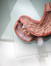
WASHINGTON – Endoscopically placed devices that include intragastric balloons and gastrointestinal liners are being evaluated for the treatment of obesity and could eventually fill some of the current void in treatment options that fall between lifestyle changes and surgical treatments.
These types of devices are the new paradigm for obesity treatments, and many are already available in Europe and elsewhere, Dr. Richard Rothstein said at a public workshop cosponsored by the Food and Drug Administration and the American Gastroenterological Association. Some have been evaluated in studies that use diabetes control measures as the main outcome endpoint, rather than weight loss, he noted.
Dr. Rothstein, the Joseph M. Huber Professor of Medicine and chair of the department of medicine, Geisel School of Medicine at Dartmouth, Hanover, N.H., provided an overview of some of the devices, which include sleeves, techniques, and devices used to reduce gastric volume such as balloons, and surgical tools used to sew or staple the stomach together.
Endoscopic treatments for obesity are less invasive, less expensive, reversible, and are performed as outpatient procedures, he said, noting that patients may be willing to pay for these procedures out of pocket.
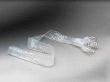
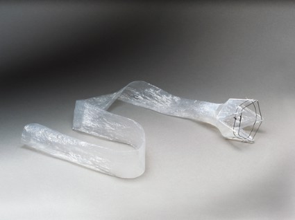
In early studies, the endoscopically delivered EndoBarrier resulted in weight loss and was well tolerated, said Dr. Rothstein, who is an adviser to the manufacturer, GI Dynamics. This is a flexible, tube-shaped liner that forms a physical barrier between food and the intestinal wall, according to the manufacturer. The device, which mimics malabsorptive surgical procedures, has been available for 3 years in Europe, Australia, and South America and is being evaluated in a pivotal U.S. study, the ENDO trial, for treating patients who are obese and have uncontrolled type 2 diabetes.
Andy Levine, who was chief technology officer at GI Dynamics and is now an adviser to the company, said that to date, the EndoBarrier, which completely covers the duodenum and the proximal jejunum and is anchored in the duodenal bulb distal to the pylorus, has been placed in more than 1,300 people worldwide. The device creates a "a complete bypass," so the food from the stomach goes through the flexible soft liner, driven by peristalsis, and the bile and pancreatic enzymes pass on the outside of the liner, with all mixing in the bowel, said Mr. Levine, who is now the leader of business development for medical devices and robotics at the Wyss Institute for Biologically Inspired Engineering at Harvard University, Boston.
In studies, improvements in type 2 diabetes measures are being used as endpoints, since showing improvement in obesity comorbidities was determined to be important for reimbursement, "and the technology had a unique impact on diabetes."
Based on experience with the device to date, Mr. Levine said that for the first 9-10 months, patients lose about 10%-20% of their body weight and many have been able to discontinue insulin and stay on oral therapy. Following removal of the device at 1 year, there is a slow rebound in weight and hemoglobin A1c values. While this might appear to be a temporary solution for a chronic problem, "we’re certainly treating it, and in some ways may be reversing the disease at least for awhile," he said, pointing out that the device addresses the need to break the cycle of continuing weight gain and helps patients discontinue diabetes medications, especially insulin.
A variety of newer, improved versions of endoscopically placed balloons are also being studied, including the ReShape, a double balloon device, Dr. Rothstein said.
During the same session, Richard Thompson, president and CEO of ReShape Medical, a small venture capital–funded company, said that the ReShape Duo intragastric device is made up of two balloons that contain a total of about 900 mL of fluid, which occupy space in the stomach without the discomfort and distension that can occur with one large balloon. This device is designed to reduce the risk of migration, which can occurs when a balloon deflates and migrates to the intestine where it can cause a blockage. If one balloon deflates the other remains inflated, and because there is blue dye in the balloons, when one deflates, the dye is visible in urine to alert the patient, he noted. ReShape Duo has been available in Europe for more than 2 years.
The U.S. trial evaluating the device in 325 patients is almost completed, and the company recently announced that the primary endpoints that assessed weight loss and responder rates had been met. The company plans to file for FDA approval in the second quarter of 2014.
Dr. Rothstein said that other nonsurgical devices being developed include those that do not require endoscopic placement, such as bezoars or balloons that are swallowed by the patient and function as temporary "space-occupying devices," he said. One example is a capsule that contains beads, which swell up in the stomach and take up space after the capsule is swallowed – which may affect satiety and result in weight loss if ingested before a meal, he said.
Techniques that bring aspects of the proximal stomach together represent another approach, using devices to sew and stitch up the stomach, compartmentalizing the stomach, creating "if you will, an endoscopic gastric sleeve," he said, noting there are not much data available on this approach.
Data from studies that are sham-controlled and have adequate sample sizes are needed to determine the role of these therapies, as they are developed, and they need to be shown to have "reasonable durability," and a safety profile with minimal complications, with cost-effectiveness data, Dr. Rothstein said. And as they become available, it will be necessary to "tease out" which patient groups respond best to which therapy, such as balloons or bezoars, or gastric bypass surgery, he added.
AT AN FDA/AGA WORKSHOP
Obesity as a disease: Implications for treatment and reimbursement
WASHINGTON – Despite the recent attention given to considering obesity a disease, there are still many limitations to providing treatments for obesity and receiving adequate reimbursement for those treatments, Dr. Lee Kaplan said at a public workshop cosponsored by the Food and Drug Administration and the American Gastroenterological Association.
The American Medical Association resolution to recognize obesity as a disease in 2013 has no legal authority, and therefore does not represent as much progress as suggested by the media attention surrounding the announcement, said Dr. Kaplan, director of the Massachusetts General Hospital Weight Center, Boston.
The AMA resolution was preceded by the Internal Revenue Service’s determination that obesity could qualify as a medical deduction in 2007, and in 2008, the Obesity Society provided support for considering obesity as a medical disorder.
But despite these apparent signs of progress, limitations remain. Weight loss drugs are excluded under Medicare Part D coverage, and while Medicare has started coverage for nutritional counseling, only services provided in a primary care setting are covered, he pointed out. An estimated 1 of every 400 people with obesity in the United States has bariatric surgery every year, but 1 in 100-150 of every 400 meets the criteria for surgery, which "doesn’t sound like we are addressing the problem very seriously," he said.
One problem is that obesity, which has many causes, is lumped together "into one big group," raising the issue of whether treating obesity would require an investment that is so huge, "that even as a society we cannot make it."
A critical issue is how to define obesity, Dr. Kaplan said. A simple definition is excess fat accumulation that presents a risk to health, but this definition does not include ways of measuring excess fat. One measurement, body mass index, is a marker, not a definition of obesity.
A major barrier is the development of an effective therapy, he said, noting that other than gastric bands, there are no devices on the market to treat obesity, and the four drugs approved by the FDA have modest efficacy overall and are not widely used. Another major barrier is "provider utilization," getting the provider to prescribe treatments and obtain reimbursement for the costs of treatment.
"There is also a large gap between the relatively noninvasive treatments and the invasive, more effective therapies, which needs to be filled by midlevel therapies," he said. Those therapies should be gastrointestinal therapies almost exclusively, he added. The current treatment strategy is fairly simplistic: Patients are asked to change their lifestyle and when that does not work, the next step is to see a dietician, psychologist, or another professional to help with their lifestyle changes, followed by the addition of medication if that does not work. Surgery is broached in a small number of patients.
"Obesity is a disease whether we call it one or not, because it behaves like a disease," and there are implications to viewing it as a disease, he said. Therapy should be physiologically based, and the heterogeneity in the underlying causes of obesity, which explains the variable treatment responses, provides an opportunity to benefit selected subpopulations.
Considering the number of disorders that are associated with obesity, it is not surprising that treatment responses vary widely, Dr. Kaplan said. People with one defect may do better than others with gastric bypass, and some people may respond better than others to the approved combination of phentermine and topiramate (Qsymia) – as reflected in the ranges of response rates associated with these therapies, he pointed out.
The average excess weight loss with gastric bypass, the most effective therapy for obesity, is about 65%, but it can be as low as 20% in some people. With banding, some patients lose all or most of their excess weight, but most lose about 25%-30%, he said. Similarly, Dr. Kaplan found some patients responded well to treatment with sibutramine (which is no longer marketed), while others did not respond well; the same pattern was seen with lorcaserin.
"Whether you’re talking about a drug, a device, surgery, or lifestyle modifications, ... you see this very broad response," because there are subtypes of obesity, he said. Identifying the subgroups of patients that are highly responsive to any of these treatments could increase the success rate of the treatment, he noted.
In December 2013, the AGA's Center for Gastrointestinal Innovation and Technology (CGIT), along with the Food and Drug Administration, held a workshop on "Changing Regulatory and Reimbursement Paradigms for Medical Devices in the Treatment of Metabolic Diseases: How to Estimate and Reward True Patient-Centric Value."

|
| Dr. Jay Pasricha |
The theme of the workshop was broad, but obesity and reflux were chosen as representative conditions that highlighted the challenges that companies and innovators face. The FDA showcased some truly innovative GI devices. These initiatives were praised by the other groups, and it is hoped the workshop will inspire similar introspection into coverage policies as well.
It also became clear at the workshop that continuing with current regulatory and reimbursement procedures may result in the throttling of medical innovation. Therefore, taking ownership of the reform process is a task that all parties need to embrace.
The workshop was very successful in bringing together representatives from all walks, and members in the audience enthusiastically joined vigorous discussions by the panel. As a follow-up, the CGIT and FDA are working on a white paper that will lay out a road map for much needed changes.
Dr. Jay Pasricha is director of the Center for Neurogastroenterology and professor (PAR) of medicine at Johns Hopkins University, Baltimore. He has no relevant conflicts of interest.
In December 2013, the AGA's Center for Gastrointestinal Innovation and Technology (CGIT), along with the Food and Drug Administration, held a workshop on "Changing Regulatory and Reimbursement Paradigms for Medical Devices in the Treatment of Metabolic Diseases: How to Estimate and Reward True Patient-Centric Value."

|
| Dr. Jay Pasricha |
The theme of the workshop was broad, but obesity and reflux were chosen as representative conditions that highlighted the challenges that companies and innovators face. The FDA showcased some truly innovative GI devices. These initiatives were praised by the other groups, and it is hoped the workshop will inspire similar introspection into coverage policies as well.
It also became clear at the workshop that continuing with current regulatory and reimbursement procedures may result in the throttling of medical innovation. Therefore, taking ownership of the reform process is a task that all parties need to embrace.
The workshop was very successful in bringing together representatives from all walks, and members in the audience enthusiastically joined vigorous discussions by the panel. As a follow-up, the CGIT and FDA are working on a white paper that will lay out a road map for much needed changes.
Dr. Jay Pasricha is director of the Center for Neurogastroenterology and professor (PAR) of medicine at Johns Hopkins University, Baltimore. He has no relevant conflicts of interest.
In December 2013, the AGA's Center for Gastrointestinal Innovation and Technology (CGIT), along with the Food and Drug Administration, held a workshop on "Changing Regulatory and Reimbursement Paradigms for Medical Devices in the Treatment of Metabolic Diseases: How to Estimate and Reward True Patient-Centric Value."

|
| Dr. Jay Pasricha |
The theme of the workshop was broad, but obesity and reflux were chosen as representative conditions that highlighted the challenges that companies and innovators face. The FDA showcased some truly innovative GI devices. These initiatives were praised by the other groups, and it is hoped the workshop will inspire similar introspection into coverage policies as well.
It also became clear at the workshop that continuing with current regulatory and reimbursement procedures may result in the throttling of medical innovation. Therefore, taking ownership of the reform process is a task that all parties need to embrace.
The workshop was very successful in bringing together representatives from all walks, and members in the audience enthusiastically joined vigorous discussions by the panel. As a follow-up, the CGIT and FDA are working on a white paper that will lay out a road map for much needed changes.
Dr. Jay Pasricha is director of the Center for Neurogastroenterology and professor (PAR) of medicine at Johns Hopkins University, Baltimore. He has no relevant conflicts of interest.
WASHINGTON – Despite the recent attention given to considering obesity a disease, there are still many limitations to providing treatments for obesity and receiving adequate reimbursement for those treatments, Dr. Lee Kaplan said at a public workshop cosponsored by the Food and Drug Administration and the American Gastroenterological Association.
The American Medical Association resolution to recognize obesity as a disease in 2013 has no legal authority, and therefore does not represent as much progress as suggested by the media attention surrounding the announcement, said Dr. Kaplan, director of the Massachusetts General Hospital Weight Center, Boston.
The AMA resolution was preceded by the Internal Revenue Service’s determination that obesity could qualify as a medical deduction in 2007, and in 2008, the Obesity Society provided support for considering obesity as a medical disorder.
But despite these apparent signs of progress, limitations remain. Weight loss drugs are excluded under Medicare Part D coverage, and while Medicare has started coverage for nutritional counseling, only services provided in a primary care setting are covered, he pointed out. An estimated 1 of every 400 people with obesity in the United States has bariatric surgery every year, but 1 in 100-150 of every 400 meets the criteria for surgery, which "doesn’t sound like we are addressing the problem very seriously," he said.
One problem is that obesity, which has many causes, is lumped together "into one big group," raising the issue of whether treating obesity would require an investment that is so huge, "that even as a society we cannot make it."
A critical issue is how to define obesity, Dr. Kaplan said. A simple definition is excess fat accumulation that presents a risk to health, but this definition does not include ways of measuring excess fat. One measurement, body mass index, is a marker, not a definition of obesity.
A major barrier is the development of an effective therapy, he said, noting that other than gastric bands, there are no devices on the market to treat obesity, and the four drugs approved by the FDA have modest efficacy overall and are not widely used. Another major barrier is "provider utilization," getting the provider to prescribe treatments and obtain reimbursement for the costs of treatment.
"There is also a large gap between the relatively noninvasive treatments and the invasive, more effective therapies, which needs to be filled by midlevel therapies," he said. Those therapies should be gastrointestinal therapies almost exclusively, he added. The current treatment strategy is fairly simplistic: Patients are asked to change their lifestyle and when that does not work, the next step is to see a dietician, psychologist, or another professional to help with their lifestyle changes, followed by the addition of medication if that does not work. Surgery is broached in a small number of patients.
"Obesity is a disease whether we call it one or not, because it behaves like a disease," and there are implications to viewing it as a disease, he said. Therapy should be physiologically based, and the heterogeneity in the underlying causes of obesity, which explains the variable treatment responses, provides an opportunity to benefit selected subpopulations.
Considering the number of disorders that are associated with obesity, it is not surprising that treatment responses vary widely, Dr. Kaplan said. People with one defect may do better than others with gastric bypass, and some people may respond better than others to the approved combination of phentermine and topiramate (Qsymia) – as reflected in the ranges of response rates associated with these therapies, he pointed out.
The average excess weight loss with gastric bypass, the most effective therapy for obesity, is about 65%, but it can be as low as 20% in some people. With banding, some patients lose all or most of their excess weight, but most lose about 25%-30%, he said. Similarly, Dr. Kaplan found some patients responded well to treatment with sibutramine (which is no longer marketed), while others did not respond well; the same pattern was seen with lorcaserin.
"Whether you’re talking about a drug, a device, surgery, or lifestyle modifications, ... you see this very broad response," because there are subtypes of obesity, he said. Identifying the subgroups of patients that are highly responsive to any of these treatments could increase the success rate of the treatment, he noted.
AT AN FDA/AGA WORKSHOP
Despite benefits, ‘bundling’ endoscopy is not norm
In more than one-third of cases, same-day bidirectional endoscopy is not performed in Medicare beneficiaries, despite the fact that so-called "bundled" endoscopies offer significant cost savings to Medicare, wrote Dr. Hashem B. El-Serag and colleagues in the January issue of Clinical Gastroenterology and Hepatology (doi:10.1016/j.cgh.2013.07.021).
Indeed, "Although in some instances the referral patterns and clinical indications may have precluded bundling, the persistence of the findings in analyses that adjusted for indications and the considerable geographic variation in practice suggest a component of physician discretion," they wrote.
Dr. El-Serag of Baylor College of Medicine, Houston, and colleagues looked at data from 12,982 Medicare beneficiaries with claims for colonoscopy and EGD within 180 days of each other captured by the Surveillance Epidemiology and End Results (SEER) Program.
Overall, 8,404 of these patients (64.7%) had "bundled" procedures, meaning that both upper and lower endoscopy occurred on the same day during the same session.
On the other hand, 2,359 patients (18.2%) did not have their procedures bundled, but rather had both procedures within 30 days of each other.
And an additional 2,219 patients (17.1%) underwent the procedures at an even greater interval, between 30 and 180 days of each other.
"Patients with bundled procedures were slightly younger, more likely to be white, more likely to reside in an urban area with a higher median educational level, and more likely to have low comorbidity scores," wrote the authors.
However, there was also a "strong and significant" geographic component to the frequency of bundling, whereby patients undergoing procedures in the Northeastern United States had the lowest rates, while patients in the Western portion of the country had the highest rates.
They also found that patients with GI bleeding were significantly more likely to have bundled procedures compared with patients undergoing screening or surveillance.
"This association between indications and bundling status persisted in a subgroup analysis of patients with a comorbidity score of 2 or greater (n = 2,961)," they wrote.
In an attempt to explain their findings, the researchers postulated that since Medicare reimburses bundled procedures at a rate that is less than the sum of each charged separately, physicians have a financial disincentive to bundle procedures.
To that end, "it is worth considering that insurers reimburse physicians fully for bundled procedures," they wrote, pending a formal cost-effectiveness analysis.
They conceded, however, that the data used in this study were collected primarily for billing purposes, such that clinical details – for example, procedural findings – were lacking.
"For example, in an open-access endoscopy system a patient may have been referred for only one procedure, and, based on the outcome, a second procedure may have been requested at a later time," they explained.
Nevertheless, "The missed opportunities related to nonbundled EGD and colonoscopy are likely to be associated with considerable increase in cost related to physician (gastroenterology, anesthesia, pathology) and facility fees," concluded the authors, as well as indirect costs of work days lost.
Dr. El-Serag and his colleagues stated that they had no conflicts of interest. They disclosed grants from the National Institute of Diabetes and Digestive and Kidney Diseases, the National Institutes of Health/National Cancer Institute, the Houston VA Health Services Research & Development Center of Excellence, and the Texas Digestive Disease Center National Institutes of Health, as well as the National Center for Advancing Translational Sciences.
Major finding: Of more than 12,000 Medicare patients who underwent upper and lower endoscopy, fewer than two-thirds had the procedures "bundled" on the same day, despite significant cost savings for Medicare and convenience for patients.
Data source: A total of 12,982 Medicare patients in the Surveillance Epidemiology and End Results (SEER) Program.
Disclosures: Dr. El-Serag and his colleagues stated that they had no conflicts of interest. They disclosed grants from the National Institute of Diabetes and Digestive and Kidney Diseases, the National Institutes of Health/National Cancer Institute, the Houston VA Health Services Research & Development Center of Excellence, and the Texas Digestive Disease Center National Institutes of Health, as well as the National Center for Advancing Translational Sciences.
Gastric bypass induces diabetes remission in obese patients
WASHINGTON – Gastric bypass surgery resulted in significantly more weight loss and also improved measures of glycemic control significantly more than did other forms of bariatric surgery, Dr. John Morton reported at the annual clinical congress of the American College of Surgeons.
However, he noted, while remission did correlate with weight loss in patients who had gastric banding or sleeving, it appeared to be independent of weight loss in those who had the bypass surgery. While he didn’t speculate on the reasons for this finding, he did affirm his belief that gastric bypass is the best option for most obese patients with comorbid diabetes.
"I feel very comfortable recommending it" for these patients, said Dr. Morton of the Stanford (Calif.) University. "There are, of course, other clinical conditions to consider when deciding [among] bypass, banding, and sleeve, but if the only consideration is around diabetes, I’m 100% comfortable in recommending it for obese diabetics."
He presented the 1-year follow-up data on 1,792 obese patients who underwent bariatric surgery. Of these, 1,364 had a Roux-en-Y bypass; 264 had a sleeve gastrectomy; and 164 had adjustable gastric banding.
The patients were a mean of about 46 years old. Body mass index was statistically, but not clinically, different between the groups (bypass 47 kg/m2; band 44 kg/m2; sleeve 44 kg/m2). Waist circumference ranged from 51 to 53 inches. About 75% of the group was female and more than half, white.
Overall, about one-third of each group had type 2 diabetes. Most were taking only oral medications. About 5% took only insulin, and about a quarter took both oral agents and insulin. At baseline, the mean HbA1c was more than 7% in each group. The mean fasting insulin was 36 microU/mL in the bypass group, 28 microU/mL in the band group, and 32 microU/mL in the sleeve group.
By 12 months after surgery, patients with diabetes who had bypass had lost the most weight – a mean of 71% of their excess body weight, compared with 38% in the banding group and 50% in the sleeve group.
Those who had the bypass surgery also experienced the biggest change in their HbA1c – dropping almost 16% to a mean of 5.8%. Patients in the other two groups experienced a mean drop of 10%, resulting in HbA1c levels of right around 6%.
Fasting insulin levels also improved significantly more in the bypass group, falling from a baseline mean of 56 microU/mL to 7.8 microU/mL – a decrease of 68%. In the band group, the level fell from 28 microU/mL to 12 microU/mL – a 52% decrease. In the sleeve group, levels fell from 32 microU/mL to 10 microU/mL – a 61% decrease.
"Fasting insulin is also considered an independent marker of cardiac risk," Dr. Morton added, indicating that the risk of cardiovascular problems would fall along with insulin levels.
Blood glucose improved significantly more in the bypass group, falling from a baseline mean of 149 mg/dL to 101 mg/dL – a 22% change. In the band group, the level fell from 140 mg/dL to 125 mg/dL (8%) and in the sleeve group, from 130 mg/dL to 118 mg/dL (5.6%).
A multivariate analysis controlled for surgery type, sex, body mass index, race, age, and insurance status. Of these factors, surgery type was the strongest predictor, with bypass patients three times more likely to achieve that goal than those undergoing banding or sleeve placement.
Dr. Morton did not present his complication data. However, during the discussion period, he said the three procedures were similarly safe. The gastric sleeve group had a higher leak rate than did the other groups, but that remained less than 1%. Readmission rates were comparable, he said, but he did not provide that number.
"Any time you consider this, it has to be a risk/benefit analysis," he said. "It’s our philosophy that for obese patients with severe diabetes, we approach them first with the bypass because it has the most proven track record over time."
Dr. Morton has up to 7 years of data on some of the patients, and said he is now analyzing that. But when questioned about durability, he agreed that diabetes can recur in the rather common scenario of a patient regaining weight. "At the end of the day, though, what’s important is that the obese patient with diabetes gets treatment. All three surgeries demonstrated some improvement, and I believe that any surgery is better than no surgery at all."
Dr. Morton has received research support from Covidien.
AT THE ACS CLINICAL CONGRESS
Major finding: Obese patients with diabetes lost significantly more weight and had significantly better glycemic control after gastric bypass than after gastric banding or sleeve gastrectomy.
Data source: The 1-year follow-up study contained data on 1,792 patients.
Disclosures: Dr. Morton has received research support from Covidien.
Medicare drops certification requirement for bariatric surgery
Medicare is dropping its requirement that bariatric surgery facilities be certified.
In a controversial move, officials at the Centers for Medicare and Medicaid Services (CMS) announced Sept. 24 that the evidence is sufficient to conclude that certification does not improve health outcomes for Medicare beneficiaries. As a result, the agency will no longer make certification a condition of Medicare coverage.
The decision reverses the agency’s February 2006 requirements. Since then, Medicare has covered bariatric procedures only when performed at facilities that were either certified by the American College of Surgeons (ACS) as a Level 1 Bariatric Surgery Center or certified by the American Society for Metabolic and Bariatric Surgery (ASMBS) as a Bariatric Surgery Center of Excellence.
In their announcement, CMS officials said they were leaning in this direction in June when they proposed to lift the certification requirement and asked for public comments.
The response overwhelmingly supported certification. Of the 483 comments received, only 92 favored eliminating the certification requirement.
The change was opposed by physician groups including the ASMBS and the ACS, which operate the certification programs referenced in the previous CMS coverage policy. The groups warned the CMS that dropping the certification requirement would put the safety of vulnerable Medicare patients at risk.
Dr. Jaime Ponce, ASMBS President, said in a statement that he was "disappointed" in the Medicare decision but encouraged that private insurers such as Blue Cross Blue Shield, Aetna, Cigna, and Optum/United Healthcare continue to support accreditation.
The CMS agreed that there is a role for accreditation programs going forward, but said that they are not necessary to ensure safe outcomes for Medicare beneficiaries.
"The removal of a coverage requirement does not require facilities to discontinue practices which they find beneficial," according to the decision memo.
Facilities may choose to continue with certification in order to distinguish themselves from the competition, for instance.
"While CMS agrees with the value of the multidisciplinary team approach and structure, we do not believe that every valued endeavor needs to be buttressed by a Medicare mandate," the memo states. "We expect all facilities to strive to provide the proper equipment and services to meet the needs of its patient population."
CMS officials reviewed nine studies to determine if certification meaningfully improved health outcomes for Medicare beneficiaries. The results were "mixed," the agency said, but overall the evidence showed "no consistent statistical or clinically meaningful difference." Further, nothing in the literature suggested a worsening of outcomes without certification.
The factors that led to the original certification requirements – the rapid growth in bariatric procedures and concerns about higher mortality rates – have changed, the CMS wrote.
The policy switch was requested by health services researchers at the University of Michigan led by Dr. John D. Birkmeyer, professor of surgery and director of the Center for Healthcare Outcomes and Policy at the University of Michigan, Ann Arbor. The scientists asserted that certified facilities were no safer than noncertified ones and that mortality and serious complication rates for bariatric surgery had declined across the country.
The CMS coverage decision did not make changes to the bariatric procedures covered by the agency. Medicare will continue to cover open and laparoscopic Roux-en-Y gastric bypass; laparoscopic adjustable gastric banding; and open and laparoscopic biliopancreatic diversion with duodenal switch for Medicare beneficiaries with a body mass index of 35 kg/m2 or greater in those with at least one comorbidity related to obesity who previously have been unsuccessful with medical treatment for obesity.
Bariatric surgery doesn’t cut health care costs
Bariatric surgery doesn’t reduce long-term health care costs, according to a report in the June issue of JAMA Surgery.
In a 6-year follow-up study comparing nearly 30,000 patients who underwent bariatric surgery against the same number of well-matched patients who did not have the surgery, the surgery group showed decreases in costs for office visits and prescriptions, but these were offset by their significant increases in inpatient costs, said Jonathan P. Weiner, Dr.P.H., of the department of health policy and management, Johns Hopkins Bloomberg School of Public Health, Baltimore, and his associates.
Health care costs for the bariatric surgery patients did fall off somewhat during the fourth, fifth, and sixth years following the operation, but not enough to outweigh the expenses incurred during the first 3 postoperative years, the investigators said.
Taken together with the results of two other recent studies of the total health care costs related to bariatric surgery, these findings indicate that "to assess the value of bariatric surgery, future studies should focus on the potential benefit of improved health and well-being of persons undergoing the procedure rather than on cost savings," they noted.
It seems intuitive that bariatric surgery, which produces considerable weight loss and alleviates or eliminates many obesity-related disorders such as hypertension and diabetes, would of course save future health care costs, at least in the long term. But studies of the impact of the procedure on such costs have shown mixed results.
"Many uncertainties remain about whether and when a return on investment can be expected, which type of bariatric surgical procedure produces the greatest cost reduction, and whether cost reductions are sustained over time," Dr. Weiner and his colleagues said.
To clarify the issue, they analyzed health care costs in a large cohort of privately insured people covered by seven Blue Cross Blue Shield plans in seven states, of which 29,820 underwent a variety of bariatric procedures during a 6-year period. Each of these subjects was matched for age, sex, area of residence, and 33 markers of obesity with a control subject who did not have bariatric surgery.
Surgical trends shifted during the course of the study. At the beginning, 72% of the procedures were open gastric bypass operations; by the end, laparoscopic procedures were predominant.
Although laparoscopic operations were associated with lower costs in the short term than other procedures, this advantage lasted only for a brief period. So overall, health care costs were not significantly different by type of bariatric surgery.
In an unadjusted analysis of the data, the surgical group showed an approximately 30% decrease in pharmacy costs during the first 3 years after the procedure. The control group showed no such drop.
However, the surgical group had significantly more hospital admissions for GI-related diagnoses than the control group during all 6 years of follow-up, which were concentrated during the second and third years following the procedure. Most of these admissions likely were for surgery-related complications, the investigators said.
In an analysis that adjusted for multiple possible confounders, inpatient costs remained higher for the surgical group than the control group throughout follow-up, but particularly during year 2 and year 3 following the procedure. Pharmacy and office visit costs were significantly lower for the surgery group but did not offset the excess in inpatient costs, Dr. Weiner and his associates reported (JAMA Surg. 2013;148:555-61).
This study "adds substantially to the existing literature on cost of bariatric surgery" because the study population was the largest to date and was representative of a broad cross section of the commercially insured U.S. population. The study also boasts one of the longest follow-ups of bariatric surgery outcomes in the United States, since previous studies generally had follow-ups of only 6 months to 2 years, the researchers said.
In a Clinical Review & Education piece accompanying this report (JAMA Surg. 2013;310:742-3), Matthew L. Maciejewski, Ph.D., and Dr. David E. Arterburn said that bariatric surgery may still be cost-effective even if it is not cost-saving.
"Does bariatric surgery need to be cost-effective (i.e., more effective but more costly than usual care), or does it need to achieve the higher standard of cost savings (i.e., more effective and less costly than usual care) to justify broader insurance coverage?" they asked.
The procedures are so expensive that they are unlikely to meet the threshold of "cost saving" for most patients. Even if a patient’s total health care costs are cut by half after the surgery, "it may take up to 20 years to achieve cost neutrality," said Dr. Maciejewski of the Center for Health Services Research in Primary Care, Durham (N.C.) VA Medical Center, and Dr. Arterburn of the Group Health Research Institute, Seattle.
This lack of cost savings shouldn’t be surprising, given that Roux-en-Y gastric bypass and laparoscopic adjustable gastric banding are invasive procedures that carry high early complication rates. It is possible that newer procedures such as sleeve gastrectomy might eventually yield cost savings because of relatively low complication rates, but these operations haven’t yet undergone long-term economic evaluation, they added.
This study was supported in part by Ethicon Endo-Surgery (a division of Johnson & Johnson), Pfizer, and GlaxoSmithKline, as well as by the National BlueCross BlueShield Association and the seven local plans that participated. No other financial conflicts of interest were reported.
"Bariatric surgery has dramatic short-term results, but on a population level its outcomes are far less impressive," said Dr. Edward H. Livingston.
The study by Weiner et al. adds to "the accumulating evidence" that weight-loss surgery carries no particular economic benefit. "Coupled with findings that bariatric surgery confers little to no long-term survival benefit, these observations show that bariatric surgery does not provide an overall societal benefit," he noted.
"In other words, the indications for bariatric surgery should be viewed in terms of individual patient benefit without anticipating that there will be cost savings to a health care system offering this treatment," Dr. Livingston said.
Dr. Edward H. Livingston is with the JAMA Network in Chicago. He is deputy editor of JAMA. These remarks were taken from his Invited Critique accompanying Dr. Weiner’s report (JAMA Surg. 2013;148:561).
Bariatric surgery doesn’t reduce long-term health care costs, according to a report in the June issue of JAMA Surgery.
In a 6-year follow-up study comparing nearly 30,000 patients who underwent bariatric surgery against the same number of well-matched patients who did not have the surgery, the surgery group showed decreases in costs for office visits and prescriptions, but these were offset by their significant increases in inpatient costs, said Jonathan P. Weiner, Dr.P.H., of the department of health policy and management, Johns Hopkins Bloomberg School of Public Health, Baltimore, and his associates.
Health care costs for the bariatric surgery patients did fall off somewhat during the fourth, fifth, and sixth years following the operation, but not enough to outweigh the expenses incurred during the first 3 postoperative years, the investigators said.
Taken together with the results of two other recent studies of the total health care costs related to bariatric surgery, these findings indicate that "to assess the value of bariatric surgery, future studies should focus on the potential benefit of improved health and well-being of persons undergoing the procedure rather than on cost savings," they noted.
It seems intuitive that bariatric surgery, which produces considerable weight loss and alleviates or eliminates many obesity-related disorders such as hypertension and diabetes, would of course save future health care costs, at least in the long term. But studies of the impact of the procedure on such costs have shown mixed results.
"Many uncertainties remain about whether and when a return on investment can be expected, which type of bariatric surgical procedure produces the greatest cost reduction, and whether cost reductions are sustained over time," Dr. Weiner and his colleagues said.
To clarify the issue, they analyzed health care costs in a large cohort of privately insured people covered by seven Blue Cross Blue Shield plans in seven states, of which 29,820 underwent a variety of bariatric procedures during a 6-year period. Each of these subjects was matched for age, sex, area of residence, and 33 markers of obesity with a control subject who did not have bariatric surgery.
Surgical trends shifted during the course of the study. At the beginning, 72% of the procedures were open gastric bypass operations; by the end, laparoscopic procedures were predominant.
Although laparoscopic operations were associated with lower costs in the short term than other procedures, this advantage lasted only for a brief period. So overall, health care costs were not significantly different by type of bariatric surgery.
In an unadjusted analysis of the data, the surgical group showed an approximately 30% decrease in pharmacy costs during the first 3 years after the procedure. The control group showed no such drop.
However, the surgical group had significantly more hospital admissions for GI-related diagnoses than the control group during all 6 years of follow-up, which were concentrated during the second and third years following the procedure. Most of these admissions likely were for surgery-related complications, the investigators said.
In an analysis that adjusted for multiple possible confounders, inpatient costs remained higher for the surgical group than the control group throughout follow-up, but particularly during year 2 and year 3 following the procedure. Pharmacy and office visit costs were significantly lower for the surgery group but did not offset the excess in inpatient costs, Dr. Weiner and his associates reported (JAMA Surg. 2013;148:555-61).
This study "adds substantially to the existing literature on cost of bariatric surgery" because the study population was the largest to date and was representative of a broad cross section of the commercially insured U.S. population. The study also boasts one of the longest follow-ups of bariatric surgery outcomes in the United States, since previous studies generally had follow-ups of only 6 months to 2 years, the researchers said.
In a Clinical Review & Education piece accompanying this report (JAMA Surg. 2013;310:742-3), Matthew L. Maciejewski, Ph.D., and Dr. David E. Arterburn said that bariatric surgery may still be cost-effective even if it is not cost-saving.
"Does bariatric surgery need to be cost-effective (i.e., more effective but more costly than usual care), or does it need to achieve the higher standard of cost savings (i.e., more effective and less costly than usual care) to justify broader insurance coverage?" they asked.
The procedures are so expensive that they are unlikely to meet the threshold of "cost saving" for most patients. Even if a patient’s total health care costs are cut by half after the surgery, "it may take up to 20 years to achieve cost neutrality," said Dr. Maciejewski of the Center for Health Services Research in Primary Care, Durham (N.C.) VA Medical Center, and Dr. Arterburn of the Group Health Research Institute, Seattle.
This lack of cost savings shouldn’t be surprising, given that Roux-en-Y gastric bypass and laparoscopic adjustable gastric banding are invasive procedures that carry high early complication rates. It is possible that newer procedures such as sleeve gastrectomy might eventually yield cost savings because of relatively low complication rates, but these operations haven’t yet undergone long-term economic evaluation, they added.
This study was supported in part by Ethicon Endo-Surgery (a division of Johnson & Johnson), Pfizer, and GlaxoSmithKline, as well as by the National BlueCross BlueShield Association and the seven local plans that participated. No other financial conflicts of interest were reported.
FROM JAMA SURGERY
Major finding: Inpatient costs were significantly higher for the surgical group than the control group, particularly during year 2 and year 3 following the procedure; pharmacy and office visit costs were significantly lower for the surgery group but did not offset the excess in inpatient costs.
Data source: A 6-year longitudinal analysis of private insurance claims data for 29,820 patients who underwent bariatric surgery and 29,820 matched nonsurgical control subjects.
Disclosures: This study was supported in part by Ethicon Endo-Surgery (a division of Johnson & Johnson), Pfizer, and GlaxoSmithKline, as well as by the National BlueCross BlueShield Association and the seven local plans that participated. No other financial conflicts of interest were reported.
Endoscopy, surgery for pancreatic pseudocysts show equal efficacy
Endoscopic cystogastrostomy was as effective as surgical cystogastrostomy for pancreatic pseudocyst drainage in a randomized trial comparing the two approaches.
None of the 20 patients randomized to undergo endoscopic treatment, and 1 of 20 patients randomized to undergo surgery, experienced pseudocyst recurrence within 24 months of follow-up, Dr. Shyam Varadarajulu of the University of Alabama at Birmingham and his colleagues reported online May 31, ahead of print in Gastroenterology.
Source: American Gastroenterological Association
Moreover, those in the endoscopy group had a shorter hospital length of stay than did the patients in the surgery group (median of 2 vs. 6 days) and a lower mean cost of care ($7,011 vs. $15,052), the investigators reported (Gastroenterology 2013 May 31 [doi: 10.1053/j.gastro.2013.05.046]).
Patients included in the study were adults with intrapancreatic or extrapancreatic pseudocysts who were enrolled between Jan. 20 and Dec. 28, 2009, following evaluation by a gastroenterologist or surgeon in an outpatient clinic or inpatient setting.
The 20 patients in the endoscopy group underwent cystogastrostomy using endoscopic ultrasound guidance and fluoroscopy while they were under conscious sedation.
"Once the pseudocyst was identified, it was accessed using a 19-gauge needle, and the gastric wall was dilated up to 15 mm using a wire-guided balloon. Two plastic stents then were deployed to facilitate the drainage of pseudocyst contents into the stomach," the investigators explained, noting that endoscopy patients were discharged following the procedure.
No procedural complications occurred in any of the 20 patients. However, one patient presented to the hospital 13 days later with persistent abdominal pain; a computed tomography scan showed a residual 7-cm pseudocyst, which was successfully treated by deployment of additional stents. At 8-week follow-up, abdominal CT scans showed pseudocyst resolution in all 20 patients.
Endoscopic retrograde cholangiopancreatography (ERCP), which was performed in all of the endoscopy patients to assess and treat any pancreatic duct leaks, was successful in 18 of the 20 patients. Magnetic resonance cholangiopancreatography (MRCP), performed in those two patients, showed a normal pancreatic duct in one and a disconnected duct in the other, the investigators said.
The 20 patients in the surgery group were all treated by the same pancreatic surgeon, who used an endovascular stapler to create at least a 6-cm cystogastrostomy after obtaining entry to the pseudocyst.
"A nasogastric tube then was left in the stomach and passed into the pseudocyst cavity to allow for intermittent irrigation until postoperative day 1 ... the nasogastric tube was removed on postoperative day 1 and clear liquids were started on day 2," they said.
Patients were discharged once a soft diet was tolerated and pain adequately controlled.
One patient with ongoing alcohol consumption developed pseudocyst recurrence at 4 months and was managed by endoscopic cystogastrostomy.
Two surgery patients experienced complications, including a wound infection treated by local debridement and antibiotics in one patient, and a case of hematemesis in one patient who was on anticoagulation and who was readmitted 9 days after discharge. "At endoscopy, a visible clot was noted at the site of surgical anastomosis, and hemostasis was achieved by application of electrocautery," the investigators said.
Two other patients were not able to tolerate oral intake postoperatively; one of them was managed conservatively, and one required surgical placement of a temporary enteral feeding tube. In addition, one patient presented at 6 months with abdominal pain and was found on ERCP to have a stricture in the pancreatic tail that required management by distal pancreatectomy.
Overall, there were no differences in the rates of treatment success, treatment failure, complications, or reinterventions between the endoscopy and surgery groups.
However, in addition to the shorter hospital stay and lower costs in the endoscopy group, patients in that group had significantly greater improvement over time in physical and mental health component scores on the Medical Outcomes Study 36-Item Short-Form General Survey. Although the scores improved for both cohorts, they were 4.48 points and 4.41 points lower, respectively, in the surgery group than the endoscopy group, the investigators said.
The findings are of note because although endoscopic drainage of pancreatic pseudocysts is increasingly performed, surgical cystogastrostomy is still considered the gold standard for treatment, as randomized trials comparing the two approaches had not previously been performed.
"The clinical relevance of this study is substantial because it shows that endoscopically managed patients can be discharged home earlier with a better health-related quality of life, and treatment can be delivered at a lower cost," the investigators said.
The authors reported having no disclosures.
There has been marked evolution in the understanding and management of acute and chronic pancreatitis over the last decade. Walled-off necroses and pseudocysts are consequences of pancreatitis that may be intrapancreatic, extrapancreatic, or both. These two entities are often confused. Fortunately, a recent international consensus has clarified that pseudocysts are liquid-filled, are almost always extrapancreatic, and rarely occur as the consequence of severe pancreatitis or involve "disconnected duct" (Gut 2013;62:102-11).

|
| Dr. Martin L. Freeman |
Dr. Varadarajulu and his colleagues are to be congratulated for performing a landmark study comparing surgery and endoscopy for internal drainage of pseudocysts (Gastroenterology 2013 May 31 [doi: 10.1053/j.gastro.2013.05.046]). They covered all the bases for an outstanding efficacy trial, including performance by experts at a tertiary center, and careful definitions of endpoints. Although the title of the paper is "Equal efficacy … [of the two approaches]," based on the primary endpoint of recurrence at 24 months, they addressed cost, hospital stay, and quality of life measures, all increasingly important in the current health care environment. In the latter regard, endoscopic ultrasound-guided cystgastrostomy emerged to be clearly superior to open surgery. If patients with more comorbidity such as portal hypertension were included, the differences would likely have been even more striking.
Thus, for pseudocysts, as for walled-off necroses, the picture is becoming increasingly clear: Minimally invasive and in particular endoscopic techniques are superior to open surgical approaches. This represents a paradigm shift in clinical practice. However, to be effective and safe in widespread applicability, it is incumbent that endoscopists attempting to manage these conditions have highly specialized expertise in pancreatic diseases and techniques, and manage these complex patients in close collaboration with their colleagues in surgery and interventional radiology.
Dr. Martin L. Freeman, FACG, FASGE, is professor of medicine at the University of Minnesota, Minneapolis. He disclosed receiving speaking honoraria from Boston Scientific and Cook, and consulting for Boston Scientific.
There has been marked evolution in the understanding and management of acute and chronic pancreatitis over the last decade. Walled-off necroses and pseudocysts are consequences of pancreatitis that may be intrapancreatic, extrapancreatic, or both. These two entities are often confused. Fortunately, a recent international consensus has clarified that pseudocysts are liquid-filled, are almost always extrapancreatic, and rarely occur as the consequence of severe pancreatitis or involve "disconnected duct" (Gut 2013;62:102-11).

|
| Dr. Martin L. Freeman |
Dr. Varadarajulu and his colleagues are to be congratulated for performing a landmark study comparing surgery and endoscopy for internal drainage of pseudocysts (Gastroenterology 2013 May 31 [doi: 10.1053/j.gastro.2013.05.046]). They covered all the bases for an outstanding efficacy trial, including performance by experts at a tertiary center, and careful definitions of endpoints. Although the title of the paper is "Equal efficacy … [of the two approaches]," based on the primary endpoint of recurrence at 24 months, they addressed cost, hospital stay, and quality of life measures, all increasingly important in the current health care environment. In the latter regard, endoscopic ultrasound-guided cystgastrostomy emerged to be clearly superior to open surgery. If patients with more comorbidity such as portal hypertension were included, the differences would likely have been even more striking.
Thus, for pseudocysts, as for walled-off necroses, the picture is becoming increasingly clear: Minimally invasive and in particular endoscopic techniques are superior to open surgical approaches. This represents a paradigm shift in clinical practice. However, to be effective and safe in widespread applicability, it is incumbent that endoscopists attempting to manage these conditions have highly specialized expertise in pancreatic diseases and techniques, and manage these complex patients in close collaboration with their colleagues in surgery and interventional radiology.
Dr. Martin L. Freeman, FACG, FASGE, is professor of medicine at the University of Minnesota, Minneapolis. He disclosed receiving speaking honoraria from Boston Scientific and Cook, and consulting for Boston Scientific.
There has been marked evolution in the understanding and management of acute and chronic pancreatitis over the last decade. Walled-off necroses and pseudocysts are consequences of pancreatitis that may be intrapancreatic, extrapancreatic, or both. These two entities are often confused. Fortunately, a recent international consensus has clarified that pseudocysts are liquid-filled, are almost always extrapancreatic, and rarely occur as the consequence of severe pancreatitis or involve "disconnected duct" (Gut 2013;62:102-11).

|
| Dr. Martin L. Freeman |
Dr. Varadarajulu and his colleagues are to be congratulated for performing a landmark study comparing surgery and endoscopy for internal drainage of pseudocysts (Gastroenterology 2013 May 31 [doi: 10.1053/j.gastro.2013.05.046]). They covered all the bases for an outstanding efficacy trial, including performance by experts at a tertiary center, and careful definitions of endpoints. Although the title of the paper is "Equal efficacy … [of the two approaches]," based on the primary endpoint of recurrence at 24 months, they addressed cost, hospital stay, and quality of life measures, all increasingly important in the current health care environment. In the latter regard, endoscopic ultrasound-guided cystgastrostomy emerged to be clearly superior to open surgery. If patients with more comorbidity such as portal hypertension were included, the differences would likely have been even more striking.
Thus, for pseudocysts, as for walled-off necroses, the picture is becoming increasingly clear: Minimally invasive and in particular endoscopic techniques are superior to open surgical approaches. This represents a paradigm shift in clinical practice. However, to be effective and safe in widespread applicability, it is incumbent that endoscopists attempting to manage these conditions have highly specialized expertise in pancreatic diseases and techniques, and manage these complex patients in close collaboration with their colleagues in surgery and interventional radiology.
Dr. Martin L. Freeman, FACG, FASGE, is professor of medicine at the University of Minnesota, Minneapolis. He disclosed receiving speaking honoraria from Boston Scientific and Cook, and consulting for Boston Scientific.
Endoscopic cystogastrostomy was as effective as surgical cystogastrostomy for pancreatic pseudocyst drainage in a randomized trial comparing the two approaches.
None of the 20 patients randomized to undergo endoscopic treatment, and 1 of 20 patients randomized to undergo surgery, experienced pseudocyst recurrence within 24 months of follow-up, Dr. Shyam Varadarajulu of the University of Alabama at Birmingham and his colleagues reported online May 31, ahead of print in Gastroenterology.
Source: American Gastroenterological Association
Moreover, those in the endoscopy group had a shorter hospital length of stay than did the patients in the surgery group (median of 2 vs. 6 days) and a lower mean cost of care ($7,011 vs. $15,052), the investigators reported (Gastroenterology 2013 May 31 [doi: 10.1053/j.gastro.2013.05.046]).
Patients included in the study were adults with intrapancreatic or extrapancreatic pseudocysts who were enrolled between Jan. 20 and Dec. 28, 2009, following evaluation by a gastroenterologist or surgeon in an outpatient clinic or inpatient setting.
The 20 patients in the endoscopy group underwent cystogastrostomy using endoscopic ultrasound guidance and fluoroscopy while they were under conscious sedation.
"Once the pseudocyst was identified, it was accessed using a 19-gauge needle, and the gastric wall was dilated up to 15 mm using a wire-guided balloon. Two plastic stents then were deployed to facilitate the drainage of pseudocyst contents into the stomach," the investigators explained, noting that endoscopy patients were discharged following the procedure.
No procedural complications occurred in any of the 20 patients. However, one patient presented to the hospital 13 days later with persistent abdominal pain; a computed tomography scan showed a residual 7-cm pseudocyst, which was successfully treated by deployment of additional stents. At 8-week follow-up, abdominal CT scans showed pseudocyst resolution in all 20 patients.
Endoscopic retrograde cholangiopancreatography (ERCP), which was performed in all of the endoscopy patients to assess and treat any pancreatic duct leaks, was successful in 18 of the 20 patients. Magnetic resonance cholangiopancreatography (MRCP), performed in those two patients, showed a normal pancreatic duct in one and a disconnected duct in the other, the investigators said.
The 20 patients in the surgery group were all treated by the same pancreatic surgeon, who used an endovascular stapler to create at least a 6-cm cystogastrostomy after obtaining entry to the pseudocyst.
"A nasogastric tube then was left in the stomach and passed into the pseudocyst cavity to allow for intermittent irrigation until postoperative day 1 ... the nasogastric tube was removed on postoperative day 1 and clear liquids were started on day 2," they said.
Patients were discharged once a soft diet was tolerated and pain adequately controlled.
One patient with ongoing alcohol consumption developed pseudocyst recurrence at 4 months and was managed by endoscopic cystogastrostomy.
Two surgery patients experienced complications, including a wound infection treated by local debridement and antibiotics in one patient, and a case of hematemesis in one patient who was on anticoagulation and who was readmitted 9 days after discharge. "At endoscopy, a visible clot was noted at the site of surgical anastomosis, and hemostasis was achieved by application of electrocautery," the investigators said.
Two other patients were not able to tolerate oral intake postoperatively; one of them was managed conservatively, and one required surgical placement of a temporary enteral feeding tube. In addition, one patient presented at 6 months with abdominal pain and was found on ERCP to have a stricture in the pancreatic tail that required management by distal pancreatectomy.
Overall, there were no differences in the rates of treatment success, treatment failure, complications, or reinterventions between the endoscopy and surgery groups.
However, in addition to the shorter hospital stay and lower costs in the endoscopy group, patients in that group had significantly greater improvement over time in physical and mental health component scores on the Medical Outcomes Study 36-Item Short-Form General Survey. Although the scores improved for both cohorts, they were 4.48 points and 4.41 points lower, respectively, in the surgery group than the endoscopy group, the investigators said.
The findings are of note because although endoscopic drainage of pancreatic pseudocysts is increasingly performed, surgical cystogastrostomy is still considered the gold standard for treatment, as randomized trials comparing the two approaches had not previously been performed.
"The clinical relevance of this study is substantial because it shows that endoscopically managed patients can be discharged home earlier with a better health-related quality of life, and treatment can be delivered at a lower cost," the investigators said.
The authors reported having no disclosures.
FROM GASTROENTEROLOGY
Major finding: Pseudocysts recurred in 0 of 20 endoscopy patients, and 1 of 20 surgery patients.
Data source: An open-label randomized trial involving 40 patients.
Disclosures: The authors reported having no disclosures.
Jack Vennes M.D. and Stephen E. Silvis M.D. Endowed Lecture: Optical biopsy
Recent advances in endoscopic imaging technology increasingly have allowed highly accurate in vivo assessment of tissue, including specific pathologic diagnoses of inflammation and neoplasia. As endoscopic imaging technology advances, we are increasingly asked whether tissue biopsy of suspicious lesions is critical or can be replaced with "optical" biopsy.
Histology will remain a key component of gastrointestinal diagnoses, but there will be important changes in how we can integrate with pathology services. These will include reduction in the number of low-yield biopsies such as random biopsies in Barrett’s esophagus, inflammatory bowel disease, and small distal non-neoplastic colorectal polyps and increasing targeting of biopsy using advanced imaging. Lastly, health care reform will increasingly drive us to reform our practices towards lower-cost, higher-value services, which include reductions in the histological evaluation of low-risk tissues. The main value of histologic analysis is the very high level of magnification, resolution, and contrast provided by standard light microscopy. Increasingly, endoscopes are capable of achieving similar levels of magnification, resolution, and contrast.
As the number of endoscopies has increased over the last few decades – over 20 million procedures are now performed annually – the cost associated with these has increased substantially. Although the cost of the endoscopy itself is an important component, the cost of ancillary services, including histology as well as anesthesia, is becoming increasingly important. This is particularly true in screening and surveillance colonoscopies, where now more than 50% of all procedures generate a histologic sample.
Advances in endoscopic optics have achieved remarkable levels of resolution and magnification over the last several decades. Important advances include the development of high-definition optical enhanced imaging and, more recently, confocal endomicroscopy. Also, inexpensive technologies such as chromoendoscopy have dramatically increased the yield of neoplasia in inflammatory bowel disease, compared with random biopsies alone. In Barrett’s esophagus, use of high-definition optical enhanced endoscopes has been shown to increase the yield of dysplasia with fewer biopsies. Confocal endomicroscopy also has been shown to be highly accurate for detection of neoplasia in Barrett’s esophagus, with overall accuracy of use exceeding 95%. A limitation of highly focused imaging technology is the small field of view that is imaged. A recent major advance is volumetric laser endomicroscopy, which provides high-resolution, high magnification imaging of large surface areas, including the entire esophagus.
Apart from advances in technology, a major impetus for adoption of optical biopsy technique will be payment reform such as bundled payments which provide payments for the endoscopy and all ancillary services such as pathology. In summary, optical biopsy with advanced endoscopic instruments now rivals the accuracy and resolution of ex vivo microscopy. The increasing cost of health care will drive reform with pressure to reduce the utilization and cost of low-value histology services. Lastly, the community of endoscopists must increasingly learn the skills to make highly accurate endomicroscopic diagnoses.
Dr. Michael B. Wallace, MPH, Professor of Medicine, Mayo Clinic, Jacksonville, Fla.
High BMI appears to cause gallstones
An elevated body mass index is not just associated with symptomatic gallstone disease, it appears to cause the disease, according to a report published in Hepatology (doi:10.1002/hep.26563).
Many epidemiologic and observational studies have noted a clear association between a high BMI and an increased risk for gallstones, but have not been able to pin down a causal effect. It was impossible to rule out confounding by some other factor, such as a high-fat diet, that simultaneously caused both the elevation in BMI and the elevation in risk for gallstones. Similarly, it was impossible to rule out reverse causation, such as the colicky pain of gallstone disease caused the physical inactivity that then led to a high BMI.
A new epidemiologic statistical approach known as Mendelian randomization is thought to avert both confounding and reverse causation by pinpointing the genetic variants that are linked to a high BMI (which are a constant throughout the lifespan) but that are not related to confounding factors, then determining whether they are also linked to gallstone disease. "If raised BMI truly is a causal factor in the development of gallstone disease, genetic variants that increase BMI would be expected to also increase risk of gallstone disease," said Dr. Stefan Stender of the department of clinical biochemistry, Rigshospitalet, University of Copenhagen, and his associates.
They studied 77,679 Danish adults from the general population who participated in two large prospective studies: the Copenhagen General Population Study (67,314 subjects) and the Copenhagen City Heart Study (10,365 subjects). All the participants had donated blood samples that could be used for DNA extraction and genotyping.
A total of 4,106 of these subjects developed symptomatic gallstone disease during follow-up of up to 34 years.
The researchers used genotyping to identify study subjects who carried any of the three polymorphisms that have the largest known effect sizes for association with BMI in European populations: FTO (rs9939609), MC4R (rs17782313), or TMEM18 (rs6548238). Each of these can be carried on two possible alleles, so any given subject could carry one to six affected alleles. The number of BMI-increasing alleles, from one to six, was determined for each study subject.
In an initial analysis of the data, the mean baseline BMI was 55% higher (11 kg/m2) in subjects carrying the most alleles compared with those carrying the fewest.
Increasing BMI was associated with a stepwise increase in the risk of developing symptomatic gallstone disease.
In the overall cohort, the risk of symptomatic gallstone disease was increased 7% for every 1-kg/m2 increase in BMI. In the Mendelian randomization cohort, the risk of gallstone disease increased 17% with every 1-kg/m2 increase in BMI. The concordance between these two estimates indicates that BMI itself is a causal risk factor for symptomatic gallstone disease, Dr. Stender and his associates said.
This study did not include data on gallstone composition and was not designed to examine the pathophysiologic mechanisms by which a high BMI causes gallstone formation. However, numerous other studies have proposed several possible mechanisms, they noted.
Obesity may raise cholesterol synthesis and hepatobiliary cholesterol efflux, "a key event in the development of cholesterol gallstones." High abdominal fat mass may induce gallbladder hypomotility and bile stasis, "another risk factor for gallstone formation."
In addition, substances secreted by or metabolized by adipocytes could influence gallstone formation. For example, estrogen is produced by adipocytes and may promote gallstone formation by raising the rate of hepatobiliary cholesterol efflux. And leptin, which is also secreted by adipocytes, may have lithogenic effects.
Adinopectin, another hormone secreted by adipocytes, has been linked to gallstones in both animal and human studies. And obesity-associated hyperinsulinemia may induce gallstone formation by causing the secretion of more lithogenic bile.
This study was limited in that it included only white people of Danish descent. "Because ethnic differences in gallstone prevalence are well known, the results reported here may not necessarily translate to other ethnicities," Dr. Stender and his colleagues said.
This study was supported by the Danish Medical Research Council, the Rigshospitalet at Copenhagen University, and the Odd Fellow Order. No financial conflicts of interest were reported.
FROM HEPATOLOGY
Major finding: In the overall cohort, the risk of symptomatic gallstone disease was increased 7% for every 1-kg/m2 increase in BMI, and in the Mendelian randomization cohort, the risk of gallstone disease increased 17% with every 1-kg/m2 increase in BMI.
Data source: A Mendelian randomization study involving 77,679 adults from the Danish general population who were genotyped to identify carriers of three BMI-increasing polymorphisms and who were followed for up to 34 years for the development of symptomatic gallstone disease.
Disclosures: This study was supported by the Danish Medical Research Council, the Rigshospitalet at Copenhagen University, and the Odd Fellow Order. No financial conflicts of interest were reported.
Medicare may scrap certification for bariatric surgery centers
Should bariatric surgery be performed in certified centers only? Medicare officials think the answer might be "no."
In a proposed decision memo issued in June, the Centers for Medicare and Medicaid Services said the evidence was sufficient to end the certification requirement and that continuing the current requirements would not improve health outcomes for Medicare beneficiaries.
While some physician experts support the proposal as improving access to bariatric surgery, others express concerns that quality and data collection efforts will suffer.
The proposal does not change which procedures are covered by Medicare. The agency will continue to cover open and laparoscopic Roux-en-Y gastric bypass; laparoscopic adjustable gastric banding; and open and laparoscopic biliopancreatic diversion with duodenal switch for Medicare beneficiaries with a body-mass index of 35 kg/m2 or greater who have at least one comorbidity related to obesity and have been unsuccessful with medical treatment for obesity.
If the proposal is accepted, it would reverse the requirements the CMS put in place in February 2006. Since then, Medicare has covered only bariatric procedures performed at facilities that were certified by the American College of Surgeons as a Level 1 Bariatric Surgery Center or by the American Society for Metabolic and Bariatric Surgery as a Bariatric Surgery Center of Excellence.
CMS officials reviewed eight studies to determine if facility certification meaningfully improved health outcomes for Medicare beneficiaries. The studies had "mixed" results, the agency said, but overall, the evidence showed "no consistent statistical or clinically meaningful difference" and there was nothing in the literature to suggest a worsening of outcomes.
The factors that led to the original certification requirements – the rapid growth in bariatric procedures and concerns about higher mortality rates – have changed.
"Since that 2006 determination, bariatric surgery has experienced a trend toward less invasive procedures and lower mortality and complication rates," according to the proposed decision memo.
The policy switch was requested by a trio of health services researchers at the University of Michigan led by Dr. John D. Birkmeyer, professor of surgery and director of the university’s Center for Healthcare Outcomes and Policy. They asserted that certified hospitals were no safer than other facilities and that mortality and serious complication rates for bariatric surgery had declined across the country.
They called on the CMS to scrap the certification requirements but to encourage bariatric surgeons to participate in registry programs through their professional societies or through payer-supported collaborative improvement programs such as the one supported by Blue Cross Blue Shield of Michigan.
The Michigan researchers suggested that the agency incentivize physicians to participate in these quality improvement activities by tying them to programs such as the Physician Quality Reporting System.
Dr. Birkmeyer pointed to a study published earlier this year showing that the current certification policy has not led to better outcomes for patients (JAMA 2013;309:792-9). In the study, which Dr. Birkmeyer coauthored, the researchers found no statistically significant improvements in complications or reoperation rates after implementation of the CMS certification requirements after accounting for patient factors, changes in procedure type, and pre-existing time trends toward improved outcomes.
"There is absolutely no controversy about the fact that [the policy] didn’t steer people to safer hospitals," Dr. Birkmeyer said.
Access could improve under the proposed decision memo, Dr. Birkmeyer said. The current certification framework makes it harder for some Medicare patients to undergo bariatric surgery without traveling hours from home. The policy may also be disproportionately affecting minorities and low-income beneficiaries, Dr. Birkmeyer said.
The American College of Surgeons and the American Society for Metabolic and Bariatric Surgery continue to support certification; however, they are working to revise their processes to create a single, unified program that will include a lower volume threshold for certification, dropping from a minimum of 125 cases a year to 50 cases a year, according to Dr. Jaime Ponce, president of the American Society for Metabolic and Bariatric Surgery and a bariatric surgeon in Chattanooga, Tenn.
The volume change should help to address some of the concerns about access by opening up certification to more programs, he said.
But Dr. Ponce said his group plans to fight the "dangerous" CMS proposal to drop certification requirements completely.
"We do believe that this could be very harmful for Medicare patients because if a patient that is very sick shows up in a community hospital that doesn’t have the right structure to take care of obese patients ... accidents are going to start happening again," he said.
The majority of the literature supports facility certification for bariatric surgery, Dr. Ponce said. As for those studies showing no difference in outcomes, the results weren’t reliable because the studies were conducted at a time when most of the facilities included were pursuing certification, he noted. Even those facilities that fell short of meeting certification requirements would have been developing a culture of safety and would likely have met many of the standards.
Dr. Ponce singled out a 2011 study in Annals of Surgery, which evaluated mortality among Medicare patients before and after the certification requirements took effect (Ann. Surg. 2011;254: 860-5). The study, which used Medicare data from 2004-2008, showed that mortality, readmission, and reoperation rates all declined after the coverage decision was implemented. The 90-day mortality rate dropped from 1.5% to 0.7%, while 90-day readmission rates decreased from 19.9% to 15.4%. The researchers also noted a decrease in payments for the procedures.
But gains in safety and cost-effectiveness could be lost if the CMS removes the certification requirements, Dr. Ponce said. Without clear requirements in place, hospitals will be unlikely to make the investments they are making today in equipment, personnel, and training, he said.
Another issue is what will happen to data collection if certification requirements are eliminated. Dr. Ponce said he’s concerned that data collection efforts would dry up without those mandates from the CMS.
"We will not have that data in order to improve the quality that we need to," he said.
A final decision on the coverage memo is expected in the fall.
