User login
Thyroid cancer rise mostly overdiagnosis
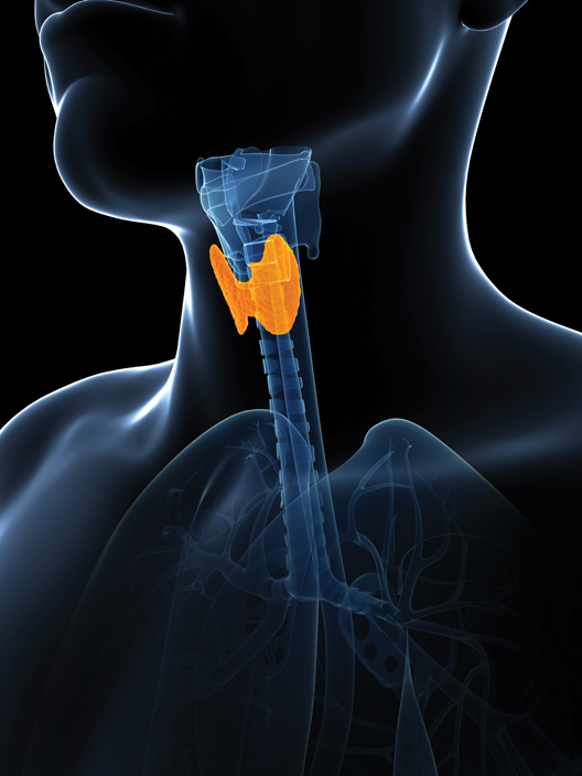
The incidence of thyroid cancer has nearly tripled in the United States since the 1970s. However, this is mainly an epidemic of diagnosis, researchers reported.
Small papillary cancers are not likely to cause death or disease, and women are four times more likely to receive a diagnosis than men, even though autopsy findings show that these cancers occur more frequently in men.
For the research, published online Feb. 20 in JAMA Otolaryngology–Head & Neck Surgery, Dr. Louise Davies and Dr. H. Gilbert Welch reviewed diagnostic trends from the population-based Surveillance, Epidemiology, and End Results (SEER) 9 program, which covers four large U.S. metropolitan areas along with five states. They also reviewed mortality records from the National Vital Statistics System between 1975 and 2009 for the same areas, reported Dr. Davies of the Veterans Affairs Medical Center in White River Junction, Vt., and Dr. Welch of the Dartmouth Institute for Health Policy and Clinical Practice in Hanover, N.H.

The researchers found that thyroid cancer incidence nearly tripled, from 4.9 to 14.3 per 100,000 individuals, in that time period (relative rate, 2.9) and that nearly all of the increase was attributable to diagnoses of small papillary cancers, the least aggressive form of thyroid cancer. The mortality rate from thyroid cancer remained stable – at 0.5 deaths per 100,000 – during the same time, Dr. Davies and Dr. Welch reported (JAMA Otolaryngol. Head Neck Surg. 2014 Feb. 20 [doi: 10.1001/jamaoto.2014.1]).
The investigators saw a much greater absolute increase in thyroid cancer in women, at 3.3-fold (from 6.5 to 21.4 cases per 100,000), than in men, at 2.2-fold (from 3.1 to 6.9), during the study period, which suggests that the burden of overdiagnosis fell heavily on women, they wrote.
Moreover, most thyroid cancers are treated "as though they are destined to cause real problems for the people who have them," Dr. Davies and Dr. Welch wrote, usually with total thyroidectomy, radiation, or both, putting patients at risk for complications and secondary cancers.
Patients – particularly women – might be better served with a less intensive diagnostic and treatment approach to these cancers, and even by relabeling them using a term other than cancer. Clinicians should take care to advise patients of the uncertainty surrounding the small papillary cancers and encourage them to consider the risks of treatment compared with active surveillance, the researchers said.
Dr. Davies and Dr. Welch received support from their institutions for their research; neither declared conflicts of interest.
This is an interesting and important study, but one that is difficult to interpret. We don't yet know which of these cancers, no matter what size, will ultimately prove to be important. Once a diagnosis of cancer is made, it is difficult for patients and doctors to simply continue to observe the cancer. Most patients and doctors are uncomfortable with that.
In addition, the follow-up itself becomes burdensome, with annual ultrasounds and, possibly, multiple needle biopsies over time. Although much of this increased incidence seems related to increased use of imaging studies, several authors have also reported an absolute increase in the incidence of thyroid cancer.
Other issues related to this topic are the extent of surgery that is necessary for these small early cancers. The authors point out that many surgeons perform total thyroidectomy and postoperative radioactive iodine ablation, but there are some who advocate for lesser surgery. This becomes problematic when patients have other smaller nodules in the opposite lobe of the thyroid of uncertain significance. Some national guidelines recommend total or near-total thyroidectomy for T1 and T2 well-differentiated thyroid cancers, and it is difficult to go against these guidelines. What is really needed are better molecular and genetic tests to better define which well-differentiated thyroid cancers are likely to act in a more aggressive manner, and which are not.
Mark C. Weissler, M.D., FACS, is the J.P. Riddle Distinguished Professor of Otolaryngology-Head and Neck Surgery at the University of North Carolina, Chapel Hill. Dr. Weissler had no disclosures.
The incidence of thyroid cancer has nearly tripled in the United States since the 1970s. However, this is mainly an epidemic of diagnosis, researchers reported.
Small papillary cancers are not likely to cause death or disease, and women are four times more likely to receive a diagnosis than men, even though autopsy findings show that these cancers occur more frequently in men.
For the research, published online Feb. 20 in JAMA Otolaryngology–Head & Neck Surgery, Dr. Louise Davies and Dr. H. Gilbert Welch reviewed diagnostic trends from the population-based Surveillance, Epidemiology, and End Results (SEER) 9 program, which covers four large U.S. metropolitan areas along with five states. They also reviewed mortality records from the National Vital Statistics System between 1975 and 2009 for the same areas, reported Dr. Davies of the Veterans Affairs Medical Center in White River Junction, Vt., and Dr. Welch of the Dartmouth Institute for Health Policy and Clinical Practice in Hanover, N.H.
The researchers found that thyroid cancer incidence nearly tripled, from 4.9 to 14.3 per 100,000 individuals, in that time period (relative rate, 2.9) and that nearly all of the increase was attributable to diagnoses of small papillary cancers, the least aggressive form of thyroid cancer. The mortality rate from thyroid cancer remained stable – at 0.5 deaths per 100,000 – during the same time, Dr. Davies and Dr. Welch reported (JAMA Otolaryngol. Head Neck Surg. 2014 Feb. 20 [doi: 10.1001/jamaoto.2014.1]).
The investigators saw a much greater absolute increase in thyroid cancer in women, at 3.3-fold (from 6.5 to 21.4 cases per 100,000), than in men, at 2.2-fold (from 3.1 to 6.9), during the study period, which suggests that the burden of overdiagnosis fell heavily on women, they wrote.
Moreover, most thyroid cancers are treated "as though they are destined to cause real problems for the people who have them," Dr. Davies and Dr. Welch wrote, usually with total thyroidectomy, radiation, or both, putting patients at risk for complications and secondary cancers.
Patients – particularly women – might be better served with a less intensive diagnostic and treatment approach to these cancers, and even by relabeling them using a term other than cancer. Clinicians should take care to advise patients of the uncertainty surrounding the small papillary cancers and encourage them to consider the risks of treatment compared with active surveillance, the researchers said.
Dr. Davies and Dr. Welch received support from their institutions for their research; neither declared conflicts of interest.
FROM JAMA OTOLARYNGOLOGY – HEAD & NECK SURGERY
Sorafenib approval now includes late-stage thyroid cancer
Sorafenib’s approval has been expanded to include late-stage differentiated thyroid cancer, the Food and Drug Administration announced on Nov. 22.
The kinase inhibitor was approved for the treatment of "locally recurrent or metastatic, progressive, differentiated thyroid carcinoma refractory to radioactive iodine treatment," according to the prescribing information.
The expedited approval –completed in 6 months – was based on a study of 417 people with locally recurrent or metastatic, progressive differentiated thyroid cancer that had not responded to radioactive iodine treatment. Subjects were randomized to 400 mg of sorafenib twice a day or placebo. The median progression-free survival was 10.8 months among those on sorafenib and 5.8 months among those on placebo, a statistically significant 41% difference.
Diarrhea, fatigue, infection, alopecia, hand-foot skin reaction, rash, weight loss, decreased appetite, nausea, gastrointestinal and abdominal pains, and hypertension were among the most common adverse effects associated with treatment, according to the FDA. In addition, thyroid stimulating hormone, which can promote thyroid cancer, "is more likely to become elevated while on treatment with Nexavar, requiring adjustment of thyroid hormone replacement therapy," the statement said.
Sorafenib, marketed as Nexavar by Bayer HealthCare Pharmaceuticals was approved for treating advanced renal cell carcinoma in 2005 and for unresectable hepatocellular carcinoma in 2007.
Delays in esophagectomy yield more postop complications
WASHINGTON – Delays in esophageal cancer surgery after a course of neoadjuvant chemotherapy and radiation were associated with more surgical complications and worse survival, based on the results of a retrospective study presented at the annual clinical congress of the American College of Surgeons.
Clinicians should focus on "prehabilitating" their patients, completing their neoadjuvant therapy and recommending esophagectomy as soon as clinically feasible, Dr. Nicholas Teman said.
Dr. Teman and his colleagues at the University of Michigan, Ann Arbor, reviewed prospectively collected data from the period of 1999-2010 on 457 patients treated at a single site. All patients underwent neoadjuvant chemotherapy and radiation with a subsequent esophagectomy; patients who underwent salvage esophagectomies were excluded from the analysis.
Outcome measures included postoperative pulmonary adverse events, anastomotic leaks, pathologic response, and mortality.
The mean time to surgery after chemotherapy and radiation was 50 days, ranging between 10 and 523 days. The most common reasons for surgical delays were patient deconditioning, noncompliance, seeking a second opinion, and complications stemming from neoadjuvant therapies.
When the time from completion of neoadjuvant therapy to surgery was analyzed as a continuous variable, there were no differences in postoperative complications and mortality. Similarly, postoperative staging and pathologic response were not significantly different.
Additionally, outcomes did not significantly differ for those who had surgery within 8 weeks of completing chemotherapy and radiation and those who had surgery after 8 weeks.
However, when time to surgery was used to place patients into quintiles of 8 weeks or less (n = 345), 9-12 weeks (n = 58), 13-16 weeks (n = 27), 17-26 weeks (n = 19), and 27 or more weeks (n = 8), there were significant differences in pulmonary complications (P = .05) and anastomotic leaks (P = .02), and a trend toward worse mortality between the quintiles (P = .09). No significant differences in pathologic response were noted in the quintiles.
Predictors of higher long-term mortality were lower pretreatment weight (P = .04), tobacco use (P = .05), higher pretreatment stage (P = .004), and failure to complete neoadjuvant treatment (P = .003).
One limitation of the study was that the neoadjuvant therapies were not standardized. A shorter time to surgery was predicted if chemotherapy included cisplatin (P = .04) or taxol (P = .001), and if there were increasing chemotherapy cycles. Chemotherapy that included 5-flourouracil was associated with longer times to surgery (P less than .001).
Dr. Teman and his associates reported no relevant disclosures.
AT THE ACS CLINICAL CONGRESS
Major finding: When time to surgery was used to place patients into quintiles of 8 weeks or less (n = 345), 9-12 weeks (n = 58), 13-16 weeks (n = 27), 17-26 weeks (n = 19), and 27 or more weeks (n = 8), there were significant differences in pulmonary complications (P = .05) and anastomotic leaks (P = .02), and a trend toward worse mortality between the quintiles (P = .09).
Data source: Retrospective review of prospective data on 457 patients treated from 1999-2010 at a single surgical site.
Disclosures: Dr. Teman and his associates reported no relevant disclosures.

Ultrasound helped reveal high-risk thyroid nodules
Thyroid nodules that include microcalcifications, are larger than 2 cm in size, and have an entirely solid composition on ultrasound imaging are the most likely to be cancerous, according to a report published online Aug. 26 in JAMA Internal Medicine.
A patient’s risk of having primary thyroid cancer ranges from 0.2% when a nodule’s ultrasound image has none of the three characteristics to 1.8% if a nodule has one of the characteristics, 6.2% if a nodule has two of the characteristics, and 96% if a nodule has all three characteristics, said Dr. Rebecca Smith-Bindman of the department of radiology and biomedical imaging, University of California, San Francisco, and her associates.
"Ours is the first study, to our knowledge, that permits estimating this risk," they noted (JAMA Intern. Med. 2013 Aug. 26 [doi:10.1001/jamainternmed.2013.9245]).
Thyroid nodules are extremely common, but almost all of them are benign, so it is crucial to identify which ones may be malignant and to biopsy them, and to avoid subjecting low-risk patients to unnecessary biopsy.

To determine which features on ultrasound correspond with cancer risk, Dr. Smith-Bindman and her colleagues performed a retrospective case-control study involving 8,806 consecutive patients who underwent 11,618 thyroid ultrasound examinations at the university from January 2000 through March 2005.
The patients who were found to have primary thyroid cancer during up to 7 years of follow-up were identified using data in a comprehensive cancer registry. Those 96 patients were matched for age, sex, and year of ultrasound examination to 369 control subjects who did not have primary thyroid cancer.
Two of the researchers who were blinded to the subjects’ cancer status independently assessed the number and size of all the thyroid nodules that were imaged in the study population and performed a detailed analysis of numerous ultrasound characteristics that might possibly correlate with the presence of malignancy. The agreement between those two reviewers in the categorization of the specific ultrasound image characteristics was "good to outstanding."
"We considered many nodule characteristics endorsed by other authors, but when put into the multiple-predictor models, most of the characteristics were not significantly associated with cancer risk," the researchers said.
In the initial univariable analysis of the data, several ultrasonographic traits were significantly associated with the likelihood that a thyroid nodule harbored cancer.
Microcalcifications had the strongest association with malignancy: They were found in 38.2% of cancerous nodules, compared with only 5.4% of benign nodules. Thus, if microcalcifications were present in a nodule, the chances were seven times greater that the nodule was cancerous, Dr. Smith-Bindman and her coworkers reported.
In the study analysis, the size of the nodule on ultrasound also correlated with cancer risk, with the odds of malignancy increasing as nodule size increased. A size of 2 cm appeared to be a good cutoff point, because nodules larger than 2 cm were much more likely than nodules smaller than 1 cm to be cancerous, with an odds ratio of 3.1.
Several other characteristics correlated with cancer risk in the analysis, but did so to a lesser degree. Coarse calcifications, solid (vs. cystic or mixed) nodule composition, hyperechoic nodule echogenicity, central vascularity, ill-defined or lobulated nodule margins, and taller-than-wide nodule shape all raised the risk of malignancy, with odds ratios ranging from 1.6 to 2.9.
Traits not associated with cancer risk included rim calcifications, comet-tail artifacts, peripheral vascularity, and the presence of a "halo," they said.
In a multivariable analysis, only three nodule characteristics remained significantly associated with cancer risk. The presence of microcalcifications had an odds ratio of 8.1, size larger than 2 cm had an odds ratio of 3.6, and an entirely solid composition had an odds ratio of 4.0, Dr. Smith-Bindman and her associates said.
The findings remained robust through a series of sensitivity analyses.
The investigators found that performing a biopsy only if two of the three characteristics were present would yield a much greater diagnostic sensitivity and specificity than the current practice of biopsying all thyroid nodules larger than 5 mm.
"Compared with existing guidelines ... adoption of this more stringent rule requiring two abnormal [ultrasound] characteristics to prompt biopsy would reduce unnecessary biopsies by 90% while maintaining a low risk of cancer [0.5%] in patients in whom biopsy is deferred," they wrote.
In contrast, requiring all three nodule characteristics to be present before performing a biopsy would detect only a small proportion of thyroid cancers, they added.
In the study, simple cysts never indicated the presence of cancer. Such thyroid cysts should be considered "essentially never malignant" and should not be biopsied, Dr. Smith-Bindman and her associates said.
"The main strength of our study is the large sample size and the linkage of the cohort with data from a comprehensive cancer registry, which allows accurate assessment of the true underlying prevalence of cancer," the researchers noted.
The overall prevalence of primary thyroid cancer was 1.5% in the study population. In contrast, most previous studies have cited a prevalence rate closer to 20%. "All previous studies have inflated the association between nodule characteristics and cancer risk because they limited their analyses to nodules that underwent biopsy," the investigators said.
The National Cancer Institute and the University of California, San Francisco, supported the study. No financial conflicts of interest were reported.
Thyroid nodules that include microcalcifications, are larger than 2 cm in size, and have an entirely solid composition on ultrasound imaging are the most likely to be cancerous, according to a report published online Aug. 26 in JAMA Internal Medicine.
A patient’s risk of having primary thyroid cancer ranges from 0.2% when a nodule’s ultrasound image has none of the three characteristics to 1.8% if a nodule has one of the characteristics, 6.2% if a nodule has two of the characteristics, and 96% if a nodule has all three characteristics, said Dr. Rebecca Smith-Bindman of the department of radiology and biomedical imaging, University of California, San Francisco, and her associates.
"Ours is the first study, to our knowledge, that permits estimating this risk," they noted (JAMA Intern. Med. 2013 Aug. 26 [doi:10.1001/jamainternmed.2013.9245]).
Thyroid nodules are extremely common, but almost all of them are benign, so it is crucial to identify which ones may be malignant and to biopsy them, and to avoid subjecting low-risk patients to unnecessary biopsy.
To determine which features on ultrasound correspond with cancer risk, Dr. Smith-Bindman and her colleagues performed a retrospective case-control study involving 8,806 consecutive patients who underwent 11,618 thyroid ultrasound examinations at the university from January 2000 through March 2005.
The patients who were found to have primary thyroid cancer during up to 7 years of follow-up were identified using data in a comprehensive cancer registry. Those 96 patients were matched for age, sex, and year of ultrasound examination to 369 control subjects who did not have primary thyroid cancer.
Two of the researchers who were blinded to the subjects’ cancer status independently assessed the number and size of all the thyroid nodules that were imaged in the study population and performed a detailed analysis of numerous ultrasound characteristics that might possibly correlate with the presence of malignancy. The agreement between those two reviewers in the categorization of the specific ultrasound image characteristics was "good to outstanding."
"We considered many nodule characteristics endorsed by other authors, but when put into the multiple-predictor models, most of the characteristics were not significantly associated with cancer risk," the researchers said.
In the initial univariable analysis of the data, several ultrasonographic traits were significantly associated with the likelihood that a thyroid nodule harbored cancer.
Microcalcifications had the strongest association with malignancy: They were found in 38.2% of cancerous nodules, compared with only 5.4% of benign nodules. Thus, if microcalcifications were present in a nodule, the chances were seven times greater that the nodule was cancerous, Dr. Smith-Bindman and her coworkers reported.
In the study analysis, the size of the nodule on ultrasound also correlated with cancer risk, with the odds of malignancy increasing as nodule size increased. A size of 2 cm appeared to be a good cutoff point, because nodules larger than 2 cm were much more likely than nodules smaller than 1 cm to be cancerous, with an odds ratio of 3.1.
Several other characteristics correlated with cancer risk in the analysis, but did so to a lesser degree. Coarse calcifications, solid (vs. cystic or mixed) nodule composition, hyperechoic nodule echogenicity, central vascularity, ill-defined or lobulated nodule margins, and taller-than-wide nodule shape all raised the risk of malignancy, with odds ratios ranging from 1.6 to 2.9.
Traits not associated with cancer risk included rim calcifications, comet-tail artifacts, peripheral vascularity, and the presence of a "halo," they said.
In a multivariable analysis, only three nodule characteristics remained significantly associated with cancer risk. The presence of microcalcifications had an odds ratio of 8.1, size larger than 2 cm had an odds ratio of 3.6, and an entirely solid composition had an odds ratio of 4.0, Dr. Smith-Bindman and her associates said.
The findings remained robust through a series of sensitivity analyses.
The investigators found that performing a biopsy only if two of the three characteristics were present would yield a much greater diagnostic sensitivity and specificity than the current practice of biopsying all thyroid nodules larger than 5 mm.
"Compared with existing guidelines ... adoption of this more stringent rule requiring two abnormal [ultrasound] characteristics to prompt biopsy would reduce unnecessary biopsies by 90% while maintaining a low risk of cancer [0.5%] in patients in whom biopsy is deferred," they wrote.
In contrast, requiring all three nodule characteristics to be present before performing a biopsy would detect only a small proportion of thyroid cancers, they added.
In the study, simple cysts never indicated the presence of cancer. Such thyroid cysts should be considered "essentially never malignant" and should not be biopsied, Dr. Smith-Bindman and her associates said.
"The main strength of our study is the large sample size and the linkage of the cohort with data from a comprehensive cancer registry, which allows accurate assessment of the true underlying prevalence of cancer," the researchers noted.
The overall prevalence of primary thyroid cancer was 1.5% in the study population. In contrast, most previous studies have cited a prevalence rate closer to 20%. "All previous studies have inflated the association between nodule characteristics and cancer risk because they limited their analyses to nodules that underwent biopsy," the investigators said.
The National Cancer Institute and the University of California, San Francisco, supported the study. No financial conflicts of interest were reported.
FROM JAMA INTERNAL MEDICINE
Major Finding: Three nodule characteristics were significantly associated with cancer risk: presence of microcalcifications (odds ratio, 8.1), size larger than 2 cm (OR, 3.6), and an entirely solid composition (OR, 4.0).
Data Source: A retrospective case-control study involving 8,806 consecutive patients who underwent 11,618 ultrasound thyroid examinations at a single medical center in a 5-year period.
Disclosures: The National Cancer Institute and the University of California, San Francisco, supported the study. No financial conflicts of interest were reported.
Poor oral health a risk factor in oncogenic HPV infection
Poor oral health is an independent risk factor of oral human papillomavirus infection regardless of smoking and oral sex practices, analysis of a large, nationally representative sample showed.
Poor oral health was associated with a 56% higher prevalence of oral HPV infection in 3,439 study participants aged 30-69 years, Thanh Cong Bui, Dr.P.H., of the University of Texas Health Sciences Center, Houston, and his colleagues wrote in Cancer Prevention Research.
For those with gum disease and dental problems, prevalence of oral HPV infection was 51% and 28% higher, respectively. More men (12%) than women (3%) were infected with oral HPV (Cancer Prev. Res. 2013 Aug. 21 [doi:10.1158/1940-6207.CAPR-13-008]).

Marijuana use also was linked to higher rates of infection: 5% in nonusers, 8% in former users, and 14% in current users.
The findings were derived from the 2009-2010 National Health and Nutrition Examination Survey (NHANES), a nationally representative sampling conducted by the Centers for Disease Control and Prevention. Oral health data were taken from participants’ self-assessments of overall oral health, presence of gum disease, use of mouthwash to treat dental problems within past 7 days of the survey, and number of teeth lost.
"The good news is, this risk factor is modifiable – by maintaining good oral hygiene and good oral health, one can prevent HPV infection and subsequent HPV-related cancers," Dr. Bui said in a statement.
Previous studies have linked oral sex, number of sex partners, and cigarettes smoked per day with infection with high-risk oral HPV. Because oral sex and smoking previously had been found to have similar predictive value, Dr. Bui and colleagues examined NHANES data for both low-risk and high-risk HPV type. Using multivariable logistic regression models, they determined oral HPV infection was significantly linked with self-rated overall poor oral health, independent of tobacco use and oral sex.
According to the researchers, the study was limited because the temporal relationship between variables could not be established because of the cross-sectional nature of the data; however, because oral HPV infection normally is asymptomatic, "it is unlikely to affect self-reported oral health." Still, they noted that the data, based on self-report, could be subject to recall bias or under-reporting.
"Public health interventions may aim to promote oral hygiene and oral health as additional preventative measures for HPV-related oral cancers," Dr. Bui and his colleagues said.
The study was supported by a grant from the University of Texas Health Innovation for Cancer Prevention Research. The authors reported no conflicts of interest.
Poor oral health is an independent risk factor of oral human papillomavirus infection regardless of smoking and oral sex practices, analysis of a large, nationally representative sample showed.
Poor oral health was associated with a 56% higher prevalence of oral HPV infection in 3,439 study participants aged 30-69 years, Thanh Cong Bui, Dr.P.H., of the University of Texas Health Sciences Center, Houston, and his colleagues wrote in Cancer Prevention Research.
For those with gum disease and dental problems, prevalence of oral HPV infection was 51% and 28% higher, respectively. More men (12%) than women (3%) were infected with oral HPV (Cancer Prev. Res. 2013 Aug. 21 [doi:10.1158/1940-6207.CAPR-13-008]).
Marijuana use also was linked to higher rates of infection: 5% in nonusers, 8% in former users, and 14% in current users.
The findings were derived from the 2009-2010 National Health and Nutrition Examination Survey (NHANES), a nationally representative sampling conducted by the Centers for Disease Control and Prevention. Oral health data were taken from participants’ self-assessments of overall oral health, presence of gum disease, use of mouthwash to treat dental problems within past 7 days of the survey, and number of teeth lost.
"The good news is, this risk factor is modifiable – by maintaining good oral hygiene and good oral health, one can prevent HPV infection and subsequent HPV-related cancers," Dr. Bui said in a statement.
Previous studies have linked oral sex, number of sex partners, and cigarettes smoked per day with infection with high-risk oral HPV. Because oral sex and smoking previously had been found to have similar predictive value, Dr. Bui and colleagues examined NHANES data for both low-risk and high-risk HPV type. Using multivariable logistic regression models, they determined oral HPV infection was significantly linked with self-rated overall poor oral health, independent of tobacco use and oral sex.
According to the researchers, the study was limited because the temporal relationship between variables could not be established because of the cross-sectional nature of the data; however, because oral HPV infection normally is asymptomatic, "it is unlikely to affect self-reported oral health." Still, they noted that the data, based on self-report, could be subject to recall bias or under-reporting.
"Public health interventions may aim to promote oral hygiene and oral health as additional preventative measures for HPV-related oral cancers," Dr. Bui and his colleagues said.
The study was supported by a grant from the University of Texas Health Innovation for Cancer Prevention Research. The authors reported no conflicts of interest.
FROM CANCER PREVENTION RESEARCH
Major finding: Poor oral health was associated with a 56% higher prevalence of oral infection with human papillomavirus.
Data source: A nationally representative sampling of 3,439 people aged 30-69 years for whom reliable oral health data were available.
Disclosures: The study was supported by a grant from the University of Texas. The researchers reported no relevant conflicts of interest.

Radioisotope therapy slowed metastatic pheochromocytoma, paraganglioma
SAN FRANCISCO – A majority of 22 patients with metastatic, inoperable pheochromocytoma or paraganglioma had stable disease or complete resolution 6 months after receiving the first dose of treatment with the radioisotope 131I-meta-iodobenzylguanidine.
Treatment with 131I-meta-iodobenzylguanidine (131I-MIBG) appears to be safe and was associated with disease stabilization or improvement, which makes it a useful therapeutic tool either alone or with external beam radiotherapy and chemotherapy for patients with metastatic pheochromocytoma or paraganglioma, according to Dr. Matthew A. Rutherford of Glasgow (Scotland) Royal Infirmary and his associates.
In general, approximately 10% of cases of pheochromocytoma and paraganglioma metastasize. Optimal treatment of these cases has been controversial as the tumors are rare and there is no standard therapy.
The researchers reviewed the experience of one tertiary center in offering treatment with 131I-MIBG from 1986 to 2011.
The 12 patients with metastatic pheochromocytoma and 10 patients with metastatic paraganglioma received a total of 68 doses of 131I-MIBG, a type of radioactive iodine. The average dose was 9,835 MBq (range, 5,000-11,300).
One year after starting therapy, 16 of the 22 patients were alive. The average survival time after the first dose of 131I-MIBG was more than 7 years (86 months), Dr. Rutherford reported in a featured poster presentation at the Endocrine Society’s Annual Meeting.
At the 6-month follow-up after starting 131I-MIBG treatment, one patient had complete resolution of disease as assessed by symptoms, biochemical assessment of catecholamine excretion rate, and tumor size, Dr. Rutherford reported.
Four patients (18%) had partial responses in symptoms. A partial biochemical or tumor response, defined as greater than a 50% reduction in catecholamine excretion rate or tumor bulk, was seen in one patient (5%) biochemically and three patients (14%) by tumor size.
Stable disease was defined as no change in symptoms, biochemical markers, or tumor bulk, or less than a 25% increase in tumor bulk or catecholamine excretion rate. Thirteen patients had stable symptoms, 11 had stable biochemical results, and 13 had stable tumor size.
Progressive disease was identified by increased symptoms in two patients, by a greater than a 25% increase in catecholamine excretion rate in two patients, and by a greater than 50% increase in tumor size in two patients. There were no 6-month data for two patients regarding symptoms, for seven patients regarding biochemical assessment, and for three patients regarding tumor bulk.
Eleven (50%) reported having no adverse effects from 131I-MIBG, seven (32%) had nausea and vomiting, and five (23%) developed transient bone marrow defects. There were no severe adverse events related to treatment.
Fourteen patients had no treatment other than 131I-MIBG (64%), while five patients also underwent chemotherapy, one had radiotherapy, and two patients underwent chemoradiotherapy.
Bone was the most common site of metastasis (nine patients), followed by lymph nodes (eight patients) and liver (five patients), with local invasion in two patients.
The patients’ mean age was 45 years (range, 15-77 years). Nine of the 22 were male. They were followed according to a defined protocol, with repeat hormonal evaluation and imaging within 6 months of starting 131I-MIBG treatment when possible.
Genetic testing identified four patients who were positive for the succinate dehydrogenase B gene (SDHB) and four with no identified genetic mutation. The other 14 patients were not tested for genetic mutations; 7 of them were older than the institution’s age cut-off for routine genetic testing.
Dr. Rutherford reported having no financial disclosures.
On Twitter @sherryboschert
AT ENDO 2013
Major finding: Disease completely resolved in one patient and was stable or improved in a majority of patients 6 months after starting 131I-MIBG therapy.
Data source: Review of 22 patients treated with 131I-MIBG for metastatic pheochromocytoma or paraganglioma at one tertiary care center in 1986-2011.
Disclosures: Dr. Rutherford reported having no financial disclosures.
Racial disparities found in thyroid cancer care
SAN FRANCISCO – Nonwhite patients with well-differentiated thyroid cancer were at least 36% more likely than were whites to present with metastatic disease, and these significant racial disparities could not be explained fully by differences in socioeconomic status or other obvious factors in a study of 25,945 California cases.
The risk of remote (metastatic) disease at presentation was 89% higher in Hispanics, 82% higher in Asians or Pacific Islanders, and 36% higher in non-Hispanic blacks than in whites, Dr. Avital Harari and her associates reported at the Endocrine Society’s Annual Meeting. The investigators adjusted for the effects of age, socioeconomic status, sex, and type of insurance when assessing the effects of race on disease stage at presentation.
The odds of regional-stage disease at presentation also were significantly higher compared with whites among Hispanics (a 59% higher likelihood of regional disease) and Asian/Pacific Islanders (32% higher).

Compared with patients with the highest socioeconomic status, those with the lowest socioeconomic status were 45% more likely to present with remote disease, a significant difference. Patients who were poorly insured, uninsured, or covered by Medicaid insurance were more than twice as likely to present with remote disease as were privately insured patients. Age and sex increased risk too, with double the odds of metastatic disease at presentation in patients who were male or at least 45 years old.
"Despite the fact that three races seem to have presented with more remote disease than white patients, their survival analysis differs from what we might expect," Dr. Harari said. Asian/Pacific Islanders were significantly less likely than whites to die (a 14% lower odds ratio), and Hispanics had essentially the same overall survival rates as whites. The risk of death was significantly higher in black patients, who were 38% more likely than whites to die, after adjustment for the effects of age, sex, and comorbidities, said Dr. Harari, an endocrine surgeon at the University of California, Los Angeles.
Remote disease quadrupled the odds of dying and regional disease increased the risk of death by 46% compared with localized disease at presentation. Older age significantly increased the risk of death by 7%.
Among patients with metastatic disease at presentation, overall survival rates were not significantly different between racial groups after adjustment for age, sex, and comorbidity. Older age significantly increased the risk of death by 5%.
The chances of dying of thyroid cancer, however, were significantly greater for blacks than for patients of other races.
Differences in the biology of thyroid disease by race, disparities in access to care and resources that might delay diagnosis and treatment, and inherent provider bias are likely at play, said Dr. Harari.
"I challenge you to think about how you might change your practice in regard to the information noted here," she said.
Dr. Harari and her colleagues analyzed data on all new cases of thyroid cancer during 1999-2008 from the California Cancer Registry, a population-based cancer surveillance system. Cases were excluded if they were not well differentiated, had unknown stage at diagnosis, or were second cases in patients already in the registry. The researchers scored socioeconomic status using Yost’s index and scored comorbidity using the Charlson system.
The cohort was 57% white, 24% Hispanic, 15% Asian/Pacific Islander, and 4% black. The racial groups differed significantly by mean age, sex, mean comorbidity score, and socioeconomic score.
Hispanics were younger at diagnosis (mean age, 44 years) compared with Asian/Pacific Islanders (48 years) or whites or Hispanics (50 years each). Hispanics were less likely to be male (18%) than were whites (26%). Mean Charlson comorbidity scores were highest for blacks (0.69) compared with whites (0.41), Hispanics (0.39), and Asian/Pacific Islanders (0.34). Overall, black patients were more likely to be older and to have higher comorbidity scores than other patients, she said.
The two highest quintiles of socioeconomic scores included 60% of whites and 55% of Asian/Pacific Islanders. The two lowest quintiles of socioeconomic scores included 55% of Hispanics and 47% of blacks.
The results support a 2012 review by the Endocrine Society in which people with low socioeconomic status were more likely than more affluent groups to present with advanced thyroid cancer. That study also found that racial minorities had less access to high-volume thyroid surgeons (J. Clin. Endocrinol. Metab. 2012;97:E1579-639).
Dr. Harari reported having no financial disclosures.
On Twitter @sherryboschert
SAN FRANCISCO – Nonwhite patients with well-differentiated thyroid cancer were at least 36% more likely than were whites to present with metastatic disease, and these significant racial disparities could not be explained fully by differences in socioeconomic status or other obvious factors in a study of 25,945 California cases.
The risk of remote (metastatic) disease at presentation was 89% higher in Hispanics, 82% higher in Asians or Pacific Islanders, and 36% higher in non-Hispanic blacks than in whites, Dr. Avital Harari and her associates reported at the Endocrine Society’s Annual Meeting. The investigators adjusted for the effects of age, socioeconomic status, sex, and type of insurance when assessing the effects of race on disease stage at presentation.
The odds of regional-stage disease at presentation also were significantly higher compared with whites among Hispanics (a 59% higher likelihood of regional disease) and Asian/Pacific Islanders (32% higher).
Compared with patients with the highest socioeconomic status, those with the lowest socioeconomic status were 45% more likely to present with remote disease, a significant difference. Patients who were poorly insured, uninsured, or covered by Medicaid insurance were more than twice as likely to present with remote disease as were privately insured patients. Age and sex increased risk too, with double the odds of metastatic disease at presentation in patients who were male or at least 45 years old.
"Despite the fact that three races seem to have presented with more remote disease than white patients, their survival analysis differs from what we might expect," Dr. Harari said. Asian/Pacific Islanders were significantly less likely than whites to die (a 14% lower odds ratio), and Hispanics had essentially the same overall survival rates as whites. The risk of death was significantly higher in black patients, who were 38% more likely than whites to die, after adjustment for the effects of age, sex, and comorbidities, said Dr. Harari, an endocrine surgeon at the University of California, Los Angeles.
Remote disease quadrupled the odds of dying and regional disease increased the risk of death by 46% compared with localized disease at presentation. Older age significantly increased the risk of death by 7%.
Among patients with metastatic disease at presentation, overall survival rates were not significantly different between racial groups after adjustment for age, sex, and comorbidity. Older age significantly increased the risk of death by 5%.
The chances of dying of thyroid cancer, however, were significantly greater for blacks than for patients of other races.
Differences in the biology of thyroid disease by race, disparities in access to care and resources that might delay diagnosis and treatment, and inherent provider bias are likely at play, said Dr. Harari.
"I challenge you to think about how you might change your practice in regard to the information noted here," she said.
Dr. Harari and her colleagues analyzed data on all new cases of thyroid cancer during 1999-2008 from the California Cancer Registry, a population-based cancer surveillance system. Cases were excluded if they were not well differentiated, had unknown stage at diagnosis, or were second cases in patients already in the registry. The researchers scored socioeconomic status using Yost’s index and scored comorbidity using the Charlson system.
The cohort was 57% white, 24% Hispanic, 15% Asian/Pacific Islander, and 4% black. The racial groups differed significantly by mean age, sex, mean comorbidity score, and socioeconomic score.
Hispanics were younger at diagnosis (mean age, 44 years) compared with Asian/Pacific Islanders (48 years) or whites or Hispanics (50 years each). Hispanics were less likely to be male (18%) than were whites (26%). Mean Charlson comorbidity scores were highest for blacks (0.69) compared with whites (0.41), Hispanics (0.39), and Asian/Pacific Islanders (0.34). Overall, black patients were more likely to be older and to have higher comorbidity scores than other patients, she said.
The two highest quintiles of socioeconomic scores included 60% of whites and 55% of Asian/Pacific Islanders. The two lowest quintiles of socioeconomic scores included 55% of Hispanics and 47% of blacks.
The results support a 2012 review by the Endocrine Society in which people with low socioeconomic status were more likely than more affluent groups to present with advanced thyroid cancer. That study also found that racial minorities had less access to high-volume thyroid surgeons (J. Clin. Endocrinol. Metab. 2012;97:E1579-639).
Dr. Harari reported having no financial disclosures.
On Twitter @sherryboschert
AT ENDO 2013
Major finding: Nonwhite patients with thyroid cancer were 36%-89% more likely to present with metastatic disease, compared with whites.
Data source: Analysis of data on 25,945 cases of well-differentiated thyroid cancer in a California registry in 1999-2008.
Disclosures: Dr. Harari reported having no financial disclosures.
Cetuximab and radiotherapy no better than chemoradiotherapy in SCC of head and neck
CHICAGO – Cetuximab combined with radiotherapy was no better than conventional chemoradiotherapy for the treatment of locally advanced squamous cell carcinomas of the head and neck, based on data from a phase II/III study presented at the annual meeting of the American Society of Clinical Oncology.
"No differences in activity and efficacy were observed between the two treatments," said Dr. Maria Grazia Ghi, of Ospedale Civile Venezia in Venice, Italy. However, while the treatments did not significantly differ in severe toxicities, compliance was better with concomitant therapy than with chemoradiotherapy (CRT), with more patients completing treatment and fewer having unplanned treatment interruptions, she noted.
The phase II/III study compared concomitant CRT with cetuximab and radiotherapy (CET/RT) – with and without induction therapy – with docetaxel, cisplatin, and 5-fluorouracil (TPF) in treatment-naive patients with unresectable squamous cell carcinomas of the head and neck. In 2010, phase II results were reported indicating that TPF induction was feasible in this population and did not compromise subsequent CRT. In addition, TPF induction chemotherapy significantly increased the chance of complete response (P = .004). Preliminary in-field toxicity results were reported at the 2012 ASCO meeting.
At this year’s meeting, data were presented on overall survival with or without induction therapy, and on grade 3 or worse in-field toxicities with CRT vs. CET/RT.
A total of 420 patients with squamous cell cancers of the oral cavity, hypopharynx, or oropharynx with good performance status (Eastern Cooperative Oncology Group 0 or 1) were randomized in a 2 x 2 factorial design to receive either TPF induction every 3 weeks for three cycles, or no induction, with each group then further randomized to either two cycles of cisplatin and 5-fluorouracil (5-FU) delivered concomitantly with 70 Gy of radiation divided into 35 fractions, or 7 weeks of cetuximab and the same radiotherapy dose.
There were no significant differences in the primary endpoint, in-field toxicities, with grade 3 mucositis occurring in 35% of patients on CRT, compared with 34% on CET/RT. Grade 4 mucositis occurred in 3% and 2% of patients, respectively. Grade 3 or 4 skin toxicities were also similar between the groups, with grade 3 adverse events seen in 12% of patients on CRT, vs. 19% on CET/RT, and grade 4 events occurring in 1% and 0.6%, respectively.
However, treatment compliance was significantly better with CRT, with 91% of patients assigned to this therapy completing both cycles, compared with 81% of patients assigned to CET/RT (P = .005). In addition, 23% of patients on CRT required interruption of radiotherapy for more than 3 consecutive days, compared with 38% of patients on CET/RT (P less than .001).
Median progression-free survival rates were 20.9 months for CRT vs. 20.7 months for CET/RT, a difference that was not significant. At 2 years, 48% of patients on CRT and 43% on CET/RT were alive without progression.
Median overall survival durations were 39.5 vs. 38.2 months, and overall survival at 2 years’ follow-up was 65% and 59%, respectively; the differences were not statistically significant.
There was an evident trend toward better progression-free survival and overall survival on CRT for patients with oropharyngeal cancers, but these differences were not significant at most recent follow-up, Dr. Ghi said.
Response rates to therapy, regardless of induction status, did not differ significantly, with 34% on CRT and 38% on CET/RT having a complete response, 47% and 46% having partial responses, 5% and 6% having stable disease, and 14% and 11% experiencing disease progression.
Invited discussant Dr. Danny Rischin, professor of oncology at the Peter MacCallum Cancer Centre in Melbourne, noted that the study was limited by its inclusion of patients from the randomized phase II induction comparison who were not later randomized to cetuximab, thereby confounding results of the phase III portion of the study. He added that it is still unknown whether there is an interaction between induction chemotherapy and the type of subsequent concomitant treatment, since the phase II data on the induction phase have not yet been reported.
Additionally, it is uncertain whether cisplatin/5-FU with a lower dose of cisplatin as given in this study – 20 mg/m2 on days 1-4 – is equivalent to three cycles of cisplatin when given with conventionally fractionated radiation, Dr. Rischin said.
"We really need to wait for the final study results, including outcomes with TPF, and HPV [human papillomavirus] analyses, particularly for understanding what was happening in oropharyngeal cancer patients, and we need to see interaction results before drawing any final conclusions," he added.
Dr. Ghi reported having no relevant disclosures. Dr. Rischin reported receiving research funding from Genentech and Serono.
AT THE ASCO ANNUAL MEETING 2013
Major finding: There were no significant differences in either overall or progression-free survival in patients with squamous cell carcinomas of the head and neck treated with either cisplatin/5-FU chemoradiotherapy or cetuximab plus radiotherapy.
Data source: Randomized phase II/III study in 420 patients.
Disclosures: Dr. Ghi reported having no relevant disclosures. Dr. Rischin reported receiving research funding from Genentech and Serono.
Sorafenib emerges as option for advanced thyroid cancer
CHICAGO – The long wait for a new medical therapy for advanced radioactive-refractory thyroid cancer is drawing to a close.
Twice-daily oral sorafenib (Nexavar) nearly doubled the median time to disease progression from 5.8 months with placebo to 10.8 months (hazard ratio, 0.587; P less than .0001) among 417 patients in the double-blind, international phase III DECISION study.
The benefit of sorafenib on this primary endpoint was seen across all patient characteristics, including age, histology, and geographic region, principal investigator Dr. Marcia S. Brose said during the plenary session at the annual meeting of the American Society of Clinical Oncology.
Median overall survival has not been reached for either arm, and will be strongly affected by the 71% of controls who crossed over to open-label treatment with the multikinase inhibitor.
No standard therapy exists for the 5%-15% of patients with thyroid cancer who are refractory to standard treatment with surgery and radioactive iodine (RAI). Doxorubicin (Adriamycin) chemotherapy was approved some 30 years ago, but it is ineffective and toxic.
"Sorafenib is a potential new treatment option for patients with locally advanced or metastatic radioactive-refractory differentiated thyroid cancer," said Dr. Brose, director of thyroid cancer therapeutics and head and neck clinical trials at the University of Pennsylvania, Philadelphia.
Approval sought in U.S. and abroad
DECISION (Study of sorafenib in locally advanced or metastatic patients with radioactive iodine refractory thyroid cancer) is the first phase III study to demonstrate treatment efficacy in this setting. Based on its results, a supplemental New Drug Application will be submitted for sorafenib by midyear in the United States, with global submissions to follow, according to study sponsors Bayer Healthcare Pharmaceuticals and Onyx Pharmaceuticals. The drug is already approved in the United States for advanced renal and liver cancer.
DECISION involved 417 patients in the United States, Europe, or Asia who had locally advanced or metastatic RAI-refractory differentiated thyroid cancer that had progressed within the previous 14 months and had received no prior chemotherapy, targeted therapy, or thalidomide. Patients received twice-daily sorafenib 400 mg or placebo. The majority had distant metastases (96%), 66% had papillary histology, and 61% had an ECOG performance status of 0. Their median age was 63 years.
Sorafenib did not induce any complete responses. Partial responses, defined by at least a 30% reduction in tumor size, occurred in 12.2% of sorafenib patients vs. 0.5% of controls, and lasted for a median of 10.2 months, Dr. Brose said. An additional 42% of patients experienced stable disease for more than 6 months, resulting in a disease control rate of 54% in the sorafenib arm vs. 34% in the placebo arm.
In all, 73% of sorafenib-treated patients had some tumor shrinkage, compared with 27% of placebo-treated patients, and this was often sufficient to relieve symptoms, she said.
It would have been nice to see more partial responses, but all responses were clinically meaningful, Dr. Brose said in an interview.
"Unlike other cancers, what’s interesting is that these patients can have 100 nodules in their chest and actually experience very few symptoms, but what will kill them is when those grow," she said. "So the more clinically important number that is going to prevent the hospitalizations and other complications is not so much the issue of response, but how long it lasts."
Whom and when to treat
Invited discussant Dr. Ezra Cohen, codirector of the head and neck cancer program at the University of Chicago Comprehensive Cancer Center, said not all iodine-refractory patients need treatment, noting that at 400 days, 25% of placebo-treated patients did not progress in DECISION.
The real question going forward is not whether all refractory differentiated thyroid cancer patients should receive sorafenib, but whom and when to treat, he said. Factors that will come into play are whether the patient is symptomatic, the location of the disease and whether that location will portend the onset of symptoms in the near future, and the growth rate of their anatomic disease.
"What you don’t see on this list is the change in chemical factors, and that is specifically thyroid globulin," Dr. Cohen said. "The rise in thyroid globulin, while often important to patients, in my opinion should not be used as a criterion to initiate therapy."
He pointed out that other vascular endothelial growth factor (VEGF) receptor inhibitors are currently being evaluated in iodine-refractory differentiated thyroid cancer, including the ongoing phase III trial of lenvatinib (NCT01321554). "If I had to guess, it will reinforce the data we just saw with sorafenib," he added.
Finally, Dr. Cohen said clinicians are already seeing a wave of patients in the clinic who are refractory to VEGF inhibitors, and that this emerging entity needs to be addressed and patients encouraged to enter clinical trials.
During a press briefing at the meeting, Dr. Gregory A. Masters, of the Helen F. Graham Cancer Center in Newark, Del., told reporters that apart from entering patients in clinical trials, there are no good options for patients who progress.
"Now we have an option where we know that we can prolong progression-free survival, and I think using this for our patients will become a very attractive option and I think, yes, it will become the standard of care," he said.
DECISION was funded by Bayer HealthCare Pharmaceuticals and Onyx Pharmaceuticals. Dr. Brose reported consulting fees, honoraria, and research funding from these companies. Dr. Cohen disclosed relationships with other companies.
Overall, thyroid cancer is very treatable and has an excellent prognosis. However, a subset of patients with thyroid cancer has disease that is resistant to radioactive iodine, and for those patients, especially those with metastatic disease, we have very limited treatment options. Traditional chemotherapy has had little to no efficacy against most thyroid cancers, leaving patients with the only option of enrolling in a clinical trial.
|
| Dr. Rebecca Sippel |
Thankfully, several phase II clinical trials of multikinase inhibitors have recently shown partial response rates in up to 50% of patients. Sorefenib is the first of these drugs to move on to a phase III clinical trial. This study showed disease stabilization in over 50% of patients and a significant improvement in progression-free survival (10.8 vs. 5.8 months). The results of this study will hopefully lead to sorafenib being the first FDA-approved treatment for radioactive iodine-resistant thyroid cancer, which will be a welcome addition to those of us treating patients with advanced thyroid cancer.
While the results of this study are exciting, it is clear that this is not going to be a cure for metastatic thyroid cancer. As with all multikinase inhibitors, not all patients will respond, and of those that do, it is likely that they will develop resistance to treatment, and that the benefits will not be long-standing. Therefore it is important that we continue to pursue our efforts to examine other drugs that have shown efficacy in early studies and also try to better understand the mechanisms behind multikinase drug resistance, so that we can continue to provide treatments and hope to our patients with radioactive iodine-resistant thyroid cancer.
Rebecca S. Sippel, M.D., FACS, is chief of endocrine surgery, department of surgery, University of Wisconsin-Madison.
Overall, thyroid cancer is very treatable and has an excellent prognosis. However, a subset of patients with thyroid cancer has disease that is resistant to radioactive iodine, and for those patients, especially those with metastatic disease, we have very limited treatment options. Traditional chemotherapy has had little to no efficacy against most thyroid cancers, leaving patients with the only option of enrolling in a clinical trial.
|
|
| Dr. Rebecca Sippel |
Thankfully, several phase II clinical trials of multikinase inhibitors have recently shown partial response rates in up to 50% of patients. Sorefenib is the first of these drugs to move on to a phase III clinical trial. This study showed disease stabilization in over 50% of patients and a significant improvement in progression-free survival (10.8 vs. 5.8 months). The results of this study will hopefully lead to sorafenib being the first FDA-approved treatment for radioactive iodine-resistant thyroid cancer, which will be a welcome addition to those of us treating patients with advanced thyroid cancer.
While the results of this study are exciting, it is clear that this is not going to be a cure for metastatic thyroid cancer. As with all multikinase inhibitors, not all patients will respond, and of those that do, it is likely that they will develop resistance to treatment, and that the benefits will not be long-standing. Therefore it is important that we continue to pursue our efforts to examine other drugs that have shown efficacy in early studies and also try to better understand the mechanisms behind multikinase drug resistance, so that we can continue to provide treatments and hope to our patients with radioactive iodine-resistant thyroid cancer.
Rebecca S. Sippel, M.D., FACS, is chief of endocrine surgery, department of surgery, University of Wisconsin-Madison.
CHICAGO – The long wait for a new medical therapy for advanced radioactive-refractory thyroid cancer is drawing to a close.
Twice-daily oral sorafenib (Nexavar) nearly doubled the median time to disease progression from 5.8 months with placebo to 10.8 months (hazard ratio, 0.587; P less than .0001) among 417 patients in the double-blind, international phase III DECISION study.
The benefit of sorafenib on this primary endpoint was seen across all patient characteristics, including age, histology, and geographic region, principal investigator Dr. Marcia S. Brose said during the plenary session at the annual meeting of the American Society of Clinical Oncology.
Median overall survival has not been reached for either arm, and will be strongly affected by the 71% of controls who crossed over to open-label treatment with the multikinase inhibitor.
No standard therapy exists for the 5%-15% of patients with thyroid cancer who are refractory to standard treatment with surgery and radioactive iodine (RAI). Doxorubicin (Adriamycin) chemotherapy was approved some 30 years ago, but it is ineffective and toxic.
"Sorafenib is a potential new treatment option for patients with locally advanced or metastatic radioactive-refractory differentiated thyroid cancer," said Dr. Brose, director of thyroid cancer therapeutics and head and neck clinical trials at the University of Pennsylvania, Philadelphia.
Approval sought in U.S. and abroad
DECISION (Study of sorafenib in locally advanced or metastatic patients with radioactive iodine refractory thyroid cancer) is the first phase III study to demonstrate treatment efficacy in this setting. Based on its results, a supplemental New Drug Application will be submitted for sorafenib by midyear in the United States, with global submissions to follow, according to study sponsors Bayer Healthcare Pharmaceuticals and Onyx Pharmaceuticals. The drug is already approved in the United States for advanced renal and liver cancer.
DECISION involved 417 patients in the United States, Europe, or Asia who had locally advanced or metastatic RAI-refractory differentiated thyroid cancer that had progressed within the previous 14 months and had received no prior chemotherapy, targeted therapy, or thalidomide. Patients received twice-daily sorafenib 400 mg or placebo. The majority had distant metastases (96%), 66% had papillary histology, and 61% had an ECOG performance status of 0. Their median age was 63 years.
Sorafenib did not induce any complete responses. Partial responses, defined by at least a 30% reduction in tumor size, occurred in 12.2% of sorafenib patients vs. 0.5% of controls, and lasted for a median of 10.2 months, Dr. Brose said. An additional 42% of patients experienced stable disease for more than 6 months, resulting in a disease control rate of 54% in the sorafenib arm vs. 34% in the placebo arm.
In all, 73% of sorafenib-treated patients had some tumor shrinkage, compared with 27% of placebo-treated patients, and this was often sufficient to relieve symptoms, she said.
It would have been nice to see more partial responses, but all responses were clinically meaningful, Dr. Brose said in an interview.
"Unlike other cancers, what’s interesting is that these patients can have 100 nodules in their chest and actually experience very few symptoms, but what will kill them is when those grow," she said. "So the more clinically important number that is going to prevent the hospitalizations and other complications is not so much the issue of response, but how long it lasts."
Whom and when to treat
Invited discussant Dr. Ezra Cohen, codirector of the head and neck cancer program at the University of Chicago Comprehensive Cancer Center, said not all iodine-refractory patients need treatment, noting that at 400 days, 25% of placebo-treated patients did not progress in DECISION.
The real question going forward is not whether all refractory differentiated thyroid cancer patients should receive sorafenib, but whom and when to treat, he said. Factors that will come into play are whether the patient is symptomatic, the location of the disease and whether that location will portend the onset of symptoms in the near future, and the growth rate of their anatomic disease.
"What you don’t see on this list is the change in chemical factors, and that is specifically thyroid globulin," Dr. Cohen said. "The rise in thyroid globulin, while often important to patients, in my opinion should not be used as a criterion to initiate therapy."
He pointed out that other vascular endothelial growth factor (VEGF) receptor inhibitors are currently being evaluated in iodine-refractory differentiated thyroid cancer, including the ongoing phase III trial of lenvatinib (NCT01321554). "If I had to guess, it will reinforce the data we just saw with sorafenib," he added.
Finally, Dr. Cohen said clinicians are already seeing a wave of patients in the clinic who are refractory to VEGF inhibitors, and that this emerging entity needs to be addressed and patients encouraged to enter clinical trials.
During a press briefing at the meeting, Dr. Gregory A. Masters, of the Helen F. Graham Cancer Center in Newark, Del., told reporters that apart from entering patients in clinical trials, there are no good options for patients who progress.
"Now we have an option where we know that we can prolong progression-free survival, and I think using this for our patients will become a very attractive option and I think, yes, it will become the standard of care," he said.
DECISION was funded by Bayer HealthCare Pharmaceuticals and Onyx Pharmaceuticals. Dr. Brose reported consulting fees, honoraria, and research funding from these companies. Dr. Cohen disclosed relationships with other companies.
AT THE ASCO ANNUAL MEETING 2013
Major finding: Median progression-free survival was 10.8 months with sorafenib and 5.8 months with placebo (P less than .0001).
Data source: Double-blind, phase III, randomized trial in 417 patients with radioactive iodine–refractory thyroid cancer that was locally advanced or metastatic.
Disclosures: DECISION was funded by Bayer HealthCare Pharmaceuticals and Onyx Pharmaceuticals. Dr. Brose reported consulting fees, honoraria, and research funding from these companies. Dr. Cohen disclosed relationships with other companies.
Sorafenib extends survival in differentiated thyroid cancer
CHICAGO – A new trial has found that sorafenib may help extend the survival of patients with differentiated thyroid cancer. In the phase-III DECISION trial, progression-free survival was extended by 5.8 months in patients treated with placebo and by 10.8 months in patients treated with sorafenib.
Dr. Marcia Brose discusses the new trial results in an interview at the annual meeting of the American Society of Clinical Oncology.
AT THE ASCO ANNUAL MEETING 2013