User login
Global study reveals healthcare inequity, preventable deaths

A global study has revealed inequity of access to and quality of healthcare among and within countries and suggests people are dying from causes with well-known treatments.
“What we have found about healthcare access and quality is disturbing,” said Christopher Murray, MD, DPhil, of the University of Washington in Seattle.
“Having a strong economy does not guarantee good healthcare. Having great medical technology doesn’t either. We know this because people are not getting the care that should be expected for diseases with established treatments.”
Dr Murray and his colleagues reported these findings in The Lancet.
For this study, the researchers assessed access to and quality of healthcare services in 195 countries from 1990 to 2015.
The group used the Healthcare Access and Quality Index, a summary measure based on 32 causes* that, in the presence of high-quality healthcare, should not result in death. Leukemia and Hodgkin lymphoma are among these causes.
Countries were assigned scores for each of the causes, based on estimates from the annual Global Burden of Diseases, Injuries, and Risk Factors study (GBD), a systematic, scientific effort to quantify the magnitude of health loss from all major diseases, injuries, and risk factors by age, sex, and population.
In addition, data were extracted from the most recent GBD update and evaluated using a Socio-demographic Index based on rates of education, fertility, and income.
Results
The 195 countries were assigned scores on a scale of 1 to 100 for healthcare access and quality. They received scores for the 32 causes as well as overall scores.
In 2015, the top-ranked nation was Andorra, with an overall score of 95. Its lowest treatment score was 70, for Hodgkin lymphoma.
The lowest-ranked nation was Central African Republic, with a score of 29. Its highest treatment score was 65, for diphtheria.
Nations in much of sub-Saharan Africa, as well as in south Asia and several countries in Latin America and the Caribbean, also had low rankings.
However, many countries in these regions, including China (score: 74) and Ethiopia (score: 44), have seen sizeable gains since 1990.
‘Developed’ nations falling short
The US had an overall score of 81 (in 2015), tied with Estonia and Montenegro. As with many other nations, the US scored 100 in treating common vaccine-preventable diseases, such as diphtheria, tetanus, and measles.
However, the US had 9 treatment categories in which it scored in the 60s: lower respiratory infections (60), neonatal disorders (69), non-melanoma skin cancer (68), Hodgkin lymphoma (67), ischemic heart disease (62), hypertensive heart disease (64), diabetes (67), chronic kidney disease (62), and the adverse effects of medical treatment itself (68).
“America’s ranking is an embarrassment, especially considering the US spends more than $9000 per person on healthcare annually, more than any other country,” Dr Murray said.
“Anyone with a stake in the current healthcare debate, including elected officials at the federal, state, and local levels, should take a look at where the US is falling short.”
Other nations with strong economies and advanced medical technology are falling short in some areas as well.
For example, Norway and Australia each scored 90 overall, among the highest in the world. However, Norway scored 65 in its treatment for testicular cancer, and Australia scored 52 for treating non-melanoma skin cancer.
“In the majority of cases, both of these cancers can be treated effectively,” Dr Murray said. “Shouldn’t it cause serious concern that people are dying of these causes in countries that have the resources to address them?” ![]()
*The 32 causes are:
- Adverse effects of medical treatment
- Appendicitis
- Breast cancer
- Cerebrovascular disease (stroke)
- Cervical cancer
- Chronic kidney disease
- Chronic respiratory diseases
- Colon and rectum cancer
- Congenital anomalies
- Diabetes mellitus
- Diarrhea-related diseases
- Diphtheria
- Epilepsy
- Gallbladder and biliary diseases
- Hodgkin lymphoma
- Hypertensive heart disease
- Inguinal, femoral, and abdominal hernia
- Ischemic heart disease
- Leukemia
- Lower respiratory infections
- Maternal disorders
- Measles
- Neonatal disorders
- Non-melanoma skin cancer
- Peptic ulcer disease
- Rheumatic heart disease
- Testicular cancer
- Tetanus
- Tuberculosis
- Upper respiratory infections
- Uterine cancer
- Whooping cough.
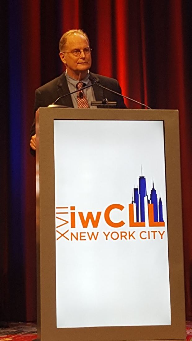
In good-candidate CLL, don’t wait too long for alloHCT
NEW YORK – Allogeneic hematopoietic stem cell transplantation (alloHCT) using HLA-compatible donors results in excellent long-term progression-free survival in younger high-risk chronic lymphocytic leukemia (CLL) patients, an analysis of data from a European Society for Blood and Marrow Transplantation registry cohort suggests.
AlloHCT may, in some patients, be preferable to sequential targeted therapy, according to Michel van Gelder, MD.
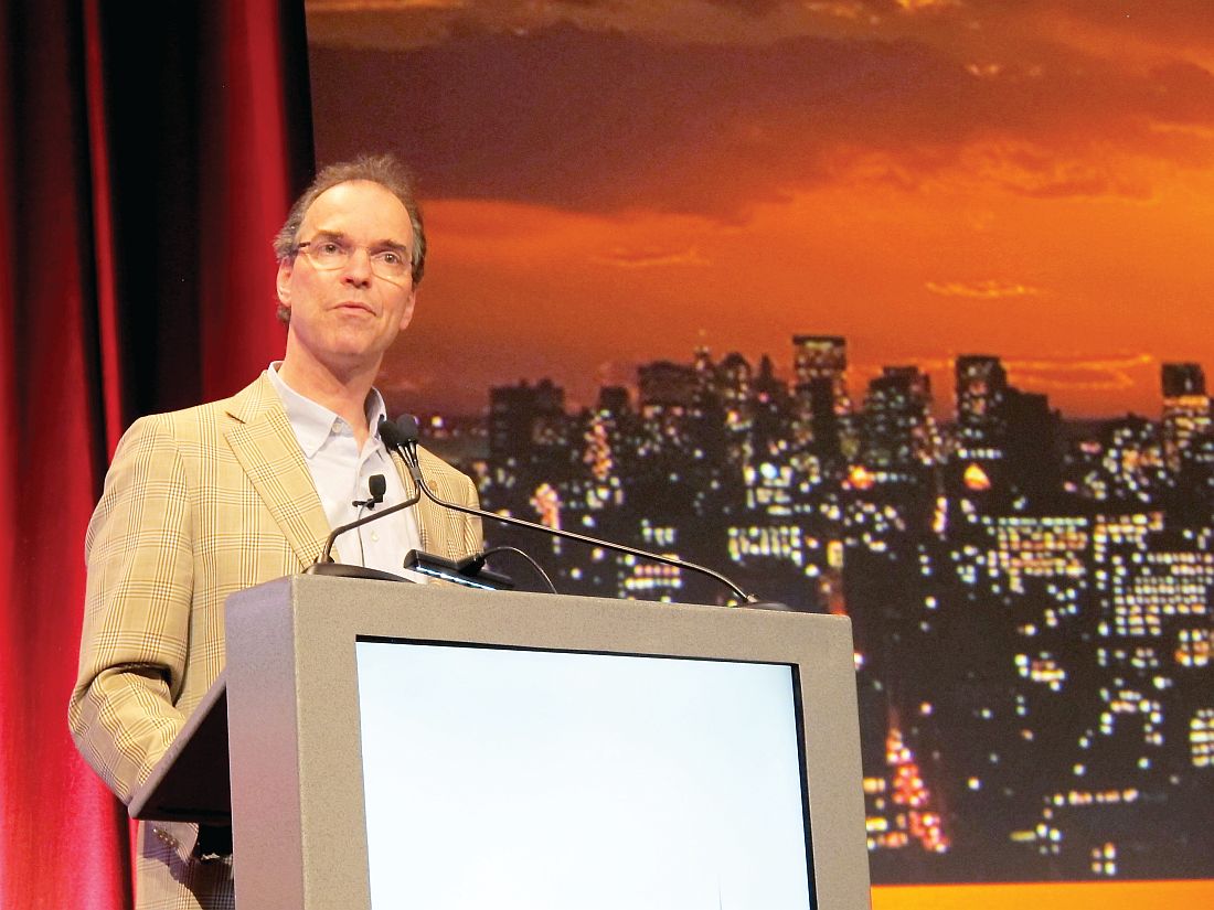
This is especially true for those progressing with Richter’s syndrome, who comprise about one-third of patients, he noted.
“On the other hand, allogeneic stem cell transplantation can induce prolonged progression-free survival,” said Dr. van Gelder of Maastricht (the Netherlands) University Medical Center.
Further, most alloHCT patients become minimal residual disease negative, which predicts prolonged progression-free survival (PFS).
“The down-side, of course, is nonrelapse mortality,” he said, noting that NRM depends on factors such as age, performance status, and HLA match.
In a recent risk factor analysis currently pending publication, he and his colleagues found, in a large group of patients, that age, performance status, remission at time of transplant, donor relationship, HLA and sex match each had an impact on 5-year PFS after alloHCT.
The more risk factors a patient had, the worse the outcome, he said.
Based on current knowledge, the place for alloHCT in CLL treatment is in patients with high-risk cytogenetics. Patients can be treated first with a kinase inhibitor or venetoclax followed by transplant, or they can wait for progression and then do the transplant, he said.
Those without high risk cytogenetics but with short PFS after treatment with a kinase inhibitor or venetoclax may also be candidates for alloHCT, he added.
“Preferably they should be young [and] have a good matched donor and low comorbidity,” he said.
In the current study, the focus was on younger CLL patients. “We tried to identify factors that predict for a low 2-year NRM and a high 8-year PFS. We studied the impact of high risk cytogenetics, and, for this study, we chose del(17p) and del(11q), and we tried to officialize the PFS, the relapse incidence, and the nonrelapse mortality of so-called ‘good transplant risk CLL patients’ with these high cytogenetic risk factors,” he explained.
In 197 patients under age 50 years (median 46 years) with a median follow-up of 90 months in an updated EBMT registry cohort, the most important relevant prognostic factor for 2-year NRM was the donor HLA match (adjusted hazard ratio, 2.5 for a matched unrelated donor, 4.0 for a partially matched unrelated donor, both vs. a matched sibling), and predictors for poor 8-year PFS were no remission at the time of alloHCT (hazard ratio, 1.7), and partially HLA matched unrelated donor (HR, 2.8).
High-risk cytogenetics did not significantly impact 8-year PFS, Dr. van Gelder said, noting that this confirms findings from prior studies.
Most of the patients included in the analysis were fludarabine refractory, 70% had del(17p), 35% had del(11q), and the median number of prior treatments was 3. Additionally, 12% had previous autologous transplant, 62% had remission at time of transplant, and most had good performance status, he said.
Conditioning regimens varied by site, 42% of patients had an HLA-matched sibling donor, and 50% had a matched unrelated donor.
Based on the regression model, a reference patient with high risk cytogenetics (del[17p] and/or del[11q]) and good transplant characteristics (age 46 years, no prior autologous stem cell transplantation, remission at the time of alloHCT and HLA- and sex-matched sibling donor) was created. A reference patient with poor transplant characteristics (not in remission at the time of transplant, with an unrelated, non-sex-matched donor) was also created. The predicted 2-year NRM for the good transplant risk patient was 12.1%, and 8-year PFS was more than 50%, Dr. van Gelder said.
For the poor risk patient, 2-year NRM was 37%, and PFS was below 50%, he said.
“So, in conclusion ... good transplant risk young patients with a low nonrelapse mortality and high 8-year progression-free survival can be identified,” he said.
The problem in clinical practice is determining whether – and when – to do a transplant in a young patient, he continued.
“There are a lot of possibilities. Nobody knows, of course, what is the best regimen, but a problem in these patients is that, if they have progression with Richter’s transformation, then you are lost,” he said. “So, if you would like to prevent this, and you have a patient with a low nonrelapse mortality risk, maybe it is better to do the transplant before.”
As for whether alloHCT can be done after kinase inhibitor therapy, the data are limited, but data presented at EBMT 2017 suggest the approach is feasible and effective. In 43 younger patients who underwent alloHCT after ibrutinib treatment, including 37% with TP53 mutation, the 1-year NRM and PFS rates were 9% and 63%, which is “in the same range as in the era before kinase inhibitors,” Dr. van Gelder said regarding the abstract presented by Peter Dreger, MD.
In 32 patients who underwent alloHCT after idelalisib treatment, including 44% with del(17p)/del(11q) and 85% in remission at the time of alloHCT, early follow-up showed that 6-month NRM and PFS was 7% and 83%, respectively, according to another abstract presented by Johannes Schetelig, MD.
“It’s all about balancing the risks. On the one hand you can use sequential therapies. On the other, if you have patients with high-risk cytogenetics [and] CLL in remission and you have a well-matched donor, maybe you should consider the transplant earlier, Dr. van Gelder said. “If you have a good transplant patient in remission, I would propose [that you] don’t wait too long.”
Dr. van Gelder reported having no relevant disclosures.
NEW YORK – Allogeneic hematopoietic stem cell transplantation (alloHCT) using HLA-compatible donors results in excellent long-term progression-free survival in younger high-risk chronic lymphocytic leukemia (CLL) patients, an analysis of data from a European Society for Blood and Marrow Transplantation registry cohort suggests.
AlloHCT may, in some patients, be preferable to sequential targeted therapy, according to Michel van Gelder, MD.
This is especially true for those progressing with Richter’s syndrome, who comprise about one-third of patients, he noted.
“On the other hand, allogeneic stem cell transplantation can induce prolonged progression-free survival,” said Dr. van Gelder of Maastricht (the Netherlands) University Medical Center.
Further, most alloHCT patients become minimal residual disease negative, which predicts prolonged progression-free survival (PFS).
“The down-side, of course, is nonrelapse mortality,” he said, noting that NRM depends on factors such as age, performance status, and HLA match.
In a recent risk factor analysis currently pending publication, he and his colleagues found, in a large group of patients, that age, performance status, remission at time of transplant, donor relationship, HLA and sex match each had an impact on 5-year PFS after alloHCT.
The more risk factors a patient had, the worse the outcome, he said.
Based on current knowledge, the place for alloHCT in CLL treatment is in patients with high-risk cytogenetics. Patients can be treated first with a kinase inhibitor or venetoclax followed by transplant, or they can wait for progression and then do the transplant, he said.
Those without high risk cytogenetics but with short PFS after treatment with a kinase inhibitor or venetoclax may also be candidates for alloHCT, he added.
“Preferably they should be young [and] have a good matched donor and low comorbidity,” he said.
In the current study, the focus was on younger CLL patients. “We tried to identify factors that predict for a low 2-year NRM and a high 8-year PFS. We studied the impact of high risk cytogenetics, and, for this study, we chose del(17p) and del(11q), and we tried to officialize the PFS, the relapse incidence, and the nonrelapse mortality of so-called ‘good transplant risk CLL patients’ with these high cytogenetic risk factors,” he explained.
In 197 patients under age 50 years (median 46 years) with a median follow-up of 90 months in an updated EBMT registry cohort, the most important relevant prognostic factor for 2-year NRM was the donor HLA match (adjusted hazard ratio, 2.5 for a matched unrelated donor, 4.0 for a partially matched unrelated donor, both vs. a matched sibling), and predictors for poor 8-year PFS were no remission at the time of alloHCT (hazard ratio, 1.7), and partially HLA matched unrelated donor (HR, 2.8).
High-risk cytogenetics did not significantly impact 8-year PFS, Dr. van Gelder said, noting that this confirms findings from prior studies.
Most of the patients included in the analysis were fludarabine refractory, 70% had del(17p), 35% had del(11q), and the median number of prior treatments was 3. Additionally, 12% had previous autologous transplant, 62% had remission at time of transplant, and most had good performance status, he said.
Conditioning regimens varied by site, 42% of patients had an HLA-matched sibling donor, and 50% had a matched unrelated donor.
Based on the regression model, a reference patient with high risk cytogenetics (del[17p] and/or del[11q]) and good transplant characteristics (age 46 years, no prior autologous stem cell transplantation, remission at the time of alloHCT and HLA- and sex-matched sibling donor) was created. A reference patient with poor transplant characteristics (not in remission at the time of transplant, with an unrelated, non-sex-matched donor) was also created. The predicted 2-year NRM for the good transplant risk patient was 12.1%, and 8-year PFS was more than 50%, Dr. van Gelder said.
For the poor risk patient, 2-year NRM was 37%, and PFS was below 50%, he said.
“So, in conclusion ... good transplant risk young patients with a low nonrelapse mortality and high 8-year progression-free survival can be identified,” he said.
The problem in clinical practice is determining whether – and when – to do a transplant in a young patient, he continued.
“There are a lot of possibilities. Nobody knows, of course, what is the best regimen, but a problem in these patients is that, if they have progression with Richter’s transformation, then you are lost,” he said. “So, if you would like to prevent this, and you have a patient with a low nonrelapse mortality risk, maybe it is better to do the transplant before.”
As for whether alloHCT can be done after kinase inhibitor therapy, the data are limited, but data presented at EBMT 2017 suggest the approach is feasible and effective. In 43 younger patients who underwent alloHCT after ibrutinib treatment, including 37% with TP53 mutation, the 1-year NRM and PFS rates were 9% and 63%, which is “in the same range as in the era before kinase inhibitors,” Dr. van Gelder said regarding the abstract presented by Peter Dreger, MD.
In 32 patients who underwent alloHCT after idelalisib treatment, including 44% with del(17p)/del(11q) and 85% in remission at the time of alloHCT, early follow-up showed that 6-month NRM and PFS was 7% and 83%, respectively, according to another abstract presented by Johannes Schetelig, MD.
“It’s all about balancing the risks. On the one hand you can use sequential therapies. On the other, if you have patients with high-risk cytogenetics [and] CLL in remission and you have a well-matched donor, maybe you should consider the transplant earlier, Dr. van Gelder said. “If you have a good transplant patient in remission, I would propose [that you] don’t wait too long.”
Dr. van Gelder reported having no relevant disclosures.
AT THE IWCLL MEETING
Key clinical point:
Major finding: The predicted 2-year nonrelapse mortality was 12.1% for a patient who is a good transplant risk and predicted 8-year PFS was more than 50%.
Data source: An analysis of updated registry data for 197 patients.
Disclosures: Dr. van Gelder reported having no relevant disclosures
Ibrutinib response in CLL/SLL less affected by select risk factors
NEW YORK – Risk factors associated with poor outcomes in chronic lymphocytic leukemia/small lymphocytic leukemia patients treated with standard therapies appear to have less relevance with ibrutinib treatment, according to an integrated analysis of data from the randomized, phase III RESONATE, RESONATE 2, and HELIOS trials.
In the combined analysis, at a median follow-up of 21 months, progression-free survival (PFS), overall survival (OS), overall response rate (ORR), and complete response rate (CRR) were better in ibrutinib-treated patients than in comparator-treated patients – and, in the ibrutinib-treated patients, the outcomes did not differ based on the adverse genomic factors examined, Thomas J. Kipps, MD, PhD, said at the annual International Workshop on Chronic Lymphocytic Leukemia.
The trials compared well with each other, but differed in terms of number of prior therapies received by patients, he said. Furthermore, the analysis did not examine the effect of del(17p); patients with that deletion were included only in the RESONATE trial.
In RESONATE, ibrutinib was superior to ofatumumab in relapsed/refractory CLL/SLL. In RESONATE 2, ibrutinib was superior to chlorambucil in treatment-naive patients with CLL/SLL. In HELIOS, ibrutinib with bendamustine/rituximab was superior to placebo with bendamustine/rituximab in patients with relapsed/refractory CLL/SLL.
In the new multivariate analysis of the pooled data from these trials – adjusting for the four genomic risk factors and age, sex, ECOG performance status, cytopenia, lactate dehydrogenase (LDH), bulky disease, and number of prior therapies – only having had one or more vs. no prior therapies, and having two or more vs. one prior therapies was associated with shorter PFS and OS in ibrutinib-treated patients, with a trend toward significance.
In comparator-treated patients, however, unmutated IGHV, del(11q), complex karyotype, male sex, two or more prior therapies, and bulky disease all were associated with significantly shorter PFS. Complex karyotype, male sex, bulky disease, ECOG performance status greater than 1, and elevated LDH were associated with significantly shorter OS.
“We need to debate on what the significance of this is and how that can be incorporated into our idea about first-line therapies,” said Dr. Kipps, who was an investigator in both RESONATE trials and is a professor of medicine at the University of California, San Diego.
In univariate analysis of data from ibrutinib-treated patients, unmutated IGHV, del(11q), trisomy 12, and complex karyotype were generally not associated with shorter PFS, OS, or lower ORR or CRR.
Overall survival with and without unmutated IGHV was 78% and 84%, respectively; with and without trisomy 12 was 82% and 80%, respectively; and with and without complex karyotype was 77% and 78%, respectively.
ORR, for example, was comparable in the presence (90%) and absence (89%) of unmutated IGHV in ibrutinib-treated patients, as was CRR, at 29% and 26%, respectively.
In the presence and absence of trisomy 12, ORR was 85% and 91%, respectively; CRR was 33% and 22%.
In the presence and absence of complex karyotype, ORR was 88% and 89%, respectively, and CRR was 18% and 24%.
In the presence and absence of del(11q), ORR was 91% and 90%, respectively, and CRR was 22% and 27%.
The only difference that reached statistical significance was the complete response rate with trisomy 12, which favored the presence of trisomy 12.
Interestingly, the ibrutinib-treated patients with del(11q) had a trend toward longer PFS and OS, compared with those without del(11q), said Dr. Kipps.
At 36 months, PFS was 74% with the presence of del(11q) and 68% with the absence of del(11q) (hazard ratio, 0.73 vs. 1.88 in comparator-treated patients), and overall survival at 42 months was 80% in patients with del(11q) and 78% in those without del(11q) (HR, 0.73), Dr. Kipps said.
“The [finding in the] patients with the complex karyotype was a bit surprising, and I think this requires further analysis,” he said, explaining that complex karyotype actually was associated with a shorter PFS in patients treated on the comparator arm, and that this finding conflicts with earlier data.
The findings suggest ibrutinib-treated patients with trisomy 12, for reasons that are unclear, had a significantly higher complete response rate, but not greater PFS or OS vs. those without trisomy 12, Dr. Kipps said.
“It’s also interesting that ... unmutated antibody genes or del(11q) or complex karyotype were adverse prognostic factors in patients treated with comparator treatments, but not necessarily in patients treated with ibrutinib-based therapy,” he said.
Furthermore, although a prior phase II study involving heavily pretreated patients suggested that del(11q) may have adverse prognostic influence on PFS, that finding may not be borne out in patients with fewer lines of prior therapy.
The findings suggest that genomic risk factors associated with poor outcomes with initial chemoimmunotherapy may be less apparent in patients treated with ibrutinib.
“I think this is important, because it may then turn a prognostic factor into a predictive factor, meaning, a predictor of adverse outcomes for a given type of therapy as opposed to adverse prognostic value overall,” he concluded.
Ibrutinib (Imbruvica, Pharmacyclics) was approved by the Food and Drug Administration in January 2015 for the treatment of CLL after previous therapy. Dr. Kipps has received research funding from and/or served as a consultant or advisor for AbbVie, Genentech, Gilead, and Pharmacyclics, an AbbVie company.
[email protected]
AT THE IWCLL MEETING
Key clinical point:
Major finding: In ibrutinib-treated patients, overall survival with and without unmutated IGHV was 78% and 84%, respectively; with and without trisomy 12 was 82% and 80%, respectively; and with and without complex karyotype was 77% and 78%, respectively.
Data source: A pooled analysis of data from 1,210 patients from three randomized phase III trials.
Disclosures: Dr. Kipps has received research funding from and/or served as a consultant or advisor for AbbVie, Genentech, Gilead, and Pharmacyclics, an AbbVie company.
Drug receives breakthrough designation for relapsed/refractory AML

The US Food and Drug Administration (FDA) has granted GMI-1271 breakthrough therapy designation for the treatment of adults with relapsed/refractory acute myeloid leukemia (AML).
GMI-1271 is an E-selectin antagonist being developed by GlycoMimetics, Inc.
The product already has orphan and fast track designations from the FDA for the treatment of AML.
GMI-1271 is currently being evaluated in a phase 1/2 trial of patients with relapsed or refractory AML and patients age 60 and older with newly diagnosed AML.
The patients are receiving GM-1271 in combination with chemotherapy. The relapsed/refractory group is receiving mitoxantrone, etoposide, and cytarabine. The newly diagnosed patients are receiving cytarabine and idarubicin (7+3).
GlycoMimetics plans to present data from this trial at the 2017 American Society of Clinical Oncology (ASCO) Annual Meeting as abstracts 2520 and 2560.
The company also plans to present the research at the 22nd Congress of the European Hematology Association (EHA) as abstracts P547 and P203.
About breakthrough designation
The FDA’s breakthrough therapy designation is intended to expedite the development and review of new treatments for serious or life-threatening conditions.
Breakthrough designation entitles the company developing a therapy to more intensive FDA guidance on an efficient and accelerated development program, as well as eligibility for other actions to expedite FDA review, such as a rolling submission and priority review.
To earn breakthrough designation, a treatment must show encouraging early clinical results demonstrating substantial improvement over available therapies with regard to a clinically significant endpoint, or it must fulfill an unmet need.
About fast track designation
The FDA’s fast track program is designed to facilitate the development and expedite the review of products intended to treat or prevent serious or life-threatening conditions and address unmet medical need.
Through the fast track program, a product may be eligible for priority review. In addition, the company developing the product may be allowed to submit sections of the new drug application or biologic license application on a rolling basis as data become available.
Fast track designation also provides the company with opportunities for more frequent meetings and written communications with the FDA.
About orphan designation
The FDA grants orphan designation to products intended to treat, diagnose, or prevent diseases/disorders that affect fewer than 200,000 people in the US.
The designation provides incentives for sponsors to develop products for rare diseases. This may include tax credits toward the cost of clinical trials, prescription drug user fee waivers, and 7 years of market exclusivity if the product is approved. ![]()
Single-cell analysis reveals TKI-resistant CML stem cells

Researchers say they’ve developed a technique for single-cell analysis that has revealed a population of treatment-resistant stem cells in patients with chronic myeloid leukemia (CML).
“It is increasingly recognized that tumors contain a variety of different cell types, including so-called cancer stem cells, that drive the growth and relapse of a patient’s cancer,” said study author Adam Mead, BM BCh, PhD, of University of Oxford in the UK.
“These cells can be very rare and extremely difficult to find after treatment as they become hidden within the normal tissue. We used a new genetic technique to identify and analyze single cancer stem cells in leukemia patients before and after treatment.”
Dr Mead and his colleagues detailed this research in Nature Medicine.
The team’s single-cell analysis technique combines high-sensitivity mutation detection with whole-transcriptome analysis.
The researchers used the method to analyze stem cells from patients with CML and found the cells to be heterogeneous.
In addition, the team was able to identify a subset of CML stem cells that proved resistant to treatment with tyrosine kinase inhibitors (TKIs).
“We found that, even in individual cases of leukemia, there are various types of cancer stem cell that respond differently to the treatment,” Dr Mead said.
“A small number of these cells are highly resistant to the treatment and are likely to be responsible for disease recurrence when the treatment is stopped. Our research allowed us uniquely to analyze these crucial cells that evade treatment so that we might learn how to more effectively eradicate them.”
The researchers said these TKI-resistant CML stem cells were “transcriptionally distinct” from normal hematopoietic stem cells.
The TKI-resistant cells were characterized by dysregulation of specific genes and pathways—TGF-β, TNF-α, JAK–STAT, CTNNB1, and NFKB1A—that could potentially be targeted to improve the treatment of CML.
The researchers also said their single-cell analysis technique can be used beyond CML.
“This technique could be adapted to analyze a range of different cancers to help predict both the likely response to treatment and the risk of the disease returning in the future,” Dr Mead said. “This should eventually enable treatment to be tailored to target each and every type of cancer stem cell that may be present.” ![]()
First generic version of clofarabine available in US

Clofarabine Injection, the first-to-market generic version of Sanofi Genzyme’s Clolar, is now available in the US.
The generic, a product of Fresenius Kabi, is available as a single dose vial containing 20 mg per 20 mL clofarabine.
Clofarabine is a purine nucleoside metabolic inhibitor indicated for the treatment of patients ages 1 to 21 with relapsed or refractory acute lymphoblastic leukemia (ALL) who received at least 2 prior treatment regimens.
Clolar was granted accelerated approval for this indication in the US in 2004.
The approval was based on response rates observed in ALL patients. There are no trials verifying that clofarabine confers improvement in survival or disease-related symptoms in ALL patients.
Clofarabine was assessed in a single-arm, phase 2 trial of 61 pediatric patients with relapsed/refractory ALL.
The patients’ median age was 12 (range, 1 to 20 years), and their median number of prior treatment regimens was 3 (range, 2 to 6).
The patients received clofarabine at 52 mg/m2 intravenously over 2 hours daily for 5 days, every 2 to 6 weeks.
The overall response rate was 30%. Seven patient achieved a complete response (CR), 5 had a CR without platelet recovery, and 6 patients had a partial response.
The median duration of CR in patients who did not go on to hematopoietic stem cell transplant was 6 weeks.
The most common grade 3 or higher adverse events were febrile neutropenia, anorexia, hypotension, and nausea.
These results were published in the Journal of Clinical Oncology in 2006. ![]()
Antibody shows potential for treating AML, B-ALL

Endoglin may be a promising therapeutic target in acute myeloid leukemia (AML) and B-cell acute lymphoblastic leukemia (B-ALL), according to researchers.
The group identified endoglin expression on the majority of blasts from patients with AML and B-ALL.
The team also found that an endoglin antibody, TRC105 (carotuximab), exhibited activity against AML and B-ALL in vivo, and combining the drug with chemotherapeutic agents enhanced this activity.
Rita Perlingeiro, PhD, of the University of Minnesota in Minneapolis, and her colleagues reported these findings in Blood.
The researchers first discovered that endoglin, which is also known as CD105, was “highly expressed” in leukemic blasts.
In samples from AML patients, 47.6% to 98.5% of blasts were CD105+. In samples from B-ALL patients, 92.6% to 99% of blasts were CD105+.
“We have been studying the function of endoglin in hematopoiesis for more than a decade, and the consistent expression of this receptor in the majority of acute leukemias was intriguing,” Dr Perlingeiro said.
She and her colleagues also found that CD105+ blasts had superior leukemogenic activity and reduced survival in mice when compared to CD105- blasts.
Mice injected with AML CD105+ blasts had all died at day 110 after injection, but mice injected with CD105- AML blasts survived until day 140.
Mice injected with CD105+ ALL blasts died 3 months after injection, but mice injected with CD105- ALL blasts were still alive and showed no signs of disease at the time of sacrifice, which was 5 months after injection.
TRC105 monotherapy
Several experiments showed that TRC105 could reduce leukemic activity in vivo.
TRC105 reduced blast counts in the peripheral blood of mice that had been injected with AML blasts. The drug also reduced blasts in the bone marrow initially, though blast counts were comparable in treated mice and controls by week 12.
On the other hand, mice treated with TRC105 did not experience the weight loss and splenomegaly observed in control mice. And TRC105 suppressed the ability of AML blasts to give rise to leukemia in secondary recipient mice.
In mice injected with ALL blasts, TRC105 initially decreased blast counts. However, by week 8, blast counts in the peripheral blood, bone marrow, and spleen of treated mice were similar to those observed in controls. The researchers said this suggests that TRC105 only slows the development of ALL.
The team then evaluated the effects of TRC105 after disease had been established. Mice with established AML received TRC105 for 8 weeks, and mice with established ALL received TRC105 for 4 weeks.
In mice with AML, TRC105 reduced blasts in the peripheral blood and spleen but not the bone marrow. The treatment also prevented splenomegaly and weight loss and prolonged survival.
“Our hypothesis that endoglin expression was linked to leukemia-forming activity was proven to be true, and it was even more rewarding to witness the robust anti-leukemogenic effect of blocking endoglin signaling with TRC105, even when leukemia had already been established in the mouse,” Dr Perlingeiro said.
However, in mice with established ALL, TRC105 had no effect on leukemia progression.
The researchers said this could be due to expression of soluble endoglin (sENG), which would titrate the TRC105 antibody, limiting its ability to bind to membrane-bound endoglin on leukemic cells. Results of additional experiments supported this idea.
TRC105 in combination
The researchers also tested TRC105 in combination with chemotherapy in the mouse models. The team combined the antibody with cytarabine to treat AML and cyclophosphamide to treat ALL.
In mice with AML, cytarabine and TRC105 significantly reduced levels of leukemic cells in the peripheral blood.
In mice with ALL, cyclophosphamide and TRC105 suppressed leukemia development more effectively and more quickly than cyclophosphamide alone.
The researchers detected high levels of sENG in untreated mice with ALL, but levels were lower in the TRC105-treated mice. And there was “no significant detection” of sENG in mice that received cyclophosphamide and TRC105 or cyclophosphamide alone.
Dr Perlingeiro and her colleagues said this suggests the inhibitory effects of sENG can be circumvented by suppressing tumor burden, which results in the combination therapy demonstrating potent antileukemic activity. ![]()
Study: Most oncologists don’t discuss exercise with patients

Results of a small, single-center study suggest oncologists may not provide cancer patients with adequate guidance on exercise.
A majority of the patients and oncologists surveyed for this study placed importance on exercise during cancer care, but most of the oncologists failed to give patients recommendations on exercise.
“Our results indicate that exercise is perceived as important to patients with cancer, both from a patient and physician perspective,” said study author Agnes Smaradottir, MD, of Gundersen Health System in La Crosse, Wisconsin.
“However, physicians are reluctant to consistently include [physical activity] recommendations in their patient discussions.”
Dr Smaradottir and her colleagues reported these findings in JNCCN.
The researchers surveyed 20 cancer patients and 9 oncologists for this study.
The patients’ mean age was 64. Ten patients had stage I-III non-metastatic cancer after adjuvant therapy, and 10 had stage IV metastatic disease and were undergoing palliative treatment. Most patients had solid tumor malignancies, but 1 had chronic lymphocytic leukemia.
The oncologists’ mean age was 45, 56% were male, and they had a mean of 12 years of practice. Most (89%) said they exercise on a regular basis.
Discussions
Nineteen (95%) of the patients surveyed felt they benefited from exercise during treatment, but only 3 of the patients recalled being instructed to exercise.
Exercise was felt to be an equally important part of treatment and well-being for patients with early stage cancer treated with curative intent as well as patients receiving palliative therapy.
Although all the oncologists noted that exercise can benefit cancer patients, only 1 of the 9 surveyed documented discussion of exercise in patient charts.
Preferences and concerns
More than 80% of the patients said they would prefer a home-based exercise regimen that could be performed in alignment with their personal schedules and symptoms.
Patients also noted a preference that exercise recommendations come from their oncologists, as they have an established relationship and feel their oncologists best understand the complexities of their personalized treatment plans.
The oncologists, on the other hand, wanted to refer patients to specialist care for exercise recommendations. Reasons for this included the oncologists’ mounting clinic schedules and a lack of education about appropriate physical activity recommendations for patients.
The oncologists also expressed concern about asking patients to be more physically active during chemotherapy and radiation and expressed trepidation about prescribing exercise to frail patients with limited mobility.
“We were surprised by the gap in expectations regarding exercise recommendation between patients and providers,” Dr Smaradottir said. “Many providers, ourselves included, thought patients would prefer to be referred to an exercise center, but they clearly preferred to have a home-based program recommended by their oncologist.”
“Our findings highlight the value of examining both patient and provider attitudes and behavioral intentions. While we uncovered barriers to exercise recommendations, questions remain on how to bridge the gap between patient and provider preferences.” ![]()
Videos reduce need for anesthesia in kids undergoing radiotherapy
VIENNA, AUSTRIA—Children with cancer may not require general anesthesia prior to radiotherapy if they can watch videos during their treatment, according to research presented at the ESTRO 36 conference (abstract OC-0546).
Allowing children to watch videos during radiotherapy reduced but did not completely eliminate the use of anesthesia in this small study.
The use of videos proved less traumatic than anesthesia for children and their families, as well as making each treatment quicker and more cost-effective, according to study investigator Catia Aguas, of the Cliniques Universitaires Saint Luc in Brussels, Belgium.
“Being treated with radiotherapy means coming in for a treatment every weekday for 4 to 6 weeks,” Aguas noted. “The children need to remain motionless during treatment, and, on the whole, that means a general anesthesia. That, in turn, means they have to keep their stomach empty for 6 hours before the treatment.”
“We wanted to see if installing a projector and letting children watch a video of their choice would allow them to keep still enough that we would not need to give them anesthesia.”
The study included 12 children, ages 1.5 to 6 years, who were treated with radiotherapy using a Tomotherapy® treatment unit at the university hospital. Six children were treated before a video projector was installed in 2014, and 6 were treated after.
Before the video was available, general anesthesia was needed for 83.3% of children’s treatments. Once the projector was installed, anesthesia was needed in 33.3% of treatments.
“Radiotherapy can be very scary for children,” Aguas noted. “It’s a huge room full of machines and strange noises, and the worst part is that they’re in the room alone during their treatment. Before their radiotherapy treatment, they have already been through a series of tests and treatments, some of them painful, so when they arrive for radiotherapy, they don’t really feel very safe or confident.”
“Since we started using videos, children are a lot less anxious. Now they know that they’re going to watch a movie of their choice, they’re more relaxed, and, once the movie starts, it’s as though they travel to another world. Sponge Bob, Cars, and Barbie have been popular movie choices with our patients.”
The research also showed that treatments that used to take 1 hour or more now take around 15 to 20 minutes. This is partly because of the time saved by not having to prepare and administer anesthesia, but it is also because the children who know they are going to watch videos are more cooperative.
“Now, in our clinic, video has almost completely replaced anesthesia, resulting in reduced treatment times and reduction of stress for the young patients and their families,” Aguas said.
She also noted that the projector was inexpensive and simple to install.
“In radiotherapy, everything is usually very expensive, but, in this case, it was not,” Aguas said. “We bought a projector, and, with the help of college students, we created a support to fix the device to the patient couch. Using video is saving money and resources by reducing the need for anesthesia.”
Aguas and her colleagues continue to study children who have been treated since the projector was installed, and the team is extending the project to include adult patients who are claustrophobic or anxious. ![]()
VIENNA, AUSTRIA—Children with cancer may not require general anesthesia prior to radiotherapy if they can watch videos during their treatment, according to research presented at the ESTRO 36 conference (abstract OC-0546).
Allowing children to watch videos during radiotherapy reduced but did not completely eliminate the use of anesthesia in this small study.
The use of videos proved less traumatic than anesthesia for children and their families, as well as making each treatment quicker and more cost-effective, according to study investigator Catia Aguas, of the Cliniques Universitaires Saint Luc in Brussels, Belgium.
“Being treated with radiotherapy means coming in for a treatment every weekday for 4 to 6 weeks,” Aguas noted. “The children need to remain motionless during treatment, and, on the whole, that means a general anesthesia. That, in turn, means they have to keep their stomach empty for 6 hours before the treatment.”
“We wanted to see if installing a projector and letting children watch a video of their choice would allow them to keep still enough that we would not need to give them anesthesia.”
The study included 12 children, ages 1.5 to 6 years, who were treated with radiotherapy using a Tomotherapy® treatment unit at the university hospital. Six children were treated before a video projector was installed in 2014, and 6 were treated after.
Before the video was available, general anesthesia was needed for 83.3% of children’s treatments. Once the projector was installed, anesthesia was needed in 33.3% of treatments.
“Radiotherapy can be very scary for children,” Aguas noted. “It’s a huge room full of machines and strange noises, and the worst part is that they’re in the room alone during their treatment. Before their radiotherapy treatment, they have already been through a series of tests and treatments, some of them painful, so when they arrive for radiotherapy, they don’t really feel very safe or confident.”
“Since we started using videos, children are a lot less anxious. Now they know that they’re going to watch a movie of their choice, they’re more relaxed, and, once the movie starts, it’s as though they travel to another world. Sponge Bob, Cars, and Barbie have been popular movie choices with our patients.”
The research also showed that treatments that used to take 1 hour or more now take around 15 to 20 minutes. This is partly because of the time saved by not having to prepare and administer anesthesia, but it is also because the children who know they are going to watch videos are more cooperative.
“Now, in our clinic, video has almost completely replaced anesthesia, resulting in reduced treatment times and reduction of stress for the young patients and their families,” Aguas said.
She also noted that the projector was inexpensive and simple to install.
“In radiotherapy, everything is usually very expensive, but, in this case, it was not,” Aguas said. “We bought a projector, and, with the help of college students, we created a support to fix the device to the patient couch. Using video is saving money and resources by reducing the need for anesthesia.”
Aguas and her colleagues continue to study children who have been treated since the projector was installed, and the team is extending the project to include adult patients who are claustrophobic or anxious.
Drug elicits responses in MDS patients

VALENCIA, SPAIN—Phase 2 results suggest luspatercept can produce erythroid responses and enable transfusion independence in patients with myelodysplastic syndromes (MDS).
Erythroid response rates were similar whether or not patients had received prior treatment with erythropoiesis-stimulating agents (ESAs).
However, patients without prior ESA exposure were more likely to achieve transfusion independence.
Most adverse events (AEs) considered possibly or probably related to luspatercept were grade 1 or 2.
Uwe Platzbecker, MD, of Universitätsklinikum Carl Gustav Carus in Dresden, Germany, presented these results at the 14th International Symposium on MDS.
The research was sponsored by Acceleron Pharma Inc., the company developing luspatercept in collaboration with Celgene Corporation.
Dr Platzbecker explained that luspatercept, formerly ACE-536, is a modified activin receptor type IIB fusion protein that acts as a ligand trap for GDF11 and other TGF-βfamily ligands to suppress Smad2/3 signaling.
He presented data from a phase 2 base study and an extension study of luspatercept. The base study included 89 patients who received luspatercept for 3 months. The long-term extension study included 52 patients who may receive luspatercept for an additional 5 years.
The patients received luspatercept at doses ranging from 0.125 mg/kg to 1.75 mg/kg in the base study and 1.0 mg/kg to 1.75 mg/kg in the extension study. They received the drug subcutaneously every 3 weeks.
There were 82 patients evaluable for efficacy. They were a median of 2.3 years from diagnosis (range, 0-14). Their median age was 72 (range, 29-90), 63% were male, and 52% had prior treatment with ESAs.
The outcome measures used in these studies were clinically meaningful erythroid hematologic improvement per the International Working Group’s criteria (IWG HI-E) and red blood cell transfusion independence (RBC-TI).
IWG HI-E was defined as hemoglobin increase ≥ 1.5 g/dL sustained for ≥ 8 weeks in patients with a transfusion burden at baseline of less than 4 RBC units every 8 weeks and baseline hemoglobin levels below 10 g/dL. For patients with a greater transfusion burden at baseline, erythroid response was defined as a reduction of ≥ 4 RBC units sustained for ≥ 8 weeks.
RBC-TI was defined as no RBC transfusions for ≥ 8 weeks in patients with a baseline transfusion burden of at least 2 RBC units every 8 weeks.
Response data
In ESA-naïve patients, 48% (11/23) achieved RBC-TI with luspatercept, and 51% (20/39) achieved an IWG HI-E response.
Among patients with prior ESA treatment, 33% (11/33) achieved RBC-TI with luspatercept, and 51% (22/43) achieved an IWG HI-E response.
In patients with baseline erythropoietin (EPO) levels ≤ 500 international units per liter (IU/L), RBC-TI and IWG HI-E response rates were positive in both ring sideroblast-positive (RS+) and RS-negative (RS-) patients, according to the researchers.
| Baseline
EPO (IU/L) |
RS status | IWG HI-E, n=82
n (%) |
RBC-TI, n=56
n (%) |
| ≤ 500 | RS+ | 30/46 (65%) | 16/29 (55%) |
| RS- | 6/14 (43%) | 4/7 (57%) | |
| > 500 | RS+ | 5/9 (56%) | 2/9 (22%) |
| RS- | 1/11 (9%) | 0/9 (0%) | |
| Unknown | 0/2 (0%) | 0/2 (0%) |
*Table includes ESA-refractory and ESA-naïve patients. Patients treated at dose levels ≥ 0.75 mg/kg.
Safety data
All 89 patients were evaluable for safety. Common AEs (occurring in at least 3 patients) that were considered possibly or probably related to study drug were fatigue (6.7%), headache (6.7%), hypertension (5.6%), diarrhea (4.5%), arthralgia (3.4%), bone pain (3.4%), injection site erythema (3.4%), myalgia (3.4%), and peripheral edema (3.4%).
Grade 3 AEs possibly or probably related to study drug were ascites, blast cell count increase, blood bilirubin increase, hypertension, platelet count increase, and pleural effusion.
Grade 3 serious AEs possibly or probably related to study drug were general physical health deterioration and myalgia.