User login
High symptom burden in advanced cancer patients
New research indicates that hospitalized patients with advanced cancer have a high burden of physical and psychological symptoms, and this burden is linked to longer hospital stays and a greater risk for unplanned hospital readmissions and death.
Researchers said these findings highlight the need to develop and test interventions to lessen patients’ symptoms.
Ryan Nipp, MD, of Massachusetts General Hospital in Boston, and his colleagues reported the findings in Cancer.
The researchers noted that patients with advanced cancer often experience frequent and prolonged hospitalizations for reasons that have not been fully explored.
To investigate, the team collected information from 1036 patients with advanced cancer as they were being admitted for an unplanned hospitalization.
The Edmonton Symptom Assessment System (ESAS) was used to assess patients’ physical symptoms, and the Patient Health Questionnaire 4 (PHQ-4) was used to assess their psychological symptoms.
The researchers examined the relationship between patients’ symptom burden and the duration of their hospital stay, risk of readmission, and death.
Many patients reported moderate or severe fatigue (86.7%), poor well-being (74.2%), drowsiness (71.7%), pain (67.7%), and lack of appetite (67.3%). Nearly 30% of patients had clinically significant symptoms of depression (28.8%) and anxiety (28.0%).
The patients’ mean hospital stay was 6.3 days, the readmission rate within 90 days of discharge was 43.1%, and the 90-day mortality rate was 41.6%.
Physical symptoms (P<0.001), total ESAS score (P<0.001), total PHQ-4 score (P=0.040), and depression symptoms (P=0.017) were significantly associated with longer hospital stays, but anxiety symptoms were not (P=0.190).
Physical symptoms (P<0.001), total ESAS score (P<0.001), total PHQ-4 score (P=0.072), and anxiety symptoms (P=0.045) were significantly associated with a higher likelihood of readmission within 90 days, but depression symptoms were not (P=0.219).
Physical symptoms (P<0.001), total ESAS score (P<0.001), total PHQ-4 score (P<0.001), depression symptoms (P<0.001), and anxiety symptoms (P=0.012) were all significantly associated with a higher likelihood of death or readmission within 90 days.
“We demonstrated that many hospitalized patients with advanced cancer experience an immense physical and psychological symptom burden,” Dr Nipp said.
“Interventions to identify and treat symptomatic patients hold great potential for improving patients’ experience with their illness, enhancing their quality of life, and reducing their healthcare utilization.” ![]()
Preserving normal hematopoietic function in AML
Preclinical research has revealed a treatment that might preserve normal hematopoietic function in patients with acute myeloid leukemia (AML).
Researchers found that AML suppresses adipocytes in the bone marrow, which leads to imbalanced regulation of endogenous hematopoietic stem and progenitor cells and impaired myelo-erythroid maturation.
However, a PPARγ agonist can induce adipogenesis, which suppresses AML cells and stimulates the regeneration of healthy blood cells.
Researchers reported these findings in Nature Cell Biology.
The team found that adipocytes in the bone marrow support myelo-erythroid maturation of hematopoietic stem and progenitor cells. But AML has a negative effect on the maturation of adipocytes, which explains why deficient myelo-erythropoiesis is “a consistent feature” of AML.
The researchers also found that pro-adipogenesis therapy—treatment with the PPARγ agonist GW1929—protects healthy myelo-erythropoiesis and limits leukemic self-renewal.
“Our approach represents a different way of looking at leukemia and considers the entire bone marrow as an ecosystem, rather than the traditional approach of studying and trying to directly kill the diseased cells themselves,” said study author Allison Boyd, PhD, of McMaster University in Hamilton, Ontario, Canada.
“These traditional approaches have not delivered enough new therapeutic options for patients. The standard of care for this disease hasn’t changed in several decades.”
“The focus of chemotherapy and existing standard of care is on killing cancer cells, but, instead, we took a completely different approach, which changes the environment the cancer cells live in,” said study author Mick Bhatia, PhD, of McMaster University.
“This not only suppressed the ‘bad’ cancer cells but also bolstered the ‘good’ healthy cells, allowing them to regenerate in the new drug-induced environment. The fact that we can target one cell type in one tissue using an existing drug makes us excited about the possibilities of testing this in patients.”
“We can envision this becoming a potential new therapeutic approach that can either be added to existing treatments or even replace others in the near future. The fact that this drug activates blood regeneration may provide benefits for those waiting for bone marrow transplants by activating their own healthy cells.” ![]()
FDA approves second CAR-T therapy
A second chimeric antigen receptor (CAR) T-cell therapy has gained FDA approval, this time for the treatment of large B-cell lymphoma in adults.
“Today marks another milestone in the development of a whole new scientific paradigm for the treatment of serious diseases,” FDA Commissioner Scott Gottlieb, MD, said in a statement. “This approval demonstrates the continued momentum of this promising new area of medicine, and we’re committed to supporting and helping expedite the development of these products.”
Approval was based on ZUMA-1, a multicenter clinical trial of 101 adults with refractory or relapsed large B-cell lymphoma. Almost three-quarters (72%) of patients responded, including 51% who achieved complete remission.
CAR-T therapy can cause severe, life-threatening side effects, most notably cytokine release syndrome (CRS) and neurologic toxicities, for which axicabtagene ciloleucel will carry a boxed warning and will come with a risk evaluation and mitigation strategy (REMS), according to the FDA.
The list price for a single treatment of axicabtagene ciloleucel is $373,000, according to the manufacturer.
“We will soon release a comprehensive policy to address how we plan to support the development of cell-based regenerative medicine,” Dr. Gottlieb said in a statement. “That policy will also clarify how we will apply our expedited programs to breakthrough products that use CAR-T cells and other gene therapies. We remain committed to supporting the efficient development of safe and effective treatments that leverage these new scientific platforms.”
Axicabtagene ciloleucel was developed by Kite Pharma, which was acquired recently by Gilead Sciences.
[email protected]
On Twitter @denisefulton
Cascade of costs could push new gene therapy above $1 million per patient
Outrage over the high cost of cancer care has focused on skyrocketing drug prices, including the $475,000 price tag for the country’s first gene therapy, Novartis’ Kymriah (tisagenlecleucel), a leukemia treatment approved in August.
But the total costs of tisagenlecleucel and the 21 similar drugs in development – known as CAR T-cell therapies – will be far higher than many have imagined, reaching $1 million or more per patient, according to leading cancer experts. The next CAR T-cell drug could be approved as soon as November.
Although Kymriah’s price tag has “shattered oncology drug pricing norms,” said Leonard Saltz, MD, chief of gastrointestinal oncology at Memorial Sloan Kettering Cancer Center in New York, “the sticker price is just the starting point.”
These therapies lead to a cascade of costs, propelled by serious side effects that require sophisticated management, Dr. Saltz said. For this class of drugs, Dr, Saltz advised consumers to “think of the $475,000 as parts, not labor.”
Hagop Kantarjian, MD, leukemia specialist and professor at the University of Texas MD Anderson Cancer Center, estimates tisagenlecleucel’s total cost could reach $1.5 million.
CAR T-cell therapy is expensive because of the unique way that it works. Doctors harvest patients’ immune cells, genetically alter them to rev up their ability to fight cancer, then reinfuse them into patients.
Taking the brakes off the immune system, Dr. Kantarjian said, can lead to life-threatening complications that require lengthy hospitalizations and expensive medications, which are prescribed in addition to conventional cancer therapy, rather than in place of it.
Keith D. Eaton, MD, a Seattle oncologist, said he ran up medical bills of $500,000 when he participated in a clinical trial of CAR T cells in 2013, even though all patients in the study received the medication for free. Dr. Eaton, who was diagnosed with acute lymphoblastic leukemia (ALL), spent nearly 2 months in the hospital.
Like Dr. Eaton, nearly half of patients who receive CAR T cells develop cytokine storm. Other serious side effects include strokelike symptoms and coma.
The cytokine storm felt like “the worst flu of your life,” said Dr. Eaton, now aged 51 years. His fever spiked so high that a hospital nurse assumed the thermometer was broken. Dr. Eaton replied, “It’s not broken. My temperature is too high to register on the thermometer.”
Although Dr. Eaton recovered, he wasn’t done with treatment. His doctors recommended a bone marrow transplant, another harrowing procedure, at a cost of hundreds of thousands of dollars.
Dr. Eaton said he feels fortunate to be healthy today, with tests showing no evidence of leukemia. His insurer paid for almost everything.
Kymriah’s sticker price is especially “outrageous” given its relatively low manufacturing costs, said Walid F. Gellad, MD, codirector of the Center for Pharmaceutical Policy and Prescribing at the University of Pittsburgh.
The gene therapy process used to create tisagenlecleucel costs about $15,000, according to a 2012 presentation by Carl H. June, MD, who pioneered CAR T-cell research at the University of Pennsylvania in Philadelphia. Dr. June could not be reached for comment.
To quell unrest about price, Novartis has offered patients and insurers a new twist on the money-back guarantee.
Novartis will charge for the drug only if patients go into remission within 1 month of treatment. In a key clinical trial, 83% of the children and young adults treated with tisagenlecleucel went into remission within 3 months. Novartis calls the plan “outcomes-based pricing.”
Novartis is “working through the specific details” of how the pricing plan will affect the Centers for Medicare & Medicaid Services, which pays for care for many cancer patients, company spokesperson Julie Masow said. “There are many hurdles” to this type of pricing plan but, Ms. Masow said, “Novartis is committed to making this happen.”
She also said that Kymriah’s manufacturing costs are much higher than $15,000, although she didn’t cite a specific dollar amount. She noted that Novartis has invested heavily in the technology, designing “an innovative manufacturing facility and process specifically for cellular therapies.”
As for Kymriah-related hospital and medication charges, “costs will vary from patient to patient and treatment center to treatment center, based on the level of care each patient requires,” Ms. Masow said. “Kymriah is a one-time treatment that has shown remarkable early, deep, and durable responses in these children who are very sick and often out of options.”
Some doctors said tisagenlecleucel, which could be used by about 600 patients a year, offers an incalculable benefit for desperately ill young people. The drug is approved for children and young adults with B-cell ALL who already have been treated with at least two other cancer therapies.
“A kid’s life is priceless,” said Michelle Hermiston, MD, director of pediatric immunotherapy at Benioff Children’s Hospital, at the University of California, San Francisco. “Any given kid has the potential to make financial impacts over a lifetime that far outweigh the cost of their cure. From this perspective, every child in my mind deserves the best curative therapy we can offer.”
Other cancer doctors say the Novartis plan is no bargain.
About 36% of patients who go into remission with tisagenlecleucel relapse within 1 year, said Vinay Prasad, MD, of Oregon Health & Science University, Portland. Many of these patients will need additional treatment, said Dr. Prasad, who wrote an editorial about tisagenlecleucel’s price Oct. 4 in Nature.
“If you’ve paid half a million dollars for drugs and half a million dollars for care, and a year later your cancer is back, is that a good deal?” asked Dr. Saltz, who cowrote a recent editorial on tisagenlecleucel’s price in JAMA.
Steve Miller, MD, chief medical officer for Express Scripts, said it would be more fair to judge Kymriah’s success after 6 months of treatment, rather than 1 month. Dr. Prasad goes even further. He said Novartis should issue refunds for any patient who relapses within 3 years.
A consumer-advocate group called Patients for Affordable Drugs also has said that tisagenlecleucel costs too much, given that the federal government spent more than $200 million over 2 decades to support the basic research into CAR T-cell therapy, long before Novartis bought the rights.
Rep. Lloyd Doggett (D-Texas) wrote a letter to the Medicare program’s director last month asking for details on how the Novartis payment deal will work.
“As Big Pharma continues to put price gouging before patient access, companies will point more and more proudly at their pricing agreements,” Rep. Doggett wrote. “But taxpayers deserve to know more about how these agreements will work – whether they will actually save the government money, defray these massive costs, and ensure that they can access lifesaving medications.”
KHN’s coverage related to aging & improving care of older adults is supported by The John A. Hartford Foundation. Kaiser Health News is a national health policy news service that is part of the nonpartisan Henry J. Kaiser Family Foundation.
Eye Hemorrhage Signals Myeloid Leukemia
A 40-year-old man suddenly began to lose vision in his left eye. The retinal exam was normal for the right eye. But the left showed isolated subinternal limited membrane hemorrhage at the fovea along with a white-centered hemorrhage above the fovea.
The patient had no history of trauma or Valsalva retinopathy. His blood pressure was normal as was his blood glucose. However, when bloodwork showed a high total count, increased platelet count, and the peripheral smear indicated myeloid hyperplasia, clinicians at LV Prasad Eye Institute in Hyderabad, India, diagnosed the patient with underlying chronic myeloid leukemia (CML).
A physical examination revealed a palpable spleenomegaly—underscoring the fact, the clinicians note, that when an ophthalmologic finding suggests a systemic disease, a general physical examination will reveal more clinical clues. The patient was referred to an oncologist and started on imatinib for CML.
White-centered or pale-centered hemorrhages are believed to represent an accumulation of leukemic cells or platelet fibrin aggregates, the clinicians say. Blood dyscrasias, such as anemias, leukemia, multiple myeloma, and other platelet disorders may present with similar features. Such hemorrhages are known to resolve spontaneously when the patient is treated for the underlying condition, and the hematologic status improves, the clinicians say. This patient’s hemorrhage gradually resolved over the next month, and his visual acuity improved to 20/20.
Ocular manifestations as a presenting sign of leukemia, especially chronic, are rare, the clinicians say. They note that retinal hemorrhages are one of the “most striking findings” in leukemia, and because they can be directly observed, they provide a “subtle but important clue toward an otherwise asymptomatic disease.” If diagnosed early and treated promptly, patients with CML have a good survival rate.
Source:
Tyagi M, Agarwal K, Paulose RM, Rani PK. BMJ Case Rep. 2017;2017: pii: bcr-2017-21974.
doi: 10.1136/bcr-2017-219741.
Flu vaccine appears ineffective in young leukemia patients
Vaccination may fail to protect young leukemia patients from developing influenza during cancer treatment, according to research published in the Journal of Pediatrics.
Researchers found that young patients with acute leukemia who received flu shots were just as likely as their unvaccinated peers to develop the flu.
The team said these results are preliminary, but they suggest a need for more research and additional efforts to prevent flu in young patients with leukemia.
“The annual flu shot, whose side effects are generally mild and short-lived, is still recommended for patients with acute leukemia who are being treated for their disease,” said study author Elisabeth Adderson, MD, of St. Jude Children’s Research Hospital in Memphis, Tennessee.
“However, the results do highlight the need for additional research in this area and for us to redouble our efforts to protect our patients through other means.”
In this retrospective study, Dr Adderson and her colleagues looked at rates of flu infection during 3 successive flu seasons (2010-2013) in 498 patients treated for acute leukemia at St. Jude.
The patients’ median age was 6 years (range, 1-21). Most patients had acute lymphoblastic leukemia (ALL, 94%), though some had acute myeloid leukemia (4.8%) or mixed-lineage leukemia (1.2%).
Most patients (n=354) received flu shots, including 98 patients who received booster doses. The remaining 144 patients were not vaccinated.
The vaccinated patients received the trivalent vaccine, which is designed to protect against 3 flu strains predicted to be in wide circulation during a particular flu season. The vaccine was a fairly good match for circulating flu viruses during the flu seasons included in this analysis.
Demographic characteristic were largely similar between vaccinated and unvaccinated patients. The exceptions were that more vaccinated patients had ALL (95.5% vs 90.3%; P=0.034) and vaccinated patients were more likely to be in a low-intensity phase of cancer therapy (90.7% vs 73.6%, P<0.0001).
Results
There were no significant differences between vaccinated and unvaccinated patients when it came to flu rates or rates of flu-like illnesses.
There were 37 episodes of flu in vaccinated patients and 16 episodes in unvaccinated patients. The rates (per 1000 patient days) were 0.73 and 0.70, respectively (P=0.874).
There were 123 cases of flu-like illnesses in vaccinated patients and 55 cases in unvaccinated patients. The rates were 2.44 and 2.41, respectively (P=0.932).
Likewise, there was no significant difference in the rates of flu or flu-like illnesses between patients who received 1 dose of flu vaccine and those who received 2 doses.
The flu rates were 0.60 and 1.02, respectively (P=0.107). And the rates of flu-like illnesses were 2.42 and 2.73, respectively (P=0.529).
Dr Adderson said additional research is needed to determine if a subset of young leukemia patients may benefit from vaccination.
She added that patients at risk of flu should practice good hand hygiene and avoid crowds during the flu season. Patients may also benefit from “cocooning,” a process that focuses on getting family members, healthcare providers, and others in close contact with at-risk patients vaccinated. ![]()
Team devises new method to analyze cells
Biophysicists have developed a new method to determine a cell’s mechanical properties, and they believe this method could provide insights regarding cancers, sickle cell anemia, and other diseases.
The method allows researchers to make standardized measurements of single cells, determine each cell’s stiffness, and assign it a number, generally between 10 and 20,000, in pascals.
“Measuring cells with our calibrated instrument is like measuring time with a standardized clock,” said Amy Rowat, PhD, of the University of California Los Angeles.
“Our method can be used to obtain stiffness measurements of hundreds of cells per second.”
Dr Rowat and her colleagues described their method in Biophysical Journal.
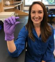
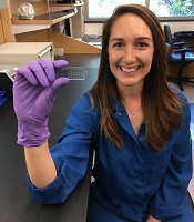
The method is called quantitative deformability cytometry (q-DC). It involves a small device (about 1 inch by 2 inches) made of a soft, flexible rubber that has integrated circuit chips like those in computers.
The researchers use gel particles containing molecules derived from seaweed to force cells through tiny pores in the device. As the cells flow through the device, the researchers take videos at thousands of frames per second—more than 100 times faster than standard video.
Dr Rowat and her colleagues used the device to analyze promyelocytic leukemia cells (HL-60) and breast cancer cells.
The researchers believe this work will provide scientists with a more precise, standardized method to distinguish cancer cells from normal cells.
The team thinks that, in the future, their method could be used to track a cancer patient over time to see how a drug is affecting the patient’s cancer cells.
“By using q-DC, we can very rapidly assess how specific drug treatments affect physical properties of single cells—such as shape, size, and stiffness—and achieve calibrated, quantitative measurements,” Dr Rowat said.
She and her colleagues believe q-DC might also help predict how invasive a cancer cell could be and which drugs might be most effective in fighting the cancer, as well as revealing which proteins are important in regulating the invasion of a cancer cell.
The researchers are now applying q-DC to other types of cancer cells. The team would like to better understand the relationship between a cancer cell’s physical properties and how easily cancer cells can spread through the body.
Dr Rowat’s hypothesis is that properties such as stiffness, size, and a cell’s ability to change shape are important in enabling cancer cells to maneuver.
The researchers said they can also use q-DC to measure other types of cells, such as normal and sickled red blood cells. ![]()
Compound induces selective apoptosis in AML
Researchers say they have discovered a compound that can overcome resistance to apoptosis in acute myeloid leukemia (AML).
The compound, BTSA1, works by activating the BCL-2 family protein BAX.
BTSA1 prompted apoptosis in leukemia cells while sparing healthy cells. It also suppressed AML in mice without producing side effects.
Evripidis Gavathiotis, PhD, of Albert Einstein College of Medicine in Bronx, New York, and his colleagues described these results in Cancer Cell.
The team knew that apoptosis occurs when BAX is activated by pro-apoptotic proteins. However, cancer cells can avoid apoptosis by producing anti-apoptotic proteins that suppress BAX and the proteins that activate it.
“Our novel compound revives suppressed BAX molecules in cancer cells by binding with high affinity to BAX’s activation site,” Dr Gavathiotis said. “BAX can then swing into action, killing cancer cells while leaving healthy cells unscathed.”
Dr Gavathiotis was the lead author of a paper published in Nature in 2008 that first described the structure and shape of BAX’s activation site. He has since looked for small molecules that can activate BAX strongly enough to overcome cancer cells’ resistance to apoptosis.
His team initially screened more than 1 million compounds to reveal those with BAX-binding potential. The most promising 500 compounds were then evaluated in the lab.
“A compound dubbed BTSA1 (short for BAX Trigger Site Activator 1) proved to be the most potent BAX activator, causing rapid and extensive apoptosis when added to several different human AML cell lines,” said Denis Reyna, a doctoral student in Dr Gavathiotis’s lab.
The researchers also tested BTSA1 in blood samples from patients with high-risk AML. BTSA1 induced apoptosis in the patients’ AML cells but did not affect healthy hematopoietic stem cells.
Finally, the researchers generated mouse models of AML. BTSA1 was given to half the mice, while the other half served as controls.
On average, the BTSA1-treated mice survived significantly longer than the control mice—55 days and 40 days, respectively (P=0.0009). In fact, 43% of BTSA1-treated mice were still alive after 60 days and showing no signs of AML.
In addition, the mice treated with BTSA1 showed no evidence of toxicity.
“BTSA1 activates BAX and causes apoptosis in AML cells while sparing healthy cells and tissues, probably because the cancer cells are primed for apoptosis,” Dr Gavathiotis said.
He and his colleagues found that AML cells contained significantly higher BAX levels than normal blood cells from healthy subjects.
“With more BAX available in AML cells, even low BTSA1 doses will trigger enough BAX activation to cause apoptotic death, while sparing healthy cells that contain low levels of BAX or none at all,” Dr Gavathiotis said.
He and his team plan to determine if BTSA1 will elicit similar results in other cancer types. ![]()
CHMP recommends new formulation of pegaspargase
The European Medicines Agency’s Committee for Medicinal Products for Human Use (CHMP) is recommending marketing authorization for lyophilized pegaspargase (ONCASPAR).
If approved, the product would be used as a component of antineoplastic therapy in patients of all ages who have acute lymphoblastic leukemia (ALL).
The product is a freeze-dried formulation of liquid pegaspargase, which is already approved for the aforementioned indication.
The CHMP’s recommendation regarding lyophilized pegaspargase will be submitted to the European Commission (EC).
The EC typically adheres to the CHMP’s recommendations and delivers its final decision within 67 days of the CHMP’s recommendation.
The EC’s decision will be applicable to the entire European Economic Area—all member states of the European Union plus Iceland, Liechtenstein, and Norway.
The CHMP’s recommendation regarding lyophilized pegaspargase is based on analytical and nonclinical studies, which indicate that lyophilized pegaspargase is comparable to the liquid formulation.
Once reconstituted, lyophilized pegaspargase demonstrates similar pharmacokinetics and pharmacodynamics as liquid pegaspargase.
“Lyophilized ONCASPAR builds on more than a decade of data and research with liquid ONCASPAR, and, with no change in dosing regimen, it offers a 3-times longer shelf life,” said Howard B. Mayer, MD, of Shire, the company that developed lyophilized pegaspargase.
“Prolonging shelf life to 24 months for this critically important therapy facilitates management of product inventory by enabling greater flexibility and longer-term planning. Once approved, with the extended shelf life of lyophilized ONCASPAR, we also hope to improve access to the medicine for ALL patients in countries currently not offering liquid ONCASPAR.”
Lyophilized pegaspargase works in the same way as the liquid formulation. It rapidly depletes serum L-asparagine levels and interferes with protein synthesis, thereby depriving lymphoblasts of asparaginase and resulting in cell death. ![]()
Cryotherapy can reduce signs of CIPN
A new study suggests cryotherapy can reduce symptoms of chemotherapy-induced peripheral neuropathy (CIPN).
Researchers found that having chemotherapy patients wear frozen gloves and socks for 90-minute periods significantly reduced the incidence of CIPN symptoms.
Hiroshi Ishiguro, MD, PhD, of International University of Health and Welfare Hospital in Tochigi, Japan, and colleagues reported these findings in the Journal of the National Cancer Institute.
The researchers prospectively evaluated the efficacy of cryotherapy for preventing CIPN. Breast cancer patients treated weekly with paclitaxel (80 mg/m2 for 1 hour) wore frozen gloves and socks on one side of their bodies for 90 minutes, including the entire duration of drug infusion.
The researchers then compared symptoms on the treated sides with those on the untreated sides.
The primary endpoint was CIPN incidence assessed by changes in tactile sensitivity from a pretreatment baseline. The researchers also assessed subjective symptoms, as reported in the Patient Neuropathy Questionnaire, and patients' manual dexterity.
Among the 40 patients studied, 4 did not reach the cumulative dose due to the occurrence of pneumonia, severe fatigue, liver dysfunction, and macular edema. Of the 36 remaining patients, none dropped out due to cold intolerance.
The incidence of objective and subjective signs of CIPN was clinically and statistically significantly lower on the intervention side than on the control side for most measurements, which includes (among other measures):
- Hand tactile sensitivity—27.8% and 80.6%, respectively (odds ratio[OR]= 20.00, P<0.001)
- Foot tactile sensitivity—25.0% and 63.9%, respectively (OR=infinite, P<0.001)
- Hand warm sense—8.8% and 32.4%, respectively (OR=9.00, P=0.02)
- Foot warm sense—33.4% and 57.6%, respectively (OR=5.00, P=0.04)
- Hand cold sense—2.8% and 13.9%, respectively (OR=infinite, P=0.13)
- Foot cold sense—12.6% and 18.8%, respectively (OR=2.00, P=0.69)
- Severe CIPN in the hand according to the Patient Neuropathy Questionnaire—2.8% and 41.7%, respectively (OR=infinite, P<0.001)
- Severe CIPN in the foot according to the Patient Neuropathy Questionnaire—2.8% and 36.1%, respectively (OR=infinite, P<0.001).


