User login
Official Newspaper of the American College of Surgeons
Medicare Part D plans get more flexibility to make midyear changes
Medicare Part D prescription drug plan sponsors will have flexibility to make maintenance changes to their formularies in 2019 as part of a broader effort to lower costs for Part D enrollees.
The ability to make so-called “maintenance changes” to a formulary can now be made prior to receiving approval from the Centers for Medicare & Medicaid Services after the agency finalized a proposal in a rule updating regulations governing Medicare Part D and Medicare Advantage.
The new rule allows plans to make formulary changes immediately upon generic approval assuming certain requirements are met, including generally advising Part D plan members beforehand that changes can occur without a specific advance notice and later providing information about any specific generic substitutions that occur.
The agency cited a Medicare Payment Advisory Commission June 2016 report to Congress as the source of the proposal. That report notes that while plan sponsors can notify beneficiaries of changes when they alert CMS, it can take up to 6 months to get formal notice of an approval, leaving some plan sponsors waiting.
CMS noted that the proposed changes drew concerns, particularly regarding changes that could be made without giving patients a chance to discuss them with their doctors about transitioning to a new medication and other concerns. However, the rule states that the policy “strikes the right balance between providing beneficiaries with access to needed drugs and Part D sponsors with flexibility to administer plans.”
Another area in the rule that CMS expects will generate savings is a new policy on biosimilars that affects beneficiaries receiving low-income subsidy benefits. Going forward, the agency will treat biosimilars and interchangeable biological products the same as generics in terms of determining copays for low-income subsidy enrollees.
Other changes in the rule eliminate requirements that sponsors eliminate plan offerings unless they “meaningfully differ” from one another, allowing plans to offer more choices to beneficiaries, and potentially more cost-saving options to meet their needs. It also clarifies rules regarding the “any willing provider” requirement to allow for more pharmacy options available to Part D enrollees and allow them to shop for best deals for their pharmaceuticals.
In combination with the final 2019 call letter that provides Medicare Advantage and Part D sponsors with the guidelines for submitting their plan designs for the coming coverage year, the rule also finalizes policies related to stemming the opioid crisis, including providing tools to help prevent opioid overprescribing and abuse. The rule implements provisions of the Comprehensive Addiction and Recovery Act of 2016 that require CMS to supply a framework that allows Part D sponsors to implement drug management programs to limit at-risk beneficiaries’ access to coverage for frequently abused drugs.
For example, plans will be allowed to limit at-risk beneficiaries to selected physicians and/or pharmacies to receive their prescriptions, although it will exempt patients who are being treated for cancer-related pain, are receiving palliative or end-of-life care, or are in hospice or long-term care from these drug management programs.
CMS also is limiting the availability of special enrollment periods for beneficiaries dually eligible for Medicare and Medicaid or eligible for the low-income subsidy who are identified as at-risk or potentially at-risk for prescription drug abuse.
Accuracy of colon cancer lymph node sampling influenced by location
CHICAGO – Clinical guidelines recommend , but those guidelines may need to be revised to take into account which side the cancer is on to accurately stage a subset of patients with colon cancer, according to results of a prospective, multicenter clinical trial presented at the Society of Surgical Oncology Annual Cancer Symposium.
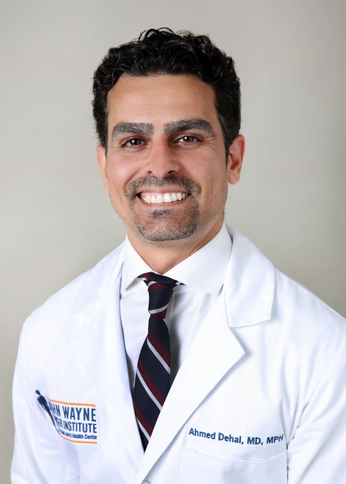
Ahmed Dehal, MD, of John Wayne Canter Institute in Santa Monica, Calif., presented results of the trial that compared nodal staging in right-sided vs. left-sided colon cancer in two cohorts with T3N0 colon cancer who had at least one lymph node examined: a group of 370 patients from the randomized, multicenter prospective trial; and a sampling of 153,945 patients in the National Cancer Database (NCDB). The latter was used to validate findings in the trial group.
The probability of achieving true nodal negativity when 12 lymph nodes were examined was 64% for left and 68% for right colon cancer in the trial group and 72% and 77% in the NCDB cohort, Dr. Dehal said.
The analysis also examined how many nodes would need to be sampled to achieve probabilities of 85%, 90% and 95% true nodal negativity. This analysis found the numbers were consistently lower for right- vs. left-sided disease, Dr. Dehal said. For example, in the trial cohort, 27 lymph nodes would need be sampled in right-sided disease to achieve 85% probability vs. 31 in left-sided. In the NCDB cohort, those numbers were 21 and 25, respectively.
“The current threshold for adequate nodal sampling does not reliably predict the true nodal negativity in this subgroup of patients,” Dr. Dehal said. “In both cohorts – the trial and NCDB – more lymph nodes are needed to predict the true nodal negativity in patients with left compared to right colon cancer.”
These findings may help to inform revisions to existing clinical guidelines, Dr. Dehal said.
“Current guidelines regarding the minimum number of nodes needed to accurately stage patients with node-negative T3 colon cancer may need to be reevaluated given that the decision to give those patients chemotherapy is largely based on the nodal status,” he said. “More studies are needed to improve our understanding of the impact of sidedness on nodal staging in the colon cancer.”
Dr. Dehal and his coauthors reported having no financial disclosures.
SOURCE: Dehal A et al. Society of Surgical Oncology Annual Cancer Symposium. Abstract #23: Accuracy of nodal staging is influenced by sidedness in colon cancer: Results of a multicenter prospective trial.
*CORRECTION, 4/4/2018; a previous version of this story misidentified the cancer type
REPORTING FROM SSO 2018
Key clinical point: Sidedness influences the number of lymph nodes needed to predict true nodal negativity in colon cancer.
Major finding: Probability of true nodal negativity when 12 lymph nodes were examined was 64% for left and 68% for right colon cancer.
Study details: Randomized, multicenter trial of ultrastaging in colon cancer in 370 patients and National Cancer Database sampling of 153,945 patients.
Disclosures: Dr. Dehal and his coauthors report having no financial disclosures.
Source: Dehal A et al. Society of Surgical Oncology Annual Cancer Symposium, Abstract 23: Accuracy of nodal staging is influenced by sidedness in colon cancer: Results of a multicenter prospective trial.
Patients who hide. Patients who seek.
Some people are more likely to seek medical care, and some people are less likely, but which type is more common? The results of a survey of over 14,000 Medicare beneficiaries suggest that the avoid-care type may be a bit more prevalent.
In the survey, 40% of respondents said that they were more likely to keep it to themselves when they got sick, but 36% visit a physician as soon as they feel bad. Almost 29% reported that they avoid going to a physician, but 25% worry about their own health more than others, the Centers for Medicare & Medicaid Services reported based on the results of the 2015 Medicare Current Beneficiary Survey.
Race and ethnicity made a big difference for some questions: 59% of Hispanics said that they visit a doctor as soon as they feel bad, compared with 44% of non-Hispanic blacks and 31% of non-Hispanic whites. That same order was seen for “worry about your health more than others” – 54% Hispanic, 38% black, and 19% white – and for “avoid going to the doctor” – 44% Hispanic, 34% black, and 26% white, the CMS reported.
The three groups, which were the only race/ethnicities included in the report, were all around 40% for “when sick, keep it to yourself,” while two of the three were the same for “had a problem and did not seek a doctor” (blacks and Hispanics at 14% and whites at 10%) and for “ever had a prescription you did not fill” (whites and Hispanics at 7% and blacks at 10%), the report said.
The estimates on propensity to seek care did not include Medicare recipients who lived part or all of the year in a long-term care facility, which was about 4% of the Medicare population in 2015. The survey included a total of 14,068 respondents.
Some people are more likely to seek medical care, and some people are less likely, but which type is more common? The results of a survey of over 14,000 Medicare beneficiaries suggest that the avoid-care type may be a bit more prevalent.
In the survey, 40% of respondents said that they were more likely to keep it to themselves when they got sick, but 36% visit a physician as soon as they feel bad. Almost 29% reported that they avoid going to a physician, but 25% worry about their own health more than others, the Centers for Medicare & Medicaid Services reported based on the results of the 2015 Medicare Current Beneficiary Survey.
Race and ethnicity made a big difference for some questions: 59% of Hispanics said that they visit a doctor as soon as they feel bad, compared with 44% of non-Hispanic blacks and 31% of non-Hispanic whites. That same order was seen for “worry about your health more than others” – 54% Hispanic, 38% black, and 19% white – and for “avoid going to the doctor” – 44% Hispanic, 34% black, and 26% white, the CMS reported.
The three groups, which were the only race/ethnicities included in the report, were all around 40% for “when sick, keep it to yourself,” while two of the three were the same for “had a problem and did not seek a doctor” (blacks and Hispanics at 14% and whites at 10%) and for “ever had a prescription you did not fill” (whites and Hispanics at 7% and blacks at 10%), the report said.
The estimates on propensity to seek care did not include Medicare recipients who lived part or all of the year in a long-term care facility, which was about 4% of the Medicare population in 2015. The survey included a total of 14,068 respondents.
Some people are more likely to seek medical care, and some people are less likely, but which type is more common? The results of a survey of over 14,000 Medicare beneficiaries suggest that the avoid-care type may be a bit more prevalent.
In the survey, 40% of respondents said that they were more likely to keep it to themselves when they got sick, but 36% visit a physician as soon as they feel bad. Almost 29% reported that they avoid going to a physician, but 25% worry about their own health more than others, the Centers for Medicare & Medicaid Services reported based on the results of the 2015 Medicare Current Beneficiary Survey.
Race and ethnicity made a big difference for some questions: 59% of Hispanics said that they visit a doctor as soon as they feel bad, compared with 44% of non-Hispanic blacks and 31% of non-Hispanic whites. That same order was seen for “worry about your health more than others” – 54% Hispanic, 38% black, and 19% white – and for “avoid going to the doctor” – 44% Hispanic, 34% black, and 26% white, the CMS reported.
The three groups, which were the only race/ethnicities included in the report, were all around 40% for “when sick, keep it to yourself,” while two of the three were the same for “had a problem and did not seek a doctor” (blacks and Hispanics at 14% and whites at 10%) and for “ever had a prescription you did not fill” (whites and Hispanics at 7% and blacks at 10%), the report said.
The estimates on propensity to seek care did not include Medicare recipients who lived part or all of the year in a long-term care facility, which was about 4% of the Medicare population in 2015. The survey included a total of 14,068 respondents.
Wound protectors lower risk of surgical site infections
particularly dual-ring devices, new research suggests.
A meta-analysis of 12 randomized, controlled trials with 3,029 participants found that the use of wound protectors during surgery was associated with 34% lower odds of surgical site infection (95% confidence interval, 0.45–0.90; P less than .01).
The study, published in the March edition of Surgical Endoscopy, also showed that dual-ring wound protectors were associated with a highly significant 69% reduction in the odds of surgical site infections (95% CI, 0.18-0.52; P less than .0001), while the benefits of single-ring wound protectors did not reach statistical significance (OR, 0.84; 95% CI, 0.67–1.04; P = .11).
Wound protectors – also known as wound guards or wound retractors – are intended to prevent the edges of a surgical wound from coming into contact with the contaminated surgical field.
The single-ring model consists of a plastic ring that sits within the wound and a protective drape that extends out from it. In the dual-ring model, one ring sits inside the wound and the other outside the wound, with the two rings joined by the protective plastic.
Of the 12 studies included in the meta-analysis, 5 involved colorectal surgery only, 5 included both colorectal and other gastrointestinal surgery, and 2 studies focused on appendectomies. All but one study involved open surgery.
“Our study is more specific in the interventions included in that we included lower gastrointestinal surgery only, which is a population that would likely benefit the most from the intervention due to the high incidence of SSIs in bowel surgery, compared to other abdominal surgeries,” wrote Lisa Zhang, MD, a general surgery resident at the department of surgery at Kingston (Ont.) General Hospital and her coauthors.
There was no difference in the effect of wound protectors based on the target organ for surgery, even with a high number of patients in one of the appendectomy trials, which the authors were concerned may have skewed the results.
Overall, the authors rated the evidence to be of moderate quality, but several studies had a high risk of bias.
The authors said that one barrier to routine use of these devices was cost. However, given that surgical site infections are estimated to cost around $3.5 billion to $10 billion annually in health care expenditures, they argued that “the use of dual-ring wound edge protectors should be considered in open lower gastrointestinal surgery, including open appendectomies.”
No funding sources or conflicts of interest were declared.
SOURCE: Zhang L et al. Surg Endosc. 2018;32:1111–22.
FROM SURGICAL ENDOSCOPY
Key clinical point: Dual-ring wound protectors significantly decrease the risk of gastrointestinal surgical site infections.
Major finding: Dual-ring wound protectors were associated with a 69% reduction in the odds of surgical site infections.
Study details: A meta-analysis of 12 randomized, controlled trials.
Disclosures: No funding source or conflicts of interest were declared.
Source: Zhang L et al. Surg Endosc. 2018;32:1111–22.
Smokers face higher infection risk after hernia operations
Jonah Stulberg, MD, FACS, is stickler about requiring patients to stop smoking at least 3 months before hernia surgery. He even uses urine tests to confirm whether they actually quit. A study by Dr. Stulberg and his colleagues supports this approach: .
The finding held up even after the researchers controlled for various factors. “Our findings are in agreement with other findings in higher risk surgeries, and they provide evidence that low-risk surgeries are not exempt from the risks associated with smoking,” said Dr. Stulberg in an interview. “Our data would suggest that there is significant clinical benefit to encouraging smoking cessation before elective hernia repair.”
The researchers launched the study to better understand how smoking affects complication rates in light of the fact that “surgeons in the U.S. tend to offer low-risk elective surgical procedures to patients who are actively smoking despite overwhelming evidence that smoking increases surgical risks,” Dr. Stulberg said.
The researchers tracked 220,629 patients in the American College of Surgeons National Surgical Quality Improvement Project (NSQIP) database who underwent several types of elective hernia repair from 2011 to 2014.
Just over 18% of the patients said they’d smoked over the past year; they were more likely to be younger (median age, 50 for smokers vs. 57 for nonsmokers). Smokers also were more likely to be black, to be underweight, and to consume two or more alcoholic beverages per day (P less than .05).
The researchers tracked serious complications in the 30 days after surgery such as death, sepsis, and readmission.
Complications developed in 6.34% of smokers and 4.72% of nonsmokers (P less than .001). Numerous kinds of complications were more common in the smokers prior to adjustment: death, return to the operating room, readmission, and transfusion plus wound, pulmonary, thromboembolic and cardiac complications.
The researchers adjusted their statistics to account for factors such as ethnicity, sex, body mass index, preexisting comorbidities, and type of hernia operation. They found that risk of all complications was higher in smokers, compared with nonsmokers (odds ratio, 1.30) as were several other complications: death (OR, 1.53), return to operating room (OR, 1.23), readmission (OR, 1.24), wound complication (OR, 1.36), sepsis/septic shock (OR, 1.31), pulmonary complication (OR 1.77-2.30) and cardiac complication (OR, 1.27-1.43).
Only transfusion (OR, 0.90) and thromboembolic (OR, 0.87) complications were less likely in smokers.
The researchers noted that the statistics don’t allow them to analyze whether it makes any difference if smokers quit shortly before their procedures. Still, Dr. Stulberg stands by his you-must-quit-smoking-before-surgery edict. “I believe that their active smoking habit is a bigger health threat than their asymptomatic hernia, and therefore feel the right thing to do as their physician is support them through their smoking cessation,” he said. “I offer counseling and nicotine replacement if needed. I have very good quit rates and would encourage other surgeons to do the same.”
Dr. Stulberg noted that he can’t point to evidence supporting his requirement that patients quit at least 3 months before surgery. “Most data out there says the farther away from your last cigarette, the better,” he said. “But there isn’t any magic to 3 months, other than I believe it will lead to a higher likelihood of permanent cessation.”
Northwestern Memorial Hospital and Northwestern University funded the study. Four of the nine authors, including Dr. Stulberg, report various disclosures that are not directly related to the study, including funding from government agencies, physician organizations, Health Care Services Corporation and Blue Cross Blue Shield of Illinois, Mallinckrodt, and Northwestern University. The other authors report no disclosures.
SOURCE: DeLancey JO et al. Am J Surg. 2018 Mar 6. doi: 10.1016/j.amjsurg.2018.03.004.
FROM AMERICAN JOURNAL OF SURGERY
Key clinical point: Smokers are more likely than are nonsmokers to develop serious complications after elective hernia surgery.
Major finding: The adjusted risk of serious complications after elective hernia surgery is higher (odds ratio, 1.30) in smokers than nonsmokers.
Study details: Retrospective study of ACS NSQIP data on 220,629 patients in the United States (18% smokers) who underwent elective hernia operations during 2011-2014.
Disclosures: Northwestern Memorial Hospital and Northwestern University funded the study. Four of the nine authors reported various disclosures. The other authors report no disclosures.
Source: DeLancey JO et al. Am J Surg. 2018 Mar 6. doi: 10.1016/j.amjsurg.2018.03.004.
VIDEO: Intestinal remodeling contributes to HbA1c drop after Roux-en-Y gastric bypass
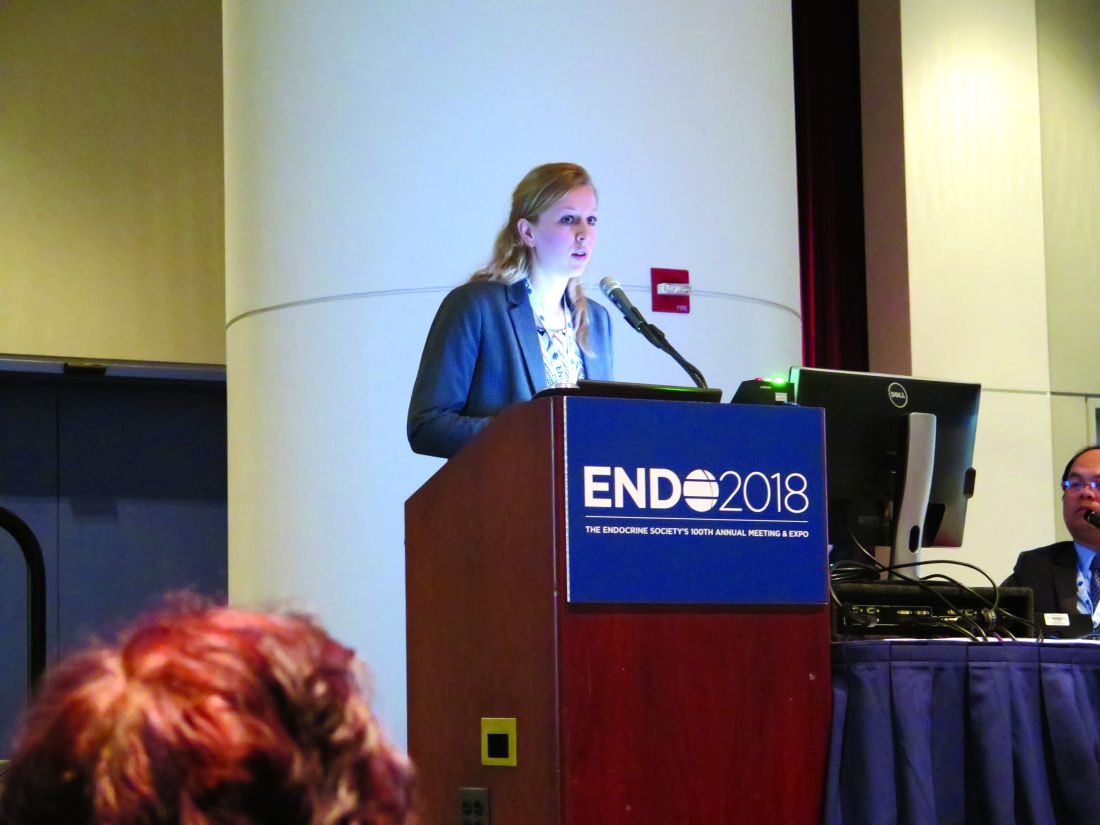
CHICAGO – Of medical and surgical tactics to tackle long-term weight loss, , and gene expression in the Roux limb may hold the key to the surgery’s efficacy, according to an ongoing study.
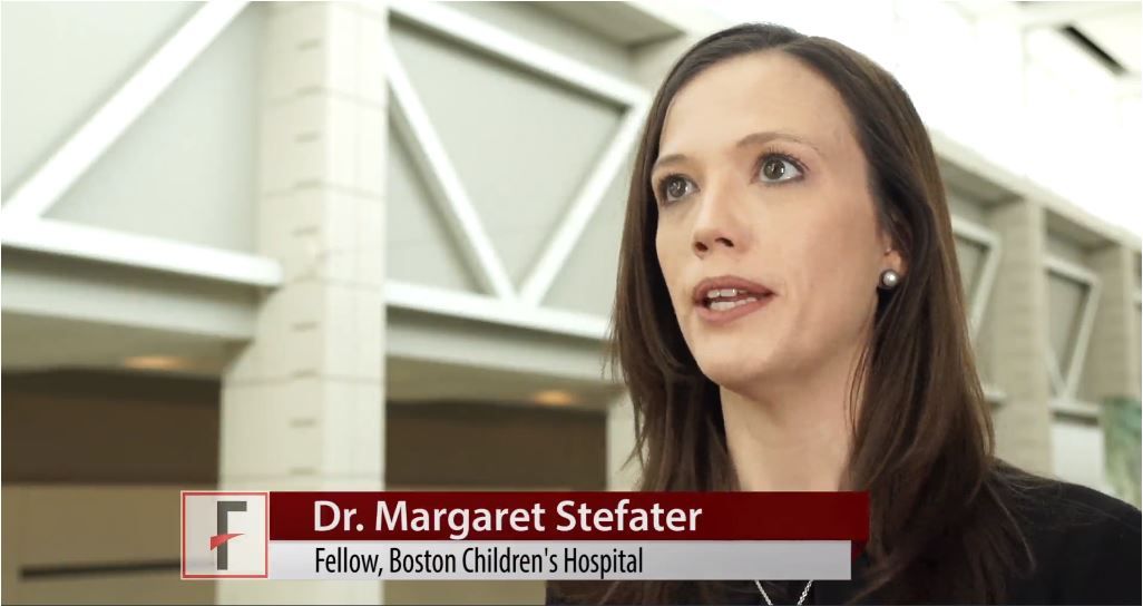
“We know that Roux-en-Y gastric bypass surgery is highly effective as not only a weight-loss therapy, but more and more we’re appreciating its role as a diabetes therapy as well,” said Margaret Stefater, MD, PhD, speaking in an interview at the annual meeting of the Endocrine Society.
The study, she said, was designed to learn more about the intestine’s contribution to the salubrious effect that Roux-en-Y surgery has on diabetes.
“We used microarray in order to characterize gene expression in the intestine” to gain a broad understanding of the processes that are altered after surgery, said Dr. Stefater, a pediatric endocrinology fellow at Boston Children’s Hospital. More specifically, though, the study looked at an individual’s changes in gene expression over time and correlated those changes with that patient’s clinical picture.
The data reported by Dr. Stefater and shared in a press conference, represent part of an ongoing longitudinal prospective study of 32 patients.
“The study aims to characterize gene expression for the first postoperative year,” and findings from the first 6 postoperative months of 19 patients were shared at the meeting, said Dr. Stefater. “This is the first look at our cohort.”
So far, she and her colleagues have compared gene expression using microarray at 1 month and 6 months post-surgery, comparing change across time and change from baseline data.
From hundreds of candidate genes, Dr. Stefater and her colleagues have developed a smaller gene list that, even in the first postoperative month, is predictive of changes in hemoglobin A1c levels over time. “Remarkably, the changes in a select list of genes out to 1 month is actually able to predict hemoglobin A1c levels out to 1 year,” she said. “This speaks to the fact that biological reprogramming in the intestine is somehow related to glycemic response in patients.
“We hope that by understanding these processes, we can home in on those processes that are most likely to be mechanistically responsible for these changes, and then to reverse-engineer this surgery to identify processes or targets which may be good places to start when we think about creating better, or nonsurgical, therapies for people who have obesity and diabetes,” said Dr. Stefater.
Dr. Stefater reported no relevant financial disclosures.
SOURCE: Stefater MA et al. ENDO 2018, Abstract OR 12-6.
REPORTING FROM ENDO 2018
Think about breast cancer surveillance for transgender patients
CHICAGO – , said Christel de Blok, MD, sharing results of a Dutch national study.
The study included 3,078 transgender people (2,064 transgender women) who began hormone therapy (HT) at age 18 years or older. The mean age at which transgender women began HT was 33 years; for transgender men, the mean age was 25 years. In all, transgender women in the study had a total of 30,699 person-years of exposure to HT; for transgender men, the figure was 13,155 person-years.
Overall, there were 16 observed cases of breast cancer in transgender women and four in transgender men. After gender-affirming surgery, the transgender women were followed for a median of 146 months, and experienced a median of 193 months of HT. Transgender men who had mastectomies were followed for a median 93 months, and those who had a hysterectomy-oophorectomy were followed for a median 144 months. Transgender men received a median 176 months of HT.
“Breast cancer can still occur after mastectomy in [transgender] men,” Dr. de Blok said at the annual meeting of the Endocrine Society. “What is interesting is that three out of the four cases of breast cancer in [transgender] men happened after mastectomy.”
In the Netherlands, one in eight women and one in 1,000 men will develop cancer at some point during their lives. In patients who have had a subtotal mastectomy and who are BRCA-1/2 carriers, there is still an approximate 5% residual risk of breast cancer, said Dr. de Blok.
A literature review conducted by Dr. de Blok and her colleagues revealed 19 cases of breast cancer in transgender women and 13 in transgender men. However, a more general study of incidence and characteristics of breast cancer in transgender people receiving hormone treatment had not been done, said Dr. de Blok, of the VU University Medical Center, Amsterdam.
The investigators examined data for adult transgender people seen at their center from 1991 to 2017 and started on hormone treatment. This clinic, said Dr. de Blok, sees about 95% of the transgender individuals in the Netherlands.
The study was able to capitalize on comprehensive information from national databases and registries. Investigators drew from a national histopathology and cytopathology registry as well as from a national vital statistics database. A comprehensive cancer database was used to establish both reference incidence values for males and females and the number of expected cases within the study group.
In both transgender men and women, exactly 50% of cases were ductal carcinoma, compared to 85% in the group of reference women.
An additional 31% of the breast cancers in transgender women were lobular, 6% were ductal carcinoma in situ (DCIS), and the remainder were of other types. Of the cancers in transgender women, 82% were estrogen receptor positive, 64% were progesterone receptor positive, and 9% were Her2/neu positive.
For transgender men, there were no lobular carcinomas; 25% were DCIS, and 25% were of other types. Half of the cancers were estrogen receptor positive, and half were progesterone receptor positive; 25% were Her2/neu positive, and there was one case of androgen receptor positive breast cancer.
Dr. de Blok explained that their analysis compared the observed cases in both transgender men and women to the expected number of cases for the same number of males and females, yielding two standardized incidence ratios (SIRs) for each transgender group.
For transgender women, the SIR for breast cancer compared with males was 50.9 (95% confidence interval, 30.1-80.9). The SIR compared to females was 0.3 (95% CI, 0.2-0.4). This reflected the expected case number of 0.3 for males and the 58 expected cases for a matched group of females.
For transgender men, the SIR for breast cancer compared with males was 59.8 (95% CI, 19-144.3), while the SIR compared to females was 0.2 (95% CI, 0.1-0.5). The expected cases for a similar group of males would be 0.1, and for females, 18.
In many cases, whether a transgender person receives standardized screening mammogram reminders will depend on which sex is assigned to that individual in insurance and other administrative databases, Mr. de Blok noted. When electronic health records and other databases have a binary system, at-risk individuals may fall through the cracks.
Dr. de Blok reported no conflicts of interest.
SOURCE: de Blok C, et al. ENDO 2018, abstract OR 25-6.
REPORTING FROM ENDO 2018
Key clinical point: Transgender individuals had increased risk of breast cancer similar to a female reference population.
Major finding: Transgender men had a standardized incidence ratio of 59.8 compared to a male reference population.
Study details: Study of 3,078 transgender adults receiving hormone therapy.
Disclosures: Dr. de Blok reported no conflicts of interest.
Source: de Blok C, et al. ENDO 2018, abstract OR 25-6.
VIDEO: Biomarker accurately predicted primary nonfunction after liver transplant
, researchers reported in Gastroenterology.
SOURCE: AMERICAN GASTROENTEROLOGICAL ASSOCIATION
Glycomic alterations of immunoglobulin G “represent inflammatory disturbances in the liver that [mean it] will fail after transplantation,” wrote Xavier Verhelst, MD, of Ghent (Belgium) University Hospital, and his associates. The new glycomarker “could be a tool to safely select high-risk organs for liver transplantation that otherwise would be discarded from the donor pool based on a conventional clinical assessment,” and also could help prevent engraftment failures. “To our knowledge, not a single biomarker has demonstrated the same accuracy today,” they wrote in the April issue of Gastroenterology.
Chronic shortages of donor livers contribute to morbidity and death worldwide. However, relaxing donor criteria is controversial because of the increased risk of primary nonfunction, which affects some 2%-10% of liver transplantation patients, and early allograft dysfunction, which is even more common. Although no reliable scoring systems or biomarkers have been able to predict these outcomes prior to transplantation, clinical glycomics of serum has proven useful for diagnosing hepatic fibrosis, cirrhosis, and hepatocellular carcinoma, and for distinguishing hepatic steatosis from nonalcoholic steatohepatitis. “Perfusate biomarkers are an attractive alternative [to] liver biopsy or serum markers, because perfusate is believed to represent the condition of the entire liver parenchyma and is easy to collect in large volumes,” the researchers wrote.
Accordingly, they studied 66 patients who underwent liver transplantation at a single center in Belgium and a separate validation cohort of 56 transplantation recipients from two centers. The most common reason for liver transplantation was decompensated cirrhosis secondary to alcoholism, followed by chronic hepatitis C or B virus infection, acute liver failure, and polycystic liver disease. Donor grafts were transported using cold static storage (21° C), and hepatic veins were flushed to collect perfusate before transplantation. Protein-linked N-glycans was isolated from these perfusate samples and analyzed with a multicapillary electrophoresis-based ABI3130 sequencer.
The four patients in the primary study cohort who developed primary nonfunction resembled the others in terms of all clinical and demographic parameters except that they had a markedly increased concentration (P less than .0001) of a single-glycan, agalacto core-alpha-1,6-fucosylated biantennary glycan, dubbed NGA2F. The single patient in the validation cohort who developed primary nonfunction also had a significantly increased concentration of NGA2F (P = .037). There were no false positives in either cohort, and a 13% cutoff for perfusate NGA2F level identified primary nonfunction with 100% accuracy, the researchers said. In a multivariable model of donor risk index and perfusate markers, only NGA2F was prognostic for developing primary nonfunction (P less than .0001).
The researchers found no specific glycomic signature for early allograft dysfunction, perhaps because it is more complex and multifactorial, they wrote. Although electrophoresis testing took 48 hours, work is underway to shorten this to a “clinically acceptable time frame,” they added. They recommended multicenter studies to validate their findings.
Funders included the Research Fund – Flanders and Ghent University. The researchers reported having no conflicts of interest.
SOURCE: Verhelst X et al. Gastroenterology 2018 Jan 6. doi: 10.1053/j.gastro.2017.12.027.
FROM GASTROENTEROLOGY
Key clinical point: A glycomarker in donor liver perfusate was 100% accurate at predicting primary nonfunction after liver transplantation.
Major finding: In a multivariable model of donor risk index and perfusate markers, only the single-glycan, NGA2F was a significant predictor of primary nonfunction (P less than .0001).
Data source: A dual-center, prospective study of 66 liver transplant patients and a 55-member validation cohort.
Disclosures: Funders included the Research Fund – Flanders and Ghent University. The researchers reported having no conflicts of interest.
Source: Verhelst X et al. Gastroenterology 2018 Jan 6. doi: 10.1053/j.gastro.2017.12.027.
From the Editors: “Okay” is not good enough
Keeping up with refinements in old procedures and adopting new procedures throughout one’s surgical career has always been a challenge.
That challenge has grown as advancements in technology have become ever more disruptive, requiring the learning of radically different skills. Surgeons are highly motivated to learn new procedures, lest they become extinct like professional “dodo birds.” But doing so requires a considerable expenditure of time and money to attend courses and then a period of being proctored in the new procedure once it has been learned.
The acquisition of technical skill in surgery is well recognized as a primary responsibility of surgical residency training programs. Surgical meetings and surgical journals have recently given a lot of space to the question of whether training programs are imparting adequate surgical skill to their learners and whether new graduates have achieved surgical competence at graduation. Some highly cited articles, having surveyed surgical teachers, maintain that a significant percentage of the new graduates have not achieved the needed skills to practice independently.1,2 Many place the blame at the feet of the much-maligned restriction in the resident work week to 80 hours, a limitation imposed by the Accreditation Council of Graduate Medical Education in 2003.
The reasons for the perceived decline in competence are many, including an increase in the number and complexity of surgical procedures, as well as institutional expectations for increased involvement of attendings in procedures. Recommended solutions include lengthening training, encouraging increased and earlier specialization, and proctoring by senior surgeons in the first year of a surgeon’s practice. Less frequently mentioned is the fact that, although the work hours are shorter and the surgery more complex, we have not compensated for these factors by appreciably changing the methods used to teach surgical technique. In many programs, faculty are performing an increasing proportion of procedures: doing more and teaching less.
Mandated surgical skills labs may be helpful to teach a basic level of skill. Simulation can be helpful in imparting the ability to perform more complex procedures and quickly adapt to unexpected intraoperative findings or occurrences. Virtual reality simulators are available, but they’re very expensive and often beyond the budgets of most residency programs. While simulation can help, it is not the whole solution to learning surgical skills.
It is now long past due for surgical training programs to rethink the process of teaching surgical skill in a more deliberate way to residents. One way to accomplish this might be through utilizing “master teachers” or “coaches” who are trained specifically to impart not only skill in performing a given procedure but also an understanding of how to critically assess one’s own performance in practice and learn how to improve that performance through self-reflection and self-assessment. Some very thoughtful and compelling studies have described how coaching might aid performance improvement of both residents and of surgeons already in practice.3,4,5
The process involves a review of videotaped procedures by both the operating surgeon (or surgical resident) and the coach to recognize points at which performance was subpar and to have a discussion about steps needed for improvement. Through further reviews of videotapes of subsequent procedures, the surgeon or resident learns to internalize the techniques of performance improvement.
While ideal in a perfect world, such a schema is far from universally feasible in our current surgical culture. Although master classes and coaching are accepted as the norm in other fields that also require technical excellence, such as classical music and athletics, our surgical culture does not readily accept that our surgical technique might be less than perfect. We tend to downplay the notion that we (and our patients) might benefit from improving our surgical skills beyond mere competence to the point of mastery. A culture change in this regard will not occur overnight and most likely must begin by making coaching a standard and accepted part of surgical training programs, both for the residents and for the teachers themselves.
We make the tacit assumption that attending surgeons are teachers, but we rarely teach them how to teach. The fact that many attendings don’t know how to give effective feedback to residents may be a reason that they fail to give specific coaching on how their learners might improve and why these attendings take over an increasing portion of the procedures themselves. In order for faculty to improve the quality of their teaching, they need training of their own. The training should be a mandatory, “protected” part of their day or it will not occur, and the “teaching the teachers” must be done by master teachers who are respected for their skill not only as a surgeons but also as a surgical educators. This role is an appropriate one for Associate Members of the new ACS Academy of Master Surgeon Educators to assume (see https://www.facs.org/education/academy/membership).
Coaching by master surgeons should become a professional norm. It is only after surgical education and coaching are incorporated all along the training continuum – from novice to competent to master during residency training – that surgeons already in practice will accept it as a regular part of their work. Refinements in procedures and new procedures would be met by continued professional improvement that would be enhanced by master surgeon coaching. We owe it to ourselves and our patients to achieve excellence, not mere competence. “Okay” is not good enough.
Dr. Deveney is a professor of surgery and the vice chair of education in the department of surgery at Oregon Health & Science University, Portland. She is the coeditor of ACS Surgery News.
1. Mattar SG et al. General surgery residency inadequately prepares trainees for fellowship: results of a survey of fellowship program directors. Ann Surg. 2013;258(3):440-9.
2. Damewood RB et al. “Taking training to the next level”: The American College of Surgeons Committee on residency training survey. J Surg Educ. 2017;74(6):e95-e105.
3. Gawande A. Coaching a surgeon: What makes top performers better? The New Yorker, Oct. 3, 2011.
4. Bonrath EM et al. Comprehensive surgical coaching enhances surgical skill in the operating room: a randomized controlled trial. Ann Surg. 2015;262:205-12.
5. Greenberg CC et al. Surgical coaching for individual performance improvement. Ann Surg. 2015;261(1):32-4.
Keeping up with refinements in old procedures and adopting new procedures throughout one’s surgical career has always been a challenge.
That challenge has grown as advancements in technology have become ever more disruptive, requiring the learning of radically different skills. Surgeons are highly motivated to learn new procedures, lest they become extinct like professional “dodo birds.” But doing so requires a considerable expenditure of time and money to attend courses and then a period of being proctored in the new procedure once it has been learned.
The acquisition of technical skill in surgery is well recognized as a primary responsibility of surgical residency training programs. Surgical meetings and surgical journals have recently given a lot of space to the question of whether training programs are imparting adequate surgical skill to their learners and whether new graduates have achieved surgical competence at graduation. Some highly cited articles, having surveyed surgical teachers, maintain that a significant percentage of the new graduates have not achieved the needed skills to practice independently.1,2 Many place the blame at the feet of the much-maligned restriction in the resident work week to 80 hours, a limitation imposed by the Accreditation Council of Graduate Medical Education in 2003.
The reasons for the perceived decline in competence are many, including an increase in the number and complexity of surgical procedures, as well as institutional expectations for increased involvement of attendings in procedures. Recommended solutions include lengthening training, encouraging increased and earlier specialization, and proctoring by senior surgeons in the first year of a surgeon’s practice. Less frequently mentioned is the fact that, although the work hours are shorter and the surgery more complex, we have not compensated for these factors by appreciably changing the methods used to teach surgical technique. In many programs, faculty are performing an increasing proportion of procedures: doing more and teaching less.
Mandated surgical skills labs may be helpful to teach a basic level of skill. Simulation can be helpful in imparting the ability to perform more complex procedures and quickly adapt to unexpected intraoperative findings or occurrences. Virtual reality simulators are available, but they’re very expensive and often beyond the budgets of most residency programs. While simulation can help, it is not the whole solution to learning surgical skills.
It is now long past due for surgical training programs to rethink the process of teaching surgical skill in a more deliberate way to residents. One way to accomplish this might be through utilizing “master teachers” or “coaches” who are trained specifically to impart not only skill in performing a given procedure but also an understanding of how to critically assess one’s own performance in practice and learn how to improve that performance through self-reflection and self-assessment. Some very thoughtful and compelling studies have described how coaching might aid performance improvement of both residents and of surgeons already in practice.3,4,5
The process involves a review of videotaped procedures by both the operating surgeon (or surgical resident) and the coach to recognize points at which performance was subpar and to have a discussion about steps needed for improvement. Through further reviews of videotapes of subsequent procedures, the surgeon or resident learns to internalize the techniques of performance improvement.
While ideal in a perfect world, such a schema is far from universally feasible in our current surgical culture. Although master classes and coaching are accepted as the norm in other fields that also require technical excellence, such as classical music and athletics, our surgical culture does not readily accept that our surgical technique might be less than perfect. We tend to downplay the notion that we (and our patients) might benefit from improving our surgical skills beyond mere competence to the point of mastery. A culture change in this regard will not occur overnight and most likely must begin by making coaching a standard and accepted part of surgical training programs, both for the residents and for the teachers themselves.
We make the tacit assumption that attending surgeons are teachers, but we rarely teach them how to teach. The fact that many attendings don’t know how to give effective feedback to residents may be a reason that they fail to give specific coaching on how their learners might improve and why these attendings take over an increasing portion of the procedures themselves. In order for faculty to improve the quality of their teaching, they need training of their own. The training should be a mandatory, “protected” part of their day or it will not occur, and the “teaching the teachers” must be done by master teachers who are respected for their skill not only as a surgeons but also as a surgical educators. This role is an appropriate one for Associate Members of the new ACS Academy of Master Surgeon Educators to assume (see https://www.facs.org/education/academy/membership).
Coaching by master surgeons should become a professional norm. It is only after surgical education and coaching are incorporated all along the training continuum – from novice to competent to master during residency training – that surgeons already in practice will accept it as a regular part of their work. Refinements in procedures and new procedures would be met by continued professional improvement that would be enhanced by master surgeon coaching. We owe it to ourselves and our patients to achieve excellence, not mere competence. “Okay” is not good enough.
Dr. Deveney is a professor of surgery and the vice chair of education in the department of surgery at Oregon Health & Science University, Portland. She is the coeditor of ACS Surgery News.
1. Mattar SG et al. General surgery residency inadequately prepares trainees for fellowship: results of a survey of fellowship program directors. Ann Surg. 2013;258(3):440-9.
2. Damewood RB et al. “Taking training to the next level”: The American College of Surgeons Committee on residency training survey. J Surg Educ. 2017;74(6):e95-e105.
3. Gawande A. Coaching a surgeon: What makes top performers better? The New Yorker, Oct. 3, 2011.
4. Bonrath EM et al. Comprehensive surgical coaching enhances surgical skill in the operating room: a randomized controlled trial. Ann Surg. 2015;262:205-12.
5. Greenberg CC et al. Surgical coaching for individual performance improvement. Ann Surg. 2015;261(1):32-4.
The Right Choice? Mixed feelings about a recent informed consent court decision
On June 20, 2017, the Supreme Court of Pennsylvania ruled on a case that may have significant implications for surgical informed consent.
Although the legal complexities of the case might be interesting to some, what got my attention was the question of whether a surgeon can delegate the informed consent discussion with a patient to someone else.
A few weeks later, the patient had a phone conversation with Dr. Tom’s physician assistant (PA) who answered several additional questions Ms. Shinal had about the surgery. Approximately one month later, the patient met with the same PA and had a preoperative history and physical examination and the informed consent form was signed.
About 2 weeks after that, the patient had an open craniotomy with total resection of the tumor. Unfortunately, the procedure was complicated by bleeding that resulted in stroke, brain injury, and partial blindness. Ms. Shinal and her husband sued Dr. Toms for malpractice, and included in the suit was a claim that Dr. Toms failed to obtain informed consent from Ms. Shinal.
At the original trial, the jury was instructed by the judge to consider information given to Ms. Shinal both by Dr. Toms and his PA as included in the informed consent process. The jury found in favor of Dr. Toms and the patient then appealed to the Pennsylvania Superior Court which upheld the decision. The case was then appealed to the Pennsylvania Supreme Court, which specifically addressed the issue of whether the informed consent discussion must be performed by the surgeon or can be delegated to others.
Several groups, including the American Medical Association, filed briefs in the case supporting Dr. Tom’s claim that the information that is conveyed in the informed consent process is what is important rather than exactly who provides that information to the patient. For many, this case seemed to be relatively straightforward. The surgeon had discussed the operation with the patient, she had agreed, and then in several additional conversations with the surgeon’s PA, the patient’s additional questions had been answered and the patient had willingly signed the informed consent document.
However, in a surprise to many, the Pennsylvania Supreme Court decision stated that “a physician may not delegate to others his or her obligation to provide sufficient information in order to obtain a patients’ informed consent. Informed consent requires direct communication between physician and patient and contemplates a back-and-forth, face-to-face exchange, which might include questions that the patient feels the physician must answer personally before the patient feels informed and becomes willing to consent. The duty to obtain the patient’s informed consent belongs solely to the physician.” Based on this finding, the case was sent back to the trial court for a new trial.
Although legal scholars may debate the legal basis of this opinion and the ramifications for future cases, I am more interested in the ethical issues that it raises. Although, in recent decades, I have become increasingly accustomed to the idea of medical care by teams, there is something almost nostalgic about this decision. It suggests to me that at least four of the seven Pennsylvania Supreme Court justices believe that there is something so special about surgical informed consent that it must involve a direct conversation between the patient and the surgeon.
This view seems ever more foreign in an environment in which we increasingly talk about processes of care and systems errors rather than individual relationships and individual responsibility. Although the supremely hierarchical concept of the surgeon as the “captain of the ship” has largely been replaced by the team approach, it is nevertheless true that, in an elective case, the patient would not be in the operating room but for the relationship and trust that the patient has in the surgeon.
As I contemplate this court case, I see how it may add to the challenges of providing surgical care to patients and how it may further the delays to see some surgeons. However, it also reemphasizes for me that informed consent for surgery is less about the information that is transferred to the patient and much more about the relationship in which a patient places his or her trust in the surgeon. The emphasis that this court ruling places on the direct relationship between a surgeon and a patient is a refreshing reminder of the personal responsibility that surgeons have for their patients’ outcomes.
Dr. Angelos is the Linda Kohler Anderson Professor of Surgery and Surgical Ethics, chief of endocrine surgery, and associate director of the MacLean Center for Clinical Medical Ethics at the University of Chicago.


