User login
The American Journal of Orthopedics is an Index Medicus publication that is valued by orthopedic surgeons for its peer-reviewed, practice-oriented clinical information. Most articles are written by specialists at leading teaching institutions and help incorporate the latest technology into everyday practice.
Bone Loss Continues Two Years After Weight Loss Surgery
Patients who have had bariatric surgery continue to lose bone for at least two years, even after their weight stabilizes, according to study results presented at the 2014 joint meeting of the International Society of Endocrinology and the Endocrine Society. “The long-term consequences of this substantial bone loss are unclear, but it might put them at increased risk of fracture, or breaking a bone,” said lead study author Elaine Yu, MD, MSc, an endocrinologist at Massachusetts General Hospital in Boston and her research colleagues.
Dr. Yu’s team previously found that patients who have gastric bypass surgery lose bone mineral density within the first year after the surgery. Because the rate of bone loss was high, they researchers continued to monitor the patients in this study.
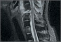
Since the standard imaging method for bone mineral density, dual-energy x-ray absorptiometry (DXA), can sometimes give inaccurate results in obese individuals, the investigators also measured bone density using quantitative computed tomography (QCT). They compared bone density at the lower spine and the hip in 50 obese adults (30 who had bariatric surgery and 20 who lost weight through nonsurgical ways but were similar to surgical patients in baseline age, sex, and body mass index). After surgery, nearly all patients received calcium and high-dose vitamin D supplementation.
Two years later, bone density was 5% to 7% lower at the spine and 7% to 10% lower at the hip in the surgical group compared with the nonsurgical control group, as shown by both DXA and QCT. In addition, the surgical patients had substantial and persistent increases in markers of bone resorption.
The bone loss in the surgical patients occurred despite the fact that the patients were not losing any more weight in the second post-surgical year and had stable blood levels of calcium and vitamin D. “Therefore, the cause of the bone loss is probably not related to weight loss itself,” Dr. Yu said.
None of the gastric bypass patients have required osteoporosis treatment, according to Dr. Yu. “The question is when is the bone loss going to stop? Over time this could be a problem in terms of fracture,” she said. Dr. Yu recommended that bariatric surgery candidates who have risk factors for osteoporosis receive bone density tests.
Patients who have had bariatric surgery continue to lose bone for at least two years, even after their weight stabilizes, according to study results presented at the 2014 joint meeting of the International Society of Endocrinology and the Endocrine Society. “The long-term consequences of this substantial bone loss are unclear, but it might put them at increased risk of fracture, or breaking a bone,” said lead study author Elaine Yu, MD, MSc, an endocrinologist at Massachusetts General Hospital in Boston and her research colleagues.
Dr. Yu’s team previously found that patients who have gastric bypass surgery lose bone mineral density within the first year after the surgery. Because the rate of bone loss was high, they researchers continued to monitor the patients in this study.
Since the standard imaging method for bone mineral density, dual-energy x-ray absorptiometry (DXA), can sometimes give inaccurate results in obese individuals, the investigators also measured bone density using quantitative computed tomography (QCT). They compared bone density at the lower spine and the hip in 50 obese adults (30 who had bariatric surgery and 20 who lost weight through nonsurgical ways but were similar to surgical patients in baseline age, sex, and body mass index). After surgery, nearly all patients received calcium and high-dose vitamin D supplementation.
Two years later, bone density was 5% to 7% lower at the spine and 7% to 10% lower at the hip in the surgical group compared with the nonsurgical control group, as shown by both DXA and QCT. In addition, the surgical patients had substantial and persistent increases in markers of bone resorption.
The bone loss in the surgical patients occurred despite the fact that the patients were not losing any more weight in the second post-surgical year and had stable blood levels of calcium and vitamin D. “Therefore, the cause of the bone loss is probably not related to weight loss itself,” Dr. Yu said.
None of the gastric bypass patients have required osteoporosis treatment, according to Dr. Yu. “The question is when is the bone loss going to stop? Over time this could be a problem in terms of fracture,” she said. Dr. Yu recommended that bariatric surgery candidates who have risk factors for osteoporosis receive bone density tests.
Denosumab Increases Bone Density in Postmenopausal Women With Osteoporosis
Postmenopausal women with osteoporosis who take denosumab long-term have increased bone density and a sustained low rate of fractures, according to study results presented at the 2014 joint meeting of the International Society of Endocrinology and the Endocrine Society. Lead study author E. Michael Lewiecki, MD, Clinical Assistant Professor of Medicine at the University of New Mexico School of Medicine in Albuquerque and research colleagues reported that long-term treatment with denosumab was safe and resulted in continuing increases in bone density over the eight years of treatment with persistently low fracture rates.
“This study provides reassurance to physicians and their patients that long-term treatment with denosumab for at least eight years leads to significant increases in bone density and is safe for appropriately selected women with postmenopausal osteoporosis.” He also noted that the overall risk of side effects did not increase over time.
Among the almost 8,000 women originally enrolled in the FREEDOM trial, denosumab reduced their risk of vertebral fractures by 68%, reduced their risk of nonvertebral fractures by 20%, and reduced their risk of hip fractures by 40%, compared with placebo. Women taking the drug had no increase in their overall risk for cancer, infection, cardiovascular disease, delayed fracture healing, or hypocalcemia.
All of the roughly 3,000 women in the long-term extension of the trial took denosumab for up to eight years. Overall, they showed a continued increase in their mean bone mineral density, with a cumulative eight-year gain of 18.4% at the lumbar spine and 8.3% at the total hip, with few fractures and a good safety profile. The tolerability profile of denosumab during this extension phase was consistent with that observed during the initial 3-year FREEDOM trial. At 12 months, denosumab treatment increased bone mineral density (BMD) at the total hip, lumbar spine and/or femoral neck and reduced markers of bone turnover to a significantly greater extent than oral bisphosphonates in women who were essentially bisphosphonate-naive and in those who had switched from alendronate to denosumab treatment.
MicroRNA Provides New Therapeutic Target for Bone Metastases and Osteoporosis
Researchers have identified miR-34a as a promising molecule that blocks bone destruction, according to a study published online ahead of print June 25 in Nature. Lead study author Yihong Wan, PhD, Assistant Professor of Pharmacology and member of the UT Southwestern Harold C. Simmons Cancer Center, and her research colleagues reported that mice with higher than normal levels of miR-34a had increased bone mass and reduced bone breakdown. According to the researchers, this is because miR-34a blocks osteoclasts, which make the bone less dense and prone to fracture.
“This new finding may lead to the development of miR-34a mimics as a new and better treatment for osteoporosis and cancers that metastasize to the bone,” said Dr. Wan.
Investigators found that injecting nanoparticles containing a mimic, or artificial version of miR-34a, into a mouse with post-menopausal osteoporosis decreased bone loss. miR-34a could also offer protection from bone metastases in a variety of cancers, Dr. Wan noted. Injecting the miR-34a mimic in the mice could prevent the metastasis of skin and breast cancer because it can disarm the metastatic niche in bone.
Study findings also suggest:
• MicroRNAs play important roles in physiology and disease, and present tremendous therapeutic potential.
• miR-34a is downregulated during osteoclast differentiation.
• Osteoclastic miR-34a-overexpressing transgenic mice exhibit lower bone resorption and higher bone mass.
• miR-34a knockout and heterozygous mice exhibit elevated bone resorption and reduced bone mass.
• Ovariectomy-induced osteoporosis, as well as bone metastasis of breast and skin cancers, are diminished in osteoclastic miR-34a transgenic mice, and can be effectively attenuated by miR-34a nanoparticle treatment.
• Transforming growth factor-β-induced factor 2 (Tgif2) as an essential direct miR-34a target that is pro-osteoclastogenic.
• Tgif2 deletion reduces bone resorption and abolishes miR-34a regulation.
Commenting on the study findings Dr. Wan said, “The mouse miR-34a is identical to that in humans, which means that our findings may apply to humans as well.”
Suggested Reading
Krzeszinski JY, Wei W, Huynh H, et al. miR-34a blocks osteoporosis and bone metastasis by inhibiting osteoclastogenesis and Tgif2. Nature. 2014 Jun 25. doi: 10.1038/nature13375. [Epub ahead of print]
Researchers have identified miR-34a as a promising molecule that blocks bone destruction, according to a study published online ahead of print June 25 in Nature. Lead study author Yihong Wan, PhD, Assistant Professor of Pharmacology and member of the UT Southwestern Harold C. Simmons Cancer Center, and her research colleagues reported that mice with higher than normal levels of miR-34a had increased bone mass and reduced bone breakdown. According to the researchers, this is because miR-34a blocks osteoclasts, which make the bone less dense and prone to fracture.
“This new finding may lead to the development of miR-34a mimics as a new and better treatment for osteoporosis and cancers that metastasize to the bone,” said Dr. Wan.
Investigators found that injecting nanoparticles containing a mimic, or artificial version of miR-34a, into a mouse with post-menopausal osteoporosis decreased bone loss. miR-34a could also offer protection from bone metastases in a variety of cancers, Dr. Wan noted. Injecting the miR-34a mimic in the mice could prevent the metastasis of skin and breast cancer because it can disarm the metastatic niche in bone.
Study findings also suggest:
• MicroRNAs play important roles in physiology and disease, and present tremendous therapeutic potential.
• miR-34a is downregulated during osteoclast differentiation.
• Osteoclastic miR-34a-overexpressing transgenic mice exhibit lower bone resorption and higher bone mass.
• miR-34a knockout and heterozygous mice exhibit elevated bone resorption and reduced bone mass.
• Ovariectomy-induced osteoporosis, as well as bone metastasis of breast and skin cancers, are diminished in osteoclastic miR-34a transgenic mice, and can be effectively attenuated by miR-34a nanoparticle treatment.
• Transforming growth factor-β-induced factor 2 (Tgif2) as an essential direct miR-34a target that is pro-osteoclastogenic.
• Tgif2 deletion reduces bone resorption and abolishes miR-34a regulation.
Commenting on the study findings Dr. Wan said, “The mouse miR-34a is identical to that in humans, which means that our findings may apply to humans as well.”
Suggested Reading
Krzeszinski JY, Wei W, Huynh H, et al. miR-34a blocks osteoporosis and bone metastasis by inhibiting osteoclastogenesis and Tgif2. Nature. 2014 Jun 25. doi: 10.1038/nature13375. [Epub ahead of print]
Suggested Reading
Krzeszinski JY, Wei W, Huynh H, et al. miR-34a blocks osteoporosis and bone metastasis by inhibiting osteoclastogenesis and Tgif2. Nature. 2014 Jun 25. doi: 10.1038/nature13375. [Epub ahead of print]