User login
Stillbirth Rates Slightly Higher With ART
Major Finding: The risk of stillbirth among children conceived by assisted reproductive technology is slightly, but significantly higher than that seen among naturally conceived children, accounting for 8 more stillbirths per 1,000 ART pregnancies.
Data Source: A Nordic population registry study of 60,650 ART pregnancies compared with a control group of 360,022 naturally conceived pregnancies.
Disclosures: The study was sponsored by the European IVF Monitoring Group, a working group of the European Society of Human Reproduction and Embryology. Dr. Henningsen said she had no relevant financial disclosures.
While the risk of stillbirth among singleton pregnancies conceived by assisted reproductive technology is marginally higher than is the risk in naturally conceived children, the difference is not large enough to warrant any changes in pregnancy management.
“Although the difference in risk is significant between the two groups, the overall risk is still very low,” Dr. Anna-Karina Henningsen said. “This means that the individual woman need not be concerned about the risk of stillbirth during pregnancy.”
The findings also contribute some data to the argument of whether to induce assisted reproductive technology (ART) pregnancies at term, rather than letting the pregnancy exceed 40 weeks, Dr. Henningsen said during a press briefing.
“In Nordic countries, mothers [can carry up to] 2 weeks past term before they are induced. Obstetricians have debated whether an ART pregnancy should be managed as high risk, with earlier induction. This study tells us that no cases of stillbirth in ART pregnancies can be avoided by inducing birth earlier,” she said.
Dr. Henningsen of the Rigshospitalet, Copenhagen, presented the results of the largest-ever Nordic investigation into this issue. The prospective database study comprised 60,650 singleton pregnancies conceived by ART, comparing them with a control group of 360,022 naturally conceived pregnancies. Information was drawn from population registries in Sweden, Finland, Norway, and Denmark, including ART registries, birth and death registries, and hospital registries. The analysis included singleton pregnancies conceived by in vitro fertilization (IVF), intracytoplasmic sperm injection (ICSI), and frozen embryo transfer.
When defining stillbirth as the birth of a dead infant after 22 weeks' gestation, the study found an overall rate of 0.4% in both groups. However, Dr. Henningsen said, after matching the two groups for maternal parity and the infants' birth year, and controlling for maternal age and the infant's gender, the overall risk was marginally higher (hazard ratio 1.1) in the ART group. This translated into an absolute rate of 8 additional stillbirths per 1,000 among ART pregnancies, compared with the control group.
The researchers also analyzed the stillbirth risk by gestational age. While there was no difference after 40 weeks' gestation, they found a 20% increased risk of stillbirth before 40 weeks among the ART group, compared with the control group. “We believe the difference in risk of stillbirth between ART and naturally conceived children seems to occur before 37 weeks' gestation,” she said. “It is likely that some of this difference in risk is related to factors affecting the subfertile mother or father.” For naturally conceived children, she said, stillbirth is more likely to be related to congenital malformations in the fetus.
In a further analysis of both stillbirth and death within the first year of life, Dr. Henningsen found that children born of ART were 40% more likely to die than were naturally conceived children. After adjusting for confounders, the rate still remained higher at 20%. However, she noted, “This can probably be explained by the higher risk of preterm birth among ART children.”
From the Annual Meeting of the European Society of Human Reproduction and Embryology
Stopping Smoking Anytime Helps Fetal Outcomes
It's never too late for a pregnant woman to stop smoking.
After reviewing the records of more than 50,000 pregnancies, Dr. Nick Macklon concluded that every day a pregnant woman doesn't smoke is a good day for her developing baby. “The more a woman smokes during pregnancy, the worse the effect on the baby,” Dr. Macklon said during a press briefing. “But stopping – even at the time a woman discovers she's pregnant – can completely ameliorate the effects of smoking” on the fetal outcomes of gestational age and birth weight. “For the baby, a mom stopping in the periconceptional phase is as good as her never having smoked at all.”
“We all know that smoking is bad for babies, increasing the rates of stillbirth, neonatal death, congenital malformations, preterm birth, and low birth weight – causing hardship to both parents and child. But it is also a significant public health issue in terms of cost,” said Dr. Macklon of the University of Southampton, England.
Dr. Macklon and his associates reviewed the records of 50,000 women who gave birth at Southampton hospitals from 2002 to 2010. Women were divided into seven groups, depending on how much they smoked: never, stopped in the last year, stopped more than 1 year ago, stopped at confirmation of pregnancy, and current smokers of up to 10 cigarettes each day, 10–20 each day, and more than 20. About 12,000 women decided to stop smoking when they discovered their pregnancy.
For nonsmokers, the mean gestational age at birth was 280 days – significantly longer than for those who smoked up to 10 cigarettes/day (279 days), 10–20/day (277 days), and 20 or more/day (276 days).
The gestational age of infants whose mothers ceased smoking a year or longer before birth was the same as those of never-smoking mothers. The surprise was that the gestational age of infants whose mothers who gave up cigarettes only when they became pregnant was exactly the same as the infants of never-smokers. This relationship remained significant even after the researchers corrected for other factors that affect gestational age, including education and socioeconomic status.
Birth weight also showed a similar relationship with smoking. The infants of current smokers were significantly smaller were than those of nonsmokers, as well as those who had quit a year or more before giving birth. Mothers who smoked up to 10 cigarettes/day had infants with a mean birth weight of 3.25 kg; mothers who smoked 10–20 cigarettes/day had infants weighing a mean 3.2 kg; and the infants of women who smoked more than 20 cigarettes/day weighed in at a mean 3.1 kg. “This effect is quite substantial, with a difference of more than 300 grams,” Dr. Macklon noted. Again, however, mothers who quit smoking as soon as they became pregnant conferred a significant benefit on their infants; these infants weighed a mean 3.4 kg – the same as those of women who had never smoked.
The findings shouldn't be construed as a free license to smoke until conception, he warned. “Many women don't plan their pregnancies and if they come in smoking and pregnant and we tell them it's too late to do anything, this sends a negative, and unnecessary, message. What we can now say is 'If you stop smoking now, you and your baby will get a major health benefit.'”
Smoking directly affects transplacental oxygen and nutrient flow, contributing to low birth weight and premature delivery. But couples who want to conceive should stop smoking for other reasons as well, he advised. “Smoking is a contraceptive. It's been shown to reduce the success of in vitro fertilization by at least 50%. Smoking affects the male partner as well, lowering fertility by impairing the DNA of sperm. Couples who want to conceive quickly and healthily should both stop smoking.” For women who want to quit before or during pregnancy, nicotine replacement therapy is “far less toxic than smoking, and even if we can't get a patient to stop completely I would support its use.” Dr. Macklon said he had no relevant financial disclosures.
From the Annual Meeting of the European Society of Human Reproduction and Embryology
Entheses on US Can Predict Spondyloarthritis
Major Finding: One vascularized enthesis seen on ultrasound predicted SpA within 2 years with a sensitivity of 76% and a specificity of 81%. An ultrasound finding of a nonvascularized enthesis, combined with positive Amor criteria, predicted the disorder at 2 years with a sensitivity of 90% and a specificity of 77%.
Data Source: A prospective cohort study of 118 patients with symptoms suggestive of spondyloarthritis.
Disclosures: The study was supported by a grant from the French Society of Rheumatology. None of the authors had any financial disclosures.
A power Doppler ultrasound examination may provide the most accurate early diagnosis of spondyloarthritis, with a sensitivity of 76% and a specificity of 81% upon the visualization of at least one vascularized enthesis.
The finding may be particularly valuable to rheumatologists because the existing spondyloarthritis diagnostic criteria have, at best, a limited ability to accurately identify the disease in its earliest stages, Dr. Maria Antoinette D'Agostino and her colleagues wrote.
Although the test itself is a “delicate technique,” it is within the reach of most ultrasound technicians, said Dr. D'Agostino of Versailles Saint-Quentin-en-Yveline (France) University.
The 2-year prospective cohort study comprised 118 patients with symptoms suggestive of spondyloarthritis. These included inflammatory back pain (48); arthritis or arthralgia (38); enthesitis or dactylitis (12); and HLA B27 plus acute anterior uveitis (20). Their median age was 40 years; the median disease duration at baseline was 2 years.
All patients underwent a standard clinical examination by a rheumatologist who was blinded to the diagnosis of the referring physician. All had provided a pelvic x-ray not more than 6 months old for the scoring of sacroiliitis; if the radiologic findings were equivocal or if there was persistent buttock pain, the patients underwent a pelvic CT scan.
Every patient had a power Doppler ultrasound examination of peripheral entheses by a sonographer who was blinded to the patients' data. Areas examined included plantar fascia, Achilles tendon, patellar ligament on the patella apex, quadriceps femoris, gluteus medius tendon, and the common extensor and common flexor tendons on the lateral and medial epicondyle of the elbow.
Important findings included any morphologic or structural abnormalities and vascularization at bony insertion points. The study evaluated three criteria: any vascularized enthesis, the number of abnormal entheses, and the global ultrasound score.
The referring physician's diagnosis was used as the clinical standard in evaluating ultrasound's diagnostic capability; after 2 years, the patients were reevaluated for a final diagnosis, which the investigators then compared with the original diagnosis to compute ultrasound's diagnostic capability. At the end of the follow-up period, patients were reclassified by their referring rheumatologist (51 diagnosed with SpA, 48 not diagnosed as SpA, and 19 unclassified).
In building the prediction model, the investigators examined the ultrasound findings in light of the final diagnoses. Ultrasound found at least one abnormal enthesis in 88 (75%) of the patients; the enthesis was vascularized in 56 of these patients. At least one vascularized enthesis occurred in 76% of those with an SpA diagnosis, 19% of those with a non-SpA diagnosis, and 42% of unclassified patients (Ann. Rheum. Dis. 2011;70:1433-40).
Ultrasound detected significantly more abnormal and vascularized entheses in SpA patients than in non-SpA patients. Those with a SpA diagnosis also had significantly higher ultrasound global scores than did the other groups.
Overall, two factors independently predicted a final diagnosis of SpA: Patients who had at least one vascularized enthesis on ultrasound were 12 times more likely to have a final diagnosis of SpA, and patients with an Amor criteria score of 6 or greater were nearly nine times more likely to have SpA than patients with a lower score. Further analysis confirmed that the baseline presence of at least one vascularized enthesis predicted SpA at 2 years with a 76.5% sensitivity and an 81% specificity. If there were no vascularized entheses at baseline, SpA could still be predicted with a combination of ultrasound and positive Amor criteria score, the authors said; this method yielded a sensitivity of 90% for SpA and a specificity of 77%. “Strikingly, we confirmed that vascularization of the enthesis insertion by [ultrasound] is a landmark feature for SpA, even in suspected cases,” the authors said.
DBS Doesn't Alter Course of Long-standing Parkinson's Disease
Subthalamic nucleus deep brain stimulation doesn’t appear to change the course of Parkinson’s disease when it is performed on patients with long-standing disease.
In the longest follow-up cohort study to date, Dr. Aristide Merola and his colleagues tracked patients who underwent the procedure at about age 60 years. They all showed similar declines in cognition, gait stability, and continence – marks of the disease’s later stage, according to their study, which was published in the July issue of Brain.
However, the study should not be construed as a negation of DBS’s potential therapeutic value, wrote Dr. Merola of the University of Turin (Italy) and his coauthors. Instead, they suggested that one can "speculate whether the subthalamic nucleus DBS surgical procedure should be proposed earlier, considering that Parkinson’s disease progression might not follow a linear course, and it is possible that age might influence the development of non-motor features more than disease duration" (Brain 2011;134:2074-84).
The study comprised 19 patients who underwent subthalamic nucleus DBS after a mean disease duration of 22 years. The investigators compared the baseline results of clinical and neuropsychological testing to those at 1 year, 3 years, and 5 years. For 14 patients who underwent testing after more than 7 years post surgery, the mean follow-up duration was 8 years.
The cohort consisted of 9 men and 10 women. The patients had developed disease symptoms relatively early in life (at a mean of 39 years). Their mean age at the intervention was 61 years.
The patients were evaluated with the UPDRS (Unified Parkinson\'s Disease Rating Scale) at baseline, which was before the DBS. After the procedure, the scale was administered at each follow-up point in four disease states: stimulation on/medication off, stimulation off/medication off, stimulation off/medication on, and stimulation on/medication on.
The authors noted that they paid "particular attention to the main axial, non-motor, and psychiatric symptoms," assessing subjects for falls, postural instability, non-levodopa responsive gait freezing, urinary incontinence, dysphagia, and speech difficulty. They used pharmacologic treatment as a measure of other symptoms, including constipation, postural hypotension, depression, and hallucinations.
Freedom from those symptoms is a prerequisite for DBS surgery; therefore at baseline, none of the patients showed evidence of them.
Long-Term DBS Treatment Evaluations
Follow-up considered not only the years out from surgery, but also the years of disease duration. The long-term follow-up evaluations showed a similar pattern of symptom progression in all patients, regardless of the disease state in which they were measured. However, the combination of stimulation on/medication on was consistently more effective at controlling symptoms than were the other states.
"The majority of patients progressively developed falls (64%), postural instability (100%), non-levodopa responsive freezing of gait (64%), dysphagia (86%), urinary incontinence (57%), severe postural hypotension (36%) and dementia (43%) during the course of follow-up," the authors wrote. "On the other hand, neuropsychological data showed a gradual decline in the performance of all the main cognitive domains, in agreement with previous findings" that showed a fivefold prevalence of dementia in patients with Parkinson’s disease, compared with the general population.
Complications of levodopa therapy significantly improved initially after DBS. But this improvement gradually decreased over the follow-up time. The same pattern occurred in activities of daily living. The mean levodopa equivalent daily dose decreased in the first year after surgery (from a mean of 890 mg to 336 mg). Over the entire follow-up, the dose continued to increase, but only rose to a mean of 435 mg/day after more than 7 years.
When the investigators examined the progression of falls, postural instability, and gait freezing, a similar pattern emerged. There was a slight increase from 1 to 3 years, and then a sharper increase at 5 years. After more than 7 years – with 14 patients still being followed – 9 of them had postural instability, all had gait freezing, and 9 had fallen.
"The initial positive effects of subthalamic nucleus DBS on balance and postural stability ... in the first years from surgery seem to be mostly related to the improvement of rigidity and bradykinesia, rather than to a specific effect on balance and gait," the authors noted.
At baseline, only one patient showed moderate dysphagia. During the follow-up period, the incidence of dysphagia increased, as did speech difficulties. After more than 7 years, 12 had dysphagia, 3 required a percutaneous gastrostomy, and 9 had speech disturbance.
Constipation and postural hypotension, as measured by the need for drug intervention, remained low for 3 years after surgery. For those who reached a follow-up observation longer than 7 years, half required a catheter or diaper.
"We found a global high rate of axial symptoms development during the course of follow-up, with a progressive lessening of both medication and stimulation effectiveness, and a global reduction in the synergistic effect of the ‘stimulation on/medication/on’ condition," the authors wrote. Other studies have suggested that this decline is a combination of the disease’s natural course and a decrease in response to dopaminergic therapy.
Dementia, depression, and hallucinations also became increasingly common over the follow-up period. After more than 7 years of follow-up, six patients required medication for depression, nine required medication for hallucinations, and six developed dementia.
"Our main findings suggest that, in spite of the prior clinical evolution and the young age at onset, non-levodopa-responsive symptoms progressively emerged" beyond 20 years of disease, the authors said. "In fact, at 30 years from the disease onset, more than 70% of subjects showed dementia, falls, dysphagia, or urinary incontinence."
None of the authors had any relevant disclosures.
FROM BRAIN
Major Finding: The classical axial and cognitive symptoms of advanced Parkinson’s disease developed in 40%-100% of patients who had undergone subthalamic nucleus DBS surgery.
Data Source: A study of 19 patients who received follow-up testing for a mean of 8 years after undergoing DBS at about age 60 years, after having had the disease for a mean of 22 years.
Disclosures: None of the authors had any relevant disclosures.
Medical, Cosmetic Dermatologists Agree: Health Before Beauty
Medical and cosmetic dermatologists may bring different views to the discussion of skin care, but they do agree on one thing – health should always come before beauty.
Basic dermatologic health procedures, like total body skin checks, mole assessment, and sun exposure advice, should be the foundation of any cosmetic dermatologist's work. No amount of wrinkle reduction, laser resurfacing, or complexion correction can ever make up for a missed skin cancer, according to cosmetic dermatologists.
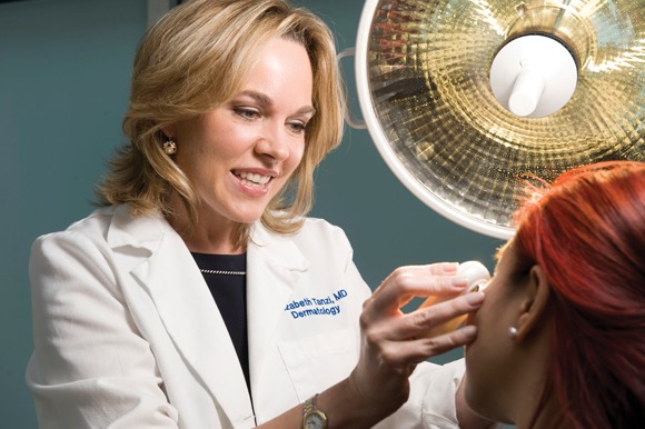
"I take this responsibility very seriously," said Dr. Elizabeth Tanzi, a Washington, D.C. dermatologist who specializes in laser surgery. "All our patients, whether new or returning, get a full head-to-toe exam for skin cancer. It would be tragic to be talking about wrinkles and crepey skin when there’s a skin cancer."
Dr. Tanzi said that health should be center of any dermatologic procedure – and that cosmetic procedures are no exception.
"We have to look at it like this: Number one, make sure your skin is healthy. Number two: beautify it."
Dermatologic practices are split into three categories: all cosmetic, all medical, or a combination of the two. Purely medical dermatologists tend to cluster in academic institutions, while purely cosmetic dermatologists are most often seen in large cities. Dual-therapy dermatologists are the workhorses who serve most communities.
All dermatologists need to know when to proceed and when to say "no," Dr. Tanzi said. Referral to a medical dermatologist may cost a patient in the short term, but will serve everyone well in the long term.
"If a patient comes to me with an issue in my area of expertise and I know I can do a great job, I’ll take it," said Dr. Tanzi, codirector of the Washington Institute of Dermatologic Laser Surgery. "If they come in with something where I’m not up on the latest, or something I haven't seen in years and feel I won’t be best for the job, I refer. An urban setting really allows this to happen. In D.C. we have the luxury of saying that there is someone nearby who is an expert in any field. It is a different story in communities where you need to be a jack of all trades."
A Jack of All Trades
Dr. Margaret "Peggy" Fitch fills that bill. "I’m a surgical and cosmetic dermatologist in the morning and a medical dermatologist in the afternoon," said Dr. Fitch, founder of Aiken Dermatology, Aiken, S.C.
She didn't always split her time between the disciplines, however. When she began practicing 30 years ago, she focused on medical and surgical dermatology. Then things began to change.
"Over time, peels, fillers, Botox, and sclerotherapy came along. They were on the front page of Cosmopolitan. Patients were asking about them. And I was seeing the results of bad outcomes – procedures done by physicians who shouldn’t have been doing them."
Taking a weekend course in a cosmetic procedure does not make an expert, Dr. Fitch said.
"Let’s be honest – there are lots of doctors out there who do this just to make a quick buck, and I was seeing the results of that. So I thought, 'I'm a dermatologist. I know skin and the anatomic structure of the face. I should be doing this.' So gradually I learned and added to my practice."
She doesn't shy away from discussing the financial benefits of cosmetic work.
"Sure, it's a revenue builder. But I let patients decide what they want. If someone comes to me for a medical issue, I will never, ever bring up a cosmetic issue. I think that is foraging in the forest for a buck. If a patient asks about Botox while we're talking, that's another issue. But as a doctor, I would never bring up someone’s wrinkles."
She isn’t afraid to say "no" either. "I say no all the time. I’m not above saying, 'Look you don’t need this.' "
For example, she said, a 27-year-old bride-to-be asked her for filler for her lips. Dr. Fitch refused, telling her the lines were too fine to be treated, and that she was too young to be looking at the procedure. "I could have made $500 for that. But I think I'm the exception to the rule here. Most people would have gone and done it."
As a surgical dermatologist, she concentrates on skin cancers, using her own in-office pathology lab to assess the specimens. "I do flaps and grafts, but I would never attempt a full Mohs. That’s all part of knowing when you must say to a patient, 'I'm a good doctor and a good surgeon, but I'm not the best person for this.' "
Taking on New Roles
Dr. Sue Ellen Cox is another dermatologist who enjoys mixing it up. While 80% of her practice is cosmetic, the rest of her patients have medical issues. "If a patient comes to me and doesn't already have a general dermatologist, I'll take on that role without a problem. I really enjoy doing that – it gives me some variety and keeps me sharp."
When she began her 16-year-old practice in Chapel Hill, N.C., she did outreach to local dermatologists. With several years of specialized training in collagen, lasers, and liposuction, and a research study on chemical peels, Dr. Cox felt like she could bring something new and valuable to the medical community.
"I went to all the dermatologists in the area and told them about my specialized training and asked them to send me any cosmetic cases they weren't comfortable with. My practice just took off from there," she said.
At the same time, she never bites the hand that feeds her. "If someone is referred to me for laser and I see a basal cell carcinoma, for example, I'll tell them to talk to their referring physician before I do anything. Referrals are a lifeline, and I don't want to cut that."
Dermatologists who combine both medical and cosmetic work need to be scrupulous in their billing, she warned.
"If I'm doing a laser treatment for rosacea, that's a fee for service procedure. If I'm diagnosing rosacea for the first time, I bill insurance. If I diagnose lentigoes and precancerous lesions and treat them, I bill insurance. But if the main benefit is cosmetic, it's self pay. The same with moles – removal of a benign mole for cosmetic reasons is one thing, removal and biopsy of a suspicious mole is another."
"We Treat Cosmetic Issues, Not Health Issues"
Dr. Eliot Battle has no issues with insurance billing. As a completely cosmetic dermatologist, his business is all self pay. And his patients know that from the beginning.

"There is no confusion about what we do. We treat cosmetic issues – not health issues. I don't take insurance, and our advertising and website are very clear about this. If we see someone with medical issues, we push them to a medical dermatologist – that saves them money, and we know that medical derms are better for these things than we would ever be."
Dr. Battle has built his practice on four areas – laser hair removal, complexion blending, textural rejuvenation, and antiaging procedures. About 100 patients, primarily women, come through his office every day, 6 days a week. Dr. Battle specializes in treating darker skin, which is much more prone to the scarring and pigmentation changes that can result from inexpert cosmetic treatment.
Dr. Battle does few of the procedures. He conducts all of the consultations and designs the treatment plans, but registered nurses perform the procedures. "I work with six full-time nurses, who undergo a strict internal training program and participate in ongoing medical education. I send them to every possible meeting that could be of help, and I support all this education financially."
Dr. Battle brings a unique set of business skills to his medical practice. After working as a marketing director for a large computer firm, he decided to enroll in medical school.
"I took my first science course at age 33 and was a dermatology resident at age 41," he said. "When I finally finished, my passion was cosmetic dermatology. At that point lasers were still in their infancy, and I worked for 3 years in research about laser safety – especially for skin of color."
He designed his cosmetic work as not only a practice but as a business, with his children's future in mind. "I wanted to make sure they could inherit my own success," he said. "Children cannot inherit a physician's practice, but they can inherit a business." Dr. Battle does not treat patients himself, so the Cultura Cosmetic Dermatology and Laser Center in Washington, is considered a business rather than a practice.
Because he offers limited procedures, his staff has become expert in performing them, he said. But when he encounters anything beyond what he is prepared to offer, Dr. Battle refers to a medical dermatologist.
"My medical oath says to do no harm, and that rules all that I do. We are always looking out for the patient's best interest. If I see a suspicious mole or a concerning flare of lupus, I refer. I am not geared to help that person."
Dr. Battle hasn't completely foresworn either medical or academic work, however. He conducts research at Howard University, Washington, and teaches residents who are interested in cosmetic dermatology.
His business background puts him in a position to offer business advice, and his age, he said, allows him to give personal counsel as well.
"One thing I tell residents is that just because they might generate a lot of revenue with a cosmetic practice doesn't necessarily mean they'll generate a lot of profit. It's also important for people to be true to themselves. I encourage them to look deeply into this and not be blinded by the idea that they'll make tons of money doing cosmetic work. A lot of residents really enjoy the academic side of medicine, and cosmetic dermatology will not bring them that. If academics are your passion, you should not forego that for the temporary dollar."
Dr. Tanzi is on the medical advisory board for Zeltiq. Dr. Cox is a consultant for Medisys and Allergan. Dr. Battle and Dr. Fitch reported having no conflicts of interest.
Medical and cosmetic dermatologists may bring different views to the discussion of skin care, but they do agree on one thing – health should always come before beauty.
Basic dermatologic health procedures, like total body skin checks, mole assessment, and sun exposure advice, should be the foundation of any cosmetic dermatologist's work. No amount of wrinkle reduction, laser resurfacing, or complexion correction can ever make up for a missed skin cancer, according to cosmetic dermatologists.
"I take this responsibility very seriously," said Dr. Elizabeth Tanzi, a Washington, D.C. dermatologist who specializes in laser surgery. "All our patients, whether new or returning, get a full head-to-toe exam for skin cancer. It would be tragic to be talking about wrinkles and crepey skin when there’s a skin cancer."
Dr. Tanzi said that health should be center of any dermatologic procedure – and that cosmetic procedures are no exception.
"We have to look at it like this: Number one, make sure your skin is healthy. Number two: beautify it."
Dermatologic practices are split into three categories: all cosmetic, all medical, or a combination of the two. Purely medical dermatologists tend to cluster in academic institutions, while purely cosmetic dermatologists are most often seen in large cities. Dual-therapy dermatologists are the workhorses who serve most communities.
All dermatologists need to know when to proceed and when to say "no," Dr. Tanzi said. Referral to a medical dermatologist may cost a patient in the short term, but will serve everyone well in the long term.
"If a patient comes to me with an issue in my area of expertise and I know I can do a great job, I’ll take it," said Dr. Tanzi, codirector of the Washington Institute of Dermatologic Laser Surgery. "If they come in with something where I’m not up on the latest, or something I haven't seen in years and feel I won’t be best for the job, I refer. An urban setting really allows this to happen. In D.C. we have the luxury of saying that there is someone nearby who is an expert in any field. It is a different story in communities where you need to be a jack of all trades."
A Jack of All Trades
Dr. Margaret "Peggy" Fitch fills that bill. "I’m a surgical and cosmetic dermatologist in the morning and a medical dermatologist in the afternoon," said Dr. Fitch, founder of Aiken Dermatology, Aiken, S.C.
She didn't always split her time between the disciplines, however. When she began practicing 30 years ago, she focused on medical and surgical dermatology. Then things began to change.
"Over time, peels, fillers, Botox, and sclerotherapy came along. They were on the front page of Cosmopolitan. Patients were asking about them. And I was seeing the results of bad outcomes – procedures done by physicians who shouldn’t have been doing them."
Taking a weekend course in a cosmetic procedure does not make an expert, Dr. Fitch said.
"Let’s be honest – there are lots of doctors out there who do this just to make a quick buck, and I was seeing the results of that. So I thought, 'I'm a dermatologist. I know skin and the anatomic structure of the face. I should be doing this.' So gradually I learned and added to my practice."
She doesn't shy away from discussing the financial benefits of cosmetic work.
"Sure, it's a revenue builder. But I let patients decide what they want. If someone comes to me for a medical issue, I will never, ever bring up a cosmetic issue. I think that is foraging in the forest for a buck. If a patient asks about Botox while we're talking, that's another issue. But as a doctor, I would never bring up someone’s wrinkles."
She isn’t afraid to say "no" either. "I say no all the time. I’m not above saying, 'Look you don’t need this.' "
For example, she said, a 27-year-old bride-to-be asked her for filler for her lips. Dr. Fitch refused, telling her the lines were too fine to be treated, and that she was too young to be looking at the procedure. "I could have made $500 for that. But I think I'm the exception to the rule here. Most people would have gone and done it."
As a surgical dermatologist, she concentrates on skin cancers, using her own in-office pathology lab to assess the specimens. "I do flaps and grafts, but I would never attempt a full Mohs. That’s all part of knowing when you must say to a patient, 'I'm a good doctor and a good surgeon, but I'm not the best person for this.' "
Taking on New Roles
Dr. Sue Ellen Cox is another dermatologist who enjoys mixing it up. While 80% of her practice is cosmetic, the rest of her patients have medical issues. "If a patient comes to me and doesn't already have a general dermatologist, I'll take on that role without a problem. I really enjoy doing that – it gives me some variety and keeps me sharp."
When she began her 16-year-old practice in Chapel Hill, N.C., she did outreach to local dermatologists. With several years of specialized training in collagen, lasers, and liposuction, and a research study on chemical peels, Dr. Cox felt like she could bring something new and valuable to the medical community.
"I went to all the dermatologists in the area and told them about my specialized training and asked them to send me any cosmetic cases they weren't comfortable with. My practice just took off from there," she said.
At the same time, she never bites the hand that feeds her. "If someone is referred to me for laser and I see a basal cell carcinoma, for example, I'll tell them to talk to their referring physician before I do anything. Referrals are a lifeline, and I don't want to cut that."
Dermatologists who combine both medical and cosmetic work need to be scrupulous in their billing, she warned.
"If I'm doing a laser treatment for rosacea, that's a fee for service procedure. If I'm diagnosing rosacea for the first time, I bill insurance. If I diagnose lentigoes and precancerous lesions and treat them, I bill insurance. But if the main benefit is cosmetic, it's self pay. The same with moles – removal of a benign mole for cosmetic reasons is one thing, removal and biopsy of a suspicious mole is another."
"We Treat Cosmetic Issues, Not Health Issues"
Dr. Eliot Battle has no issues with insurance billing. As a completely cosmetic dermatologist, his business is all self pay. And his patients know that from the beginning.
"There is no confusion about what we do. We treat cosmetic issues – not health issues. I don't take insurance, and our advertising and website are very clear about this. If we see someone with medical issues, we push them to a medical dermatologist – that saves them money, and we know that medical derms are better for these things than we would ever be."
Dr. Battle has built his practice on four areas – laser hair removal, complexion blending, textural rejuvenation, and antiaging procedures. About 100 patients, primarily women, come through his office every day, 6 days a week. Dr. Battle specializes in treating darker skin, which is much more prone to the scarring and pigmentation changes that can result from inexpert cosmetic treatment.
Dr. Battle does few of the procedures. He conducts all of the consultations and designs the treatment plans, but registered nurses perform the procedures. "I work with six full-time nurses, who undergo a strict internal training program and participate in ongoing medical education. I send them to every possible meeting that could be of help, and I support all this education financially."
Dr. Battle brings a unique set of business skills to his medical practice. After working as a marketing director for a large computer firm, he decided to enroll in medical school.
"I took my first science course at age 33 and was a dermatology resident at age 41," he said. "When I finally finished, my passion was cosmetic dermatology. At that point lasers were still in their infancy, and I worked for 3 years in research about laser safety – especially for skin of color."
He designed his cosmetic work as not only a practice but as a business, with his children's future in mind. "I wanted to make sure they could inherit my own success," he said. "Children cannot inherit a physician's practice, but they can inherit a business." Dr. Battle does not treat patients himself, so the Cultura Cosmetic Dermatology and Laser Center in Washington, is considered a business rather than a practice.
Because he offers limited procedures, his staff has become expert in performing them, he said. But when he encounters anything beyond what he is prepared to offer, Dr. Battle refers to a medical dermatologist.
"My medical oath says to do no harm, and that rules all that I do. We are always looking out for the patient's best interest. If I see a suspicious mole or a concerning flare of lupus, I refer. I am not geared to help that person."
Dr. Battle hasn't completely foresworn either medical or academic work, however. He conducts research at Howard University, Washington, and teaches residents who are interested in cosmetic dermatology.
His business background puts him in a position to offer business advice, and his age, he said, allows him to give personal counsel as well.
"One thing I tell residents is that just because they might generate a lot of revenue with a cosmetic practice doesn't necessarily mean they'll generate a lot of profit. It's also important for people to be true to themselves. I encourage them to look deeply into this and not be blinded by the idea that they'll make tons of money doing cosmetic work. A lot of residents really enjoy the academic side of medicine, and cosmetic dermatology will not bring them that. If academics are your passion, you should not forego that for the temporary dollar."
Dr. Tanzi is on the medical advisory board for Zeltiq. Dr. Cox is a consultant for Medisys and Allergan. Dr. Battle and Dr. Fitch reported having no conflicts of interest.
Genes Play Bigger Role in MI Than Stroke
People whose mother and father have both had a myocardial infarction are six times more likely to have one than are those without a parental history, according to a large, population-based study.
Strokes, on the other hand, do not seem related to genetic predisposition.

Because most risk-estimation models include both cardiovascular and cerebrovascular factors, the findings could influence the way such risks are calculated, Dr. Peter Rothwell and colleagues wrote in an article published online ahead of print in the journal Circulation: Cardiovascular Genetics.
"The way physicians predict the odds of a healthy person suffering a heart attack or stroke needs refining," Dr. Rothwell, senior author and professor of neurology at Oxford (England) University, said in a press statement. "Currently, most risk models lump a patient’s family history of stroke and heart attack together. We probably should model family history of stroke and heart attack separately in the future."
Dr. Rothwell and his coauthors reported a subanalysis of the Oxford Vascular Study (OXVASC), a population-based study that examined transient ischemic attacks, strokes, acute coronary syndrome (ACS), and acute peripheral vascular events in 91,106 people.
The investigators focused on 1,921 patients who had ACS or stroke and had information on cardiovascular and cerebrovascular events in both their parents and siblings. Among these, 906 had experienced an ACS and 1,015 some kind of cerebrovascular event (Circ. Cardiovasc. Genet. 2011;DOI:10.1161/CIRCGENETICS.110.959114).
The authors found clusters of ACS in many families: 21% of patients (191) had at least one sibling who had had an MI and 7% (64) had at least two siblings with the event. There was also a strong parental influence. Nearly a third of patients (277) had one parent with a positive history of MI and in 5% (47) both parents had a positive history.

The associations were not as strong in patients with cerebrovascular events. Of these patients, 8% (82) had least one sibling with a stroke history and 1% (14) at least two siblings with a history. Twenty-one percent (216) of patients had one parent with a positive history, while 2% (21) had two parents with a positive history.
When the investigators considered the proband families, they found significant relationships between the number of affected parents and the number of affected siblings. The ACS patients’ 2,601 siblings were 48% more likely to have had an ACS when one parent had a positive MI history and six times more likely when both parents did.
These associations were not significant among stroke patients, however. Among the 2,692 siblings of the stroke patients, there were no significant relationships with parental history of stroke (either one or both parents).
The study plays into the knowledge that atherosclerosis and plaque – the main risk factors of MI – are more heritable than thromboembolism and small-vessel disease, which are the main risk factors for stroke.
"Coronary disease may be a better indicator of generalized atherosclerosis than stroke, and therefore family history of MI may represent a greater risk factor for MI and stroke, whereas family history of stroke is not a strong risk factor for MI," Dr. Rothwell and his associates noted. "Our results have implications for clinical practice and for future research. In particular, the use of composite measures of family history of vascular disease in risk scores and in screening may not be optimal since the heritability of stroke is much less than that of MI."
The study was primarily sponsored by the U.K. Medical Research Council. None of the authors had any financial disclosures.
People whose mother and father have both had a myocardial infarction are six times more likely to have one than are those without a parental history, according to a large, population-based study.
Strokes, on the other hand, do not seem related to genetic predisposition.
Because most risk-estimation models include both cardiovascular and cerebrovascular factors, the findings could influence the way such risks are calculated, Dr. Peter Rothwell and colleagues wrote in an article published online ahead of print in the journal Circulation: Cardiovascular Genetics.
"The way physicians predict the odds of a healthy person suffering a heart attack or stroke needs refining," Dr. Rothwell, senior author and professor of neurology at Oxford (England) University, said in a press statement. "Currently, most risk models lump a patient’s family history of stroke and heart attack together. We probably should model family history of stroke and heart attack separately in the future."
Dr. Rothwell and his coauthors reported a subanalysis of the Oxford Vascular Study (OXVASC), a population-based study that examined transient ischemic attacks, strokes, acute coronary syndrome (ACS), and acute peripheral vascular events in 91,106 people.
The investigators focused on 1,921 patients who had ACS or stroke and had information on cardiovascular and cerebrovascular events in both their parents and siblings. Among these, 906 had experienced an ACS and 1,015 some kind of cerebrovascular event (Circ. Cardiovasc. Genet. 2011;DOI:10.1161/CIRCGENETICS.110.959114).
The authors found clusters of ACS in many families: 21% of patients (191) had at least one sibling who had had an MI and 7% (64) had at least two siblings with the event. There was also a strong parental influence. Nearly a third of patients (277) had one parent with a positive history of MI and in 5% (47) both parents had a positive history.
The associations were not as strong in patients with cerebrovascular events. Of these patients, 8% (82) had least one sibling with a stroke history and 1% (14) at least two siblings with a history. Twenty-one percent (216) of patients had one parent with a positive history, while 2% (21) had two parents with a positive history.
When the investigators considered the proband families, they found significant relationships between the number of affected parents and the number of affected siblings. The ACS patients’ 2,601 siblings were 48% more likely to have had an ACS when one parent had a positive MI history and six times more likely when both parents did.
These associations were not significant among stroke patients, however. Among the 2,692 siblings of the stroke patients, there were no significant relationships with parental history of stroke (either one or both parents).
The study plays into the knowledge that atherosclerosis and plaque – the main risk factors of MI – are more heritable than thromboembolism and small-vessel disease, which are the main risk factors for stroke.
"Coronary disease may be a better indicator of generalized atherosclerosis than stroke, and therefore family history of MI may represent a greater risk factor for MI and stroke, whereas family history of stroke is not a strong risk factor for MI," Dr. Rothwell and his associates noted. "Our results have implications for clinical practice and for future research. In particular, the use of composite measures of family history of vascular disease in risk scores and in screening may not be optimal since the heritability of stroke is much less than that of MI."
The study was primarily sponsored by the U.K. Medical Research Council. None of the authors had any financial disclosures.
FROM CIRCULATION: CARDIOVASCULAR GENETICS
Major Finding: Heart attacks are significantly more likely than strokes to occur in family clusters, with 21% of heart attack patients having at least one similarly affected sibling, compared with 8% of stroke patients.
Data Source: A subanalysis of more than 91,000 subjects who had experienced either a heart attack or stroke.
Disclosures: The study was primarily sponsored by the U.K. Medical Research Council. None of the authors had any financial disclosures.
Entheses Seen on Ultrasound Can Accurately Predict Spondyloarthritis
A power Doppler ultrasound examination may provide the most accurate early diagnosis of spondyloarthritis, with a sensitivity of 76% and a specificity of 81% upon the visualization of at least one vascularized enthesis.
The finding may be particularly valuable to rheumatologists because the existing spondyloarthritis diagnostic criteria have, at best, a limited ability to accurately identify the disease in its earliest stages, Dr. Maria Antoinette D’Agostino and her colleagues wrote in the August issue of Annals of the Rheumatic Diseases.
Although the test itself is a "delicate technique," it is within the reach of most ultrasound technicians. "It can be performed reliably by sonographers with varying levels of experience followed by dedicated training," said Dr. D’Agostino of Versailles Saint-Quentin-en-Yveline (France) University.
The 2-year prospective cohort study comprised 118 patients with symptoms suggestive of spondyloarthritis. These included inflammatory back pain (48); arthritis or arthralgia (38); enthesitis or dactylitis (12) and HLA B27 plus acute anterior uveitis (20). Their median age was 40 years; the median disease duration at baseline was 2 years.
All patients underwent a standard clinical examination by a rheumatologist who was blinded to the diagnosis of the referring physician. All had provided a pelvic x-ray not more than 6 months old for the scoring of sacroiliitis; if the radiologic findings were equivocal or if there was persistent buttock pain, the patients underwent a pelvic CT scan. Those with past or present inflammatory back pain also underwent MRI.
Every patient had a power Doppler ultrasound examination of peripheral entheses by a sonographer who was blinded to the patients’ data. Areas examined included plantar fascia, Achilles tendon, patellar ligament on the patella apex, quadriceps femoris, gluteus medius tendon, and the common extensor and common flexor tendons on the lateral and medial epicondyle of the elbow.
Important findings included any morphologic or structural abnormalities and vascularization at bony insertion points. The study evaluated three criteria: any vascularized enthesis, the number of abnormal entheses, and the global ultrasound score.
The referring physician’s diagnosis was used as the clinical standard in evaluating ultrasound’s diagnostic capability; after 2 years, the patients were reevaluated for a final diagnosis, which the investigators then compared with the original diagnosis to compute ultrasound’s diagnostic capability. At the end of the follow-up period, patients were reclassified by their referring rheumatologist (51 diagnosed with SpA, 48 not diagnosed as SpA, and 19 unclassified).
In building the prediction model, the investigators examined the ultrasound findings in light of the final diagnoses. Ultrasound found at least one abnormal enthesis in 88 (75%) of the patients; the enthesis was vascularized in 56 of these patients. At least one vascularized enthesis occurred in 76% of those with an SpA diagnosis, 19% of those with a non-SpA diagnosis, and 42% of unclassified patients (Ann. Rheum. Dis. 2011;70:1433-40).
Ultrasound detected significantly more abnormal and vascularized entheses in SpA patients than in non-SpA patients. Those with a SpA diagnosis also had significantly higher ultrasound global scores than did the other groups.
Overall, two factors independently predicted a final diagnosis of SpA: Patients who had at least one vascularized enthesis on ultrasound were 12 times more likely to have a final diagnosis of SpA, and patients with an Amor criteria score of 6 or greater were nearly nine times more likely to have SpA than patients with a lower score.
Increasing the number of vascularized entheses to more than one did not improve the prediction model, nor did other diagnostic criteria, including the Berlin criteria and ASAS (Assessment of Spondyloarthritis Society) classification and diagnostic criteria.
Further analysis confirmed that the baseline presence of at least one vascularized enthesis predicted SpA at 2 years with a 76.5% sensitivity and an 81% specificity. If there were no vascularized entheses at baseline, SpA could still be predicted with a combination of ultrasound and positive Amor criteria score, the authors said; this method yielded a sensitivity of 90% for SpA and a specificity of 77%.
The study points out the diagnostic importance of enthesis vascularization in early SpA, the authors noted: "Strikingly, we confirmed that vascularization of the enthesis insertion by [ultrasound] is a landmark feature for SpA, even in suspected cases ... One vascularized enthesis was sufficient to predict a diagnosis of SpA, independent of the localization and the frequency of involvement."h
The study was supported by a grant from the French Society of Rheumatology. None of the authors had any financial disclosures.
FROM ANNALS OF THE RHEUMATIC DISEASES
Major Finding: One vascularized enthesis seen on ultrasound predicted SpA within 2 years with a sensitivity of 76% and a specificity of 81%. An ultrasound finding of a nonvascularized enthesis, combined with positive Amor criteria, predicted the disorder at 2 years with a sensitivity of 90% and a specificity of 77%.
Data Source: A prospective cohort study of 118 patients with symptoms suggestive of spondyloarthritis.
Disclosures: The study was supported by a grant from the French Society of Rheumatology. None of the authors had any financial disclosures.
Frequent Migraines Not Linked to Cognitive Impairment in Women
WASHINGTON – Women with a high burden of episodic migraine don’t appear to have any related cognitive impairment.
A small prospective study has found that women with as many as 10 migraines per month scored well within the normal range on tests of learning and memory, efficiency and attention, and processing speed, Jill Jesurum, Ph.D., reported in a poster at the annual meeting of the American Headache Society.
"This is very good news for women with migraine," said Dr. Jesurum, scientific director of the Swedish Heart and Vascular Institute at the Swedish Medical Center, Seattle. "I see migraine patients in my own clinic, and many are very worried because they feel they have some type of cognitive problems from their migraines."

She presented a subanalysis of the CAMP (Comorbidities Associated With Migraine and Patent Foramen Ovale) study. The ongoing study aims to assess cognitive impairment and other comorbidities in at least 40 patients with migraine aura and a large patent foramen ovale, compared with migraineurs who do not have the heart defect.
Dr. Jesurum’s substudy comprised 28 women with a high migraine burden. The subjects’ mean age was 35 years. They had experienced migraines for a mean 19 years, with a mean of eight migraines each month. The MIDAS (Migraine Disability Assessment Test) and HIT-6 (Headache Impact Test–6) both showed that these women experienced severe disability with their migraines, with a mean MIDAS score of 40 and a mean HIT-6 score of 64. Depression and anxiety were minimal and moderate, according to mean test scores.
The women underwent a battery of cognitive testing during a headache-free period; they had no headache symptoms and no alcohol or opioid use during the 24 hours before the testing. The analysis controlled for antiepileptic agents as well as the use of NSAIDs and anticoagulant or antiplatelet drugs. A neuropsychologist who was blinded to the headache ratings administered and scored the tests.
"What we found was very encouraging," Dr. Jesurum said in an interview. "These women with a very high migraine burden were functioning at a high cognitive level during their headache-free periods. All of them scored within 1 standard deviation in all of the tests."
A subanalysis of the data showed no significant relationships between cognitive function scores and monthly migraine frequency. However, Dr. Jesurum noted, "When we looked at migraine burden and disability as measured by the HIT-6 and the MIDAS, we did see many significant inverse relationships between migraine burden, disability and cognitive function. The higher the migraine burden and the higher the disability, the lower the cognitive functions were, but that relationship was not significant when [we looked] at monthly migraine frequency."
This may reflect the perceived intensity of migraines, she said. "One woman might have two migraines a month, but if they totally wreck her life and she can’t work or take care of her children, that may negatively impact her cognitive function. On the other hand, another may have 5 or even 10 migraines a month, but if she’s able to treat them adequately and go about her daily life, they may not affect her cognitive function."
Because the study was so small did not have a comparator, it must be regarded as exploratory, Dr. Jesurum noted. Also, the women were generally very well educated, with 54% having at least a bachelor’s degree.
"The results may not be generalizable to other areas or populations with a different educational level, but it still is a very encouraging initial observation," she said.
The research was supported by grants from Coherex Medical, the John L. Locke Jr. Charitable Trust, NMT Medical, and the National Headache Foundation. Neither Dr. Jesurum not her coinvestigators had any relevant financial disclosures.
WASHINGTON – Women with a high burden of episodic migraine don’t appear to have any related cognitive impairment.
A small prospective study has found that women with as many as 10 migraines per month scored well within the normal range on tests of learning and memory, efficiency and attention, and processing speed, Jill Jesurum, Ph.D., reported in a poster at the annual meeting of the American Headache Society.
"This is very good news for women with migraine," said Dr. Jesurum, scientific director of the Swedish Heart and Vascular Institute at the Swedish Medical Center, Seattle. "I see migraine patients in my own clinic, and many are very worried because they feel they have some type of cognitive problems from their migraines."
She presented a subanalysis of the CAMP (Comorbidities Associated With Migraine and Patent Foramen Ovale) study. The ongoing study aims to assess cognitive impairment and other comorbidities in at least 40 patients with migraine aura and a large patent foramen ovale, compared with migraineurs who do not have the heart defect.
Dr. Jesurum’s substudy comprised 28 women with a high migraine burden. The subjects’ mean age was 35 years. They had experienced migraines for a mean 19 years, with a mean of eight migraines each month. The MIDAS (Migraine Disability Assessment Test) and HIT-6 (Headache Impact Test–6) both showed that these women experienced severe disability with their migraines, with a mean MIDAS score of 40 and a mean HIT-6 score of 64. Depression and anxiety were minimal and moderate, according to mean test scores.
The women underwent a battery of cognitive testing during a headache-free period; they had no headache symptoms and no alcohol or opioid use during the 24 hours before the testing. The analysis controlled for antiepileptic agents as well as the use of NSAIDs and anticoagulant or antiplatelet drugs. A neuropsychologist who was blinded to the headache ratings administered and scored the tests.
"What we found was very encouraging," Dr. Jesurum said in an interview. "These women with a very high migraine burden were functioning at a high cognitive level during their headache-free periods. All of them scored within 1 standard deviation in all of the tests."
A subanalysis of the data showed no significant relationships between cognitive function scores and monthly migraine frequency. However, Dr. Jesurum noted, "When we looked at migraine burden and disability as measured by the HIT-6 and the MIDAS, we did see many significant inverse relationships between migraine burden, disability and cognitive function. The higher the migraine burden and the higher the disability, the lower the cognitive functions were, but that relationship was not significant when [we looked] at monthly migraine frequency."
This may reflect the perceived intensity of migraines, she said. "One woman might have two migraines a month, but if they totally wreck her life and she can’t work or take care of her children, that may negatively impact her cognitive function. On the other hand, another may have 5 or even 10 migraines a month, but if she’s able to treat them adequately and go about her daily life, they may not affect her cognitive function."
Because the study was so small did not have a comparator, it must be regarded as exploratory, Dr. Jesurum noted. Also, the women were generally very well educated, with 54% having at least a bachelor’s degree.
"The results may not be generalizable to other areas or populations with a different educational level, but it still is a very encouraging initial observation," she said.
The research was supported by grants from Coherex Medical, the John L. Locke Jr. Charitable Trust, NMT Medical, and the National Headache Foundation. Neither Dr. Jesurum not her coinvestigators had any relevant financial disclosures.
FROM THE ANNUAL MEETING OF THE AMERICAN HEADACHE SOCIETY
Major Finding: Among 28 women with up to 10 migraines per month, there was no significant relationship between cognitive function and headache frequency.
Data Source: A subanalysis of the ongoing CAMP study.
Disclosures: The research was supported by grants from Coherex Medical, the John L. Locke Jr. Charitable Trust, NMT Medical, and the National Headache Foundation. Neither Dr. Jesurum not her coinvestigators had any relevant financial disclosures.
Quick Cooling With Therapeutic Hypothermia Improves Neurologic Outcomes in Certain Patients
A protocol of in-field cooling quickly followed by rapid in-hospital hypothermia positively affected neurologic outcomes in patients with cardiac arrest – even among the elderly and those with non–ventricular fibrillation arrest and cardiogenic shock.
In a prospective study of 140 patients, 56% survived to hospital discharge. Of these, 92% had positive neurologic outcomes, compared with 77% of survivors before the hospital developed its integrated hypothermia program, Dr. Michael Mooney and his colleagues reported in the July 11 issue of Circulation (doi:10.1161/circulationaha.110.986257).
The study also determined that each 1-hour delay in initiating hypothermia among out-of-hospital cardiac arrest decreased the chance of survival by 20%." Therefore, the authors wrote, "... it is recommended that therapeutic hypothermia protocols should include a prehospital cooling component. Education and resources should be directed toward emergency medical services and community hospitals to ensure execution of the simple but seemingly effective practice of initiating cooling with ice packs immediately on return of spontaneous circulation."
Dr. Mooney, director of the therapeutic hypothermia program at the Minneapolis Heart Institute, and his colleagues tracked 140 patients with out-of-hospital cardiac arrest during 2006-2009. All patients remained unresponsive despite the return of spontaneous circulation. The patients were all treated according to the hospital’s 2006 "Cool It" initiative – a multidisciplinary care system that allows EMS personnel to begin the cooling procedure after circulation returns and stresses rapid transfer to a hospital capable of therapeutic cooling.
The procedure begins with cardiac arrest guidelines established by the American Heart Association, followed by noninvasive body cooling. Ice packs are placed at the patient’s groin, head, neck, and chest during the first phase of management and transport to a Minneapolis Heart Institute–affiliated hospital.
As soon as EMS identifies a candidate patient, the complementary hospital program kicks into gear. When a patient arrives, a cardiologist and intensivist are ready to implement the standardized therapeutic cooling protocol; if the patient arrives at a nonaffiliate hospital, the emergency department staff initiates the procedure and then transfers the patient to a facility capable of complete therapeutic cooling.
The target body temperature is 33°C. After 24 hours, rewarming proceeds at a rate of 0.5° C/hour until the core temperature reaches 37°C – usually 8 hours.
The mean age of the 140 patients followed was 62 years, with 30 being older than 75 years. Most (102) had ventricular fibrillation or tachycardia; other presenting factors included asystole or pulseless electrical activity (32), ST-segment myocardial infarction (68), and cardiogenic shock (61). EMS personnel initiated some kind of cooling in 43% of the patients.
Most (75%) were taken to a hospital not equipped to administer therapeutic cooling and were transferred to one of the target facilities; the mean transfer distance was 56 miles, although some traveled up to 173 miles. The median time from return of spontaneous circulation to in-hospital cooling was 117 minutes.
Invasive cardiac interventions were performed in most of the patients, with angiography in 101 and percutaneous coronary intervention in 56.
Of the entire cohort, 78 patients (56%) survived to discharge. Of these 78 survivors, 72 (92%) had a good neurologic outcome, defined as a Cerebral Performance Category score of 1 (good) or 2 (moderate disability). Mortality and poor neurologic outcome were significantly related to advanced age (more than 75 years), asystole, or pulseless electrical activity, and cardiogenic shock.
Survival rates were not significantly different among those who arrived at a hypothermia treatment facility and those who did not (52% vs. 57%).
Two other significant prognostic factors were time between arrest to the return of spontaneous circulation and time of returned circulation to application of therapeutic cooling. A total of 36% of those with a circulatory down time of more than 30 minutes survived to hospital discharge. When therapeutic cooling began more than 2.5 hours after circulation returned, patients were 65% less likely to survive.
"When modeled continuously, the relative hazard estimate for a 1-hour increase in time from return of spontaneous circulation to first cooling was 1.20, indicating that for every 1 hour in delay to initiation of cooling, the risk of death increased by 20%," the authors wrote.
Over the study period, the authors also tracked their progress in time to cooling time. The median time from circulatory return to target temperature improved from 345 minutes to 258 minutes. The median time from circulatory return to first cooling shortened by 2 hours, "reflecting increased efforts to initiate early surface cooling," the authors said. "Through ongoing education and outreach, the proportion of cases receiving some cooling before arrival at [a therapeutic cooling facility] has risen consistently and dramatically, from [6% in year 1 to 69% in year 4]."
They said the study argues not only for expanding such a program to more regions, but also for considering a wider variety of patients as candidates, including those with poor prognostic indicators such as advanced age, nonventricular fibrillation arrest, and cardiogenic shock.
"Our work suggests that therapeutic hypothermia should be extended to these patients because, on survival, they appear to benefit substantially from the neuroprotective effects," the authors noted.
The study was sponsored by the Minneapolis Heart Institute Foundation. None of the authors declared any financial disclosures.
FROM CIRCULATION
Major Finding: Therapeutic hypothermia for cardiac arrest patients – beginning in the field and completed in a hospital – was associated with good neurologic outcomes in 98% of patients who survived to hospital discharge.
Data Source: A 4-year prospective study of 140 patients with out-of-hospital cardiac arrest.
Disclosures: The study was sponsored by the Minnesota Heart Institute Foundation. None of the authors declared any financial disclosures.