User login
Oxidized LDL May Predict CV Disease Risk
Major Finding: Patients with type 1 diabetes who have high levels of oxidized LDL circulating as antigen-antibody (immune) complexes have significantly higher odds of developing increased carotid intima-media thickness and, therefore, cardiovascular events.
Data Source: A cohort of 479 patients aged 13–39 years from the Diabetes Cohort and Complications Trial/Epidemiology of Diabetes Interventions and Complications.
Disclosures: The DCCT/EDIC cohort has been and is being studied by a group of centers around the United States, and is sponsored by the National Institute of Diabetes and Digestive and Kidney Diseases. Dr. Lopes-Virella's study was sponsored by the National Heart, Lung, and Blood Institute and by the NIDDK, as well as by the Juvenile Diabetes Research Foundation. Dr. Lopes-Virella had no financial disclosures with regard to her work.
ORLANDO — Low-density lipoproteins that have been modified by oxidative stress and their associated antibodies are strongly associated with increased carotid intima-media thickness in people with type 1 diabetes, and may predict the development of cardiovascular disease years before the symptoms become apparent.
Among a cohort of 479 young people with type 1 diabetes, those with the highest levels of oxidized LDL circulating as immune complexes were seven times more likely than those with lower levels to have high intima-media thickness, Dr. Maria Lopes-Virella reported.
The finding is “really surprising and quite significant,” said Dr. Lopes-Virella of the Medical University of South Carolina, Charleston. “These associations were much stronger than those that we see with conventional risk factors” for cardiovascular disease. Although the research is still in its early stage, she said in an interview that it could have an important clinical impact.
“In young patients with type 1 diabetes, when their lipid levels are fairly normal and their blood pressure is fine, you may think everything is okay if their blood sugar is also under good control. However, some of these patients can have a heart attack much earlier than people of the same age but without diabetes. If you could measure a biomarker that will indicate that their risk for an early heart attack is very high when they are still young — even children — then you can manage them more intensively and control the other risk factors for cardiovascular disease as tightly as you can. Society can't afford to intensively treat everyone, so measurements like this may be able to help us target the people who can get the most benefit,” she said.
Her study population comprised 479 patients from the Diabetes Cohort and Complications Trial/Epidemiology of Diabetes Interventions and Complications cohort. These patients all had type 1 diabetes, and were enrolled when they were aged 13–39 years (mean age, 28 years). The serum samples she utilized were collected when the patients were enrolled in the study. At that time, the mean duration of type 1 diabetes was 72 months. The mean HbA1c was 9%, and the mean urinary albumin excretion rate was 16 mg/24 hours. The patients underwent an ultrasound measurement of carotid intima-media thickness 8–14 years after they were enrolled in the study, and another measurement 5 years later.
Dr. Lopes-Virella and her colleagues measured the levels of oxidized LDL (Ox-LDL), advanced glycated end product LDL (AGE-LDL), and malondialdehyde-modified LDL (MDA-LDL) in immune complexes that were isolated from the baseline serum samples, and compared those levels with the ultrasound measurements at both time points.
In a recent related article, she explained the importance of these modified lipoproteins in patients with diabetes. “Enhanced oxidative stress and dyslipoproteinemia have been proposed as significant factors contributing to the accelerated development of macrovascular complications in diabetes,” Dr. Lopes-Virella and colleagues wrote (J. Diabetes Complications 2010 June 2 [doi:10.1016/j.jdiacomp.2010.03.001
The unique clinical significance of the levels of Ox-LDL in circulating immune complexes was recently reviewed by Dr. Lopes-Virella (Clin. Immunol. 2010;134:55–65).
Patients with type 1 diabetes are prone to develop immunomodified lipoproteins because of their enhanced formation of autoantibodies, she said. “Diabetic patients are also at a higher risk of forming LDL-immune complexes because LDL glycation favors oxidation and advanced glycation end-product formation. These LDL modifications promote immune complex formation detectable in both serum and atheroma.”
In her current study, Dr. Lopes-Virella found that three different modifications of LDL were associated with increasing carotid intima-media thickness. After adjusting for multiple variables (age, sex, HbA1c, retinopathy, and diabetes duration), patients in the upper quartile of the Ox-LDL measurements were 7.72 times more likely to have high vs. normal intima-media thickness than were those in the first quartile, a difference that was highly significant.
AGE-LDL levels also were significantly associated with an increasing intima-media thickness. Patients in the upper quartile of AGE-LDL were 7.82 times more likely to have high vs. normal intima-media thickness than were those in the lowest quartile; this risk was again highly significant.
In contrast, elevations in conventional biomarkers of cardiovascular disease among this cohort (unmodified LDL, diastolic blood pressure, and HbA1c levels) were much less strongly associated with increased intima-media thickness. “The majority of these associations carry an odds ratio of around a twofold increase in the risk of a heart attack, compared to normal levels. A twofold increase in risk is, in general, considered a strong predictor,” Dr. Lopes-Virella said in the interview.
But even after adjustment for all conventional risk factors, including lipids, blood pressure, and HbA1c, “Ox-LDL and AGE-LDL circulating as immune complexes have been shown to be extremely strong predictors of cardiovascular risk in type 1 diabetes. The tremendous strength of the prediction provided by Ox-LDL and AGE-LDL was quite surprising, but since we studied almost 500 patients, there is very little chance of [its] being just a random association.”
“Whether or not these findings can be applied to predict risk in patients with type 2 diabetes is unclear, since it is possible that the strength of the prediction in type 1 diabetes is related to the fact that type 1 diabetes is an autoimmune disease,” she noted in an interview. “We are doing similar measurements in the serum of patients with type 2 diabetes to see if the levels of Ox-LDL and AGE-LDL circulating as immune complexes are also strongly associated with or predictive of cardiovascular events in type 2 diabetes.”
She would also like to perform similar studies someday on patients without diabetes and in patients with autoimmune disorders other than type 1 diabetes.
The test could be commercialized, Dr. Lopes-Virella added. “Separating the immune complexes by chromatography is relatively simple. You dissociate the two parts of the complex (antigen and antibody moieties), and run an immunoassay against the modified lipoproteins in the antigen moiety.”
New Criteria May Hasten Identification of AD
Updated diagnostic criteria for Alzheimer's disease will allow physicians to identify patients in the earliest possible stages of the disease, capitalizing on the treatments now available and enriching the research into new therapies.
Unveiled at the International Conference on Alzheimer's Disease, the proposed criteria are the first updates to Alzheimer's diagnosis in 25 years, Dr. Ronald Peterson said in an interview.
“Our current criteria were established in 1984,” said Dr. Peterson, director of the Mayo Clinic Alzheimer Disease Research Center, Rochester, Minn. “They functioned well for 25 years, but they were completely syndromic. The field has moved on.
“There has been an explosion of information, including neuroimaging and biomarkers, which allows us to recognize a milder state of clinical impairment and is informing us about the underlying pathology. These need to be included in our diagnostic work–ups.”
The new criteria form the basis of a more flexible diagnostic tool – one that can be annually revisited and updated as new data demand, he said. “As the field evolves, so will these criteria, rather than waiting another 25 years to change.”
The National Institute on Aging and the Alzheimer's Association agreed last year to examine how to better incorporate new knowledge into the existing diagnostic criteria. The agencies created work groups to explore this idea in three stages of the disease process – preclinical, mild cognitive impairment, and Alzheimer's dementia.
Dr. Reisa Sperling, director of clinical research at the Memory Disorders Unit, Brigham and Women's Hospital, Boston, headed the preclinical group. “For me, this is the most exciting area, because it's the newest,” she said in an interview.
“We have never tried to set criteria to diagnose Alzheimer's before there is significant clinical impairment.”
And yet, she said, this period may be the most crucial, for two reasons. First, because the earlier existing treatments are employed, the more effective they are. Second, because identifying a prodromal stage of Alzheimer's will, eventually, be key to developing new therapies.
Alzheimer's has never been viewed as a disease with an identifiable, but asymptomatic, prodromal state. “In most other chronic diseases, we recognize that there is a preclinical stage – carcinoma in situ, for example, or high cholesterol that can be detected far in advance of a heart attack. We desperately need to move Alzheimer's to that kind of continuum, because our best chance at treating the disease and changing its course will be to treat before any symptoms appear, or when there are only very mild symptoms,” Dr. Sperling said.
The preclinical group identified three diagnostic criteria for the earliest stage of Alzheimer's:
▸ Asymptomatic amyloidosis, defined by evidence of abnormal levels of amyloid in the spinal fluid or on a brain scan, but no cognitive or functional symptoms.
▸ Amyloidosis plus one other marker of disease, which could be brain atrophy on imaging, functional abnormalities on positron–emission tomography (PET), or abnormal levels of phosphorylated tau in spinal fluid.
▸ Amyloidosis plus a biomarker and slight cognitive symptoms. “This may be the most important stage, because there is good evidence that people experience cognitive changes years before they progress to mild cognitive impairment,” Dr. Sperling said.
“Right now, we can't differentiate normal aging from the very beginning of Alzheimer's. But the combination of these biomarkers and memory trouble will allow us to predict who is on the Alzheimer's trajectory.”
Research might especially benefit from this identification, because drugs to slow or halt disease progression will be most effective in patients with the least neuronal damage, she added.
Dr. Peterson is a member of the work group that examined diagnostic criteria for mild cognitive impairment (MCI). That group also identified three criteria:
▸ The already-established clinical syndrome of MCI in which patients are aware of their memory problem and have a measurable deficit, but other cognitive and functional skills are preserved.
▸ In addition to MCI, there is some evidence of change in brain topography – either hippocampal atrophy or hypometabolic brain regions.
▸ In addition to MCI and topographical brain changes, a confirmed measure of amyloid abnormality, including reduced amyloid-beta42 in cerebrospinal fluid (indicating its accumulation in the brain) or positive amyloid brain imaging.
“This represents the progression in a perfect world,” Dr. Peterson said. “But the devil is in the details. What if you have the clinical syndrome but your biomarkers go in the opposite direction, or you have an incomplete set? That is where research is going to fill in the gaps in our knowledge.”
Dr. John Morris, director of the Alzheimer's Disease Research Center at Washington University in St. Louis, is a member of the dementia working group. Because diagnostic algorithms for dementia were already in place – albeit 25 years old – his group made modifications to the existing criteria.
“With the addition of biomarkers to support the clinical suspicion of dementia, we have been able to strengthen those criteria substantially, giving physicians the ability to be much more confident in their diagnoses,” Dr. Morris said in an interview.
Previously, the only way to obtain a definitive Alzheimer's disease diagnosis was through brain autopsy.”
The project was funded by the National Institute on Aging and the Alzheimer's Association.
Disclosures: None of the physicians reported any potential financial conflicts.
View on the News
Alzheimer's Is Coming of Age
The proposal to update Alzheimer's disease diagnostic criteria will incorporate the progress made these last 20 years in our understanding of the disease. We already have a number of promising approaches to the disease that include both drug and non-drug interventions, and much has been done to understand the basic biology and pathology of disease progression. Even though we have no cure and currently cannot prevent Alzheimer's disease, I prefer my patients to run toward a diagnosis rather than away from it, so I expect that these criteria will help.
With each advance in medicine, more sensitive and specific tests are validated and used to diagnose and treat a wide assortment of conditions. This is now the case with Alzheimer's disease. It is the inclusion of these biomarkers in the updated diagnostic criteria that will help us arrive at a diagnosis sooner and to allow us to study a variety of drug and non–drug interventions in an attempt to modify disease progression.
Clinicians use a variety of tools to assess the patient. We use laboratory tests including blood, urine and cerebrospinal fluid, imaging studies, pathologic findings, and interpretation of the history and physical to arrive at our conclusions. The more sensitive and specific the test, the more sure we are that the diagnosis is correct. Alzheimer's disease is coming of age and if new tests move us forward, then we need to incorporate these tools into our plan of care.
ERIC G. TANGALOS, M.D., is co-director of education at the Mayo Clinic Alzheimer's Disease Research Center. He is also professor of medicine at the Mayo Clinic College of Medicine in Rochester, Minn. Dr. Tangalos is a consultant to Novartis and is on the data safety board for Eli Lilly; his wife owns stock in Johnson& Johnson, all of which have Alzheimer's disease products.
Vitals
Adalimumab Improves JIA-Related Uveitis
VALENCIA, Spain – More than half of patients with juvenile idiopathic arthritis who also had uveitis had a good or moderate response to long-term adalimumab treatment, a study has shown.
By the end of the 24-month follow-up period, 35% (17) of these patients did not need any additional local treatment for their eye disease, Dr. Hanna Saila reported, in presenting the study results at the 17th European Pediatric Rheumatology Society Congress.
Another 35% (17) needed to use 1-2 corticosteroid drops per day in addition to their adalimumab treatment, while the remaining children, who still had active eye disease, used three or more drops per day.
Dr. Saila and her colleagues also observed that the arthritis symptoms in patients with both disorders tended to be more refractory to treatment than those in patients who did not also develop uveitis.
She presented the results of a 24-month prospective study of 94 children with active juvenile idiopathic arthritis (JIA), 49 of whom also had active uveitis. All of the children were started on a treatment regimen of 20-40 mg adalimumab per week. In 18 of the JIA patients without uveitis, the drug was withdrawn, she reported at the congress.
Most of the patients (52) were girls. The mean age at arthritis diagnosis was 4.5 years in patients with uveitis, versus 7 years in those without. In the group without eye disease, 9 of the 27 patients (33%) had oligoarthritis, 16 (59%) polyarthritis, and 2 (7%) psoriatic arthritis.
Among those with uveitis, 29 had oligoarthritis, 17 had polyarthritis, and 3 had psoriatic arthritis. HLA B27 positivity occurred in 10 (20%), while 33 (67%) were positive for antinuclear antibodies (significantly more than those without eye disease). The mean age at onset of eye disease was 5 years; by the time children were enrolled in the trial, they had had uveitis for a mean of 9 years.
The researchers evaluated the activity of uveitis at the beginning of treatment, and at the end, using the Standardization of Uveitis Nomenclature (SUN) criteria. Arthritis activity was evaluated by the number of swollen or active joints both at the onset of adalimumab therapy and at the end of the study.
There was a 19% adalimumab discontinuation rate during the study (18 patients). Two patients restarted therapy with good results. Fifteen patients discontinued for inefficacy; one each discontinued for vasculitis, cutaneous eruption, and remission.
By the end of the study, among the 49 patients with eye disease, 14 (29%) had a good response according to the SUN criteria, 15 (31%) had a moderate response, 13 (27%) had no change, and 7 (14%) were worse. In the 34 patients who achieved clinical control of their eye disease, 17 had no need of ophthalmic corticosteroids, and 17 needed 1-2 drops/day. The 15 children with active uveitis still used 3 or more corticosteroid drops per day.
Dr. Saila also examined adalimumab's effect on arthritis. At the beginning of the study, there was no significant difference in swollen or active joints between those with and without uveitis (34 vs. 25; 69% vs. 93%). At the study’s end, significantly more patients without eye disease were in remission with medication (36 vs. 11; 73% vs. 41%) and significantly fewer had swollen or active joints (13 vs. 16; 27% vs. 59%).
At the end of the study, significantly more patients with eye disease were taking methotrexate (30 vs. 9; 61% vs. 33%), but prednisone was used equally between the groups (9 vs. 5; 18% vs. 19%).
Disclosures: Dr. Saila had no financial disclosures.
Major Finding: A good to moderate response to adalimumab was reported in 29 of 49 children with uveitis in the setting of juvenile idiopathic arthritis.
Data Source: A 24-month prospective study of 94 children with JIA, 49 of whom also had uveitis, who took adalimumab 20-40 mg/wk.
Disclosures: Dr. Saila said she had no financial conflicts.
Attack Pyoderma Gangrenosum With an Array of Treatment Options
CHICAGO - A multimodal approach is usually necessary to shut down the destructive progress of pyoderma gangrenosum and induce wound healing.
“The primary, No. 1 issue is shutting down the inflammation that causes the wound to continually expand,” Dr. Laura Winterfield said. “Second in importance is pain control. These patients have a very high level of pain, which is sometimes out of proportion to the size of the lesion.”
Once the inflammatory process halts and pain is under control, physicians can address any behaviors or concomitant disorders that might interfere with healing.
A long list of options is available to control inflammation, “which is usually an indication that no one treatment works in everyone all the time,” said Dr. Winterfield a dermatologist at Brigham and Women’s Hospital, Boston. “Corticosteroids and cyclosporine are usually the first lines of therapy, but there are not really any data saying that they are better than anything else.”
A small randomized controlled trial examined the use of infliximab for the treatment of pyoderma gangrenosum. Thirty patients were randomized to infliximab (13) or placebo (17). At week 2, significantly more patients in the infliximab group had improved (46% vs. 6% placebo, respectively). After 2 weeks, nonresponders in the placebo group were offered infliximab; of 29 of the 30 patients being treated with infliximab at 6 weeks, 69% reported improvement. The remission rate at 6 weeks was 21% (Gut 2006; 55:505-9).
In patients with a history of pyoderma gangrenosum, early lesions can sometimes be controlled with topical or intralesional corticosteroids, calcineurin inhibitors, topical cyclosporine in the form of ophthalmic drops, or dapsone gel.
Minocylcine is also a possibility. “We’ve had a fair amount of success in our patients. It’s fairly well-tolerated and is a good choice where immunosuppression is a concern,” she said.
Dapsone gel can also be helpful. “It can be quite useful for patients who are reluctant to use immunosuppressive agents,” said Dr. Winterfield. She cited one patient who responded so well to dapsone gel that she was later able to undergo an autologous skin graft.
Shutting down pain is also critically important. “Dermatologists don’t think about treating pain as often as other specialties, so work with your pain management colleagues on this. Send patients for consult early in therapy. Controlling pain makes them feel you are much more invested in their care.”
After wound inflammation and pain are controlled, nonstick dressings are a necessity. Dry or wet-to-dry dressings can pull on healing tissue and reactive the inflammatory process. Dressing impregnated with silver or iodine can help prevent superinfection of the lesion.
Cyclosporine ophthalmic drops can also help some. “Drip the liquid right into the wound,” she said. Topical tacrolimus and dapsone gel applied to the wound edges are often helpful. “For a wound that’s ‘stuck’ and won’t heal, you can try a topical recombinant human platelet–derived growth factor.”
Although many are hesitant to surgically debride pyoderma lesions, some patients can tolerate gentle curette debridement once inflammation and pain have stabilized. Enzymatic debridement is an option for patients with a thick wound eschar interfering with topical medications. “Collagenase can soften the eschar and help you remove it so those medicines can get in there and work better,” she said.
Skin grafting may be necessary for some wounds, but autologous grafts can set up another pyoderma lesion at the harvest site. Engineered allograft may make more sense for some patients.
Finally, she said, it’s important to address other issues that might interfere with healing—nutritional status, smoking, venous insufficiency, and poorly controlled diabetes. “Sometimes patients in a lot of pain don’t take in adequate calories and protein, which can be a problem. See what you can do about mitigating these other issues, and you may start to see some improvement.”
Disclosures: Dr. Winterfield said she had no relevant financial conflicts.
Subclinical Hypothyroidism Tied to Increased Risk of Heart Disease Events, Deaths
Subclinical hypothyroidism appears to increase the risk of coronary heart events by up to 89% and coronary heart death by up to 58%, adding more fuel to the debate about whether to treat low-level thyroid dysfunction.

But although the findings clarify the level of heart disease risk associated with subclinical hypothyroidism, only a treatment trial can fully answer the question of who – and when – to treat, said Dr. Nicolas Rodondi, the study’s primary investigator.
“Now that we have clearly shown the increased risk and what we should test for, we need to know if we can decrease this risk by treating,” he said in an interview. “Unfortunately, our paper does not totally solve this issue. For that, we need a randomized, controlled interventional trial.”
Dr. Rodondi, an epidemiologist and internist at the University of Lausanne, Switzerland, presented the results Sept. 14 at the International Thyroid Congress in Paris. The full paper appeared in the Sept. 21 issue of JAMA (2010;304:1365-74).
The meta-analysis, comprising more than 55,000 patients, was intended to clarify an issue which until now has had no clear-cut answers, Dr. Rodondi said. “Before this study, there were very conflicting data as to whether there was any risk at all. Current guidelines mention this ‘magic’ cut-off number of 10 mIU/L [thyroid stimulating hormone level] as the level to have concern of increased risk for heart problems. But this number has never been supported by a lot of data. It’s been based mostly on expert opinion.”
He and his colleagues reviewed 11 prospective studies with nearly 543,000 person-years of follow-up. Because the studies used varying cut-off levels for subclinical hypothyroidism, the group defined the condition as described in the Cardiovascular Health Study: a serum thyroid stimulating hormone (TSH) of 4.5 mIU/L to 19.9 mIU/L with a normal free thyroxine (T4) level. Euthyroidism was defined as a TSH of 0.5 mIU/L to 4.49 mIU/L. Coronary heart disease events, coronary heart disease death, and overall mortality were the primary end points.
Of the 55,287 adults included in the meta-analysis, 3,450 (6%) had subclinical hypothyroidism; the rest were euthyroid. The rate of thyroid hormone replacement therapy at baseline varied among the studies, from 0% to 8%. In some studies, up to 12% of patients were taking thyroid hormone during the follow-up period. All 11 of the papers reported total and coronary heart disease mortality, while 7 also reported coronary heart disease events.
During the follow-up periods, which ranged from 2.5 to 20 years, 9,664 patients died; 2,168 of those from coronary heart disease (CHD). There were also 4,470 CHD events in the studies that examined this end point.
In an overall analysis of subclinical hypothyroidism vs. euthryoidism, adjusted for age and sex, there were no significant differences in the risk of CHD events or death, or total mortality.
However, significant differences appeared when the investigators examined these risks according to the degree of subclinical hypothyroidism: TSH 4.5-6.9 mIU/L; 7-9.9 mIU/L; and 10-19.9 mIU/L.
There were no significant between-group differences in the risk of overall mortality in any of the three TSH levels. “The finding of no increased risk of CHD among the high proportions of adults with minimal TSH elevations is also important because many patients with minimal TSH elevations are currently treated in clinical practice,” Dr. Rodondi and his colleagues noted.
However, those with a TSH of 7.0-9.9 mIU/L were a significant 42% more likely to die from CHD than were euthyroid patients.
The biggest differences emerged in the group with the highest TSH levels. The risk for CHD events was significantly higher than in euthyroid patients (hazard ratio 1.89). In this group of 235 patients, there were 70 events, for a rate of 38/1,000 person-years, compared with 20/1,000 person-years in euthyroid patients.
The highest TSH group also had a significantly increased risk of death from CHD (HR 1.58). There were 28 such deaths in that group of 333 patients, for an overall rate of 8/1,000 person-years, compared with 5 in the euthyroid patients. Although younger patients with elevated TSH appeared to have slightly higher risks, there were no significant overall associations with age.
The findings were essentially unchanged in a variety of sensitivity analyses that took into account such factors as excluding patients taking thyroid medication, adjustment for cardiovascular risk factors and drugs to manage those risks, and studies that included only cardiac patients.
The mechanism by which subclinical hypothyroidism could increase the risk of heart disease is not fully understood, Dr. Rodondi and his colleagues wrote. “Increased systemic vascular resistance, arterial stiffness, altered endothelial function, increased atherosclerosis, and altered coagulability have been reported to be associated with subclinical hypothyroidism and may accelerate CHD,” they noted. “The fact that adjustments for traditional cardiovascular risk factors did not alter risks could favor this hypothesis.”
Now that firmer data are established, the questions of screening and treatment remain to be answered, Dr. Rodondi said in the interview. Population-based screening is not warranted, but screening might someday be useful in specific groups – older patients, for example.
“Subclinical hypothyroidism is a very common finding, especially among older patients, with a prevalence of about 5% at age 50 [years] and 10% by age 65. But if we do screen these patients, and find abnormal levels, what do we do? At this point, we still don’t know,” he noted.
He also pointed out that there is no institutional support for any screening test without evidence that treatment can improve outcomes. “This is why we need an intervention trial,” he said. In fact, he and his colleagues are planning such a trial. Coauthor Dr. Douglas Bauer of the University of California, San Francisco, has secured a National Institutes of Health grant to begin its planning.
“It is likely to randomize older adults with subclinical hypothyroidism [to treatment or none] and examine not only cardiovascular disease, but musculoskeletal and cognitive outcomes as well,” Dr. Rodondi said.
In the meantime, physicians will still be left to weigh the existing evidence and apply it to each individual patient in making treatment decisions. “If a patient has a TSH above 10 mIU/L, we probably should be concerned that this person is at increased risk,” Dr. Rodondi said. “Our meta-analysis did not prove that it’s useful to treat that patient, but it is now clear that the person is likely to have a worse outcome than someone with a normal or near-normal TSH.”
Commenting on the study, Dr. Hossein Gharib said that it “confirms what is already known and applied by clinical endocrinologists – that a TSH of more than 10 mIU/L is clearly bad for the heart.” Furthermore, “it emphasizes that a borderline TSH between 5 and 10 mIU/L can also be bad, especially in a patient with other risk factors, such as antithyroid antibodies, goiter, hyperlipidemia, or pregnancy. Most clinical endocrinologists in the U.S. and in Europe would choose to treat these patients with thyroxine.”
Dr. Gharib’s personal practice “has been, and will continue to be, to favor treatment for these patients over no treatment.” Most clinical endocrinologists would agree in favor of treating a patient with a TSH of 6-10 mIU/L, he added, “once it has been confirmed on two separate occasions and in the presence of these other risk factors.”
A treatment trial such as the one Dr. Rodondi describes “will answer some of the concerns that still persist and is certainly desirable,” said Dr. Gharib, professor of medicine at the Mayo Clinic, Rochester, Minn. But until then, individual clinical judgment should override any blanket recommendations.
The study was sponsored by the National Institutes of Health. None of the authors reported any financial conflicts.
Subclinical hypothyroidism appears to increase the risk of coronary heart events by up to 89% and coronary heart death by up to 58%, adding more fuel to the debate about whether to treat low-level thyroid dysfunction.
But although the findings clarify the level of heart disease risk associated with subclinical hypothyroidism, only a treatment trial can fully answer the question of who – and when – to treat, said Dr. Nicolas Rodondi, the study’s primary investigator.
“Now that we have clearly shown the increased risk and what we should test for, we need to know if we can decrease this risk by treating,” he said in an interview. “Unfortunately, our paper does not totally solve this issue. For that, we need a randomized, controlled interventional trial.”
Dr. Rodondi, an epidemiologist and internist at the University of Lausanne, Switzerland, presented the results Sept. 14 at the International Thyroid Congress in Paris. The full paper appeared in the Sept. 21 issue of JAMA (2010;304:1365-74).
The meta-analysis, comprising more than 55,000 patients, was intended to clarify an issue which until now has had no clear-cut answers, Dr. Rodondi said. “Before this study, there were very conflicting data as to whether there was any risk at all. Current guidelines mention this ‘magic’ cut-off number of 10 mIU/L [thyroid stimulating hormone level] as the level to have concern of increased risk for heart problems. But this number has never been supported by a lot of data. It’s been based mostly on expert opinion.”
He and his colleagues reviewed 11 prospective studies with nearly 543,000 person-years of follow-up. Because the studies used varying cut-off levels for subclinical hypothyroidism, the group defined the condition as described in the Cardiovascular Health Study: a serum thyroid stimulating hormone (TSH) of 4.5 mIU/L to 19.9 mIU/L with a normal free thyroxine (T4) level. Euthyroidism was defined as a TSH of 0.5 mIU/L to 4.49 mIU/L. Coronary heart disease events, coronary heart disease death, and overall mortality were the primary end points.
Of the 55,287 adults included in the meta-analysis, 3,450 (6%) had subclinical hypothyroidism; the rest were euthyroid. The rate of thyroid hormone replacement therapy at baseline varied among the studies, from 0% to 8%. In some studies, up to 12% of patients were taking thyroid hormone during the follow-up period. All 11 of the papers reported total and coronary heart disease mortality, while 7 also reported coronary heart disease events.
During the follow-up periods, which ranged from 2.5 to 20 years, 9,664 patients died; 2,168 of those from coronary heart disease (CHD). There were also 4,470 CHD events in the studies that examined this end point.
In an overall analysis of subclinical hypothyroidism vs. euthryoidism, adjusted for age and sex, there were no significant differences in the risk of CHD events or death, or total mortality.
However, significant differences appeared when the investigators examined these risks according to the degree of subclinical hypothyroidism: TSH 4.5-6.9 mIU/L; 7-9.9 mIU/L; and 10-19.9 mIU/L.
There were no significant between-group differences in the risk of overall mortality in any of the three TSH levels. “The finding of no increased risk of CHD among the high proportions of adults with minimal TSH elevations is also important because many patients with minimal TSH elevations are currently treated in clinical practice,” Dr. Rodondi and his colleagues noted.
However, those with a TSH of 7.0-9.9 mIU/L were a significant 42% more likely to die from CHD than were euthyroid patients.
The biggest differences emerged in the group with the highest TSH levels. The risk for CHD events was significantly higher than in euthyroid patients (hazard ratio 1.89). In this group of 235 patients, there were 70 events, for a rate of 38/1,000 person-years, compared with 20/1,000 person-years in euthyroid patients.
The highest TSH group also had a significantly increased risk of death from CHD (HR 1.58). There were 28 such deaths in that group of 333 patients, for an overall rate of 8/1,000 person-years, compared with 5 in the euthyroid patients. Although younger patients with elevated TSH appeared to have slightly higher risks, there were no significant overall associations with age.
The findings were essentially unchanged in a variety of sensitivity analyses that took into account such factors as excluding patients taking thyroid medication, adjustment for cardiovascular risk factors and drugs to manage those risks, and studies that included only cardiac patients.
The mechanism by which subclinical hypothyroidism could increase the risk of heart disease is not fully understood, Dr. Rodondi and his colleagues wrote. “Increased systemic vascular resistance, arterial stiffness, altered endothelial function, increased atherosclerosis, and altered coagulability have been reported to be associated with subclinical hypothyroidism and may accelerate CHD,” they noted. “The fact that adjustments for traditional cardiovascular risk factors did not alter risks could favor this hypothesis.”
Now that firmer data are established, the questions of screening and treatment remain to be answered, Dr. Rodondi said in the interview. Population-based screening is not warranted, but screening might someday be useful in specific groups – older patients, for example.
“Subclinical hypothyroidism is a very common finding, especially among older patients, with a prevalence of about 5% at age 50 [years] and 10% by age 65. But if we do screen these patients, and find abnormal levels, what do we do? At this point, we still don’t know,” he noted.
He also pointed out that there is no institutional support for any screening test without evidence that treatment can improve outcomes. “This is why we need an intervention trial,” he said. In fact, he and his colleagues are planning such a trial. Coauthor Dr. Douglas Bauer of the University of California, San Francisco, has secured a National Institutes of Health grant to begin its planning.
“It is likely to randomize older adults with subclinical hypothyroidism [to treatment or none] and examine not only cardiovascular disease, but musculoskeletal and cognitive outcomes as well,” Dr. Rodondi said.
In the meantime, physicians will still be left to weigh the existing evidence and apply it to each individual patient in making treatment decisions. “If a patient has a TSH above 10 mIU/L, we probably should be concerned that this person is at increased risk,” Dr. Rodondi said. “Our meta-analysis did not prove that it’s useful to treat that patient, but it is now clear that the person is likely to have a worse outcome than someone with a normal or near-normal TSH.”
Commenting on the study, Dr. Hossein Gharib said that it “confirms what is already known and applied by clinical endocrinologists – that a TSH of more than 10 mIU/L is clearly bad for the heart.” Furthermore, “it emphasizes that a borderline TSH between 5 and 10 mIU/L can also be bad, especially in a patient with other risk factors, such as antithyroid antibodies, goiter, hyperlipidemia, or pregnancy. Most clinical endocrinologists in the U.S. and in Europe would choose to treat these patients with thyroxine.”
Dr. Gharib’s personal practice “has been, and will continue to be, to favor treatment for these patients over no treatment.” Most clinical endocrinologists would agree in favor of treating a patient with a TSH of 6-10 mIU/L, he added, “once it has been confirmed on two separate occasions and in the presence of these other risk factors.”
A treatment trial such as the one Dr. Rodondi describes “will answer some of the concerns that still persist and is certainly desirable,” said Dr. Gharib, professor of medicine at the Mayo Clinic, Rochester, Minn. But until then, individual clinical judgment should override any blanket recommendations.
The study was sponsored by the National Institutes of Health. None of the authors reported any financial conflicts.
Hypothyroid Treatment in Early Pregnancy Does Not Improve Childrens’ Intellect
PARIS – Screening pregnant women for hypothyroidism, and treating them early in pregnancy, doesn’t seem to improve the intellectual development of their children.
The preliminary results of the CATS (Controlled Antenatal Thyroid Screening) trial, presented Sept. 15 at the International Thyroid Congress, found no differences in the intellectual development of 3-year-olds whose hypothyroid mothers took levothyroxine during pregnancy and those who did not take the hormone. Thus, said primary investigator Dr. John Lazarus, CATS will not settle the argument that has plagued endocrinologists for more than decade.
“At the moment, I think we have data that argues against population-based screening and treatment for these women,” he said in an interview. “This leaves the decision of whether to screen and treat up to the individual physician. The Endocrine Society guidelines, so far, say that testing should only be done in early pregnancy if there is some significant risk factor. But other studies show that if you adopt this method, you miss a significant number of women who should be treated. So I think the debate will rumble on.”
CATS is the first large-scale randomized controlled trial to tackle the issue, which was first examined in 1999, with a retrospective study of serum thyrotropin samples collected from 25,216 women during 1987-1990. Only 62 of the group were found to have subclinical hypothyroidism at their first prenatal visit. Between 7 and 9 years after they gave birth, their children underwent neuropsychological testing and the results were compared to a control group of 124 children. The full-scale IQ scores of the children of hypothyroid mothers averaged 7 points lower than those of the control children: 15% had a score of 85 or less, compared with 5% of the control children (N. Engl. J. Med. 1999;341:549-55).
Because the study was retrospective, Dr. Lazarus, a professor of clinical endocrinology at the University Hospital Wales and Cardiff University, said it could not draw any direct conclusion about causation. CATS was launched in 2002 to find the answer.
He presented preliminary data on the CATS study population comprising nearly 22,000 women with singleton pregnancies whose blood was tested for free thyroxine (T4) and thyroid stimulating hormone before 16 weeks’ gestation. Before being tested, the sera were randomized to either screening (10,923) or control (10,198). Women in the screening group with low T4 and/or high TSH were given 150 mcg/day levothyroxine, and those in the control group with these results were given the drug after delivery.
Among the screening group, 4.7% were found to be hypothyroid and received treatment. Among the control group, 3.6% were found to be hypothyroid, a statistically nonsignificant difference.
When the children of these women were aged 38-40 months, they underwent intellectual testing with the Wechsler Preschool and Primary Scale of Intelligence III. A psychologist blinded to the children’s status conducted the tests at the children’s homes. Dr. Lazarus presented data on 404 children in the control group and 390 in the screening group.
In the intention-to-treat analysis, the investigators found no difference in the mean IQ scores of children randomized to screening or no screening (100 vs. 99). Nor was there a significant between-group difference in the percentage of children with an IQ of less than 85 (11.5% in the screening group vs. 15.6% in the control group).
A subgroup analysis found a different result, however. The investigators examined results only among women whose consecutive blood levels showed that they were compliant with their medication. In this analysis, the mean IQ was still similar (100 vs. 100), but the percentage of those with an IQ below 85 became significantly different (9.5% screening group vs. 15.6% control group).
Dr. Lazarus cautioned against using this finding as a case for population screening, however. “By looking at it that way, we are no longer looking at a randomized group, so this is why these are very preliminary data,” he said. “This is a secondary analysis, and in randomized controlled trials, those are always subject to criticism.”
More data and analyses will be forthcoming over the next year, he said. But if the results are similar, “The trial won’t show a benefit to the intellect of the child of generalized maternal screening for hypothyroidism in pregnancy.”
Disclosures: The trial was funded by the U.K. Wellcome Trust. Dr. Lazarus had no financial conflicts to declare.
From the International Thyroid Congress
Major Finding: Generalized screening and treatment of pregnant women for hypothyroidism does not improve intellectual outcomes for their offspring.
Data Source: CATS randomized almost 22,000 pregnant women to screening and treatment for hypothyroidism or no screening. At age 3, the children of treated women were intellectually comparable to those of nontreated women
Disclosures: The trial was funded by the U.K. Wellcome Trust. Dr. Lazarus had no financial conflicts to declare.
Initial Therapy Response Refines Risk Stratification for Thyroid Cancer
PARIS – Adding information about response to initial therapy for thyroid cancer could improve risk stratification for patients and guide more effective long-term follow-up.
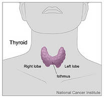
A prospective study reported Sept. 15 at the International Thyroid Congress found that among those who did not respond well to initial treatment, 50% initially classified as low risk developed persistent disease over a 5-year follow-up period. Conversely, Dr. Maria G. Castagna said, half of the initial high-risk patients remained in clinical remission throughout the study.
“This observation strongly supports the concept of ongoing risk stratification throughout the patient’s treatment and follow-up,” said Dr. Castagna of the University of Siena, Italy.
She presented data on 548 patients who were treated for differentiated thyroid cancer. All were risk stratified according to the 2006 European Thyroid Association Consensus. By this method, 284 were considered low risk (tumor 2-4 cm, no distal or nodal metastases) and 264 as high risk (tumor size larger than 4 cm, distal or nodal metastases, or tumor extension beyond the thyroid capsule).
Women made up three-fourths of the group; 90% had papillary thyroid cancer. Most (80%) underwent a total thyroidectomy, and 89% had radioiodine ablation. The average follow-up period was 5.5 years, but some had 10-year data available.
At the time of first follow-up (1 year after initial therapy), clinical remission was significantly higher in the low-risk group than in the high-risk group (85% vs. 53%), and persistent disease was significantly higher in the high-risk than the low-risk group (47% vs. 15%). “This suggests that response to initial treatment is correlated to the original risk stratification,” Dr. Castagna said.
She then evaluated the 1-year clinical outcome only in those 382 patients who were disease free at that time (140 high-risk and 242 low-risk patients). Clinical remission occurred in 98% of the low-risk group and 96% of the high risk group. “The majority of these patients in both groups continued to do well; the rate of recurrent disease was very low without difference between the groups. This suggests that patients in clinical remission after their initial treatment have a good prognosis regardless of their initial staging,” she said.
At the 5-year follow-up, most of those with a good initial response also remained well, with 96% of the low-risk group and 90% of the high-risk group being disease-free. A good prognosis continued in those available for a 10-year follow-up. Among this group, 96% of low-risk and 90% of high-risk patients remained disease free.
“We then evaluated the clinical outcomes of the 166 patients who had persistent disease at 1 year after treatment,” Dr. Castagna said. Of these, 42 had been initially classed as low risk and 124 as high risk. At an average follow-up of almost 7 years, among the low-risk patients, 52% were in remission, and 48% had persistent disease. Among the high-risk group, 52% had persistent disease, 43% were in remission, and 5% were dead from thyroid cancer. “This suggests that in this group of patients, clinical outcome was not related to the initial risk stratification.”
“Our findings suggest that when classified on the basis of response to initial therapy, 50% of patients initially classified as high risk will convert to a low-risk status, and similarly, 50% of those initially classed as low risk will convert to high risk,” she said. “While the initial risk stratification represented a good starting point for treatment, based on data available at the time, during follow-up additional clinical data are accumulated and they may increase or decrease the initial risk estimate. These data should be included so that we can more effectively guide patient follow-up.”
Dr. Castagna reported she had no financial disclosures with regard to the research.
PARIS – Adding information about response to initial therapy for thyroid cancer could improve risk stratification for patients and guide more effective long-term follow-up.
A prospective study reported Sept. 15 at the International Thyroid Congress found that among those who did not respond well to initial treatment, 50% initially classified as low risk developed persistent disease over a 5-year follow-up period. Conversely, Dr. Maria G. Castagna said, half of the initial high-risk patients remained in clinical remission throughout the study.
“This observation strongly supports the concept of ongoing risk stratification throughout the patient’s treatment and follow-up,” said Dr. Castagna of the University of Siena, Italy.
She presented data on 548 patients who were treated for differentiated thyroid cancer. All were risk stratified according to the 2006 European Thyroid Association Consensus. By this method, 284 were considered low risk (tumor 2-4 cm, no distal or nodal metastases) and 264 as high risk (tumor size larger than 4 cm, distal or nodal metastases, or tumor extension beyond the thyroid capsule).
Women made up three-fourths of the group; 90% had papillary thyroid cancer. Most (80%) underwent a total thyroidectomy, and 89% had radioiodine ablation. The average follow-up period was 5.5 years, but some had 10-year data available.
At the time of first follow-up (1 year after initial therapy), clinical remission was significantly higher in the low-risk group than in the high-risk group (85% vs. 53%), and persistent disease was significantly higher in the high-risk than the low-risk group (47% vs. 15%). “This suggests that response to initial treatment is correlated to the original risk stratification,” Dr. Castagna said.
She then evaluated the 1-year clinical outcome only in those 382 patients who were disease free at that time (140 high-risk and 242 low-risk patients). Clinical remission occurred in 98% of the low-risk group and 96% of the high risk group. “The majority of these patients in both groups continued to do well; the rate of recurrent disease was very low without difference between the groups. This suggests that patients in clinical remission after their initial treatment have a good prognosis regardless of their initial staging,” she said.
At the 5-year follow-up, most of those with a good initial response also remained well, with 96% of the low-risk group and 90% of the high-risk group being disease-free. A good prognosis continued in those available for a 10-year follow-up. Among this group, 96% of low-risk and 90% of high-risk patients remained disease free.
“We then evaluated the clinical outcomes of the 166 patients who had persistent disease at 1 year after treatment,” Dr. Castagna said. Of these, 42 had been initially classed as low risk and 124 as high risk. At an average follow-up of almost 7 years, among the low-risk patients, 52% were in remission, and 48% had persistent disease. Among the high-risk group, 52% had persistent disease, 43% were in remission, and 5% were dead from thyroid cancer. “This suggests that in this group of patients, clinical outcome was not related to the initial risk stratification.”
“Our findings suggest that when classified on the basis of response to initial therapy, 50% of patients initially classified as high risk will convert to a low-risk status, and similarly, 50% of those initially classed as low risk will convert to high risk,” she said. “While the initial risk stratification represented a good starting point for treatment, based on data available at the time, during follow-up additional clinical data are accumulated and they may increase or decrease the initial risk estimate. These data should be included so that we can more effectively guide patient follow-up.”
Dr. Castagna reported she had no financial disclosures with regard to the research.
Initial Therapy Response Refines Risk Stratification for Thyroid Cancer
PARIS – Adding information about response to initial therapy for thyroid cancer could improve risk stratification for patients and guide more effective long-term follow-up.
A prospective study reported Sept. 15 at the International Thyroid Congress found that among those who did not respond well to initial treatment, 50% initially classified as low risk developed persistent disease over a 5-year follow-up period. Conversely, Dr. Maria G. Castagna said, half of the initial high-risk patients remained in clinical remission throughout the study.
“This observation strongly supports the concept of ongoing risk stratification throughout the patient’s treatment and follow-up,” said Dr. Castagna of the University of Siena, Italy.
She presented data on 548 patients who were treated for differentiated thyroid cancer. All were risk stratified according to the 2006 European Thyroid Association Consensus. By this method, 284 were considered low risk (tumor 2-4 cm, no distal or nodal metastases) and 264 as high risk (tumor size larger than 4 cm, distal or nodal metastases, or tumor extension beyond the thyroid capsule).
Women made up three-fourths of the group; 90% had papillary thyroid cancer. Most (80%) underwent a total thyroidectomy, and 89% had radioiodine ablation. The average follow-up period was 5.5 years, but some had 10-year data available.
At the time of first follow-up (1 year after initial therapy), clinical remission was significantly higher in the low-risk group than in the high-risk group (85% vs. 53%), and persistent disease was significantly higher in the high-risk than the low-risk group (47% vs. 15%). “This suggests that response to initial treatment is correlated to the original risk stratification,” Dr. Castagna said.
She then evaluated the 1-year clinical outcome only in those 382 patients who were disease free at that time (140 high-risk and 242 low-risk patients). Clinical remission occurred in 98% of the low-risk group and 96% of the high risk group. “The majority of these patients in both groups continued to do well; the rate of recurrent disease was very low without difference between the groups. This suggests that patients in clinical remission after their initial treatment have a good prognosis regardless of their initial staging,” she said.
At the 5-year follow-up, most of those with a good initial response also remained well, with 96% of the low-risk group and 90% of the high-risk group being disease-free. A good prognosis continued in those available for a 10-year follow-up. Among this group, 96% of low-risk and 90% of high-risk patients remained disease free.
“We then evaluated the clinical outcomes of the 166 patients who had persistent disease at 1 year after treatment,” Dr. Castagna said. Of these, 42 had been initially classed as low risk and 124 as high risk. At an average follow-up of almost 7 years, among the low-risk patients, 52% were in remission, and 48% had persistent disease. Among the high-risk group, 52% had persistent disease, 43% were in remission, and 5% were dead from thyroid cancer. “This suggests that in this group of patients, clinical outcome was not related to the initial risk stratification.”
“Our findings suggest that when classified on the basis of response to initial therapy, 50% of patients initially classified as high risk will convert to a low-risk status, and similarly, 50% of those initially classed as low risk will convert to high risk,” she said. “While the initial risk stratification represented a good starting point for treatment, based on data available at the time, during follow-up additional clinical data are accumulated and they may increase or decrease the initial risk estimate. These data should be included so that we can more effectively guide patient follow-up.”
Dr. Castagna reported she had no financial disclosures with regard to the research.
Novel Alzheimer's Drug Falls Short at Phase III
Major Finding: Alzheimer's patients taking semagacestat fared worse than placebo patients, both in cognition and function.
Data Source: A phase III study of more than 2,600 patients who took semagacestat 140 mg daily.
Disclosures: Eli Lilly & Co. sponsored the studies. Dr. Sabbagh reported no financial ties with the company.
Another potential Alzheimer's disease drug failed after Eli Lilly & Co. pulled the plug last month on its phase III study of semagacestat, a gamma secretase inhibitor designed to reduce the aggregation of beta-amyloid into brain plaques.
This latest in a string of Alzheimer's drug flops carried an especially harsh sting, said Dr. Marwan Sabbagh, an investigator in the IDENTITY (Interrupting Alzheimer's Dementia by Evaluating Treatment of Amyloid Pathology) and IDENTITY-2 trials.
“Unlike some of the previous failed phase III studies, IDENTITY was a well-designed, well-powered study of a drug with a very specific mechanism,” he said in an interview.
The company announced its decision after a planned interim analysis of both trials showed that patients who took the study drug did not experience cognitive improvement and, in fact, showed a significant worsening of cognition and the ability to perform activities of daily living, compared with those taking placebo. In a press statement, the company also noted that semagacestat was associated with an increased risk of skin cancer. No data were available as to the risk ratio, crude incidence rate, or type of cancers observed.
IDENTITY and IDENTITY-2 randomized more than 2,600 patients with mild to moderate Alzheimer's disease to either placebo or 140-mg semagacestat for 24 months. Although the trials have been halted, Lilly will continue to follow patients and analyze safety and efficacy data from both studies for at least another 6 months.
Dr. Sabbagh of the Banner Sun Health Research Institute in Sun City, Ariz., said it is still too early to understand why those who took the drug fared worse than the placebo group, or how it may have affected skin cancer risk.
Semagacestat made an unusual leap from phase II to phase III, Dr. Sabbagh said. “It was a very different approach. Their phase II study met the safety end points, but did not look at [cognitive] efficacy.” Instead, he said, Lilly moved the drug forward on the basis of significantly reduced beta-amyloid levels in blood plasma. The drug does not decrease the existing Alzheimer's plaque burden. Instead, its aim is to prevent new plaques. It decreases the amount of plaque-forming beta-amyloid protein by changing the point at which gamma secretase cleaves the amyloid precursor protein. The resulting shorter peptide lengths do not aggregate into brain plaques like those with 40 and 42 amino acids—amyloid beta40 (Abeta40), and Abeta42.
In the phase II study, patients who took 100 mg semagacestat daily had a 58% reduction of Abeta40 in the plasma; those who took 140 mg daily had a 65% reduction. However, there were no significant reductions in CSF Abeta40 levels (Arch. Neurol. 2008;65:1031-8).
Although the drug was generally well tolerated, the phase II researchers, led by Dr. Adam Fleischer of University of California, San Diego, expressed some concern about safety. All gamma secretase inhibitors interfere with Notch signaling. The Notch protein is important in programmed cell death; blocking it particularly affects organ systems with high cell turnover, such as the gut and immune system. In the phase II trial, there were more—but not significantly more—gastrointestinal side effects in the active group (27% vs. 13%).
Although the drug mobilized Abeta40 in phase II, the changes did not translate into clinical benefit during phase III, Dr. Sabbagh said. But that finding may mean that semagacestat's failure is a case of poor timing rather than poor efficacy.
“By the time you have Alzheimer's symptoms, you already have a critical mass of amyloid plaque, and this stays relatively constant as you progress through the disease,” he said.
“The question is, will any antiamyloid drug have a meaningful effect if it's given after the plaques have already developed?”
It may be time, he said, to think of Alzheimer's as a biphasic disorder. “Maybe our amyloid-based therapies should be used in the presymptomatic phase, before the plaques build to that critical level. Other drugs might be more useful in the symptomatic stage.”
Dr. Sabbagh said he wondered if some of the Alzheimer's drugs that have been abandoned after failing their phase III studies might be more successful if used earlier in the disease course.
“My fear is that a drug will be shelved when in fact it might be a good choice in a presymptomatic scenario, when amyloid plaques are just beginning to develop.”
With the enormous leaps now being made in amyloid imaging, researchers are pushing back the diagnostic timeline, identifying patients at the very onset of mild cognitive impairment—and perhaps even before any memory complaints appear.
“A drug like semagacestat would be interesting to study in patients at that stage. Don't chuck the product altogether; back it up into an earlier phase and see if the results are any different.”
'My fear is that a drug will be shelved when in fact it might be a good choice in a presymptomatic scenario.'
Source DR. SABBAGH