User login
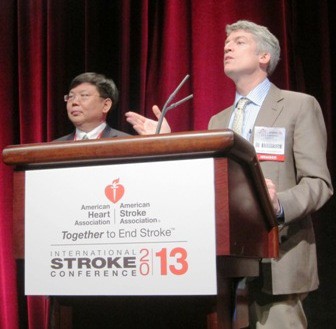
American Heart Association (AHA): International Stroke Conference
Undiagnosed prediabetes pervasive among stroke survivors
One in three stroke survivors in the United States has undiagnosed prediabetes, a national survey has shown.
Black stroke survivors are more than twice as likely as whites to have undiagnosed prediabetes or diabetes mellitus, while Hispanics have an intermediate risk, Dr. Amytis Towfighi said at the International Stroke Conference sponsored by the American Heart Association.
She presented an analysis of 1,070 adult stroke survivors included in the National Health and Nutrition Examination Survey, a Centers for Disease Control and Prevention–sponsored cross-sectional survey weighted so as to be representative of the full U.S. population. Thus, the findings in the NHANES stroke survivors can be extrapolated to the estimated 5.1 million adult stroke survivors nationwide.
The overall prevalence of prediabetes among stroke survivors as defined by a hemoglobin A1c level of 5.7%-6.4% was 32.3%. The figure was 37.8% in blacks, 31.6% in Hispanics, and 26.3% in whites. Only 2.5% of stroke survivors had physician-diagnosed prediabetes, reported Dr. Towfighi, who is chair of the neurology department and director of the acute neurology and acute stroke unit at Rancho Los Amigos National Rehabilitation Center, Downey, Calif.
The prevalence of undiagnosed diabetes mellitus was 3.7% overall. In contrast, 26.9% of stroke survivors had diagnosed diabetes, with rates ranging from 42.7% in Hispanics to 35.7% in blacks and 24.4% in whites.
Black stroke survivors with undiagnosed prediabetes or diabetes were significantly younger: a mean of 59 years old compared with 70 years in whites and 67 years in Hispanics. They were also significantly more likely to smoke and to be on antihypertensive medications. Of black stroke survivors, 58% were women compared with 42% of white stroke survivors.
Chronic hyperglycemia is a potent risk factor for vascular events. Thus, unrecognized prediabetes or diabetes may amplify an individual’s risk of recurrent stroke. Given that if left untreated, prediabetes will progress to diabetes, it makes sense to systematically screen patients for these metabolic abnormalities at the time of an index stroke as a means of improving clinical outcomes and reducing racial disparities, Dr. Towfighi said.
She reported having no relevant financial conflicts.
AT THE INTERNATIONAL STROKE CONFERENCE
Major Finding: One in three survivors of an acute stroke has undiagnosed prediabetes, and another 3.7% have undiagnosed diabetes mellitus.
Data Source: This analysis was based on 1,070 stroke survivors included in the National Health and Nutrition Examination Survey, a cross-sectional survey weighted to be representative of the full U.S. population.
Disclosures: NHANES is supported by the Centers for Disease Control and Prevention. The presenter reported having no relevant financial conflicts.
Low stroke treatment rates under fire
The relatively low U.S. rates of intravenous tissue plasminogen activator therapy for acute stroke may be due in substantial part to widespread use of additional eligibility restrictions beyond the product labeling, Fern Cudlip said at the International Stroke Conference sponsored by the American Heart Association.
Her national survey showed that 81% of U.S. certified stroke centers employ additional inclusion and exclusion criteria not contained in the TPA (Activase) labeling.
"The use of additional exclusion criteria is so common that it implies perhaps there’s a tendency to try to find a reason not to treat," observed Ms. Cudlip, a nurse practitioner who is stroke program coordinator at Eden Medical Center, Castro Valley, Calif.
Her proposed solution: Organizations that certify stroke centers should require adherence to the TPA-label–defined treatment criteria as a means of improving the nation’s low intravenous TPA treatment rates.
The impetus for the national survey presented by Ms. Cudlip was a sense of frustration that 16 years after Food and Drug Administration approval of intravenous TPA as the first and to date still the only proven therapy for acute stroke, treatment rates remain lower than in many other countries. That’s the case even though access to treatment is better than ever, since there are now more than 900 certified U.S. stroke centers.
In talking informally with colleagues around the country, Ms. Cudlip said she and her coinvestigators realized that many hospitals tack on additional nonstandard requirements for intravenous TPA beyond those specified in the labeling. The investigators decided to document these practices.
Representatives from 229 stroke centers in 43 states and Washington, D.C., completed the 15-minute survey. Sixty-nine percent of the stroke centers were located in community hospitals; the other 31%, in academic medical centers. The centers handled an average of 374 ischemic stroke patients per year, with an overall 8.7% intravenous TPA treatment rate. The rate was significantly higher in academic centers: 10.8%, compared with 8.0% in stroke centers in community hospitals.
The survey questions on nonlabel inclusion and exclusion criteria were restricted to patients who present within the 3-hour treatment window opening at stroke onset. For patients who present within 3 hours, 35% of stroke centers impose as an additional treatment criterion a National Institutes of Health Stroke Severity (NIHSS) score of at least 4, and 26% of centers will give intravenous TPA only if CT angiography shows an occlusion. Neither of these inclusion restrictions are part of the product labeling.
Numerous exclusion criteria beyond the TPA labeling are in use. For example, 44% of stroke centers won’t administer intravenous TPA to a patient who presents within 3 hours of stroke onset if the patient shows rapid improvement, even though a disabling deficit remains. Thirty percent exclude from treatment those patients with an NIHSS score greater than 22; a concurrent acute MI is a contraindication to intravenous TPA at 26% of stroke centers; 22% exclude patients on any form of anticoagulation, regardless of the coagulation laboratory results; 20% exclude patients with a suspected but unwitnessed seizure; 16% routinely exclude patients over age 80; 14% exclude patients with a large-vessel occlusion deemed amenable to intra-arterial therapy; 7% won’t treat a patient on nicardipine for blood pressure control; and 5% won’t treat an otherwise-qualified patient who is on dual-antiplatelet therapy.
The number of nonstandard inclusion and exclusion criteria in place at a stroke center showed a significant inverse relationship with the institutional intravenous TPA treatment rate. Smaller community hospitals with lower ischemic stroke admission volumes tended to have a longer list of nonstandard treatment criteria, according to Ms. Cudlip.
Several audience members rose to voice frustration at the list of off-label impediments to prompt intravenous TPA therapy at their home institutions. One physician, noting that institutional change can be difficult to achieve, asked Ms. Cudlip which two common nonlabel treatment criteria she’d make her top priority for elimination.
Ms. Cudlip answered without hesitation: The requirement that a patient has to have an NIHSS score of 4 or more, which according to her survey is in use at more than one-third of stroke centers, needs to go.
"I can have an NIHSS score of 3 and yet have completely lost my speech. That’s pretty disabling, right? I’d really want you to treat me. I need to speak," she said.
The other widely used nonlabel criterion Ms. Cudlip would target for elimination immediately is the treatment exclusion for patients with rapidly improving symptoms despite a remaining disabling deficit.
A physician audience member chimed in that he finds particularly disturbing the policy of not treating patients over age 80, which is in place at 16% of stroke centers. Ms. Cudlip was fully on board with getting rid of that restriction, as well.
"The oldest we’ve treated at our center is a woman who was 106, not that we do it all the time," she said, generating a vigorous round of applause.
She reported having no relevant financial conflicts.
AT THE INTERNATIONAL STROKE CONFERENCE
Major Finding: Eighty-one percent of U.S. stroke centers impose additional inclusion and exclusion criteria for the use of intravenous tissue plasminogen activator therapy in acute ischemic stroke patients beyond what’s contained in the labeling. The longer a center’s list of nonlabel criteria, the lower the treatment rate.
Data Source: A survey with responses from 229 of the nation’s certified stroke centers.
Disclosures: The presenter reported having no relevant financial conflicts.
Even mild kidney dysfunction raises recurrent stroke risk
Diminished renal function is an independent risk factor for recurrent stroke within the first 6 months following hospitalization for an acute ischemic stroke, Dr. Abraham Thomas reported at the International Stroke Conference.
The short-term risk of recurrent stroke climbs in stepwise fashion with decreasing renal function. Even patients categorized as having stage 2 renal function by National Kidney Foundation criteria – those with an estimated glomerular filtration rate of 60-89 mL/min/1.73 m2 – have a 60% increased risk compared with those who have an estimated GFR of 90 or greater, according to Dr. Thomas of the University of California, San Francisco.
He presented an observational study involving 2,882 patients admitted with acute ischemic stroke to 12 Northern California Kaiser Permanente hospitals during 2004-2007. Twenty-four percent had stage 1 renal function upon admission, with an eGFR of at least 90 mL/min/1.73 m2. Forty-seven percent were stage 2, 25% were stage 3 as defined by an eGFR of 30-59, and the rest had stage 4 chronic kidney disease.
In a multivariate analysis, stage 2 renal function was independently associated with a 60% greater risk of recurrent stroke within 6 months compared with those who were stage 1. Patients with stage 3 renal function were at 70% greater risk than were those who were stage 1, while stage 4 patients were at 80% increased risk.
Renal dysfunction is an established risk factor for first-time cardiovascular events, including stroke. But the relationship between renal function and short-term risk of recurrent stroke has not previously been scrutinized.
One possible mechanism by which impaired renal function might predict an increased short-term risk of recurrent stroke involves poor blood pressure control, Dr. Thomas observed. The prevalence of hypertension was 19% with stage 1 renal function, 24% in those who were stage 2, and 26% in patients with stage 3 or 4 renal function. And at 6 months’ follow-up, blood pressure control was less than half as common among stage 4 patients than in those who were stages 1-3.
The conference was sponsored by the American Heart Association. Dr. Thomas reported having no financial conflicts.
National Kidney Foundation, glomerular, he University of California
Diminished renal function is an independent risk factor for recurrent stroke within the first 6 months following hospitalization for an acute ischemic stroke, Dr. Abraham Thomas reported at the International Stroke Conference.
The short-term risk of recurrent stroke climbs in stepwise fashion with decreasing renal function. Even patients categorized as having stage 2 renal function by National Kidney Foundation criteria – those with an estimated glomerular filtration rate of 60-89 mL/min/1.73 m2 – have a 60% increased risk compared with those who have an estimated GFR of 90 or greater, according to Dr. Thomas of the University of California, San Francisco.
He presented an observational study involving 2,882 patients admitted with acute ischemic stroke to 12 Northern California Kaiser Permanente hospitals during 2004-2007. Twenty-four percent had stage 1 renal function upon admission, with an eGFR of at least 90 mL/min/1.73 m2. Forty-seven percent were stage 2, 25% were stage 3 as defined by an eGFR of 30-59, and the rest had stage 4 chronic kidney disease.
In a multivariate analysis, stage 2 renal function was independently associated with a 60% greater risk of recurrent stroke within 6 months compared with those who were stage 1. Patients with stage 3 renal function were at 70% greater risk than were those who were stage 1, while stage 4 patients were at 80% increased risk.
Renal dysfunction is an established risk factor for first-time cardiovascular events, including stroke. But the relationship between renal function and short-term risk of recurrent stroke has not previously been scrutinized.
One possible mechanism by which impaired renal function might predict an increased short-term risk of recurrent stroke involves poor blood pressure control, Dr. Thomas observed. The prevalence of hypertension was 19% with stage 1 renal function, 24% in those who were stage 2, and 26% in patients with stage 3 or 4 renal function. And at 6 months’ follow-up, blood pressure control was less than half as common among stage 4 patients than in those who were stages 1-3.
The conference was sponsored by the American Heart Association. Dr. Thomas reported having no financial conflicts.
National Kidney Foundation, glomerular, he University of California
National Kidney Foundation, glomerular, he University of California
AT THE INTERNATIONAL STROKE CONFERENCE
iScore outperforms stroke physicians in predicting patient outcomes
HONOLULU – Neurologists, internists, and emergency physicians with expertise in acute stroke care are wildly inaccurate in predicting key clinical outcomes in patients with acute ischemic stroke, a study has shown.
Indeed, in the JURASSIC (Clinician Judgment versus Risk Score to Predict Stroke Outcomes) trial, physician estimates as to whether patients would be dead or disabled at discharge were accurate in only 16.9% of cases, or one out of six times. In contrast, a validated predictive model of stroke mortality known as the iScore was on the mark 90% of the time, Dr. Gustavo Saposnik reported at the International Stroke Conference sponsored by the American Heart Association.
Dr. Saposnik and his coworkers developed the iScore because they saw the need for an objective, simple tool to stratify mortality risk in acute ischemic stroke patients. The ability to estimate prognosis with reasonable accuracy is essential to treatment decisions, discharge planning, counseling of patients and their families, and the accuracy of health care policy makers’ comparisons of outcomes between different hospitals.
For the JURASSIC trial, the investigators involved a convenience sample of 111 Ontario physicians with expertise in acute stroke care. Half were neurologists, and the rest were internists or emergency physicians. Their mean age was 40 years, and they each saw an average of 98 stroke patients annually.
Each physician was presented with case summaries for five acute ischemic stroke patients and asked to predict their likelihood of death or disability at discharge as well as 30-day mortality. The five cases were representative of the most common clinical scenarios extracted from a pool of more than 12,000 patients admitted to Ontario stroke centers, so there were no red herrings or tricks.
The 111 physicians collectively made 1,661 outcome predictions. Only 16.9% of their predictions regarding death or disability at discharge were within the 95% confidence interval for the actual observed outcomes. The physicians’ accuracy at predicting 30-day mortality was 46.9%. The iScore-based outcome estimates were within the 95% confidence interval in 90% of cases.
Only 1 of the 111 physicians was accurate in predicting clinical outcomes in four of the five cases. None got all five correct.
The iScore utilizes information easily obtainable within hours of hospital presentation without specialized lab tests or imaging. The variables incorporated into the iScore are age, sex, stroke severity and subtype, coronary artery disease, heart failure, smoking, cancer, hyperglycemia upon admission, history of atrial fibrillation, and renal disease requiring dialysis. Thus, the user-friendly iScore is suitable for use in community hospitals as well as tertiary centers, noted Dr. Saposnik of the University of Toronto.
The score enables patients to be classified into one of five categories based on the estimated risk of mortality. In a validation study (Circulation 2011;123:739-49), the 30-day mortality risk ranged from a low of 1.19% in group 1 to 41.57% in group 5.
The iScore is available as a Web-based tool.
Dr. Saposnik reported having no relevant financial conflicts.
AT THE INTERNATIONAL STROKE CONFERENCE
Cocaine use ups hospital death post subarachnoid hemorrhage
HONOLULU -- Recent cocaine use raised the risk of death nearly threefold in a cohort of more than 1,000 patients who were hospitalized for an acute aneurysmal subarachnoid hemorrhage.
The significantly elevated risk of death was observed despite a lack of difference in severity of initial presentation and remained after exclusion of deaths due to rebleeding, which was higher in cocaine users, Dr. Neeraj Naval said at the International Stroke Conference. The study will also be presented at the annual meeting of the American Academy of Neurology in San Diego on March 19.
"Patients with acute subarachnoid hemorrhage following cocaine use warrant very close monitoring," said Dr. Naval, director of Neurosciences Critical Care at Johns Hopkins Bayview Medical Center in Baltimore.
Although cocaine use and aneurysmal subarachnoid hemorrhage (SAH) is not controversial, data have been scattered on how cocaine affects presentation and outcomes, he observed. In the largest series prior to this, cocaine use had no significant effect on symptomatic vasospasm or neurologic outcome among 600 patients with SAH (World Neurosurg. 2010;73:357-60). An earlier study, however, showed a 2.8-fold higher risk of vasospasm and 3.3-fold higher risk of poor outcome among cocaine users with SAH (J. Neurosurg. 2003;99:271-5).
Dr. Naval and his colleagues reviewed 1,134 patients admitted to one of two Johns Hopkins University hospitals for ruptured brain aneurysm between 1991 and 2009. The cohort included 142 patients (12.5%) who had a history of cocaine use in the 72 hours prior to admission based on self-report or urine toxicology, and 992 with no cocaine use. Cocaine users were more likely to be younger (49 years vs. 53 years), but had similar rates of poor grade 4/5 Hunt & Hess scores (21% vs. 26%) and associated intraventricular hemorrhage (IVH, 56% vs. 51%). Their mean Glasgow Coma Scale scores at admission were also similar (15 in users vs. 14 in nonusers).
In all, 26% of cocaine users and 17% of nonusers died in the hospital (P = .01).
Significant independent predictors of in-hospital death were cocaine use (adjusted odds ratio 2.85), admission Hunt & Hess score (OR 2.33) and higher age (OR 1.03; all P less than .001), Dr. Naval said.
Cocaine users had higher rates of aneurysm re-rupture (7.7% vs. 2.7%; P = .004). Unfortunately, admission mean arterial pressure (MAP) data were unreliable from 1991 to 2005, but data available from 2006 to 2009 showed higher MAP in cocaine users, he said.
Cocaine users were more likely to have delayed cerebral ischemia (22% vs. 16%; P = .041), but the association was not statistically significant after correction for other confounding factors, including age and IVH. Delayed cerebral ischemia was defined as new clinical deterioration more than 48 hours post-SAH and more than 24 hours after surgical clipping or endovascular coiling, radiologic confirmation of cerebral infarction, and/or angiographic confirmation of vasospasm and/or clinical responsiveness (transient or sustained) to hemodynamic augmentation.
Dr. Naval suggested that it is controversial to include IVH in the model because data are available suggesting that cocaine use is independently associated with a higher rate of IVH in patients with intracerebral hemorrhage.
"If there really is a cause-effect relationship between cocaine use and IVH, one wonders whether using IVH in the multivariate analysis may mask the true impact of cocaine exposure on vasospasm-mediated cerebral infarction," he said.
The investigators did not demonstrate any difference between groups in functional outcomes at discharge or post discharge. Dr. Naval said this was not surprising given the significant difference in age between the groups, with younger patients much more likely to recover from neurological injury.
When asked during a discussion of the study to speculate on the mechanism of elevated mortality in cocaine users, Dr. Naval said subsequent analyses found no difference in rates of withdrawal of care between groups and no impact with frequency of cocaine use or delayed cerebral ischemia. Regional wall motion abnormalities have been identified in cocaine users, but electrocardiograms were not performed to tease out the impact of myocardial stunning on mortality.
In terms of its implications for management, a case could be made to use antifibrinolytic therapy in patients with SAH who are cocaine users because of the higher risk of rebleeding, but Dr. Naval said that a trial would be needed to evaluate this.
The conference was sponsored by the American Heart Association. Dr. Naval reported honoraria from EKR Therapeutics. His coauthors made no disclosures.
AT THE INTERNATIONAL STROKE CONFERENCE
Major Finding: The adjusted odds of in-hospital death were nearly threefold higher in cocaine users than in non-users (OR 2.8; P less than .001).
Data Source: Review of prospective data collected on 1,134 patients hospitalized for an acute aneurysmal subarachnoid hemorrhage.
Disclosures: Dr. Naval reported honoraria from EKR Therapeutics. His co-authors made no disclosures.
Poor outcomes seen after carotid intervention non-ST-elevation MI
HONOLULU – Just 1% of patients experienced a non-ST-elevation myocardial infarction following carotid-artery stenting or endarterectomy in a retrospective, nationally representative analysis of more than 1 million patients.
When it did occur, however, NSTEMI significantly increased periprocedural neurologic and cardiac complications, inpatient mortality, disability, and resource utilization – adding on average a full 10 days to hospital length of stay and nearly $85,000 in hospital charges, Dr. Amir Khan said during a plenary session at the International Stroke Conference. Dr. Khan is scheduled to present the study results at the annual meeting of the American Academy of Neurology in San Diego on March 20.
He observed that postoperative evaluation for MI varied in the data set, as it does nationally, but that post hoc analyses from the POISE (Perioperative Ischemic Evaluation) trial revealed that 65% of patients with an MI after noncardiac surgery did not have ischemic symptoms (Ann. Intern. Med. 2011;154:523-8).
"So, we know that hospitals that don’t have active surveillance regimens for non-STEMI will miss a fair amount of this," said Dr. Khan, an endovascular surgical neuroradiology fellow at the University of Minnesota in Minneapolis.
The majority of perioperative MI is NSTEMI. While few studies have parsed out MI types, the SAPPHIRE (Stenting and Angioplasty with Protection in Patients at High Risk for Endarterectomy) trial showed that 80% of carotid endarterectomy (CEA) patients with periprocedural MI and all carotid angioplasty or stenting (CAS) patients with periprocedural MI were NSTEMI (N. Engl. J. Med. 2004;351:1493-1501).
In an effort to assess the frequency of periprocedural NSTEMI following CEA or CAS in practice and its relationship with outcomes, the researchers used data from the Nationwide Inpatient Sample for all adult patients who underwent CEA or CAS from 2002 to 2009. From this, hospital-weighted national estimates were generated using Healthcare Utilization Project algorithms.
Overall, 11,341 patients (1%) experienced an NSTEMI and 1,072,347 did not; 92% of all patients underwent endarterectomy.
Age, gender, and racial distribution were roughly similar between groups, although patients with NSTEMI had significantly higher baseline rates of atrial fibrillation (24% vs. 8%), heart failure (24% vs. 6.5%) and chronic renal insufficiency (16% vs. 5%), Dr. Khan noted.
In terms of health care usage, rates were slightly higher among NSTEMI patients for inpatient diagnostic cerebral angiography (19% vs. 13.5%), gastrostomy (3% vs. 0.4%), and postprocedure mechanical ventilation (0.5% vs. 0.3%; all P less than .0001).
Blood transfusions were conspicuously higher in those with NSTEMI at 20% vs. 3% in those without, he said. The average length of stay also jumped with NSTEMI from 2.8 days to 12.2 days, pushing hospital charges from an average of $29,160 to $113,317 (all P less than .0001).
Neurologic complications were seen in 6% of patients with NSTEMI vs. 1.4% without, while cardiac complications occurred in 31% vs. 1.5%. In addition, 31% of NSTEMI patients were moderately or severely disabled at discharge vs. just 6% without NSTEMI (all P less than .0001), Dr. Khan reported at the conference, sponsored by the American Heart Association.
In-hospital mortality was 6% in the NSTEMI group and 0.5% in the group without. The composite endpoint of cardiac or neurologic complication and/or death was reached by 38% vs. 3% (both P less than .0001).
When these numbers were plugged into a multivariate analysis that adjusted for age, gender, and comorbidities, the odds ratios for patients with NSTEMI were 3.6 for neurologic complications, 23.2 for cardiac complications, 8.6 for in-hospital mortality, 14.6 for the composite end point, and 5.5 for moderate to severe disability (all P less than .0001), he said.
During a discussion following the presentation, an audience member expressed concern about the complication rates, rising to say, "Often we make decisions about stroke treatment based only on what’s good for the brain. Well there’s no point in making the brain better if the person can have a heart attack and die of some other complications, and I really think this [study] emphasizes that."
Dr. Khan replied, "I totally agree and I think that is one of the take-home messages here ... "
Dr. Khan and his coauthors reported no disclosures.
AT THE INTERNATIONAL STROKE CONFERENCE
Major Finding: The composite endpoint of cardiac or neurologic complication and/or death was reached by 38% of patients with NSTEMI vs. 3% of those without (P less than .0001).
Data Source: Retrospective cohort analysis of the Nationwide Inpatient Sample of 2002-2009.
Disclosures: Dr. Khan and his coauthors reported no disclosures.
In-hospital strokes fare worse despite thrombolysis
HONOLULU – One out of every five patients in the United States who receives thrombolytic therapy for an acute ischemic stroke gets it for a stroke occurring when they’re already hospitalized for another reason.
These thrombolytic-treated in-hospital strokes are associated with significantly increased complication and procedural intervention rates, longer length of stay, higher hospital charges, and greater risk of inpatient mortality, compared with out-of-hospital ischemic strokes in patients who get thrombolytic therapy upon presentation, Dr. Tenbit Emiru reported at the International Stroke Conference, which was sponsored by the American Heart Association.
Dr. Emiru’s study is also scheduled to be presented at the annual meeting of the American Academy of Neurology in San Diego on March 21.
She presented an analysis of data for the years 2002-2010 from the National Inpatient Sample, a large, nationally representative hospital database. During the 8-year study period, the database captured more than 24,000 patients who received thrombolytic therapy for an in-hospital acute ischemic stroke and 112,000 others who got a thrombolytic agent for an out-of-hospital ischemic stroke. Thus, 18% of all thrombolytic therapy for acute stroke was administered to patients whose stroke occurred in the hospital.
In-hospital mortality was 11% in thrombolytic-treated patients with in-hospital stroke and 10% in those treated for an out-of-hospital stroke. In a multivariate analysis adjusted for age, gender, and baseline risk factors, patients treated for in-hospital stroke had a statistically significant 10% increased risk of mortality, said Dr. Emiru of the Zeenat Qureshi Stroke Research Center at the University of Minnesota, Minneapolis.
The discharge rate with minimal or no disability was 36% for the in-hospital stroke group and 38% in those treated upon presentation with out-of-hospital stroke, a nonsignificant difference.
The mean length of stay was 8 days in the in-hospital stroke group and 7 days in the comparison group. The in-hospital stroke group had significantly higher rates of in-hospital pneumonia, deep venous thrombosis (DVT), sepsis, and pulmonary embolism, although the absolute differences were small. For example, pneumonia occurred in 5% of patients treated for in-hospital acute ischemic stroke, compared with 3% with out-of-hospital stroke, while DVT was a complication in 2% and 0.7%, respectively.
The disparity between in-hospital procedures for the two groups was more pronounced. For example, 31% of patients treated for in-hospital stroke underwent angiography, compared with 19% of out-of-hospital stroke patients. Those who got thrombolytic therapy for in-hospital stroke also had significantly higher rates of carotid angioplasty (5% vs. 2%) and carotid endarterectomy (3% vs. 1%), Dr. Emiru continued.
Hospital charges averaged $74,713 for patients with in-hospital stroke and $68,419 for those with out-of-hospital stroke.
Audience members indicated that they would have welcomed data on time to treatment from stroke onset for the two groups, as well as information on the reasons for hospitalization in the patients with in-hospital stroke. Neither issue was examined in the study. One audience member commented that the most obvious explanation for the worse outcomes in the thrombolytic-treated patients with in-hospital stroke is that they were sicker to begin with, as evidenced by the fact that were already hospitalized at the time of their ischemic stroke.
Dr. Emiru reported having no financial conflicts.
National Inpatient Sample,
HONOLULU – One out of every five patients in the United States who receives thrombolytic therapy for an acute ischemic stroke gets it for a stroke occurring when they’re already hospitalized for another reason.
These thrombolytic-treated in-hospital strokes are associated with significantly increased complication and procedural intervention rates, longer length of stay, higher hospital charges, and greater risk of inpatient mortality, compared with out-of-hospital ischemic strokes in patients who get thrombolytic therapy upon presentation, Dr. Tenbit Emiru reported at the International Stroke Conference, which was sponsored by the American Heart Association.
Dr. Emiru’s study is also scheduled to be presented at the annual meeting of the American Academy of Neurology in San Diego on March 21.
She presented an analysis of data for the years 2002-2010 from the National Inpatient Sample, a large, nationally representative hospital database. During the 8-year study period, the database captured more than 24,000 patients who received thrombolytic therapy for an in-hospital acute ischemic stroke and 112,000 others who got a thrombolytic agent for an out-of-hospital ischemic stroke. Thus, 18% of all thrombolytic therapy for acute stroke was administered to patients whose stroke occurred in the hospital.
In-hospital mortality was 11% in thrombolytic-treated patients with in-hospital stroke and 10% in those treated for an out-of-hospital stroke. In a multivariate analysis adjusted for age, gender, and baseline risk factors, patients treated for in-hospital stroke had a statistically significant 10% increased risk of mortality, said Dr. Emiru of the Zeenat Qureshi Stroke Research Center at the University of Minnesota, Minneapolis.
The discharge rate with minimal or no disability was 36% for the in-hospital stroke group and 38% in those treated upon presentation with out-of-hospital stroke, a nonsignificant difference.
The mean length of stay was 8 days in the in-hospital stroke group and 7 days in the comparison group. The in-hospital stroke group had significantly higher rates of in-hospital pneumonia, deep venous thrombosis (DVT), sepsis, and pulmonary embolism, although the absolute differences were small. For example, pneumonia occurred in 5% of patients treated for in-hospital acute ischemic stroke, compared with 3% with out-of-hospital stroke, while DVT was a complication in 2% and 0.7%, respectively.
The disparity between in-hospital procedures for the two groups was more pronounced. For example, 31% of patients treated for in-hospital stroke underwent angiography, compared with 19% of out-of-hospital stroke patients. Those who got thrombolytic therapy for in-hospital stroke also had significantly higher rates of carotid angioplasty (5% vs. 2%) and carotid endarterectomy (3% vs. 1%), Dr. Emiru continued.
Hospital charges averaged $74,713 for patients with in-hospital stroke and $68,419 for those with out-of-hospital stroke.
Audience members indicated that they would have welcomed data on time to treatment from stroke onset for the two groups, as well as information on the reasons for hospitalization in the patients with in-hospital stroke. Neither issue was examined in the study. One audience member commented that the most obvious explanation for the worse outcomes in the thrombolytic-treated patients with in-hospital stroke is that they were sicker to begin with, as evidenced by the fact that were already hospitalized at the time of their ischemic stroke.
Dr. Emiru reported having no financial conflicts.
National Inpatient Sample,
National Inpatient Sample,
AT THE INTERNATIONAL STROKE CONFERENCE
Major Finding: Acute ischemic stroke patients in the United States who received thrombolytic therapy for a stroke that occurred while they were already hospitalized had an adjusted 10% greater risk of in-hospital mortality than did patients who received thrombolytic therapy upon presentation with an out-of-hospital stroke.
Data Source: Analysis of roughly 136,000 acute ischemic stroke patients who received thrombolytic therapy as recorded in the National Inpatient Sample for 2002-2010.
Disclosures: The study presenter reported having no relevant financial interests.
Short course of clopidogrel, aspirin lowers stroke recurrence
A short course of clopidogrel plus aspirin safely reduced the risk of early recurrent stroke in Chinese patients with high-risk transient ischemic attack or minor stroke who participated in a randomized, double-blind, placebo-controlled trial.
The relative risk of any stroke within the first 90 days was 32% lower with aspirin and clopidogrel (Plavix) than with aspirin alone (hazard ratio, 0.68; P less than .001). The absolute reduction in ischemic stroke was 3.5% (7.9% vs. 11.4%; P less than .0001).
There was no signal to suggest that dual antiplatelet therapy was unsafe, Dr. Yongjun Wang reported during a late-breaking plenary session at the International Stroke Conference.
The risk of any bleeding was 2.3% with clopidogrel plus aspirin and 1.6% with aspirin alone (P = .09). The risk of severe bleeding was 0.2% in both groups.
Rates of hemorrhagic stroke, myocardial infarction, and cardiovascular death were the same.
Notably, most of the benefit with dual antiplatelet therapy occurred early on in the trial, called CHANCE (Clopidogrel in High-Risk Patients With Acute Non-disabling Cerebrovascular Events) before day 21, underscoring the importance of treating acutely.
"Even more aggressive interventions after acute TIA and minor stroke may be indicated but require more clinical trials," said Dr. Wang of Beijing (China) Tiantan Hospital.
A coinvestigator on the multicenter trial, Dr. S. Claiborne Johnston, director of the stroke service at the University of California, San Francisco, said the risk reduction was large in CHANCE but also observed that there are important differences in health care between China and the United States or Europe, such as undertreatment of conditions such as hypertension during poststroke follow-up. There also are differences in stroke subtyping and a higher frequency of polymorphisms that reduce clopidogrel efficacy.
He suggested that the positive results will likely spur enrollment in the North American, phase III POINT (Platelet-Oriented Inhibition in New TIA and Minor Ischemic Stroke) trial, of which he is also a coinvestigator.
POINT uses a 600-mg loading dose of clopidogrel within 12 hours of symptom onset, followed by 75 mg of clopidogrel daily plus aspirin 50-325 mg/day for 90 days. In contrast, CHANCE randomized 5,170 patients within 24 hours of symptom onset. One group received a 300-mg loading dose of clopidogrel, followed by 75 mg daily during days 2-90 plus 75-300 mg of aspirin open-label on the first day followed by 75 mg/day blinded during days 2-21 and a placebo for aspirin during days 22-90. A second group received aspirin 75-300 mg open-label on the first day, followed by 75 mg/day during days 2-90 and a placebo for clopidogrel during days 1-90.
For years, physicians have been advised not to use clopidogrel plus aspirin in stroke patients unless there’s a compelling reason because it can increase their already elevated risk of major and life-threatening bleeding. Those recommendations, however, were based on long-term use of dual antiplatelet therapy in all comers with stroke, commented Dr. Philip Gorelick, medical director of the Hauenstein Neuroscience Center in Grand Rapids, Mich., who has helped pen some of the recurrent stroke prevention guidelines.
"We’ve been beating that drum for years now and we finally got doctors to stop," he said in an interview. "But what we’re seeing here is that, in the acute period, it not only appears to be safe to have the dual combination of aspirin plus clopidogrel but it also reduces the risk of early stroke in those TIA and minor stroke patients. ...
"It’s a focused indication with good patient selection that’s made the difference here."
Dr. Gorelick predicted that guidelines will change within the next 12-18 months to reflect the current findings supporting early, short-term use of clopidogrel plus aspirin in this subgroup of patients. He suggested that the standard for now will be 21 days of aspirin plus clopidogrel, unless data emerge from POINT to suggest otherwise.
In CHANCE, 2,586 patients were treated with aspirin alone and 2,584 with clopidogrel plus aspirin. Roughly two-thirds had a nondisabling ischemic stroke (National Institutes of Health Stroke Scale score of 3 or less) and one-third had a TIA with moderate-to-high risk of stroke recurrence (ABCD2 score of 4 or more). Their average age was 62 years and 63 years, respectively. The average time to randomization was 13 hours in both groups.
The secondary combined outcome of stroke, myocardial infarction, or death from cardiovascular causes was significantly reduced with clopidogrel plus aspirin (HR, 0.69; P less than .001), Dr. Wang reported at the meeting, which was sponsored by the American Heart Association.
Rates of hemorrhagic stroke, myocardial infarction, and cardiovascular death were 0.3%, 0.1%, and 0.2% in both groups. Death from any cause occurred in 0.4% of all patients.
CHANCE was funded by the Chinese Ministry of Science and Technology. Dr. Wang and his coauthors reported having no disclosures.
AT THE INTERNATIONAL STROKE CONFERENCE
Major Finding: The relative risk of any stroke within the first 90 days was 32% lower with aspirin and clopidogrel than aspirin alone (hazard ratio 0.68; P less than .001).
Data Source: Phase III trial involving 5,170 patients with moderate-to-high-risk TIA or minor stroke.
Disclosures: CHANCE was funded by the Chinese Ministry of Science and Technology. Dr. Wang and his coauthors reported having no disclosures.
Cardiac stress testing underutilized in stroke patients
HONOLULU – Ischemic stroke patients at high risk of coronary artery disease rarely receive guideline-supported cardiac stress testing, a study found.
Among 2,377 veterans with stroke, 28% were at high risk of coronary artery disease (CAD), and only 6.2% received CAD screening within 6 months of discharge.
Moreover, 1-year all-cause mortality was significantly lower among high-risk patients who received CAD screening than among their counterparts who did not (5% vs. 19%; P = .018), Dr. Jason Sico said at the International Stroke Conference.
American Heart Association/American Stroke Association guidelines (Circulation 2003;108:1278-90) recommend that acute ischemic stroke patients at high risk of CAD, defined by a Framingham Risk Score of at least 20%, should receive cardiac screening for occult disease.
Studies have shown that 20%-40% of stroke patients have silent cardiac ischemia, and up to 6% of stroke patients die from cardiac causes or are readmitted with a myocardial infarction in the first 3 months following a stroke, said Dr. Sico, of the department of neurology at Yale New Haven (Conn.) Hospital.
Cardiac stress testing may be underutilized because most medical professionals caring for stroke survivors are not aware of the recommendation, he said in an interview. When they are made aware, one study found that providers may not be convinced that screening for cardiac disease within the stroke population will help their patients or is cost-effective (Stroke 2009;40:3407-9).
"In their favor, there has not been a large prospective study that has demonstrated that cardiac screening for stroke patients improves such important outcomes as mortality and hospital readmission," he added.
The investigators reviewed medical records for a sample of 3,965 patients from 131 Veterans Health Administration facilities admitted for a confirmed diagnosis of ischemic stroke in 2007. Framingham Risk Scores were calculated for 2,377 patients after exclusion of 1,588 patients with a prior cardiac stress test or a known history of CAD, or if they died during hospitalization or had unaccountable data.
In all, 676 (28%) patients had a high Framingham Risk Score of 20% or more, and 1,701 (72%) had a low/intermediate Framingham Risk Score.
Cardiac stress testing within 6 months of discharge from the index stroke was not performed more frequently among high-risk than among low/intermediate-risk patients (6.2% vs. 7.5%; odds ratio, 0.81), Dr. Sico said.
Patients who underwent screening had significantly lower baseline National Institutes of Health Stroke Severity scores than those who did not (mean, 3.3 vs. 4.1; P = .003) and were younger by about 2 years (64.5 years vs. 66.4 years; P = .01). Rates of hypertension, hyperlipidemia, diabetes, and white race were similar between groups, he said at the meeting, which was sponsored by the American Heart Association.
Among all patients, 1-year mortality was significantly lower at 5% in cases where screening was performed, compared with 14% when it was not (P = .001).
Dr. Sico said the strength of the study was the relatively large cohort but that the study was limited by its makeup of primarily male veterans, data from fiscal year 2007, and the inability to explain the reasons for the underutilization of guideline-concordant cardiac screening.
Future work includes understanding the barriers to cardiac testing, the reasons behind the mortality differences among patients who did and did not receive CAD screening, and how implementation of CAD screening guidelines affects outcomes.
"To borrow a page from the diabetes literature, it was dogma that if you were diabetic because it is a coronary equivalent, you should get cardiac stress testing, but when it was prospectively looked at [in the DIAD trial] it really didn’t affect outcome; so we don’t have any prospective [study] in the stroke population to answer this question," he said.
Session moderator Dr. Jennifer Juhl Majersik, of the University of Utah, Salt Lake City, said, "This is a great example of stroke neurologists’ need to look beyond the brain."
The Veterans Health Administration provided funding for the study. Dr. Sico and his coauthors reported no disclosures.
AT THE INTERNATIONAL STROKE CONFERENCE
Major Finding: Only 6.2% of high-risk stroke patients received guideline-concordant cardiac stress testing.
Data Source: Retrospective cohort study of 2,337 ischemic stroke patients.
Disclosures: The Veterans Health Administration provided funding for the study. Dr. Sico and his coauthors reported no disclosures.
Embolectomy for ischemic stroke remains unproven
Patients with large-vessel, anterior circulation ischemic stroke who underwent pretreatment neuroimaging to identify favorable penumbral patterns had similar clinical outcomes after treatment with either embolectomy or standard medical care regardless of the imaging results in the MR RESCUE trial.
"MR RESCUE underscores the importance of confirming hypotheses in randomized, controlled trials prior to implementing treatment approaches in clinical practice," co-primary investigator Dr. Chelsea Kidwell said during a late-breaking plenary session at the International Stroke Conference.
Possible reasons for the results are low recanalization rates with less-effective, first-generation devices (Merci Retriever or Penumbra System), the introduction of two imaging modalities, and the potential for favorable outcomes in penumbral patients regardless of treatment due to sufficient perfusion through collateral vessels. Another possibility is that the penumbral imaging selection hypothesis is flawed as currently conceived, she said.
The MR RESCUE (Mechanical Retrieval and Recanalization of Stroke Clots Using Embolectomy) investigators hypothesized that CT or MRI would identify patients with a favorable penumbral pattern – defined as a predicted infarct core of 90 mL or less and 70% or less of the predicted infarct tissue within the at-risk region – who would differentially benefit from embolectomy. Patients were treated within 8 hours of onset of large-vessel, anterior circulation strokes. Embolectomy is particularly attractive in these cases as intravenous tissue plasminogen activator (TPA) often is not effective on these large, frequently disabling clots.
There was no significant interaction, however, between treatment assignment and penumbral pattern by shift analysis of the 90-day modified Rankin Scale (mRS) score, reported Dr. Kidwell, professor of neurology at Georgetown University Medical Center in Washington. The mean mRS scores in patients with a favorable penumbral pattern were 3.9 with embolectomy and 3.4 with standard care, and 4.0 and 4.4, respectively, among patients without a favorable penumbral pattern (P = .14).
"As such, the trial failed to demonstrate that penumbral imaging identifies patients who will differentially benefit from endovascular therapy for acute ischemic stroke," she said.
Among all patients, mean modified Rankin scores at 90 days were similar between embolectomy and standard care (3.9 vs. 3.9; P = .99).
"Further randomized, controlled trials of new-generation devices are needed to test the full spectrum of the penumbral imaging selection hypothesis and the clinical efficacy of new-generation stent-retriever devices," she said.
That may be easier said than done, despite the lack of randomized, controlled trial data proving the benefit of penumbral imaging selection or recanalization with embolectomy.
In an editorial (N. Engl. J. Med. 2013 [doi:10.1056/NEJMe1215730]) published online with the MR RESCUE results (N. Engl. J. Med. 2013 [doi:10.1056/NEJMoa1212793]), Dr. Marc Chimowitz with the neurosciences department at Medical University of South Carolina, Charleston, points out that recruitment to the IMS (Interventional Management of Stroke) III and MR RESCUE trials was difficult "because once the Food and Drug Administration approved the devices and Medicare provided reimbursement for these procedures, endovascular treatment became widespread and many physicians who were treating patients with acute stroke felt that the ‘answer was in.’ Therefore, treatment equipoise was lost."
In all, 127 patients were enrolled between 2004 and 2011, of which 118 were fully eligible. Their mean age was 65.5 years and median National Institutes of Health Stroke Scale score was 17. Overall, 37% received intravenous TPA, and 80% underwent imaging with MRI.
Symptomatic hemorrhage occurred in 4% and 21% died, Dr. Kidwell said. The mean time to groin puncture was 6.2 hours.
"All of this means that we still need to put more and more patients in research protocols to better figure out who will benefit the most from endovascular therapy," American Heart Association past-president Dr. Ralph Sacco, chair of neurology at the University of Miami, said in an interview. "It has been shown now, including the subanalysis from IMS III, that the earlier you recanalize the better, and so we need to be still thinking about time windows. Just like with IV [intravenous] TPA, the earlier we treat, the better. Well, with endovascular therapy that may be true as well and you have to remember that sometimes penumbral imaging adds time."
Although embolectomy proponents and device manufacturers may see the results as a setback, another take was expressed by an audience member, who described the results as good news.
"The vast majority of people in the United States and the world probably have better access to primary care facilities rather than having access to academic centers, so to my patient population and to people I think in the vast majority, when we see that IV TPA is perhaps equivalent to more aggressive treatments that require more expansive techniques and depth of expertise, it’s kind of good news," she said to a round of applause.
Dr. Kidwell thanked her for the comment, but added, "I think there still are those among us who do believe that advanced neuroimaging, as well as endovascular therapy, will have a role down the line. I think we just need further trials with the new stent retriever devices to really sort this out."
MR RESCUE was funded by the National Institute of Neurological Disorders and Stroke. Concentric Medical Inc. provided study catheters and devices for part of the trial, with costs thereafter covered by study funds or third-party payers. Seven of the coauthors are employees of the University of California, which holds a patent on Merci Retriever devices. Dr. Chimowitz disclosed research support from the National Institutes of Health. Dr. Sacco reported no disclosures.
AT THE INTERNATIONAL STROKE CONFERENCE
Major Finding: Among all patients, mean modified Rankin scores at 90 days were similar between embolectomy and standard care (3.9 vs. 3.9; P = .99).
Data Source: Phase IIb study in 118 patients with ischemic stroke.
Disclosures: MR RESCUE was funded by the National Institute of Neurological Disorders and Stroke. Concentric Medical Inc. provided study catheters and devices for part of the trial, with costs thereafter covered by study funds or third-party payers. Seven of the co-authors are employees of the University of California, which holds a patent on Merci Retriever devices. Dr. Chimowitz disclosed research support from the National Institutes of Health. Dr. Sacco reported no disclosures.

