User login
EDs Admitting More Previously Hospitalized Patients
Rates of hospital readmission from the emergency department of patients who were recently hospitalized are on the rise, according to an analysis of 2005-2008 data from the National Hospital Ambulatory Medical Care Survey.
"Patients who return to the emergency department within 7 days of hospitalization have both relatively high and increasing rates of readmission. ... Policies and programs aimed at reducing hospital readmissions should consider the role of the ED in determining the disposition of recently hospitalized patients," said Dr. Zachary F. Meisel of the University of Pennsylvania, Philadelphia.
The analysis of the nationwide NHAMCS included all patients aged older than 18 years and excluded only those who were transferred, left against medical advice, or died in the ED. In all, 2.4% of ED visits – an estimated 2.3 million per year – were by patients who had been hospitalized in the previous 7 days. Admission rates for recently hospitalized patients increased for each study year, from 28.6% in 2005 to 38.0% in 2008. In contrast, admission rates for visits by patients who were not recently hospitalized increased only slightly, from 15.7% to 16.2%, Dr. Meisel reported at the annual meeting of the Society for Academic Emergency Medicine.
After adjustment for age and triage acuity, the odds ratio of hospital admission for those who had been hospitalized within the previous 7 days was 2.59, compared with those not previously hospitalized. Older age, higher triage acuity, and visits to hospitals in metropolitan areas were also associated with increased odds of admission but did not confound or modify the interaction between recent hospitalization and admission for any year.
The rising rate of readmission was somewhat surprising, given the recent financial pressures being placed on hospitals to reduce those rates. One possible explanation is that hospitals are now discharging sicker patients. "Even though I adjusted for triage acuity, that doesn’t capture all the factors that make a patient sick," Dr. Meisel said in an interview.
However, he pointed out that even though the percentages of ED patients admitted after recent hospitalization rose by about 10%, compared with the baseline admission rate over the 4-year period, two-thirds of patients who were recently hospitalized are still being evaluated in the ED and discharged rather than admitted. "So, clearly we’re doing something in the ED to mitigate readmissions. Whether it’s pain control, reassurance, addressing questions about prescriptions, arranging follow-up, or other measures, hospitals should be analyzing what’s going on in the ED and our role in preventing hospital readmission."
Dr. Meisel stated that he had no relevant financial disclosures.
FROM THE ANNUAL MEETING OF THE SOCIETY FOR ACADEMIC EMERGENCY MEDICINE
Etomidate Not Associated With Longer Vasopressor Duration
BOSTON – Etomidate did not significantly increase vasopressor requirements, compared with midazolam, in a double-blind randomized trial of 122 patients with suspected sepsis who presented to a single emergency department over an 18-month period.
Etomidate is widely used for sedation before endotracheal intubation. One recent post hoc analysis showed that prolonged use of etomidate was associated with increased mortality (N. Engl. J. Med. 2008;358:111-24), while another post hoc analysis demonstrated a significant increase in mortality in septic patients given etomidate for rapid-sequence intubation irrespective of steroid supplementation (Crit. Care Med. 2007;35:1012-8). In a small study, single-dose etomidate use was associated with adrenal suppression that may have contributed to longer hospital length of stay, more ventilator days, and more days in intensive care (J. Trauma 2008;65:573-9).
However, the clinical relevance of the adrenal suppression is not yet clear, and the studies showing increased mortality were not randomized, noted Dr. David Barounis, an emergency physician at Advocate Christ Medical Center, Oak Lawn, Ill.

In the current study, a post hoc analysis of data from a prospectively randomized double-blind study performed between November 2007 and May 2009 at a single tertiary care center’s emergency department (ED), 303 patients met the enrollment criteria of age greater than 18 years and intubation in the ED with suspected infectious etiology for their illness. Of 122 who could be enrolled in the study, 96 were later confirmed to be septic following intubation. Of those, 45 were randomized to etomidate while 51 received midazolam.
Baseline characteristics did not differ significantly between the two groups, although there was a nonsignificant trend among the etomidate patients toward greater use of vasopressors in the ED (53% vs. 43% of the midazolam group) and total vasopressor use (66% vs. 51%; 95% confidence interval of difference, -4% to 34%). The difference in total number of hours septic patients received vasopressors was not significant: 38 hours with etomidate vs. 32 hours with midazolam (P = .65).
Limiting the analysis to just the 26 etomidate and 23 midazolam patients who received vasopressors within the first 48 hours of ED arrival (95% CI, 7% to 31%) did not change the results, with 58% vasopressor use with etomidate vs. 45% with midazolam (95% CI, -7% to 31%) and with 64 vs. 51 total hours, respectively, of vasopressor use in this subgroup (P = .44).
Norepinephrine was the most common vasopressor used for both groups, and there were no differences in sedative use by type of vasopressor, Dr. Barounis said at the annual meeting of the Society for Academic Emergency Medicine.
"Based on our results, etomidate does not result in a statistically significant increase in length of vasopressor duration when used as a bolus dose during rapid sequence intubation," he said. However, he added, due to the relatively small number of patients who could be enrolled at one center over 18 months, a multicenter prospective trial powered to detect clinically significant mortality and secondary outcome differences should be undertaken.
Dr. Barounis stated that he had no financial disclosures.
BOSTON – Etomidate did not significantly increase vasopressor requirements, compared with midazolam, in a double-blind randomized trial of 122 patients with suspected sepsis who presented to a single emergency department over an 18-month period.
Etomidate is widely used for sedation before endotracheal intubation. One recent post hoc analysis showed that prolonged use of etomidate was associated with increased mortality (N. Engl. J. Med. 2008;358:111-24), while another post hoc analysis demonstrated a significant increase in mortality in septic patients given etomidate for rapid-sequence intubation irrespective of steroid supplementation (Crit. Care Med. 2007;35:1012-8). In a small study, single-dose etomidate use was associated with adrenal suppression that may have contributed to longer hospital length of stay, more ventilator days, and more days in intensive care (J. Trauma 2008;65:573-9).
However, the clinical relevance of the adrenal suppression is not yet clear, and the studies showing increased mortality were not randomized, noted Dr. David Barounis, an emergency physician at Advocate Christ Medical Center, Oak Lawn, Ill.
In the current study, a post hoc analysis of data from a prospectively randomized double-blind study performed between November 2007 and May 2009 at a single tertiary care center’s emergency department (ED), 303 patients met the enrollment criteria of age greater than 18 years and intubation in the ED with suspected infectious etiology for their illness. Of 122 who could be enrolled in the study, 96 were later confirmed to be septic following intubation. Of those, 45 were randomized to etomidate while 51 received midazolam.
Baseline characteristics did not differ significantly between the two groups, although there was a nonsignificant trend among the etomidate patients toward greater use of vasopressors in the ED (53% vs. 43% of the midazolam group) and total vasopressor use (66% vs. 51%; 95% confidence interval of difference, -4% to 34%). The difference in total number of hours septic patients received vasopressors was not significant: 38 hours with etomidate vs. 32 hours with midazolam (P = .65).
Limiting the analysis to just the 26 etomidate and 23 midazolam patients who received vasopressors within the first 48 hours of ED arrival (95% CI, 7% to 31%) did not change the results, with 58% vasopressor use with etomidate vs. 45% with midazolam (95% CI, -7% to 31%) and with 64 vs. 51 total hours, respectively, of vasopressor use in this subgroup (P = .44).
Norepinephrine was the most common vasopressor used for both groups, and there were no differences in sedative use by type of vasopressor, Dr. Barounis said at the annual meeting of the Society for Academic Emergency Medicine.
"Based on our results, etomidate does not result in a statistically significant increase in length of vasopressor duration when used as a bolus dose during rapid sequence intubation," he said. However, he added, due to the relatively small number of patients who could be enrolled at one center over 18 months, a multicenter prospective trial powered to detect clinically significant mortality and secondary outcome differences should be undertaken.
Dr. Barounis stated that he had no financial disclosures.
FROM THE ANNUAL MEETING OF THE SOCIETY FOR ACADEMIC EMERGENCY MEDICINE
Major Finding: The difference in total number of hours septic patients received vasopressors was nonsignificant: 38 hours with etomidate vs. 32 hours with midazolam (P = .65).
Data Source: A post hoc analysis of data from a double-blind study performed between November 2007 and May 2009 at a single tertiary care center’s ED, in which 96 intubated patients with confirmed sepsis were prospectively randomized to either etomidate or midazolam.
Disclosures: Dr. Barounis has no disclosures.
RSV, Rhinovirus Coinfections Common With Ped Bronchiolitis, Linked to Longer Hospital Stay
BOSTON – Coinfections with both respiratory syncytial virus and rhinovirus were common and associated with increased length of stay in a prospective multicenter study of over 2,000 children under 2 years of age who were hospitalized with bronchiolitis.
The clinical value of testing for an infectious etiology in a child with bronchiolitis is unclear. Indeed, the recommendation is not to test (Pediatrics 2006;118:1774-93). Some argue however, that testing may be useful for the influenza treatment or to identify the beginning of the viral "seasons" and which viruses are circulating, Dr. Jonathan M. Mansbach, a hospitalist physician at Children’s Hospital Boston, said at the annual meeting of the Society for Academic Emergency Medicine.
Additionally, Dr. Mansbach said that the 70% frequency of coinfection seen in this study raises questions about the effectiveness of inpatient cohorting by viral etiology, which some researchers contend is of use. Moreover, the findings suggest that hospitals consider adding RV to respiratory viral panels, he said.
The 16-center study enrolled consecutive children between November and March during 2007-2010. Of the total 2,207 children enrolled, 83% were located on the ward while 17% were admitted to the intensive care unit. Of those 377, 42% were intubated or given continuous positive airway pressure. Overall mean length of stay was 2 days. The patients had a median age of 4 months; 59% were male, 61% were white, 24% black, and 15% other races. A third (36%) were of Hispanic ethnicity.
The three most common viral etiologies identified by polymerase chain reaction were RSV-A (43%) RSV-B (30%) and RV (26%). Adenovirus, human metapneumovirus, and the coronaviruses were all 7%-8%, and only 6% of the children had no virus detected. (These figures add up to more than 100 because of a 30% rate of coinfections.) The low-frequency infections did not affect results, so subsequent analysis focused on RSV (subtypes A and B) and RV, Dr. Mansbach said.
Of the 940 children in whom RSV-A was identified, it was the only virus in 66%, while one or more additional viruses were identified in the other 34%. Similarly, 68% of the 664 RSV-B infected patients had only one virus identified, while 32% were coinfected.
Rhinovirus was somewhat different, however, in that just 30% of 564 had only that and 70% had coinfections.
For children with both RSV-A and RSV-B, the likelihood of having a length of stay of 3 or more days did not differ between those who had the single virus infection and those who were coinfected (48% vs. 49%, respectively, for RSV-A, and 47% and 54% for RSV-B). There was a significant difference with rhinovirus, however, with 28% of those with the single infection and 46% with coinfections hospitalized 3 or more days.
After adjustment for age, gender, race, eczema history, intubation history, apnea, retractions, oxygen saturation, oral intake, comorbid medical condition, and site, rhinovirus alone was associated with a lower chance of being hospitalized 3 or more days compared with RSV-A or RSV-B alone (odds ratio 0.4), while RSV-RV co-infections were associated with a significantly greater chance of being hospitalized for that duration compared with RSV-A or RSV-B alone (OR 1.3).
Clustering by site did not affect the results, and in a preliminary analysis, controlling for acute severity as defined by ICU, CPAP, or intubation also did not materially change the results, Dr. Mansbach said.
This study was conducted as part of the Multicenter Airway Research Collaboration (MARC), a program of the Emergency Medicine Network. Pediatric Research in Inpatient Settings sites collaborated with EMNet, Dr. Mansbach said.
Dr. Mansbach stated that he had no relevant financial disclosures.
FROM THE ANNUAL MEETING OF THE SOCIETY FOR ACADEMIC EMERGENCY MEDICINE
Major Finding: Of 564 children infected with rhinovirus, 70% had RSV coinfections. Rhinovirus alone was associated with a lower chance of being hospitalized 3 or more days compared with RSV-A or RSV-B alone (odds ratio 0.4). RSV-RV coinfections were associated with a significantly greater chance of being hospitalized for 3 or more days, compared with RSV-A or RSV-B alone (OR 1.3).
Data Source: Prospective multicenter study of 2,207 children under 2 years of age who were hospitalized for bronchiolitis.
Disclosures: Dr. Mansbach reported no disclosures.
Nasopharyngeal Lactate Dehydrogenase May Predict Bronchiolitis Severity
BOSTON – Higher levels of nasopharyngeal lactate dehydrogenase were associated with decreased odds of hospitalization in a prospective, observational, multicenter cohort study of samples from 277 children under 2 years of age who presented to the emergency department with bronchiolitis.
"Despite identified risk factors for severe disease, the difficulty and uncertainty of determining the appropriate level of supportive care for children with bronchiolitis is well documented by the large variability in hospital admission practices. Admission rates for infants with bronchiolitis are significantly different between pediatric and general EDs and even among pediatric ED attending physicians. There is also wide variation in thresholds for pediatric intensive care unit admission and intubation," said Dr. Jonathan M. Mansbach, a hospitalist physician at Children’s Hospital Boston at the annual meeting of the Society for Academic Emergency Medicine.
Recent studies suggest that lactate dehydrogenase (LDH), a membrane-associated protein released from injured cells that is a marker of inflammatory response, correlates with innate immunity and could therefore be used as a predictor of disease severity. In one recent single-center study of 98 children aged less than 2 years presenting to the ED with bronchiolitis, serum LDH was not predictive of admission, but higher levels of nasopharyngeal LDH were associated with an 81% reduced risk of hospital admission (Pediatrics 2010;125:e225-33).
The current study is part of the larger Multicenter Airway Research Collaboration (MARC), a program of the Emergency Medicine Network. The primary aim of the MARC-25 Virology Study is to describe virology of children presenting to the ED with bronchiolitis using nasopharyngeal aspirate (NPA) samples (Acad. Emerg. Med. 2008;15:111-8).
Nasopharyngeal aspirate samples were collected between December 2005 and March 2006 at 14 centers in 10 states. The 277 children had a median age of 6 months. More than half (61%) were boys; 37% were Hispanic, 31% white, 28% black, and 4% other. The median value of NPA LDH was 7 U/mL but the range was wide, 0-8,400 U/mL. "Nasopharyngeal LDH is a new test. Therefore, when considering the actual values for LDH, please remember that there are no known standards," Dr. Mansbach commented.
Of the study cohort, 45% (125 children) were admitted to the hospital, and of those, 74% (93) were admitted for longer than 24 hours. For the entire 0-8,400 U/mL range, higher LDH values were associated with a lower rate of admission for 24 hours or longer. However, there were two outlier patients. With those two removed, the LDH range dropped to 0-1,000 and the inverse relationship between LDH and admission remained, but there was a blunting of the association at higher levels of LDH.
The rest of the analysis was limited to patients with the two most common viral etiologies, respiratory syncytial virus (64%) and rhinovirus (16%). The low frequency of infections with human metapneumovirus and influenza did not affect the results, he noted.
In the 176 children with RSV, the median LDH values were higher but not significantly higher than in the 101 children without RSV, 8 vs. 4 U/mL (P = .53). However, in the 44 children with rhinovirus, median LDH values were significantly higher than in children without rhinovirus, 14 vs. 5 U/mL (P = .03).
Moving from the first to the third LDH quartile after adjustment for common variables associated with illness severity (age less than 2 months, sex, history of intubation, retractions, oxygen saturation 94% or below), there was a fairly linear drop in the odds of admission. With the quartile of 1.33 U/mL and below as the referent 1.0, those with LDH levels 1.34-6.6 U/mL had an adjusted odds ratio of admission of 0.58 (P = .23, confidence interval 0.24-1.4), while the adjusted odds ratio for those with LDH 6.7-43.2 U/mL was 0.23 (P = .002, confidence interval 0.09-0.58). The difference was no longer significant for LDH of 43.3 U/mL or above (P = .33, 0.28-1.5). Stratification by virus or site did not change the outcomes, Dr. Mansbach added.
"We do not know why the association is blunted at the highest level of LDH, and more research will be needed to sort through this aspect of our findings. Remember that the test still shows that higher levels of LDH are associated with fewer admissions. It was just blunted, with slightly lower magnitude, so it could still be clinically useful," he said in an interview.
Dr. Mansbach stated that he has no conflicts of interest.
FROM THE ANNUAL MEETING OF THE SOCIETY FOR ACADEMIC EMERGENCY MEDICINE
Major Finding: Moving from the first to the third LDH quartile after adjustment for common variables associated with illness severity, there was a fairly linear drop in the odds of admission. With the quartile of 1.33 U/mL and below as the referent 1.0, those with LDH levels 1.34-6.6 U/mL had an adjusted odds ratio of admission of 0.58 (P = .23, confidence interval 0.24-1.4), while the adjusted odds ratio for those with LDH 6.7-43.2 U/mL was 0.23 (P = .002, 0.09-0.58). The difference was no longer significant for LDH of 43.3 U/mL or above (P = .33, 0.28-1.5).
Data Source: Prospective, observational, multicenter cohort study of samples from 277 children under 2 years of age who presented to the emergency department with bronchiolitis.
Disclosures: Dr. Mansbach had no disclosures.
Sepsis Mortality Rate Reduced by Antibiotics Prior to Shock Recognition
BOSTON – Delaying antibiotics until after onset of septic shock was associated with increased mortality in a prospective study of 300 patients who presented to the emergency department with suspected infection and were treated with a quantitative resuscitation protocol.
However, there was no increase in mortality with hourly delays in antibiotics after shock onset up to 3 hours, said Dr. Michael A. Puskarich of the Carolinas Medical Center, Charlotte, N.C.
"When we gave antibiotics before the recognition of shock, patients did better. One of the limitations of that, however, it’s really impossible to predict when you’re seeing the patient when there will be shock recognition. Therefore, our recommendation is still to give broad-spectrum antibiotics as early as possible while not sacrificing hemodynamic resuscitation. However, also based on these data, we cannot support a specific time frame from triage or from shock recognition to suggest a time-to-antibiotic-dose core measure for septic shock at this point," Dr. Puskarich said in an interview.
International guidelines call for administration of broad-spectrum antibiotic therapy within 1 hour of diagnosis of septic shock and severe sepsis without septic shock, as part of a quantitative resuscitation protocol (Crit. Care Med. 2008;36:296-327). These guidelines were based largely on one study that found that effective antimicrobial administration within the first hour of documented hypotension was associated with increased survival to hospital discharge in adult patients with septic shock. Despite a progressive increase in mortality with increasing delays, only 50% of septic shock patients received effective antimicrobial therapy within 6 hours of documented hypotension (Crit. Care Med. 2006;34:1589-96).
The current study was a preplanned secondary analysis of a previously published study designed to test the hypothesis of noninferiority between lactate clearance and central venous oxygen saturation (ScvO2) as goals of early sepsis resuscitation (JAMA 2010;303:739-46). Patients were enrolled from three large, urban, tertiary care emergency departments. Inclusion criteria were suspected infection with two or more systemic inflammatory response syndrome criteria.
All patients received structure resuscitation with targets of central venous pressure 8 mm Hg or greater, a mean arterial pressure 65 mm Hg or greater, and either ScvO2 greater than or equal to 70% or lactate clearance greater than or equal to 10%. Broad-spectrum antibiotics were given according to local institution guidelines, as early as practical in the course of the resuscitation, Dr. Puskarich said at the annual meeting of the Society for Academic Emergency Medicine.
Of the 300 patients who had been randomized to either the lactate or ScvO2 target groups, 9 had received antibiotics prior to arrival and were excluded. Of the remaining 291 patients, 59% (172) received the initial dose of antibiotics after shock onset. Median time from triage to shock recognition was 89 minutes. The patients had a mean age of 62 years, 54% were white, and 53% were male. Median systolic blood pressure was 86 mm Hg, and median venous lactate was 3.3 mmol/L. The median ED sepsis-related organ failure assessment score was 6.
Overall mortality was 55%, and the majority of survivors and nonsurvivors received antibiotics within the same time frame. Patients who received antibiotics after shock recognition were more than twice as likely to die as were those who received them prior to shock recognition, 24% vs. 12%, with an unadjusted odds ratio of 2.3 that remained significant after adjustment for confounders. However, there was no increase in mortality by hourly delays from either triage or shock onset up to 3 hours, Dr. Puskarich reported.
"When early sepsis hemodynamic resuscitation is sufficiently refined, the strength of the association between hourly delays in antibiotic administration and mortality appears to recede. The effect of timing in the most proximal phase of septic shock remains unclear," he concluded.
Dr. Puskarich received grant support from the American Heart Association. The study for which this was a secondary analysis was funded by the National Institutes of Health. He said he had no other relevant financial disclosures.
FROM THE ANNUAL MEETING OF THE SOCIETY FOR ACADEMIC EMERGENCY MEDICINE
Major Finding: Patients who received antibiotics after shock recognition were more than twice as likely to die as were those who received them prior to shock recognition, 24% vs. 12%, with an unadjusted odds ratio of 2.3 that remained significant after adjustment for confounders.
Data Source: A preplanned secondary analysis of a previously published study designed to test the hypothesis of noninferiority between lactate clearance and central venous oxygen saturation as goals of early sepsis resuscitation in 300 ED patients.
Disclosures: Dr. Puskarich received grant support from the American Heart Association. The previous study was funded by the National Institutes of Health. He said he had no other relevant financial disclosures.
Nurse-Administered Corticosteroids in ED Reduce Admissions for Asthma
BOSTON – Emergency department administration of oral corticosteroids by a triage nurse prior to physician assessment significantly reduced the time to clinical improvement, total time in the ED, and risk of inpatient admission in a study of 644 children who presented with moderate to severe asthma exacerbations.

Medical directives allowing triage nurses to administer bronchodilator therapy are common, but nurse administration of oral corticosteroids has not previously been studied, even though earlier use of these agents in asthma patients with severe exacerbations has been shown to directly affect the risk of hospital admission. "We know that written clinical pathways improve physician ordering of oral steroids in acute asthma exacerbations, but delays to steroid administration are still long," said Dr. Roger L. Zemek, a pediatric emergency physician at Children’s Hospital of Eastern Ontario, Ottawa.
The controlled trial used a "before-after" design, in which a 4-month phase of physician-ordered oral corticosteroids was compared with a 4-month period in which triage nurses administered them. The study was done in a tertiary children’s hospital ED, with an annual patient population of about 60,000 visits a year, of which about 2,500 are for asthma, Dr. Zemek said at the annual meeting of the Society for Academic Emergency Medicine.
Eligible children were those aged 2-17 years who presented to the pediatric ED with a moderate to severe acute asthma exacerbation, with a Pediatric Respiratory Assessment Measure (PRAM) of 4 or greater. The PRAM is a validated scoring system that measures asthma severity based on the patient’s symptoms on a scale of 1-12, with 12 being most severe. It has good nurse-to-nurse and nurse-to-physician interuser reliability, he said.
All of the children had either a prior physician diagnosis of asthma or three or more episodes of wheezing responsive to beta-2 agonists. Of 1,183 children with acute asthma presenting to the ED between September 2009 and May 2010, 644 were classified as having moderate to severe disease. There were 336 children in the physician-ordered phase and 308 in the triage nurse–administered phase. The children were similar in age (mean of about 6 years in both groups). About two-thirds of both groups were boys. Both groups had been experiencing respiratory distress for about a day and a half prior to ED arrival, with an initial average PRAM score of 6.7 (moderate exacerbation).
Other than the timing of oral steroid administration, the children were treated similarly in the ED. Children in the triage nurse–administered phase received steroids at just 30 minutes, compared with 75 minutes in the physician-ordered phase, a highly significant difference.
For the primary outcome, time until clinical improvement as defined by a reduction in initial PRAM score by 3 points, children who received steroids from the triage nurse improved a statistically significant 24 minutes faster (median, 158 minutes) than those treated during the physician-ordered phase (median, 182 minutes). They also improved to "mild" status 51 minutes sooner, with medians of 211 vs. 262 minutes.
Hospital admission was required for 19.0% of the physician group, compared with 11.7% of the triage nurse group, for a significant odds ratio of 0.56. Time to ED discharge was a significant 44 minutes sooner with steroid administration by a triage nurse, at 316 vs. 360 minutes.
Adjustments for a few significant although small differences at baseline in the proportion of patients with a preceding upper respiratory tract infection and in prior use of other asthma medications did not change the results, Dr. Zemek noted.
"While 44 minutes may seem short, when you add that over thousands of visits per year at most pediatric tertiary centers, you’re talking about thousands of hours saved of nursing time [and] physician time," plus money saved from reduced hospital admissions. "If adopted broadly, this strategy to optimize multidisciplinary teams could have large health care implications with regard to reducing the burden of asthma and as a potential solution to overcrowding of our emergency rooms," he commented.
This study was funded by a grant from the Academic Health Sciences Alternate Funding Plan of Ontario. Dr. Zemek stated that he had no additional financial disclosures.
BOSTON – Emergency department administration of oral corticosteroids by a triage nurse prior to physician assessment significantly reduced the time to clinical improvement, total time in the ED, and risk of inpatient admission in a study of 644 children who presented with moderate to severe asthma exacerbations.
Medical directives allowing triage nurses to administer bronchodilator therapy are common, but nurse administration of oral corticosteroids has not previously been studied, even though earlier use of these agents in asthma patients with severe exacerbations has been shown to directly affect the risk of hospital admission. "We know that written clinical pathways improve physician ordering of oral steroids in acute asthma exacerbations, but delays to steroid administration are still long," said Dr. Roger L. Zemek, a pediatric emergency physician at Children’s Hospital of Eastern Ontario, Ottawa.
The controlled trial used a "before-after" design, in which a 4-month phase of physician-ordered oral corticosteroids was compared with a 4-month period in which triage nurses administered them. The study was done in a tertiary children’s hospital ED, with an annual patient population of about 60,000 visits a year, of which about 2,500 are for asthma, Dr. Zemek said at the annual meeting of the Society for Academic Emergency Medicine.
Eligible children were those aged 2-17 years who presented to the pediatric ED with a moderate to severe acute asthma exacerbation, with a Pediatric Respiratory Assessment Measure (PRAM) of 4 or greater. The PRAM is a validated scoring system that measures asthma severity based on the patient’s symptoms on a scale of 1-12, with 12 being most severe. It has good nurse-to-nurse and nurse-to-physician interuser reliability, he said.
All of the children had either a prior physician diagnosis of asthma or three or more episodes of wheezing responsive to beta-2 agonists. Of 1,183 children with acute asthma presenting to the ED between September 2009 and May 2010, 644 were classified as having moderate to severe disease. There were 336 children in the physician-ordered phase and 308 in the triage nurse–administered phase. The children were similar in age (mean of about 6 years in both groups). About two-thirds of both groups were boys. Both groups had been experiencing respiratory distress for about a day and a half prior to ED arrival, with an initial average PRAM score of 6.7 (moderate exacerbation).
Other than the timing of oral steroid administration, the children were treated similarly in the ED. Children in the triage nurse–administered phase received steroids at just 30 minutes, compared with 75 minutes in the physician-ordered phase, a highly significant difference.
For the primary outcome, time until clinical improvement as defined by a reduction in initial PRAM score by 3 points, children who received steroids from the triage nurse improved a statistically significant 24 minutes faster (median, 158 minutes) than those treated during the physician-ordered phase (median, 182 minutes). They also improved to "mild" status 51 minutes sooner, with medians of 211 vs. 262 minutes.
Hospital admission was required for 19.0% of the physician group, compared with 11.7% of the triage nurse group, for a significant odds ratio of 0.56. Time to ED discharge was a significant 44 minutes sooner with steroid administration by a triage nurse, at 316 vs. 360 minutes.
Adjustments for a few significant although small differences at baseline in the proportion of patients with a preceding upper respiratory tract infection and in prior use of other asthma medications did not change the results, Dr. Zemek noted.
"While 44 minutes may seem short, when you add that over thousands of visits per year at most pediatric tertiary centers, you’re talking about thousands of hours saved of nursing time [and] physician time," plus money saved from reduced hospital admissions. "If adopted broadly, this strategy to optimize multidisciplinary teams could have large health care implications with regard to reducing the burden of asthma and as a potential solution to overcrowding of our emergency rooms," he commented.
This study was funded by a grant from the Academic Health Sciences Alternate Funding Plan of Ontario. Dr. Zemek stated that he had no additional financial disclosures.
FROM THE ANNUAL MEETING OF THE SOCIETY FOR ACADEMIC EMERGENCY MEDICINE
Major Finding: Time until clinical improvement occurred 24 minutes earlier among children who received oral corticosteroids from a triage nurse prior to physician assessment, at 158 minutes compared with 182 minutes.
Data Source: Controlled "before-after" trial of 644 children comparing a 4-month phase of physician-ordered oral corticosteroids vs. 4 months of triage nurse–administered medication.
Disclosures: The study was funded by a grant from the Academic Health Sciences Alternate Funding Plan of Ontario. Dr. Zemek said he had no additional financial disclosures.
Biomarker Helps Stratify Traumatic Brain Injury Patients
BOSTON – The biomarker glial fibrillary acidic protein accurately distinguished between mild and moderate traumatic brain injury, between mild traumatic injury and controls, and between patients with and without intracranial lesions on CT in a prospective cohort study of emergency department patients with head injuries.
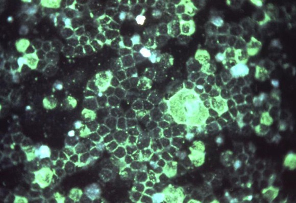
Glial fibrillary acidic protein (GFAP) is found in glial cells and is specific to the central nervous system. Found in both gray and white brain matter, it has recently been identified in serum, which makes it an attractive candidate as a clinical biomarker. Currently, the Glasgow Coma Scale (GCS) is the primary measure used to assess patients with head injury, but it can be influenced by intoxicants, medication, other injuries, or hypoperfusion and was not actually intended as an emergency department tool. "Really what we’d like is to develop some kind of biomarker that can be [measured] quickly and accurately, just like we do troponin for ischemia, or creatinine or bilirubin. Some type of blood test for the brain is our ultimate goal," said Dr. Linda Papa, director of academic clinical research, graduate medical education, Orlando Health, and an attending emergency physician at Orlando (Fla.) Regional Medical Center.

The study enrolled adult patients at three level 1 trauma centers who presented with mild or moderate blunt traumatic brain injury (TBI) and loss of consciousness or change in sensorium. Patients were enrolled from the ED and had blood samples collected within 4 hours of injury. All received CT scans. Control groups included patients with orthopedic trauma or motor vehicle trauma without head injury, and normal healthy people recruited from an ad. One aim of the study was to collect a large amount of normative data, she noted at the annual meeting of the Society for Academic Emergency Medicine.
A total of 307 patients were enrolled in the study, of whom 108 had a TBI. The TBI patients had a mean age of 39 years (range 18-89), and 65% were men. A total of 97 had GCS scores of 13-15 (mild), and 11 had GCS scores of 9-12 (moderate). Of the 97 with GCS 13-15, 24 (25%) had CT scans positive for intracranial lesions, and of the 11 patients with GCS scores 9-12, 8 (73%) had positive CTs. Controls included 176 normal individuals, 16 with non–head injury motor vehicle accidents, and 7 with orthopedic injuries.
Blood samples were drawn within a mean of 2.7 hours after injury. The GFAP biomarker first appeared in the serum within an hour (among those who had blood drawn that soon), increased until 3-4 hours, then leveled off. "It’s very interesting that so early in the course of injury, you can see a marker appearing in the blood," Dr. Papa commented.
Early GFAP levels were able to distinguish TBI patients from uninjured controls with an area under the curve (AUC) of 0.90 (1.0 is perfect), and differentiated those with mild TBI (GCS 15) from all controls with an AUC of 0.88. Mean GFAP levels in patients with negative CT scans versus those with positive CT scans were 0.335 ng/mL and 2.168 ng/mL, a highly significant difference with an AUC of 0.79.
The findings, if validated, suggest that early elevations of GFAP can be a potentially useful clinical tool in determining whether to image patients who are intoxicated or sedated, to admit or discharge patients from the ED, to assess severity of brain injury in a multiple trauma victim, to seek neurosurgical consultation and/or transfer to a neurosurgical facility, and to assess whether the patient can return to play or duty, she said.
Dr. Papa disclosed that she is a scientific consultant for Banyan Biomarkers, the company that is developing GFAP. However, she does not own stock or hold patents with the company. This study was funded by a grant from the Department of Defense and the National Institute of Neurological Disorders and Stroke.
BOSTON – The biomarker glial fibrillary acidic protein accurately distinguished between mild and moderate traumatic brain injury, between mild traumatic injury and controls, and between patients with and without intracranial lesions on CT in a prospective cohort study of emergency department patients with head injuries.
Glial fibrillary acidic protein (GFAP) is found in glial cells and is specific to the central nervous system. Found in both gray and white brain matter, it has recently been identified in serum, which makes it an attractive candidate as a clinical biomarker. Currently, the Glasgow Coma Scale (GCS) is the primary measure used to assess patients with head injury, but it can be influenced by intoxicants, medication, other injuries, or hypoperfusion and was not actually intended as an emergency department tool. "Really what we’d like is to develop some kind of biomarker that can be [measured] quickly and accurately, just like we do troponin for ischemia, or creatinine or bilirubin. Some type of blood test for the brain is our ultimate goal," said Dr. Linda Papa, director of academic clinical research, graduate medical education, Orlando Health, and an attending emergency physician at Orlando (Fla.) Regional Medical Center.
The study enrolled adult patients at three level 1 trauma centers who presented with mild or moderate blunt traumatic brain injury (TBI) and loss of consciousness or change in sensorium. Patients were enrolled from the ED and had blood samples collected within 4 hours of injury. All received CT scans. Control groups included patients with orthopedic trauma or motor vehicle trauma without head injury, and normal healthy people recruited from an ad. One aim of the study was to collect a large amount of normative data, she noted at the annual meeting of the Society for Academic Emergency Medicine.
A total of 307 patients were enrolled in the study, of whom 108 had a TBI. The TBI patients had a mean age of 39 years (range 18-89), and 65% were men. A total of 97 had GCS scores of 13-15 (mild), and 11 had GCS scores of 9-12 (moderate). Of the 97 with GCS 13-15, 24 (25%) had CT scans positive for intracranial lesions, and of the 11 patients with GCS scores 9-12, 8 (73%) had positive CTs. Controls included 176 normal individuals, 16 with non–head injury motor vehicle accidents, and 7 with orthopedic injuries.
Blood samples were drawn within a mean of 2.7 hours after injury. The GFAP biomarker first appeared in the serum within an hour (among those who had blood drawn that soon), increased until 3-4 hours, then leveled off. "It’s very interesting that so early in the course of injury, you can see a marker appearing in the blood," Dr. Papa commented.
Early GFAP levels were able to distinguish TBI patients from uninjured controls with an area under the curve (AUC) of 0.90 (1.0 is perfect), and differentiated those with mild TBI (GCS 15) from all controls with an AUC of 0.88. Mean GFAP levels in patients with negative CT scans versus those with positive CT scans were 0.335 ng/mL and 2.168 ng/mL, a highly significant difference with an AUC of 0.79.
The findings, if validated, suggest that early elevations of GFAP can be a potentially useful clinical tool in determining whether to image patients who are intoxicated or sedated, to admit or discharge patients from the ED, to assess severity of brain injury in a multiple trauma victim, to seek neurosurgical consultation and/or transfer to a neurosurgical facility, and to assess whether the patient can return to play or duty, she said.
Dr. Papa disclosed that she is a scientific consultant for Banyan Biomarkers, the company that is developing GFAP. However, she does not own stock or hold patents with the company. This study was funded by a grant from the Department of Defense and the National Institute of Neurological Disorders and Stroke.
FROM THE ANNUAL MEETING OF THE SOCIETY FOR ACADEMIC EMERGENCY MEDICINE
Major Finding: Early GFAP levels distinguished TBI from uninjured controls with an area under the curve (AUC) of 0.90 (1.0 is perfect), and differentiated mild TBI from all controls with an AUC of 0.88. Mean GFAP levels in patients with negative CT scans versus those with positive CT scans were 0.335 ng/mL and 2.168 ng/mL, a highly significant difference with an AUC of 0.79.
Data Source: Prospective cohort study of 108 TBI patients and 199 controls, including 176 normal controls and 23 with injuries not involving the head.
Disclosures: Dr. Papa is a scientific consultant for Banyan Biomarkers but does not own stock or hold patents with the company. The study was funded by a grant from the Department of Defense and the National Institute of Neurological Disorders and Stroke.
Biomarker Helps Stratify Traumatic Brain Injury Patients
BOSTON – The biomarker glial fibrillary acidic protein accurately distinguished between mild and moderate traumatic brain injury, between mild traumatic injury and controls, and between patients with and without intracranial lesions on CT in a prospective cohort study of emergency department patients with head injuries.
Glial fibrillary acidic protein (GFAP) is found in glial cells and is specific to the central nervous system. Found in both gray and white brain matter, it has recently been identified in serum, which makes it an attractive candidate as a clinical biomarker. Currently, the Glasgow Coma Scale (GCS) is the primary measure used to assess patients with head injury, but it can be influenced by intoxicants, medication, other injuries, or hypoperfusion and was not actually intended as an emergency department tool. "Really what we’d like is to develop some kind of biomarker that can be [measured] quickly and accurately, just like we do troponin for ischemia, or creatinine or bilirubin. Some type of blood test for the brain is our ultimate goal," said Dr. Linda Papa, director of academic clinical research, graduate medical education, Orlando Health, and an attending emergency physician at Orlando (Fla.) Regional Medical Center.
The study enrolled adult patients at three level 1 trauma centers who presented with mild or moderate blunt traumatic brain injury (TBI) and loss of consciousness or change in sensorium. Patients were enrolled from the ED and had blood samples collected within 4 hours of injury. All received CT scans. Control groups included patients with orthopedic trauma or motor vehicle trauma without head injury, and normal healthy people recruited from an ad. One aim of the study was to collect a large amount of normative data, she noted at the annual meeting of the Society for Academic Emergency Medicine.
A total of 307 patients were enrolled in the study, of whom 108 had a TBI. The TBI patients had a mean age of 39 years (range 18-89), and 65% were men. A total of 97 had GCS scores of 13-15 (mild), and 11 had GCS scores of 9-12 (moderate). Of the 97 with GCS 13-15, 24 (25%) had CT scans positive for intracranial lesions, and of the 11 patients with GCS scores 9-12, 8 (73%) had positive CTs. Controls included 176 normal individuals, 16 with non–head injury motor vehicle accidents, and 7 with orthopedic injuries.
Blood samples were drawn within a mean of 2.7 hours after injury. The GFAP biomarker first appeared in the serum within an hour (among those who had blood drawn that soon), increased until 3-4 hours, then leveled off. "It’s very interesting that so early in the course of injury, you can see a marker appearing in the blood," Dr. Papa commented.
Early GFAP levels were able to distinguish TBI patients from uninjured controls with an area under the curve (AUC) of 0.90 (1.0 is perfect), and differentiated those with mild TBI (GCS 15) from all controls with an AUC of 0.88. Mean GFAP levels in patients with negative CT scans versus those with positive CT scans were 0.335 ng/mL and 2.168 ng/mL, a highly significant difference with an AUC of 0.79.
The findings, if validated, suggest that early elevations of GFAP can be a potentially useful clinical tool in determining whether to image patients who are intoxicated or sedated, to admit or discharge patients from the ED, to assess severity of brain injury in a multiple trauma victim, to seek neurosurgical consultation and/or transfer to a neurosurgical facility, and to assess whether the patient can return to play or duty, she said.
Dr. Papa disclosed that she is a scientific consultant for Banyan Biomarkers, the company that is developing GFAP. However, she does not own stock or hold patents with the company. This study was funded by a grant from the Department of Defense and the National Institute of Neurological Disorders and Stroke.
FROM THE ANNUAL MEETING OF THE SOCIETY FOR ACADEMIC EMERGENCY MEDICINE
Major Finding: Early GFAP levels distinguished TBI from uninjured controls with an area under the curve (AUC) of 0.90 (1.0 is perfect), and differentiated mild TBI from all controls with an AUC of 0.88. Mean GFAP levels in patients with negative CT scans versus those with positive CT scans were 0.335 ng/mL and 2.168 ng/mL, a highly significant difference with an AUC of 0.79.
Data Source: Prospective cohort study of 108 TBI patients and 199 controls, including 176 normal controls and 23 with injuries not involving the head.
Disclosures: Dr. Papa is a scientific consultant for Banyan Biomarkers but does not own stock or hold patents with the company. The study was funded by a grant from the Department of Defense and the National Institute of Neurological Disorders and Stroke.
Rule Predicts Which ED Patients Need an Immediate ECG
BOSTON – A simple, validated rule based on age and presenting symptoms was highly sensitive at identifying which emergency department patients need an immediate triage 12-lead ECG to identify ST-segment elevation myocardial infarction in a study population of more than 3 million patients.
According to current guidelines from the American College of Cardiology and the American Heart Association, "ECG should be performed within 10 minutes of ED arrival for all patients with chest discomfort or other symptoms suggestive of STEMI" (Circulation 2004;110:588-636). "These are common-sense guidelines, but we often get caught up in the latter part," said Dr. Seth Glickman of the University of North Carolina at Chapel Hill, noting that about one-third of patients with STEMI do not have a complaint of chest pain.
The study data came from a statewide public health surveillance system in North Carolina comprising about 8.1 million ED visits from 2007-2008. The patients were divided into a derivation cohort in 2007 and a validation cohort in 2008. After the exclusion of those aged 18 years or younger, those with missing data, and all injury, bleeding, or pregnancy-related visits, the final data set included 1,685,633 visits in 2007 and 1,889,545 in 2008.
Of the 6,464 STEMI patients, 78% presented with chest pain. However, this varied dramatically by age. Although more than 90% of the 18- to 49-year-old group diagnosed with STEMI had chest pain, only 53% of those aged 80 years and older did. In contrast, the frequency of other chief complaints increased steadily with age, including dyspnea, syncope, weakness, abdominal pain, and altered mental status, Dr. Glickman said.
Using those factors, the investigators derived the following simplified rule: Immediate ECG is required for patients aged 30 years and older with chest pain; those aged 50 years and older with shortness of breath, altered mental status, upper extremity pain, weakness, or syncope; and patients aged 80 years and older. The rule is meant to identify those who should be prioritized for immediate ECG, not the total group of patients who may ultimately receive one, Dr. Glickman noted.
In the validation cohort, this rule was 92.7% sensitive (95% confidence interval, 91.8-93.5), was 74% specific (CI, 74.0-74.1), had a positive predictive value of 0.62% (CI, 0.60-0.64), and had a negative predictive value of 99.98% (CI, 99.98-99.98).
Regarding the 80-plus population, "clearly, there’s an opportunity to use clinical judgment in the rule. But on the flip side, I can say that the chief complaints of the advanced elderly who are diagnosed with STEMI are so across the map that it is challenging to develop a more specific rule," he said, adding that he and his associates are working on incorporating other elements into the model such as abdominal pain and nausea, which are also common complaints in very elderly patients.
In response to an audience member’s question about gender, Dr. Glickman said that they tried the rule with and without gender and it performed similarly, despite the fact that women are less likely to present with chest pain. "The real dominant factor in the model was age, which trumps gender across the entire spectrum. We thought it would be a lot easier to implement a rule that wasn’t gender-specific in the ambulance or the triage setting."
This study was funded by the American Heart Association Pharmaceutical Roundtable, the Robert Wood Johnson Foundation, the North Carolina Bio-Preparedness Collaborative, and Department of Homeland Security Office of Health Affairs. Dr. Glickman stated that he had no other personal disclosures.
The triage rule for who needs an ECG is long overdue. It addresses a question that plagues every emergency department in the country: Which of the millions of people flooding our emergency departments (EDs) need an immediate ECG? While it may appear simple to answer, to my knowledge no one has attempted to answer it scientifically. It’s easy to do one ECG, but consider the following everyday scenario: Eight people show up to your front door all at once, and an ECG takes about 3 minutes. There is a significant time pressure because of national guidelines mandating an ECG within 10 minutes. This is rigorously reviewed, and the ED is critiqued when a patient ends up having a ST-segment elevation myocardial infarction. So you really do need a rational way to decide. Furthermore, all of those ECGs must be immediately reviewed, causing frequent interruptions to the emergency physician and creating potential errors. This rule also is applicable to the many emergency medical service staff who perform prehospital ECGs. A paramedic has many actions to take and little time, so knowing who actually needs an ECG can be helpful.
Dr. Alexander T. Limkakeng Jr. is a director of acute care research, division of emergency medicine, Duke University, Durham, N.C. Dr. Limkakeng receives financial support from Roche to conduct a cardiac biomarker study and from the National Institutes of Health, for which he is a trial site investigator.
BOSTON – A simple, validated rule based on age and presenting symptoms was highly sensitive at identifying which emergency department patients need an immediate triage 12-lead ECG to identify ST-segment elevation myocardial infarction in a study population of more than 3 million patients.
According to current guidelines from the American College of Cardiology and the American Heart Association, "ECG should be performed within 10 minutes of ED arrival for all patients with chest discomfort or other symptoms suggestive of STEMI" (Circulation 2004;110:588-636). "These are common-sense guidelines, but we often get caught up in the latter part," said Dr. Seth Glickman of the University of North Carolina at Chapel Hill, noting that about one-third of patients with STEMI do not have a complaint of chest pain.
The study data came from a statewide public health surveillance system in North Carolina comprising about 8.1 million ED visits from 2007-2008. The patients were divided into a derivation cohort in 2007 and a validation cohort in 2008. After the exclusion of those aged 18 years or younger, those with missing data, and all injury, bleeding, or pregnancy-related visits, the final data set included 1,685,633 visits in 2007 and 1,889,545 in 2008.
Of the 6,464 STEMI patients, 78% presented with chest pain. However, this varied dramatically by age. Although more than 90% of the 18- to 49-year-old group diagnosed with STEMI had chest pain, only 53% of those aged 80 years and older did. In contrast, the frequency of other chief complaints increased steadily with age, including dyspnea, syncope, weakness, abdominal pain, and altered mental status, Dr. Glickman said.
Using those factors, the investigators derived the following simplified rule: Immediate ECG is required for patients aged 30 years and older with chest pain; those aged 50 years and older with shortness of breath, altered mental status, upper extremity pain, weakness, or syncope; and patients aged 80 years and older. The rule is meant to identify those who should be prioritized for immediate ECG, not the total group of patients who may ultimately receive one, Dr. Glickman noted.
In the validation cohort, this rule was 92.7% sensitive (95% confidence interval, 91.8-93.5), was 74% specific (CI, 74.0-74.1), had a positive predictive value of 0.62% (CI, 0.60-0.64), and had a negative predictive value of 99.98% (CI, 99.98-99.98).
Regarding the 80-plus population, "clearly, there’s an opportunity to use clinical judgment in the rule. But on the flip side, I can say that the chief complaints of the advanced elderly who are diagnosed with STEMI are so across the map that it is challenging to develop a more specific rule," he said, adding that he and his associates are working on incorporating other elements into the model such as abdominal pain and nausea, which are also common complaints in very elderly patients.
In response to an audience member’s question about gender, Dr. Glickman said that they tried the rule with and without gender and it performed similarly, despite the fact that women are less likely to present with chest pain. "The real dominant factor in the model was age, which trumps gender across the entire spectrum. We thought it would be a lot easier to implement a rule that wasn’t gender-specific in the ambulance or the triage setting."
This study was funded by the American Heart Association Pharmaceutical Roundtable, the Robert Wood Johnson Foundation, the North Carolina Bio-Preparedness Collaborative, and Department of Homeland Security Office of Health Affairs. Dr. Glickman stated that he had no other personal disclosures.
FROM THE ANNUAL MEETING OF THE SOCIETY FOR ACADEMIC EMERGENCY MEDICINE
Major Finding: In the validation cohort, this rule was 92.7% sensitive (95% confidence interval, 91.8-93.5), was 74% specific (CI, 74.0-74.1), had a positive predictive value of 0.62% (CI 0.60-0.64), and had a negative predictive value of 99.98% (CI, 99.98-99.98).
Data Source: A statewide public health surveillance system in North Carolina comprising about 8.1 million ED visits during 2007-2008.
Disclosures: This study was funded by the American Heart Association Pharmaceutical Roundtable, the Robert Wood Johnson Foundation, the North Carolina Bio-Preparedness Collaborative, and Department of Homeland Security Office of Health Affairs. Dr. Glickman has no other personal disclosures.