User login
When Can Exercise Supplant Surgery for Degenerative Meniscal Tears?
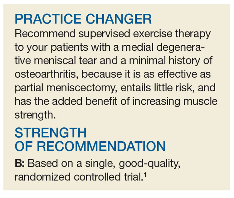
A 48-year-old man presents to your office for follow-up of right knee pain that has been bothering him for the past 12 months. He denies any trauma or inciting incident for the pain. On physical exam, he does not have crepitus but does have medial joint line tenderness of his right knee. An MRI shows a partial medial meniscal tear. Do you refer him to physical therapy (PT) or to orthopedics for arthroscopy and repair?
The meniscus—cartilage in the knee joint that provides support, stability, and lubrication to the joint during activity—can tear during a traumatic event or as a result of degeneration over time. Traumatic meniscal tears typically occur in those younger than 30 during sports (eg, basketball, soccer), whereas degenerative meniscal tears generally occur in patients ages 40 to 60.2,3 The annual incidence of all meniscal tears is 79 per 100,000.4 While some clinicians can diagnose traumatic meniscal tears based on history and physical examination, degenerative meniscal tears are more challenging and typically warrant an MRI for confirmation.3
Meniscal tears can be treated either conservatively, with supportive care and exercise, or surgically. Unfortunately, there are no national orthopedic guidelines available to help direct care. In one observational study, 95 of 117 patients (81.2%) were generally satisfied with surgical treatment at four-year follow-up; satisfaction was higher among those with a traumatic meniscal tear than in those with a degenerative tear.5
Two systematic reviews of surgery versus nonoperative management or sham therapies found no additional benefit of surgery for meniscal tears in a variety of patients with and without osteoarthritis.6,7 However, both studies were of only moderate quality, because of the number of patients in the nonoperative groups who ultimately underwent surgery. Neither of the studies directly compared surgery to nonoperative management.6,7Another investigation—a multicenter, randomized, double-blind, sham-controlled study conducted in Finland involving 1
Clinical practice recommendations devised from a vast systematic review of the literature recommend that the decision for surgery be based on patient-specific factors, such as symptoms, age, mechanism of tear, extent of damage, and occupational/social/activity needs.9
STUDY SUMMARY
Exercise is as good as surgery
The current superiority RCT compared exercise therapy to arthroscopic partial meniscectomy. Subjects (ages 35 to 60) presented to the orthopedic department of two hospitals in Norway with unilateral knee pain of more than two months’ duration and an MRI-delineated medial meniscal tear. They were included in the study only if they had radiographic evidence of minimal osteoarthritis (Kellgren-Lawrence classification grade ≤ 2). Exclusion criteria included acute trauma, locked knee, ligament injury, and knee surgery in the same knee within the previous two years.
The primary outcomes were change in patient-reported knee function (as determined by overall Knee injury and Osteoarthritis Outcome Score [KOOS] after two years) and thigh muscle strength at three months (as measured by physiotherapists). The researchers used four of the five KOOS subscales for this analysis: pain, other symptoms (swelling, grinding/noise from the joint, ability to straighten and bend), function in sports/recreation, and knee-related quality of life (QOL). The average score of each subscale was used.
Secondary outcomes included the five individual KOOS subscales (the four previously mentioned, plus activities of daily living [ADLs]), as well as thigh muscle strength and lower-extremity performance test results.
Methods. Testing personnel were blinded to group allocation; participants wore pants or neoprene sleeves to cover surgical scars. A total of 140 patients were randomized to either 12 weeks (24-36 sessions) of exercise therapy alone or a standardized arthroscopic partial meniscectomy; upon discharge, those in the latter group received written and oral encouragement to perform simple exercises at home, two to four times daily, to regain range of motion and reduce swelling.
Results. At two years, the overall mean improvement in KOOS4 score from baseline was similar between the exercise group and the meniscectomy group (25.3 pts vs 24.4 pts, respectively; mean difference [MD], 0.9). Additionally, muscle strength (measured as peak torque flexion and extension and total work flexion and extension) at both three and 12 months showed significant objective improvements favoring exercise therapy.
In the secondary analysis of the KOOS subscale scores, change from baseline was nonsignificant for four of the five (pain, ADL, sports/recreation, and QOL). Only the symptoms subscale had a significant difference favoring exercise therapy (MD, 5.3 pts); this was likely clinically insignificant on a grading scale of 0 to 100.
Of the patients allocated to exercise therapy alone, 19% crossed over and underwent surgery during the two-year study period.
WHAT’S NEW
Head-to-head comparison adds evidence
This is the first trial to directly compare exercise therapy to surgery in patients with meniscal tears. Interestingly, exercise therapy was as effective after a two-year follow-up period and was superior in the short term for thigh muscle strength.1
The results of this study build on those from the aforementioned smaller study conducted in Finland.8 In that study, both groups received instruction for the same graduated exercise plan. The researchers found that exercise was comparable to surgery for meniscal tears in patients with no osteoarthritis.
CAVEATS
What about more severe osteoarthritis?
This trial included patients with no to mild osteoarthritis in addition to their meniscal tear.1 It is unclear if the results would be maintained in those with more advanced disease. Additionally, 19% of patients crossed over from the exercise group to the surgery group, even though muscle strength improved. Therefore, education about the risks of surgery and the potential lack of benefit is important.
CHALLENGES TO IMPLEMENTATION
Cost and effort of PT
The cost of PT can be a barrier for patients who have adequate insurance coverage for surgery but inadequate coverage for PT. Additionally, exercise therapy requires significant and ongoing time and effort, which may deter those with busy lifestyles. Patients and clinicians may view surgery as an “easier” fix.
ACKNOWLEDGEMENT
The PURLs Surveillance System was supported in part by Grant Number UL1RR024999 from the National Center For Research Resources, a Clinical Translational Science Award to the University of Chicago. The content is solely the responsibility of the authors and does not necessarily represent the official views of the National Center For Research Resources or the National Institutes of Health.
Copyright © 2017. The Family Physicians Inquiries Network. All rights reserved.
Reprinted with permission from the Family Physicians Inquiries Network and The Journal of Family Practice (2017;66[4]:250-252).
1. Kise NJ, Risberg MA, Stensrud S, et al. Exercise therapy versus arthroscopic partial meniscectomy for degenerative meniscal tear in middle aged patients: randomised controlled trial with two year follow-up. BMJ. 2016;354:i3740.
2. Beals CT, Magnussen RA, Graham WC, et al. The prevalence of meniscal pathology in asymptomatic athletes. Sports Med. 2016;46:1517-1524.
3. Maffulli N, Longo UG, Campi S, et al. Meniscal tears. Open Access J Sports Med. 2010;1:45-54.
4. Peat G, Bergknut C, Frobell R, et al. Population-wide incidence estimates for soft tissue knee injuries presenting to healthcare in southern Sweden: data from the Skåne Healthcare Register. Arthritis Res Ther. 2014;16:R162.
5. Ghislain NA, Wei JN, Li YG. Study of the clinical outcome between traumatic and degenerative (non-traumatic) meniscal tears after arthroscopic surgery: a 4-years follow-up study. J Clin Diagn Res. 2016;10:RC01-RC04.
6. Khan M, Evaniew N, Bedi A, et al. Arthroscopic surgery for degenerative tears of the meniscus: a systematic review and meta-analysis. CMAJ. 2014;186:1057-1064.
7. Monk P, Garfjeld Roberts P, Palmer AJ, et al. The urgent need for evidence in arthroscopic meniscal surgery: a systematic review of the evidence for operative management of meniscal tears. Am J Sports Med. 2017;45:965-973.
8. Sihvonen R, Paavola M, Malmivaara A, et al; Finnish Degenerative Meniscal Lesion Study (FIDELITY) Group. Arthroscopic partial meniscectomy versus sham surgery for a degenerative meniscal tear. N Engl J Med. 2013;369:2515-2524.
9. Beaufils P, Hulet C, Dhénain M, et al. Clinical practice guidelines for the management of meniscal lesions and isolated lesions of the anterior cruciate ligament of the knee in adults. Orthop Traumatol Surg Res. 2009;95:437-442.
A 48-year-old man presents to your office for follow-up of right knee pain that has been bothering him for the past 12 months. He denies any trauma or inciting incident for the pain. On physical exam, he does not have crepitus but does have medial joint line tenderness of his right knee. An MRI shows a partial medial meniscal tear. Do you refer him to physical therapy (PT) or to orthopedics for arthroscopy and repair?
The meniscus—cartilage in the knee joint that provides support, stability, and lubrication to the joint during activity—can tear during a traumatic event or as a result of degeneration over time. Traumatic meniscal tears typically occur in those younger than 30 during sports (eg, basketball, soccer), whereas degenerative meniscal tears generally occur in patients ages 40 to 60.2,3 The annual incidence of all meniscal tears is 79 per 100,000.4 While some clinicians can diagnose traumatic meniscal tears based on history and physical examination, degenerative meniscal tears are more challenging and typically warrant an MRI for confirmation.3
Meniscal tears can be treated either conservatively, with supportive care and exercise, or surgically. Unfortunately, there are no national orthopedic guidelines available to help direct care. In one observational study, 95 of 117 patients (81.2%) were generally satisfied with surgical treatment at four-year follow-up; satisfaction was higher among those with a traumatic meniscal tear than in those with a degenerative tear.5
Two systematic reviews of surgery versus nonoperative management or sham therapies found no additional benefit of surgery for meniscal tears in a variety of patients with and without osteoarthritis.6,7 However, both studies were of only moderate quality, because of the number of patients in the nonoperative groups who ultimately underwent surgery. Neither of the studies directly compared surgery to nonoperative management.6,7Another investigation—a multicenter, randomized, double-blind, sham-controlled study conducted in Finland involving 1
Clinical practice recommendations devised from a vast systematic review of the literature recommend that the decision for surgery be based on patient-specific factors, such as symptoms, age, mechanism of tear, extent of damage, and occupational/social/activity needs.9
STUDY SUMMARY
Exercise is as good as surgery
The current superiority RCT compared exercise therapy to arthroscopic partial meniscectomy. Subjects (ages 35 to 60) presented to the orthopedic department of two hospitals in Norway with unilateral knee pain of more than two months’ duration and an MRI-delineated medial meniscal tear. They were included in the study only if they had radiographic evidence of minimal osteoarthritis (Kellgren-Lawrence classification grade ≤ 2). Exclusion criteria included acute trauma, locked knee, ligament injury, and knee surgery in the same knee within the previous two years.
The primary outcomes were change in patient-reported knee function (as determined by overall Knee injury and Osteoarthritis Outcome Score [KOOS] after two years) and thigh muscle strength at three months (as measured by physiotherapists). The researchers used four of the five KOOS subscales for this analysis: pain, other symptoms (swelling, grinding/noise from the joint, ability to straighten and bend), function in sports/recreation, and knee-related quality of life (QOL). The average score of each subscale was used.
Secondary outcomes included the five individual KOOS subscales (the four previously mentioned, plus activities of daily living [ADLs]), as well as thigh muscle strength and lower-extremity performance test results.
Methods. Testing personnel were blinded to group allocation; participants wore pants or neoprene sleeves to cover surgical scars. A total of 140 patients were randomized to either 12 weeks (24-36 sessions) of exercise therapy alone or a standardized arthroscopic partial meniscectomy; upon discharge, those in the latter group received written and oral encouragement to perform simple exercises at home, two to four times daily, to regain range of motion and reduce swelling.
Results. At two years, the overall mean improvement in KOOS4 score from baseline was similar between the exercise group and the meniscectomy group (25.3 pts vs 24.4 pts, respectively; mean difference [MD], 0.9). Additionally, muscle strength (measured as peak torque flexion and extension and total work flexion and extension) at both three and 12 months showed significant objective improvements favoring exercise therapy.
In the secondary analysis of the KOOS subscale scores, change from baseline was nonsignificant for four of the five (pain, ADL, sports/recreation, and QOL). Only the symptoms subscale had a significant difference favoring exercise therapy (MD, 5.3 pts); this was likely clinically insignificant on a grading scale of 0 to 100.
Of the patients allocated to exercise therapy alone, 19% crossed over and underwent surgery during the two-year study period.
WHAT’S NEW
Head-to-head comparison adds evidence
This is the first trial to directly compare exercise therapy to surgery in patients with meniscal tears. Interestingly, exercise therapy was as effective after a two-year follow-up period and was superior in the short term for thigh muscle strength.1
The results of this study build on those from the aforementioned smaller study conducted in Finland.8 In that study, both groups received instruction for the same graduated exercise plan. The researchers found that exercise was comparable to surgery for meniscal tears in patients with no osteoarthritis.
CAVEATS
What about more severe osteoarthritis?
This trial included patients with no to mild osteoarthritis in addition to their meniscal tear.1 It is unclear if the results would be maintained in those with more advanced disease. Additionally, 19% of patients crossed over from the exercise group to the surgery group, even though muscle strength improved. Therefore, education about the risks of surgery and the potential lack of benefit is important.
CHALLENGES TO IMPLEMENTATION
Cost and effort of PT
The cost of PT can be a barrier for patients who have adequate insurance coverage for surgery but inadequate coverage for PT. Additionally, exercise therapy requires significant and ongoing time and effort, which may deter those with busy lifestyles. Patients and clinicians may view surgery as an “easier” fix.
ACKNOWLEDGEMENT
The PURLs Surveillance System was supported in part by Grant Number UL1RR024999 from the National Center For Research Resources, a Clinical Translational Science Award to the University of Chicago. The content is solely the responsibility of the authors and does not necessarily represent the official views of the National Center For Research Resources or the National Institutes of Health.
Copyright © 2017. The Family Physicians Inquiries Network. All rights reserved.
Reprinted with permission from the Family Physicians Inquiries Network and The Journal of Family Practice (2017;66[4]:250-252).
1. Kise NJ, Risberg MA, Stensrud S, et al. Exercise therapy versus arthroscopic partial meniscectomy for degenerative meniscal tear in middle aged patients: randomised controlled trial with two year follow-up. BMJ. 2016;354:i3740.
2. Beals CT, Magnussen RA, Graham WC, et al. The prevalence of meniscal pathology in asymptomatic athletes. Sports Med. 2016;46:1517-1524.
3. Maffulli N, Longo UG, Campi S, et al. Meniscal tears. Open Access J Sports Med. 2010;1:45-54.
4. Peat G, Bergknut C, Frobell R, et al. Population-wide incidence estimates for soft tissue knee injuries presenting to healthcare in southern Sweden: data from the Skåne Healthcare Register. Arthritis Res Ther. 2014;16:R162.
5. Ghislain NA, Wei JN, Li YG. Study of the clinical outcome between traumatic and degenerative (non-traumatic) meniscal tears after arthroscopic surgery: a 4-years follow-up study. J Clin Diagn Res. 2016;10:RC01-RC04.
6. Khan M, Evaniew N, Bedi A, et al. Arthroscopic surgery for degenerative tears of the meniscus: a systematic review and meta-analysis. CMAJ. 2014;186:1057-1064.
7. Monk P, Garfjeld Roberts P, Palmer AJ, et al. The urgent need for evidence in arthroscopic meniscal surgery: a systematic review of the evidence for operative management of meniscal tears. Am J Sports Med. 2017;45:965-973.
8. Sihvonen R, Paavola M, Malmivaara A, et al; Finnish Degenerative Meniscal Lesion Study (FIDELITY) Group. Arthroscopic partial meniscectomy versus sham surgery for a degenerative meniscal tear. N Engl J Med. 2013;369:2515-2524.
9. Beaufils P, Hulet C, Dhénain M, et al. Clinical practice guidelines for the management of meniscal lesions and isolated lesions of the anterior cruciate ligament of the knee in adults. Orthop Traumatol Surg Res. 2009;95:437-442.
Perioperative pharmacological thromboprophylaxis in patients with cancer: a systematic review and meta-analysis
CLINICAL QUESTION: What are the benefits and harms of perioperative pharmacological thromboprophylaxis in cancer patients undergoing surgery?
STUDY DESIGN: Systematic review and meta-analysis.
SYNOPSIS: Thirty-nine trials were deemed eligible for inclusion in the meta-analysis. Twenty-five of these were prospective and 14 were retrospective. The overall incidence of deep venous thrombosis (DVT) and pulmonary embolism was 0.9% (across 20 studies) and 0.3% (across 19 studies), respectively. Pharmacologic prophylaxis overall reduced DVT incidence (0.5% vs. 1.2%; relative risk, 0.51; P = .03). Subgroup analysis demonstrated this was significant for abdominal/pelvic surgeries and with low molecular weight heparin. Six studies compared duration of standard prophylaxis (10 days) with extended prophylaxis (4 weeks), with a lower VTE rate in the extended group. Bleeding events were noted in 13 studies and pharmacologic prophylaxis significantly increased bleeding risk (2.7% vs. 8%; RR, 2.51; P less than .0001).
BOTTOM LINE: Perioperative pharmacologic prophylaxis reduces DVT risk in patients with cancer, with greatest risk reduction seen in patients undergoing abdominal/pelvic surgeries. This comes at the cost of increased bleeding complications.
CITATIONS: Guo Q, Huang B, Zhao J, et al. Perioperative pharmacological thromboprophylaxis in patients with cancer: a systematic review and meta-analysis. Ann Surg. 2016 Nov. doi: 10.1097/SLA.0000000000002074.
Dr. Patil is a clinical instructor, Division of Hospital Medicine, University of Colorado School of Medicine, Aurora.
Readmission rates after passage of the hospital readmissions reduction program
CLINICAL QUESTION: Did hospitals receiving the highest penalties for readmissions have accelerated improvement in this metric after passage of Medicare Hospital Readmissions Reduction Program (HRRP)?
BACKGROUND: Medicare passed the HRRP to incentivize reductions in readmission rates. The impact of penalties on relative hospital improvement rates remains unknown.

SETTING: Query of national Medicare Provider Analysis and Review files.
SYNOPSIS: 2,868 hospitals were identified as candidates for analysis and were stratified into four risk groups based on penalty size under HRRP: highest-performing, average-performing, low-performing, and lowest-performing. The primary outcomes were hospital-specific, 30-day, all-cause risk-standardized readmission rates (RSRRs) for patients discharged with acute MI, HF, or pneumonia. The investigators separated data into a pre-law period and post-law period. They fitted a logistic regression model to pre-law RSRRs and developed a piecewise linear model on post-law RSRRs with pre-law data as the dependent variable. All hospital groups had reductions in RSRRs, with the lowest quartile demonstrating greatest improvement.
BOTTOM LINE: HRRP has resulted in reductions in RSRRs with greatest improvement in hospitals with lowest pre-law performance.
CITATIONS: Wasfy JH, Zigler CM, Choirat C, et al. Readmission rates after passage of the hospital readmissions reduction program: a pre-post analysis. Ann Intern Med. 2017 Mar;166(5):324-31.
Dr. Patil is a clinical instructor, Division of Hospital Medicine, University of Colorado School of Medicine, Aurora.
CLINICAL QUESTION: Did hospitals receiving the highest penalties for readmissions have accelerated improvement in this metric after passage of Medicare Hospital Readmissions Reduction Program (HRRP)?
BACKGROUND: Medicare passed the HRRP to incentivize reductions in readmission rates. The impact of penalties on relative hospital improvement rates remains unknown.
SETTING: Query of national Medicare Provider Analysis and Review files.
SYNOPSIS: 2,868 hospitals were identified as candidates for analysis and were stratified into four risk groups based on penalty size under HRRP: highest-performing, average-performing, low-performing, and lowest-performing. The primary outcomes were hospital-specific, 30-day, all-cause risk-standardized readmission rates (RSRRs) for patients discharged with acute MI, HF, or pneumonia. The investigators separated data into a pre-law period and post-law period. They fitted a logistic regression model to pre-law RSRRs and developed a piecewise linear model on post-law RSRRs with pre-law data as the dependent variable. All hospital groups had reductions in RSRRs, with the lowest quartile demonstrating greatest improvement.
BOTTOM LINE: HRRP has resulted in reductions in RSRRs with greatest improvement in hospitals with lowest pre-law performance.
CITATIONS: Wasfy JH, Zigler CM, Choirat C, et al. Readmission rates after passage of the hospital readmissions reduction program: a pre-post analysis. Ann Intern Med. 2017 Mar;166(5):324-31.
Dr. Patil is a clinical instructor, Division of Hospital Medicine, University of Colorado School of Medicine, Aurora.
Assessment of goals of care in nursing home reduces hospitalization for patients with dementia
CLINICAL QUESTION: For patients with advanced dementia, does a goals-of-care intervention improve communication and care outcomes?
STUDY DESIGN: Single-blind cluster randomized trial.
SETTING: Twenty-two nursing homes in North Carolina.
SYNOPSIS: Three hundred and two patient/families enrolled. Intervention included video and print decision aids followed by a structured goals of care discussion with trained nursing home staff. Quality of communication results, the primary outcome, at 3 months were mixed. Family perception of communication with nursing home staff was better in the intervention. Family–health care provider concordance on primary goal of care and treatment consistent with preferences were not significantly different. By the end of the study at 9 months there was no difference in symptom control but some secondary outcomes were encouraging including greater completion of MOST advanced directives (35% vs. 16%; P = .05) and half as many hospital transfers. Multiple comparisons merits future verification of secondary outcome findings.
BOTTOM LINE: Goals of care discussions for patients with advanced dementia appears to reduce hospitalizations.
CITATIONS: Hanson LC, Zimmerman S, Song MK, et al. Effect of the goals of care intervention for advanced dementia: a randomized clinical trial. JAMA Intern Med. 2017 Jan;177:24-31.
Dr. Cumbler is the associate chief of hospital medicine, Division of Hospital Medicine, University of Colorado School of Medicine, Aurora.
Daratumumab, elotuzumab eyed for initial treatment of myeloma
The emerging role for immunotherapies as an essential component of multiple myeloma therapy is examined in a review article in Leukemia by Cyrille Touzeau, MD, and his colleagues.
The reviewers detail research examining a string of monoclonal antibodies that fell short in earlier evaluations. They focus on the two approved agents that target CD38 (daratumumab) and SLAMF7 (elotuzumab) and have succeeded in combination therapies for patients with relapsed myeloma. These two antibodies, and other immunotherapy possibilities in the pipeline, are expected to have a strong impact on treatment modalities and outcomes in patients with multiple myeloma, including transplant eligible and elderly patients, Dr. Touzeau, of the service d’hématologie clinique, Nantes, France, and his fellow researchers wrote.

In two phase III randomized studies, ELO 1 (NCT01891643) and ELOQUENT 1 (NCT01335399), previously untreated myeloma patients are receiving lenalidomide/dexamethasone with or without elotuzumab.
In another ongoing trial, elotuzumab is being evaluated in combination with the anti-KIR antibody lirilumab and the anti-CD137 antibody urelumab (NCT02252263). Elotuzumab also is being studied in combination with lenalidomide as maintenance after high-dose therapy (NCT02420860).
Additionally, elotuzumab in combination with pomalidomide-dexamethasone is being examined for relapsed myeloma in an ongoing phase II randomized trial (NCT02654132). SLAMF7 is also being evaluated as a target for immunoconjugate therapy, with an ongoing trial of an auristatin E conjugate (ABBV-838) in patients with relapsed or refractory disease (NCT02462525), the reviewers note.
Daratumumab is being examined in combination with VTD [bortezomib (Velcade)/thalidomide/dexamethasone] as induction therapy and for its role as maintenance after high-dose therapy, among previously untreated transplant-eligible myeloma patients in the phase III randomized Cassiopeia study (NCT02541383).
In patients not eligible for transplant, the phase III randomized trial, MAIA, is evaluating the addition of daratumumab to lenalidomide-dexamethasone (NCT02252172). In high-risk smoldering myeloma, daratumumab is being evaluated in the phase III randomized CENTAURUS trial (NCT02316106). PAVO is a phase 1b study of the subcutaneous administration of daratumumab (NCT02519452). Preliminary results determined that the fixed subcutaneous dose of 1800 mg was consistent with the 16 mg/kg IV dose in terms of pharmacokinetics.
Dr. Touzeau declared no conflicts of interest. His coauthors participate in advisory boards and receive honoraria from several drug makers including the makers of immunotherapies.
[email protected]
On Twitter @maryjodales
The emerging role for immunotherapies as an essential component of multiple myeloma therapy is examined in a review article in Leukemia by Cyrille Touzeau, MD, and his colleagues.
The reviewers detail research examining a string of monoclonal antibodies that fell short in earlier evaluations. They focus on the two approved agents that target CD38 (daratumumab) and SLAMF7 (elotuzumab) and have succeeded in combination therapies for patients with relapsed myeloma. These two antibodies, and other immunotherapy possibilities in the pipeline, are expected to have a strong impact on treatment modalities and outcomes in patients with multiple myeloma, including transplant eligible and elderly patients, Dr. Touzeau, of the service d’hématologie clinique, Nantes, France, and his fellow researchers wrote.
In two phase III randomized studies, ELO 1 (NCT01891643) and ELOQUENT 1 (NCT01335399), previously untreated myeloma patients are receiving lenalidomide/dexamethasone with or without elotuzumab.
In another ongoing trial, elotuzumab is being evaluated in combination with the anti-KIR antibody lirilumab and the anti-CD137 antibody urelumab (NCT02252263). Elotuzumab also is being studied in combination with lenalidomide as maintenance after high-dose therapy (NCT02420860).
Additionally, elotuzumab in combination with pomalidomide-dexamethasone is being examined for relapsed myeloma in an ongoing phase II randomized trial (NCT02654132). SLAMF7 is also being evaluated as a target for immunoconjugate therapy, with an ongoing trial of an auristatin E conjugate (ABBV-838) in patients with relapsed or refractory disease (NCT02462525), the reviewers note.
Daratumumab is being examined in combination with VTD [bortezomib (Velcade)/thalidomide/dexamethasone] as induction therapy and for its role as maintenance after high-dose therapy, among previously untreated transplant-eligible myeloma patients in the phase III randomized Cassiopeia study (NCT02541383).
In patients not eligible for transplant, the phase III randomized trial, MAIA, is evaluating the addition of daratumumab to lenalidomide-dexamethasone (NCT02252172). In high-risk smoldering myeloma, daratumumab is being evaluated in the phase III randomized CENTAURUS trial (NCT02316106). PAVO is a phase 1b study of the subcutaneous administration of daratumumab (NCT02519452). Preliminary results determined that the fixed subcutaneous dose of 1800 mg was consistent with the 16 mg/kg IV dose in terms of pharmacokinetics.
Dr. Touzeau declared no conflicts of interest. His coauthors participate in advisory boards and receive honoraria from several drug makers including the makers of immunotherapies.
[email protected]
On Twitter @maryjodales
FROM LEUKEMIA
Two doses HPV vaccine are as good as three against genital warts
Receiving two doses of human papillomavirus (HPV) vaccine at 5-month intervals or longer appears to provide similar protection against genital warts as three doses among girls initiating the series before age 15 years, reported Rebecca B. Perkins, MD, of Boston University, and her associates.
Of 387,906 adolescent females, 8% received 1 dose of HPV vaccine, 9% received 2 doses, 31% received 3 doses, and 52% remained unvaccinated. The mean age of the girls in the study was 15 years, and average length of follow-up was 6 years. The girls were aged 9-18 years on Jan.1, 2007, and the exposure period began at that time for unvaccinated girls or on the date of the last HPV vaccine injection for those receiving the vaccine. Among girls receiving more than 1 dose, 60% received their second dose within 3 months of their first dose (on time), and 47% received their third dose within 5 months of their second dose.

“Although reductions in genital warts are an important early marker of vaccine effectiveness, reductions in cervical dysplasia and cancers are far more important vaccine-related outcomes,” Dr. Perkins and her associates said. “Human papillomavirus vaccine protection must last many years to provide adequate cancer protection, therefore ongoing studies are paramount,” they noted.
The study used data from the Truven Health Analytics MarketScan Commercial Claims Database, covering enrollees and dependents from about half of provider-sponsored U.S. health insurance plans.
Read more at (Sex Transm Dis. 2017 Jun. doi: 10.1097/OLQ.0000000000000615).
Receiving two doses of human papillomavirus (HPV) vaccine at 5-month intervals or longer appears to provide similar protection against genital warts as three doses among girls initiating the series before age 15 years, reported Rebecca B. Perkins, MD, of Boston University, and her associates.
Of 387,906 adolescent females, 8% received 1 dose of HPV vaccine, 9% received 2 doses, 31% received 3 doses, and 52% remained unvaccinated. The mean age of the girls in the study was 15 years, and average length of follow-up was 6 years. The girls were aged 9-18 years on Jan.1, 2007, and the exposure period began at that time for unvaccinated girls or on the date of the last HPV vaccine injection for those receiving the vaccine. Among girls receiving more than 1 dose, 60% received their second dose within 3 months of their first dose (on time), and 47% received their third dose within 5 months of their second dose.
“Although reductions in genital warts are an important early marker of vaccine effectiveness, reductions in cervical dysplasia and cancers are far more important vaccine-related outcomes,” Dr. Perkins and her associates said. “Human papillomavirus vaccine protection must last many years to provide adequate cancer protection, therefore ongoing studies are paramount,” they noted.
The study used data from the Truven Health Analytics MarketScan Commercial Claims Database, covering enrollees and dependents from about half of provider-sponsored U.S. health insurance plans.
Read more at (Sex Transm Dis. 2017 Jun. doi: 10.1097/OLQ.0000000000000615).
FROM SEXUALLY TRANSMITTED DISEASES
House barely passes ACA repeal/replace bill
A few additional tweaks to the American Health Care Act helped garner just enough Republican votes to pass the first phase of the party’s three-part effort to repeal and replace the Affordable Care Act.
The bill passed May 4 by a 217-213 margin, with one Republican member not voting. The vote came after a false start in March when House Speaker Paul Ryan (R-Wisc.) canceled consideration because Republicans could not muster enough votes to pass it. All votes in favor of the bill came from GOP members, while all House Democrats plus 20 Republicans voted against passage.
Two amendments helped to make the bill palatable enough to gain enough votes for passage. New amendments from Rep. Gary Palmer (R-Ala.) and Rep. Fred Upton (R-Mich.) targeted high-risk pools. They were added to an April amendment from Rep. Tom MacArthur (R-N.J.) that would allow states to seek waivers for coverage of the essential health benefits and from community rating provisions to allow for higher premiums for those who are sicker or older.
The American Health Care Act (H.R. 1629) in its amended form has not been yet been scored by the Congressional Budget Office (CBO) to determine its effects on the federal budget. However, an earlier CBO analysis of the original, unamended bill predicted an estimated 58 million people would be uninsured by 2026, compared with 28 million under the Affordable Care Act. It is also predicting large premium increases for some groups of patients.
A CBO score is required of all legislation because, under Senate rules, anything that adds to the budget must be offset by additional revenue-generating provisions or cuts elsewhere in the federal budget. The bill, if passed and sent to the president’s desk for signature, is at minimum budget neutral if not deficit reducing.
The amended AHCA was roundly rejected by most physician organizations, including the American Medical Association and many specialty societies.
“The bill passed by the House today will result in millions of Americans losing access to quality, affordable health insurance and those with preexisting health conditions face the possibility of going back to the time when insurers could charge them premiums that made access to coverage out of the question,” AMA President Andrew Gurman, MD, said in a statement. “The AMA urges the Senate and the Administration to work with physician, patient, hospital, and other provider groups to craft bipartisan solutions so all American families can access affordable and meaningful coverage, while preserving the safety net for vulnerable patients.”
House Minority Leader Nancy Pelosi (D-Calif.), in addition to criticizing the bill for its predicted effects in increasing premiums and decreasing coverage for those with preexisting conditions, called out the bill for being nothing more than a means to help cover the cost of a separate tax bill when she described the AHCA as providing “tax cuts for the rich at the expense of health insurance for tens of millions of working families across America” during the debate prior to passage. “Trumpcare is a billionaire’s tax cut disguised as a health care bill.”
Speaker Ryan continued to highlight the continual coverage issues that have been growing in the individual health insurance marketplace, spotlighting Iowa, where insurer Medica has announced that it will likely discontinue providing coverage in 2018, leaving most counties in the state with no option to purchase coverage following the announced withdrawal of Aetna and Wellmark Blue Cross Blue Shield.
“This is a crisis,” Speaker Ryan said during the debate. “What protection is Obamacare if there is no health care plan to purchase in your state? This is the direction Obamacare is rapidly heading.”
In helping to get the AHCA barely over the final hurdle, the Palmer amendment creates an “invisible risk-sharing program” under which the federal government would subsidize insurers to the tune of $15 billion over 9 years. The program would allow insurers to make a prospective determination of who might be a cost-intensive user and move them to the high-risk pool with the federal funding joining insurer funding to help pay for coverage. The amendment also would change the ACA’s reinsurance provisions, which retroactively reimburse insurers for high-utilizing customers.
Neither invisible risk sharing nor retroactive reinsurance are “inherently superior at reducing premiums,” according to an April 12 blog post by the journal Health Affairs. “Premium reductions depend entirely on how much funding the program receives in relation to the risks being insured. Rep. Palmer’s amendment leaves all the critical details of this new, invisible program unspecified, making it hard to generate precise estimate.”
Authors Mark Hall, senior fellow at Brookings Institute, and Nicholas Bagely, law professor at the University of Michigan, Ann Arbor, note that the funding in the amendment “is no more than 2% of total premiums in the market.”
The Upton amendment would add an additional $8 billion in funding for high-risk pools, bringing the total amount potentially available to help cover people with preexisting conditions to $123 billion.
Analysis from Avalere finds that the funding specifically allocated to assist those with preexisting conditions ($23 billion, including the additional $8 billion from the Upton amendment) “will only cover 110,000 individuals with preexisting, chronic condition. If states were to allocate all the other funds [available in the AHCA] toward providing insurance to people with preexisting conditions ... 600,000 with preexisting chronic conditions could be covered.” The analysis notes approximately “2.2 million enrollees in the individual market today have some sort of preexisting chronic condition.”
“Given the amount of funding in the bill, the program can only afford a few small states to opt into medical underwriting,” Caroline Pearson, senior vice president at Avalere, said in a statement. “If any large states receive a waiver, many chronically ill individuals could be left without access to insurance.”
Because the AHCA focuses solely on reforming revenue-related aspects of the ACA and was passed using budget reconciliation procedures, it will need only a simple majority to pass the Senate. However, keeping the bill budget neutral will make it difficult to pass in its current form, even though Republicans hold 52 of the chamber’s 100 seats.
Phase two of the repeal and replace plan will be a full examination of federal regulations and phase three, which will require at least 60 votes in the Senate, will be changes to other provisions in the health care law that are not directly revenue generating. President Donald Trump in the past has suggested that this will include his goals of opening the sale of insurance across state lines and other key priorities.
VIDEO: Incoming AATS president outlines goals
BOSTON – Strengthening member engagement is a top goal for incoming AATS President Duke E. Cameron, MD.
In this video, Dr. Cameron, of Massachusetts General Hospital, Boston, shared his objectives as the next AATS leader and the direction he envisions for the specialty over the next 100 years. Dr. Cameron also discussed his hope for new online educational efforts and the importance of physician collaboration with other health professionals.
On Twitter @legal_med
AT THE AATS ANNUAL MEETING

Point/Counterpoint: Should all suspected mucinous cystic neoplasms be resected?
Dr. Pawlik is a pretty clever guy and a strong adversary. Mucinous cystic neoplasm is literally the only topic in hepato-pancreato-biliary surgery he has not written about – yet.1
But I will argue that all suspected mucinous cystic neoplasms (MCNs) should be surgically removed. Reasons include: the natural history of a benign MCN in a typical 45-year-old remains unknown, clinical features are not always reliable, and it’s difficult to distinguish benign from malignant neoplasms without surgery.

Although many MCNs are benign when resected, all have malignant potential.
The prevalence of cancer at time of diagnosis with an MCN is fairly low, about 15%. This means most of these cysts are benign when resected. However, discrimination between benign and malignant is difficult without surgery, and the degree of epithelial dysplasia at time of resection ranges from mild to severe with invasive carcinoma.
When cancer is present, patients tend to do poorly. In one study where 44 out of 344 patients developed invasive cancer, the group with cancer had a 3-year overall survival rate of 59%.2
In addition, aspiration of cyst fluid is often of limited utility. It is poor at distinguishing whether a cyst is benign or malignant. For example, in a series of 55 patients with MCNs that underwent fine-needle aspiration of cyst epithelium, 71% of assays were nondiagnostic.3 So this diagnostic test is very insensitive and may miss at least half of cancers – another point in favor of surgical resection for all MCNs.
It is true that evidence in the literature associates certain clinical factors with a higher risk for malignancy in suspected MCNs. These include male gender, larger cysts, and location in the pancreatic head or neck or larger cyst diameter and presence of nodules.2,4 However, use of clinical features is not perfect.
In another study, of 163 resected MCNs, those with invasive cancer were often – but not always – larger than 4 cm with nodules.5 These same series revealed that those with invasive cancer were often, but again not always, larger than 4 cm.
We see this in our practice as well – that a large cyst or presence of nodules is not a foolproof indicator of malignancy. We had a 38-year-old woman with a large cyst and nodules who did not have cancer. In contrast, a 45-year-old woman at our institution presented with small cysts and no cancer on fine-needle aspiration. However, final pathology in her case revealed high-grade dysplasia.
Also, we only know what happens to cysts that have been resected. We know nothing about duration or other best practices for following patients who do not undergo surgery. For this reason, we really don’t know what these tumors will do if left alone.
Surgery is curative and carries a pretty low risk. Following resection, these patients do well. We’re talking about curative operations.
Dr. Katz is an Associate Professor, Department of Surgical Oncology, Division of Surgery at the University of Texas MD Anderson Cancer Center in Houston. He is also Chief of the Pancreas Surgery Service at MD Anderson. Dr. Katz noted he was asked to provide the pro side of the argument, and he may not necessarily uphold these positions in his own practice. Dr. Katz had no relevant financial disclosures.
References:
1. Dr. Pawlik’s list of selected publications: http://pathology.jhu.edu/liver/pawlik.cfm.
2. JAMA Surg. 2017;152:19-25.
3. Cancer. 2017;125:169-77.
4. Pancreas. 2011;40:67-71.
5. Ann Surg. 2008;247:571-9.
Some mucinous cystic neoplasms can be safely followed.
Data in the literature suggest some of these suspected mucinous cystic neoplasms can be followed; surgery may not be indicated solely because “as surgeons we tend to take all masses out” and because operative complications occur. Therefore, resection is not a benign procedure.
Dr. Katz, you were done before you got started. The evidence shows that some of the MCNs can be followed rather than resected.

Most of the published studies focus on IPMN, but I think the topic of MCNs is becoming increasingly important; they follow IPMNs as the second most common type of cystic neoplasms. So we’re going to be finding more small MCNs. In this debate, we are not talking about an 8-cm MCN, but rather what do we do when we see a 2- to 3-cm MCN. Do these patients all need to undergo resection?
What Dr. Katz is concerned about is that we are going to miss a cancer. We should operate on all patients because – at least as Dr. Katz’s argues – the surgery can always be done without complications.
However, even at the very, very experienced centers, morbidity was 30%-50%, and a pancreatic fistula developed for 1 in 10 patients. So it’s not a benign procedure.1
As surgeons, we tend to take every mass out. Although operative mortality is low, it is still in a measurable range, 1%-2%, and that is even at expert centers. Many of these small cysts are being found at smaller and community hospitals, and undue morbidity and mortality may weigh more heavily in these settings.2
Rather than “being a hammer and seeing everything as a nail,” we need a more rational approach. For example, we should identify a subgroup that will the most benefit from resection.
Investigators from Memorial Sloan Kettering Cancer Center reported that certain clinical factors identify patients at higher risk for mortality, such as nodules.3 Patients with invasive cancer almost all had nodules. And all patients with invasive cancer without nodules had a tumor larger than 4 cm.
Maybe using both presence of nodules and size is the right approach to identifying underling malignancy in suspected MCNs. Again, size is important, as is male gender, presence of solid nodules, and duct dilatation.4
We are arguing whether all MCNs should be resected. I’m positing that many 2-cm to 3-cm MCNs can be followed. There is only a small chance they will grow over time, and it’s unlikely they are harboring a malignancy.
Dr. Pawlik is Chair of Surgery at the Ohio State Wexner Medical Center in Columbus. He is also the Urban Meyer III and Shelley Meyer Chair for Cancer Research at Ohio State. He has no disclosures.
References
1. HPB (Oxford). 2007;9:8-15.
2. Diagnosis and Management of Cystic Lesions of the Pancreas. Diagnostic and Therapeutic Endoscopy Volume 2011 (2011), Article ID 478913.
3. JAMA Surg. 2017;152:19-25.
4. Ann Surg. 2006;244:572-82.
This Point/Counterpoint feature is based on comments Dr. Katz and Dr. Pawlik made during a debate at AHPBA 2017, the annual meeting of the Americas Hepato-Pancreato-Biliary Association.
Dr. Pawlik is a pretty clever guy and a strong adversary. Mucinous cystic neoplasm is literally the only topic in hepato-pancreato-biliary surgery he has not written about – yet.1
But I will argue that all suspected mucinous cystic neoplasms (MCNs) should be surgically removed. Reasons include: the natural history of a benign MCN in a typical 45-year-old remains unknown, clinical features are not always reliable, and it’s difficult to distinguish benign from malignant neoplasms without surgery.
Although many MCNs are benign when resected, all have malignant potential.
The prevalence of cancer at time of diagnosis with an MCN is fairly low, about 15%. This means most of these cysts are benign when resected. However, discrimination between benign and malignant is difficult without surgery, and the degree of epithelial dysplasia at time of resection ranges from mild to severe with invasive carcinoma.
When cancer is present, patients tend to do poorly. In one study where 44 out of 344 patients developed invasive cancer, the group with cancer had a 3-year overall survival rate of 59%.2
In addition, aspiration of cyst fluid is often of limited utility. It is poor at distinguishing whether a cyst is benign or malignant. For example, in a series of 55 patients with MCNs that underwent fine-needle aspiration of cyst epithelium, 71% of assays were nondiagnostic.3 So this diagnostic test is very insensitive and may miss at least half of cancers – another point in favor of surgical resection for all MCNs.
It is true that evidence in the literature associates certain clinical factors with a higher risk for malignancy in suspected MCNs. These include male gender, larger cysts, and location in the pancreatic head or neck or larger cyst diameter and presence of nodules.2,4 However, use of clinical features is not perfect.
In another study, of 163 resected MCNs, those with invasive cancer were often – but not always – larger than 4 cm with nodules.5 These same series revealed that those with invasive cancer were often, but again not always, larger than 4 cm.
We see this in our practice as well – that a large cyst or presence of nodules is not a foolproof indicator of malignancy. We had a 38-year-old woman with a large cyst and nodules who did not have cancer. In contrast, a 45-year-old woman at our institution presented with small cysts and no cancer on fine-needle aspiration. However, final pathology in her case revealed high-grade dysplasia.
Also, we only know what happens to cysts that have been resected. We know nothing about duration or other best practices for following patients who do not undergo surgery. For this reason, we really don’t know what these tumors will do if left alone.
Surgery is curative and carries a pretty low risk. Following resection, these patients do well. We’re talking about curative operations.
Dr. Katz is an Associate Professor, Department of Surgical Oncology, Division of Surgery at the University of Texas MD Anderson Cancer Center in Houston. He is also Chief of the Pancreas Surgery Service at MD Anderson. Dr. Katz noted he was asked to provide the pro side of the argument, and he may not necessarily uphold these positions in his own practice. Dr. Katz had no relevant financial disclosures.
References:
1. Dr. Pawlik’s list of selected publications: http://pathology.jhu.edu/liver/pawlik.cfm.
2. JAMA Surg. 2017;152:19-25.
3. Cancer. 2017;125:169-77.
4. Pancreas. 2011;40:67-71.
5. Ann Surg. 2008;247:571-9.
Some mucinous cystic neoplasms can be safely followed.
Data in the literature suggest some of these suspected mucinous cystic neoplasms can be followed; surgery may not be indicated solely because “as surgeons we tend to take all masses out” and because operative complications occur. Therefore, resection is not a benign procedure.
Dr. Katz, you were done before you got started. The evidence shows that some of the MCNs can be followed rather than resected.
Most of the published studies focus on IPMN, but I think the topic of MCNs is becoming increasingly important; they follow IPMNs as the second most common type of cystic neoplasms. So we’re going to be finding more small MCNs. In this debate, we are not talking about an 8-cm MCN, but rather what do we do when we see a 2- to 3-cm MCN. Do these patients all need to undergo resection?
What Dr. Katz is concerned about is that we are going to miss a cancer. We should operate on all patients because – at least as Dr. Katz’s argues – the surgery can always be done without complications.
However, even at the very, very experienced centers, morbidity was 30%-50%, and a pancreatic fistula developed for 1 in 10 patients. So it’s not a benign procedure.1
As surgeons, we tend to take every mass out. Although operative mortality is low, it is still in a measurable range, 1%-2%, and that is even at expert centers. Many of these small cysts are being found at smaller and community hospitals, and undue morbidity and mortality may weigh more heavily in these settings.2
Rather than “being a hammer and seeing everything as a nail,” we need a more rational approach. For example, we should identify a subgroup that will the most benefit from resection.
Investigators from Memorial Sloan Kettering Cancer Center reported that certain clinical factors identify patients at higher risk for mortality, such as nodules.3 Patients with invasive cancer almost all had nodules. And all patients with invasive cancer without nodules had a tumor larger than 4 cm.
Maybe using both presence of nodules and size is the right approach to identifying underling malignancy in suspected MCNs. Again, size is important, as is male gender, presence of solid nodules, and duct dilatation.4
We are arguing whether all MCNs should be resected. I’m positing that many 2-cm to 3-cm MCNs can be followed. There is only a small chance they will grow over time, and it’s unlikely they are harboring a malignancy.
Dr. Pawlik is Chair of Surgery at the Ohio State Wexner Medical Center in Columbus. He is also the Urban Meyer III and Shelley Meyer Chair for Cancer Research at Ohio State. He has no disclosures.
References
1. HPB (Oxford). 2007;9:8-15.
2. Diagnosis and Management of Cystic Lesions of the Pancreas. Diagnostic and Therapeutic Endoscopy Volume 2011 (2011), Article ID 478913.
3. JAMA Surg. 2017;152:19-25.
4. Ann Surg. 2006;244:572-82.
This Point/Counterpoint feature is based on comments Dr. Katz and Dr. Pawlik made during a debate at AHPBA 2017, the annual meeting of the Americas Hepato-Pancreato-Biliary Association.
Adjunctive rifampicin doesn’t improve any outcome in S. aureus bacteremia
VIENNA – When given in conjunction with an antibiotic, rifampicin didn’t improve treatment response or mortality in patients with Staphylococcus aureus bacteremia, either in an overall analysis or in any of 18 subgroups.
The only hints of benefit associated with the drug were decreases in treatment failure and recurrence, but the numbers needed to treat were excessive (29 and 26, respectively). They didn’t translate into any long-term survival benefit and couldn’t balance out other findings that rifampicin increased drug interactions and complicated treatment, Guy Thwaites, MD, said at the European Conference on Clinical Microbiology and Infectious Diseases.

He presented the results of the randomized, placebo-controlled ARREST (Adjunctive Rifampicin to Reduce Early Mortality From Staphylococcus aureus bacteremia) study. ARREST was conducted at 29 sites in the United Kingdom. It enrolled 758 adults with proven S. aureus bacteremia who were already on standard antibiotic therapy and switched them to either adjunctive rifampicin or placebo for 2 weeks. Clinicians could choose rifampicin in either 600 mg or 900 mg, oral or IV formulations, once or twice daily doses.
Patients were followed with clinical assessments and blood cultures for 12 weeks and for overall mortality for 102 weeks. The primary endpoint was bacteriologically confirmed treatment failure or recurrence by week 12.
Patients were a mean of 65 years old. Most infections (64%) were community acquired, with the remainder associated with a stay in a health care facility, and 6% were methicillin resistant. Serious comorbidities were common, including cancer (17%), chronic lung disease (12%), kidney disease (18%), and diabetes (30%).
The largest portion of infections (40%) had a deep focus, including native cardiac valve or joint, prosthetic cardiac valve or implant, and deep tissue infections. Other sites of infection were indwelling lines, skin/soft tissue, surgical sites, pneumonia, and urinary tract. For 18%, no specific focus was ever established.
Rifampicin was initiated a mean of 68 hours after main antibiotic therapy. Most patients (86%) received it orally, in the 900-mg dose (78%). The mean rifampicin treatment duration was 13 days.
Treatment failure rates through week 12 were practically identical for rifampicin and placebo (17.5% vs. 18.9%) in the overall analysis. Clinical failure or recurrence through week 12 was also similar for rifampicin and placebo (21.4% vs. 22.9%). Dr. Thwaites didn’t present all 18 subgroup analyses but said the results were similar no matter how patients were divided.
There was no significant difference in 12-week mortality for rifampicin vs. placebo (15.7% vs. 14.8%). There were 112 deaths, 56 in each group. Of these, 28 were directly related to the S. aureus infection. There was no difference in long-term survival measured at 102 weeks.
When an independent endpoint review committee examined some of the composite endpoints separately, it determined that rifampicin did confer a significant advantage in both bacterial and clinical recurrence. However, 29 patients needed to be treated to avoid a bacteriologic recurrence and 26 to avoid a clinical recurrence. Two cases of rifampicin resistance developed.
One-quarter of the group experienced serious adverse events. Dr. Thwaites didn’t review these but said they were evenly distributed between the groups. He also said that rifampicin was associated with an increase in drug-drug interactions, some of which required changing the backbone antibiotic.
There was a small, but nonsignificant, increase in acute kidney injury in the rifampicin group.
The study was funded by the National Institute of Health Research in the United Kingdom. Dr. Thwaites had no financial disclosures.
[email protected]
On Twitter @alz_gal
VIENNA – When given in conjunction with an antibiotic, rifampicin didn’t improve treatment response or mortality in patients with Staphylococcus aureus bacteremia, either in an overall analysis or in any of 18 subgroups.
The only hints of benefit associated with the drug were decreases in treatment failure and recurrence, but the numbers needed to treat were excessive (29 and 26, respectively). They didn’t translate into any long-term survival benefit and couldn’t balance out other findings that rifampicin increased drug interactions and complicated treatment, Guy Thwaites, MD, said at the European Conference on Clinical Microbiology and Infectious Diseases.
He presented the results of the randomized, placebo-controlled ARREST (Adjunctive Rifampicin to Reduce Early Mortality From Staphylococcus aureus bacteremia) study. ARREST was conducted at 29 sites in the United Kingdom. It enrolled 758 adults with proven S. aureus bacteremia who were already on standard antibiotic therapy and switched them to either adjunctive rifampicin or placebo for 2 weeks. Clinicians could choose rifampicin in either 600 mg or 900 mg, oral or IV formulations, once or twice daily doses.
Patients were followed with clinical assessments and blood cultures for 12 weeks and for overall mortality for 102 weeks. The primary endpoint was bacteriologically confirmed treatment failure or recurrence by week 12.
Patients were a mean of 65 years old. Most infections (64%) were community acquired, with the remainder associated with a stay in a health care facility, and 6% were methicillin resistant. Serious comorbidities were common, including cancer (17%), chronic lung disease (12%), kidney disease (18%), and diabetes (30%).
The largest portion of infections (40%) had a deep focus, including native cardiac valve or joint, prosthetic cardiac valve or implant, and deep tissue infections. Other sites of infection were indwelling lines, skin/soft tissue, surgical sites, pneumonia, and urinary tract. For 18%, no specific focus was ever established.
Rifampicin was initiated a mean of 68 hours after main antibiotic therapy. Most patients (86%) received it orally, in the 900-mg dose (78%). The mean rifampicin treatment duration was 13 days.
Treatment failure rates through week 12 were practically identical for rifampicin and placebo (17.5% vs. 18.9%) in the overall analysis. Clinical failure or recurrence through week 12 was also similar for rifampicin and placebo (21.4% vs. 22.9%). Dr. Thwaites didn’t present all 18 subgroup analyses but said the results were similar no matter how patients were divided.
There was no significant difference in 12-week mortality for rifampicin vs. placebo (15.7% vs. 14.8%). There were 112 deaths, 56 in each group. Of these, 28 were directly related to the S. aureus infection. There was no difference in long-term survival measured at 102 weeks.
When an independent endpoint review committee examined some of the composite endpoints separately, it determined that rifampicin did confer a significant advantage in both bacterial and clinical recurrence. However, 29 patients needed to be treated to avoid a bacteriologic recurrence and 26 to avoid a clinical recurrence. Two cases of rifampicin resistance developed.
One-quarter of the group experienced serious adverse events. Dr. Thwaites didn’t review these but said they were evenly distributed between the groups. He also said that rifampicin was associated with an increase in drug-drug interactions, some of which required changing the backbone antibiotic.
There was a small, but nonsignificant, increase in acute kidney injury in the rifampicin group.
The study was funded by the National Institute of Health Research in the United Kingdom. Dr. Thwaites had no financial disclosures.
[email protected]
On Twitter @alz_gal
Key clinical point:
Major finding: Treatment failure was nearly identical with rifampicin and placebo (17.5% vs. 18.9%).
Data source: A placebo-controlled trial enrolling 758 adults.
Disclosures: The study was funded by the National Institute of Health Research in the United Kingdom. Dr. Thwaites had no financial disclosures.