User login
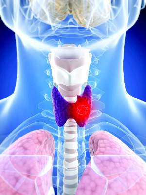
Thyroglobulin can’t predict pazopanib response in differentiated thyroid cancer
Lake BUENA VISTA, FLA. – Pazopanib is an effective therapy for advanced thyroid cancer, but at present, there seems to be no way to predict which patients will respond to it.
An investigation into the predictive value of thyroglobulin found that the level during treatment did change, but it did so in parallel with response; there was no way to use the protein to parse out which patients would do well, Dr. Keith Bible said at the International Thyroid Congress.

“We had hoped that it might be a predictor as early as 4 weeks, so we could assign patients into categories of response and perhaps stop treatment earlier,” said Dr. Bible of the Mayo Clinic, Rochester, Minn. “Unfortunately, there was no way to do that.”
Pazopanib (Votrient) is a tyrosine kinase inhibitor of vascular endothelial growth factor receptors. It is approved for advanced soft tissue sarcoma and advanced renal cell carcinoma. The Mayo Clinic Consortium has been investigating pazopanib in phase II trials for advanced differentiated and medullary thyroid cancers. In a 2010 report, it was shown to induce at least a partial response in about half of the patients who received it (Lancet Oncol. 2010 Oct;11[10]:962-72).
Last year, it was also shown to be effective in advanced medullary thyroid cancer. Five of 35 patients attained partial Response Evaluation Criteria In Solid Tumors (RECIST) responses (14%) (J Clin Endocrinol Metab. 2014 May;99[5]:1687-93).
The drug has not been successful in treating advanced anaplastic thyroid cancer, however.
Because of pazopanib’s proclivity to induce sometimes-severe hypertension, investigators were hoping for some way to stratify potential responders. Thyroglobulin levels could be one marker, Dr. Bible said at the meeting, which was held by the American Thyroid Association, Asia-Oceania Thyroid Association, European Thyroid Association, and Latin American Thyroid Society.
He and the consortium investigators examined how thyroglobulin levels correlated with RECIST scores in 60 patients with metastatic differentiated thyroid cancers. The patients received a median of 10 cycles, but the range was wide (1-53 cycles). Most of them (92%) had already received systemic therapy, including a tyrosine kinase inhibitor and/or radioactive iodine.
The most common side effect was hypertension, which occurred in 75% patients, and was severe in 23%. Of those with a severe reaction, 53% required a new prescription for an antihypertensive medication. No one left the study or required a pazopanib dose reduction because of a blood pressure elevation, however. “We responded with a aggressive treatment, but it was a prominent issue,” Dr. Bible said.
Other adverse events were fatigue (83%; 8% severe); decrease in neutrophils (47%; 8% severe); diarrhea (78%; 7% severe); hand-foot syndrome (17%; 7% severe); and elevations of liver enzymes (53%; 6% severe). There were no deaths related to the study drug.
Partial RECIST responses occurred in 22 patients (37%). Thyroglobulin change did not differ by response after cycle 1, although its nadir was lower among patients who attained a partial response than among those who maintained disease stability (–87% vs. –69%).
“There was this correlation of nadir with maximum RECIST response, but this occurred in parallel with the response, so it was not capable of providing a prediction of response,” Dr. Bible said.
Prior therapy also was not a response predictor, he added.
Genomic profiling was available for 16 patients; of these, 11 had mutations of BRAF, p53, JAK3 or HRAS. Thyroglobulin change and response to treatment was not significantly correlated with any of these mutations. Nor did it correlate with any type of prior tumor therapy.
The finding that pazopanib can benefit patients previously treated with a kinase inhibitor is an interesting one, Dr. Bible noted.
“Most kinase inhibitors are very promiscuous – they work on a number of pathways and have a footprint which is very messy. Most of them seem to have some activity in differentiated thyroid cancer, but we are still struggling to understand how that footprint varies. In theory they are all targeting VEGF receptors, but it’s striking that we can go from one kinase inhibitor to the next and still get a response.”
Dr. Bible had no financial declarations.
On Twitter @Alz_Gal
Lake BUENA VISTA, FLA. – Pazopanib is an effective therapy for advanced thyroid cancer, but at present, there seems to be no way to predict which patients will respond to it.
An investigation into the predictive value of thyroglobulin found that the level during treatment did change, but it did so in parallel with response; there was no way to use the protein to parse out which patients would do well, Dr. Keith Bible said at the International Thyroid Congress.
“We had hoped that it might be a predictor as early as 4 weeks, so we could assign patients into categories of response and perhaps stop treatment earlier,” said Dr. Bible of the Mayo Clinic, Rochester, Minn. “Unfortunately, there was no way to do that.”
Pazopanib (Votrient) is a tyrosine kinase inhibitor of vascular endothelial growth factor receptors. It is approved for advanced soft tissue sarcoma and advanced renal cell carcinoma. The Mayo Clinic Consortium has been investigating pazopanib in phase II trials for advanced differentiated and medullary thyroid cancers. In a 2010 report, it was shown to induce at least a partial response in about half of the patients who received it (Lancet Oncol. 2010 Oct;11[10]:962-72).
Last year, it was also shown to be effective in advanced medullary thyroid cancer. Five of 35 patients attained partial Response Evaluation Criteria In Solid Tumors (RECIST) responses (14%) (J Clin Endocrinol Metab. 2014 May;99[5]:1687-93).
The drug has not been successful in treating advanced anaplastic thyroid cancer, however.
Because of pazopanib’s proclivity to induce sometimes-severe hypertension, investigators were hoping for some way to stratify potential responders. Thyroglobulin levels could be one marker, Dr. Bible said at the meeting, which was held by the American Thyroid Association, Asia-Oceania Thyroid Association, European Thyroid Association, and Latin American Thyroid Society.
He and the consortium investigators examined how thyroglobulin levels correlated with RECIST scores in 60 patients with metastatic differentiated thyroid cancers. The patients received a median of 10 cycles, but the range was wide (1-53 cycles). Most of them (92%) had already received systemic therapy, including a tyrosine kinase inhibitor and/or radioactive iodine.
The most common side effect was hypertension, which occurred in 75% patients, and was severe in 23%. Of those with a severe reaction, 53% required a new prescription for an antihypertensive medication. No one left the study or required a pazopanib dose reduction because of a blood pressure elevation, however. “We responded with a aggressive treatment, but it was a prominent issue,” Dr. Bible said.
Other adverse events were fatigue (83%; 8% severe); decrease in neutrophils (47%; 8% severe); diarrhea (78%; 7% severe); hand-foot syndrome (17%; 7% severe); and elevations of liver enzymes (53%; 6% severe). There were no deaths related to the study drug.
Partial RECIST responses occurred in 22 patients (37%). Thyroglobulin change did not differ by response after cycle 1, although its nadir was lower among patients who attained a partial response than among those who maintained disease stability (–87% vs. –69%).
“There was this correlation of nadir with maximum RECIST response, but this occurred in parallel with the response, so it was not capable of providing a prediction of response,” Dr. Bible said.
Prior therapy also was not a response predictor, he added.
Genomic profiling was available for 16 patients; of these, 11 had mutations of BRAF, p53, JAK3 or HRAS. Thyroglobulin change and response to treatment was not significantly correlated with any of these mutations. Nor did it correlate with any type of prior tumor therapy.
The finding that pazopanib can benefit patients previously treated with a kinase inhibitor is an interesting one, Dr. Bible noted.
“Most kinase inhibitors are very promiscuous – they work on a number of pathways and have a footprint which is very messy. Most of them seem to have some activity in differentiated thyroid cancer, but we are still struggling to understand how that footprint varies. In theory they are all targeting VEGF receptors, but it’s striking that we can go from one kinase inhibitor to the next and still get a response.”
Dr. Bible had no financial declarations.
On Twitter @Alz_Gal
AT ITC 2015
Key clinical point: Falling thyroglobulin levels cannot predict early response to pazopanib in differentiated thyroid cancers.
Major finding: There was a 37% response rate for patients taking pazopanib for differentiated thyroid cancer, but thyroglobulin decline was not an early predictor of response.
Data source: A prospective study of 60 patients.
Disclosures: Dr. Bible had no financial disclosures.

Cabozantinib shows promise in refractory differentiated thyroid cancer
LAKE BUENA VISTA, FLA. – Most patients with differentiated thyroid cancer who had shown progression on previous courses of targeted chemotherapy either maintained stable disease or responded to the oral multikinase inhibitor cabozantinib (Cometriq), according to a small multicenter phase II trial presented at the International Thyroid Congress.
This is important, according to Dr. Manisha H. Shah, because there has been no standard of care for patients with differentiated thyroid cancer whose cancer progresses on first- or second-line vascular endothelial growth factor receptor (VEGFR) inhibitors.
Nine of the 25 enrolled patients (36%; 95% confidence interval, 18%-57%) showed confirmed partial response, 12 patients (48%) had stable disease, and one patient had disease progression, according to Dr. Shah, director of the neuroendocrine tumor program at Ohio State University’s Wexner Medical Center. The trial enrolled patients with radioiodine–refractory differentiated thyroid cancer who had progression of their disease after one or two previous VEGFR agents.

Cabozantinib targets VEGFR and MET and is approved as first-line treatment for medullary thyroid cancer. The majority of the response to cabozantinib occurs in the first several months of treatment, so the study used a Simon minimax two-stage design, enrolling an initial 16 patients, then opening enrollment to an additional 9 when at least 2 of the initial cohort showed partial or complete response within the first 6 months.
The primary outcome measure was the number of patients showing objective response (partial or complete response) within the first 6 months of therapy.
Median patient age was 64 years, and 64% of patients were male. Just over half of the patients previously had been treated with sorafenib, and just over a quarter had received pazopanib. Five patients had received two previous VEGFR-targeted therapies, while the remaining 20 had received one.
Nine patients (36%) had papillary thyroid cancer, seven (28%) had poorly differentiated thyroid cancer, five (20%) had Hurthle cell cancer, and four (16%) had follicular thyroid cancer. The most common metastasis sites were lymph node, bone, and lung.
Patients received continuous treatment until they showed disease progression, had an unacceptable adverse event or an illness precluding further treatment, or withdrew consent.
Disease progression was measured by serum tumor markers and CT or MRI scan every 8 weeks while in the study; patients also received bone scans and 18F-FDG and 18F-fluoride PET scans before the study and while in the study.
Side effects were common and generally mild, with two instances each of grade 3 events related to fatigue, hand-foot skin reactions, and diarrhea. One death occurred and was adjudicated as possibly study related; there were no grade 4 events, and no grade 3 bleeding events.
Dr. Shah noted that the starting cabozantinib dose of 60 mg/day was considerably lower than that used in previous trials for thyroid cancer. With time, investigators have learned that the sometimes debilitating side effects of cabozantinib are somewhat dose dependent, she noted. The study design permitted dose escalation to 80 mg for nonresponders to the lower dose, and permitted a decrease to 40 or 20 mg/day as needed to manage side effects. Investigators were able to tell the patients what to expect, and to be proactive in anticipating side effects. “We have learned to manage these drugs much better with time,” she said.
“Cabozantinib was effective in inducing a durable partial response,” said Dr. Shah. Future directions, in addition to phase III clinical trials, may include combining cabozantinib with immune checkpoint–targeted therapies such as lenvatinib, a strategy that has been effective for other cancers, she said at the meeting, which was held by the American Thyroid Association, Asia-Oceania Thyroid Association, European Thyroid Association, and Latin American Thyroid Society.
The multisite study was sponsored by the National Cancer Institute with participation by eight International Thyroid Oncology Group centers. Dr. Shah reported being on the advisory board for Exelixis and Eisai, and receiving research funding from those two organizations and Bayer.
On Twitter @karioakes
LAKE BUENA VISTA, FLA. – Most patients with differentiated thyroid cancer who had shown progression on previous courses of targeted chemotherapy either maintained stable disease or responded to the oral multikinase inhibitor cabozantinib (Cometriq), according to a small multicenter phase II trial presented at the International Thyroid Congress.
This is important, according to Dr. Manisha H. Shah, because there has been no standard of care for patients with differentiated thyroid cancer whose cancer progresses on first- or second-line vascular endothelial growth factor receptor (VEGFR) inhibitors.
Nine of the 25 enrolled patients (36%; 95% confidence interval, 18%-57%) showed confirmed partial response, 12 patients (48%) had stable disease, and one patient had disease progression, according to Dr. Shah, director of the neuroendocrine tumor program at Ohio State University’s Wexner Medical Center. The trial enrolled patients with radioiodine–refractory differentiated thyroid cancer who had progression of their disease after one or two previous VEGFR agents.
Cabozantinib targets VEGFR and MET and is approved as first-line treatment for medullary thyroid cancer. The majority of the response to cabozantinib occurs in the first several months of treatment, so the study used a Simon minimax two-stage design, enrolling an initial 16 patients, then opening enrollment to an additional 9 when at least 2 of the initial cohort showed partial or complete response within the first 6 months.
The primary outcome measure was the number of patients showing objective response (partial or complete response) within the first 6 months of therapy.
Median patient age was 64 years, and 64% of patients were male. Just over half of the patients previously had been treated with sorafenib, and just over a quarter had received pazopanib. Five patients had received two previous VEGFR-targeted therapies, while the remaining 20 had received one.
Nine patients (36%) had papillary thyroid cancer, seven (28%) had poorly differentiated thyroid cancer, five (20%) had Hurthle cell cancer, and four (16%) had follicular thyroid cancer. The most common metastasis sites were lymph node, bone, and lung.
Patients received continuous treatment until they showed disease progression, had an unacceptable adverse event or an illness precluding further treatment, or withdrew consent.
Disease progression was measured by serum tumor markers and CT or MRI scan every 8 weeks while in the study; patients also received bone scans and 18F-FDG and 18F-fluoride PET scans before the study and while in the study.
Side effects were common and generally mild, with two instances each of grade 3 events related to fatigue, hand-foot skin reactions, and diarrhea. One death occurred and was adjudicated as possibly study related; there were no grade 4 events, and no grade 3 bleeding events.
Dr. Shah noted that the starting cabozantinib dose of 60 mg/day was considerably lower than that used in previous trials for thyroid cancer. With time, investigators have learned that the sometimes debilitating side effects of cabozantinib are somewhat dose dependent, she noted. The study design permitted dose escalation to 80 mg for nonresponders to the lower dose, and permitted a decrease to 40 or 20 mg/day as needed to manage side effects. Investigators were able to tell the patients what to expect, and to be proactive in anticipating side effects. “We have learned to manage these drugs much better with time,” she said.
“Cabozantinib was effective in inducing a durable partial response,” said Dr. Shah. Future directions, in addition to phase III clinical trials, may include combining cabozantinib with immune checkpoint–targeted therapies such as lenvatinib, a strategy that has been effective for other cancers, she said at the meeting, which was held by the American Thyroid Association, Asia-Oceania Thyroid Association, European Thyroid Association, and Latin American Thyroid Society.
The multisite study was sponsored by the National Cancer Institute with participation by eight International Thyroid Oncology Group centers. Dr. Shah reported being on the advisory board for Exelixis and Eisai, and receiving research funding from those two organizations and Bayer.
On Twitter @karioakes
AT ITC 2015
Key clinical point: Cabozantinib shows promise for refractory differentiated thyroid cancer.
Major finding: Twenty-one of 25 patients with differentiated thyroid cancer showed stable disease or partial response to cabozantinib after disease progression on previous targeted therapies.
Data source: Multisite, open-label phase II clinical trial of 25 patients.
Disclosures: The study was sponsored by the National Cancer Institute with participation by eight International Thyroid Oncology Group centers. Dr. Shah reported being on the advisory board for Exelixis and Eisai, and receiving research funding from those two organizations and Bayer.
GI bleeds in obese patients more complicated but no more fatal
HONOLULU – Obese patients who develop an upper GI bleed receive more treatment, are more likely to develop hemorrhagic shock, and are more likely to require admission to an intensive care unit than those who are not obese, but they do not have higher in-hospital mortality, according to analysis of a large national database that was presented at the annual meeting of the American College of Gastroenterology.
The significantly greater odds ratio of most major complications from upper GI bleeds in patients with obesity relative to those who are not obese was expected but so was an increased rate of in-hospital mortality, according to the first author of the study, Dr. Marwan S. Abou Gergi of Catalyst Medical Consulting, Baltimore.

“In this study, patients with obesity received more frequent endoscopic interventions, which could explain why mortality rates were not significantly higher,” Dr. Abou Gergi reported.
In the analysis, characterized as the first study to evaluate the impact of obesity on outcomes in upper GI hemorrhage, data were drawn from the 2012 Nationwide Inpatient Sample database, which is considered to provide a representative sample of U.S. hospital experience. Drawn from more than 7 million hospitalizations, the study focused on patients 18 or over with a primary ICD-9 code for upper GI hemorrhage. The primary outcome was mortality. Secondary outcomes included interventions, ICU admissions, and length of stay.
Of the 132,545 discharges with upper GI hemorrhage, 11,220 (8.5%) were identified as obese. The in-hospital mortality overall was 1.97%, but the proportion of those who died was slightly lower among patients identified as obese, producing a nonsignificant adjusted odds ratio (OR) of 0.87 (P greater than .1). Yet the rates of hemorrhage shock (OR 1.31; P = .02) and admission to the ICU (OR 1.35; P less than .02) were greater in the obese. The median length of stay of 0.35 days for obese patients was also significantly longer.
One reason for the lower rate of mortality may be more aggressive treatment. In particular, repeat endoscopy therapy was more common in those who were obese (P less than .01), suggesting, “Our treatments are working,” Dr. Abou Gergi said.
The proportion of patients with a score of 3 or greater on the Charlson comorbidity index was higher in the obese than in those not identified as obese (49% vs. 39%; P less than .01). This along with previously published evidence that obese patients take more anticoagulants, take more antiplatelets, and may face delays in endoscopy due to greater difficulty in administering sedation, were among considerations predicting a higher mortality, according to Dr. Abou Gergi.
However, there are several potential explanations for these unexpected findings. One is that mortality rates overall were low, making it difficult to show differences on this outcome. In addition, ICD-9 codes may be effective for isolating a group with obesity but not in identifying a control group without obesity.
“It is likely that not all patients who are obese received this code, so we may be seeing a population of obese patients be compared to another population that includes at least some patients who also have obesity,” explained Dr. John R. Saltzman, director of endoscopy, Brigham and Women’s Hospital, Boston. A coauthor of this study, Dr. Saltzman noted that despite a comparable rate of in-hospital mortality, most of the findings in the study argued that patients who develop upper GI bleeding have a more difficult course. He noted that this is reflected in the cost of care, which was significantly higher in those who were obese.
Although he acknowledged that he was surprised that this was “essentially a negative study,” he believes that mortality may have been “too tough” as a primary endpoint for demonstrating a difference. However, he also believes that it may be appropriate to give credit for effective treatments.
“I think that may be the key. We are just getting better at taking care of these patients,” Dr. Saltzman said.
Dr. Abou Gergi reported he has no relevant financial relationships.
HONOLULU – Obese patients who develop an upper GI bleed receive more treatment, are more likely to develop hemorrhagic shock, and are more likely to require admission to an intensive care unit than those who are not obese, but they do not have higher in-hospital mortality, according to analysis of a large national database that was presented at the annual meeting of the American College of Gastroenterology.
The significantly greater odds ratio of most major complications from upper GI bleeds in patients with obesity relative to those who are not obese was expected but so was an increased rate of in-hospital mortality, according to the first author of the study, Dr. Marwan S. Abou Gergi of Catalyst Medical Consulting, Baltimore.
“In this study, patients with obesity received more frequent endoscopic interventions, which could explain why mortality rates were not significantly higher,” Dr. Abou Gergi reported.
In the analysis, characterized as the first study to evaluate the impact of obesity on outcomes in upper GI hemorrhage, data were drawn from the 2012 Nationwide Inpatient Sample database, which is considered to provide a representative sample of U.S. hospital experience. Drawn from more than 7 million hospitalizations, the study focused on patients 18 or over with a primary ICD-9 code for upper GI hemorrhage. The primary outcome was mortality. Secondary outcomes included interventions, ICU admissions, and length of stay.
Of the 132,545 discharges with upper GI hemorrhage, 11,220 (8.5%) were identified as obese. The in-hospital mortality overall was 1.97%, but the proportion of those who died was slightly lower among patients identified as obese, producing a nonsignificant adjusted odds ratio (OR) of 0.87 (P greater than .1). Yet the rates of hemorrhage shock (OR 1.31; P = .02) and admission to the ICU (OR 1.35; P less than .02) were greater in the obese. The median length of stay of 0.35 days for obese patients was also significantly longer.
One reason for the lower rate of mortality may be more aggressive treatment. In particular, repeat endoscopy therapy was more common in those who were obese (P less than .01), suggesting, “Our treatments are working,” Dr. Abou Gergi said.
The proportion of patients with a score of 3 or greater on the Charlson comorbidity index was higher in the obese than in those not identified as obese (49% vs. 39%; P less than .01). This along with previously published evidence that obese patients take more anticoagulants, take more antiplatelets, and may face delays in endoscopy due to greater difficulty in administering sedation, were among considerations predicting a higher mortality, according to Dr. Abou Gergi.
However, there are several potential explanations for these unexpected findings. One is that mortality rates overall were low, making it difficult to show differences on this outcome. In addition, ICD-9 codes may be effective for isolating a group with obesity but not in identifying a control group without obesity.
“It is likely that not all patients who are obese received this code, so we may be seeing a population of obese patients be compared to another population that includes at least some patients who also have obesity,” explained Dr. John R. Saltzman, director of endoscopy, Brigham and Women’s Hospital, Boston. A coauthor of this study, Dr. Saltzman noted that despite a comparable rate of in-hospital mortality, most of the findings in the study argued that patients who develop upper GI bleeding have a more difficult course. He noted that this is reflected in the cost of care, which was significantly higher in those who were obese.
Although he acknowledged that he was surprised that this was “essentially a negative study,” he believes that mortality may have been “too tough” as a primary endpoint for demonstrating a difference. However, he also believes that it may be appropriate to give credit for effective treatments.
“I think that may be the key. We are just getting better at taking care of these patients,” Dr. Saltzman said.
Dr. Abou Gergi reported he has no relevant financial relationships.
AT ACG 2015

NICE plans to recommend device for SCD patients
Image by Graham Beards
The UK’s National Institute for Health and Care Excellence (NICE) is asking for views on its plans to recommend a device that automatically replaces sickled red blood cells (RBCs) with healthy RBCs in patients with sickle cell disease (SCD).
NICE has issued a draft medical technology guidance supporting use of the Spectra Optia Apheresis System for automated RBC exchange in patients with SCD who need regular transfusions.
The guidance is open for comment until November 16.
The Spectra Optia system is made up of 3 components: the apheresis machine, embedded software, and a single-use disposable blood tubing set. The system is manufactured by Terumo.
NICE said the evidence examined indicates that the Spectra Optia system speeds up the process of RBC exchange compared to manual RBC exchange.
The evidence also suggests that patients require RBC exchange less frequently when using this system than they do with manual RBC exchange.
Furthermore, the Spectra Optia system is estimated to provide cost savings for most patients, when compared to manual RBC exchange or top-up transfusion.
Potential savings depend on the patient’s clinical circumstance and if devices already owned by the National Health Service can also be used to treat SCD.
However, NICE’s draft guidance also highlights a need for additional data on treatment outcomes, as there is limited clinical evidence for some outcomes.
“In particular, the independent Medical Technologies Advisory Committee would like to see long-term data on how manual and automated cell exchange affects the amount of iron in the body and the need to treat this complication,” said Carole Longson, director of the NICE Centre for Health Technology Evaluation. “We welcome comments on the draft guidance as part of this consultation.” ![]()
Drug-resistant malaria could spread to Africa, team says

of Plasmodium falciparum
Image by Mae Melvin/CDC
New research indicates that drug-resistant forms of the malaria parasite Plasmodium falciparum can infect the Anopheles coluzzii mosquito (formerly Anopheles gambiae M form), which is the main transmitter of malaria in Africa.
The discovery suggests that Africa is more at risk for drug-resistant malaria infections than researchers previously thought, and this could further compromise efforts to prevent and eliminate the disease.
Rick Fairhurst, MD, PhD, of the National Institute of Allergy and Infectious Diseases in Rockville, Maryland, and his colleagues reported the discovery in Nature Communications.
The team noted that P falciparum parasites that are resistant to artemisinin, the main drug used to treat malaria, have been rapidly spreading in parts of Southeast Asia.
Malaria experts are concerned that artemisinin-resistant parasites could spread to Africa. However, there were no scientific indications to suggest these parasites could infect A coluzzii mosquitoes—until now.
Dr Fairhurst and his colleagues investigated the transmission potential of these parasites in 3 mosquito species—A coluzzii and the Southeast Asian mosquito species Anopheles dirus and Anopheles minimus.
The researchers evaluated whether these mosquitoes became infected after feeding on blood containing any of 6 artemisinin-resistant parasite strains and 3 artemisinin-sensitive parasite strains previously isolated from malaria patients in Cambodia.
The team found parasites in the mosquitoes’ midguts and salivary glands in almost all cases, showing that the artemisinin-resistant and artemisinin-sensitive Cambodian parasites can infect a variety of mosquito species.
The researchers also discovered a shared genetic background among artemisinin-resistant parasites that may enable them to infect diverse mosquito species by evading the insects’ immune systems.
This study did not show that infected mosquitoes can effectively transmit the disease to humans. However, the results support the idea that artemisinin-resistant parasites could potentially spread beyond Cambodia and to Africa, the researchers said.
They plan to investigate other potential genetic determinants of parasite infection in mosquitoes and further examine which Anopheles species from Cambodia are naturally transmitting artemisinin-resistant parasites in the wild. ![]()
Older RBCs don’t increase risks, study suggests

Photo by Elise Amendola
Patients undergoing cardiac surgery can safely receive transfusions with older red blood cells (RBCs), according to new research.
In many countries, RBCs can be stored for as long as 6 weeks before transfusion.
But a study published in 2008 suggested that transfusing RBCs stored for more than 2 weeks could increase the risk of serious complications.
Results of subsequent studies both supported and contradicted that finding.
The new study, published in JAMA, adds to the debate. The results suggest the duration of RBC storage does not affect the risk of death or serious complications after transfusion.
“There have literally been hundreds of studies conducted on this topic the past 5 or 6 years, none of which have been able to provide a definitive answer,” said Gustaf Edgren, MD, PhD, of Karolinska Institutet in Stockholm, Sweden.
In an attempt to change that, Dr Edgren and his colleagues conducted a large-scale study of transfusions among cardiac surgery patients in Sweden. National guidelines there require that the oldest available blood unit is allocated first.
The researchers analyzed registry data on patients who underwent coronary artery bypass graft surgery, heart valve surgery, or both between 1997 and 2012.
There were 47,071 patients who received transfusions at 9 different hospitals. Of these patients, 36.6% received RBCs stored for less than 14 days, 26.8%
received RBCs stored 14 to 27 days, 8.9% received RBCs stored 28 to 42
days, and 27.8% received RBCs of mixed age.
The researchers compared these patient groups, looking at the incidence of serious complications at 30 days and mortality at 30 days, 2 years, and 10 years.
They adjusted their analyses for potential confounding factors such as sex, age, blood group, and hospital. And they found no association between RBC storage duration and mortality or serious complications.
| RBC storage duration and adverse outcomes at 30 days | ||||||
| RBCs stored 1-13 days (n=17,224) | 14-27 days (n=12,602) | 28-42 days
(n=4173) |
||||
| Adverse outcome | No. of events | Adjusted
odds ratio (OR) |
No. of events | Adjusted OR | No. of events | Adjusted OR |
| Acute kidney injury | 202 | 1 (reference) | 121 | 0.94 | 38 | 0.97 |
| ARDS/respiratory
failure |
228 | 1 (ref) | 157 | 1.16 | 42 | 1.00 |
| Serious infection | 524 | 1 (ref) | 351 | 0.99 | 133 | 1.13 |
| Stroke | 403 | 1 (ref) | 295 | 1.04 | 106 | 1.13 |
| Thrombosis/embolism | 70 | 1 (ref) | 49 | 1.01 | 17 | 1.09 |
| Composite
adverse outcome (including death) |
1670 | 1 (ref) | 1151 | 1.02 | 371 | 1.03 |
| RBC storage duration and mortality | |||||||
| Storage age | Patient No. | Deaths at
30 days |
Adjusted
hazard ratio (HR) |
Deaths at
2 years |
2-year HR | Deaths at
10 years |
2-year HR |
| 1-13 days | 17,224 | 615 | 1 (ref) | 1593 | 1 (ref) | 5897 | 1 (ref) |
| 14-27 days | 12,602 | 410 | 1.06 | 1074 | 1.02 | 4358 | 1.02 |
| 28-42 days | 4173 | 103 | 0.90 | 325 | 0.98 | 1403 | 0.99 |
| mixed age | 13,072 | 911 | 0.86 | 1954 | 0.94 | 5655 | 0.99 |
“This study is by far the largest investigation focusing on the issue of blood storage in this very sensitive patient group, and we find absolutely no hint of negative health effects associated with stored blood,” said Ulrik Sartipy, MD, PhD, of Karolinska University Hospital.
“[W]e have been able to provide very firm reassurance that the current blood storage practices are safe,” Dr Edgren added. ![]()
Antibody can turn AML cells against each other

Image by Joshua Stokes
Scientists say they have discovered an antibody that can transform leukemic cells into immune cells that target their former “siblings.”
This agonist antibody, which is highly specific for the thrombopoietin receptor, can turn acute myeloid leukemia (AML) cells into dendritic cells.
With extended exposure to the antibody, these dendritic cells can then transform into natural killer (NK)-like cells that target AML cells.
The scientists described these discoveries in PNAS.
“It’s a totally new approach to cancer, and we’re working to test it in human patients as soon as possible,” said study author Richard A. Lerner, MD, of The Scripps Research Institute in La Jolla, California.
Unexpected transformation
Dr Lerner’s lab has pioneered techniques to generate and screen large libraries of antibodies to find therapeutic antibodies that bind to a desired target or activate a desired receptor on cells.
During the course of this work, the team discovered a phenomenon they call “receptor pleiotropism,” in which agonist antibodies against known receptors cause cell fates that are extremely different from those induced by the natural agonist to the receptor.
Why this happens is unclear, but the discovery led the scientists to wonder if they could use the method to convert leukemia cells into non-cancerous cells.
To find out, they tested 20 of their recently discovered receptor-activating antibodies against AML cells from patients. This revealed the transformative properties of the agonist antibody that is highly specific for the thrombopoietin receptor.
When this antibody was applied to healthy, immature bone marrow cells, it caused them to mature into megakaryocytes. However, when the antibody was applied to AML cells, they were converted into dendritic cells.
With longer exposures to the antibody and certain other in vitro conditions, the induced dendritic cells morphed into cells that closely resemble NK cells.
“That antibody could have turned those acute myeloid leukemia cells into a lot of other cell types, but, somehow, we were lucky enough to get NK cells,” Dr Lerner said.
The team examined these induced NK cells with electron microscopy and observed that many of the cells had extended tendrils through the outer membranes of neighboring AML cells. In in vitro tests, a modest number of these NK cells wiped out about 15% of the surrounding AML cell population in 24 hours.
The scientists also noted that the induced NK cells’ cancer-killing effects appeared to be purely “fratricidal.” Unrelated breast cancer cells did not die off in large numbers in the presence of the NK cells.
Fratricidin therapy
Why the induced NK cells appear to target only closely related cells isn’t yet clear. However, the finding suggests there may be antibodies—and even small-molecule compounds—that would turn other cancerous cell types into fratricidal NK cells by activating other receptors expressed on those cells.
Such fratricidal therapies, which Dr Lerner calls “fratricidins,” would have several potential advantages. First, especially if they are antibodies, they could be clinically useful with little or no further modification.
Second, their high specificity for their target receptors, and the resulting NK cells’ specificity for related cancer cells, should reduce the likelihood of adverse effects.
Finally, the peculiar dynamics of fratricidin therapy, in which every cancerous cell is potentially convertible to a cancer-killing NK cell, suggests that—if the strategy works—it might not just reduce the targeted cancer-cell population in a patient but eliminate it altogether.
“We’re in discussions with pharmaceutical companies to take this straight into humans after the appropriate preclinical toxicity studies,” Dr Lerner said.
This research was supported by the JPB Foundation and Zebra Biologics. ![]()
Malpractice premiums flat in 2015, but changes could be ahead
Physicians paid about the same in liability insurance premiums in 2015 as in 2014, and analysts don’t see costs changing anytime soon. A nationwide survey of insurers by the Medical Liability Monitor shows that 71% of insurance premiums did not change this year, while 17% of rates rose and 12% fell.
Internists experienced an average premium increase of 0.6% in 2015, while general surgeons saw a 0.2% average rate decrease, and ob.gyns experienced an average 0.5% rate increase.

The static premium market is being largely driven by the low number of lawsuits filed by patients and family members in recent years, said survey coauthor Paul Greve Jr., executive vice president/senior consultant for the Willis Health Care Practice, a global risk management consultant firm.
“It’s amazing to see the continuing stability in claim frequency,” Mr. Greve said in an interview. “The claims counts are just not rising. Its great for the industry, and it’s great for physicians, but it is puzzling because you wonder what has caused what amounts to a sea change in the attitudes of the general public toward malpractice litigation such that the claim counts were drop off.”
Premiums continue to vary geographically. Southern Florida internists for example, will pay $47,707 for malpractice insurance this year, while their counterparts in Minnesota will pay $3,375. For ob.gyns., premiums range from $214,999 in southern New York to $16,240 in central California. General surgeons in Southern Florida will pay $190,829 this year, while Wisconsin surgeons will pay $10,868.

Various factors influence premium amounts, including the overall legal climate and the rate of insurer competition in each state, said Susan J. Forray, principal and consulting actuary with the Milwaukee office of Milliman, a global provider of actuarial services.
“The dollar amounts themselves are a function of the litigation environment [and] the cost level of medicine or living within the state,” Ms. Forray said in an interview. “In terms of rate changes, we are seeing certain environments where there is more competition. Obviously, those more competitive markets are more likely to have rate decreases or perhaps, stable rates, where perhaps markets with less competition are more likely to see increased rates.”
On a regional basis, Southern physicians experienced the largest rate increases, while doctors in the Northeast, West, and Midwest continued to see decreases. The Midwest’s 0.8% rate decrease was the largest decline, while Western states experienced a 0.2% average rate decrease. On average, the South showed a rate increase of 0.9% and the Northeast experienced a 0.1% average decrease. Doctors in Georgia, North Carolina, and Texas saw rate increases in excess of 5%, while Iowa physicians experienced an 11% rate decrease. Only three western states experienced rate increases: New Mexico at 2.5%, Oregon at 2%, and Idaho at 1%. Premium changes for Northeastern doctors fluctuated from Rhode Island’s 7% increase to Pennsylvania’s 8% decrease. Additionally, for the first time in 8 years, the premium market experienced an average overall increase of 0.3% in 2015, compared with an average overall decrease of 1.5% last year.
The jury is still out on how the Affordable Care Act and other health reforms will impact the malpractice premium market, according to Mr. Greve. He said that he believes the majority of upcoming health reforms will improve patient safety, thus reducing liability for doctors. However, as more physicians become part of larger networks to deliver new models of care, their contractual liability spreads, he said. However, as value-based care becomes the law of the land, new claims could arise.
“We’re just beginning to see the tip of the iceberg here,” Mr. Greve said. “In the past, it was overutilization, [the claim] that you did something in order to put money in your pocket. With putting providers at financial risk with capitated or bundled payments or global payments, then the argument is going to be, ‘You didn’t deliver enough care,’ or ‘You [used that device] because it was less expensive.’ ”
The MLM survey, published yearly in October, gathered July 1 premium data from the major malpractice insurers and examines rates for mature, claims-made policies with $1 million/$3 million limits for internists, general surgeons, and ob.gyns.
On Twitter @legal_med
Physicians paid about the same in liability insurance premiums in 2015 as in 2014, and analysts don’t see costs changing anytime soon. A nationwide survey of insurers by the Medical Liability Monitor shows that 71% of insurance premiums did not change this year, while 17% of rates rose and 12% fell.
Internists experienced an average premium increase of 0.6% in 2015, while general surgeons saw a 0.2% average rate decrease, and ob.gyns experienced an average 0.5% rate increase.
The static premium market is being largely driven by the low number of lawsuits filed by patients and family members in recent years, said survey coauthor Paul Greve Jr., executive vice president/senior consultant for the Willis Health Care Practice, a global risk management consultant firm.
“It’s amazing to see the continuing stability in claim frequency,” Mr. Greve said in an interview. “The claims counts are just not rising. Its great for the industry, and it’s great for physicians, but it is puzzling because you wonder what has caused what amounts to a sea change in the attitudes of the general public toward malpractice litigation such that the claim counts were drop off.”
Premiums continue to vary geographically. Southern Florida internists for example, will pay $47,707 for malpractice insurance this year, while their counterparts in Minnesota will pay $3,375. For ob.gyns., premiums range from $214,999 in southern New York to $16,240 in central California. General surgeons in Southern Florida will pay $190,829 this year, while Wisconsin surgeons will pay $10,868.
Various factors influence premium amounts, including the overall legal climate and the rate of insurer competition in each state, said Susan J. Forray, principal and consulting actuary with the Milwaukee office of Milliman, a global provider of actuarial services.
“The dollar amounts themselves are a function of the litigation environment [and] the cost level of medicine or living within the state,” Ms. Forray said in an interview. “In terms of rate changes, we are seeing certain environments where there is more competition. Obviously, those more competitive markets are more likely to have rate decreases or perhaps, stable rates, where perhaps markets with less competition are more likely to see increased rates.”
On a regional basis, Southern physicians experienced the largest rate increases, while doctors in the Northeast, West, and Midwest continued to see decreases. The Midwest’s 0.8% rate decrease was the largest decline, while Western states experienced a 0.2% average rate decrease. On average, the South showed a rate increase of 0.9% and the Northeast experienced a 0.1% average decrease. Doctors in Georgia, North Carolina, and Texas saw rate increases in excess of 5%, while Iowa physicians experienced an 11% rate decrease. Only three western states experienced rate increases: New Mexico at 2.5%, Oregon at 2%, and Idaho at 1%. Premium changes for Northeastern doctors fluctuated from Rhode Island’s 7% increase to Pennsylvania’s 8% decrease. Additionally, for the first time in 8 years, the premium market experienced an average overall increase of 0.3% in 2015, compared with an average overall decrease of 1.5% last year.
The jury is still out on how the Affordable Care Act and other health reforms will impact the malpractice premium market, according to Mr. Greve. He said that he believes the majority of upcoming health reforms will improve patient safety, thus reducing liability for doctors. However, as more physicians become part of larger networks to deliver new models of care, their contractual liability spreads, he said. However, as value-based care becomes the law of the land, new claims could arise.
“We’re just beginning to see the tip of the iceberg here,” Mr. Greve said. “In the past, it was overutilization, [the claim] that you did something in order to put money in your pocket. With putting providers at financial risk with capitated or bundled payments or global payments, then the argument is going to be, ‘You didn’t deliver enough care,’ or ‘You [used that device] because it was less expensive.’ ”
The MLM survey, published yearly in October, gathered July 1 premium data from the major malpractice insurers and examines rates for mature, claims-made policies with $1 million/$3 million limits for internists, general surgeons, and ob.gyns.
On Twitter @legal_med

APA-IPS: Disaster psychiatry – Nepal, Ebola, and beyond
NEW YORK – After the earthquake in Nepal earlier this year, Disaster Psychiatry Outreach sent in volunteers who found preexisting issues that made their mental health response challenging at best, Dr. Ram Suresh Mahato reported at the American Psychiatric Association’s Institute on Psychiatric Services.
Nepal was recovering from armed conflict that lasted from 1996 to 2006 and resulted in what some have called a “postconflict identity crisis” (Int J Educ Dev. 2014;34:42-50). The caste system in the country was abolished in 1963, but social inequality continued to persist. In addition, more than 60 languages are spoken in Nepal, and at least 25% of the population lives below the poverty line, said Dr. Mahato, a Disaster Psychiatry Outreach (DPO) volunteer who was part of the needs-assessment team dispatched to the country in May.

Other complicating factors included high rates of domestic violence. Nepali women are at greater risk of depression, anxiety, and posttraumatic stress than are males (Lancet. 2008 May;371[9625]:1664) and (J Affect Disord. 2007 Sep;102[1-3]:219-25), and a culture of silence prevails, Dr. Mahato said. The literature describes informal social networks in Nepal in which community members share their distress and symptoms, “as well as traditional (shamanistic) healing practices for those suffering mental health complaints in relation to political violence” (Soc Sci Med. 2010 Jan;70[1]:35-44).
Dr. Mahato spoke at a workshop, sponsored by DPO, aimed at urging psychiatrists to be prepared in providing mental health services to disaster survivors across the globe and here at home. “The room was full last year,” said Dr. Sander Koyfman, DPO’s president, referring to the intense interest in Ebola at the height of the outbreak in 2014. “This year, it’s more of a challenge, as interest wanes from disaster to disaster,” but their organization would like to “sustain the desire in mental health providers and disaster responders to learn how to help most effectively,” Dr. Koyfman said in an interview.
The presentation focused on the mental health aspects of the Ebola response and the more recent DPO work following the earthquake in Nepal that killed 10,000 people. In a striking similarity, about 10,900 people died in the wake of the Ebola epidemic in West Africa and its rolling impact across many regions. (In May, the World Health Organization declared Liberia free of Ebola but said on Oct. 14 that a preliminary study published in the New England Journal of Medicine shows that the virus can persist in the semen of some survivors for at least 9 months.)
Vulnerable suffer most
Over the last 10 years, more than 1.4 million people have been injured and about 23 million have been left homeless across the globe because of man-made and natural disasters, according to a 2015 United Nations report. “Overall, more than 1.5 billion people have been affected by disasters in various ways, with women, children, and people in vulnerable situations disproportionately affected,” the report says.
DPO, a New York–based nonprofit, launched in 1998, has sent volunteers to an average of one disaster per year, said Dr. Koyfman, also medical director for EmblemHealth Insurance, New York.
“We at DPO learn to caution folks and say, ‘Look, it’s important and it’s critical to do everything you can, but do appreciate one thing: The key is what happens 3 to 6 months from today,’ ” he said. “The mental health component will happen then. This is very different from a typical disaster mentality.”
Before the earthquake in Nepal, manpower and resources were limited: The country has about 80 psychiatrists, or about 1 for every million people, said Dr. Mahato, chief psychiatry resident, PGY-4, at Mount Sinai/Elmhurst Hospital Center, New York. After the earthquake and more than 300 aftershocks, about 2.8 million people were in need of humanitarian assistance. The DPO team partnered with the Psychiatrists’ Association of Nepal by visiting affected districts and participating in health camps. “The challenges we saw involved developing communication and training materials in a culturally appropriate framework,” Dr. Mahato said.
Portable intervention used
One intervention used by DPO teams in Nepal was Psychological First Aid (PFA), Dr. Javier Garcia said.
PFA has grown in popularity and acceptance, especially when it became increasingly clear after the attacks of Sept. 11, 2001, that psychological debriefing was not as universally useful or safe modality as it was once thought to be, said Dr. Garcia of Richmond University Medical Center, New York.
In contrast, PFA is an intervention based on principles of resilience that focuses on safety, calming, connectedness, self-community, efficacy, and hope. “PFA assumes that people can have maladaptive reactions,” Dr. Garcia said. “ But is designed to reduce the initial distress and foster short- and long-term adaptive functioning.” He said all first responders, including fire, police and crisis response teams, health care professionals, and paraprofessionals can be trained to use PFA. In fact, another model of PFA was created for school staff in the 1990s in response to school shootings.
The first goal after a disaster is to ensure physical safety. After that, teams try to protect those traumatized from additional trauma. Emotionally overwhelmed and disoriented survivors must be stabilized, and medications generally are not recommended during this part of the process. Medications might be helpful in cases involving addiction or sleep, but such cases are exceptions, Dr. Garcia said. In general, the same strict clinical criteria for use of psychiatric medications are applicable in postdisaster environments and are specific to the episode and the individual. PFA attempts to be culturally informed and delivered in a flexible manner, Dr. Garcia said. “It’s evidence informed but not evidence based. So, we need more research.”
PFA, along with effective risk communications, frequently are the mainstay of an effective mental health response. Where PFA informs the “what” of the mental health conversation, risk communications, as Dr. Grant H. Brenner pointed out at the meeting, is the key “how” of getting the right message out the right way. Dr. Brenner, DPO board member, is a faculty member of Mount Sinai Hospital, director of the William Alanson White Institute Trauma Service, and an editor of Creating Spiritual and Psychological Resilience: Integrating Care in Disaster Relief Work (New York: Routledge, 2009).
After the Ebola work on the ground, volunteers often found complicated terrain in the United States. As the example of the single New York City Ebola patient showed, medical and psychological preparedness and the ability of the authorities to effectively communicate safety information to the public were tested. DPO worked with a nonprofit group called More Than Me to offer mental health support services to returning volunteers and to the few people who were under quarantine orders in New York.
Each disaster is different, but a few common themes are apparent. “There’s huge value in presence and human touch,” Dr. Koyfman said.
DPO offers training sessions for new volunteers. Psychiatrists interested in volunteering can send a message to [email protected] or call 646-867-3514. For more on risk communication, check out the information on emergency preparedness and response provided by the Centers for Disease Control and Prevention. Other useful resources are the American Psychiatric Association’s Committee on Psychiatric Dimensions of Disaster and Resiliency in the Face of Disaster and Terrorism: 10 Things to Do to Survive (Personhood Press, 2005).
On Twitter @ginalhenderson
NEW YORK – After the earthquake in Nepal earlier this year, Disaster Psychiatry Outreach sent in volunteers who found preexisting issues that made their mental health response challenging at best, Dr. Ram Suresh Mahato reported at the American Psychiatric Association’s Institute on Psychiatric Services.
Nepal was recovering from armed conflict that lasted from 1996 to 2006 and resulted in what some have called a “postconflict identity crisis” (Int J Educ Dev. 2014;34:42-50). The caste system in the country was abolished in 1963, but social inequality continued to persist. In addition, more than 60 languages are spoken in Nepal, and at least 25% of the population lives below the poverty line, said Dr. Mahato, a Disaster Psychiatry Outreach (DPO) volunteer who was part of the needs-assessment team dispatched to the country in May.
Other complicating factors included high rates of domestic violence. Nepali women are at greater risk of depression, anxiety, and posttraumatic stress than are males (Lancet. 2008 May;371[9625]:1664) and (J Affect Disord. 2007 Sep;102[1-3]:219-25), and a culture of silence prevails, Dr. Mahato said. The literature describes informal social networks in Nepal in which community members share their distress and symptoms, “as well as traditional (shamanistic) healing practices for those suffering mental health complaints in relation to political violence” (Soc Sci Med. 2010 Jan;70[1]:35-44).
Dr. Mahato spoke at a workshop, sponsored by DPO, aimed at urging psychiatrists to be prepared in providing mental health services to disaster survivors across the globe and here at home. “The room was full last year,” said Dr. Sander Koyfman, DPO’s president, referring to the intense interest in Ebola at the height of the outbreak in 2014. “This year, it’s more of a challenge, as interest wanes from disaster to disaster,” but their organization would like to “sustain the desire in mental health providers and disaster responders to learn how to help most effectively,” Dr. Koyfman said in an interview.
The presentation focused on the mental health aspects of the Ebola response and the more recent DPO work following the earthquake in Nepal that killed 10,000 people. In a striking similarity, about 10,900 people died in the wake of the Ebola epidemic in West Africa and its rolling impact across many regions. (In May, the World Health Organization declared Liberia free of Ebola but said on Oct. 14 that a preliminary study published in the New England Journal of Medicine shows that the virus can persist in the semen of some survivors for at least 9 months.)
Vulnerable suffer most
Over the last 10 years, more than 1.4 million people have been injured and about 23 million have been left homeless across the globe because of man-made and natural disasters, according to a 2015 United Nations report. “Overall, more than 1.5 billion people have been affected by disasters in various ways, with women, children, and people in vulnerable situations disproportionately affected,” the report says.
DPO, a New York–based nonprofit, launched in 1998, has sent volunteers to an average of one disaster per year, said Dr. Koyfman, also medical director for EmblemHealth Insurance, New York.
“We at DPO learn to caution folks and say, ‘Look, it’s important and it’s critical to do everything you can, but do appreciate one thing: The key is what happens 3 to 6 months from today,’ ” he said. “The mental health component will happen then. This is very different from a typical disaster mentality.”
Before the earthquake in Nepal, manpower and resources were limited: The country has about 80 psychiatrists, or about 1 for every million people, said Dr. Mahato, chief psychiatry resident, PGY-4, at Mount Sinai/Elmhurst Hospital Center, New York. After the earthquake and more than 300 aftershocks, about 2.8 million people were in need of humanitarian assistance. The DPO team partnered with the Psychiatrists’ Association of Nepal by visiting affected districts and participating in health camps. “The challenges we saw involved developing communication and training materials in a culturally appropriate framework,” Dr. Mahato said.
Portable intervention used
One intervention used by DPO teams in Nepal was Psychological First Aid (PFA), Dr. Javier Garcia said.
PFA has grown in popularity and acceptance, especially when it became increasingly clear after the attacks of Sept. 11, 2001, that psychological debriefing was not as universally useful or safe modality as it was once thought to be, said Dr. Garcia of Richmond University Medical Center, New York.
In contrast, PFA is an intervention based on principles of resilience that focuses on safety, calming, connectedness, self-community, efficacy, and hope. “PFA assumes that people can have maladaptive reactions,” Dr. Garcia said. “ But is designed to reduce the initial distress and foster short- and long-term adaptive functioning.” He said all first responders, including fire, police and crisis response teams, health care professionals, and paraprofessionals can be trained to use PFA. In fact, another model of PFA was created for school staff in the 1990s in response to school shootings.
The first goal after a disaster is to ensure physical safety. After that, teams try to protect those traumatized from additional trauma. Emotionally overwhelmed and disoriented survivors must be stabilized, and medications generally are not recommended during this part of the process. Medications might be helpful in cases involving addiction or sleep, but such cases are exceptions, Dr. Garcia said. In general, the same strict clinical criteria for use of psychiatric medications are applicable in postdisaster environments and are specific to the episode and the individual. PFA attempts to be culturally informed and delivered in a flexible manner, Dr. Garcia said. “It’s evidence informed but not evidence based. So, we need more research.”
PFA, along with effective risk communications, frequently are the mainstay of an effective mental health response. Where PFA informs the “what” of the mental health conversation, risk communications, as Dr. Grant H. Brenner pointed out at the meeting, is the key “how” of getting the right message out the right way. Dr. Brenner, DPO board member, is a faculty member of Mount Sinai Hospital, director of the William Alanson White Institute Trauma Service, and an editor of Creating Spiritual and Psychological Resilience: Integrating Care in Disaster Relief Work (New York: Routledge, 2009).
After the Ebola work on the ground, volunteers often found complicated terrain in the United States. As the example of the single New York City Ebola patient showed, medical and psychological preparedness and the ability of the authorities to effectively communicate safety information to the public were tested. DPO worked with a nonprofit group called More Than Me to offer mental health support services to returning volunteers and to the few people who were under quarantine orders in New York.
Each disaster is different, but a few common themes are apparent. “There’s huge value in presence and human touch,” Dr. Koyfman said.
DPO offers training sessions for new volunteers. Psychiatrists interested in volunteering can send a message to [email protected] or call 646-867-3514. For more on risk communication, check out the information on emergency preparedness and response provided by the Centers for Disease Control and Prevention. Other useful resources are the American Psychiatric Association’s Committee on Psychiatric Dimensions of Disaster and Resiliency in the Face of Disaster and Terrorism: 10 Things to Do to Survive (Personhood Press, 2005).
On Twitter @ginalhenderson
EXPERT ANALYSIS AT THE INSTITUTE ON PSYCHIATRIC SERVICES

Worsening of lesions on MRI predicts knee OA
People without x-ray evidence of osteoarthritis in their knees but with MRI-identified lesions that worsen over time are significantly likelier to develop knee OA, compared with people whose lesions remain stable, according to results from a prospective cohort study.
For their research, published Oct. 14 in Annals of the Rheumatic Diseases, Dr. Leena Sharma of Northwestern University in Chicago and her colleagues recruited more than 1,000 patients at elevated risk of knee OA but with no radiographic evidence yet of disease (Kellgren/Lawrence measures of 0 [KL0] in both knees) to test their hypothesis that lesions seen worsening over time on MRI were predictive of knee OA within 4 years and of persistent symptoms between 4 and 7 years. Patients in the cohort (56% women, mean age 59.6 years) were assessed for cartilage damage, meniscal tears, meniscal extrusions, and bone marrow lesions at 12 and 48 months. Study inclusion required that patients remain at KL0 in both knees at 12 months to continue, and 849 patients had complete data at 12 and 48 months.

Patients with lesions that had worsened on MRI between 12 and 48 months had significantly higher risk of incident radiographic KL1 and KL2 by 48 months, compared with patients whose lesions had not worsened. For example, 6.3% of patients with tibiofemoral cartilage damage that was stable at 48 months developed mild (KL1) disease at 48 months, compared with 9.5% of patients whose damage had worsened in that interval (odds ratio, 2.69; 95% confidence interval, 1.50-4.84). Half of patients with worsening meniscal extrusion developed mild knee OA by the endpoint, compared with 13.6% of patients with a stable lesion (OR, 5.73; 95% CI, 2.94-11.16). Higher risk of KL1 or KL2 at 48 months was significant for all the lesion types studied except bone marrow lesions. Worsening of these lesions between 12 and 48 months was also significantly associated with having persistent symptoms between 4 and 7 years. Having more lesion types that worsened was significantly associated with worse outcomes.
The findings, Dr. Sharma and her colleagues wrote, support the idea of stable and progressive disease phases with early indicators of each and that worsening lesions represent early osteoarthritis. “Given the absence of disease-modifying therapy for OA, widespread clinical application of MRI is difficult to justify,” the investigators wrote in their analysis (Ann Rheum Dis. 2015 Oct 14. doi: 10.1136/annrheumdis-2015-208129).
Nevertheless, they concluded, “prevention or delay of worsening of early-stage lesions should be considered as a target for emerging pharmacological and nonpharmacological treatments in an effort to prevent or delay full-blown disease. Candidate interventions should be studied at this stage, when they are more likely to be effective.” Investigators acknowledged that one limitation of the study was that its findings may not apply in populations not already at higher risk for knee OA.
The study was funded by the Osteoarthritis Initiative, a public-private partnership of the National Institutes of Health and Merck, Novartis, GlaxoSmithKline, and Pfizer, with industry funding administered by NIH.
People without x-ray evidence of osteoarthritis in their knees but with MRI-identified lesions that worsen over time are significantly likelier to develop knee OA, compared with people whose lesions remain stable, according to results from a prospective cohort study.
For their research, published Oct. 14 in Annals of the Rheumatic Diseases, Dr. Leena Sharma of Northwestern University in Chicago and her colleagues recruited more than 1,000 patients at elevated risk of knee OA but with no radiographic evidence yet of disease (Kellgren/Lawrence measures of 0 [KL0] in both knees) to test their hypothesis that lesions seen worsening over time on MRI were predictive of knee OA within 4 years and of persistent symptoms between 4 and 7 years. Patients in the cohort (56% women, mean age 59.6 years) were assessed for cartilage damage, meniscal tears, meniscal extrusions, and bone marrow lesions at 12 and 48 months. Study inclusion required that patients remain at KL0 in both knees at 12 months to continue, and 849 patients had complete data at 12 and 48 months.
Patients with lesions that had worsened on MRI between 12 and 48 months had significantly higher risk of incident radiographic KL1 and KL2 by 48 months, compared with patients whose lesions had not worsened. For example, 6.3% of patients with tibiofemoral cartilage damage that was stable at 48 months developed mild (KL1) disease at 48 months, compared with 9.5% of patients whose damage had worsened in that interval (odds ratio, 2.69; 95% confidence interval, 1.50-4.84). Half of patients with worsening meniscal extrusion developed mild knee OA by the endpoint, compared with 13.6% of patients with a stable lesion (OR, 5.73; 95% CI, 2.94-11.16). Higher risk of KL1 or KL2 at 48 months was significant for all the lesion types studied except bone marrow lesions. Worsening of these lesions between 12 and 48 months was also significantly associated with having persistent symptoms between 4 and 7 years. Having more lesion types that worsened was significantly associated with worse outcomes.
The findings, Dr. Sharma and her colleagues wrote, support the idea of stable and progressive disease phases with early indicators of each and that worsening lesions represent early osteoarthritis. “Given the absence of disease-modifying therapy for OA, widespread clinical application of MRI is difficult to justify,” the investigators wrote in their analysis (Ann Rheum Dis. 2015 Oct 14. doi: 10.1136/annrheumdis-2015-208129).
Nevertheless, they concluded, “prevention or delay of worsening of early-stage lesions should be considered as a target for emerging pharmacological and nonpharmacological treatments in an effort to prevent or delay full-blown disease. Candidate interventions should be studied at this stage, when they are more likely to be effective.” Investigators acknowledged that one limitation of the study was that its findings may not apply in populations not already at higher risk for knee OA.
The study was funded by the Osteoarthritis Initiative, a public-private partnership of the National Institutes of Health and Merck, Novartis, GlaxoSmithKline, and Pfizer, with industry funding administered by NIH.
FROM ANNALS OF THE RHEUMATIC DISEASES
Key clinical point: People with knee cartilage damage, meniscal tear, meniscal extrusion, and bone marrow lesions on MRI were likelier to have developed knee osteoarthritis at 48 months, compared with people with stable lesions; more lesion types at baseline were associated with worse outcomes.
Major finding: Higher-risk mild or moderate radiographic knee OA at 48 months was significant for most types of lesions that had worsened after 12 months.
Data source: A prospective cohort study of 849 people at high risk of knee osteoarthritis evaluated on radiography (for evidence of knee OA) and MRI (for lesions) at baseline, 12 months, and 48 months and followed up for symptoms through 84 months.
Disclosures: The study was funded by the Osteoarthritis Initiative, a public-private partnership of the National Institutes of Health and Merck, Novartis, GlaxoSmithKline, and Pfizer, with industry funding administered by NIH. The study authors disclosed no conflicts of interest.
