User login
CASE STUDY: Management Decisions in a Comorbid Patient With Type 2 Diabetes Having Primary Hyperlipidemia
A supplement to Clinical Endocrinology News. This supplement was sponsored by Daiichi Sankyo, Inc.
• Background
• Current Visit
• Laboratory Results
• Clinical Discussion
• Endocrinologist Consultation
• New Treatment Regimen With Add-On Therapy
• Conclusions
Faculty
Yehuda Handelsman, MD, FACP, FACE
Medical Director, Metabolic Institute of America
Chair and Program Director, 7th World Congress on Insulin Resistance Chair, International Committee for Insulin Resistance
18372 Clark Street, Suite 212
Tarzana, CA 91356
E-mail:[email protected]
Web site:www.TheMetabolicCenter.com
Dr Handelsman is a consultant for Bristol-Myers Squibb Company, Daiichi Sankyo, Inc., GlaxoSmithKline, Medtronic, Merck, Xoma, and Tethys;he has received clinical research grant funding from Takeda, Daiichi Sankyo Inc., GlaxoSmithKline, and Novo Nordisk; and he is on the speakers bureau for AstraZeneca, Bristol-Myers Squibb, Daiichi Sankyo Inc., GlaxoSmithKline, Merck, and Novartis. He also serves on the advisory board for CLINICAL ENDOCRINOLOGY NEWS.
Copyright © 2010 Elsevier Inc.
Work-Life Balance for Hospitalists a People Issue, Not a Women's Issue
When recounting her HM career, Janet Nagamine, RN, MD, SFHM, often tells people she went from being the chief of everything to the chief of nothing, by choice. She can remember the whirlwind of being a quality-improvement (QI) chief, patient safety officer, risk management team member, and new mother who felt more married to her beeper than to her husband.
“I felt incredibly stressed and pulled in so many different directions,” says Dr. Nagamine, a hospitalist at Kaiser Permanente Medical Center in Santa Clara, Calif., and an SHM board member. “What really concerned me was that I was starting to feel that I wasn’t doing anything well, that I was dropping balls here and there.”
A revelation came to Dr. Nagamine at a time-management workshop. The speaker asked participants to list the three most important things in their lives, then add up the hours spent doing them.
“There was obviously a disconnect when I looked at the actual waking hours I spent with my family,” says Dr. Nagamine, who placed family at the top of her list. “That’s where I made the decision that I could always come back to doing these leadership things at a later time.”
Now pursuing an MBA, she intends to return to leadership positions to focus on QI, patient safety, and hospitalist work-life issues—for both women and men.
“Work-life balance is a key anchor for us,” says Dr. Nagamine, who helped organize a “Women in Hospital Medicine” session at HM12 last month in San Diego. “Whether you’re male or female, work-life [balance] is a challenge. We need to do better on that.”
In today’s era of ever-increasing healthcare demands, the future of hospitalist practice rests, in no small part, on the work-life satisfaction of its physicians. Recent studies suggest hospitalists are experiencing more stress and burnout now than in the past, a phenomenon HM groups would be wise to address by offering more flexible work options and workload support, regardless of gender, experts say. But individual hospitalists can mitigate strain and dissatisfaction by assessing their life and goals, and developing a work-life balance that is right for them.
“Work-life balance is really not something that is ‘a nice thing to have,’” says Iris Grimm, creator of the Atlanta-based Balanced Physician program, which helps physicians meet work, life, and leadership challenges. “It is a necessity for them if they want to sustain a long-term career.”
Defining Balance
So what causes tension between work and life outside of work? The list is long and growing.
“We still define the ideal worker as someone who starts to work in early adulthood and works full time, full force, for 40 years straight, available for overtime as needed,” says Joan Williams, distinguished professor of law, founding director of the Center of WorkLife Law at the University of California’s Hastings College of the Law in San Francisco, and author of “Unbending Gender: Why Family and Work Conflict and What To Do About It.” “That basically describes a man married to a homemaker, and that’s not who the work force is.”
In general, workplaces—including those in the hospital work environment—have been slow to adjust to the changing work force, Williams says. In time, friction arises, which leads to dissatisfied workers.
Work-life balance is when a person can rise above the conflict and align their responsibilities with their values and priorities, says Maria Bailey, founder and CEO of BlueSuitMom.com, a Pompano Beach, Fla.-based company that provides work-life balance information for professional working mothers and their employers.
“It is being satisfied with one’s entire life, with the work side as well as with the personal side,” Grimm says.

—Janet Nagamine, RN, MD, SFHM, Kaiser Permanente Medical Center, Santa Clara, Calif., SHM board member
Young physicians of both genders view work-life balance as essential, and are willing to risk career advancement to achieve it, according to a 2006 survey of U.S. doctors under age 50 conducted by the Association of American Medical Colleges and the American Medical Association.1 When asked to rate factors that are very important to a desirable position, 71% identified work-life balance. Two out of 3 young physicians said they were not interested in working longer hours for more money—a sharp contrast from previous generations.
“It started in the 1990s, but I think in the early 2000s was when the medical world began to take a much more honest appraisal of the long-term impact of an unbalanced life and what that meant for physicians,” says Erin Stucky Fisher, MD, MHM, medical director for quality at Rady Children’s Hospital San Diego, associate program director for the University of California at San Diego Pediatric Residency Program, and an SHM board member.
Dr. Nagamine agrees the tide has shifted in terms of physician attitudes toward work hours, compensation, and personal time. “Now that we have work-hour rules in residency, the doctors coming out don’t buy that you have to be on 24/7, 365 days a year,” she says.
The Survey Says...
Recent research on hospitalist work-life satisfaction indicates that while hospitalists generally are satisfied with their job and specialty, burnout rates appear higher than the 13% previously reported in 2001.2
Earlier this year, a study in the Journal of General Internal Medicine found that 29.9% of respondents to a national survey of hospitalists reported job burnout symptoms.3 Hospitalists surveyed also reported low satisfaction with personal time (28.3%), autonomy (17.4%), organizational climate (10.7%), and organizational fairness (31.2%). The results are somewhat alarming to longtime hospitalists, in that hospitalist work models might be less flexible and less sustainable than originally thought.
Results from an email survey published in 2011 showed that 67% of academic hospitalists reported high levels of stress, and 23% described some level of burnout.4 Additionally, 57% of the respondents had 20% or less of protected time for scholarly activity—a disconnect between career goals and actual work that could lead to career dissatisfaction. More than half of the academic hospitalists surveyed, however, did express high or somewhat high satisfaction with personal and family time, and control over work schedules.
“Hospital medicine is still a new field, and people are trying to find the right balance in the work,” says Rebecca Harrison, MD, associate professor of medicine and section chief of the division of hospital medicine at Oregon Health & Science University in Portland. “I think we’re in a very critical time now where we have to look at scheduling, patient load, fulfillment factors. A physician’s sense of commitment, happiness, and enjoyment in their work is going to be much higher if you pay attention to the things they want to pursue.”

What Women Really Want
Female physicians are far less able to control their work environments than men, says Mark Linzer, MD. He helped design and conduct the Society of General Internal Medicine’s Physician Work Life Study, which found that aside from less work control, the female doctors surveyed said they faced a more difficult patient mix, more time pressure in patient examinations, and a 60% greater chance of burnout compared with their male counterparts.5
“One of those factors, we think, is what has been called ‘gendered expectations for listening,’” says Dr. Linzer, division director for general internal medicine at Hennepin County Medical Center in Minneapolis and professor of medicine at the University of Minnesota. Patients prefer female doctors because they believe women are better listeners than men, he explains. But listening takes time, and female physicians generally aren’t afforded more time for patient visits than male physicians are.
“This is an issue I see many times with female physicians, with hospitalists in particular,” Grimm says. “They just can’t seem to stay in the time frame that has been given to them for their patients. They think that the more time they spend with their patients, the more the patients appreciate them and feel like they are heard.”
Another factor is extra work outside the office. “If you measure the total number of work hours performed, including work and home, it is considerably higher for women,” Dr. Linzer says.
Even if a working woman has help at home or a spouse who works part time or stays at home, she never really relinquishes responsibility of the home, Bailey says.
“A woman carries social pressures that she needs to—even if she has a career—carry out the role of a traditional wife and mother,” she says.

Regardless of whether they have a spouse and children, women generally feel a responsibility to care for their loved ones’ needs, whether it’s an aging parent, an ailing sibling, or a friend facing a difficult situation, says Jennifer Owens, director of the Working Mother Research Institute in New York City. One thing working in female physicians’ favor, however, is they are less likely to lose their careers due to work-family conflicts than are women in such high-skill professions as finance or law, because part-time work is readily available for female doctors, Williams says.
—Rebecca Harrison, MD, associate professor of medicine, section chief, division of hospital medicine, Oregon Health & Science University, Portland
“The number of hours that women work has been increasing. So there’s incredible stress on women,” says Owens. “Just to have the support that you’re not stigmatized for dialing back and not working a 60-hour-plus work week means a lot.”
The Flip Side
Men also have partners, families, children, and outside interests. Therefore, if hospitalist groups are going to create flexible work opportunities, they have to market them and make them available to everyone, regardless of gender, Dr. Fisher says.
The key work-life balance battle today, Williams says, centers around male workers and the stereotypes surrounding masculinity. Increasing numbers of young men want to participate in the day-to-day caregiving of their children. Most workplaces, however, have been slow to adjust.
“Women have the cultural room to make workplace adjustments to ease work-family conflict, and men often don’t,” Williams says. “The ideas of masculinity are closely intertwined with the idea of being a provider. So if a man leaves work to care for his child or ailing mother, people not only think of him as a poor worker, they often think of him as less of a man. The stereotypes that hit people who make their caregiving responsibilities salient on the job are extremely hostile and even more powerful for men than for women.”
Similarly, if male physicians want to structure their schedules around personal interests or take extended time off to pursue a life passion, they often are viewed unfavorably because the culture of medicine for years has been complete dedication to patients and career over personal needs, Dr. Harrison says.
“For example, a single person or someone who doesn’t have a family might want to go climbing the Himalayas and take three months off. Or perhaps a staff member wants to go part time in order to go back to school for an MBA,” Dr. Nagamine says. “We’re not prepared to deal with those types of requests.”

Stop the Churn
HM groups around the country—big and small, academic and community—deal with work-life balance issues on a regular basis. Some have solved the issue; many have not. Too many hospitalist groups are stuck in a churn cycle: hire hospitalist, fail to meet their needs, see them leave after a year or two, repeat.
“When you see people churning their staff, especially when they’re losing good ones, it’s a financial and human capital drain,” Dr. Nagamine says. “Think about the care that’s being delivered within the system, what’s happening to the other members in the group, the return on investment for keeping your staff happy. We argue over pennies sometimes, but we don’t calculate these types of losses of personnel.”
Dr. Nagamine says hospitalist groups should approach work-life balance not just on a day-to-day or week-to-week basis, but also in terms of extended leave for child or elder care, travel, volunteer work, professional development, etc.
Compensation and workload are used to recruit and retain hospitalists. But recent research suggests that leaders might find more nuanced approaches to improving their hospitalists’ overall satisfaction.6 For example, leaders of local community-based hospitalist groups might find their hospitalists tolerant of heavier workloads, provided they are financially rewarded and given autonomy over their work. And rather than using higher salaries to be competitive, leaders of academic programs might find it more effective to provide their hospitalists with time and training to pursue scholarly work.
“Physicians and faculty are the most valuable commodities for moving the work forward,” Dr. Harrison says, “and good leaders pay attention to this data.”
HM groups should think about surveying their employees to find out where problems exist, Bailey says. Once you determine what work-life benefits and/or flexible employment opportunities will work, train supervisors to manage workplace flexibility, then hold them accountable for executing the policies, Owens says.
“It makes sense to take care of your people,” Dr. Nagamine says. “First, it’s the right thing to do. Second, it’s financially and fiscally a good move. It’s not just work-life balance for the sake of work-life balance; it’s critically important to your operations and your overall success in delivering good care.”
Lisa Ryan is a freelance writer in New Jersey.
Take Ownership of Your Time Off

The only way to fully achieve work-life balance is if you’re trying to achieve balance, according to experts in physician career satisfaction. In other words, work-life balance starts with you. Here are six steps you can take to improve your balancing act:
Define balance. Draw a picture of what you look like when your life is in balance, Bailey says. Or write a description in a journal. The goal is to determine what balance means for you at this point in your life, Grimm says.
Conduct a personal assessment. Look at every area of your life to assess what’s working and what’s not working, Owens says. Where things aren’t working, identify what you would have to add or subtract from your life to make improvements, says Grimm.
Eliminate stressors. Pinpoint your primary stressors and work to resolve or mitigate them, Grimm says. If it’s new software at work, find ways to master the technology. If it’s lack of spousal support, practice effective communication and teamwork.
Maximize energy. Create a workplace with the least amount of friction by investing energy in areas where you can make a positive impact, Grimm says. Don’t waste time or effort on things that cannot be changed.
Practice self-care. To be effective at caring for loved ones and patients, you have to care for yourself, Dr. Harrison says. Self-care can include protecting time with friends and family, taking short breaks during the workday, exercising, and getting regular sleep and meals. Hospitalists also should schedule vacations.
Consider job fit. Read recent research on the job characteristics of hospitalist practice models.6 If you’re someone who’s less concerned about workload but want to be paid well and have more autonomy, a local, community-based hospitalist group might be right for you. Academic HM might fit better if you’re willing to sacrifice some compensation for a variety of activities beyond direct clinical care.
—Lisa Ryan
References
- Kirch DG, Salsberg E. The physician workforce challenge: response of the academic community. Ann Surg. 2007;246(4):535-540.
- Hoff TH, Whitcomb WF, Williams K, Nelson JR, Cheesman RA. Characteristics and work experiences of hospitalists in the United States. Arch Intern Med. 2001;161(6):851-8.
- Hinami K, Whelan CT, Wolosin RJ, Miller JA, Wetterneck TB. Worklife and satisfaction of hospitalists: toward flourishing careers. J Gen Intern Med. 2012;27(1):28-36.
- Glasheen JJ, Misky GJ, Reid MB, Harrison RA, Sharpe B, Auerbach A. Career satisfaction and burnout in academic hospital medicine. Arch Intern Med. 2011;171(8):782-785.
- McMurray JE, Linzer M, Konrad TR, Douglas J, Shugerman R, Nelson K. The work lives of women physicians results from the physician work life study. The SGIM Career Satisfaction Study Group. J Gen Intern Med. 2000;15(6):372-380.
- Hinami K, Whelan CT, Miller JA, Wolosin RJ, Wetterneck TB. Job characteristics, satisfaction, and burnout across hospitalist practice models. Journal of Hospital Medicine website. Available at: http://onlinelibrary.wiley.com/doi/10.1002/jhm.1907/full. Accessed March 21, 2012.
When recounting her HM career, Janet Nagamine, RN, MD, SFHM, often tells people she went from being the chief of everything to the chief of nothing, by choice. She can remember the whirlwind of being a quality-improvement (QI) chief, patient safety officer, risk management team member, and new mother who felt more married to her beeper than to her husband.
“I felt incredibly stressed and pulled in so many different directions,” says Dr. Nagamine, a hospitalist at Kaiser Permanente Medical Center in Santa Clara, Calif., and an SHM board member. “What really concerned me was that I was starting to feel that I wasn’t doing anything well, that I was dropping balls here and there.”
A revelation came to Dr. Nagamine at a time-management workshop. The speaker asked participants to list the three most important things in their lives, then add up the hours spent doing them.
“There was obviously a disconnect when I looked at the actual waking hours I spent with my family,” says Dr. Nagamine, who placed family at the top of her list. “That’s where I made the decision that I could always come back to doing these leadership things at a later time.”
Now pursuing an MBA, she intends to return to leadership positions to focus on QI, patient safety, and hospitalist work-life issues—for both women and men.
“Work-life balance is a key anchor for us,” says Dr. Nagamine, who helped organize a “Women in Hospital Medicine” session at HM12 last month in San Diego. “Whether you’re male or female, work-life [balance] is a challenge. We need to do better on that.”
In today’s era of ever-increasing healthcare demands, the future of hospitalist practice rests, in no small part, on the work-life satisfaction of its physicians. Recent studies suggest hospitalists are experiencing more stress and burnout now than in the past, a phenomenon HM groups would be wise to address by offering more flexible work options and workload support, regardless of gender, experts say. But individual hospitalists can mitigate strain and dissatisfaction by assessing their life and goals, and developing a work-life balance that is right for them.
“Work-life balance is really not something that is ‘a nice thing to have,’” says Iris Grimm, creator of the Atlanta-based Balanced Physician program, which helps physicians meet work, life, and leadership challenges. “It is a necessity for them if they want to sustain a long-term career.”
Defining Balance
So what causes tension between work and life outside of work? The list is long and growing.
“We still define the ideal worker as someone who starts to work in early adulthood and works full time, full force, for 40 years straight, available for overtime as needed,” says Joan Williams, distinguished professor of law, founding director of the Center of WorkLife Law at the University of California’s Hastings College of the Law in San Francisco, and author of “Unbending Gender: Why Family and Work Conflict and What To Do About It.” “That basically describes a man married to a homemaker, and that’s not who the work force is.”
In general, workplaces—including those in the hospital work environment—have been slow to adjust to the changing work force, Williams says. In time, friction arises, which leads to dissatisfied workers.
Work-life balance is when a person can rise above the conflict and align their responsibilities with their values and priorities, says Maria Bailey, founder and CEO of BlueSuitMom.com, a Pompano Beach, Fla.-based company that provides work-life balance information for professional working mothers and their employers.
“It is being satisfied with one’s entire life, with the work side as well as with the personal side,” Grimm says.
—Janet Nagamine, RN, MD, SFHM, Kaiser Permanente Medical Center, Santa Clara, Calif., SHM board member
Young physicians of both genders view work-life balance as essential, and are willing to risk career advancement to achieve it, according to a 2006 survey of U.S. doctors under age 50 conducted by the Association of American Medical Colleges and the American Medical Association.1 When asked to rate factors that are very important to a desirable position, 71% identified work-life balance. Two out of 3 young physicians said they were not interested in working longer hours for more money—a sharp contrast from previous generations.
“It started in the 1990s, but I think in the early 2000s was when the medical world began to take a much more honest appraisal of the long-term impact of an unbalanced life and what that meant for physicians,” says Erin Stucky Fisher, MD, MHM, medical director for quality at Rady Children’s Hospital San Diego, associate program director for the University of California at San Diego Pediatric Residency Program, and an SHM board member.
Dr. Nagamine agrees the tide has shifted in terms of physician attitudes toward work hours, compensation, and personal time. “Now that we have work-hour rules in residency, the doctors coming out don’t buy that you have to be on 24/7, 365 days a year,” she says.
The Survey Says...
Recent research on hospitalist work-life satisfaction indicates that while hospitalists generally are satisfied with their job and specialty, burnout rates appear higher than the 13% previously reported in 2001.2
Earlier this year, a study in the Journal of General Internal Medicine found that 29.9% of respondents to a national survey of hospitalists reported job burnout symptoms.3 Hospitalists surveyed also reported low satisfaction with personal time (28.3%), autonomy (17.4%), organizational climate (10.7%), and organizational fairness (31.2%). The results are somewhat alarming to longtime hospitalists, in that hospitalist work models might be less flexible and less sustainable than originally thought.
Results from an email survey published in 2011 showed that 67% of academic hospitalists reported high levels of stress, and 23% described some level of burnout.4 Additionally, 57% of the respondents had 20% or less of protected time for scholarly activity—a disconnect between career goals and actual work that could lead to career dissatisfaction. More than half of the academic hospitalists surveyed, however, did express high or somewhat high satisfaction with personal and family time, and control over work schedules.
“Hospital medicine is still a new field, and people are trying to find the right balance in the work,” says Rebecca Harrison, MD, associate professor of medicine and section chief of the division of hospital medicine at Oregon Health & Science University in Portland. “I think we’re in a very critical time now where we have to look at scheduling, patient load, fulfillment factors. A physician’s sense of commitment, happiness, and enjoyment in their work is going to be much higher if you pay attention to the things they want to pursue.”
What Women Really Want
Female physicians are far less able to control their work environments than men, says Mark Linzer, MD. He helped design and conduct the Society of General Internal Medicine’s Physician Work Life Study, which found that aside from less work control, the female doctors surveyed said they faced a more difficult patient mix, more time pressure in patient examinations, and a 60% greater chance of burnout compared with their male counterparts.5
“One of those factors, we think, is what has been called ‘gendered expectations for listening,’” says Dr. Linzer, division director for general internal medicine at Hennepin County Medical Center in Minneapolis and professor of medicine at the University of Minnesota. Patients prefer female doctors because they believe women are better listeners than men, he explains. But listening takes time, and female physicians generally aren’t afforded more time for patient visits than male physicians are.
“This is an issue I see many times with female physicians, with hospitalists in particular,” Grimm says. “They just can’t seem to stay in the time frame that has been given to them for their patients. They think that the more time they spend with their patients, the more the patients appreciate them and feel like they are heard.”
Another factor is extra work outside the office. “If you measure the total number of work hours performed, including work and home, it is considerably higher for women,” Dr. Linzer says.
Even if a working woman has help at home or a spouse who works part time or stays at home, she never really relinquishes responsibility of the home, Bailey says.
“A woman carries social pressures that she needs to—even if she has a career—carry out the role of a traditional wife and mother,” she says.
Regardless of whether they have a spouse and children, women generally feel a responsibility to care for their loved ones’ needs, whether it’s an aging parent, an ailing sibling, or a friend facing a difficult situation, says Jennifer Owens, director of the Working Mother Research Institute in New York City. One thing working in female physicians’ favor, however, is they are less likely to lose their careers due to work-family conflicts than are women in such high-skill professions as finance or law, because part-time work is readily available for female doctors, Williams says.
—Rebecca Harrison, MD, associate professor of medicine, section chief, division of hospital medicine, Oregon Health & Science University, Portland
“The number of hours that women work has been increasing. So there’s incredible stress on women,” says Owens. “Just to have the support that you’re not stigmatized for dialing back and not working a 60-hour-plus work week means a lot.”
The Flip Side
Men also have partners, families, children, and outside interests. Therefore, if hospitalist groups are going to create flexible work opportunities, they have to market them and make them available to everyone, regardless of gender, Dr. Fisher says.
The key work-life balance battle today, Williams says, centers around male workers and the stereotypes surrounding masculinity. Increasing numbers of young men want to participate in the day-to-day caregiving of their children. Most workplaces, however, have been slow to adjust.
“Women have the cultural room to make workplace adjustments to ease work-family conflict, and men often don’t,” Williams says. “The ideas of masculinity are closely intertwined with the idea of being a provider. So if a man leaves work to care for his child or ailing mother, people not only think of him as a poor worker, they often think of him as less of a man. The stereotypes that hit people who make their caregiving responsibilities salient on the job are extremely hostile and even more powerful for men than for women.”
Similarly, if male physicians want to structure their schedules around personal interests or take extended time off to pursue a life passion, they often are viewed unfavorably because the culture of medicine for years has been complete dedication to patients and career over personal needs, Dr. Harrison says.
“For example, a single person or someone who doesn’t have a family might want to go climbing the Himalayas and take three months off. Or perhaps a staff member wants to go part time in order to go back to school for an MBA,” Dr. Nagamine says. “We’re not prepared to deal with those types of requests.”
Stop the Churn
HM groups around the country—big and small, academic and community—deal with work-life balance issues on a regular basis. Some have solved the issue; many have not. Too many hospitalist groups are stuck in a churn cycle: hire hospitalist, fail to meet their needs, see them leave after a year or two, repeat.
“When you see people churning their staff, especially when they’re losing good ones, it’s a financial and human capital drain,” Dr. Nagamine says. “Think about the care that’s being delivered within the system, what’s happening to the other members in the group, the return on investment for keeping your staff happy. We argue over pennies sometimes, but we don’t calculate these types of losses of personnel.”
Dr. Nagamine says hospitalist groups should approach work-life balance not just on a day-to-day or week-to-week basis, but also in terms of extended leave for child or elder care, travel, volunteer work, professional development, etc.
Compensation and workload are used to recruit and retain hospitalists. But recent research suggests that leaders might find more nuanced approaches to improving their hospitalists’ overall satisfaction.6 For example, leaders of local community-based hospitalist groups might find their hospitalists tolerant of heavier workloads, provided they are financially rewarded and given autonomy over their work. And rather than using higher salaries to be competitive, leaders of academic programs might find it more effective to provide their hospitalists with time and training to pursue scholarly work.
“Physicians and faculty are the most valuable commodities for moving the work forward,” Dr. Harrison says, “and good leaders pay attention to this data.”
HM groups should think about surveying their employees to find out where problems exist, Bailey says. Once you determine what work-life benefits and/or flexible employment opportunities will work, train supervisors to manage workplace flexibility, then hold them accountable for executing the policies, Owens says.
“It makes sense to take care of your people,” Dr. Nagamine says. “First, it’s the right thing to do. Second, it’s financially and fiscally a good move. It’s not just work-life balance for the sake of work-life balance; it’s critically important to your operations and your overall success in delivering good care.”
Lisa Ryan is a freelance writer in New Jersey.
Take Ownership of Your Time Off
The only way to fully achieve work-life balance is if you’re trying to achieve balance, according to experts in physician career satisfaction. In other words, work-life balance starts with you. Here are six steps you can take to improve your balancing act:
Define balance. Draw a picture of what you look like when your life is in balance, Bailey says. Or write a description in a journal. The goal is to determine what balance means for you at this point in your life, Grimm says.
Conduct a personal assessment. Look at every area of your life to assess what’s working and what’s not working, Owens says. Where things aren’t working, identify what you would have to add or subtract from your life to make improvements, says Grimm.
Eliminate stressors. Pinpoint your primary stressors and work to resolve or mitigate them, Grimm says. If it’s new software at work, find ways to master the technology. If it’s lack of spousal support, practice effective communication and teamwork.
Maximize energy. Create a workplace with the least amount of friction by investing energy in areas where you can make a positive impact, Grimm says. Don’t waste time or effort on things that cannot be changed.
Practice self-care. To be effective at caring for loved ones and patients, you have to care for yourself, Dr. Harrison says. Self-care can include protecting time with friends and family, taking short breaks during the workday, exercising, and getting regular sleep and meals. Hospitalists also should schedule vacations.
Consider job fit. Read recent research on the job characteristics of hospitalist practice models.6 If you’re someone who’s less concerned about workload but want to be paid well and have more autonomy, a local, community-based hospitalist group might be right for you. Academic HM might fit better if you’re willing to sacrifice some compensation for a variety of activities beyond direct clinical care.
—Lisa Ryan
References
- Kirch DG, Salsberg E. The physician workforce challenge: response of the academic community. Ann Surg. 2007;246(4):535-540.
- Hoff TH, Whitcomb WF, Williams K, Nelson JR, Cheesman RA. Characteristics and work experiences of hospitalists in the United States. Arch Intern Med. 2001;161(6):851-8.
- Hinami K, Whelan CT, Wolosin RJ, Miller JA, Wetterneck TB. Worklife and satisfaction of hospitalists: toward flourishing careers. J Gen Intern Med. 2012;27(1):28-36.
- Glasheen JJ, Misky GJ, Reid MB, Harrison RA, Sharpe B, Auerbach A. Career satisfaction and burnout in academic hospital medicine. Arch Intern Med. 2011;171(8):782-785.
- McMurray JE, Linzer M, Konrad TR, Douglas J, Shugerman R, Nelson K. The work lives of women physicians results from the physician work life study. The SGIM Career Satisfaction Study Group. J Gen Intern Med. 2000;15(6):372-380.
- Hinami K, Whelan CT, Miller JA, Wolosin RJ, Wetterneck TB. Job characteristics, satisfaction, and burnout across hospitalist practice models. Journal of Hospital Medicine website. Available at: http://onlinelibrary.wiley.com/doi/10.1002/jhm.1907/full. Accessed March 21, 2012.
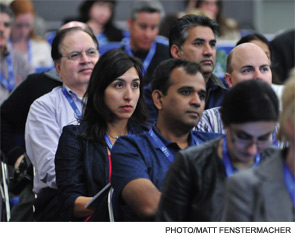
HM12 Highlights Hospitalists' Excellence, Importance to Healthcare's Future
A four-day bazaar of HM’s top minds and clinicians, all with a spectacular view of the Pacific Ocean. What wasn’t to like?
SHM’s annual meeting, held April 1-4 at the San Diego Convention Center, drew thousands to educational sessions, CME-eligible pre-courses, and plenary addresses delivered by CMS bigwig Patrick Conway, MD, MSc, FAAP, SFHM; political guru Norman Ornstein, PhD, MA, BA; and hospitalist pioneer Robert Wachter, MD, MHM.
Yet for all of the meeting’s individual branches, hospitalist Steven Pestka, MD, chief of the hospitalist service at Newton-Wellesley Hospital in Newton, Mass., sees the sum of its parts as its highest value.
“It is a great pause to recognize that there is excellence in hospitalist medicine,” he says. “Through the plenary sessions and the lectures, and various, very-high-quality presentations, it’s become very clear that this is a community to really respect.”
Richard Quinn is a freelance writer in New Jersey.
Hospitalist Programs Climb Aboard Palliative-Care Bandwagon
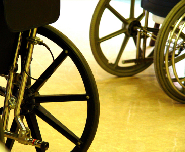
Palliative care in U.S. hospitals is growing, with 1,568 operational programs in nearly 2,500 hospitals, according to the most recent tally from the American Hospital Association and the Center to Advance Palliative Care. And as palliative care becomes a staple of inpatient care, hospitalists across the country become more involved in end-of-life care planning..jpg)
At Kaiser Permanente’s San Rafael Medical Center in California, most of the 21-member hospitalist group has been learning palliative-care concepts through grand rounds, practice updates, and self-study. Hospitalists are incorporating the concepts into routine practice and doing palliative-care consults and family meetings, says Robert Lavaysse, MD, who started the inpatient palliative-care team at San Rafael. About 10 hospitalists will join nephrologists, oncologists, and pulmonologists and sit for board certification in hospice and palliative medicine (HPM), a subspecialty recognized by 10 medical boards of the American Board of Medical Specialties. The Oct. 4 board exam is the last time physicians can earn the recognition without first completing a full-year HPM fellowship.
At Monarch Healthcare, a large physician group in Southern California, a dozen employed hospitalists and “SNFists” have been working with the palliative-care team at the University of California Irvine (UCI) Medical Center, says Vincent Nguyen, DO, CMD, Monarch’s medical director for geriatrics and palliative care. The hospitalists, who work seven-on, seven-off schedules, are using “off” weeks to train at UCI. Nine have completed six weeks of training and plan to sit for the HPM boards in October. Dr. Nguyen also pulled in palliative-care experts for 26 hours of didactic presentations, and invited hospices from the community to hold their interdisciplinary team meetings at the medical group’s office so that interested hospitalists could sit in and observe how hospice cases are managed.
—Edward Merrens, MD, FHM, hospital medicine section chief, Dartmouth-Hitchcock Medical Center, Hanover, N.H.
“Every physician who has gone through this experience is utilizing it in daily practice and influencing their colleagues,” Dr. Nguyen says. He also says hospitalists need to learn to “slow down a bit” with seriously ill patients, many of whom are good candidates for palliative care. He suggests hospitalists make certain that patient goals of care are elicited and advance directives are captured, and that they are 100% ready for the next care transition.
At Dartmouth-Hitchcock Medical Center in Hanover, N.H., hospitalists and palliative care collaborate in many areas, says HM section chief Edward Merrens, MD, FHM. “I made it a priority to broaden palliative care’s role in the organization, across all subspecialties,” says Dr. Merrens, who started the program in 2004.
Palliative-care consults are embedded in the ICUs at Dartmouth-Hitchcock, and the palliative-care team is involved in the assessment process at its affiliated outpatient cancer center.
“If a cancer patient is admitted to the hospital for reasons other than to receive chemotherapy, we take on the care of that patient, which provides an opportunity for us to collaborate with the inpatient palliative-care team,” Dr. Merrens says. “We do an initial conversation with patients about decision-making and code status within our service, and then work closely with the palliative-care team.”
Palliative care is part of the hospital’s current conversation about preventing unnecessary hospital readmissions. One example is end-stage renal patients, who come from a broad catchment area, and have high rates of mortality. “[They] can get caught in a vortex of readmissions,” he says.
The collaboration is just one example of how the “robust” 25-member hospitalist program “covers virtually everything that hospitalists do,” Dr. Merrens says. “Palliative care is a big part of how we envision the right care for these patients. There are no miracles about what palliative care does, but [caretakers] take the time to sit down and have these conversations. As we continue to take care of sicker, older patients, palliative care will play an ever-larger role.”
Larry Beresford is a freelance writer in Oakland, Calif.
Palliative care in U.S. hospitals is growing, with 1,568 operational programs in nearly 2,500 hospitals, according to the most recent tally from the American Hospital Association and the Center to Advance Palliative Care. And as palliative care becomes a staple of inpatient care, hospitalists across the country become more involved in end-of-life care planning.
At Kaiser Permanente’s San Rafael Medical Center in California, most of the 21-member hospitalist group has been learning palliative-care concepts through grand rounds, practice updates, and self-study. Hospitalists are incorporating the concepts into routine practice and doing palliative-care consults and family meetings, says Robert Lavaysse, MD, who started the inpatient palliative-care team at San Rafael. About 10 hospitalists will join nephrologists, oncologists, and pulmonologists and sit for board certification in hospice and palliative medicine (HPM), a subspecialty recognized by 10 medical boards of the American Board of Medical Specialties. The Oct. 4 board exam is the last time physicians can earn the recognition without first completing a full-year HPM fellowship.
At Monarch Healthcare, a large physician group in Southern California, a dozen employed hospitalists and “SNFists” have been working with the palliative-care team at the University of California Irvine (UCI) Medical Center, says Vincent Nguyen, DO, CMD, Monarch’s medical director for geriatrics and palliative care. The hospitalists, who work seven-on, seven-off schedules, are using “off” weeks to train at UCI. Nine have completed six weeks of training and plan to sit for the HPM boards in October. Dr. Nguyen also pulled in palliative-care experts for 26 hours of didactic presentations, and invited hospices from the community to hold their interdisciplinary team meetings at the medical group’s office so that interested hospitalists could sit in and observe how hospice cases are managed.
—Edward Merrens, MD, FHM, hospital medicine section chief, Dartmouth-Hitchcock Medical Center, Hanover, N.H.
“Every physician who has gone through this experience is utilizing it in daily practice and influencing their colleagues,” Dr. Nguyen says. He also says hospitalists need to learn to “slow down a bit” with seriously ill patients, many of whom are good candidates for palliative care. He suggests hospitalists make certain that patient goals of care are elicited and advance directives are captured, and that they are 100% ready for the next care transition.
At Dartmouth-Hitchcock Medical Center in Hanover, N.H., hospitalists and palliative care collaborate in many areas, says HM section chief Edward Merrens, MD, FHM. “I made it a priority to broaden palliative care’s role in the organization, across all subspecialties,” says Dr. Merrens, who started the program in 2004.
Palliative-care consults are embedded in the ICUs at Dartmouth-Hitchcock, and the palliative-care team is involved in the assessment process at its affiliated outpatient cancer center.
“If a cancer patient is admitted to the hospital for reasons other than to receive chemotherapy, we take on the care of that patient, which provides an opportunity for us to collaborate with the inpatient palliative-care team,” Dr. Merrens says. “We do an initial conversation with patients about decision-making and code status within our service, and then work closely with the palliative-care team.”
Palliative care is part of the hospital’s current conversation about preventing unnecessary hospital readmissions. One example is end-stage renal patients, who come from a broad catchment area, and have high rates of mortality. “[They] can get caught in a vortex of readmissions,” he says.
The collaboration is just one example of how the “robust” 25-member hospitalist program “covers virtually everything that hospitalists do,” Dr. Merrens says. “Palliative care is a big part of how we envision the right care for these patients. There are no miracles about what palliative care does, but [caretakers] take the time to sit down and have these conversations. As we continue to take care of sicker, older patients, palliative care will play an ever-larger role.”
Larry Beresford is a freelance writer in Oakland, Calif.
HM12's Clinical Pearls
SESSION
DVT Prophylaxis: Don’t Forget the Pediatric Patients
Most would agree that hospitalists have seen more thrombosis in children over the past decade, and although it isn’t known why, it is likely due to multifactorial causes, said Leslie Raffini, MD, MSCE, director of the Hemostasis and Thrombosis Center at Children’s Hospital of Philadelphia.
Central venous catheters remain a significant risk factor for venous thromboembolism (VTE), and our knowledge of inherited risk factors has expanded in recent years. While it is likely that inherited risk factors increase the risk of thrombosis in children, the question of testing has engendered debate, due in large part to the lack of clear benefit of that information in the majority of situations.
“The decision to test should be made on an individual basis, after counseling,” said Dr. Raffini. “Results should be interpreted by an experienced physician with adolescent females most likely to benefit from the testing. There are no recommendations for what to do with pediatric patients” despite the fact that this is an important cause of morbidity in high-risk patients.
Dr. Raffini described efforts at Children’s Hospital of Philadelphia that led to a VTE prophylaxis guideline. Successful implementation of the guideline required significant multidisciplinary collaboration, and an analysis of outcomes is under way.
KEY TAKEAWAYS

- The decision to test for inherited risk factors should be individualized.
- Adolescent females are most likely to benefit from testing for inherited risk factors.
- Implementation of guidelines requires intentional multidisciplinary collaboration.
A program that is structured in such a way as to hire or retain experienced hospitalists is likely to have a higher cost savings than one that doesn't.
—Mark Shen, MD, SFHM, FAAP, medical director of hospital medicine, assistant professor of pediatrics, Dell Children’s Medical Center, Austin, Texas
SESSION
Updates from the 9th ACCP Antithrombotic Therapy Guidelines
The evidence-based, rapid-fire presentation by Catherine Curley, MD, of MetroHealth Medical Center in Cleveland on the brand-new antithrombotic therapy from the ACCP took attendees through key aspects of the new guidelines. Dr. Curley used the more controversial topics as examples: treatment of submassive PE, use of catheter-directed thrombolysis in patients with acute DVT, recommended VTE prophylaxis. She even threw in some anatomy lessons for clinicians.
KEY TAKEAWAYS

- Major innovations in the methodology in the AT9: Focusing on the absolute effects to allow the provider to weigh the benefits and risks of therapy easily; rigorous conflict-of-interest reviews of the editors; re-analysis of many older studies; and simplified recommendations with emphasis on summary-of-finding tables as opposed to texts.
- A strong focus on patient-centered outcomes recommends specifically focusing on patients’ preferences.
A program that is structured in such a way as to hire or retain experienced hospitalists is likely to have a higher cost savings than one that doesn't.
—Larry W. Holder, MD, FACP, FHM, medical director of hospitalist services, chief medical information officer, Decatur (Ill.) Memorial Hospital
SESSION
HM12 Pre-Course: Medical Procedures for the Hospitalist
Bradley Rosen, MD, MBA, FHM, of Cedars Sinai Medical Center in Los Angeles, Sally Wang, MD, FHM, of Brigham and Women’s Hospital’s in Boston, and Joshua Lenchus, DO, RPh, SFHM, of the University of Miami Miller School of Medicine led a motivated group of hospitalists through hands-on training in bedside invasive procedures during two half-day pre-course sessions at HM12. With emphasis on ultrasound guidance and evidence-based practices, the faculty provided the sold-out audience with lively discussions and small-group experiential education in central venous catheter placement, paracentesis, thoracentsis, lumbar puncture (LP), and other bedside procedures.
Although bedside procedures have long been a staple of internal-medicine practice, the field of procedural medicine has increasingly become the domain of hospitalists, many of whom call themselves proceduralists. Nearly all procedures can be aided by ultrasound guidance, and for many procedures, ultrasound guidance is the standard of care.
KEY TAKEAWAYS

- Performing bedside procedures safely requires specific training and steady experience that is well-suited to healthcare providers in hospital medicine.
- Ultrasound guidance is considered the standard of care for central venous catheter placement, paracentesis, and thoracentesis.
- Widely accepted limitations in fluid removal thought to prevent re-expansion pulmonary edema (RPE) after thoracentesis might not prove to be valid.
- Arbitrary cutoffs for INR and platelet count in paracentesis are based on data that might not be valid in bedside paracentesis.
- Use of non-traumatic lumbar puncture needles, such as the Gertie-Marx and Sprotte needles, may reduce the incidence of post-LP headache.
- Fine-needle aspiration, punch skin biopsy, and arthrocentesis are bedside procedures that can be mastered by hospitalists and used regularly in their practices.
- Establishing a proceduralist group or center initially requires showing to hospital administrators benefits other than revenue, such as reduction in CLABSIs and off-loading other procedural services.
A program that is structured in such a way as to hire or retain experienced hospitalists is likely to have a higher cost savings than one that doesn't.
—Weijen Chang, MD, SFHM, pediatric hospitalist, University of San Diego Medical Center and Rady Children’s Hospital in San Diego
SESSION
ACCP Antithrombotic Therapy Guideline: The Questions that Remain Unanswered
Daniel Brotman, MD, FACP, FHM, of Johns Hopkins University School of Medicine in Baltimore addressed questions all hospitalists wonder about: Is warfarin still the best anticoagulant in atrial fibrillation (afib)?; Should DVT prevention extend beyond hospitalization?; When should anticoagulation be started in stroke patients with afib?
Warfarin, Dr. Brotman explained, has many disadvantages, and new oral anticoagulants (e.g. dabigatran, apixaban, rivaoxaban) offer many advantages with lower side-effect profiles. All of the new agents appear to have either better efficacy or trend toward better efficacy; none require monitoring, and all have lower rates of ICH.
Prices are higher for new agents but are competitive with other drugs currently on the market for other diseases. Use dabigatran with caution in patients with renal failure, and realize that there is no antidote for any of these drugs. Dabigatran is acidic and causes gastrointestinal (GI) upset, thus has a higher rate of GI bleeding. Stop any of these five days prior to planned procedures, longer if patients are at high risk of bleeding.
Evidence from RCTs in hospitalized surgical patients suggests that VTE prophylaxis should be continued in patients undergoing hip surgery and surgery for abdominal or pelvic malignancy. Patients admitted for acute medical illness do not benefit from VTE prophylaxis beyond acute hospitalization, even if immobilized, unless they have solid tumors with additional risk factors (hormone use, prior VTE, etc.) and are at low risk for bleeding. Chronically immobilized patients do not benefit from VTE prophylaxis beyond the acute hospitalization.
Oral anticoagulants can be started within one to two weeks of stroke onset. The larger the stroke, the greater the risk of hemorrhagic transformation with early anticoagulation, so the smaller the stroke, the safer it is to start early. VTE prophylaxis is important regardless.
KEY TAKEAWAYS

- We’ll be using the new oral anticoagulants in place of warfarin in the coming years, although there is no safe anticoagulant. Be cautious and aware of the side-effect profiles of each.
- Don’t sweat VTE prophylaxis in chronically immobilized patients unless they are acutely hospitalized.
- VTE prophylaxis is critical in stroke patients, but the larger the stroke in afib patients, the longer the wait to start oral anticoagulation.
A program that is structured in such a way as to hire or retain experienced hospitalists is likely to have a higher cost savings than one that doesn't.
—Caitlin Foxley, MD, FHM, medical director of Inpatient Management Inc., Nebraska Medical Center Hospitals, Omaha
SESSION
Update in Hospital Medicine
Facing a packed house in the main auditorium, Kevin O’Leary, MD, of Northwestern University’s Feinberg School of Medicine in Chicago and Efren Manjarrez, MD, from the University of Miami’s Miller School of Medicine synthesized dozens of research articles that are clinically relevant to hospitalists everywhere. “We looked for articles that would change or modify your current practice,” Dr. O’Leary said.
KEY TAKEAWAYS

- Regarding optimal diuretic dosing strategy in patients hospitalized with acute heart failure: While there was no difference between IV bolus versus continuous infusion of diuretics in the primary outcome of global symptoms or change in creatinine, patients treated with a dose 2.5 times their home dose saw significant improvement in their global symptoms, but it came at the expense of a significant increase in creatinine.
- VTE prophylaxis is routinely used in all medical and stroke patients without a clear understanding of the benefits and harms for each patient, and hospitalists should make individual VTE prophylaxis decisions for each patient.
- New options for anticoagulation in atrial fibrillation: While rivaroxiban and dabigatran were both found to be non-inferior to warfarin in the prevention of stroke and systemic emboli and in major bleeding complications, only apixiban was found to have a statistically significant improvement over warfarin.
- Identifying risk of perioperative cardiac death or non-fatal MI after surgery: The use of BNP along with the Revised Cardiac Risk Index improved the risk stratification of patients by achieving better separation between low-, intermediate-, and high-risk patients.
A program that is structured in such a way as to hire or retain experienced hospitalists is likely to have a higher cost savings than one that doesn't.
—Michelle Mourad, MD, director for quality, division of hospital medicine; assistant professor, department of medicine, University of California at San Francisco Medical Center
SESSION
Complicated Pneumonia and Acute Hematogenous Osteomyelitis: New Insights into Diagnosis and Management
The etiologic agents for complicated pneumonias and osteomyelitis have changed recently, according to Vanderbilt University School of Medicine’s Derek Williams, MD, MPH, and C. Buddy Creech, MD, MPH, who assisted pediatric hospitalists in updated diagnosis and intervention strategies.
The increase in complicated pneumonias and empyemas is mostly due to the increase in Streptococcus pneumoniae serotype 19a. After introduction of the PCV-7 vaccine, incidence of serotype 19a infections increased to 98% of infections. Serotype 19a is now included in the PCV-13 vaccine, approved by the FDA in 2011. There are multiple interventions available for empyemas, including chest tube alone, chest tube with fibrinolysis, and VATS. Current research is being done to assess efficacy for these measures.
Osteomyelitis might be caused by direct inoculation, spread from local infection, or hematogenous spread. S. aureus is a causative agent in 80% to 90% of patients. MRSA infection has a more complicated course. Based on patient response and inflammatory markers, a short course of intravenous antibiotics followed by oral antibiotics might be appropriate.
KEY TAKEAWAYS

- Surgical intervention for empyemas is patient specific and depends on clinical status, effusion, status, presence of loculations, and expertise of consultants.
- Hematogenous spread is the most common cause of osteomyelitis in children.
- MRI is the diagnostic modality of choice for osteomyelitis.
- Bone aspiration and blood cultures are very helpful in the treatment of osteomyelitis.
A program that is structured in such a way as to hire or retain experienced hospitalists is likely to have a higher cost savings than one that doesn't.—Dan Hale, MD, FAAP, pediatric hospitalist, Floating Hospital for Children at Tufts Medical Center, Boston
SESSION
DVT Prophylaxis: Don’t Forget the Pediatric Patients
Most would agree that hospitalists have seen more thrombosis in children over the past decade, and although it isn’t known why, it is likely due to multifactorial causes, said Leslie Raffini, MD, MSCE, director of the Hemostasis and Thrombosis Center at Children’s Hospital of Philadelphia.
Central venous catheters remain a significant risk factor for venous thromboembolism (VTE), and our knowledge of inherited risk factors has expanded in recent years. While it is likely that inherited risk factors increase the risk of thrombosis in children, the question of testing has engendered debate, due in large part to the lack of clear benefit of that information in the majority of situations.
“The decision to test should be made on an individual basis, after counseling,” said Dr. Raffini. “Results should be interpreted by an experienced physician with adolescent females most likely to benefit from the testing. There are no recommendations for what to do with pediatric patients” despite the fact that this is an important cause of morbidity in high-risk patients.
Dr. Raffini described efforts at Children’s Hospital of Philadelphia that led to a VTE prophylaxis guideline. Successful implementation of the guideline required significant multidisciplinary collaboration, and an analysis of outcomes is under way.
KEY TAKEAWAYS
- The decision to test for inherited risk factors should be individualized.
- Adolescent females are most likely to benefit from testing for inherited risk factors.
- Implementation of guidelines requires intentional multidisciplinary collaboration.
A program that is structured in such a way as to hire or retain experienced hospitalists is likely to have a higher cost savings than one that doesn't.
—Mark Shen, MD, SFHM, FAAP, medical director of hospital medicine, assistant professor of pediatrics, Dell Children’s Medical Center, Austin, Texas
SESSION
Updates from the 9th ACCP Antithrombotic Therapy Guidelines
The evidence-based, rapid-fire presentation by Catherine Curley, MD, of MetroHealth Medical Center in Cleveland on the brand-new antithrombotic therapy from the ACCP took attendees through key aspects of the new guidelines. Dr. Curley used the more controversial topics as examples: treatment of submassive PE, use of catheter-directed thrombolysis in patients with acute DVT, recommended VTE prophylaxis. She even threw in some anatomy lessons for clinicians.
KEY TAKEAWAYS
- Major innovations in the methodology in the AT9: Focusing on the absolute effects to allow the provider to weigh the benefits and risks of therapy easily; rigorous conflict-of-interest reviews of the editors; re-analysis of many older studies; and simplified recommendations with emphasis on summary-of-finding tables as opposed to texts.
- A strong focus on patient-centered outcomes recommends specifically focusing on patients’ preferences.
A program that is structured in such a way as to hire or retain experienced hospitalists is likely to have a higher cost savings than one that doesn't.
—Larry W. Holder, MD, FACP, FHM, medical director of hospitalist services, chief medical information officer, Decatur (Ill.) Memorial Hospital
SESSION
HM12 Pre-Course: Medical Procedures for the Hospitalist
Bradley Rosen, MD, MBA, FHM, of Cedars Sinai Medical Center in Los Angeles, Sally Wang, MD, FHM, of Brigham and Women’s Hospital’s in Boston, and Joshua Lenchus, DO, RPh, SFHM, of the University of Miami Miller School of Medicine led a motivated group of hospitalists through hands-on training in bedside invasive procedures during two half-day pre-course sessions at HM12. With emphasis on ultrasound guidance and evidence-based practices, the faculty provided the sold-out audience with lively discussions and small-group experiential education in central venous catheter placement, paracentesis, thoracentsis, lumbar puncture (LP), and other bedside procedures.
Although bedside procedures have long been a staple of internal-medicine practice, the field of procedural medicine has increasingly become the domain of hospitalists, many of whom call themselves proceduralists. Nearly all procedures can be aided by ultrasound guidance, and for many procedures, ultrasound guidance is the standard of care.
KEY TAKEAWAYS
- Performing bedside procedures safely requires specific training and steady experience that is well-suited to healthcare providers in hospital medicine.
- Ultrasound guidance is considered the standard of care for central venous catheter placement, paracentesis, and thoracentesis.
- Widely accepted limitations in fluid removal thought to prevent re-expansion pulmonary edema (RPE) after thoracentesis might not prove to be valid.
- Arbitrary cutoffs for INR and platelet count in paracentesis are based on data that might not be valid in bedside paracentesis.
- Use of non-traumatic lumbar puncture needles, such as the Gertie-Marx and Sprotte needles, may reduce the incidence of post-LP headache.
- Fine-needle aspiration, punch skin biopsy, and arthrocentesis are bedside procedures that can be mastered by hospitalists and used regularly in their practices.
- Establishing a proceduralist group or center initially requires showing to hospital administrators benefits other than revenue, such as reduction in CLABSIs and off-loading other procedural services.
A program that is structured in such a way as to hire or retain experienced hospitalists is likely to have a higher cost savings than one that doesn't.
—Weijen Chang, MD, SFHM, pediatric hospitalist, University of San Diego Medical Center and Rady Children’s Hospital in San Diego
SESSION
ACCP Antithrombotic Therapy Guideline: The Questions that Remain Unanswered
Daniel Brotman, MD, FACP, FHM, of Johns Hopkins University School of Medicine in Baltimore addressed questions all hospitalists wonder about: Is warfarin still the best anticoagulant in atrial fibrillation (afib)?; Should DVT prevention extend beyond hospitalization?; When should anticoagulation be started in stroke patients with afib?
Warfarin, Dr. Brotman explained, has many disadvantages, and new oral anticoagulants (e.g. dabigatran, apixaban, rivaoxaban) offer many advantages with lower side-effect profiles. All of the new agents appear to have either better efficacy or trend toward better efficacy; none require monitoring, and all have lower rates of ICH.
Prices are higher for new agents but are competitive with other drugs currently on the market for other diseases. Use dabigatran with caution in patients with renal failure, and realize that there is no antidote for any of these drugs. Dabigatran is acidic and causes gastrointestinal (GI) upset, thus has a higher rate of GI bleeding. Stop any of these five days prior to planned procedures, longer if patients are at high risk of bleeding.
Evidence from RCTs in hospitalized surgical patients suggests that VTE prophylaxis should be continued in patients undergoing hip surgery and surgery for abdominal or pelvic malignancy. Patients admitted for acute medical illness do not benefit from VTE prophylaxis beyond acute hospitalization, even if immobilized, unless they have solid tumors with additional risk factors (hormone use, prior VTE, etc.) and are at low risk for bleeding. Chronically immobilized patients do not benefit from VTE prophylaxis beyond the acute hospitalization.
Oral anticoagulants can be started within one to two weeks of stroke onset. The larger the stroke, the greater the risk of hemorrhagic transformation with early anticoagulation, so the smaller the stroke, the safer it is to start early. VTE prophylaxis is important regardless.
KEY TAKEAWAYS
- We’ll be using the new oral anticoagulants in place of warfarin in the coming years, although there is no safe anticoagulant. Be cautious and aware of the side-effect profiles of each.
- Don’t sweat VTE prophylaxis in chronically immobilized patients unless they are acutely hospitalized.
- VTE prophylaxis is critical in stroke patients, but the larger the stroke in afib patients, the longer the wait to start oral anticoagulation.
A program that is structured in such a way as to hire or retain experienced hospitalists is likely to have a higher cost savings than one that doesn't.
—Caitlin Foxley, MD, FHM, medical director of Inpatient Management Inc., Nebraska Medical Center Hospitals, Omaha
SESSION
Update in Hospital Medicine
Facing a packed house in the main auditorium, Kevin O’Leary, MD, of Northwestern University’s Feinberg School of Medicine in Chicago and Efren Manjarrez, MD, from the University of Miami’s Miller School of Medicine synthesized dozens of research articles that are clinically relevant to hospitalists everywhere. “We looked for articles that would change or modify your current practice,” Dr. O’Leary said.
KEY TAKEAWAYS
- Regarding optimal diuretic dosing strategy in patients hospitalized with acute heart failure: While there was no difference between IV bolus versus continuous infusion of diuretics in the primary outcome of global symptoms or change in creatinine, patients treated with a dose 2.5 times their home dose saw significant improvement in their global symptoms, but it came at the expense of a significant increase in creatinine.
- VTE prophylaxis is routinely used in all medical and stroke patients without a clear understanding of the benefits and harms for each patient, and hospitalists should make individual VTE prophylaxis decisions for each patient.
- New options for anticoagulation in atrial fibrillation: While rivaroxiban and dabigatran were both found to be non-inferior to warfarin in the prevention of stroke and systemic emboli and in major bleeding complications, only apixiban was found to have a statistically significant improvement over warfarin.
- Identifying risk of perioperative cardiac death or non-fatal MI after surgery: The use of BNP along with the Revised Cardiac Risk Index improved the risk stratification of patients by achieving better separation between low-, intermediate-, and high-risk patients.
A program that is structured in such a way as to hire or retain experienced hospitalists is likely to have a higher cost savings than one that doesn't.
—Michelle Mourad, MD, director for quality, division of hospital medicine; assistant professor, department of medicine, University of California at San Francisco Medical Center
SESSION
Complicated Pneumonia and Acute Hematogenous Osteomyelitis: New Insights into Diagnosis and Management
The etiologic agents for complicated pneumonias and osteomyelitis have changed recently, according to Vanderbilt University School of Medicine’s Derek Williams, MD, MPH, and C. Buddy Creech, MD, MPH, who assisted pediatric hospitalists in updated diagnosis and intervention strategies.
The increase in complicated pneumonias and empyemas is mostly due to the increase in Streptococcus pneumoniae serotype 19a. After introduction of the PCV-7 vaccine, incidence of serotype 19a infections increased to 98% of infections. Serotype 19a is now included in the PCV-13 vaccine, approved by the FDA in 2011. There are multiple interventions available for empyemas, including chest tube alone, chest tube with fibrinolysis, and VATS. Current research is being done to assess efficacy for these measures.
Osteomyelitis might be caused by direct inoculation, spread from local infection, or hematogenous spread. S. aureus is a causative agent in 80% to 90% of patients. MRSA infection has a more complicated course. Based on patient response and inflammatory markers, a short course of intravenous antibiotics followed by oral antibiotics might be appropriate.
KEY TAKEAWAYS
- Surgical intervention for empyemas is patient specific and depends on clinical status, effusion, status, presence of loculations, and expertise of consultants.
- Hematogenous spread is the most common cause of osteomyelitis in children.
- MRI is the diagnostic modality of choice for osteomyelitis.
- Bone aspiration and blood cultures are very helpful in the treatment of osteomyelitis.
A program that is structured in such a way as to hire or retain experienced hospitalists is likely to have a higher cost savings than one that doesn't.—Dan Hale, MD, FAAP, pediatric hospitalist, Floating Hospital for Children at Tufts Medical Center, Boston
HM12 Experts Teach Hospitalists to Deal with Practice-Management Issues
If such a thing exists, hospitalist Dwayne Gard, MD, of Memorial Health in Savannah, Ga., has a good practice management problem: In recent years, his HM group started collaborating with Memorial’s gastrointestinal team to admit their patients. Now, Dr. Gard and his colleagues are in similar discussions with neurosurgery. And ments provide steady revenue for the hospitalist group, too much growth, too fast, can be as dangerous as too little.
“There are a lot of demands, from an administrative standpoint, to cater to the needs of some of the subspecialists at a time where we really need to grow the number of hospitalists within our program before we even consider expanding our services,” says Dr. Gard, whose group has 10 FTE hospitalists and has an average patient census of 90-110 at the 500-bed hospital. “I let our administration know it’s a challenge we’re willing to take on as long as we have the support to get the staff in that we need to safely see those patients.”
Just how to let them know is the art of practice management, a topic that dominates much of SHM’s annual meeting. From a popular, daylong CME pre-course to a dedicated practice-management track, hospitalists looking for real-time advice on the business side of medicine came away from HM12 last month with brown bags full of tips.
“This is a huge pause, a huge opportunity to pause and remind ourselves what’s important,” says Steven Pestka, MD, chief of the hospitalist service at Newton-Wellesley Hospital in Newton, Mass. “And then recognizing that all those other things … need to be kept in context and need to be limited so that the core running of the group can be performed.”
For Dr. Gard, gaining management skills at this year’s annual meeting was a two-step process. First, they sent seven members of their HM groupthree hospitalists, two non-physician providers (NPPs), an administrator, and an office managerto San Diego. The annual-meeting team spread across as many sessions as they could, including pre-courses on value-based purchasing and practice management. Second, and perhaps more important, the team members didn’t decide ahead of time what advice they were going to glean; they came with agendas and schedules of what breakout sessions
they viewed as most valuable while remaining flexible.
“It always seems like while you’re here, you learn something you weren’t expecting,” Dr. Gard adds, “and something that’s actually maybe more pertinent than the reason you maybe thought about coming to the meeting to begin with. You can take that home as well.
“It’s important for our group because a lot of our new hires, the majority of our new hires, are residents within our own training program. Unless we hire hospitalists from outside our own network, it does tend to be the same people in the same system—not thinking outside the box, like we need to do in this day and age.”
Advanced Degrees of Hospital Medicine
Benjamin Frizner, MD, director of the hospitalist program at Saint Agnes Hospital in Baltimore, wonders whether earning an MBA would give him an advantage in “getting into the mind of the CEO and the C-suite.”
“What’s their frame of mind when I go into meetings, so I can talk their language, frame things in a different way?” Dr. Frizner says during a break in a practice-management pre-course. “The topics [at HM12] are focused to a lot of the problems we are facing.”
The issues are specific to individual HM groups. Dr. Frizner also wants to know how to better speak to colleagues and care team members in the hospitals his group services. Madonna Ringswald, DO, medical director of the hospitalist program at Baptist Hospital Northeast in La Grange, Ky., took over management of her group a little more than two years ago and came to San Diego to bounce questions off people who have more experience than she does. Dr. Pestka took home suggestions on how to recognize hospitalist performance in non-compensated ways.
“This is the best meeting I’ve ever been to,” Dr. Ringswald says. “If you can’t find a lecture [that appeals to you], there’s something wrong with you.”
Renewed Focus

In the annual meeting’s wrap-up address, Michael Pistoria, DO, FACP, SFHM, hospitalist at Lehigh Valley Health Network in Allentown, Pa., described the future of practice management as an amalgam of all the issues HM faces. Improved communication, a renewed focus on costs and high-value care, and continued adoption of best practices found at institutions across the country are all ways to better operate individual practices.
“We move ahead by doing what we do best in hospital medicine,” says Dr. Pistoria, the course director for HM13, which will be May 16-19, 2013, just outside of Washington, D.C. “We get together as teams, we collaborate with each other across our institutions, within our own institution, and professionally. We come up with that one small or big idea that improves care at the level of the patient and at the macro level.”
CMS’ CMO Offers Practice-Management Tips
Patrick Conway, MD, MSc, FAAP, SFHM, a pediatric hospitalist and chief medical officer of the Centers for Medicare & Medicaid Services (CMS), has advice for hospitalists looking to better manage their practice:
- Understand your hospital’s data. What is collected? What is analyzed? Where are the gaps in that research? What does it all mean for you?
- Take leadership roles. Lead multidisciplinary teams, teach others around you, become known for a focus on evidence-based approaches.
- Familiarize yourself with resources. CMS, SHM, and myriad other medical organizations and private agencies offer free or low-cost resources. When looking to solve problems, it’s possible someone already has done so.
- Create collaborative forums. Physicians feel engaged when they believe they can voice opinions in an accepting environment. The reverse is also true.
- Stay connected to clinical care, even if it’s just occasional shifts to help out.
“My challenge to you is please don’t sit on the sidelines,” Dr. Conway says. “Please be actively engaged in your local system in creating this change. This is not for me or CMS, but stating the obvious: It’s why we went into medicine.”
If such a thing exists, hospitalist Dwayne Gard, MD, of Memorial Health in Savannah, Ga., has a good practice management problem: In recent years, his HM group started collaborating with Memorial’s gastrointestinal team to admit their patients. Now, Dr. Gard and his colleagues are in similar discussions with neurosurgery. And ments provide steady revenue for the hospitalist group, too much growth, too fast, can be as dangerous as too little.
“There are a lot of demands, from an administrative standpoint, to cater to the needs of some of the subspecialists at a time where we really need to grow the number of hospitalists within our program before we even consider expanding our services,” says Dr. Gard, whose group has 10 FTE hospitalists and has an average patient census of 90-110 at the 500-bed hospital. “I let our administration know it’s a challenge we’re willing to take on as long as we have the support to get the staff in that we need to safely see those patients.”
Just how to let them know is the art of practice management, a topic that dominates much of SHM’s annual meeting. From a popular, daylong CME pre-course to a dedicated practice-management track, hospitalists looking for real-time advice on the business side of medicine came away from HM12 last month with brown bags full of tips.
“This is a huge pause, a huge opportunity to pause and remind ourselves what’s important,” says Steven Pestka, MD, chief of the hospitalist service at Newton-Wellesley Hospital in Newton, Mass. “And then recognizing that all those other things … need to be kept in context and need to be limited so that the core running of the group can be performed.”
For Dr. Gard, gaining management skills at this year’s annual meeting was a two-step process. First, they sent seven members of their HM groupthree hospitalists, two non-physician providers (NPPs), an administrator, and an office managerto San Diego. The annual-meeting team spread across as many sessions as they could, including pre-courses on value-based purchasing and practice management. Second, and perhaps more important, the team members didn’t decide ahead of time what advice they were going to glean; they came with agendas and schedules of what breakout sessions
they viewed as most valuable while remaining flexible.
“It always seems like while you’re here, you learn something you weren’t expecting,” Dr. Gard adds, “and something that’s actually maybe more pertinent than the reason you maybe thought about coming to the meeting to begin with. You can take that home as well.
“It’s important for our group because a lot of our new hires, the majority of our new hires, are residents within our own training program. Unless we hire hospitalists from outside our own network, it does tend to be the same people in the same system—not thinking outside the box, like we need to do in this day and age.”
Advanced Degrees of Hospital Medicine
Benjamin Frizner, MD, director of the hospitalist program at Saint Agnes Hospital in Baltimore, wonders whether earning an MBA would give him an advantage in “getting into the mind of the CEO and the C-suite.”
“What’s their frame of mind when I go into meetings, so I can talk their language, frame things in a different way?” Dr. Frizner says during a break in a practice-management pre-course. “The topics [at HM12] are focused to a lot of the problems we are facing.”
The issues are specific to individual HM groups. Dr. Frizner also wants to know how to better speak to colleagues and care team members in the hospitals his group services. Madonna Ringswald, DO, medical director of the hospitalist program at Baptist Hospital Northeast in La Grange, Ky., took over management of her group a little more than two years ago and came to San Diego to bounce questions off people who have more experience than she does. Dr. Pestka took home suggestions on how to recognize hospitalist performance in non-compensated ways.
“This is the best meeting I’ve ever been to,” Dr. Ringswald says. “If you can’t find a lecture [that appeals to you], there’s something wrong with you.”
Renewed Focus
In the annual meeting’s wrap-up address, Michael Pistoria, DO, FACP, SFHM, hospitalist at Lehigh Valley Health Network in Allentown, Pa., described the future of practice management as an amalgam of all the issues HM faces. Improved communication, a renewed focus on costs and high-value care, and continued adoption of best practices found at institutions across the country are all ways to better operate individual practices.
“We move ahead by doing what we do best in hospital medicine,” says Dr. Pistoria, the course director for HM13, which will be May 16-19, 2013, just outside of Washington, D.C. “We get together as teams, we collaborate with each other across our institutions, within our own institution, and professionally. We come up with that one small or big idea that improves care at the level of the patient and at the macro level.”
CMS’ CMO Offers Practice-Management Tips
Patrick Conway, MD, MSc, FAAP, SFHM, a pediatric hospitalist and chief medical officer of the Centers for Medicare & Medicaid Services (CMS), has advice for hospitalists looking to better manage their practice:
- Understand your hospital’s data. What is collected? What is analyzed? Where are the gaps in that research? What does it all mean for you?
- Take leadership roles. Lead multidisciplinary teams, teach others around you, become known for a focus on evidence-based approaches.
- Familiarize yourself with resources. CMS, SHM, and myriad other medical organizations and private agencies offer free or low-cost resources. When looking to solve problems, it’s possible someone already has done so.
- Create collaborative forums. Physicians feel engaged when they believe they can voice opinions in an accepting environment. The reverse is also true.
- Stay connected to clinical care, even if it’s just occasional shifts to help out.
“My challenge to you is please don’t sit on the sidelines,” Dr. Conway says. “Please be actively engaged in your local system in creating this change. This is not for me or CMS, but stating the obvious: It’s why we went into medicine.”
Vanderbilt Hospitalist Impresses RIV Judges with Sample Size, Takes Home Research Prize
Eduard Vasilevskis, MD, is a member of SHM’s Research Committee and was a judge for the annual Research, Innovations, and Clinical Vignettes (RIV) poster competition at HM11 outside Dallas. But when the committee members discussed who would judge the HM12 contest, Dr. Vasilevskis passed. He had a poster he wanted to enter instead.
Smart move.
His poster took the top prize in the Research portion of the annual contest, as judged by a panel of hospitalist leaders and visiting professor Pamela Lipsett, MD, MHPE, FACS, FCCM, a professor in the department of surgery, anesthesiology, and critical-care medicine at Johns Hopkins University Schools of Medicine and Nursing in Baltimore.
“When you work in a specific research area and you’re really passionate about the subject you’re studying, you sometimes lose sight of how other people might care about it,” says Dr. Vasilevskis, a hospitalist and assistant professor of medicine in the section of hospital medicine at Vanderbilt University School of Medicine and Tennessee Valley-Nashville VA Hospital in Nashville. “It’s nice to have an audience to validate that.”
HM has “a really outstanding core of researchers,” he adds. “The people who are walking around in that group, I really look up to.”
The feeling was mutual, as a team of judges led by SHM Education Committee Chair Daniel Brotman, MD, FACP, FHM, set out to pick a winner in a contest that has grown so large, the Clinical Vignettes portion was broken into a separate session.
First, SHM selects which abstracts to accept. Then, 186 hospitalists or HM-led teams were invited to give poster presentations at the San Diego Convention Center. Then, 23 finalists were chosen for judging rounds. All of that happened before anyone got to the annual meeting. Once in the exhibit hall, two-clinician judging teams worked their way through two dozen research posters deemed the best of the best. Each team recommended a finalist except for one, which Dr. Brotman dubbed “the Russian judges.”
But from the get-go, it was clear that the work completed by Dr. Vasilevskis and his colleagues would be the winner. As the two-member teams went around naming the posters they wanted the full judging panel to visit, one of those who visited Dr. Vasilevskis was brief but enthusiastic in the description: “I really liked this one.”
So the judges went back to poster No. 63, “Veterans Administration Acute Care 30-Day Mortality Model: Development, Validation, and Performance Variation.” Dr. Brotman was impressed by the breadth of the study, which aimed to develop a hybrid administrative/clinical risk model to address the shortcomings of risk models that rely on only administrative data or clinical data.
The first number that caught the judges’ attention was 1,114,327. That’s how many patients were used in retrospective cohort study of 131 VA hospitals. Then it was the results, which were impressively accurate given the size of the sample.
“It was a strong survey with incredible methodological rigor, addressing an incredibly important issue,” Dr. Brotman says. Dr. Vasilevskis notes that few hospitals or health systems currently have access to the level of data available to his research team. But as electronic health records (EHR) capture more clinical and administrative data, the ability to build models like the one he presented will only increase. “The better your model, the more you can believe your results,” he adds. “And we want our clinicians and CEOs and hospital boards to believe those numbers.”
Hospitalist Investigators Impress Judges at HM12’s Annual RIV Competition
Sometimes the first impression really is the right one.
Nearly 150 HM-focused innovations abstracts were submitted for the 2012 Research, Innovations, and Clinical Vignettes poster competition at HM12, and more than 60 of the submitters were invited to display posters in San Diego. A judging committee reviewed 24 poster finalists, and the consensus was that one poster stood out from the start: “Structured Interdisciplinary Bedside Rounds at Emory University School of Medicine in Atlanta, Ga.,” presented by Emory hospitalist Christina Payne, MD.
One of the Innovations judges noted the 73% reduction in mortality rates realized via Emory’s Accountable Care Unit, along with a one-day reduction in length of stay and a 90% reduction in catheter-related bloodstream infections. “My God, what’s going on there?” one judge exclaimed.
Added a second judge: “We tried something like this and didn’t succeed—but we didn’t have a champion like [Dr. Payne].”
SHM’s Innovations co-chair, Andrew Modest, MD, assistant clinical professor of medicine at Harvard Medical School and Mount Auburn Hospital in Cambridge, Mass., explained that the poster contests are a real stepping-stone opportunity for young clinicians—particularly junior faculty and residents. “You can go on to do more elaborate work and, with some statistical analysis, all of a sudden, you have a research paper,” Dr. Modest said, “or else turn it into a true quality-improvement project for your hospital. And if you’re a poster finalist at SHM, that goes on your resume.”
Innovation implies either something new or a new and effective application of existing methods for addressing an issue that all hospitalists deal with, Dr. Modest said. At HM12, posters featured innovative approaches to such issues as scheduling, readmissions, care transitions, and team communication.
“What I like to see is a buzz in the room,” Dr. Modest said. “Something that people recognize they can just take home and start using.”
In addition to Dr. Payne’s winning poster, the judges also chose five abstracts for oral presentations on Day Two of the annual meeting; one abstract was chosen and orally presented to start the Day Two plenary session before all HM12 attendees.
The Innovations judges, in teams of two, each met with four or five poster finalists in front of their panels; each principal investigator was asked to briefly summarize the innovation.
For example, at the University of Texas Health Sciences Center at San Antonio, financial support to add a fourth chief resident dedicated to quality led to the launch of a procedural service, explained by principal investigator David Schmit, MD. Medical interns now are taught in a one-month rotation to perform a number of medical procedures, aided by ultrasound, using a standardized curriculum, checklists, video training, and practice on a simulator. In Month Two, they perform the procedures on actual patients. Trained interns have performed 342 procedures, with 100% success rates for paracentesis and thoracentesis, Dr. Schmit said.
“This has to be incorporated into physicians’ training,” noted judge James Yturri, MD, a hospitalist from Great Falls, Mont. “How many programs are actually doing this?”
The service, Dr. Schmit said, has required a lot of coordination, infrastructure, and investment from the hospitalist group, residency program, and three participating hospitals.
“How did you get them all to buy in?” Dr. Yturri asked.
“I think the data on patient safety was persuasive,” Dr. Schmit said. “If you look at our low rate of pneumothorax, we saved money.”
When the 10 judges reconvened, discussion focused on innovative telemedicine and real-time reporting of VTE. A statewide collaborative to improve care transitions in Michigan was singled out for its active participation by the state’s health payors.
“I’m partial to the procedural project,” said Michael Pistoria, DO, SFHM, a hospitalist at Lehigh Valley Health Network in Allentown, Pa., and HM13 course director.
Kimberly Tartaglia, MD, was complimented for a succinct presentation of her poster about improving the inpatient management of neonatal jaundice. “Nicely done,” Dr. Modest told the hospitalist from Ohio State University Medical Center in Columbus.
But the judges eventually circled back to the Emory poster about rounds done on a nonteaching, medical accountable-care unit, defined as a unit that is consistently responsible for the outcomes it produces. On the unit, interns are trained to lead team-based, patient-centered rounds.
“They really have learned to love it,” Dr. Payne said.
Rounds start punctually each morning, with five minutes spent in each room for updates from the intern, nurse, social worker, patient and family, leading to a plan for the day with a safety checklist and discharge planning checklist, all entered in real time into the medical record by a second intern.
“How hard is it to get through all that in five minutes?” Dr. Modest queried.
“Every month, I get a new set of learners,” Dr. Payne replied. “At the beginning of the month, it doesn’t work as well. But by Week Two, we’re a well-oiled machine.”
The judges huddled again, but quickly agreed to award the poster prize to Dr. Payne.
HM12’s Professional Development Offerings Have Singular Focus
Informatics specialist David Knoepfler, MD, sat in the back row of a daylong HM12 pre-course that aimed to prepare hospitalists for the American Board of Internal Medicine’s (ABIM) Maintenance of Certification (MOC) examination. The question-and-answer session was intended to help the longtime clinician pass his second certification renewal.
But it was utterly unnecessary.
Dr. Knoepfler is the chief medical information officer at Overlake Hospital Medical Center (http://www.overlakehospital.org/about-us/) in the Seattle suburb of Bellevue, Wash. As much as 90% of his time is spent on information technology (IT) duties, and since he is grandfathered, he does not need to get recertified. So why did he spend seven hours scribbling notes as though he was preparing for his first boards?
Pride in his career development, plain and simple.
“Despite my heavily administrative role, I want to stay in touch with the clinical side,” says Dr. Knoepfler, a practicing hospitalist for 20 years who hadn’t attended an SHM annual meeting in more than 10 years before participating in HM12 last month at the San Diego Convention Center. “I feel like I have to for my patients’ sake. Even if I’m doing 10 percent or 15 percent [of my time in clinical], I absolutely have to be practicing quality medicine.”
SHM’s annual meeting is a hotbed of lifelong learning. A majority of attendees each year say that the continuing medical education (CME) credits are a major draw. This year’s pre-course lineup included the MOC test preparation, hands-on classes in medical procedures and ultrasound usage, and a debut class on “How to Improve Performance in CMS’s Value-Based Purchasing Program,” which was led by SHM senior vice president Joseph Miller and
Patrick Torcson, MD, MMM, FAACP, SFHM, chair of SHM’s Performance Measurement and Reporting Committee.

Career development, of course, means different things to different people. Dr. Knoepfler might have been focused on updating his clinical knowledge, but he is a technology expert whose main duties are to integrate health IT into the daily routine of his 349-bed hospital. So whether listening in a pre-course, attending a workshop, or meeting old or new colleagues for a drink, he spent the four-day conference constantly looking for ways to improve his job performance.
“My ears are constantly listening for how can I tie this into our electronic health record,” Dr. Knoepfler adds. “Whether it’s a clinical topic, an administrative topic, an insurance topic, a governmental topic, for me, I’m going to take all those back and see how I can make our electronic record work with the practitioners rather than fight them.”
A Learning Process
Former SHM president Jeff Weise, MD, professor of medicine and residency program director at Tulane University Health Sciences Center in New Orleans, feels so strongly about the value of professional development and the ABIM’s MOC process for hospital medicine that he and several society staffers held an impromptu session between plenary addresses to tackle perceived confusion with the Focused Practice in Hospital Medicine (FPHM) MOC pathway.

Dr. Weise says that the FPHM pathway does not mean doctors give up board certification in internal medicine, nor does it apply only to academic or community physicians.
Dr. Torcson, who works at St. Tammany Parish Hospital in Covington, La., says that the certification should be viewed as a “meaningful affirmation” of HM’s solidified place in the healthcare system.
“It’s really the right thing to do for our developing specialty,” he says. “We really have to distinguish ourselves, and there’s really no better brand distinction than a professional designation based on board certification and maintenance of certification.”
Ethan Cumbler, MD, FACP, of the University of Colorado Denver, led a portion of the MOC pre-course at HM12. He says that those who attend SHM’s annual meeting are able to take advantage of communal learning, which provides benefits that those who study alone can’t absorb. Taking a pre-test just teaches whether participants answered correctly or not. Questioning why the answer is what it is, with interactive feedback, is “a learning process,” he says.
“There’s a very different feel to learning in a group setting with your peers than to sitting down with a journal or a magazine or a textbook or an online module,” Dr. Cumbler says. “Part of what makes this conference engaging is not just content, it’s the milieu. It’s a social milieu of our peers, and we’re learning together and we’re exploring these difficult issues together.
“My sense is that the people who come to this are probably a cut above,” adds Dr. Cumbler, whose University of Colorado In-Hospital Stroke QI Team was the 2012 recipient of SHM’s Award for Excellence in Teamwork in Quality Improvement. “They are people who are really committed to being more evidence-based and understanding what the evidence is and how they apply that to their patients.”
Career Decisions
They are physicians like James Eppinette, MD, who became a hospitalist three years ago after 20 years in private practice as a primary-care physician (PCP). He joined a hospital-owned group at St. Francis Medical Center in Monroe, La., and HM12 was his first annual meeting—he attended because the meeting’s clinical focus provided him with professional development opportunities. He began with the pre-operative-care pre-course, an area in which he felt he wanted more exposure. From there, he bounced around clinical workshops.
“[HM12] has an incredible variety of tracks that you can pursue, so there’s something here for everybody,” Dr. Eppinette says, later adding that “my primary responsibility is clinical. That’s all I do is take care of people. I don’t manage people, I don’t run a practice, nor do I want to. That’s why I left 20 years of private practice to be a hospitalist. I just think it’s well set up structurally for an educational event.”
Dr. Eppinette, who took his notes back to the 12 hospitalists and nine pulmonologists in his hospitalist group, says that the decision to attend HM12 came from his increasing satisfaction with his late-career job change.
“I didn’t know how I would like going from an independent practitioner to being employed, hospital-owned. But I’ve done it three years,” he says, and “[I think this is] how I will finish my career. After three years, I’m kind of liking this road I’m on, so I’ve started looking into more about it.”
That level of personal accountability is a next step for hospitalists nationwide, according to new SHM president Shaun Frost, MD, SFHM, FACP, chief medical officer for the Northeast region of Brentwood, Tenn.-based Cogent HMG. Dr. Frost says that personal and professional accountability will be the drivers for HM’s continued growth and success, and that without individual physicians pushing their career development goals to new heights, the fastest-growing medical specialty in history could stall.
“We must all strive to consistently hold each other and hold ourselves personally accountable for embracing the work that is necessary to realize the potential of hospital medicine to be a true healthcare reform effector,” Dr. Frost says. “It’s time, I think, for each of us to put our money where our mouths have been.”
Informatics specialist David Knoepfler, MD, sat in the back row of a daylong HM12 pre-course that aimed to prepare hospitalists for the American Board of Internal Medicine’s (ABIM) Maintenance of Certification (MOC) examination. The question-and-answer session was intended to help the longtime clinician pass his second certification renewal.
But it was utterly unnecessary.
Dr. Knoepfler is the chief medical information officer at Overlake Hospital Medical Center (http://www.overlakehospital.org/about-us/) in the Seattle suburb of Bellevue, Wash. As much as 90% of his time is spent on information technology (IT) duties, and since he is grandfathered, he does not need to get recertified. So why did he spend seven hours scribbling notes as though he was preparing for his first boards?
Pride in his career development, plain and simple.
“Despite my heavily administrative role, I want to stay in touch with the clinical side,” says Dr. Knoepfler, a practicing hospitalist for 20 years who hadn’t attended an SHM annual meeting in more than 10 years before participating in HM12 last month at the San Diego Convention Center. “I feel like I have to for my patients’ sake. Even if I’m doing 10 percent or 15 percent [of my time in clinical], I absolutely have to be practicing quality medicine.”
SHM’s annual meeting is a hotbed of lifelong learning. A majority of attendees each year say that the continuing medical education (CME) credits are a major draw. This year’s pre-course lineup included the MOC test preparation, hands-on classes in medical procedures and ultrasound usage, and a debut class on “How to Improve Performance in CMS’s Value-Based Purchasing Program,” which was led by SHM senior vice president Joseph Miller and
Patrick Torcson, MD, MMM, FAACP, SFHM, chair of SHM’s Performance Measurement and Reporting Committee.
Career development, of course, means different things to different people. Dr. Knoepfler might have been focused on updating his clinical knowledge, but he is a technology expert whose main duties are to integrate health IT into the daily routine of his 349-bed hospital. So whether listening in a pre-course, attending a workshop, or meeting old or new colleagues for a drink, he spent the four-day conference constantly looking for ways to improve his job performance.
“My ears are constantly listening for how can I tie this into our electronic health record,” Dr. Knoepfler adds. “Whether it’s a clinical topic, an administrative topic, an insurance topic, a governmental topic, for me, I’m going to take all those back and see how I can make our electronic record work with the practitioners rather than fight them.”
A Learning Process
Former SHM president Jeff Weise, MD, professor of medicine and residency program director at Tulane University Health Sciences Center in New Orleans, feels so strongly about the value of professional development and the ABIM’s MOC process for hospital medicine that he and several society staffers held an impromptu session between plenary addresses to tackle perceived confusion with the Focused Practice in Hospital Medicine (FPHM) MOC pathway.
Dr. Weise says that the FPHM pathway does not mean doctors give up board certification in internal medicine, nor does it apply only to academic or community physicians.
Dr. Torcson, who works at St. Tammany Parish Hospital in Covington, La., says that the certification should be viewed as a “meaningful affirmation” of HM’s solidified place in the healthcare system.
“It’s really the right thing to do for our developing specialty,” he says. “We really have to distinguish ourselves, and there’s really no better brand distinction than a professional designation based on board certification and maintenance of certification.”
Ethan Cumbler, MD, FACP, of the University of Colorado Denver, led a portion of the MOC pre-course at HM12. He says that those who attend SHM’s annual meeting are able to take advantage of communal learning, which provides benefits that those who study alone can’t absorb. Taking a pre-test just teaches whether participants answered correctly or not. Questioning why the answer is what it is, with interactive feedback, is “a learning process,” he says.
“There’s a very different feel to learning in a group setting with your peers than to sitting down with a journal or a magazine or a textbook or an online module,” Dr. Cumbler says. “Part of what makes this conference engaging is not just content, it’s the milieu. It’s a social milieu of our peers, and we’re learning together and we’re exploring these difficult issues together.
“My sense is that the people who come to this are probably a cut above,” adds Dr. Cumbler, whose University of Colorado In-Hospital Stroke QI Team was the 2012 recipient of SHM’s Award for Excellence in Teamwork in Quality Improvement. “They are people who are really committed to being more evidence-based and understanding what the evidence is and how they apply that to their patients.”
Career Decisions
They are physicians like James Eppinette, MD, who became a hospitalist three years ago after 20 years in private practice as a primary-care physician (PCP). He joined a hospital-owned group at St. Francis Medical Center in Monroe, La., and HM12 was his first annual meeting—he attended because the meeting’s clinical focus provided him with professional development opportunities. He began with the pre-operative-care pre-course, an area in which he felt he wanted more exposure. From there, he bounced around clinical workshops.
“[HM12] has an incredible variety of tracks that you can pursue, so there’s something here for everybody,” Dr. Eppinette says, later adding that “my primary responsibility is clinical. That’s all I do is take care of people. I don’t manage people, I don’t run a practice, nor do I want to. That’s why I left 20 years of private practice to be a hospitalist. I just think it’s well set up structurally for an educational event.”
Dr. Eppinette, who took his notes back to the 12 hospitalists and nine pulmonologists in his hospitalist group, says that the decision to attend HM12 came from his increasing satisfaction with his late-career job change.
“I didn’t know how I would like going from an independent practitioner to being employed, hospital-owned. But I’ve done it three years,” he says, and “[I think this is] how I will finish my career. After three years, I’m kind of liking this road I’m on, so I’ve started looking into more about it.”
That level of personal accountability is a next step for hospitalists nationwide, according to new SHM president Shaun Frost, MD, SFHM, FACP, chief medical officer for the Northeast region of Brentwood, Tenn.-based Cogent HMG. Dr. Frost says that personal and professional accountability will be the drivers for HM’s continued growth and success, and that without individual physicians pushing their career development goals to new heights, the fastest-growing medical specialty in history could stall.
“We must all strive to consistently hold each other and hold ourselves personally accountable for embracing the work that is necessary to realize the potential of hospital medicine to be a true healthcare reform effector,” Dr. Frost says. “It’s time, I think, for each of us to put our money where our mouths have been.”
HM Leaders Call for Thoughtful, Budget-Minded Advancement of Patient-Safety Reforms
In a Venn diagram, patient handoffs would fall at the intersection of quality improvement (QI), patient safety, and technology.
Vineet Arora, MD, MPP, FACP, FHM, associate professor of medicine at the University of Chicago, wants hospitalists to understand that best practices in transitional care only work efficiently if they incorporate pieces from all three spheres. Hospitalists who focus too much on the outcome of an individual case might fail to utilize technology that could create a systemic protocol that would improve outcomes across multiple cases, she says. Similarly, hospitalists who leave notes in an electronic health record (EHR) can mistakenly believe that the next physician will interpret that information perfectly despite a lack of verbal and physical cues that guide verbal communication.

“People may fall into the fallacy that electronic health records actually equal good handoffs,” says Dr. Arora, a faculty member at HM12 last month in San Diego who led a breakout session that focused on the obstacles of handoffs. “The challenge is that handoffs actually are about a verbal exchange of informationa conversation and a dialogue, in conjunction with and supplemented with written communication. Technology certainly has the ability to revolutionize the way we review information and access information, but it will not change the way we speak about the information.”
The crossroads of patient safety, QI, and the use of technology from medical devices to iPhones was a major theme at SHM’s annual meeting, April 1-4 at the San Diego Convention Center. Hospitalist leaders agree that clinicians must focus on all three tenets to improve healthcare delivery, particularly in the face of generational healthcare reform. But they also acknowledge that finding a balance between best practices, budget and staffing constraints, and the rigors of daily practice is difficult.
Hospitalist Mark Lyons, MD, who works on residency programs and a patient-handoff initiative at Methodist Health System of Dallas, says that sessions on QI and patient safety provide new ideas from industry leaders who are in the trenches. To wit, he was impressed by the suggestions from Dr. Arora’s handoffs session as she described cases in which she was involved.
“They understand the limitations and the other things that go along with doing the actual act itself,” Dr. Lyons says. “They understand those things and take them into consideration. And you appreciate that.”
He and other hospitalists at HM12 agree that when leading practices are promoted at national forums, they take on the imprimatur of evidence-based procedures that physicians want to bring back to their respective HM groups. And QI suggestions promoted by physicians, not management, often tend to be more readily accepted.
“When you force change upon people, then it really leads to resistance,” Dr. Lyons says. “These are good opportunities to learn different strategies, to learn easier ways to present new ideas to people, to encourage them to do better, and to make it easier for them to do those things.”
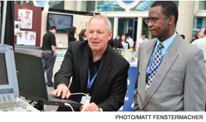
Anuj Dalal, MD, FHM, a hospitalist at Brigham and Women’s Hospital in Boston, says technology can range from global EHRs across a multiple-institutional health system to microblogs to practice-centered wikis that promote communication between physicians. But integrating those technologies into care delivery that improves outcomes hinges not on technical hurdles, but on physicians accepting their validity and utility, he says. Once clinicians start digitally codifying the informal communication tools they currently use, patient-specific communication can improve.
And while Dr. Dalal, who co-led a session on the use of wikis as a communication-management strategy, adds that despite the little research linking those technological tacks to patient outcomes, he believes they are related.
“We’re using our devices, our iPads, our iPhones, our Android devices, to send information to other providers, but that information may not be transparent to other people,” says Dr. Dalal, who heads an SHM technology subcommittee. “But if you have it on a blog, a patient-specific Facebook, or a patient-specific Twitter, than everyone who subscribes to that will have access to the whole discussion.”
Dr. Dalal cautions that the details of legal compliance are important to ensure private patient information is safely transmitted. He anticipates vendors will market products that will balance the need for that privacy with a wider dissemination of information to ensure all involved physicians know what they need to know. One example: He expects the internal-messaging components of EHRs to improve in the next few years.
“It wouldn’t surprise me if these companies start realizing we need to support patient-specific communication in informal dialogues using microblogs and mobile devices,” he adds. “If they don’t do that, they’re probably going to miss the boat.”
The power of technology, large and small, must be balanced with what hospitalist pioneer Robert Wachter, MD, MHM, calls “foundational knowledge.” Dr. Wachter, who counts chair-elect of the American Board of Internal Medicine (ABIM) among his myriad titles, says that hospitalists can’t be slaves to technology. He relates it to a well-known adage in the aviation industry in which planes have become so advanced that some pilots would be incapable of flying them should the technology fail. In one quip, it is said that two people will soon populate cockpits: a pilot and a dog. The pilot’s job is to feed the dog, while the dog’s job is to bite the pilot’s hand if he tries to touch the controls.

“As we move quickly, briskly, appropriately into this new world of computerization, if we aren’t constantly asking ourselves what have we lost in terms of our relationship with patients, we will really screw things up,” Dr. Wachter says. “This is not an argument to be Luddites, it’s not an argument to send back your meaningful use and get rid of the computers. We need them, but we have to be very thoughtful about how we maintain this balance.”
Patrick Conway, MD, MSc, FAAP, SFHM, a pediatric hospitalist and chief medical officer of the Centers for Medicare & Medicaid Services (CMS), says that to tie quality, safety, and technology together, government has to be willing to back therapies, then determine whether their efficacy deserves financial coverage.
One example Dr. Conway cites is the use of intravascular shunts. After backing their use initially for Medicare patients, it was determined that compared with medical management, the therapy had greater risks for death and stroke—and cost about 100 times more. Still, Dr. Conway says, determining how well the shunt works is exactly the process medicine should go through.
“When we have these new technologies that made a promise, we want to get them to market where they might help patients, but we also want to make sure evidence is developed to make sure they truly benefit patients,” he says, later adding, “I think this catalyst for health system transformation is important, and it’s teaching vision coupled with that execution.”
A well-planned blending of quality and technology can lead to improved patient safety, Dr. Arora says. But physicians and HM group leaders have to be proactive. Those with a clinical focus must seek out their IT counterparts and provide guidance on what information users need to access from electronic interfaces. Chief medical information officers and their staffs must seek feedback from providers before creating templates that don’t serve the clinical functions of end users.
Put simply: The fight to improve outcomes requires all parties to be properly armed. “You can choose a weapon,” she adds, “but you still have to choose it wisely and customize it to fit the needs of your organization.”
In a Venn diagram, patient handoffs would fall at the intersection of quality improvement (QI), patient safety, and technology.
Vineet Arora, MD, MPP, FACP, FHM, associate professor of medicine at the University of Chicago, wants hospitalists to understand that best practices in transitional care only work efficiently if they incorporate pieces from all three spheres. Hospitalists who focus too much on the outcome of an individual case might fail to utilize technology that could create a systemic protocol that would improve outcomes across multiple cases, she says. Similarly, hospitalists who leave notes in an electronic health record (EHR) can mistakenly believe that the next physician will interpret that information perfectly despite a lack of verbal and physical cues that guide verbal communication.
“People may fall into the fallacy that electronic health records actually equal good handoffs,” says Dr. Arora, a faculty member at HM12 last month in San Diego who led a breakout session that focused on the obstacles of handoffs. “The challenge is that handoffs actually are about a verbal exchange of informationa conversation and a dialogue, in conjunction with and supplemented with written communication. Technology certainly has the ability to revolutionize the way we review information and access information, but it will not change the way we speak about the information.”
The crossroads of patient safety, QI, and the use of technology from medical devices to iPhones was a major theme at SHM’s annual meeting, April 1-4 at the San Diego Convention Center. Hospitalist leaders agree that clinicians must focus on all three tenets to improve healthcare delivery, particularly in the face of generational healthcare reform. But they also acknowledge that finding a balance between best practices, budget and staffing constraints, and the rigors of daily practice is difficult.
Hospitalist Mark Lyons, MD, who works on residency programs and a patient-handoff initiative at Methodist Health System of Dallas, says that sessions on QI and patient safety provide new ideas from industry leaders who are in the trenches. To wit, he was impressed by the suggestions from Dr. Arora’s handoffs session as she described cases in which she was involved.
“They understand the limitations and the other things that go along with doing the actual act itself,” Dr. Lyons says. “They understand those things and take them into consideration. And you appreciate that.”
He and other hospitalists at HM12 agree that when leading practices are promoted at national forums, they take on the imprimatur of evidence-based procedures that physicians want to bring back to their respective HM groups. And QI suggestions promoted by physicians, not management, often tend to be more readily accepted.
“When you force change upon people, then it really leads to resistance,” Dr. Lyons says. “These are good opportunities to learn different strategies, to learn easier ways to present new ideas to people, to encourage them to do better, and to make it easier for them to do those things.”
Anuj Dalal, MD, FHM, a hospitalist at Brigham and Women’s Hospital in Boston, says technology can range from global EHRs across a multiple-institutional health system to microblogs to practice-centered wikis that promote communication between physicians. But integrating those technologies into care delivery that improves outcomes hinges not on technical hurdles, but on physicians accepting their validity and utility, he says. Once clinicians start digitally codifying the informal communication tools they currently use, patient-specific communication can improve.
And while Dr. Dalal, who co-led a session on the use of wikis as a communication-management strategy, adds that despite the little research linking those technological tacks to patient outcomes, he believes they are related.
“We’re using our devices, our iPads, our iPhones, our Android devices, to send information to other providers, but that information may not be transparent to other people,” says Dr. Dalal, who heads an SHM technology subcommittee. “But if you have it on a blog, a patient-specific Facebook, or a patient-specific Twitter, than everyone who subscribes to that will have access to the whole discussion.”
Dr. Dalal cautions that the details of legal compliance are important to ensure private patient information is safely transmitted. He anticipates vendors will market products that will balance the need for that privacy with a wider dissemination of information to ensure all involved physicians know what they need to know. One example: He expects the internal-messaging components of EHRs to improve in the next few years.
“It wouldn’t surprise me if these companies start realizing we need to support patient-specific communication in informal dialogues using microblogs and mobile devices,” he adds. “If they don’t do that, they’re probably going to miss the boat.”
The power of technology, large and small, must be balanced with what hospitalist pioneer Robert Wachter, MD, MHM, calls “foundational knowledge.” Dr. Wachter, who counts chair-elect of the American Board of Internal Medicine (ABIM) among his myriad titles, says that hospitalists can’t be slaves to technology. He relates it to a well-known adage in the aviation industry in which planes have become so advanced that some pilots would be incapable of flying them should the technology fail. In one quip, it is said that two people will soon populate cockpits: a pilot and a dog. The pilot’s job is to feed the dog, while the dog’s job is to bite the pilot’s hand if he tries to touch the controls.
“As we move quickly, briskly, appropriately into this new world of computerization, if we aren’t constantly asking ourselves what have we lost in terms of our relationship with patients, we will really screw things up,” Dr. Wachter says. “This is not an argument to be Luddites, it’s not an argument to send back your meaningful use and get rid of the computers. We need them, but we have to be very thoughtful about how we maintain this balance.”
Patrick Conway, MD, MSc, FAAP, SFHM, a pediatric hospitalist and chief medical officer of the Centers for Medicare & Medicaid Services (CMS), says that to tie quality, safety, and technology together, government has to be willing to back therapies, then determine whether their efficacy deserves financial coverage.
One example Dr. Conway cites is the use of intravascular shunts. After backing their use initially for Medicare patients, it was determined that compared with medical management, the therapy had greater risks for death and stroke—and cost about 100 times more. Still, Dr. Conway says, determining how well the shunt works is exactly the process medicine should go through.
“When we have these new technologies that made a promise, we want to get them to market where they might help patients, but we also want to make sure evidence is developed to make sure they truly benefit patients,” he says, later adding, “I think this catalyst for health system transformation is important, and it’s teaching vision coupled with that execution.”
A well-planned blending of quality and technology can lead to improved patient safety, Dr. Arora says. But physicians and HM group leaders have to be proactive. Those with a clinical focus must seek out their IT counterparts and provide guidance on what information users need to access from electronic interfaces. Chief medical information officers and their staffs must seek feedback from providers before creating templates that don’t serve the clinical functions of end users.
Put simply: The fight to improve outcomes requires all parties to be properly armed. “You can choose a weapon,” she adds, “but you still have to choose it wisely and customize it to fit the needs of your organization.”