User login
3-D images aid total body mole mapping
EDINBURGH – The advent of three-dimensional total body photography overcomes many, but not all, of the drawbacks of two-dimensional imaging as an aid to melanoma detection.
“There are challenges to using two-dimensional images, both in terms of acquiring them and reviewing them, but the simplest of which is a 5-mm lesion that’s perfectly perpendicular to the camera will not look like 5 mm if it’s at an angle to the camera, an inherent problem of consistency in looking at size over time on curved surfaces,” Dr. Allan Halpern said at the 15th World Congress on Cancers of the Skin.
Over the last several months, the dermatology service Dr. Halpern heads at Memorial Sloan-Kettering Cancer Center in New York has shifted to 3-D total body photography in patients at high risk of melanoma.
“The new system (Vectra WB360, Canfield Imaging Systems) uses 40 some odd cameras behind a series of panels, and in 3 milliseconds allows the clinician to image the entire patient in 3-D,” he said.
A touch-screen interface allows for additional close-up pictures of areas of particular interest and tags their location to the corresponding body map photographs. The onscreen 3-D images also can be used for consultation and patient education.
In a recent open-ended survey of 199 of his patients who had already undergone 2-D imaging, participants said 3-D mole mapping was faster, easier, less invasive, and required fewer poses, Dr. Halpern said.
Among their concerns, the admittedly motivated patient cohort wanted even better resolution and more images to catch moles in body folds or on the soles of their feet.
Dr. Halpern was quick to acknowledge that, like population-based melanoma screening, total body photography (TBP) has not been shown in a definitive trial to reduce mortality. In fact, clinical practice guidelines issued this year in Germany and Australia strongly recommend training and use of dermoscopy for examination of pigmented lesions, but note that the value of TBP in melanoma-risk patients remains ‘unproven.’
Research also has shown that uptake of TBP in clinical practice has remained relatively flat in the United States since 2000 (J. Am. Acad. Dermatol. 2010:62:794-803). Obstacles include expense, privacy, and security of stored images, and a lack of imaging standards.
The International Society for Digital Imaging of the Skin, however, is working with dermatology specialists, informatics experts, and imaging technology developers through its Melanoma Project to develop international imaging standards and a public-access archive of clinical and dermoscopic images of skin lesions, Dr. Halpern said.
“One of the encouraging things to me is that the CEOs of six of those imaging developers have already signed on. They recognize the importance of having international standards and interchangeability of images,” he said at the meeting sponsored by the Skin Cancer Foundation.
Dr. Halpern has consulted for and shared research grants with Canfield Scientific and consulted for Caliber I.D., Scibase, and DermTech.
AT WCCS 2014
More people die from thin melanomas than thick melanomas
EDINBURGH – Contrary to the perception that most melanoma deaths result from thick melanomas, long-term data from Australia show that more people die who initially present with thin melanomas.
Among 4,218 Australians who died from melanoma between 1990 and 2009, thin melanomas (1 mm or less) accounted for 19% of melanoma deaths overall, but increased steadily from 14% of deaths in 1990-1994 to 23% in 2005-2009.
During the most recent time period (2005-2009), more people died from thin melanomas (296 deaths, 23%) than from thick melanomas more than 4 mm in thickness (186 deaths, 14%) or from metastatic presentations (207 deaths, 16%).
The number of deaths in the intermediate thickness categories was also higher for lesions of thickness 1.01-2.0 mm (272 deaths, 21%) than for lesions of thickness 2.01-4.0 mm (267 deaths, 20%).
The patterns of mortality were essentially unchanged when the analyses were restricted to patients with only one primary melanoma, Dr. David Whiteman and Dr. Catherine Olsen of the QIMR Berghofer Medical Research Institute, Brisbane, Australia, reported at the 15th World Congress on Cancers of the Skin.
The melanoma incidence has been rising steadily around the world, mostly because of the greater numbers of thin lesions being diagnosed. The perception that most people who die from melanoma present initially with thick lesions is widespread, but the veracity of this proposition has never been tested because population-based data describing total mortality by the thickness of the primary tumor are scarce, according to the researchers. They used linked histology and death data from the Queensland Cancer Registry, where notification of melanoma has been compulsory since 1982, to calculate age-standardized mortality rates for each year for all melanomas, and by thickness of the first primary lesion. Overall, 67% of patients were male, 68% presented with a single primary lesion, and 68% of all melanomas were thin (1 mm or less).
Deaths from melanoma were most common among those who were in the seventh and eighth decades of life, male, or had a melanoma arising on the trunk. As expected, the intervals from diagnosis to death were significantly shorter for thicker tumors than thinner tumors, Dr. Whiteman and Dr. Olsen noted in the poster presentation.
The average annual rate of change in melanoma mortality increased significantly for men for thin lesions and those of intermediate thickness. Mortality rates from metastatic lesions, however, declined during the observation period.
“From a public health perspective, it can be argued that primary prevention activities aimed at reducing the occurrence of melanoma in the entire population should be accorded the highest priority,” the authors concluded at the meeting, sponsored by the Skin Cancer Foundation.
AT WCCS 2014
Key clinical point: Thin lesions are associated with substantial mortality, so primary prevention of melanoma should remain the principal strategy.
Major finding: From 2005 to 2009, 23% of residents of Queensland, Australia, died from thin melanomas versus 14% with thick melanomas.
Data source: State cancer registry analysis of 4,218 melanoma deaths in Queensland.
Disclosures: The researchers had no relevant financial conflicts to disclose.
Moles quadruple risk for melanoma
EDINBURGH – Patients with moles had more than four times the risk of developing melanoma, compared with those without moles in a large record-linkage study.
The overall rate ratio for melanoma, based on person-years at risk, was 4.68 among patients with moles recorded in their medical record (95% confidence interval, 4.39-4.98), Dr. Eugene Ong reported at the 15th World Congress on Cancers of the Skin.
Rate ratios were also significantly higher for individuals with moles of both sexes and in all age groups, including those aged younger than 25 years (RR, 3.79), 25-59 years (RR, 5.02), and at least 60 years (RR, 4.68).
Prior research has shown that high numbers of melanocytic or dysplastic nevi are strong risk factors for the development of melanoma. The investigators sought to further characterize the risk of melanoma in persons with melanocytic nevus (MN) using linked hospital and mortality records covering the entire population of England from 1999 to 2011.
The analysis included 271,656 patients with a hospital or day-case record of moles and a control cohort of 10,130,417 persons with no moles recorded. Anyone diagnosed with melanoma within 1 year of study entry was excluded.
Patients with a record of moles had a significantly higher risk of developing melanoma both around the site of the mole and elsewhere on their body, and therefore may benefit from increased surveillance, said Dr. Ong of the University of Oxford, England. For patients with a mole on the trunk, the rate ratio for a melanoma on the trunk was 8.99 (95% CI, 7.69-10.46) and 5.66 for a melanoma elsewhere (95% CI, 4.97-6.42).
The investigators were unable to distinguish between different types of moles or to determine the number of moles in each patient. Further, a mole or moles were the principal reason for hospital contact for 91% of patients, so it’s likely they presented with unusual appearing moles in order for them to warrant recording, Dr. Ong acknowledged.
“So while this study does not suggest that everyone with a single mole is far more likely to develop melanoma, it does illustrate the link between moles and skin cancer. This is why it is vital people check their moles regularly and report any changes to their doctor,” he said in a statement released during the meeting, sponsored in part by the Skin Cancer Foundation.
AT THE WCCS 2014
Key clinical point: Individuals with moles are at significantly increased risk for developing melanoma in the same body region as the mole, and in other regions, and could benefit from increased surveillance.
Major finding: The rate ratio for melanoma, based on person-years at risk, was 4.68 times among patients with moles than those without moles (95% CI 4.39-4.98).
Data source: A record-linkage study in 271,656 patients with a diagnosis of moles and 10,130,417 controls without moles.
Disclosures: Dr. Ong had no financial conflicts to disclose.
VIDEO: How red hair and freckles might raise your skin cancer risk
EDINBURGH – Variants in the pigment-associated MC1R gene have been implicated in an increased risk for melanoma and nonmelanoma skin cancers, although the extent of that risk has been inconsistent across studies, according to Dr. Eugene Healy of the University of Southampton (England). In an interview at the 15th World Congress on Cancers of the Skin sponsored by the Skin Cancer Foundation, Dr. Healy discussed how the MC1R gene variants might impact skin cancer risk and the challenges of pinning down genetic data into practical applications for patients.
The video associated with this article is no longer available on this site. Please view all of our videos on the MDedge YouTube channel
EXPERT ANALYSIS FROM WCCS 2014
Veterans at increased risk of skin cancer
EDINBURGH – U.S. military personnel deployed overseas during the past decade have increased risk factors for skin cancer, a new survey suggests.
Researchers reported that 77% of veterans were exposed to 4 or more hours of bright sun during a typical day, but only 27% had ready access to sunscreen while working. Another 32% had no access to sunscreen.
A full 62% of veterans reported getting sunburned while deployed, with 42% having two or more sunburns, 16% four or more sunburns, and 15% at least one blistering sunburn.
Twenty-nine percent of veterans noticed changes in size, shape, or color of moles since deployment, but only 4% reported receiving a skin exam by a physician since returning home.
“This study demonstrates room for improvement for skin cancer prevention in veterans’ populations,” dermatologist Jennifer Powers of Vanderbilt University, Nashville, Tenn., reported at the 15th World Congress on Cancers of the Skin.
The 30-question survey was given to U.S. military workers engaged in Operation Enduring Freedom, Operation Iraqi Freedom, or Operation New Dawn presenting at the Nashville postdeployment clinic, Tennessee Valley Healthcare System, U.S. Department of Veterans. The combat missions occurred at a more equatorial latitude than the mean center of the United States population, increasing the potential for ultraviolet exposure and the development of skin cancer, Dr. Powers noted.
Of the 197 respondents, 64% identified as white, 22% as African American, and 8% as Hispanic. Almost half (48%) were fair-skinned.
Only 22% of veterans reported being made very aware of the risks of skin cancer, compared with 41% who reported not being made aware at all.
Dr. Powers highlighted a prior retrospective review of tumor registries showing that the melanoma incidence increased from 1990-1994 to 2000-2004 among white active-duty military personnel and that rates were significantly higher among those 45 years or older (Cancer Epidemiol. Biomarkers Prev. 2011;20:318-23).
The congress was sponsored by the Skin Cancer Foundation.
AT WCCS 2014
Key clinical point: Skin cancer prevention can be improved among U.S. military personnel.
Major finding: Seventy-seven percent of veterans were exposed to 4 or more hours of bright sun during a typical day, but 27% had ready access to sunscreen.
Data source: Survey of 197 recent veterans.
Disclosures: Dr. Powers reported no conflicts of interest. The study was supported by the Nashville Tennessee Valley Healthcare System, which employs some of the coauthors.
Photodynamic therapy clears thin AKs better than cryotherapy
Thin actinic keratoses on the face or scalp were 14% more likely to clear completely in patients treated with photodynamic therapy, compared with cryotherapy, in a meta-analysis of four studies including 641 patients.
Complete clearance 3 months after treatment was significantly more likely in the 2,170 actinic keratoses treated by photodynamic therapy (PDT), compared with 2,174 actinic keratoses treated by cryotherapy, Dr. Gayatri Patel and her associates reported.
The data came from randomized trials with 10 or more participants in which the PDT used topical aminolevulinic acid hydrochloride or methyl aminolevulinate hydrochloride, the most widely available PDT stabilizers in North America and Europe. Methyl aminolevulinate recently was withdrawn from the U.S. market but remains common in Europe, noted Dr. Patel of the University of California, Davis, and her associates.
The study results were published online in JAMA Dermatology (2014 Aug. 27 [doi:10.1001/jamadermatol.2014.1253]).
The results suggested that PDT works better on thinner actinic keratoses. Grade 1 (thin) actinic keratoses on the face or scalp were 86% more likely to clear by 12 weeks after PDT, compared with cryotherapy, the investigators reported.
Only one of the fours studies found higher efficacy rates for cryotherapy, compared with PDT, and more than 60% of lesions in that study were grade 2 (moderately thick, easily felt) or grade 3 (very thick and/or obvious) actinic keratoses. The other three studies in the meta-analysis excluded thicker lesions or favored thinner ones, the researchers noted.
They excluded from the meta-analysis two other studies that compared PDT with cryotherapy for actinic keratoses because of incompatible follow-up times. They reviewed 13 studies in all, including studies involving treatment of actinic keratosis with imiquimod, fluorouracil, or carbon dioxide laser, but could not meta-analyze data on these other treatments because of different outcome measures and follow-up times or lack of a comparator.
Photosensitivity, pain, erythema, and pruritus were common after PDT. Cryotherapy induced pain and pruritus, but at lower rates than did PDT. Hypopigmentation occurred in 33% of patients after cryotherapy and in 9% after PDT in one study.
Satisfaction ratings by patients and unblinded investigators tended to favor PDT over cryotherapy, perhaps because PDT may produce ancillary cosmetic improvements when treating actinic keratosis, Dr. Patel and her associates speculated.
The findings were limited by the poor quality of the studies, which were lacking double-blind design and description of randomization methods, but no sources of bias were evident, and the large number of patients and relatively similar treatment locations were strengths of the analysis, they said.
Dr. Patel reported having no financial disclosures.
On Twitter @sherryboschert
Several limitations associated with photodynamic therapy (PDT) make cryotherapy the first-line treatment choice for most practicing dermatologists, Dr. Harvey Lui commented in an article that accompanied Dr. Patel’s report.
Dr. Patel’s meta-analysis found a 14% better chance of complete clearance of actinic keratosis lesions, compared with cryotherapy, but the data are not clear enough to claim better cosmesis or patient acceptance, Dr. Lui said (JAMA Dermatol. 2014 Aug. 27 [doi:10.1001/jamadermatol.2014.1869]).

|
|
PDT costs more in time and equipment than cryotherapy. PDT may seem simple, but achieving optimal results can require longer drug incubation times and/or light-dose fractionation. Local pain is a bigger problem with PDT that requires anticipation and management by clinicians, he said. Cryotherapy, on the other hand, requires relatively brief outpatient visits.
The future of PDT for actinic keratosis may lie in further development of a currently off-label treatment – exposure to ambient outdoor light after application of topical aminolevulinic acid, Dr. Lui suggested: "Perhaps the most tantalizing irony of daylight PDT is the specter of treating a solar-induced neoplasm with sunlight itself."
Dr. Lui is head of the department of dermatology and skin science at the University of British Columbia in Vancouver. He disclosed financial associations with Galderma, LEO Pharma, Janssen, Novartis, Valeant Pharmaceuticals, RepliCel Life Sciences, Lumen Health Technologies, and Verisante Technology.
Several limitations associated with photodynamic therapy (PDT) make cryotherapy the first-line treatment choice for most practicing dermatologists, Dr. Harvey Lui commented in an article that accompanied Dr. Patel’s report.
Dr. Patel’s meta-analysis found a 14% better chance of complete clearance of actinic keratosis lesions, compared with cryotherapy, but the data are not clear enough to claim better cosmesis or patient acceptance, Dr. Lui said (JAMA Dermatol. 2014 Aug. 27 [doi:10.1001/jamadermatol.2014.1869]).

|
|
PDT costs more in time and equipment than cryotherapy. PDT may seem simple, but achieving optimal results can require longer drug incubation times and/or light-dose fractionation. Local pain is a bigger problem with PDT that requires anticipation and management by clinicians, he said. Cryotherapy, on the other hand, requires relatively brief outpatient visits.
The future of PDT for actinic keratosis may lie in further development of a currently off-label treatment – exposure to ambient outdoor light after application of topical aminolevulinic acid, Dr. Lui suggested: "Perhaps the most tantalizing irony of daylight PDT is the specter of treating a solar-induced neoplasm with sunlight itself."
Dr. Lui is head of the department of dermatology and skin science at the University of British Columbia in Vancouver. He disclosed financial associations with Galderma, LEO Pharma, Janssen, Novartis, Valeant Pharmaceuticals, RepliCel Life Sciences, Lumen Health Technologies, and Verisante Technology.
Several limitations associated with photodynamic therapy (PDT) make cryotherapy the first-line treatment choice for most practicing dermatologists, Dr. Harvey Lui commented in an article that accompanied Dr. Patel’s report.
Dr. Patel’s meta-analysis found a 14% better chance of complete clearance of actinic keratosis lesions, compared with cryotherapy, but the data are not clear enough to claim better cosmesis or patient acceptance, Dr. Lui said (JAMA Dermatol. 2014 Aug. 27 [doi:10.1001/jamadermatol.2014.1869]).

|
|
PDT costs more in time and equipment than cryotherapy. PDT may seem simple, but achieving optimal results can require longer drug incubation times and/or light-dose fractionation. Local pain is a bigger problem with PDT that requires anticipation and management by clinicians, he said. Cryotherapy, on the other hand, requires relatively brief outpatient visits.
The future of PDT for actinic keratosis may lie in further development of a currently off-label treatment – exposure to ambient outdoor light after application of topical aminolevulinic acid, Dr. Lui suggested: "Perhaps the most tantalizing irony of daylight PDT is the specter of treating a solar-induced neoplasm with sunlight itself."
Dr. Lui is head of the department of dermatology and skin science at the University of British Columbia in Vancouver. He disclosed financial associations with Galderma, LEO Pharma, Janssen, Novartis, Valeant Pharmaceuticals, RepliCel Life Sciences, Lumen Health Technologies, and Verisante Technology.
Thin actinic keratoses on the face or scalp were 14% more likely to clear completely in patients treated with photodynamic therapy, compared with cryotherapy, in a meta-analysis of four studies including 641 patients.
Complete clearance 3 months after treatment was significantly more likely in the 2,170 actinic keratoses treated by photodynamic therapy (PDT), compared with 2,174 actinic keratoses treated by cryotherapy, Dr. Gayatri Patel and her associates reported.
The data came from randomized trials with 10 or more participants in which the PDT used topical aminolevulinic acid hydrochloride or methyl aminolevulinate hydrochloride, the most widely available PDT stabilizers in North America and Europe. Methyl aminolevulinate recently was withdrawn from the U.S. market but remains common in Europe, noted Dr. Patel of the University of California, Davis, and her associates.
The study results were published online in JAMA Dermatology (2014 Aug. 27 [doi:10.1001/jamadermatol.2014.1253]).
The results suggested that PDT works better on thinner actinic keratoses. Grade 1 (thin) actinic keratoses on the face or scalp were 86% more likely to clear by 12 weeks after PDT, compared with cryotherapy, the investigators reported.
Only one of the fours studies found higher efficacy rates for cryotherapy, compared with PDT, and more than 60% of lesions in that study were grade 2 (moderately thick, easily felt) or grade 3 (very thick and/or obvious) actinic keratoses. The other three studies in the meta-analysis excluded thicker lesions or favored thinner ones, the researchers noted.
They excluded from the meta-analysis two other studies that compared PDT with cryotherapy for actinic keratoses because of incompatible follow-up times. They reviewed 13 studies in all, including studies involving treatment of actinic keratosis with imiquimod, fluorouracil, or carbon dioxide laser, but could not meta-analyze data on these other treatments because of different outcome measures and follow-up times or lack of a comparator.
Photosensitivity, pain, erythema, and pruritus were common after PDT. Cryotherapy induced pain and pruritus, but at lower rates than did PDT. Hypopigmentation occurred in 33% of patients after cryotherapy and in 9% after PDT in one study.
Satisfaction ratings by patients and unblinded investigators tended to favor PDT over cryotherapy, perhaps because PDT may produce ancillary cosmetic improvements when treating actinic keratosis, Dr. Patel and her associates speculated.
The findings were limited by the poor quality of the studies, which were lacking double-blind design and description of randomization methods, but no sources of bias were evident, and the large number of patients and relatively similar treatment locations were strengths of the analysis, they said.
Dr. Patel reported having no financial disclosures.
On Twitter @sherryboschert
FROM JAMA DERMATOLOGY
Key clinical point: Clearance of thin actinic keratosis lesions on the face or head is more likely with photodynamic therapy vs. cryotherapy, but the impact of either treatment on reducing the incidence of squamous cell carcinomas remains unknown.
Major finding: Clearance was 14% more likely at 3 months after PDT, compared with cryotherapy.
Data source: Meta-analysis of four studies including 641 patients with 2,170 actinic keratosis lesions treated by PDT and 2,174 treated by cryotherapy.
Disclosures: Dr. Patel reported having no financial disclosures.
Field therapy preferred when treating actinic keratoses
VANCOUVER, B.C. – In the clinical opinion of Dr. Mariusz Sapijaszko, treating actinic keratosis without field therapy creates a disadvantage "because this is not an individual lesion disease," he maintained at the annual meeting of the Pacific Dermatologic Association.
Actinic keratosis "is a field concept disease. I tell patients ‘the sun has not been shining only on your left temple. It’s been shining all over your face and scalp, neck, and arms. ... It’s time to start looking after your skin with sun protection and lesion-directed field therapies.’"
An estimated 11% of all dermatologic visits in the United States are for actinic keratosis (AK) and "we worry about it because the natural course of AK lesions is unpredictable," said Dr. Sapijaszko of the Western Canada Dermatology Institute, Edmonton, Alta. It’s not easy to predict which lesions will progress to in situ or invasive squamous cell carcinomas (SCCs).
An estimated 40%-80% of cutaneous SCCs arise from, or near, an AK lesion, which supports the concept of field UV damage. AK lesions may persist, regress, or progress, depending on the patient’s immune status. Some lesions that regress will recur, from 32% within 1 year to 92% within 5 years. Progression can lead to hypertrophic AKs, in situ SCC, or invasive SCC. It can be difficult to distinguish AKs and early forms of SCC or even other nonmelanoma skin cancers, "so it’s important to treat all AKs," Dr. Sapijaszko said. Lesions that can progress to SCC include those that are hyperkeratotic, painful, have atypical features such as broader or deeper presentations, as well as those difficult to clear with standard therapies and those that occur in immunocompromised patients.
Locally destructive, mechanical ways to treat AKs include liquid nitrogen cryosurgery, electrodessication and curettage, and excision. "All of these treatments are highly operator dependent, because clearly if you use liquid nitrogen cryotherapy enough you will destroy that lesion but you will not destroy the surrounding DNA damage that has been present," he said.
Field-directed therapies, however, provide an opportunity for a more complete treatment effect. Options include 5-FU (5-fluorouracil), imiquimod, ingenol mebutate, and photodynamic therapy as well as chemical peels and laser resurfacing. Chemical peels and laser resurfacing "have less robust data, but they’re operator dependent, because you can do laser resurfacing with 100 microns or 300 microns," Dr. Sapijaszko said.
"That can depend on the technique you use, and the laser you use, and the patient in front of you." Some of the field treatment options are easier to apply than others. For example, 5-FU is applied twice daily, while imiquimod is applied twice weekly; yet all boast complete clearance rates in the 40%-50% range. "Side effects are also similar between these agents," he said. "Pain is not a big issue except with 5-FU; some patients experience a significant burning sensation."
The newest approved field therapy option, ingenol mebutate, has a dual mechanism of action: it causes cell death within 24 hours and it has been shown to reduce the number of UV-induced mutated p53 patches in mice. "This is important because we’re not just treating the lesions that we see, we want to treat the molecular changes that lead to the actual problem," Dr. Sapijaszko said. "Having decreased mutations is a huge advantage. Direct cell death leads to secondary inflammation. The immune response is characterized by cytokine release and activation of endothelial cells, leading to infiltration of lymphocytes and neutrophils, which contributes to clearance of tumor cells."
Before ingenol mebutate came on the market, investigators randomized patients with AKs to one of three treatment groups: imiquimod 5% cream, 5-FU 5% ointment, or cryotherapy (Br. J. Dermatol. 2007; 157 [suppl. 2]:34-40). Compared with their counterparts, patients in the imiquimod group fared significantly better in terms of sustained clearance of cleared lesions (73% vs. 54% in the 5-FU group, vs. 28% in the cryosurgery group (P less than .01).
"I wish we had comparative data to ingenol mebutate, but to me, of these three modalities, imiquimod stands out as the favorite," Dr. Sapijaszko said.
He went on to note that combining the available mechanical and field treatments for AK simultaneously or sequentially can lead to optimal outcomes. "Combination therapy, in particular cryotherapy, has been successfully used with a variety of topicals and has been shown to be highly advantageous, compared with placebo or to some of these agents alone," he said. "In addition, cryotherapy can be used with PDT, chemical peels, and laser resurfacing. Almost nobody in my practice gets one treatment, unless it’s a single individual lesion. Everybody gets recommendations on appropriate sun protection – just being sun smart."
Dr. Sapijaszko disclosed that he has received honoraria from and is an advisor to Galderma, Leo Pharma, and Valeant.
On Twitter @dougbrunk
EXPERT ANALYSIS AT THE PDA ANNUAL MEETING
Surgeon General: Tanning, indoor tanning must stop
The U.S. Surgeon General’s office is calling on Americans to do more to help prevent skin cancer, saying that it is a growing public health problem.
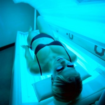
The five-point call to action singled out ultraviolet light exposure – from indoor and outdoor tanning – as a major culprit in the growing incidence of all cancers, and melanoma, in particular.
"Tanned skin is damaged skin," Acting Surgeon General, Boris D. Lushniak, a dermatologist, said at a briefing to release the report.
"When people tan or get sunburned, they increase their risk of getting skin cancer later in life," he said, adding that the nation needs to change its cultural acceptance of tanning as a sign of health.
The report noted that about 5 million Americans are treated for skin cancer each year, at a cost of $8 billion. Melanoma is of the greatest concern; its incidence has tripled over the past 3 decades, with 63,000 new cases per year. Some 9,000 Americans die from melanoma each year, and death rates are on the rise in men, Dr. Howard Koh, assistant secretary for health at the U.S. Department of Health & Human Services, said at the briefing.
Both Dr. Koh and Dr. Lushniak singled out indoor tanning as particularly dangerous. The Surgeon General’s report cited research showing that indoor tanning may be associated with more than 400,000 cases of skin cancer, including 6,000 melanomas, per year.
Forty-four states and the District of Columbia have some type of indoor tanning laws or regulations. Eleven states – California, Delaware, Hawaii, Illinois, Louisiana, Minnesota, Nevada, Oregon, Texas, Vermont and Washington – prohibit indoor tanning by individuals under age 18. But indoor tanning remains popular: One in three white women aged 16-25 years engages in indoor tanning each year, according to the report.
In May, the Food and Drug Administration issued stricter rules for sunlamps used with indoor tanning, requiring a boxed warning that they should not be used on anyone younger than 18 years. The agency also required that more information on risks and contraindications be provided to sunlamp users, FDA Commissioner Margaret Hamburg noted in a statement on the Surgeon General’s report.
The five main points in the Surgeon General’s call to action are:
• Increase opportunities for sun protection in outdoor settings, such as increasing shade in recreational areas, and providing more protection for school children and outdoor workers.
• Give individuals information to make informed, healthy choices about UV exposure, including providing materials in workplaces and schools, and partnering with physicians and health systems.
• Promote policies that advance the goal of preventing skin cancer, such as encouraging electronic reporting of reportable skin cancers.
• Reduce harms from indoor tanning by enforcing existing laws and considering additional restrictions.
• Strengthen research, surveillance, monitoring, and evaluation.
The American Academy of Dermatology Association applauded the report.
"The American public needs to be aware that the dangers of ultraviolet radiation exposure are real," Dr. Brett M. Coldiron, president of the AADA, said in a statement. "The AADA is particularly pleased that the HHS and the Office of the Surgeon General have highlighted methods for the public to prevent skin cancer in this white paper, that include seeking shade, wearing protective clothing, applying sunscreen, and avoiding dangerous indoor tanning devices."
John Seffrin, Ph.D., chief executive officer of the American Cancer Society Cancer Action Network, noted that this is the first time there has been a national action plan regarding skin cancer.
"By bringing national attention to this growing public health crisis, the Surgeon General is calling on all of us to reinvigorate the fight against skin cancer," he said. "The Surgeon General’s Call to Action outlines achievable goals and strategies to support more Americans in making healthy choices about protecting their skin," Dr. Seffrin said.
On Twitter @aliciaault
Adding ingenol mebutate after cryosurgery enhances clearance of actinic keratoses
Complete clearance occurred at 12 months in 31% of patients who received field treatment with ingenol mebutate gel after cryotherapy for actinic keratoses, compared with 19% of placebo patients in a phase III trial.
The findings were published in the June issue of the Journal of Drugs in Dermatology.
Recurrence rates for AKs treated with cryotherapy alone are high, possibly because the treatment is lesion-directed and fails to address field cancerization, wrote Dr. Brian Berman of the University of Miami.
The FIELD study 1, a phase III, multicenter, randomized trial, showed that ingenol mebutate gel was safe and effective as a follow-up to cryosurgery for AKs. Short-term efficacy data (11 weeks) have been previously reported.
In this study, the patients were followed for 12 months (J. Drugs Dermatol. 2014;13:741-7).
"In phase III clinical trials, 2 or 3 days of field-directed therapy with ingenol mebutate demonstrated high and sustained clearance of AKs on the head and body, and was well tolerated," noted Dr. Berman and his coauthors.
The percentage reduction in the number of AKs was significantly higher at 12 months in the treatment group compared with the placebo group (68% vs. 54%), and significantly fewer patients in the treatment group developed new lesions in the treatment area compared with the placebo group (39% vs. 52%). In addition, the probability of sustained clearance of AKs at 12 months was 55% in the treatment group vs. 40% in the placebo group.
The study included 329 adults aged 18 years and older with 4-8 clinically typical, visible, and discrete AKs within a 25-cm2 treatment area on the face or scalp. The patients underwent cryosurgery, and 3 weeks later underwent field therapy with 0.015% ingenol mebutate gel or vehicle gel once a day for 3 consecutive days. Patients with suspected basal cell carcinoma or squamous cell carcinoma were excluded.
The findings were limited by the possible unblinding of treatment because of localized skin reactions, the researchers noted. However, few drug-related adverse events and no drug-related serious adverse events were reported, they said.
Dr. Berman has served as a consultant, received honoraria, and served on the speakers bureau for Leo Pharma, which funded the study.
FROM THE JOURNAL OF DRUGS IN DERMATOLOGY
Key clinical point: The findings support the safety and effectiveness of adding field therapy to cryosurgery when treating actinic keratosis lesions.
Major finding: Complete clearance rates at 12 months were significantly higher in patients treated with ingenol mebutate gel vs. a vehicle (31% vs. 19%).
Data source: A phase III, multicenter, randomized trial of adults with clinically typical AKs.
Disclosures: Dr. Berman has served as a consultant, received honoraria, and served on the speakers bureau for Leo Pharma, which funded the study.
What's working now for actinic keratoses
WAIKOLOA, HAWAII – Half of patients whose face or scalp actinic keratoses clear completely in response to 3 consecutive days of topical field therapy with ingenol mebutate gel 0.015% will remain clear 12 months later with no further treatment.
That finding from a multicenter prospective study is just one of several recent developments of note in the treatment of actinic keratoses. Other recent studies have addressed the effectiveness of fractional laser therapy and cryotherapy followed by ingenol mebutate, and there have been new developments in phototherapy, Dr. James E. Sligh said in a review of actinic keratosis research at the Hawaii Dermatology Seminar sponsored by the Global Academy for Medical Education/Skin Disease Education Foundation.
• Long-term follow-up of ingenol mebutate therapy. In a multicenter study, dermatologists followed 100 patients with complete clearance of AKs on the face or scalp in response to 3 consecutive days of ingenol mebutate gel (Picato) and 71 patients whose AKs on the trunk or limbs cleared after 2 consecutive days of therapy. The sustained lesion reduction rate, compared with pretreatment baseline was 87.2% for the face and scalp lesions and 86.8% for AKs on the trunk or extremities.
The median time to recurrence was 365 days for AKs on the face and scalp, and shorter at 274 days for lesions on the trunk or extremities, which notably had received 1 less day of treatment. No safety concerns arose during follow-up (JAMA Dermatol. 2013;149:666-70).
• Sequential cryosurgery followed by ingenol mebutate. In a randomized, double-blind, multicenter phase-III trial, 329 patients were randomized to field therapy of AKs on the face or scalp with ingenol mebutate or vehicle 3 weeks after cryosurgery. The complete clearance rate in the treated area at week 11 was significantly higher in the sequential therapy group: 60.5%, compared with 49.4% with cryosurgery alone (J. Drugs Dermatol. 2014;13:154-60).
"I think this is the first in what will be a number of studies where you start to see assessment of combined therapies. But I think the target to look at is not so much what happens at 11 weeks, as in this study, but what happens 1 or 2 years down the road. If you can maintain complete clearance for that long I think it’s worth the extra effort to combine treatment cycles at the beginning," commented Dr. Sligh, chief of the division of dermatology at the University of Arizona, Tucson.
• Laser therapy. Investigators at the Laser and Skin Surgery Center of New York investigated treatment of facial AKs using a fractionated 1927-nm nonablative thulium laser. Twenty-four patients received up to four fractional resurfacing treatments in this prospective trial. At 6 months follow-up, the AK lesion count was down 87%, compared with baseline. Patient satisfaction was high – all participants reported marked or noticeable improvement in overall photodamage – and treatment was well tolerated (J. Am. Acad. Dermatol. 2013;68:98-102).
"If you look at the reduction in AK counts, it stands up very nicely to some of our medical therapies in terms of overall clearance at 6 months, with an 87% reduction – provided you have this special thulium laser in your office," Dr. Sligh observed.
• Photodynamic therapy. What’s new in photodynamic therapy for AKs is not currently available in the United States: methyl aminolevulinate cream 16.8% (Metvixia) for use with red light illumination. This photosensitizer does remain available, however, in Canada and Mexico, according to Dr. Sligh. And aminolevulinic acid (Levulan) is still available as an approved photosensitizer for use with blue light.
"There are many people I know who will activate the Levulan with either a red or blue light source," he said.
Dr. Sligh is on the advisory board for and a consultant to Genentech and has received research grants from DermSpectra and Scibase.
The SDEF and this news organization are owned by the same parent company.
EXPERT ANALYSIS FROM THE SDEF HAWAII DERMATOLOGY SEMINAR


