User login
FDA issues draft guidance on MRD

The U.S. Food and Drug Administration (FDA) has issued a draft guidance on the use of minimal residual disease (MRD) assessment in trials of patients with hematologic malignancies.
The FDA said it developed this guidance to assist sponsors who are planning to use MRD as a biomarker in clinical trials conducted under an investigational new drug application or to support FDA approval of products intended to treat hematologic malignancies.
“As a result of important workshops where we’ve heard from stakeholders and an analysis of marketing applications showing inconsistent quality of MRD data, the FDA identified a need to provide sponsors with guidance on the use of MRD as a biomarker in regulatory submissions,” said FDA Commissioner Scott Gottlieb, MD.
The guidance explains how MRD might be used in clinical trials, highlights considerations for MRD assessment that are specific to certain hematologic malignancies, and lists requirements for regulatory submissions that utilize MRD.
The full document, “Hematologic Malignancies: Regulatory Considerations for Use of Minimal Residual Disease in Development of Drug and Biological Products for Treatment,” is available for download from the FDA website.
How MRD can be used
The guidance notes that MRD could potentially be used as a biomarker in clinical trials, specifically, as a diagnostic, prognostic, predictive, efficacy-response, or monitoring biomarker.
MRD could also be used as a surrogate endpoint, and there are two mechanisms for obtaining FDA feedback on the use of a novel surrogate endpoint to support approval of a product:
- The drug development tool qualification process
- Discussions with the specific Center for Drug Evaluation and Research or Center for Biologics Evaluation and Research review division.
Furthermore, a sponsor can use MRD “to select patients at high risk or to enrich the trial population,” according to the guidance.
Disease specifics
The guidance also details specific considerations for MRD assessment in individual hematologic malignancies. For example:
- In acute lymphoblastic leukemia, a patient with an MRD level of 0.1% or more in first or second complete remission has a high risk of relapse.
- In trials of acute myeloid leukemia, the sponsor should provide data showing that the marker selected to assess MRD “reflects the leukemia and not underlying clonal hematopoiesis.”
- Patients with low-risk acute promyelocytic leukemia who achieve MRD negativity after arsenic/tretinoin-based therapy are generally considered cured.
- In chronic lymphocytic leukemia, MRD can be assessed in the peripheral blood or bone marrow, but the sample source should remain the same throughout a trial.
- In chronic myeloid leukemia, MRD can be used to select and monitor patients who are eligible to discontinue treatment with tyrosine kinase inhibitors.
- In multiple myeloma, imaging techniques may be combined with MRD assessment of the bone marrow to assess patient response to treatment.
Types of technology
The guidance lists the four general technologies used for MRD assessment in hematologic malignancies:
- Multiparametric flow cytometry
- Next-generation sequencing
- Quantitative reverse transcription polymerase chain reaction of specific gene fusions
- Allele-specific oligonucleotide polymerase chain reaction.
The FDA said it does not have a preference as to which technology is used in a trial. However, the sponsor must pre-specify the technology used and should utilize the same technology throughout a trial.
The FDA also said it “does not foresee the need for co-development of an MRD assay with a drug product.” However, the assay must be analytically valid for results important to the trial, and MRD assessment must be a clinically valid biomarker in the context in which it’s used.
If the MRD assay used is not FDA-cleared or -approved, additional information about the assay must be provided to the FDA.
Virus-specific T-cell infusion may resolve progressive multifocal leukoencephalopathy
, according to investigators from the University of Texas MD Anderson Cancer Center, Houston.
The infusion cleared JC virus from the cerebrospinal fluid (CSF) of two patients and reduced viral load in the third, reported lead author Muharrem Muftuoglu, MD, of MD Anderson’s department of stem cell transplantation and cellular therapy and colleagues. One of the patients completely recovered and returned to work.
“Several approaches for the treatment of PML, including the use of antiviral medications and mirtazapine, have been tested, with poor results,” the investigators wrote in the New England Journal of Medicine. Although virus-specific T-cell infusion is a novel approach to treating PML, this method has been used for other conditions.
“Several groups, including ours, have successfully used viral-specific T cells to treat BK virus infection after stem-cell transplantation,” the investigators wrote. “Because BK virus and JC virus are genetically similar to one another and share a number of immunogenic proteins with a substantial degree of sequence homology ... we hypothesized that T cells developed against BK virus may also be effective against JC virus infection.”
This hypothesis proved accurate. The investigators infused three PML patients with “cryopreserved, third-party–produced, viral-specific T cells that had been designed for the treatment of patients with BK virus infection after stem-cell transplantation.” Each patient presented with a different condition and PML-precipitating therapy. The first patient was a 32-year-old woman with high-risk acute myeloid leukemia who had received a cord-blood transplantation, the second a 73-year-old woman with JAK2-positive myeloproliferative neoplasia on ruxolitinib (Jakafi) therapy, and the third a 35-year-old man with HIV who had received highly active antiretroviral therapy.
T-cell infusions cleared JC virus from the CSF of the woman with leukemia (three infusions) and the man with HIV (four infusions). These patients recovered to different degrees: The woman had full resolution of symptoms, while the man had slurred speech and walked with a cane. Treatment reduced JC viral load in the elderly woman with myeloproliferative neoplasia (two infusions), but she did not clear the virus and died about 8 months later.
No adverse events occurred, but two patients developed immune reconstitution inflammatory syndrome. This was likely caused by the T-cell infusion, since absolute T-cell counts remained steady and white matter enhancement was detected on MRI within 4 weeks of treatment. Still, the investigators were optimistic about future potential.
“Third-party–produced, ‘off-the-shelf,’ partially HLA-matched, BK virus–specific T cells may serve as therapy for PML,” the investigators concluded. “Further study in a larger group of patients is required to determine the success rate, durability, and longer-term adverse events associated with this treatment.”
The study was funded by the MD Anderson Cancer Center Moon Shots Program and the National Institutes of Health.
SOURCE: Muftuoglu M et al. N Engl J Med. 2018 Oct 11;379:1443-51
This article was updated 3/22/19.
FROM NEW ENGLAND JOURNAL OF MEDICINE
Key clinical point: Infusion of allogeneic BK virus-specific T cells may be an effective treatment for patients with PML.
Major finding: Two of three patients cleared JC virus from cerebrospinal fluid after infusion.
Study details: A case series involving three patients with PML.
Disclosures: The study was funded by the MD Anderson Cancer Center Moon Shots Program and the National Institutes of Health.
Source: Muftuoglu M et al. N Engl J Med. 2018 Oct 11;379:1443-51.
Genomic profiling predicts outcomes in patients with MPN
Genomic characteristics of patients with myeloproliferative neoplasms (MPN) can predict clinical outcomes, a recent study found.

Eight genomic subgroups of MPN were recognized, each with distinct clinical features, including event-free survival, risk of leukemic transformation, and blood counts, according to Jacob Grinfeld, MD, of the Wellcome-MRC Cambridge (England) Stem Cell Institute and Cambridge Institute for Medical Research and his colleagues.
“Current classification schemes distinguish among the subtypes of myeloproliferative neoplasms according to clinical and laboratory features, but uncertainty clouds where and how to draw dividing lines among them,” the investigators wrote in the New England Journal of Medicine. “In blood cancers, a progressive shift is under way, from clinical and morphologic classification schemes to those that are based on genomics.”
MPNs are often driven by mutations in CALR, MPL, or JAK2 genes, but classification is not confined to just three genomic types; many patients have additional driver mutations throughout a variety of cancer genes, and it is these additional mutations that are responsible for the wide range of disease phenotypes and clinical outcomes.
This study included 2,035 patients with MPNs, including essential thrombocythemia, polycythemia vera, myelofibrosis, and other MPN diagnoses. The investigators performed targeted sequencing for the full coding sequence of 69 genes and genomewide copy-number information in 1,887 patients. Another 148 patients underwent whole-exome sequencing.
By sequencing coding exons from 69 myeloid cancer genes, the investigators were able to survey the diversity of mutations across a population of patients with MPNs and identify mutation-associated clinical outcomes.
The results showed that slightly less than half (45%) of the patients had a solitary abnormality in CALR, MPL, or JAK2, while the remaining patients had additional driver mutations. In some instances, additional mutations were numerous, particularly in older patients with advanced disease. In at least five cases, 33 genes had driver mutations.
Further analysis identified eight genomic subgroups that could predict clinical outcomes based on shared chromosomal abnormalities and mutations. For example, one subgroup included patients with TP53 mutations; these individuals had a “dismal prognosis” and were 15.5 times more likely to transform to acute myeloid leukemia (AML), compared with the JAK2-heterozygous subgroup (P less than .001).
Because prognosis is “a key determinant of the treatment of patients with MPNs,” genomic subgrouping may one day guide clinical decision making, the investigators concluded.
To further this cause, the investigators have made available an online calculator of individualized patient outcomes, which can be accessed at https://cancer.sanger.ac.uk/mpn-multistage/.
The study was funded by the Wellcome Trust, the National Institute for Health Research Cambridge Biomedical Research Centre, Cancer Research UK, and others. Some study authors reported fees from Celgene, Novartis, Gilead, Shire, and others outside of the study.
SOURCE: Grinfeld J et al. N Engl J Med. 2018;379:1416-30.
Genomic characteristics of patients with myeloproliferative neoplasms (MPN) can predict clinical outcomes, a recent study found.
Eight genomic subgroups of MPN were recognized, each with distinct clinical features, including event-free survival, risk of leukemic transformation, and blood counts, according to Jacob Grinfeld, MD, of the Wellcome-MRC Cambridge (England) Stem Cell Institute and Cambridge Institute for Medical Research and his colleagues.
“Current classification schemes distinguish among the subtypes of myeloproliferative neoplasms according to clinical and laboratory features, but uncertainty clouds where and how to draw dividing lines among them,” the investigators wrote in the New England Journal of Medicine. “In blood cancers, a progressive shift is under way, from clinical and morphologic classification schemes to those that are based on genomics.”
MPNs are often driven by mutations in CALR, MPL, or JAK2 genes, but classification is not confined to just three genomic types; many patients have additional driver mutations throughout a variety of cancer genes, and it is these additional mutations that are responsible for the wide range of disease phenotypes and clinical outcomes.
This study included 2,035 patients with MPNs, including essential thrombocythemia, polycythemia vera, myelofibrosis, and other MPN diagnoses. The investigators performed targeted sequencing for the full coding sequence of 69 genes and genomewide copy-number information in 1,887 patients. Another 148 patients underwent whole-exome sequencing.
By sequencing coding exons from 69 myeloid cancer genes, the investigators were able to survey the diversity of mutations across a population of patients with MPNs and identify mutation-associated clinical outcomes.
The results showed that slightly less than half (45%) of the patients had a solitary abnormality in CALR, MPL, or JAK2, while the remaining patients had additional driver mutations. In some instances, additional mutations were numerous, particularly in older patients with advanced disease. In at least five cases, 33 genes had driver mutations.
Further analysis identified eight genomic subgroups that could predict clinical outcomes based on shared chromosomal abnormalities and mutations. For example, one subgroup included patients with TP53 mutations; these individuals had a “dismal prognosis” and were 15.5 times more likely to transform to acute myeloid leukemia (AML), compared with the JAK2-heterozygous subgroup (P less than .001).
Because prognosis is “a key determinant of the treatment of patients with MPNs,” genomic subgrouping may one day guide clinical decision making, the investigators concluded.
To further this cause, the investigators have made available an online calculator of individualized patient outcomes, which can be accessed at https://cancer.sanger.ac.uk/mpn-multistage/.
The study was funded by the Wellcome Trust, the National Institute for Health Research Cambridge Biomedical Research Centre, Cancer Research UK, and others. Some study authors reported fees from Celgene, Novartis, Gilead, Shire, and others outside of the study.
SOURCE: Grinfeld J et al. N Engl J Med. 2018;379:1416-30.
FROM THE NEW ENGLAND JOURNAL OF MEDICINE
Key clinical point:
Major finding: Eight genomic subgroups of MPN were recognized, each with distinct clinical features, including event-free survival, risk of leukemic transformation, and blood counts.
Study details: A gene sequencing study involving 2,035 patients with MPN.
Disclosures: The study was funded by the Wellcome Trust, the National Institute for Health Research Cambridge Biomedical Research Centre, Cancer Research UK, and others. Some study authors reported fees from Celgene, Novartis, Gilead, Shire, and others outside of the study.
Source: Grinfeld J et al. N Engl J Med. 2018;379:1416-30.
CDK8 inhibitor can fight AML, though it’s unclear how
DUBROVNIK, CROATIA—The CDK8 inhibitor SEL120 has demonstrated preclinical activity against acute myeloid leukemia (AML), but the agent’s mechanism of action is still unclear.
Researchers found that several AML cell lines were “highly sensitive” to SEL120, and the inhibitor was active in primary patient samples.
SEL120 also reduced tumor growth in mouse models of AML and demonstrated synergy with venetoclax.
The researchers believe SEL120 works by affecting the maintenance of AML cells and leukemic stem cells (LSCs), inducing differentiation and, sometimes, apoptosis. However, the mechanism is not well defined.
Eliza Majewska, PhD, of Selvita S.A. in Krakow, Poland, discussed research with SEL120 at Leukemia and Lymphoma: Europe and the USA, Linking Knowledge and Practice.
Dr. Majewska explained that CDK8 is a transcriptional kinase working in the context of the Mediator complex, and previous research1 indicated that CDK8 drives oncogenic transcription in AML.
In a prior study2, researchers found that SEL120 inhibits CDK8 activity in AML cells with high levels of STAT phosphorylation.
Dr. Majewska said the MV4-11 cell line responds particularly well to SEL120, and other sensitive cell lines include SKNO-1, Oci-AML5, GDM-1, KG-1, MOLM-16, and Oci-AML3.
“The fact that STAT signaling was upregulated in those cell lines that were very sensitive to SEL120 gave us the hint that perhaps we are looking at a mechanism of action of the compound that has something to do with leukemic stem cells,” Dr. Majewska said.
In fact, she and her colleagues found that cell lines sensitive to SEL120 had upregulation of genes linked to LSCs and high levels of CD34 surface expression.
Experiments in CD34+ TEX cells showed that SEL120 specifically depletes CD34+ cells, leads to downregulation of stemness-related genes, and induces myeloid differentiation.
After 6 days of treatment with SEL120, TEX cells showed decreased expression of the LSC-linked genes MEIS1 and LILRB2, enrichment of gene sets downregulated in LSCs and linked to differentiation, and increased expression of differentiation markers and immune response genes.
SEL120 also demonstrated antileukemic activity in vivo. The researchers tested SEL120 in a CD34+ model of AML (KG-1) and a FLT3-ITD model of AML (MV4-11).
In both models, SEL120 induced “significant tumor regression” of about 80%. In some cases, the researchers observed apoptosis.
Toxicities observed in the mice included weight loss and upregulation of inflammation.
The researchers also found that SEL120 was synergistic with venetoclax. In fact, the combination of these drugs resulted in “almost complete remission cures” in the MV4-11 model, according to Dr. Majewska.
Finally, she and her colleagues discovered that SEL120 was active against primary patient cells. Samples from 3 of 4 AML patients had a significant reduction in cell numbers after 7 days of treatment with SEL120. For one patient, there were no viable cells on day 7.
Dr. Majewska said a phase 1 trial of SEL120 is planned for 2019 or 2020, and SEL120’s mechanism of action is still under investigation.
“The mechanism of action . . . is, in our mind, at least in some cases, linked to the fact that CDK8 functions within the context of the Mediator complex, which contributes to gene expression related to leukemic stem cells,” Dr. Majewska said.
“And when we inhibit this specific transcription, of course, the Mediator complex still works because this is just one of the components of the complex. However, the function that it has is suddenly very different, and it’s actually linked to lack of maintenance of leukemic stem cells, resulting in differentiation [and], in some cases, the induction of apoptosis, but we do not fully understand the mechanism of this induction.”
Dr. Majewska works for Selvita, the company developing SEL120. This research was funded by Selvita, the Leukemia & Lymphoma Society, and the National Centre for Research and Development.
1. Pelish HE et al. Nature. 2015 Oct 8;526(7572):273-276. doi: 10.1038/nature14904
2. Rzymski T et al. Oncotarget. 2017 May 16;8(20):33779-33795. doi: 10.18632/oncotarget.16810.
Five “can’t miss” oncologic emergencies
SAN DIEGO – Acute promyelocytic leukemia is one of five “can’t miss” oncologic emergencies, Megan Boysen Osborn, MD, MHPE, told a standing-room-only crowd at the annual meeting of the American College of Emergency Physicians.
In our exclusive video interview, Dr. Osborn, vice chair of education and the residency program director in the department of emergency medicine at the University of California, Irvine, offered tips on how to recognize acute promyelocytic leukemia, leukostasis, neutropenic fever, tumor lysis syndrome, and disseminated intravascular coagulation.
“All patients with suspected leukemias should be admitted,” she said. “Time is of the essence.”
Dr. Osborn reported having no financial disclosures related to her presentation.
REPORTING FROM ACEP18

Venetoclax promising in unfit elderly AML patients
New York – For unfit elderly patients with acute myeloid leukemia (AML), venetoclax may be one of the most promising potential options that is emerging, according to an expert in the field.
The oral B-cell lymphoma 2 (BCL-2) inhibitor is the treatment that some in the AML community are “most excited about” for this population, Eunice S. Wang, MD, of Roswell Park Comprehensive Cancer Center in Buffalo, N.Y., said at the National Comprehensive Cancer Network (NCCN) Annual Congress: Hematologic Malignancies.
Venetoclax is currently approved by the Food and Drug Administration for previously treated chronic lymphocytic leukemia (CLL), alone or in combination with rituximab. It has been granted four Breakthrough Therapy designations from the FDA in AML. In July 2018, AbbVie submitted a Supplemental New Drug Application to the FDA for its use in combination with a hypomethylating agent (HMA) or in combination with low-dose cytarabine (LDAC) for the treatment of newly diagnosed AML patients who are ineligible for intensive chemotherapy.
“This agent doesn’t work on its own but had worked in the refractory setting and can be a great option upfront,” Dr. Wang said.
About half the patients were alive at 1 year following treatment with venetoclax plus low-dose chemotherapy, whether that was LDAC or an HMA, she said, commenting on recently reported results.
In a phase 1b trial, venetoclax was evaluated in combination with either azacitidine or decitabine. In recently reported preliminary results that included 57 patients aged 65 years or older who were ineligible for standard induction therapy, the combination was well tolerated and had promising activity (Lancet Oncol. 2018 Feb;19[2]:216-28).
Overall, 35 patients (61%) had complete remission (CR) or complete remission with incomplete marrow recovery (CRi).
In another report on this same trial, which included 33 patients from a single participating center who received venetoclax and azacytidine, the overall response rate was 91%, including 19 (58%) with CR and 9 (27%) with CRi (Blood. 2017 Dec;130 [Suppl 1]:181).
A separate phase 1/2 trial examined venetoclax plus LDAC in treatment-naive elderly patients unfit for intensive chemotherapy. In the 1-year outcomes that have been reported, the observed CR/CRi rate was 62%, median overall survival was an “encouraging” 11.4 months, and the observed 12-month overall survival was 46% in 61 patients treated at a venetoclax dose of 600 mg (Blood. 2017 Dec;130 [Suppl 1]:890).
In those 61 patients, treatment-related grade 3/4 adverse events included thrombocytopenia in 59%, neutropenia in 46%, febrile neutropenia in 36%, anemia in 28%, decreased white blood cell count in 26%, and one case of tumor lysis syndrome, according to the report.
Based on those findings in a cohort of patients with poor risk features, venetoclax 600 mg plus LDAC was carried forward to be evaluated in an ongoing phase 3 study, investigators noted at the time.
Dr. Wang reported financial relationships with AbbVie, Amgen, ImmunoGen, Incyte, Novartis, and Otsuka.
EXPERT ANALYSIS FROM THE NCCN ANNUAL CONGRESS: HEMATOLOGIC MALIGNANCIES
Novel agents changing treatment algorithm in AML
NEW YORK—Recent drug approvals for acute myeloid leukemia (AML) have greatly expanded options for treating patients, according to a presentation at the NCCN 13th Annual Congress: Hematologic Malignancies.
Richard M. Stone, MD, of the Dana-Farber Cancer Institute in Boston, M.A., gave this presentation, providing some guidance for how to incorporate newly approved AML drugs into practice.
Dr. Stone also discussed therapies and treatment strategies that are under investigation.
Midostaurin
Midostaurin was approved by the U.S. Food and Drug Administration (FDA) in 2017. It is approved for use in combination with standard cytarabine and daunorubicin induction, followed by cytarabine consolidation, in adults with newly diagnosed AML who are FLT3-mutation-positive, as detected by an FDA-approved test.
Even though FLT3-ITD mutation confers a bad prognosis in AML, midostaurin capitalizes on the activated enzyme by inhibiting it.
The RATIFY trial (CALGB 10603) enrolled only FLT3-mutated AML patients younger than 60 and randomized them to induction and consolidation with or without midostaurin.
“Bottom line, being on midostaurin was a good thing,” Dr. Stone said.
Midostaurin led to a 23% reduction in the risk of death, and the 4-year survival rate was improved by about 7%.
“What is meaningful, if you happen to get a transplant in CR1 [first complete remission], you may be going to transplant with lower disease burden,” Dr. Stone explained. “Adding midostaurin is probably a good thing for mutated FLT3.”
The addition of midostaurin to induction may also be appropriate for fit older adults with FLT3 mutations, Dr. Stone said.
Gemtuzumab ozogamicin
The antibody-drug conjugate gemtuzumab ozogamicin has been around for almost 20 years and has an “interesting” history, according to Dr. Stone.
Gemtuzumab ozogamicin was first approved by the FDA in 2000, based on a 30% remission rate in relapsed AML.
However, the agent was voluntarily withdrawn from the market in 2010 because it was used in the upfront setting with chemotherapy and didn’t show a benefit.
Recent studies of the agent suggested use of the agent should be revisited but with lower doses.
Data from the ALFA 0701 study was key for the FDA’s re-approval of gemtuzumab in 2017.
According to Dr. Stone, the most important finding of this trial was the major event-free survival benefit for those on gemtuzumab, which was 41% at 2 years, compared to 17% for the control group.
Overall survival, however, was not significantly superior with gemtuzumab.
A meta-analysis of 5 trials showed that adding gemtuzumab to treatment of patients with favorable and intermediate risk conferred a survival advantage, but this was not the case in patients with adverse-risk cytogenetics.
Fit older adults with CBF mutation may benefit from the addition of gemtuzumab, Dr. Stone said.
He also pointed out that gemtuzumab has a big “booby prize,” which is veno-occlusive disease, shown to be a problem with high doses of gemtuzumab and transplant.
CPX-351
CPX-351 is a liposomal co-formulation of cytarabine and daunorubicin, the two drugs delivered separately in the standard induction chemotherapy referred to as 7+3.
Last year, the FDA approved CPX-351 to treat adults who have AML with myelodysplasia-related changes or newly diagnosed, therapy-related AML.
In a phase 3 trial, CPX-351 conferred a survival advantage over standard 7+3. There was a 31% reduction in the risk of death with CPX-351.
Patients who went on to transplant after CPX-351 did much better than patients who received 7+3 and transplant, with a hazard ratio of 0.51.
Dr. Stone noted that minimal residual disease was not measured, “but it’s quite possible that patients who had CPX went into transplant with a lower level of disease burden.”
Dr. Stone also said CPX-351 may be added to induction for fit older patients with secondary AML.
Enasidenib and ivosidenib
In relapsed AML, the treatment approach is to induce a second complete remission and then proceed to allogeneic stem cell transplant.
Traditionally, FLAG-IDA (fludarabine, cytarabine, idarubicin, and G-CSF) and MEC (mitoxantrone, etoposide, and cytarabine) have been used as salvage, and another course of 7+3 is an option if the patient has been disease-free for over a year.
Now, however, enasidenib and ivosidenib may be an option for IDH2- and IDH1-mutated patients, respectively.
The FDA approved enasidenib in 2017 to treat adults with relapsed or refractory AML and an IDH2 mutation, as detected by an FDA-approved test.
Ivosidenib was approved by the FDA this year to treat adults with relapsed or refractory AML who have an IDH1 mutation, as detected by an FDA-approved test.
New approaches on the horizon
Dr. Stone noted that gilteritinib and quizartinib are currently in development for FLT3-mutated patients, and he anticipates these therapies will be approved by the FDA in 2019.
Dr. Stone also touched on other new approaches under investigation, such as hedgehog pathway inhibition, dysregulation of the spliceosome complex, inhibiting MDM2, and strengthening the immune system.
However, he believes the most important is BCL-2 inhibition with venetoclax.
Venetoclax combined with a hypomethylator (azacitidine or decitabine) produced a response rate of 75% with azacitidine, double what one would expect with azacitidine or decitabine alone, Dr. Stone noted.
And venetoclax with low-dose cytarabine may enable elderly good-risk patients to avoid 7+3.
Dr. Stone’s presentation also covered genes commonly mutated in AML, the increasing scrutiny of complete remission, and minimal residual disease assessment. An account of this part of the presentation can be found here: “Current management of AML patients.”
Current management of AML patients

NEW YORK—A presentation at the NCCN 13th Annual Congress: Hematologic Malignancies outlined current practices for managing patients with acute myeloid leukemia (AML).
Richard M. Stone, MD, of the Dana-Farber Cancer Institute in Boston, M.A., noted that management of AML now includes increased screening of commonly mutated genes, greater scrutiny of complete remission (CR), and a focus on minimal residual disease (MRD).
Genetics and cytogenetics
Genome sequencing projects in AML have revealed about 30 genes commonly mutated in AML patients, which can be further divided into 9 subcategories, Dr. Stone said.
He noted that the list of genes to screen for is getting longer every year, from FLT3-ITD, NPM1, and CEBPA last year, to RUNX1, TP53, ASXL1, KIT, and CBF mutations this year.
“Just looking at genetics, you can tell if you have CEBPA mutation, you are most likely going to be free of AML, and if you have TP53, you are in big trouble,” Dr. Stone said.
He added that the mutation status of IDH1/2, DNMT3A, and TET2 will be important in the future.
“So, basically, I like to have an NGS [next-generation sequencing] panel on all of our patients with AML today,” Dr. Stone said. “It’s probably going to be cheaper than trying to do the shotgun approach we were recently used to.”
Achieving CR
Dr. Stone noted that the first goal of AML treatment is to reduce the gross leukemia to undetectable levels with induction therapy and to achieve a CR.
“But there are real good remissions and not so good remissions,” he said. “But once we get into remission, that’s just the beginning of the curative approach.”
The second goal is to reduce patients’ leukemic cells still present at CR to a level low enough to achieve prolonged disease-free survival.
“CR is coming under more scrutiny,” according to Dr. Stone, because some patients with CR have low blasts, but the blood counts aren’t returning to normal.
“[T]here’s CRc, CRi, CRp, you name it, all the little subscripts, which mean the remission really isn’t as strong as it should be,” Dr. Stone said.
“But if you do have a complete remission with low blast counts in the marrow and blood and normal blood counts, you might have an MRD-negative remission, which is the goal.”
Quantitation techniques and MRD
The European LeukemiaNet recommends all AML patients have an MRD assessment at the time of remission by either multiparameter flow cytometry (MFC) or molecular means.
The most sensitive techniques to determine disease burden are MFC and polymerase chain reaction (PCR)-based methods, both of which are sensitive enough to find one leukemic cell in 10,000 cells (10-4).
NGS techniques are becoming more sensitive, Dr. Stone added, but probably not down to the level of 10-4.
In a study by Ivey et al, MRD levels based on PCR of NPM1-mutated patients after two rounds of chemotherapy could independently predict clinical outcome. Overall survival was 73% for MRD-negative patients and 24% for MRD-positive patients.
MFC and NGS data have been shown in a study by Jongen-Lavrencic et al to be mutually helpful. If a patient is positive by neither method, the relapse rate is low. If the patient is positive by both, the relapse rate is high. And if the patient is positive by one and negative by the other, the relapse rate is intermediate.
“So what do we do with this?” Dr. Stone asked. “The problem is, I don’t know what to do with that data except get worried.”
The dilemma is that if patients are MRD-positive in remission, they are probably not going to respond well to a transplant. And if they are MRD-negative, they don’t need a transplant.
A retrospective study by Milano et al indicated that MRD-positive patients seem to fare better than expected with a double umbilical cord blood transplant. However, Dr. Stone noted that this finding has not been confirmed prospectively.
The most exciting aspect of MRD in AML, Dr. Stone said, is it may allow us to get to new drugs quicker by revealing whether a drug is actually lowering MRD burden.
Dr. Stone also discussed newly approved drugs for AML and new treatment approaches under investigation. Details on that portion of his presentation are available here: “Novel agents changing treatment algorithm in AML.”
FLT3 inhibitor approved for rel/ref AML in Japan

The Japanese Ministry of Health, Labour and Welfare (MHLW) has approved the FLT3 inhibitor gilteritinib (Xospata®) to treat patients with FLT3-positive relapsed or refractory acute myeloid leukemia (AML).
Gilteritinib is available in 40 mg tablets. The usual recommended starting dose of gilteritinib for an adult is 120 mg once daily.
The dosage may be adjusted depending on the patient’s condition. However, the daily maximum dose should be 200 mg.
Gilteritinib has demonstrated inhibitory activity against FLT3 internal tandem duplication and FLT tyrosine kinase domain mutation. These two mutations are present in approximately one-third of AML patients.
The MHLW approval of gilteritinib is based on interim results from the phase 3 ADMIRAL study (NCT02421939).
ADIMRAL is designed to compare gilteritinib to salvage chemotherapy in adults who have AML with FLT3 mutations and have relapsed after or are refractory to frontline therapy.
Patients are randomized in a 2:1 ratio to receive gilteritinib (120 mg) or salvage chemotherapy, which may consist of low-dose cytarabine, azacitidine, MEC (mitoxantrone, etoposide, and cytarabine), or FLAG-IDA (fludarabine, cytarabine, and granulocyte colony-stimulating factor [G-CSF] with idarubicin).
Results from this trial have not yet been presented or published. However, a description of the trial was presented at the 2018 ASCO Annual Meeting (abstract TPS7075).
Results from a phase 1/2 study of gilteritinib in AML were published in The Lancet Oncology in 2017.
Orphan and SAKIGAKE designations
The MHLW previously granted SAKIGAKE designation and orphan drug designation to gilteritinib.
To receive orphan designation, a product (drug or medical device) must be intended for use in less than 50,000 patients in Japan. Furthermore, orphan products must be indicated for the treatment of serious diseases for which there are high medical needs.
Companies granted orphan designation can receive preferential tax treatment as well as subsidies through the National Institute of Biomedical Innovation (NIBIO) to reduce the financial burden of product development.
Companies with orphan designation can also receive guidance and consultation from the MHLW, the Pharmaceuticals and Medical Devices Agency (PMDA), and NIBIO on research and development activities.
Orphan designation also allows for priority review and an extension of the re-examination period—up to 10 years for drugs and up to 7 years for medical devices.
SAKIGAKE designation can shorten the review period for a product via prioritized consultation, substantial pre-application consultation, and prioritized review.
SAKIGAKE designation also helps promote development with the review partner system (to be conducted by the PMDA) and “substantial” post-marketing safety measures.
NGS can predict AML relapse after HSCT
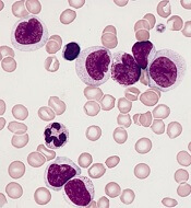
Next-generation sequencing (NGS) can be used to predict relapse in acute myeloid leukemia (AML) patients undergoing hematopoietic stem cell transplant (HSCT), according to research published in Blood.
Researchers found that patients with a higher variant allele frequency (VAF) 21 days after HSCT had a higher risk of relapse and death.
“We can detect mutations in patients’ bone marrow cells 3 weeks after the transplant and, based on that, predict the likelihood of their relapse,” explained study author Zhaolei Zhang, PhD, of the University of Toronto in Ontario, Canada.
Dr. Zhang and his colleagues performed NGS on 529 bone marrow samples from 104 AML patients who underwent chemotherapy and HSCT.
The samples were collected at the time of diagnosis, during the chemotherapy-induced remission, and 3 weeks after HSCT. A subset of patients also gave samples at 3 months, 6 months, and 12 months after HSCT.
The researchers identified 256 mutations that were present in 90 patients at diagnosis and looked for those same mutations at each sampling point.
Chemotherapy and HSCT eliminated most AML cells, leading to a reduction in mutation frequency. However, in some patients, mutations observed at diagnosis could still be detected after chemotherapy and at day 21 after HSCT, indicating the presence of treatment-resistant AML cells.
Allelic burdens at day 21 post-HSCT were higher in patients who ultimately relapsed, and the mutations observed at day 21 expanded at relapse.
The 3-year relapse rate was 56.2% in patients with a VAF greater than 0.2% at day 21 post-HSCT, compared to 16.0% in patients with a lower or no mutational burden (P<0.001).
The 3-year overall survival rates were 36.5% in patients with a VAF greater than 0.2% and 67.0% for patients with a lower or no mutational burden (P=0.006).
In multivariate analyses, VAF0.2% at day 21 was an independent adverse prognostic factor for relapse—hazard ratio, 4.75 (P<0.001)—and overall survival—hazard ratio, 3.07 (P=0.003).
Dr. Zhang and his colleagues said these results suggest NGS-based monitoring after HSCT “provides valuable information” that, together with a patient’s baseline mutational profile and clinical evaluation, can be used to predict outcomes of transplant.
This study was supported by research grants from the Natural Science and Engineering Council of Canada, Leukemia and Lymphoma Society of Canada, Princess Margaret Foundation, National Research Foundation of Korea, and National Natural Science Foundation of China.