User login
FDA approves first biosimilar pegfilgrastim
The US Food and Drug Association (FDA) has approved pegfilgrastim-jmdb (Fulphila™) as the first biosimilar to Neulasta®.
The agents reduce the risk of infection or the duration of febrile neutropenia in patients treated with immunosuppressive chemotherapy for non-myeloid hematologic malignancies.
The FDA approved Fulphila based on evidence that included extensive structural and functional characterization, animal study data, human pharmacokinetic and pharmacodynamic data, clinical immunogenicity data, and other clinical safety and effectiveness data.
The evidence demonstrated that Fulphila is biosimilar to Amgen’s Neulasta. The FDA, in its announcement, noted that Fulphila has been approved as a biosimilar and not as an interchangeable product.
A biosimilar is a biological product approved based on data showing it is highly similar to a biological product already approved by the FDA, termed the reference product.
A biosimilar has no clinically meaningful differences from the reference product in terms of safety, purity, and effectiveness.
Common side effects of Fulphila include bone pain and pain in extremities.
The FDA cautions that patients with a history of serious allergic reaction to human granulocyte colony-stimulating factors, such as pegfilgrastim or filgrastim products, should not take Fulphila.
Serious side effects from Fulphila include:
- rupture of the spleen
- acute respiratory distress syndrome
- serious allergic reactions including anaphylaxis
- glomerulonephritis
- leukocytosis
- capillary leak syndrome
- potential for tumor growth
Fatal sickle cell crises have also occurred with Fulphila use.
Fulphila is not indicated for the mobilization of peripheral blood progenitor cells for hematopoietic stem cell transplantation.
The FDA is planning to release a comprehensive new plan to advance policy efforts that promote biosimilar product development, according to FDA Commissioner Scott Gotlieb, MD.
“We want to make sure that the pathway for developing biosimilar versions of approved biologics is efficient and effective, so that patients benefit from competition to existing biologics once lawful intellectual property has lapsed on these products,” he said in the announcement.
The FDA granted approval of Fulphila to Mylan GmbH. Mylan is co-developing Fulphila with Biocon.
Last fall, the agency had issued a complete response letter saying it could not approve the proposed biosimilar pending an update to the application.
The complete response letter did not raise any questions on the biosimilarity of Fulphila (investigational drug product MYL-1401H), pharmacokinetic/pharmacodynamic data, clinical data, or immunogenicity, however.
Mylan anticipates launching Fulphila in the coming weeks.
Eltrombopag receives priority review designation for SAA
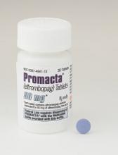
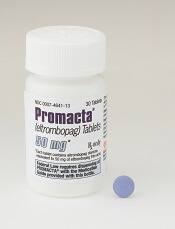
Eltrombopag (Promacta®) in combination with standard immunosuppressive therapy (IST) has received priority review designation from the US Food and Drug Administration (FDA) for first-line treatment of severe aplastic anemia (SAA).
The drug is already approved for SAA in the refractory setting for patients who have had an insufficient response to IST.
And it is approved for adults and children with chronic immune thrombocytopenia (ITP) who are refractory to other treatments and for patients with chronic hepatitis C virus infection who are thrombocytopenic.
Eltrombopag, an oral thrombopoietin receptor agonist, had received breakthrough therapy designation from the FDA earlier this year for use in combination with IST as first-line treatment of SAA.
The priority review designation for the agent as a first-line treatment for SAA is supported by data from a phase 1/2 trial published in NEJM in April 2017 and subsequent data on file with Novartis.
The FDA intends to take action on a priority review application within 6 months of receiving it, rather than the standard 10 months.
The agency grants priority review to applications for products that may provide significant improvements in the treatment, diagnosis, or prevention of serious conditions.
Trial data
Ninety-two patients with previously untreated SAA were enrolled on the trial and received IST and eltrombopag in 3 different cohorts.
Cohorts varied by start day of eltrombopag and duration of eltrombopag therapy. Patients in cohort 1 received eltrombopag from day 14 to 6 months. Patients in cohort 2 received the drug from day 14 to 3 months. And patients in cohort 3 received eltrombopag from day 1 to 6 months.
The overall response rate (ORR) at 6 months was 80% (cohort 1), 87% (cohort 2), and 94% (cohort 3).
The complete response rate at 6 months was 33%, 26%, and 58% in the 3 cohorts, respectively.
At a median follow-up of 2 years, the overall survival rate was 97%.
In the corporate announcement of the priority review designation, Novartis reported an ORR of 85% at 6 months.
Adverse events included transient elevations in liver enzyme levels (7 patients) and 2 severe adverse events—grades 2 and 3 cutaneous eruption—related to eltrombopag that resulted in patients stopping the drug.
Eltrombopag is marketed as Revolade in countries outside the US.
FDA grants priority review to first-line SAA treatment
The Food and Drug Administration has granted priority review to Novartis for their severe aplastic anemia drug.
in combination with standard immunosuppressive therapy (IST). The drug is already approved in the United States for treatment of refractory SAA patients. It is also approved for treatment of chronic immune thrombocytopenia in adults and children who are refractory to other treatments or patients with chronic hepatitis C virus. [[{"fid":"","view_mode":"","fields":{"format":"","field_file_image_alt_text[und][0][value]":"","field_file_image_credit[und][0][value]":"","field_file_image_caption[und][0][value]":"","field_file_image_caption[und][0][format]":"filtered_html"},"type":"media","attributes":{"class":"media-element file-"},"field_deltas":{"1":{"field_file_image_caption[und][0][format]":"filtered_html"}}}]]
The priority review status was granted based on preliminary findings showing that eltrombopag plus IST outperformed IST alone in treatment-naïve patients. The study showed that 52% of newly diagnosed patients achieved a complete response at 6 months with eltrombopag plus IST, which was 35% higher than patients treated with IST alone. The overall response rate was 85% at 6 months in the eltrombopag group, according to Novartis.
The drugmaker received a breakthrough therapy designation from the FDA for eltrombopag for first-line use in SAA in January 2018.
FDA places CTX001 for SCD on clinical hold
The US Food and Drug Administration (FDA) has placed a clinical hold on the investigational new drug application (IND) for CTX001. The agent is being developed for the treatment of sickle cell disease (SCD) and β-thalassemia.
The IND was submitted to the FDA in April to support the initiation of a phase 1/2 trial in the US in adult patients with SCD. The hold will be in place pending the resolution of questions as part of the FDA review.
The phase 1/2 trial in Europe in adult patients with transfusion-dependent β-thalassemia is expected to proceed according to schedule. Trial initiation is planned for the second half of 2018.
CTX001 is being co-developed and co-commercialized by CRISPR Therapeutics and Vertex Pharmaceuticals Incorporated.
The agent is an investigational ex vivo CRISPR gene edited therapy for patients with β-thalassemia or sickle cell disease.
The patient’s hematopoietic stem cells are engineered to produce high levels of fetal hemoglobin (HbF; hemoglobin F) in red blood cells.
The increase in HbF levels by CTX001 may potentially alleviate transfusion requirements for β-thalassemia patients and sickle crises for SCD patients.
Hematocrit improvement with SGLT2 inhibitor: Not just a diuretic effect?
BOSTON – The SGLT2 inhibitor dapagliflozin may increase red blood cell production by suppressing plasma levels of hepcidin, a proinflammatory inhibitor of iron transport, according to results of a randomized study.
This reduction in hepcidin provides a new mechanistic explanation for the improvement in hematocrit seen with SGLT2 inhibitor treatment and suggests a role for use of these drugs beyond their current indications, according to researcher Husam A. Ghanim, PhD, of the State University of New York at Buffalo.
To see whether there were other mechanisms involved beyond hemoconcentration caused by diuretic effects of the drugs, Dr. Ghanim and his colleagues investigated the possibility that dapagliflozin might suppress concentrations of hepcidin concentrations, thereby increasing erythropoiesis.
Their study included 22 patients with type 2 diabetes and normal renal function randomized to dapagliflozin 10 mg daily or placebo for 12 weeks.
They found that the plasma concentration of hepcidin fell significantly over that time period, from 265 to 215 ng/mL in dapagliflozin-treated patients. They also saw significant decreases in hemoglobin A1c, hemoglobin concentration, and hematocrit, as well as an increase in transferrin, the major transporter of iron in the circulation, over 12 weeks.
No such significant changes in those measures were seen in the placebo group, Dr. Ghanim said.
There was a modest but nonsignificant increase in erythropoietin concentrations in the dapagliflozin-treated group, according to the researcher.
Circulating ferritin also fell by about 40% over the course of the study. “Circulating ferritin doesn’t have a clear indication or implication on iron transport,” Dr. Ghanim said. “However, it gets secreted from macrophages and from the liver, and it gets used as a marker for inflammation, and it’s also used as a marker of liver function. So a reduction in ferritin levels may have some clinical implication to what’s going on in the liver.”
On the basis of these findings, it appears that SGLT2 inhibition might increase hematocrit via anti-inflammatory effects and increased erythropoiesis, Dr. Ghanim said.
The increase in oxygenated blood available to tissues might contribute to the beneficial effects of SGLT2 inhibitors on cardiovascular disease, he added.
Also, it’s possible that SGLT2 inhibitors could have a “major impact” on the liver since hepcidin and ferritin are secreted mainly by the liver: “This could also lead us to think that it is possible that we could use SGLT2 inhibitors in conditions of liver inflammation like nonalcoholic steatohepatitis and fatty liver disease,” Dr. Ghanim said in his presentation. “These are future ideas we could explore, based on our data.”
Dr. Ghanim had no disclosures to report.
SOURCE: Ghanim HA et al. AACE 2018, Abstract 228.
REPORTING FROM AACE 2018
Key clinical point: The SGLT2 inhibitor dapagliflozin suppressed hepcidin, a proinflammatory inhibitor of iron transport.
Major finding: Hepcidin plasma concentration fell from 265 to 215 ng/mL (P < 0.05) in dapagliflozin-treated patients.
Study details: A study of 22 patients with type 2 diabetes randomized to either dapagliflozin 10 mg daily or placebo for 12 weeks.
Disclosures: Dr. Ghanim had no disclosures related to the presentation.
Source: Ghanim HA et al. AACE 2018, Abstract 228.
FDA approves first epoetin alfa biosimilar
The US Food and Drug Administration (FDA) has approved epoetin alfa-epbx (Retacrit), a biosimilar to epoetin alfa (Epogen/Procrit).
Epoetin alfa-epbx is approved for the treatment of anemia caused by chronic kidney disease, the use of zidovudine in patients with HIV infection, and myelosuppressive chemotherapy in patients who have a minimum of 2 additional months of planned chemotherapy.
Epoetin alfa-epbx is also approved for use before and after surgery to reduce the chance that red blood cell transfusions will be needed because of blood loss during elective, noncardiac, or nonvascular surgery.
As with epoetin alfa, the prescribing information for epoetin alfa-epbx contains a Boxed Warning noting that erythropoiesis-stimulating agents increase the risk of death, myocardial infarction, stroke, venous thromboembolism, thrombosis of vascular access, and tumor progression or recurrence.
The FDA granted approval of epoetin alfa-epbx to Hospira Inc., a Pfizer company.
The agency’s approval is based on a review of evidence that included structural and functional characterization, animal study data, human pharmacokinetic and pharmacodynamic data, clinical immunogenicity data, and other clinical safety and effectiveness data.
This evidence is available in an FDA briefing document on the biologics license application for epoetin alfa-epbx.
FDA approves epoetin alfa biosimilar to treat anemia
, a treatment for anemia brought on by chronic kidney disease, chemotherapy, or use of zidovudine.
The biosimilar product is also approved to reduce the chance of red blood cell transfusion before and after surgery.
FDA’s approval, issued on May 15, is based on review of structural and functional characterization, animal study data, human pharmacokinetic and pharmacodynamic data, clinical immunogenicity data, and other safety and effectiveness information showing that the epoetin alfa-epbx is biosimilar to the reference product epoetin alfa. By approving epoetin alfa-epbx as a biosimilar, the FDA is saying that there are “no clinically meaningful differences in safety, purity, and potency” from epoetin alfa.
The agency’s approval comes almost a year after the Oncologic Drugs Advisory Committee voted 14-1 to support approval of the biosimilar. The FDA had rejected the application in 2017, citing manufacturing issues at a facility in Kansas, before ultimately approving the product in 2018.
The biosimilar product must be dispensed with a patient Medication Guide with information about uses and risks and carries a boxed warning about an increased risk of death, heart problems, stroke, and tumor growth or recurrence.
The biosimilar product is marketed by Hospira Inc., a Pfizer company.
IV superior to oral treatment for iron deficiency during pregnancy
AUSTIN, TEX. – Utilizing intravenous treatment for iron deficiency in anemic pregnant women was more efficacious than oral iron supplements, according to a study presented at the annual clinical and scientific meeting of the American College of Obstetricians and Gynecologists.
With 42% of pregnancies worldwide affected by anemia, according to the World Health Organization, improving treatment beyond the standard oral treatment could have a large effect on decreasing pregnancy complications.
“Women with bariatric surgery and inflammatory bowel disease are at higher risk of failure,” said Shravya Govindappagari, MD, a gynecologist affiliated with New York–Presbyterian Hospital. “Intravenous iron overcomes the limited intestinal absorption of oral formulations, and may increase iron stores more quickly.”
Dr. Govindappagari and her colleagues conducted a meta-analysis of 11 randomly controlled trials published between 2002 and 2017 to uncover the possible benefits of intravenous iron over oral treatment.
Studies were conducted in India, Egypt, France, and Turkey, with one additional multicenter study that gathered patients from seven different countries. Participants were given iron sucrose, ferric carboxymaltose, or low molecular weight iron dextran, according to Dr. Govindappagari.
In an overall assessment of subjects who achieved target hemoglobin levels, patients receiving intravenous iron were 2.66 times more likely to reach target levels than those given oral treatment (P less than .001). After 4 weeks of treatment, patients in the intravenous groups had a mean hemoglobin increase of 0.84 g/dl higher than those in the oral group (P less than .001).
Some clinicians may be wary about switching treatment modality from oral to intravenous; however, Dr. Govindappagari and fellow investigators found those taking oral treatment were 35% more likely to experience adverse effects than those receiving intravenous treatment.
While the analysis, according to Dr. Govindappagari, has merit, she and her team did not have access to relevant blinded, randomly controlled trials, which may have affected the findings. Maternal and neonatal outcomes were also not included in any of the studies analyzed, nor was a cost analysis of the financial burden of switching from oral to intravenous treatment.
Despite these limitations, Dr. Govindappagari and her colleagues assert the use of intravenous iron could have a significant effect on this problem.
“Intravenous iron compared to oral iron has a higher number reach target, a greater increase in hemoglobin, and has fewer side effects,” Dr. Govindappagari said to attendees. “This could be particularly useful in women in labor, during the third trimester, and women who are iron deficient and are at risk for postpartum hemorrhage.”
Dr. Govindappagari and her colleagues reported no relevant financial disclosures.
SOURCE: Govindappagari S et al. ACOG 2018, Abstract 10OP.
REPORTING FROM ACOG 2018
Key clinical point: Intravenous iron treatment is better for pregnant women with anemia.
Major finding: Hemoglobin levels in women with intravenous iron increased by 1.2 g/dl more than in those using oral supplements after 4 weeks (P less than .001).
Data source: A meta-analysis of 11 randomized, controlled trials comparing intravenous with oral iron treatment.
Disclosures: Dr. Govindappagari and her colleagues reported no relevant financial disclosures.
Source: Govindappagari S et al. ACOG 2018, Abstract 10OP.
VIDEO: Anemia more than doubles risk of postpartum depression
AUSTIN, TEX. – The risk of depression was more than doubled in women who were anemic during pregnancy, according to a recent retrospective cohort study of nearly 1,000 women. Among patients who had anemia at any point, the relative risk of screening positive for postpartum depression was 2.25 (95% confidence interval, 1.22-4.16).
The video associated with this article is no longer available on this site. Please view all of our videos on the MDedge YouTube channel
“This was an unexpected finding,” said Shannon Sutherland, MD, of the University of Connecticut, Farmington, in an interview after she presented the findings at the annual clinical and scientific meeting of the American College of Obstetricians and Gynecologists.
“Maternal suicide exceeds hemorrhage and hypertensive disease as a cause of U.S. maternal mortality,” wrote Dr. Sutherland and her collaborators in the poster accompanying the presentation. And anemia is common: “Anemia in pregnancy can be as high as 27.4% in low-income minority pregnant women in the third trimester,” they wrote.
“If we can find something like this that affects depression, and screen for it and correct for it, we can make a real big difference in patients’ lives,” said Dr. Sutherland in a video interview. “Screening for anemia ... is such a simple thing for us to do, and I also think it’s very easy for us to correct, and very cheap for us to correct.”
The 922 study participants were at least 16 years old and receiving postpartum care at an outpatient women’s health clinic. Patients who had diseases that disrupted iron metabolism or were tobacco users, and those on antidepressants, anxiolytics, or antipsychotics were excluded from the study. Other exclusion criteria included anemia that required transfusion, and intrauterine fetal demise or neonatal mortality.
To assess depression, Dr. Sutherland and her colleagues administered the Edinburgh Postnatal Depression Scale at routine postpartum visits. Dr. Sutherland and her coinvestigators calculated the numbers of respondents who fell above and below the cutoff for potential depression on the 10-item self-report scale. They then looked at the proportion of women who scored positive for depression among those who were, and those who were not, anemic.
Possible depression was indicated by depression scale scores of 9.2% of participants, while three quarters (75.2%) were anemic either during pregnancy or in the immediate postpartum period. Among anemic patients, 10.8% screened positive for depression, while 4.8% of those without anemia met positive screening criteria for postpartum depression (P = .007).
Dr. Sutherland and her collaborators noted that fewer women in their cohort had postpartum depression than the national average of 19%. They may have missed some patients who would later develop depression since the screening occurred at the first postpartum visit; also, “it is possible that women deeply affected by [postpartum depression] may have been lost to follow-up,” they wrote.
Participants had a mean age of about 26 years, and body mass index was slightly higher for those with anemia than without (mean, 32.2 vs 31.2 kg/m2; P = .025).
Postpartum depression was not associated with marital status, substance use, ethnicity, parity, or the occurrence of postpartum hemorrhage, in the investigators’ analysis.
Dr. Sutherland said that, in their analysis, she and her coinvestigators did not find an association between degree of anemia and the likelihood, or severity, of postpartum depression. However, they did find that anemia of any degree in the immediate peripartum period was most strongly associated with postpartum depression.
Though the exact mechanism of the anemia-depression link isn’t known, the fatigue associated with anemia may help predispose women to postpartum depression, said Dr. Sutherland. Also, she said, “iron can make a difference in synthesizing neurotransmitters” such as serotonin, “so it may follow that you might have some depressive symptoms.”
“The next step after this study, which was a launching point, is to see if we correct the degree of anemia and bring them to normal levels, if that can help decrease the risk of postpartum depression,” said Dr. Sutherland.
Dr. Sutherland and her coinvestigators reported that they had no relevant financial disclosures.
SOURCE: Sutherland S et al. ACOG 2018. Abstract 35C.
REPORTING FROM ACOG 2018
Too few Michigan children with SCD receive pneumococcal, meningococcal vaccines
Substantial percentages of children with sickle cell disease are not receiving certain recommended vaccines on time or at all, found a study examining receipt of pneumococcal and meningococcal vaccines among children born in Michigan.
Although these children were more likely to be up-to-date on their pneumococcal vaccines than others their age without sickle cell disease (SCD), nearly one-third had not received all their pneumococcal vaccines by 36 months old. These children are at higher risk of meningococcal and invasive pneumococcal disease because they lack normal spleen function.
ACIP has recommended since February 2010 that all children receive the 13-valent pneumococcal conjugate vaccine (PCV13), which replaced the 7-valent pneumococcal conjugate vaccine (PCV7) that had been recommended since October 2000.
But ACIP also recommends that children with SCD receive two doses of the 23-valent polysaccharide vaccine (PPV23), starting at 2 years old. These children also should receive a PCV13 dose before age 18 years, even if they received the full PCV7 vaccine series.
“By directly including SCD status in a child’s immunization record, an immunization information system could use a specialized algorithm to indicate to healthcare providers which vaccines should be given to a patient with SCD, which may differ from a typical patient,” Dr. Wagner and his colleagues wrote in The Journal of Pediatrics.
“Educational campaigns targeted to parents of these children and their providers could also help advance the importance of vaccination, particularly as more vaccines enter the market, many of which may be highly recommended for children with SCD,” they said.
The researchers matched 1,022 children with SCD to 3,725 children without SCD based on age, sex, race, and zip code. The data was based on the Michigan Care Improvement Registry (MCIR), Michigan Vital Records live birth file, and the Michigan Newborn Screening Program for children born in the state between April 1, 1995, and January 1, 2014.
At age 36 months, 69% of children with SCD had been fully vaccinated with the pneumococcal conjugate vaccine series, compared with 45% of children without SCD. The meningococcal vaccine had been administered to 59% of children with SCD.
Children with SCD were more likely than those without the disease to be up-to-date on their pneumococcal vaccine(s) at 5, 7 and 16 months old.
Nevertheless, substantial percentages of children with SCD who received the complete series of the 7-valent pneumococcal conjugate vaccine had not received two other pneumococcal vaccines. Just over 29% were missing a dose of PCV13, 21.8% of children over 2 years old had not received any dose of PPV23, and 50.7% had not received a second dose of PPV23 by the age of 10 years.
The authors drew attention to the complexity of ACIP recommendations, however: ACIP released 7 recommendations a year, on average, between 2006 and 2015.
“Although providers have a responsibility to educate themselves on how best to protect children with high-risk conditions, these figures speak to the need for MCIR, the state’s immunization information system, to provide additional information on children, such as those who have sickle cell disease, who have special vaccination recommendations,” the authors wrote.
The authors reported no conflicts of interest. No external funding was noted.
SOURCE: Wagner AL et al. J Pediatr. J Pediatr. 2018 May;196:223-9.
This study is particularly valuable because of the “depth, breadth and completeness” of data from across an entire state, a control group that is socioeconomically matched, and a study that was done during a time when new, life-saving vaccines were licensed and recommended. The many changes in the recommendations because of new vaccines and new understanding of the best use of these vaccines make for a complex schedule, but we health care providers need to keep current and to educate parents so their children are protected against infectious diseases. For parents of children with sickle cell disease, the schedule is more complex and the need is greater because of their extreme vulnerability. Wagner et al. suggest that “a proactive electronic prompt to providers [and parents] for vaccines needed for children with special conditions [as exists for the general immunization schedule] is needed – and seems doable.”
Sarah S. Long, MD, is a professor of pediatrics at Drexel University, Philadelphia. She is an associate editor of the Journal of Pediatrics and the Red Book Report of the Committee on Infectious Diseases of the American Academy of Pediatrics. She reported no disclosures. This is a summary of her editorial accompanying the article by Wagner et al. (J. Pediatr. 2018;196:3).
Substantial percentages of children with sickle cell disease are not receiving certain recommended vaccines on time or at all, found a study examining receipt of pneumococcal and meningococcal vaccines among children born in Michigan.
Although these children were more likely to be up-to-date on their pneumococcal vaccines than others their age without sickle cell disease (SCD), nearly one-third had not received all their pneumococcal vaccines by 36 months old. These children are at higher risk of meningococcal and invasive pneumococcal disease because they lack normal spleen function.
ACIP has recommended since February 2010 that all children receive the 13-valent pneumococcal conjugate vaccine (PCV13), which replaced the 7-valent pneumococcal conjugate vaccine (PCV7) that had been recommended since October 2000.
But ACIP also recommends that children with SCD receive two doses of the 23-valent polysaccharide vaccine (PPV23), starting at 2 years old. These children also should receive a PCV13 dose before age 18 years, even if they received the full PCV7 vaccine series.
“By directly including SCD status in a child’s immunization record, an immunization information system could use a specialized algorithm to indicate to healthcare providers which vaccines should be given to a patient with SCD, which may differ from a typical patient,” Dr. Wagner and his colleagues wrote in The Journal of Pediatrics.
“Educational campaigns targeted to parents of these children and their providers could also help advance the importance of vaccination, particularly as more vaccines enter the market, many of which may be highly recommended for children with SCD,” they said.
The researchers matched 1,022 children with SCD to 3,725 children without SCD based on age, sex, race, and zip code. The data was based on the Michigan Care Improvement Registry (MCIR), Michigan Vital Records live birth file, and the Michigan Newborn Screening Program for children born in the state between April 1, 1995, and January 1, 2014.
At age 36 months, 69% of children with SCD had been fully vaccinated with the pneumococcal conjugate vaccine series, compared with 45% of children without SCD. The meningococcal vaccine had been administered to 59% of children with SCD.
Children with SCD were more likely than those without the disease to be up-to-date on their pneumococcal vaccine(s) at 5, 7 and 16 months old.
Nevertheless, substantial percentages of children with SCD who received the complete series of the 7-valent pneumococcal conjugate vaccine had not received two other pneumococcal vaccines. Just over 29% were missing a dose of PCV13, 21.8% of children over 2 years old had not received any dose of PPV23, and 50.7% had not received a second dose of PPV23 by the age of 10 years.
The authors drew attention to the complexity of ACIP recommendations, however: ACIP released 7 recommendations a year, on average, between 2006 and 2015.
“Although providers have a responsibility to educate themselves on how best to protect children with high-risk conditions, these figures speak to the need for MCIR, the state’s immunization information system, to provide additional information on children, such as those who have sickle cell disease, who have special vaccination recommendations,” the authors wrote.
The authors reported no conflicts of interest. No external funding was noted.
SOURCE: Wagner AL et al. J Pediatr. J Pediatr. 2018 May;196:223-9.
Key clinical point: Too few children with sickle cell disease (SCD) are receiving Advisory Committee on Immunization Practices–recommended meningococcal and pneumococcal vaccines, including PCV13 and PPSV23.
Major finding:
Study details: The findings are based on a cohort study of children with and without SCD born in Michigan between April 1, 1995, and January 1, 2014.
Disclosures: The authors reported no conflicts of interest. No external funding was noted.
Source: Wagner AL et al. J Pediatr. 2018 May;196:223-9.
