User login
FDA: Programmable heart failure device approved
. Specifically, these patients are unsuited for other treatments, have marked physical limitations related to their heart failure, and have remained symptomatic despite optimal medical therapy. They also have a regular heart rhythm, are not candidates for resynchronization, and possess a left ventricular ejection fraction of 25%-45%.
The cardiac contractility modulation system is indicated to improve 6-minute hall walk distance, quality of life, and functional status in these patients.
The system is made up of several components, including the implantable pulse generator, a programmer, and software. The pulse generator is connected to three leads that have been implanted in the heart, after which the device is tested and programmed to deliver pulses during normal heartbeats, which improves the heart’s squeezing capability. In randomized, multicenter clinical trials, the system plus optimal medical therapy demonstrated improvements in distance during 6-minute walking tests and standard assessments of heart failure symptoms when compared with optimal medical therapy alone.
The Breakthrough Device designation means this system treats a life-threatening disease and addresses unmet medical needs among some patients. “The FDA recognized the unmet need for these patients and worked with the manufacturer through our Breakthrough Device Program to efficiently bring this product to market, while ensuring it meets our regulatory requirements for safety and effectiveness,” Bram Zuckerman, MD, the director of the division of cardiovascular devices in the FDA’s Center for Devices and Radiological Health said in a news release from the agency.
Abstinence by moderate drinkers improves their AFib
NEW ORLEANS – Abstinence from alcohol on the part of moderate drinkers with atrial fibrillation resulted in clinically meaningful improvement in their arrhythmia in the randomized controlled Alcohol-AF trial, Aleksandr Voskoboinik, MBBS, reported at the annual meeting of the American College of Cardiology.
The benefits of abstinence included significant reductions in the AFib recurrence rate, total AFib burden, and symptom severity. And the payoff extended beyond the arrhythmia: The abstinent group also averaged a 12–mm Hg drop in systolic blood pressure and dropped 3 kg of body weight over the course of the 6-month trial, added Dr. Voskoboinik, a cardiologist at the Baker Heart & Diabetes Institute at the University of Melbourne.
“We would conclude that significant reduction in alcohol intake should be considered as part of the lifestyle intervention in moderate drinkers with AFib,” he said.
Some physicians already advise alcohol abstinence for moderate drinkers with AFib, but until now such recommendations were not evidence based. Instead, the guidance was based on extrapolation from the well-established harmful effects of heavy drinking and binge drinking on AFib as well as epidemiologic studies.
“We wanted to provide some evidence base for physicians to say, ‘If I have motivated patients, here’s what’s potentially achievable,’” he explained.
The key word here is “motivated.” Conducting the Alcohol-AF trial made clear that many moderate drinkers with AFib enjoy their beverages more than they hate having their arrhythmia. Indeed, out of 697 moderate-drinking patients with paroxysmal or persistent AFib who were deemed good candidates and were approached about study participation, more than 500 were excluded because they were flat out unwilling to consider abstinence. After Dr. Voskoboinik and his coinvestigators shortened the planned trial duration from 12 months to 6 because so many recruits were unwilling to abstain for a full year, 140 of the initial 697 patients were randomized to abstinence or continuation of their usual consumption. Thirty percent of them had previously undergone AFib ablation.
The Alcohol-AF study was a multicenter, prospective, open-label study conducted at six Australian hospitals. All subjects underwent comprehensive rhythm monitoring via an implantable loop recorder or an existing pacemaker. Twice daily they triggered the AliveCor mobile phone app together with Holter monitoring. Patients assigned to abstinence were counseled to abstain completely, got written and verbal advice to help with compliance, and had contact with the investigators on a monthly basis. They also underwent urine testing to monitor compliance. All study participants kept a weekly alcohol intake diary, reviewed monthly by investigators.
Participants averaged 17 standard drinks per week at enrollment. Two-thirds were primarily wine drinkers. At the 6-month mark, patients in the abstinence arm averaged two drinks per week, for an 88% reduction from baseline, and 43 of the 70, or 61%, had remained completely abstinent. The control arm averaged a 20% reduction in weekly consumption.
One of the two co-primary endpoints was recurrent AFib episodes lasting for at least 30 seconds. The rate was 53% in the abstinence group and 73% in controls. The time to first recurrence averaged 118 days in the abstinence group, compared to 86 days in controls, for a 37% prolongation through abstinence.
The other primary endpoint was total AFib burden. During the 6-month study, the abstinence group spent a mean of 5.6% of their time in AFib, compared to 8.2% in controls. The median times were 0.5% and 1.2%.
In a multivariate analysis, neither AFib type, duration, history of AFib ablation, age, nor gender predicted AFib recurrence. In fact, the only significant predictor was alcohol abstinence, which conferred a 48% reduction in risk.
Symptom severity, a secondary endpoint, showed an impressive between-group difference at follow-up. Ten percent of patients in the abstinence group had moderate or severe symptoms at follow-up as assessed via the European Heart Rhythm Association score of atrial fibrillation, compared to 32% of controls, for an absolute 22% difference. And 90% of the abstinence group had no or mild symptoms, as did 68% of controls. Nine percent of the abstinence group had an AFib-related hospitalization, as did 20% of controls.
Cardiac MRI, performed in all subjects, showed that the abstinence group experienced a significant reduction in left atrial area, from 29.5 cm2 at baseline to 27.1 cm2 at follow-up. They also experienced significant improvement in left atrial mechanical function, with their left atrial emptying fraction climbing from 42% to 50%.
The 3-kg weight loss from a baseline of 90 kg in the abstinent group represented a 0.7 kg/m2 reduction in body mass index.
In terms of the likely mechanism of benefit of alcohol abstinence in moderate drinkers, Dr. Voskoboinik noted that he and his colleagues recently demonstrated that regular moderate alcohol consumption, but not mild intake, is associated with potentially explanatory atrial electrical and structural changes, including conduction slowing, lower global bipolar atrial voltage, and an increase in complex atrial potentials (Heart Rhythm. 2019 Feb;16[2]:251-9).
Alcohol has been shown to be linked with AFib through multiple mechanisms. Alcohol has adverse effects on the atrial substrate, including promotion of left atrial remodeling, dilation, and fibrosis via oxidative stress, and alcohol has a contributory role in hypertension and obstructive sleep apnea. Alcohol can act as an acute trigger of AFib through binge drinking – the holiday heart syndrome – which causes autonomic changes, electrolyte abnormalities, and electrophysiologic effects, he continued.
Epidemiologic evidence in support of the notion that moderate drinking increases the risk of AFib includes a Swedish meta-analysis of seven prospective studies comprising 12,554 patients with AFib. The researchers concluded that the relative risk of AFib rose by about 8% with each drink per day, compared with the risk of a reference group composed of current drinkers at a rate of less than one drink per week (J Am Coll Cardiol. 2014 Jul 22;64[3]:281-9).
Although discussants were wowed by the 61% complete abstinence rate in the trial, Dr. Voskoboinik cautioned that the study participants were a highly motivated subset of the universe of moderate drinkers with AFib. And even so, “It was an incredibly challenging study to run,” he said.
“I think lifestyle change is a challenge, and with alcohol particularly so, because alcohol is so ubiquitous in our society. I think the important key message for us as physicians is to take an alcohol history and have a discussion with the patient. And we now have some data to show them. But at the end of the day, it’s up to the patient in conjunction with the physician,” he observed.
Discussant Annabelle S. Volgman, MD, noted that 85% of study participants were men, and because of important differences in AFib between men and women, she doesn’t consider the findings applicable to women.
“You need to redo the study in women,” advised Dr. Volgman, professor of medicine at Rush University, Chicago, and director of the Rush Heart Center for Women.
She suggested that an interventional trial such as this one is a great opportunity to utilize wearable device technology, such as the Apple Watch, which can detect irregular pulse rhythms as demonstrated in the Apple Heart Study. This technology is especially popular with younger individuals.
“A lot of young men and women drink a lot of alcohol,” Dr. Volgman noted.
“That’s a great idea. It gets around a problem with AliveCor, which can miss episodes while you’re sleeping,” Dr. Voskoboinik replied.
He reported having no financial conflicts regarding the investigator-initiated and -funded Alcohol-AF trial.
SOURCE: Voskoboinik A. ACC 19, Session 413-08.
REPORTING FROM ACC 19
AUGUSTUS: Dual surpasses triple therapy when AFib patients have PCI or ACS
NEW ORLEANS – For patients with atrial fibrillation and either a recent acute coronary syndrome or percutaneous coronary intervention, combined treatment for 6 months with the anticoagulant apixaban and a P2Y12 inhibitor antiplatelet drug, but without aspirin, was safer than and as effective as a regimen that either also included aspirin or that substituted a vitamin K antagonist, such as warfarin, for the direct-acting oral anticoagulant, based on results from a multicenter, randomized trial with more than 4,600 patients.
The apixaban plus P2Y12 inhibitor (typically, clopidogrel) combination “resulted in less bleeding and fewer hospitalizations without significant differences in ischemic events than regimens that included a vitamin K antagonist, aspirin, or both,” Renato D. Lopes, MD, said at the annual meeting of the American College of Cardiology. Concurrently, his report of the results also appeared in an online article.
This finding in the AUGUSTUS trial gives clinicians more guidance for the long-standing dilemma of how to best prevent arterial thrombus formation in patients with atrial fibrillation (AFib). To prevent a stroke, these patients routinely receive treatment with an anticoagulant when they have an acute coronary syndrome (ACS) event or undergo percutaneous coronary intervention (PCI). Typically, they receive several months of dual antiplatelet therapy with aspirin plus a P2Y12 inhibitor to prevent a clot from forming in the stented or unstable region of a coronary artery.
These patients are not uncommon; this circumstance occurs for about 20% of all AFib patients, and poses the question of what is the safest and most effective way to treat them. Should they get triple therapy with an anticoagulant, aspirin, and a P2Y12 inhibitor, an option that could cause excess bleeding; or should one of the three drugs drop out with the potential for an increased rate of ischemic events? The AUGUSTUS findings suggest that one solution is treatment with a combination of the direct-acting oral anticoagulant apixaban (Eliquis) and the P2Y12 inhibitor clopidogrel (Plavix) but without aspirin.
For the majority of patients like the ones enrolled, “less is more.” By dropping aspirin from the treatment mix, patients did better, said Dr. Lopes, a professor of medicine at Duke University in Durham, N.C.
Dr. Lopes and his associates designed AUGUSTUS (A Study of Apixaban in Patients With Atrial Fibrillation, Not Caused by a Heart Valve Problem, Who Are at Risk for Thrombosis [Blood Clots] Due to Having Had a Recent Coronary Event, Such as a Heart Attack or a Procedure to Open the Vessels of the Heart) as a two-by-two factorial study to address two different questions: During 6 months of treatment, how did apixaban compare with a vitamin K antagonist (usually warfarin) in these patients for safety and efficacy, and how did aspirin compare with placebo in this setting for the same endpoints?
The trial enrolled 4,614 patients at 492 sites in 33 countries. All patients in the study received a P2Y12 inhibitor, with 93% treated with clopidogrel. The study had roughly as many patients as the combined total of patients enrolled in two smaller, prior studies that had looked at roughly the same questions in similar patients.
“The aspirin part is the more interesting, and probably more unique and important finding,” John H. Alexander, MD, a coinvestigator on the study, said in a video interview. Regardless of the anticoagulant used, patients who received aspirin had a 16% rate of major bleeds or clinically relevant non-major bleeds, compared with a 9% rate among those on placebo, a statistically significant result that underscored the bleeding risk posed by adding aspirin to an anticoagulant and a P2Y12 inhibitor.
The results also showed no statistically significant difference in any efficacy measure with or without aspirin, including the rate of death or hospitalization, or of any individual ischemic endpoint. However the results showed a signal of a small increase in the rates of each of three types of ischemic events – stent thrombosis, MI, and need for urgent revascularization, each of which showed a numerical increase when aspirin was dropped. But the increase was small.
Dr. Lopes calculated that, for example, to prevent one episode of stent thrombosis by treating with aspirin also would cause 15 major or clinically relevant non-major bleeds, which makes inclusion of aspirin something of a judgment call for each patient, said Dr. Alexander, a professor of medicine at Duke. An AFib patient with a high risk for thrombosis but a low risk for bleeding following PCI or an ACS event might be a reasonable patient to treat with aspirin along with apixaban and a P2Y12 inhibitor, he explained.
The rate of major or clinically relevant bleeds was 11% with apixaban and 15% with a vitamin K antagonist, a statistically significant difference. Patients treated with apixaban also had a significantly reduced rate of death or hospitalization, 24%, compared with 27% among those on the vitamin K antagonist, as well as a significantly lower rate of stroke.
Overall the lowest bleeding rate was in patients on apixaban but no aspirin, a 7% rate, while the highest rate was in patients on a vitamin K antagonist plus aspirin, a 19% rate.
Dr. Alexander said that it would be an overreach to extrapolate these findings to other direct-acting oral anticoagulants, compared with a vitamin K antagonist, but he believed that the findings the study generated about aspirin were probably relevant regardless of the anticoagulant used.
[email protected]
On Twitter @mitchelzoler
It’s very reassuring to see that you can use a direct-acting oral anticoagulant like apixaban along with a P2Y12 inhibitor, but with no aspirin, and have no statistically significant increase in ischemic events. This is a fantastic finding. The finding shows once again that warfarin is a problematic drug. As the cost for direct-acting oral anticoagulants has decreased, their use has increased.
These results were not unexpected and also are probably the final nail in the coffin for using a combination of warfarin and aspirin. Prior findings from the PIONEER AF-PCI study that used rivaroxaban (N Engl J Med. 2016 Dec 22;375[25]:2423-34) and from the RE-DUAL PCI study that used dabigatran (N Engl J Med. 2017 Oct 19;377[16]:1513-24) also showed the advantages of using a direct-acting oral anticoagulant when compared with a vitamin K antagonist in this setting, The AUGUSTUS trial, with just over 4,600 patients, had nearly as many patients as the roughly 4,850 enrolled in these two prior studies put together.
Dhanunjaya Lakkireddy, MD , is medical director of the Kansas City Heart Rhythm Institute in Overland Park. He had no disclosures. He made these comments as the designated discussant during a press briefing.
NEW ORLEANS – For patients with atrial fibrillation and either a recent acute coronary syndrome or percutaneous coronary intervention, combined treatment for 6 months with the anticoagulant apixaban and a P2Y12 inhibitor antiplatelet drug, but without aspirin, was safer than and as effective as a regimen that either also included aspirin or that substituted a vitamin K antagonist, such as warfarin, for the direct-acting oral anticoagulant, based on results from a multicenter, randomized trial with more than 4,600 patients.
The apixaban plus P2Y12 inhibitor (typically, clopidogrel) combination “resulted in less bleeding and fewer hospitalizations without significant differences in ischemic events than regimens that included a vitamin K antagonist, aspirin, or both,” Renato D. Lopes, MD, said at the annual meeting of the American College of Cardiology. Concurrently, his report of the results also appeared in an online article.
This finding in the AUGUSTUS trial gives clinicians more guidance for the long-standing dilemma of how to best prevent arterial thrombus formation in patients with atrial fibrillation (AFib). To prevent a stroke, these patients routinely receive treatment with an anticoagulant when they have an acute coronary syndrome (ACS) event or undergo percutaneous coronary intervention (PCI). Typically, they receive several months of dual antiplatelet therapy with aspirin plus a P2Y12 inhibitor to prevent a clot from forming in the stented or unstable region of a coronary artery.
These patients are not uncommon; this circumstance occurs for about 20% of all AFib patients, and poses the question of what is the safest and most effective way to treat them. Should they get triple therapy with an anticoagulant, aspirin, and a P2Y12 inhibitor, an option that could cause excess bleeding; or should one of the three drugs drop out with the potential for an increased rate of ischemic events? The AUGUSTUS findings suggest that one solution is treatment with a combination of the direct-acting oral anticoagulant apixaban (Eliquis) and the P2Y12 inhibitor clopidogrel (Plavix) but without aspirin.
For the majority of patients like the ones enrolled, “less is more.” By dropping aspirin from the treatment mix, patients did better, said Dr. Lopes, a professor of medicine at Duke University in Durham, N.C.
Dr. Lopes and his associates designed AUGUSTUS (A Study of Apixaban in Patients With Atrial Fibrillation, Not Caused by a Heart Valve Problem, Who Are at Risk for Thrombosis [Blood Clots] Due to Having Had a Recent Coronary Event, Such as a Heart Attack or a Procedure to Open the Vessels of the Heart) as a two-by-two factorial study to address two different questions: During 6 months of treatment, how did apixaban compare with a vitamin K antagonist (usually warfarin) in these patients for safety and efficacy, and how did aspirin compare with placebo in this setting for the same endpoints?
The trial enrolled 4,614 patients at 492 sites in 33 countries. All patients in the study received a P2Y12 inhibitor, with 93% treated with clopidogrel. The study had roughly as many patients as the combined total of patients enrolled in two smaller, prior studies that had looked at roughly the same questions in similar patients.
“The aspirin part is the more interesting, and probably more unique and important finding,” John H. Alexander, MD, a coinvestigator on the study, said in a video interview. Regardless of the anticoagulant used, patients who received aspirin had a 16% rate of major bleeds or clinically relevant non-major bleeds, compared with a 9% rate among those on placebo, a statistically significant result that underscored the bleeding risk posed by adding aspirin to an anticoagulant and a P2Y12 inhibitor.
The results also showed no statistically significant difference in any efficacy measure with or without aspirin, including the rate of death or hospitalization, or of any individual ischemic endpoint. However the results showed a signal of a small increase in the rates of each of three types of ischemic events – stent thrombosis, MI, and need for urgent revascularization, each of which showed a numerical increase when aspirin was dropped. But the increase was small.
Dr. Lopes calculated that, for example, to prevent one episode of stent thrombosis by treating with aspirin also would cause 15 major or clinically relevant non-major bleeds, which makes inclusion of aspirin something of a judgment call for each patient, said Dr. Alexander, a professor of medicine at Duke. An AFib patient with a high risk for thrombosis but a low risk for bleeding following PCI or an ACS event might be a reasonable patient to treat with aspirin along with apixaban and a P2Y12 inhibitor, he explained.
The rate of major or clinically relevant bleeds was 11% with apixaban and 15% with a vitamin K antagonist, a statistically significant difference. Patients treated with apixaban also had a significantly reduced rate of death or hospitalization, 24%, compared with 27% among those on the vitamin K antagonist, as well as a significantly lower rate of stroke.
Overall the lowest bleeding rate was in patients on apixaban but no aspirin, a 7% rate, while the highest rate was in patients on a vitamin K antagonist plus aspirin, a 19% rate.
Dr. Alexander said that it would be an overreach to extrapolate these findings to other direct-acting oral anticoagulants, compared with a vitamin K antagonist, but he believed that the findings the study generated about aspirin were probably relevant regardless of the anticoagulant used.
[email protected]
On Twitter @mitchelzoler
REPORTING FROM ACC 19
Antibiotic-eluting envelope reduces CIED infections
An absorbable, antibiotic-eluting envelope around cardiac implantable electronic devices could significantly reduce the incidence of infection, according to a presentation at the annual meeting of the American College of Cardiology.
The WRAP-IT trial, which was simultaneously published online March 17 in the New England Journal of Medicine, involved 6,983 patients undergoing cardiac implantable electronic device (CIED) implantation, replacement, revision, or upgrade. Patients were randomized either to receive the TYRX Absorbable Antibacterial Envelope or not.
After a mean follow-up of 20.7 months, there was a significant 40% lower rate of major infections in the envelope group compared to the control group, which met the efficacy objective of the study. Researchers saw 30 major infections in 25 patients in the envelope group; in the control group, there were 45 major infections in 42 patients (P = 0.04). The trial excluded patients at high risk of systemic infection due to other sources and patients with existing infection.
“CIED infection is a rare but serious event, and its management requires prolonged hospitalization, which involves device and lead extraction with adjunctive antibiotic therapy,” wrote Dr. Khaldoun G. Tarakji, from The Cleveland Clinic, and co-authors. “Despite proper management of CIED infection, both short- and long-term mortality remains high.”
One previous randomized study had shown that intravenous administration of antibiotics during CIED procedures can reduce the risk of infection, while a different study failed to find a benefit. The vast majority of patients in this study (98.7%) received periprocedural antibiotics, 74.5% received pocket wash and 29.6% received post-procedural antibiotics. These strategies were not controlled, but there is no clear evidence that any particular strategy influenced the infection rate, the authors wrote.
Patients in the envelope group experienced numerically fewer pocket infections but more endocarditis or bacteremia compared to those in the control group, a finding that the authors could not explain.
The most common pathogen responsible was staphylococcus, but data on antibiotic susceptibility was not collected. The authors stressed that this limited their ability to assess the risk of antibiotic resistance developing.
In this study, the researchers noted that the reduction in the risk of infection was greater among individuals who were implanted with higher-power devices, compared to those implanted with low-power devices or an initial cardiac resynchronization therapy device.
However, they said, the rate of infections was generally lower among those receiving low-power devices.
There was no increase in complications related to use of the envelope. The rate of complications occurring within 12 months of the procedure and relating to the CIED procedure or envelope was 6% in the envelope group and 6.9% in the control group.
When major infections were excluded, the rate of complications in each group was 5.7% and 5/9% respectively. There was also no significant difference in mortality rates between the two groups (17.4% and 17.8% respectively).
The authors wrote that while use of the envelope can require a slightly larger CIED dissection pocket, this was not associated with increased procedural time or complications. The envelope was successfully implanted in 99.7% of procedure attempts.
“There were fewer system revisions in the envelope group than in the control group and no complications due to allergy to the envelope mesh, polymer, or antibiotics,” they wrote.
The study was supported by Medtronic, the maker of the TYRX Absorbable Antibacterial Envelope. Twenty-four authors declared institutional funding or research grants from Medtronic, thirteen declared fees, consultancies and other support from private industry outside the submitted work. Three authors were employees of Medtronic.
SOURCE: Tarakji K et al. NEJM, 2019, March 17. DOI: 10.1056/NEJMoa1901111
FROM ACC 19
Apple Watch algorithm showed 84% positive predictive value for Afib
, investigators reported at the annual meeting of the American College of Cardiology.
This was a single-arm, prospective, open-label, observational study of unprecedented size and speediness of completion. It included nearly 420,000 self-enrolled adults living in the U.S., with 8 months of monitoring. But despite the study’s flashy size and trendy digital health theme, the researchers were careful not to oversell the findings.
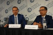
“This study was just meant to be a very, very first step in trying to learn if this kind of technology can help us to prevent stroke. It was not a randomized trial of a public health intervention for screening. This is the first half of the first inning. Rigorous investigation of this technology and its potential use in clinical settings will need to happen. But we do think from a trial and operational standpoint the Apple Heart Study provides a solid foundation upon which further research in digital health can be conducted,” according to Mintu Turakhia, MD, co-principal investigator and an electrophysiologist as well as executive director of the Center for Digital Health at Stanford (Calif.) University, which conducted the study.
The study was conducted virtually. Screening, consent, and data gathering were performed electronically by smartphone. Participants had to have an Apple Watch Series 1, 2, or 3, and an Apple iPhone 5 or more recent model in order to join. The majority of subjects were under age 40, and just 6% were age 65 or older, when the risks of atrial fibrillation (AFib) and stroke are higher. All participants self-reported having no history of AFib nor currently being on anticoagulation.
The study algorithm utilized the Apple Watch’s built-in light sensor technology to opportunistically sample the time interval between pulses when the wearer was still. An irregular time interval triggered a cascade of more frequent sampling. If 5 of 6 samples were irregular within a 48-hour period, the wearer received an irregular rhythm notification along with a prompt to contact a participating physician via telemedicine. The physician could then arrange for an ECG patch to be mailed to the participant, who wore it for up to 7 days before mailing it back for analysis.
Among the key findings in the Apple Heart Study: the irregular pulse notification rate was low overall, at 0.5%, ranging from 0.16% in the under-40 group to 3.2% in subjects age 65 or older. As a result, the study population of particular interest nosedived from an initial 419,297 to the less than 2,100 who received an irregular pulse notification. Of the 658 participants who were subsequently sent an ECG patch, 450 returned it for analysis.
An average of 13 days went by between an irregular pulse notification and ECG patch receipt and activation, so it wasn’t particularly surprising that only 34% of the patches were positive for AFib, since early-stage paroxysmal AFib comes and goes. However, of the 86 subjects who received a new notification of an irregular rhythm while they were wearing a patch, 72 simultaneously showed AFib on their patch. That translates to an 84% positive predictive value for an irregular rhythm notification as an indicator of AFib.
Of the 153 subjects with evidence of AFib on their ECG patch, 20% proved to be in AFib for the full week they wore it. Of those with AFib, 89% had a longest episode of at least 1 hour in duration.
Several discussants expressed reservations about this approach to finding individuals with previously undetected AFib. Jeanne E. Poole, MD, an electrophysiologist and professor of medicine at the University of Washington in Seattle, observed that the question of whether patients with asymptomatic AFib should receive oral anticoagulation therapy is as-yet unanswered and is the focus of ongoing randomized trials. The Apple Heart Study approach, she said, “might lead a lot of patients into being treated unnecessarily or prematurely, or flooding doctors’ offices with a lot of young people.”
Co-principal investigator Marco Perez, MD, an electrophysiologist at Stanford, replied, “Stroke is important, and we all worry about it. But it’s also important that there are other things atrial fibrillation is associated with, like cardiomyopathy and heart failure. So finding atrial fibrillation in a young population might be important. Maybe they don’t need anticoagulation, but maybe there’s something else going on.”
Patrick T. O’Gara, MD, professor of medicine at Harvard University, Boston, was concerned about what he called “the signal-to-noise ratio – the noise that will come in when there is an irregularity detected on the watch that could range from anything from ventricular premature beats to atrial fibrillation.” He is also leery of what he considers to be at this point the excessive hype surrounding direct-to-consumer wearable digital health technology.
“I applaud your circumspection,” he told Dr. Turakhia and Dr. Perez. “I understand very directly from you that these are limited observations. But it’s a good step forward.”
Dr. Perez reported receiving research funding from and serving as a consultant to Apple. Dr. Turakhia reported serving as a consultant to AliveCor and Cardiva Medical.
Their presentation was immediately followed by a related panel discussion titled, “Digital Disruption at Our Doorstep – Implications for Clinicians and Patients.” Session moderator John Rumsfeld, MD, chief innovation officer at the ACC and professor of medicine at the University of Colorado, Denver, kicked things off by observing, “Digital health technology certainly exists. There’ve been billions of dollars invested in digital health. Outside of health care there’s been successful digital transformation of almost every other sector of the economy except for health care. But we deliver care pretty much the same as we have for the past 50 or more years.”
Paul Stoeffels, MD, chief scientific officer at Johnson & Johnson, said physicians and payers want to see evidence of benefit before adopting change. Towards that end, he announced that Johnson & Johnson and Apple are collaborating on a randomized controlled trial called the HEARTLINE study. The active intervention arm in the study involves utilization of the Apple Watch’s irregular pulse notification algorithm, with confirmation of AFib to be achieved using the ECG app incorporated in the latest version of the watch, coupled with a medication adherence app developed by Johnson & Johnson. Enrollment of 150,000 U.S. adults age 65 and older is planned to begin this summer. The study, conducted on a digital platform akin to the Apple Heart Study, will look at the intervention’s impact on rates of stroke, MI, and death as well as AF detection.
Maulik Majmudar, MD, a cardiologist and chief health officer for health and wellness at Amazon, declared, “There’s no doubt in my mind that digital solutions will become a mainstay in our care delivery going forward. The question is really not if, but when.”
He predicted that just as the past two decades have seen the birth of new medical specialties, including hospitalists and cardiovascular intensivists, the next 10 years or so will see the creation of a new field within cardiovascular medicine, whose skilled practitioners might be called ‘digitalists’ – experts in collecting, moving, and safeguarding massive quantities of digital health data.
Robert Califf, MD, vice chancellor for health data science at Duke Health in Durham, N.C., addressed the issue of how society is going to pay for a shift to digital health: “It’s very simple. I don’t see any way that fee-for-service medicine can deal with this. It’s just not possible. If you think we’re going to add on more cost to the system by doing virtual visits, I just do not see that happening. Our solution to the payment system is to get rid of fee-for-service medicine and go to pay-for-value. The minute you’re in pay-for-value, virtual visits and digital information will become the way to do it – to move the treatment and the interaction more to home and less of having people wait in doctors’ offices and spending time in hospitals.”
SOURCE: Turakhia M, ACC 19 NCT03335800
REPORTING FROM ACC 19
Apple Watch algorithm brings wearables closer to clinical practice
NEW ORLEANS – , Matthew W. Martinez, MD, medical director of the Sports Cardiology and Hypertrophic Cardiomyopathy Center at the Lehigh Valley Health Network in Allentown, Pa., said in a video interview.
The Apple Heart Study, presented at the annual meeting of the American College of Cardiology, evaluated a mobile app that uses the watch’s existing light sensor technology to detect subtle changes that might indicate an arrhythmia.
The Apple Watch generates a tachogram, which is a plot of time between heart beats. If an abnormal tachogram occurs five out of six times, they are analyzed by an algorithm and sent to the Apple Watch.
The positive predictive value for the tachogram was 71%, and the positive predictive value for the notification was 84%.
Dr. Martinez, who is lead cardiologist for U.S. Major League Soccer and is also heavily involved with the National Football League, said that the study helps clinicians understand the utility of wearable technology.
His take home from the study is that, when people are notified by their watch, they should notify their health care provider, and the provider should take it seriously.
Dr. Martinez was not involved in the Apple Heart Study, and had no relevant disclosures.
REPORTING FROM ACC 19
Infective endocarditis isn’t what it used to be
SNOWMASS, COLO. – Infective endocarditis in 2019 is very different from the disease most physicians encountered in training, both in terms of epidemiology and clinical presentation, Patrick T. O’Gara, MD, observed at the Annual Cardiovascular Conference at Snowmass sponsored by the American College of Cardiology.
The classic description of infective endocarditis provided by Sir William Osler, MD, was of a subacute bacterial infection characterized by a long latent phase of low-grade fever, back pain, weight loss, and night sweats. It was mainly a right-heart disease of younger individuals with an infected native valve, and the predominant pathogens were streptococci, Dr. O’Gara said.
“I think in the current era endocarditis is more often characterized by an acute illness with toxic features in the context of adults with a high burden of degenerative diseases – for example, patients with rheumatoid arthritis or psoriatic arthritis on immunosuppressive therapy, or diabetes, end-stage renal disease, and risk factors for hospital-acquired infection. Injectable drug use is through the roof, there’s a wider prevalence of cardiac implanted electronic devices, which are a wonderful place for bacteria to hide, and Staphylococcus aureus has certainly become the leading pathogen with regard to endocarditis in the United States, especially MRSA, often multidrug resistant,” said Dr. O’Gara, professor of medicine at Harvard Medical School, Boston.
“Also, no talk about endocarditis is sufficient without paying some attention to the opioid crisis in which we find ourselves. It’s one of the top three causes of death among young men in the United States, along with accidents and gun violence. No region of the country is spared. This has completely inundated our ER and hospitalist services and our inpatient cardiology services with folks who are often repeat offenders when it comes to the difficulty in being able to give up an injectable drug use habit. They have multiple infections and hospitalizations, tricuspid valve involvement, and depending upon the aggressiveness of the Staphylococcus organism, typically they have left-sided disease with multiple complications, including aortic regurgitation and heart failure,” the cardiologist continued.
This description underscored one of Dr. O’Gara’s major points about the challenges posed by infective endocarditis in contemporary practice: “Expect the unexpected,” he advised. “When you’ve seen one case of infective endocarditis, you’ve seen one case of infective endocarditis.”
Outcomes are ‘sobering’
In the current era, outcomes are “sobering,” the cardiologist noted. Infective endocarditis carries a 6-month mortality rate of 20%-25% despite early surgery being performed during the index hospitalization in up to 60% of patients, with a relatively high perioperative mortality rate of about 10%. However, the risk of reinfection occurring in a newly implanted cardiac valve is impressively low at about 2%.
Refer early for multimodality imaging and surgical consultation
Transesophageal echocardiography is valuable in assessment of the infected valve. However, when extravalvular extension of the infection is suspected and the echo assessment is nondiagnostic or indeterminate, it’s time to quickly move on to advanced imaging, such as PET-CT.
The ACC/American Heart Association class I recommendations for early surgery in infected native valves haven’t changed substantially in over a decade. Based largely on observational data, there is an association between early surgery and lower in-hospital mortality (Lancet. 2012 Mar 10;379[9819]:965-975).
Class IIa recommendations for native valve surgery include recurrent emboli and a persistent vegetation despite appropriate antibiotic therapy. A “very controversial” class IIb recommendation for surgery because of weak supporting data is the identification of a mobile vegetation larger than 10 mm, particularly if it’s located on an anterior mitral valve leaflet, he said.
If the decision is made to forgo early surgery, be sure to repeat transesophageal echocardiography on day 7-10 to reassess the size of the patient’s vegetation.
“There is an association between size of vegetation and 1-year mortality, with a cut point of greater than 15 mm. Some would argue this constitutes a reasonable indication for early surgery,” Dr. O’Gara noted.
The embolization rate in patients with infective endocarditis is highest during the day before presentation, the day of presentation, and through the first 2 days afterward. The rate drops precipitously within 2 weeks after initiation of appropriate antibiotic therapy. Thus, to utilize early surgery to maximum effect in order to decrease the risk of embolization, it makes sense to operate within the first several days following presentation, before antibiotics have had sufficient time to catch up with the evolving disease process.
Don’t use half measures when it comes to removal of cardiac implanted electronic devices
The guidelines are clear regarding infected pacemakers, implanted cardioverter-defibrillators, and cardiac resynchronization devices: “It all needs to come out,” Dr. O’Gara emphasized. That includes all leads and the generator in patients with documented infection of only one portion of the device system, as a class I, level of evidence B recommendation. Moreover, complete removal of a pacemaker or defibrillator system is deemed “reasonable” as a class IIa recommendation in all patients with valvular infection caused by S. aureus or fungi even in the absence of evidence of device infection.
“I think we as general cardiologists have become increasingly impressed about how sick and festering these kinds of patients can become, even when we’re not able to prove that the lead is infected. The lead looks okay on transesophageal echo or PET-CT, blood cultures are negative, the valvular heart disease is really not that advanced, but several days go by and the patient is just not responding. We should have a high index of suspicion that there’s an infection we cannot appreciate. But obviously, you make these difficult decisions in consultation with your electrophysiology colleagues,” he added.
Know when the cardiologist should say ‘no’ to early aggressive surgery
While an aggressive early surgical approach often pays off in terms of prevention of embolic sequelae and a reduction in heart failure, the timing of surgery in the 20%-40% of patients with infective endocarditis who present with stroke or other neurologic complications remains controversial. An international group of Canadian and French cardiac surgeons and neurologists developed a useful algorithm regarding the types of neurologic complications for which early cardiac surgery is a poor idea because of the high risk of neurologic exacerbation. For example, a mycotic neuroaneurysm is grounds for postponement of cardiac surgery for at least 4 weeks (Circulation. 2016 Oct 25;134[17]:1280-92).
Dr. O’Gara reported receiving funding from the National Heart, Lung and Blood Institute, the National Institute of Dental and Craniofacial Research, from Medtronic in conjunction with the ongoing pivotal APOLLO transcatheter mitral valve replacement trial, and from Edwards Lifesciences for the ongoing EARLY TAVR trial.
REPORTING FROM ACC SNOWMASS 2019
Not all AF maze operations are aMAZE-ing
SNOWMASS, COLO. – The term “maze procedure” for surgical ablation of atrial fibrillation is bandied about rather loosely these days, but as far as Hartzell V. Schaff, MD, is concerned, the operation of choice remains the classic cut-and-sew maze III procedure developed by James L. Cox, MD, while at Washington University, St. Louis.
“The classic Cox maze III, the cut-and-sew maze, is the best procedure for getting rid of atrial fibrillation and is in my view the gold standard. Some people argue that transmurality isn’t important, but it can occur because of gap lesions,” he said at the Annual Cardiovascular Conference at Snowmass sponsored by the American College of Cardiology.
Unlike modifications of the Cox maze III – such as the mini maze or the maze IV, which utilizes radiofrequency energy or cryoablation to create scars in an effort to achieve pulmonary vein isolation – the maze III cannot be done as a minimally invasive procedure. After all, it requires making incisions in both atria, along with aortic cross-clamping and cardiopulmonary bypass. But it has a significantly higher long-term rate of freedom from recurrent atrial fibrillation (AF) than the other operations. And crucially, it enables the surgeon to readily obliterate the left atrial appendage.
“The most important thing when you do any surgical procedure for atrial fibrillation, I think, is getting rid of the left atrial appendage. When you do cut-and-sew maze, that’s done 100% of the time,” explained Dr. Schaff, professor of surgery at the Mayo Clinic in Rochester, Minn.
“We really have a lot of work left to do as surgeons in improving the outcome of surgery for atrial fibrillation. One of the things we as surgeons don’t do well is getting rid of the left atrial appendage. This ought to be done in every patient that has surgical ablation for atrial fibrillation,” according to the cardiothoracic surgeon.
And yet, he continued, in a series of nearly 87,000 patients with AF who underwent nonemergent cardiac surgery in the Society of Thoracic Surgeons database, 48.0% of whom underwent surgical ablation for AF, only 63.9% of those who had standalone ablation for lone AF got their left atrial appendage dealt with, compared with 86%-89% of those who underwent concomitant cardiac surgery, such as mitral valve repair or replacement (Ann Thorac Surg. 2017 Aug;104[2]:493-500).
“That’s awful, really. And the reason for that low left atrial appendage obliteration rate is this: For many of those patients who had surgery for lone atrial fibrillation, the surgeons were trying to do minimally invasive surgery, where they do pulmonary vein isolation on the right side, so they don’t have access to the left atrial appendage,” Dr. Schaff said.
“In the past,” he recalled, “we would ligate the left atrial appendage. Nowadays because of echocardiographic studies that show there’s persistent patency in a sizable percentage of patients, we amputate the left atrial appendage in almost all of the patients.”
The terminology surrounding surgical ablation for AF, in his view, has become rather confusing. “Most of you, when you refer a patient for surgical ablation for AF, the surgeons will just say they do a maze procedure,” Dr. Schaff cautioned. “Somehow, all of that [maze IV, mini maze] today is lumped together as a classic maze procedure, but it’s really not. We have different lesion sets and energy sources.”
And different outcomes as well. In a series of 1,189 adults who underwent surgical ablation for AF at the Mayo Clinic, of whom 44% had a biatrial cut-and-sew maze while the rest had surgical cryotherapy, radiofrequency ablation, or a combination of the two, the rate of freedom from AF 1 year post surgery was 85% with the cut-and-sew maze versus 71% with the alternatives. At 5 years or more, the rates were 78% and 52%, respectively. In a multivariate analysis, freedom from AF was independently associated with preoperative paroxysmal rather than permanent AF, performance of the classic maze III procedure, concomitant treatment of associated mitral valve disease, and younger age.
Moreover, rates of the major early postoperative complications – stroke, bleeding, and renal failure – were similar in the cut-and-sew maze III and other groups.
“So a lesser procedure doesn’t necessarily mean fewer complications,” Dr. Schaff noted.
One of the criticisms levied against the maze III is that it’s too much surgery for AF. But it’s actually relatively inexpensive because the disposables – suture, needles, scalpel – are those used in the commonly performed concomitant cardiac surgical procedures. “The Cox maze III does take extra time, but with experience it’s not much extra time,” he asserted.
Indeed, in a series of 452 Mayo Clinic maze III patients, the cross-clamp and cardiopulmonary bypass times were 52 and 73 minutes, respectively, for those undergoing an isolated maze III, compared with 73 and 86 minutes for patients whose maze III was done in conjunction with other procedures, most commonly mitral valve repair or replacement.
An underrecognized group of patients who benefit from a standalone cut-and-sew maze are those with tachycardia-induced cardiomyopathy marked by AF or atrial flutter, rapid uncontrolled ventricular response, a decreased left ventricular ejection fraction, and no associated valvular or congenital heart disease. In a series of 37 such patients identified and treated with a maze III operation at the Mayo Clinic, their average preoperative left ventricular ejection fraction of 43% improved to about 55% at discharge, a benefit sustained at last follow-up a median of 63 months later. The outcome was particularly impressive in the 11 patients with a severely depressed left ventricular ejection fraction averaging 31% preoperatively, which jumped to 53% at discharge (Ann Thorac Surg. 2006 Aug;82[2]:494-500).
“Their ejection fraction goes up when you control the tachycardia-induced cardiomyopathy,” he observed. “So reduced left ventricular ejection fraction may be an indication for surgery rather than a contraindication.”
Dr. Schaff emphasized that it’s important for cardiologists and surgeons not to overpromise what surgical ablation of AF can accomplish. The only randomized trial of surgical ablation of AF versus no ablation during mitral valve surgery, sponsored by the National Institutes of Health and Canadian Institutes of Health Research and carried out by the Cardiothoracic Surgical Trials Network, showed no significant between-group differences at 1 year in any of numerous quality of life measures, nor was there a survival benefit for ablation (N Engl J Med. 2015 Apr 9;372(15):1399-409).
“We must point out that there’s no indication that controlling atrial fibrillation has anything to do with improving survival. It has to do with symptomatic benefit and perhaps reducing risk of stroke,” he said.
Dr. Schaff reported having no financial conflicts regarding his presentation.
REPORTING FROM ACC SNOWMASS 2019
ICYMI: Andexanet alfa reduces anti–factor Xa activity from apixaban, rivaroxaban
Patients with acute major bleeding associated with factor Xa inhibitor usage who received andexanet alfa experienced a significant decrease in anti–factor Xa activity, with more than three-quarters of patients experiencing good or excellent hemostatic efficiency after 12 hours. That finding emerged from the multicenter, prospective, open-label, single-group ANNEXA-4 trial published in the New England Journal of Medicine (2019 Feb 11. doi: 10.1056/NEJMoa1814051).
We reported this story at the annual meeting of the American College of Cardiology before it was published in the journal. Find our coverage at the link below.
FROM THE NEW ENGLAND JOURNAL OF MEDICINE
What cardiologists need to know about ARVC
SNOWMASS, COLO. – Electrophysiologist N.A. Mark Estes III, MD, has a piece of advice for his general cardiology colleagues regarding arrhythmogenic right ventricular cardiomyopathy (ARVC), an inherited arrhythmia syndrome that’s one of the most common causes of sudden death in athletes under the age of 40 years.
“When you have a patient with syncope and an ECG abnormality that suggests ARVC, you immediately restrict activity and immediately go on to imaging. And then it’s time to get that patient off to somebody who deals with these patients all the time,” he said at the Annual Cardiovascular Conference at Snowmass sponsored by the American College of Cardiology.
ARVC is thought to affect 1 in 5,000 people. It’s the cause of sudden death in roughly 5% of young athletes in the United States and 25% in Italy, with the disparity believed to be largely caused by underrecognition of the disease.
Dr. Estes was a coauthor of a 2015 international task force consensus statement on the treatment of ARVC (Eur Heart J. 2015 Dec 7;36[46]:3227-37). An updated report that incorporates much new information is due to be released in May 2019.
“The new guidelines are going to put some very clear limits on physical activity: Less than 7 METs [metabolic equivalents], which is about a brisk walk. Exercise is not good with ARVC,” said Dr. Estes, professor of medicine at the University of Pittsburgh.
Patients with ARVC absolutely must be restricted from participation in competitive sports, a step whose importance was demonstrated in the North American Multidisciplinary Study of ARVC, for which Dr. Estes was a coauthor. That study, too recent for inclusion in the 2015 task force consensus statement, showed that competitive sport was associated with a 100% increased risk of ventricular tachyarrhythmias and death, as well as earlier presentation with symptoms, compared with patients with ARVC who engaged in much less intense recreational athletic activity, who in turn didn’t have a risk level any different from inactive patients. That being said, however, the investigators also noted that the absolute risk of ventricular tachyarrhythmias or death remained high even in the inactive and recreational sports participants, at 23% by 2 years post diagnosis (Eur Heart J. 2015 Jul 14;36[27]:1735-43).
ARVC has three phases: An early concealed phase, with an associated high rate of syncope or sudden cardiac death; an intermediate phase marked by frequent arrhythmias; and a late phase involving cardiomyopathy and heart failure. Pathologically, the disease involves progressive loss of myocytes caused by abnormalities of plakoglobin and plakophilin, which Dr. Estes described as “essentially the glue that holds the sarcomeres together.” As myocytes are lost they are replaced by fibrofatty deposits, mostly in the right ventricle, creating a substrate for ventricular arrhythmias.
When to suspect ARVC?
“Anyone who has syncope with exertion automatically has a red flag because there is almost certainly something causing that event that’s potentially malignant,” according to the cardiologist.
An early, sensitive, and reasonably specific hallmark of ARVC on the 12-lead ECG is T wave inversion in right precordial leads V1, V2, and V3 or beyond. An epsilon wave – a small positive deflection at the end of the QRS complex – is present in about one in five patients with ARVC and is diagnostic of the condition.
“It’s an ECG you want to commit to memory. And when you see it, think ARVC,” Dr. Estes advised.
Abnormalities on MRI and 2D echocardiography include right ventricular enlargement and wall motion abnormalities, with left ventricular involvement becoming common in more advanced disease.
The detailed structural, electrographic, functional imaging, histologic, and clinical diagnostic criteria for ARVC, known as the 2010 Task Force Criteria, are readily available (Circulation. 2010 Apr 6;121[13]:1533-41). So are guidelines on genetic testing, which practitioners need to understand is now the standard of care for patients with ARVC and, in the event they are found to have a pathogenic mutation, in first-degree family members as well (Europace. 2011 Aug;13(8):1077-109).
“These days all you need to do is simply Google ‘syncope’ and ‘ARVC’ and you will get to the guidelines,” Dr. Estes advised. “It’s virtually impossible to keep up with all this information, but you should know how to access it and apply it in an individual patient. And learn the role of genetic testing. It’s very much an evolving area, quite mature for the long QT syndromes, but not a robust database for ARVC now. Yet there’s no doubt it’s going to be used more and more for diagnosis, risk stratification, and therapy of ARVC in the future.”
Once the diagnosis of ARVC is made in accordance with the 2010 Task Force Criteria, risk stratification and decision making about definitive therapy become paramount. The international task force consensus statement gives a Class I recommendation to implantable cardioverter defibrillator (ICD) therapy in high-risk ARVC patients, defined as those who’ve had a cardiac arrest, sustained ventricular tachycardia, or have severe right and/or left ventricular dysfunction. ICD therapy gets a Class IIa recommendation in those with at least one of the major risk factors: syncope, nonsustained ventricular tachycardia, and moderate ventricular dysfunction. For ARVC patients with one or more minor risk factors, which include frequent premature ventricular contractions and inducible ventricular tachycardia upon electrophysiological testing, the strength of the recommendation for ICD therapy drops to Class IIb, or “may be considered.” And for low-risk patients, those with confirmed ARVC but no phenotypic expressions of the disorder, the ICD recommendation is Class III, meaning nonindicated.
“There’s a very low threshold for putting in an ICD in patients with ARVC because if you eliminate the arrhythmic mortality, progression to heart failure needing advanced therapies or transplant is extremely rare as long as they’re restricted from athletics,” Dr. Estes said.
He reported serving as a consultant to Boston Scientific, Medtronic, and St. Jude Medical.[email protected]
REPORTING FROM ACC SNOWMASS 2019
