User login
HCV testing stagnant among baby boomers
Despite the urging of the United States Preventive Services Task Force and other organizations in 2013, the percentage of baby boomers who underwent testing for hepatitis C (HCV) infection had barely changed 2 years later – from 12.3% in 2013 to 13.8% in 2015.
The numbers are particularly troubling because new and improved antiviral drugs offer cures that could forestall liver cancer, cirrhosis, and other potential complications, with shorter regimens and fewer side effects than older regimens.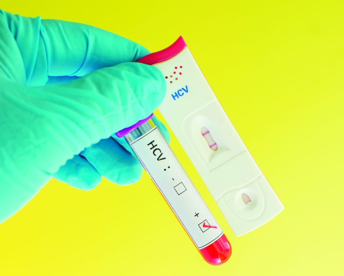
Other reactions were more forceful. “Kind of pathetic, isn’t it?” said John D. Scott, MD, assistant director of the Hepatitis and Liver Clinic at Harborview Medical Center, and an associate professor of medicine at the University of Washington, Seattle.
The researchers analyzed 2013 and 2015 data from the National Health Interview Survey, which included records for 21,827 baby boomers with HCV testing data.
The slight increase overall of 12.3% to 13.8% was small but also statistically significant (P = .013). Some populations fared better: Compared with the privately insured, those with Medicare plus Medicaid were more likely to have been tested (prevalence ratio, 1.83; 95% confidence interval, 1.32-2.53), as were those only on Medicaid (PR, 1.35; 95% CI, 1.04-1.76), and those with military insurance (PR, 1.62; 95% CI, 1.16-2.26).
The study could be subject to recall bias, since it relied on participants’ self-reports.
The authors speculate that the higher prevalence of testing in those with military insurance may reflect efforts by the Veterans Health Administration to reduce the high prevalence of HCV-associated disease among veterans.
It’s entirely possible to increase testing rates, according to Dr. Scott, who has a grant from the Centers for Disease Control and Prevention to study ways to increase uptake. “Probably the easiest thing to do is just incorporate this information into your electronic medical record and make it part of your alerts and standard preventative practices. Try to automate a lot of this rather than remind a very busy primary care doctor of all the things they have to do,” he said.
For example, one strategy that Seattle’s King County has employed is to automatically notify the testing laboratory if an antibody test is positive. “The lab knows to keep that blood and run a second (nucleic acid) test without the patient having to come back. That has helped to get our confirmatory rates up,” said Dr. Scott.
More broadly, the importance of testing needs to be emphasized, according to Paul J. Thuluvath, MD, medical director at the Institute of Digestive Health and Liver Disease at Mercy Medical Center, Baltimore, and a professor of medicine and surgery at the University of Maryland. “We need everybody to buy into this: the primary care physicians, internists, and gynecologists. If they are not convinced of the importance of this, it’s not going to happen. And I don’t think many primary care physicians and internists are convinced yet,” he said.
AGA Resource
Through the HCV Clinical Service Line, AGA offers tools to help you become more efficient, understand quality standards and improve the process of care for patients. Learn more at http://www.gastro.org/patient-care/conditions-diseases/hepatitis-c
FROM AMERICAN JOURNAL OF PREVENTIVE MEDICINE
Herbal/dietary supplements linked to liver injury requiring transplant
Herbal or dietary supplements are the fourth most common cause of drug-induced acute hepatic necrosis requiring liver transplantation in the United States, according to a study of liver transplant registry data.
Researchers analyzed registry data for 2,408 adults who underwent urgent liver transplantation for acute hepatic necrosis between 2003 and 2015, 625 of whom were recorded as having drug-induced liver injury. Of these, 21 cases were reportedly due to herbal or dietary supplements, all of which occurred after 2007. Eight cases were attributed to Lipolyze, Hydroxycut, testosterone, and OxyElite Pro, a muscle-building and weight-loss dietary supplement containing a blend of plant-derived extracts. The remaining cases did not list a specific product (Transplantation Proc 2017;49[2]:322-5).
“The potential hepatotoxicity of these HDS [herbal/dietary supplement] agents can result in the need for liver transplantation for irreversible liver failure,” wrote Dr. Linda L. Wong of the University of Hawaii and coauthors, citing two cases in Hawaii where patients needed liver transplant after taking OxyElite Pro.
They also referred to other case reports and case series of liver transplantation occurring in individuals who had taken the supplements usnic acid, Herbalife, Hydroxycut, linoleic acid, black cohosh, Chinese herbs, kava kava, skullcap, ma huang, or bai fang herb. “The benefits of the current regulatory oversight of medications breaks down when patients misuse non-FDA [Food and Drug Administration] approved herbal/dietary supplements (HDS) or ‘natural remedies’ with their inherent unknown side effects, interactions, and complications,” they wrote.
The mean age of individuals for which an herbal/dietary supplement was thought to be the cause of the hepatotoxicity was 36.8 years, 14 of the 21 were female, and the mean waiting time for liver transplantation was 4.7 days. One of the 21 patients died.
Overall, the most common drug associated with drug-induced liver injury was acetaminophen (300 cases), followed by antituberculosis medications (30), and antibiotics (30).
The authors suggested the true figure for HDS-induced liver transplantation may be underestimated, pointing to the fact that in this study, a further 154 cases were recorded as drug-induced injury but no drug was listed.
“Transplant centers and physicians may have difficulty identifying the exact inciting agent because patients do not typically report HDS when queried about medication use,” the authors wrote.
Given this, they recommended that transplant professionals specifically ask potential transplant recipients about their use of HDS, and request a detailed list of these along with the time course of use. They noted that family members and friends may not be privy to this information, and the patients themselves may not be in a position to answer because of decompensation with encephalopathy.
“It is critical to obtain this information as early as possible in the evaluation of the potential liver transplant recipient with an unknown etiology of liver disease or who presents with acute hepatic decompensation,” they wrote.
They also called for transplant professionals to report any information about confirmed HDS use in their patients to the FDA.
“It is imperative that transplant professionals inform the FDA of these dangerous compounds, with the ultimate intent of enforcing regulatory oversight,” they wrote. “This adherence to strict reporting will in turn translate to a reduction in the need for liver transplantation from HDS use.”
No conflicts of interest were declared.
FROM TRANSPLANTATION PROCEEDINGS
Key clinical point: Herbal or dietary supplements are the fourth most common cause of drug-induced acute hepatic necrosis requiring liver transplantation in the United States.
Major finding: Twenty-one cases of liver transplantation linked to the use of herbal or dietary supplements were recorded between 2003 and 2015.
Data source: Analysis of registry data from 2,408 adults who underwent urgent liver transplantation for acute hepatic necrosis.
Disclosures: No conflicts of interest were declared.
VIDEO: Point-of-care assay caught acetaminophen toxicity
A rapid point-of-care assay for acetaminophen-related liver toxicity had a sensitivity of 100% and a specificity of 86%, compared with etiologic diagnosis, based on the results of a multicenter study published in the April issue of Clinical Gastroenterology and Hepatology.
The test might help guide treatment decisions for these patients in the emergency department and intensive care unit, said Dean W. Roberts, PhD, of the University of Arkansas, Little Rock, and his associates.
About 45% of acute liver failure cases in the United States stem from acetaminophen toxicity, but the diagnosis can be hard to confirm because the drug has a short half-life and patients often cannot or will not report an overdose, which also may consist of multiple exposures, limiting the interpretability of the Rumack nonogram. High-pressure liquid chromatography with electrochemical detection (HPLC-EC) accurately detects acetaminophen-protein adducts (3-[cysteine-S-yl] acetaminophen) released by lysed hepatocytes into the peripheral circulation, but this test requires specialized equipment and skilled personnel, the researchers noted (Clin Gastroenterol Hepatol. 2016 Sep 15. doi: 10.1016/j.cgh.2016.09.007).
The point-of-care assay was positive in all 33 patients diagnosed with acetaminophen toxicity, for a test sensitivity of 100%, the researchers reported. The median band amplitude for cases was 584 (range, 222-1,027), significantly lower than that for patients with nonacetaminophen acute liver failure (3,678; range, 394-8,289; P less than .001) or for controls (8,971; range, 5,151-11,108; P less than .001). Band amplitude correlated inversely with adduct levels because AcetaSTAT is a competitive immunoassay – the presence of adducts decreases reactions at the test band, the investigators reported.
AcetaSTAT results were negative for 25 of 29 patients who were initially diagnosed with nonacetaminophen liver failure, for a test specificity of 86%, a positive predictive value of 89%, and a negative predictive value of 100%. Among the remaining four “false positives,” three tested near or above the toxicity threshold on HPLC-EC and were considered positive after further review, the investigators said. The fourth false-positive case was HPLC-EC–negative autoimmune hepatitis.
AcetaSTAT might not catch cases very early after acetaminophen overdose or that have only mild toxicity, the researchers noted. Nonetheless, it can help guide treatment decisions “at the point of clinical care,” they said. “Because the survival rate of acetaminophen acute liver failure is more favorable than that of other causes of acute live failure, assay results could impact future physician referral patterns and reduce medical costs associated with additional tests to determine the etiology of liver injury.”
The National Institute of Diabetes and Digestive and Kidney Diseases funded the study. Dr. Roberts and two coinvestigators are part owners of Acetaminophen Toxicity Diagnostics and have submitted a patent application for the AcetaSTAT serum assay used in this study. There were no other disclosures.
Source: American Gastroenterological Association
FROM CLINICAL GASTROENTEROLOGY AND HEPATOLOGY
Key clinical point:
Major finding: Compared with etiologic diagnosis, its sensitivity was 100%, specificity was 86%, positive predictive value was 89%, and negative predictive value was 100%.
Data source: Competitive immunoassays of serum samples from 19 healthy controls, 29 patients with nonacetaminophen acute liver failure, and 33 patients with acetaminophen-induced acute liver failure.
Disclosures: The National Institute of Diabetes and Digestive and Kidney Diseases funded the study. Dr. Roberts and two coinvestigators are part owners of Acetaminophen Toxicity Diagnostics and have submitted a patent application for the AcetaSTAT serum assay used in this study. There were no other disclosures.
VIDEO: Bacterial DNA predicted infections associated with prednisolone in severe alcoholic hepatitis
High baseline levels of circulating bacterial DNA increased the odds of serious infections by nearly fivefold in patients receiving prednisolone for severe alcoholic hepatitis, even after controlling for MELD score and white blood cell count, investigators reported in the April issue of Gastroenterology (2016 Dec 31. doi: 10.1053/j.gastro.2016.08.029).
“Patients with severe alcoholic hepatitis given prednisolone are at greater risk for developing serious infections and infections after treatment than patients not given prednisolone, which may offset its therapeutic benefit,” Nikhil Vergis, MD, and his associates wrote in Gastroenterology. “Level of circulating bacterial DNA before treatment could identify patients at high risk of infection if given prednisolone, which could be used to select therapies for patients with severe alcoholic hepatitis.”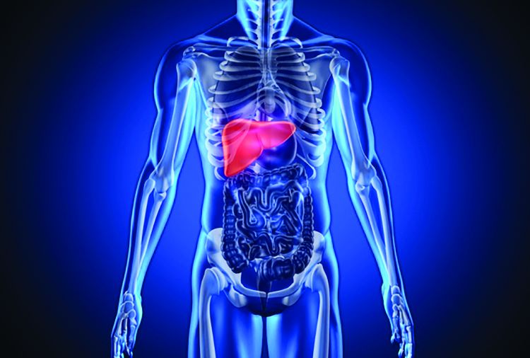
To further explore rates and predictors of infections in STOPAH, the researchers analyzed longitudinal data on incident infections for 1,092 trial participants who received either prednisolone (40 mg daily) or pentoxifylline (400 mg three times daily). For 731 patients, they also examined whether baseline circulating levels of 16s ribosomal bacterial DNA were associated with infections.
A total of 135 patients (12%) had an infection at baseline, 251 (23%) developed infections during treatment, and 89 (8%) developed infections after treatment, the investigators reported. Prednisolone therapy was not associated with infections during treatment, but was associated with a nearly 30% rise in the odds of serious posttreatment infections compared with pentoxifylline (odds ratio, 1.27; 95% confidence interval, 1.27-2.92; P = .002). Prednisolone recipients who developed infections were significantly more likely to die within 90 days than those who did not, even after controlling for end-stage liver disease or Lille score (OR, 2.5; 95% CI, 1.4-4.3; P = .002). Antibiotic therapy appeared to significantly reduce the risk of mortality among infected prednisolone recipients (13% vs. 52%; OR, 0.13; 95% CI 0.04-0.47; P = .002).
There was “a striking association between bacterial DNA and the development of infection within 7 days in patients treated with prednisolone,” the researchers reported. These patients had a median baseline circulating DNA level of 20.9 pg/mL, while prednisolone recipients who did not develop infections had a median baseline bacterial DNA level of 8.3 pg/mL (P = .004). Bacterial DNA predicted infections with an area under receiver operating characteristic curve of 0.70 (95% CI, 0.58-0.83; P = .003), which substantially exceeded the curve for white blood cell count (0.58).
A cut-off value of 18.5 pg/mL was 80% specific for predicting infection within 7 days of prednisolone therapy, the investigators also reported. Bacterial DNA level did not, however, predict infections within 7 days of pentoxifylline therapy, and pentoxifylline was not linked with infections that were serious, infections during treatment, or infections after treatment. (P =.08).
Using bacterial DNA levels to guide prednisolone prescription also appeared to reduce 90-day mortality in this patient population, although the effect achieved borderline statistical significance, the researchers said. “Larger prospective randomized studies are needed to definitely report whether bacterial DNA-guided therapy can [have an] impact on mortality in severe alcoholic hepatitis, and perhaps in other acute inflammatory conditions” in which immunosuppression is required, they added.
The National Institute for Health Research and Wellcome Trust and Medical Research Council provided funding. Dr. Vergis and 10 coinvestigators disclosed no conflicts of interest. Senior author Dr. Mark Thursz and one coinvestigator disclosed ties to Gilead, Bristol-Myers Squibb, AbbVie, Abbott, and Norgine.
Source: American Gastroenterological Association
FROM GASTROENTEROLOGY
Key clinical point: High baseline levels of circulating bacterial DNA predicted serious infections in patients receiving prednisolone for severe alcoholic hepatitis.
Major finding: The odds of serious posttreatment infections were significantly higher for prednisolone compared with pentoxifylline (OR, 1.27; P = .002). High baseline levels of circulating bacterial DNA predicted infection within 7 days of prednisolone therapy (adjusted OR, 4.68; P = .001).
Data source: An analysis of 1,092 patients with severe alcoholic hepatitis from the randomized, double-blind STOPAH trial.
Disclosures: The National Institute for Health Research and Wellcome Trust and Medical Research Council provided funding. Dr. Vergis and 10 coinvestigators reported having no competing interests. Senior author Mark Thursz and one coinvestigator disclosed ties to Gilead, Bristol-Myers Squibb, AbbVie, Abbott, and Norgine.
Unrestricted DAA access halved Dutch HCV incidence in HIV
SEATTLE – New hepatitis C infections among HIV-positive men who have sex with men (MSM) were halved in the Netherlands by unrestricted access to direct-acting antivirals (DAAs), primarily ledipasvir/sofosbuvir tablets (Harvoni), according to Dutch investigators.
Since 2015, DAAs have been available to all newly acquired hepatitis C virus (HCV) patients without restriction. Due to the high cost of the drugs, payers in the United States and some other Western countries limit access to only patients with severe liver disease.*
The Dutch government, however, requires insurers to cover them, and has negotiated price discounts with makers. “The price that is paid is secret,” but it’s less than the standard cost of, for instance, €45,000 for a 3-month course of [ledipasvir/sofosbuvir] in the Netherlands, said senior investigator Bart Rijnders, MD, an infectious diseases assistant professor at Erasmus University Medical Center, Rotterdam.
The study compared the incidence of acute HCV (aHCV) in HIV-positive MSM in 2014, before unrestricted access to DAAs, to the incidence of aHCV in 2016, after limits were lifted. The investigators used data from 18 HIV treatment centers spread across the Netherlands, capturing about 80% of Dutch MSM being treated for HIV.
In 2014, there were 93 aHCV infections diagnosed among the men, translating to an incidence of 11.2 cases per 1,000 person-years of follow-up (95% CI, 9.1-13.7 cases). In 2016, there were 49 aHCV cases, an incidence of 5.5 cases per 1,000 person-years (95% CI, 4.1–7.2, P less than .001). At the same time, there was a substantial increase in new syphilis cases, indicating that the 51% reduction in aHCV over 2 years was not due to changes in behavior.
Meanwhile, “within 14 months after these drugs became available to all, 75% of the HIV-positive MSM in the Netherlands were cured of their infection,” Dr. Rijnders said at the Conference on Retroviruses & Opportunistic Infections in partnership with the International Antiviral Society.
Ledipasvir/sofosbuvir was the DAA used by about 90% of the men.
In short, unrestricted access to DAAs wiped out the infection so that men were no longer passing it to other men. The results are “an example of what is possible if you search for HCV and treat it as soon as you find it. You cure patients and prevent new infections. In the long run, you may save money,” he said, especially as more DAA options come on the market and prices fall.
Almost all the subjects were seen in their HIV clinic at least twice a year. An uptick in liver enzymes triggered HCV testing. The investigators checked positive results against patients’ own stored blood samples to distinguish new from chronic infections.
The study wasn’t funded, but Dr. Rijnders is a paid researcher for Merck’s DAA option, elbasvir/grazoprevir (Zepatier).
*This story was updated on February 24, 2017.
This is the first proof that early treatment of acute HCV could be a form of prevention. By removing the fibrosis requirement and restrictions forbidding treatment of people who are actively engaged in high-risk behaviors, they are reducing new infections.
We are far behind in the United States; 90% of states have restrictions that don’t allow uniform uptake of hepatitis C treatment and many forbid treatment for people who are actively using drugs. Having restrictions on people that don’t have enough liver disease is like telling a person with HIV they can’t be treated because their CD4 count isn’t below 200.
David Thomas, MD, is professor of medicine and director of the division of infectious diseases at Johns Hopkins University, Baltimore. He wasn’t involved with the work.
SEATTLE – New hepatitis C infections among HIV-positive men who have sex with men (MSM) were halved in the Netherlands by unrestricted access to direct-acting antivirals (DAAs), primarily ledipasvir/sofosbuvir tablets (Harvoni), according to Dutch investigators.
Since 2015, DAAs have been available to all newly acquired hepatitis C virus (HCV) patients without restriction. Due to the high cost of the drugs, payers in the United States and some other Western countries limit access to only patients with severe liver disease.*
The Dutch government, however, requires insurers to cover them, and has negotiated price discounts with makers. “The price that is paid is secret,” but it’s less than the standard cost of, for instance, €45,000 for a 3-month course of [ledipasvir/sofosbuvir] in the Netherlands, said senior investigator Bart Rijnders, MD, an infectious diseases assistant professor at Erasmus University Medical Center, Rotterdam.
The study compared the incidence of acute HCV (aHCV) in HIV-positive MSM in 2014, before unrestricted access to DAAs, to the incidence of aHCV in 2016, after limits were lifted. The investigators used data from 18 HIV treatment centers spread across the Netherlands, capturing about 80% of Dutch MSM being treated for HIV.
In 2014, there were 93 aHCV infections diagnosed among the men, translating to an incidence of 11.2 cases per 1,000 person-years of follow-up (95% CI, 9.1-13.7 cases). In 2016, there were 49 aHCV cases, an incidence of 5.5 cases per 1,000 person-years (95% CI, 4.1–7.2, P less than .001). At the same time, there was a substantial increase in new syphilis cases, indicating that the 51% reduction in aHCV over 2 years was not due to changes in behavior.
Meanwhile, “within 14 months after these drugs became available to all, 75% of the HIV-positive MSM in the Netherlands were cured of their infection,” Dr. Rijnders said at the Conference on Retroviruses & Opportunistic Infections in partnership with the International Antiviral Society.
Ledipasvir/sofosbuvir was the DAA used by about 90% of the men.
In short, unrestricted access to DAAs wiped out the infection so that men were no longer passing it to other men. The results are “an example of what is possible if you search for HCV and treat it as soon as you find it. You cure patients and prevent new infections. In the long run, you may save money,” he said, especially as more DAA options come on the market and prices fall.
Almost all the subjects were seen in their HIV clinic at least twice a year. An uptick in liver enzymes triggered HCV testing. The investigators checked positive results against patients’ own stored blood samples to distinguish new from chronic infections.
The study wasn’t funded, but Dr. Rijnders is a paid researcher for Merck’s DAA option, elbasvir/grazoprevir (Zepatier).
*This story was updated on February 24, 2017.
AT CROI
Key clinical point:
Major finding: In 2014, there were 93 acute HCV infections diagnosed among HIV-positive men who have sex with men, translating to an incidence of 11.2 cases per 1,000 person-years of follow-up (95% CI, 9.1-13.7 cases). In 2016, there were 49 cases, an incidence of 5.5 cases per 1,000 person years (95% CI, 4.1–7.2, P less than .001).
Data source: HIV treatment centers in the Netherlands
Disclosures: The study wasn’t funded, but the senior investigator is a paid researcher for Merck’s DAA option, elbasvir/grazoprevir (Zepatier).
Biannual HCC ultrasound cost-effective, lifesaving in cirrhosis
Twice-yearly ultrasound screening for liver cancer increases survival an average of almost 5 months in cirrhosis patients and costs about $32,415 per life-year gained, according to new economic modeling.
“The overall gain in life expectancy might be considered modest,” but it’s “good by cancer screening standards.” The cost, meanwhile, is within the accepted threshold in the United States of $30,000-50,000 per life-year gained (LYG), said French investigators led by statistician Benjamin Cadier of the French National Institute of Health and Medical Research, Paris (Hepatology. 2017 Feb 8. doi:10.1002/hep.28961).
To estimate probabilities and costs for various scenarios, the team combined data from two large French cohorts – one of viral cirrhosis, another of HCC – with French and U.S. pricing data, among other information. French costs with biannual screening were far less, at $1,754 per LYG, because of a 4-10–fold difference in the price of surveillance and first-line curative treatment. The team estimated that 10-year overall survival was 67% with current monitoring practices, and 76% with biannual ultrasound.
The mean survival increase from 6.8 to 7.2 years was attributed to earlier detection, higher access to curative first-line treatment, and better treatment results. Radiofrequency ablation (RFA), as opposed to liver resection or transplant, provided the best value for the money. “Our results indicate that [guideline-directed] monitoring for patients with cirrhosis is cost-effective,” the team said.
“Later detection not only reduced the likelihood of curative treatment, but also increased the proportion of [liver transplants] among the curative treatments,” they said.
In the modeling, when biannual ultrasound surveillance detected a suspicious nodule, the recall policy included magnetic resonance imaging or computerized tomography, and liver biopsy, if needed, based on recent international guidelines, with subsequent treatment.
There was no outside funding for the work. The lead investigator had no conflicts. Other investigators were consultants for Bayer, General Electric, AbbVie, Janssen, Bristol-Meyer Squibb, Gilead, and other companies.
FROM HEPATOLOGY
Key clinical point:
Major finding: Twice-a-year ultrasound to catch liver cancer early increases survival an average of nearly 5 months in cirrhosis patients, and costs about $32,415 per life-year gained.
Data source: Economic modeling of two French cohorts.
Disclosures: There was no outside funding for the work. The lead investigator had no conflicts. Other investigators were consultants for Bayer, General Electric, AbbVie, Janssen, Bristol-Meyer Squibb, Gilead, and other companies.
Liver transplantation largely effective in critically ill children
The use of advanced critical care in children and infants with liver failure is justified because orthotopic liver transplantation can be performed on the sickest children and achieve acceptable outcomes, results from a large analysis demonstrated.
“Hand in hand with improved care for critically ill children with liver failure, posttransplant critical care has made tremendous strides,” Abbas Rana, MD, wrote in a study published online in the Journal of the American College of Surgeons. “Our recipients have gotten sicker while our postoperative outcomes have improved. The question then becomes, have our operative skills and postoperative critical care management kept up with the abilities to keep sick children with liver failure alive? Just because transplantation is now possible in our sickest children, is it justified?”
To find out, Dr. Rana of the division of abdominal transplantation and hepatobiliary surgery at Baylor College of Medicine, Houston, and colleagues retrospectively analyzed United Network for Organ Sharing data from all orthotopic liver transplantation (OLT) recipients between Sept. 1, 1987, and June 30, 2015. The analysis paired the liver registry data with data collected by the Organ Procurement and Transplantation Network, and was limited to transplant recipients younger than age 18. The researchers followed a total of 13,723 recipients from date of transplant until either death or the date of last known follow-up (J Am Coll Surg. 2016 Dec 25. doi: 10.1016/j.jamcollsurg.2016.12.025).
In another part of the study, the researchers retrospectively reviewed the charts of 354 patients under 18 years of age who underwent OLT between March 1, 2002, and June 30, 2015, at Texas Children’s Hospital, including 65 who were admitted to the ICU at the time of transplantation.
In the analysis of national data, the researchers found that the rates of 1-year survival following OLT in children in the ICU improved from 60% in 1987 to 92% in 2013 (P less than .001). The rates of 1-year survival also improved for children on dialysis at the time of transplant (from 50% in 1995 to 95% in 2013; P less than .001) and for those dependent on a mechanical ventilator at the time of transplant (from 49% in 1994 to 94% in 2013; P less than .001). The significant risk factors were two previous transplants (hazard ratio, 4.2), one previous transplant (HR, 2.5), serum sodium greater than 150 mEq/L (HR, 2.0), dialysis or glomerular filtration rate less than 30 mL/min per 1.73 m2 (HR, 2.0), mechanical ventilator dependence (HR, 1.8), body weight under 6 kg (HR, 1.8), encephalopathy (HR, 1.8), and annual center volume of fewer than five cases (HR, 1.7).
In the experience at Texas Children’s Hospital, the researchers observed “preserved and successful patient survival outcomes” in many markers of acuity. For example, the 10-year survival rates for patients dependent on mechanical ventilation and dialysis were 85% and 96%, respectively, and reached 100% for those requiring therapeutic plasma exchange, molescular adsorbent recirculating system (MARS) liver dialysis, and vasopressors.
“Our collective ability to keep sick children alive with liver failure has improved considerably over the years,” the researchers wrote. “Keeping pace, this analysis demonstrates that the posttransplant outcomes have also improved dramatically. The survival outcomes are comparable to the general population, justifying the use of scarce donors. Although we cannot declare in absolute that no child should be left behind, we can demonstrate acceptable outcomes to date and urge the continual revisiting of our concepts of futility.
“We have learned throughout our experience that almost every child with end-stage liver disease and acute liver failure should be offered liver replacement, as long as the vasoactive medication and mechanical support are not maximized prior to the initiation of the OLT procedure. Every effort should be made to transplant our sickest children.”
This study was supported by the Cade R. Alpard Foundation. The researchers reported having no relevant financial disclosures.
FROM THE JOURNAL OF THE AMERICAN COLLEGE OF SURGEONS
Key clinical point:
Major finding: The rates of 1-year survival following orthotopic liver transplantation in ICU children improved from 60% in 1987 to 92% in 2013 (P less than .001).
Data source: An analysis of 13,723 patients under the age of 18 years who underwent OLT between Sept. 1, 1987, and June 30, 2015.
Disclosures: The study was supported by the Cade R. Alpard Foundation. The researchers reported having no relevant financial disclosures.
Don't rule out liver transplant grafts from octogenarians
Donors aged 80 years or older are not necessarily inferior for a liver transplantation (LT) graft, compared with young ideal donors (aged 18-39 years), according to an analysis of the perioperative LT period.
While “the potential risks and benefits associated with the use of livers from octogenarian donors must be closely weighed, with careful donor evaluation, selective donor-to-recipient matching and skilled perioperative care, octogenarian grafts do not affect the short-term course of patients undergoing LT,” concluded Gianni Biancofiore, MD, of Azienda Ospedaliera-Universitaria Pisana, Pisa, Italy, and his coauthors (Dig Liver Dis. 2017. doi: 10.1016/j.dld.2017.01.149).
Perioperative differences were insubstantial in terms of cardiovascular complications (P = .2), respiratory complications (P = 1.0), coagulopathy (P = .5), and incidence of perfusion syndrome (P = .3). Median ICU length of stay of the two groups was identical (P = .4). No differences in terms of death or retransplant were observed during the ICU stay.
“Accordingly, anesthesiologists and intensivists should not label liver allografts from donors aged 80 years [or older] as ‘unusable’ or ‘high risk’ ” based on age alone, the authors concluded.
The authors declared no sources of funding and no conflicts of interest.
FROM DIGESTIVE AND LIVER DISEASE
Nivolumab shows promise for pretreated advanced HCC
SAN FRANCISCO – Nivolumab was associated with durable responses in heavily pretreated patients with advanced hepatocellular carcinoma in the dose-escalation and dose-expansion phases of the CheckMate 040 study.
Further, the safety profile of nivolumab, a fully human IgG4 monoclonal antibody inhibitor of programmed death-1 (PD-1), was consistent with that observed in other solid tumors; events were manageable, and no new safety signals were detected, Ignacio Melero, MD, of Clinica Universidad de Navarra, Pamplona, Spain, reported at the symposium, sponsored by ASCO, ASTRO, the American Gastroenterological Association, and Society of Surgical Oncology.
In all, 262 patients with histologically confirmed advanced disease not amenable to curative resection were treated across the CheckMate 040 dose-escalation phase (phase I), which evaluated 0.1-10 mg/kg of nivolumab given every 2 weeks, and the dose-expansion phase (phase II), which involved nivolumab at a dose of 3 mg/kg every 2 weeks in four cohorts: sorafenib naive/intolerant patients, sorafenib progressors, hepatitis C virus-infected patients, and hepatitis B virus-infected patients.
“Because of the inflammation that often afflicts the liver, we were very afraid of precipitating hyper-acute or fulminant hepatitis in these patients, and that was the reason we undertook a very careful dose escalation,” Dr. Melero said, adding that due to efficacy and safety results in the dose escalation phase, the 3-mg/kg dose used in other clinical trials was expanded to all subjects, including uninfected patients and those with HCV or HBV infection.
In regard to safety and tolerability – the primary endpoint of phase I – grade 3/4 treatment-related adverse events occurred in 20% of patients. No maximum tolerated dose was reached during dose escalation, Dr. Melero said.
The most common reason for treatment discontinuation was disease progression; very few patients discontinued for toxicity, he said, noting that “the safety profile was very consistent with what has been reported in other indications.”
In fact, most grade 3/4 events involved laboratory abnormalities without clinical repercussions, he said.
The safety profile in phase II was consistent with that observed in phase I, and no differences in safety were seen in the three categories of hepatocellular carcinoma patients in the study.
In regard to objective response – the primary endpoint of phase II – the investigator-assessed rates were 16.2% in phase I and 18.6% in phase II, and the rates assessed by blinded independent central review were 18.9% and 14.5% in the phases, respectively. Based on imaging assessed using modified RESIST criteria, the rates were 21.6% and 18.6%, respectively.
The median duration of response was 17.1 months in phase I, and had not yet been reached in phase II, Dr. Melero said, adding that in uninfected patients, an “important number of patients developed stable disease that was durable.”
This also was seen in patients with chronic hepatitis, he noted.
“We believe that stabilization of disease is a driver of survival, because we have seen that in dose escalation, 45% of patients were alive 18 months after treatment onset, and at last data base log [in dose escalation], 71% were alive 9 months after starting treatment,” he said.
Of note, objective response rates were similar in patients treated with sorafenib and in those who received nivolumab as first-line treatment, and they were similar regardless of tumor programmed death-ligand 1 (PD-L1) expression.
Quality of life, as reported by patients via EQ-5D questionnaire, remained stable over time.
Study subjects had a median age of 63 years, and were heavily pretreated – most often by sorafenib.
Hepatocellular carcinoma diagnosed at advanced stages has a poor prognosis, and those who progress on sorafenib – the only systemic therapy option in such patients – have few options, Dr. Melero said.
Nivolumab, which has been shown to restore T-cell–mediated antitumor activity, has demonstrated clinical and survival benefit in a number of tumor types.
The current findings suggest that it also is a promising alternative for advanced hepatocellular carcinoma.
“Nivolumab demonstrated objective responses and long-term survival in both sorafenib-experienced and sorafenib-naive patients. Nivolumab monotherapy provided early, stable and durable responses when they took place, efficacy was observed irrespective of chronic viral infection by hepatitis viruses, and responses were also observed in PD-L1-negative cases upon examination of biopsies,” he concluded, noting that a randomized phase III trial comparing nivolumab with systemic sorafenib is ongoing.
CheckMate 040 was sponsored by Bristol Myers Squibb. Dr. Melero reported receiving honoraria from and serving as a consultant or adviser for AstraZeneca, Bayer, Bristol-Myers Squibb, Incyte, Merck Serono, MSD, and Roche, and reported research funding to his institution from Alligator Bioscience, Pfizer, and Tusk Therapeutics.
AT THE 2017 GASTROINTESTINAL CANCERS SYMPOSIUM
Key clinical point:
Major finding: Investigator-assessed objective response rates were 16.2% in phase I and 18.6% in phase II.
Data source: The CheckMate 040 phase I and phase II clinical studies involving 262 patients.
Disclosures: CheckMate 040 was sponsored by Bristol Myers Squibb. Dr. Melero reported receiving honoraria from and serving as a consultant or adviser for AstraZeneca, Bayer, Bristol-Myers Squibb, Incyte, Merck Serono, MSD, and Roche, and reported research funding to his institution from Alligator Bioscience, Pfizer, and Tusk Therapeutics.
AGA Guideline: Acute liver failure
Physicians should avoid routinely testing patients with acute liver failure for Wilson’s disease unless there is “high clinical suspicion” for the disorder, according to a new guideline from the AGA Institute.
Wilson’s disease so rarely accompanies acute liver failure that a positive test will have low predictive value, Steven L. Flamm, MD, of Northwestern University, Chicago, and his associates wrote in the February issue of Gastroenterology (doi: 10.1053/j.gastro.2016.12.026). Diagnosing Wilson’s disease also is unlikely to change treatment “because liver transplantation is the ultimate outcome,” they emphasize.
The guideline grades seven recommendations as “conditional” based on “very-low” quality evidence. These include the statement on Wilson’s disease testing, plus suggestions to test and treat patients with ALF for herpes simplex virus (HSV) infection, to test pregnant patients for hepatitis E virus infection, and to perform autoantibody testing for autoimmune hepatitis. Case series report only about a 1% prevalence of HSV infection in ALF, and there is little information on diagnostic accuracy or treatment in this setting, the guidelines state. Although acyclovir is relatively safe and inexpensive, data on efficacy is limited to “a suggestion on a case-report level that patients with acute hepatitis secondary to HSV do better with treatment than without.”
The guideline also conditionally recommends against routine testing for varicella zoster virus infection and routine liver biopsy in ALF. The authors note only about 10 case reports of varicella zoster–associated ALF and few data on how liver biopsy results in ALF alter treatment plan, outcome, or the choice to seek liver transplantation. The experts do recommend prognostic scoring with Model for End-Stage Liver Disease, which pooled analyses have found to be more sensitive than King’s College Criteria, they wrote.
The guideline conditionally recommends against empirically treating elevated intracranial pressure in ALF, on the basis of five randomized trials that found no overall mortality benefit of moderate hypothermia, hypertonic saline, L-ornithine, L-aspartate, intravenous mannitol, or hyperventilation.
The experts cite insufficient evidence to recommend using N-acetyl cysteine in patients whose ALF is not associated with acetaminophen exposure. Likewise, they find inadequate data to make any recommendation about using extracorporeal liver support systems outside of the setting of clinical trials. Although such systems can “potentially” buy time for patients to either spontaneously recover without transplant or survive longer on the transplantation list, three systematic reviews found “no clear effect on mortality,” and randomized trials reported either null results or a “marginally significant survival benefit” in the face of steep costs and potentially significant toxicities, the authors emphasize.
None of the experts had relevant financial disclosures.
FROM GASTROENTEROLOGY