User login
EULAR nears first recommendations for managing Sjögren’s syndrome
AMSTERDAM – , and they divide the treatment targets into sicca syndrome and systemic manifestations of the disease.
“In Sjögren’s, we always have two subtypes of patients: those who have sicca syndrome only, and those with sicca syndrome plus systemic disease,” explained Soledad Retamozo, MD, who presented the current version of the recommendations at the European Congress of Rheumatology. “We wanted to highlight that there are two types of patients,” said Dr. Retamozo, a rheumatologist at the University of Córdoba (Argentina). “It’s hard to treat patients with sicca syndrome plus fatigue and pain because there is no high-level evidence on how to do this; all we have is expert opinion,” Dr. Retamozo said in an interview.
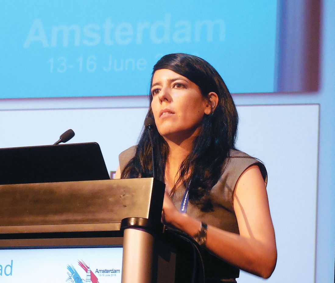
In fact, roughly half of the recommendations have no supporting evidence base, as presented by Dr. Retamozo. That starts with all three general recommendations she presented:
• Patients with Sjögren’s should be managed at a center of expertise using a multidisciplinary approach, which she said should include ophthalmologists and dentists to help address the mouth and ocular manifestations of sicca syndrome.
• Patients with sicca syndrome should receive symptomatic relief with topical treatments.
• Systemic treatments – glucocorticoids, immunosuppressants, and biologicals – can be considered for patients with active systemic disease.
The statement’s specific recommendations start with managing oral dryness, an intervention that should begin by measuring salivary gland (SG) dysfunction. The document next recommends nonpharmacologic interventions for mild SG dysfunction, pharmacological stimulation for moderate SG dysfunction, and a saliva substitute for severe SG dysfunction. All three recommendations are evidence based, relying on results from either randomized trials or controlled studies.
The second target for topical treatments is ocular dryness, which starts with artificial tears, or ocular gels or ointments, recommendations based on randomized trials. Refractory or severe ocular dryness should receive eye drops that contain a nonsteroidal anti-inflammatory drug or a glucocorticoid, based on controlled study results, or autologous serum eye drops, a strategy tested in a randomized trial.
The recommendations then shift to dealing with systemic manifestations, starting with fatigue and pain, offering the expert recommendation to evaluate the contribution of comorbid diseases and assess their severity with tools such as the Eular Sjögren’s Syndrome Patient-Reported Index (ESSPRI) (Ann Rheum Dis. 2011 June;70[6]:968-72), the Profile of Fatigue, and the Brief Pain Inventory.
Using evidence from randomized trials, the recommendations tell clinicians to consider treatment with analgesics or pain-modifying agents for musculoskeletal pain by weighing the potential benefits and adverse effects from this treatment.
For other forms of systemic disease, the recommendations offer the expert opinion to tailor treatment to the organ-specific severity using the ESSPRI definitions. If using glucocorticoids to treat systemic disease, they should be given at the minimum effective dose and for the shortest period of time needed to control active systemic disease, a recommendation based on retrospective or descriptive studies. Expert opinion called for using immunosuppressive treatments as glucocorticoid-sparing options for systemic disease, and this recommendation added that no particular immunosuppressive agent stands out as best compared with all available agents. In more than 95% of reported cases of systemic disease treatment in Sjögren’s patients, clinicians used the immunosuppressive drugs in association with glucocorticoids, Dr. Retamozo noted.
Finally, for systemic disease the recommendations cited evidence from controlled studies that B-cell targeted therapies, such as rituximab (Rituxan) and belimumab (Benlysta), may be considered in patients with severe, refractory systemic disease. An additional expert opinion was that the systemic, organ-specific approach should sequence treatments by using glucocorticoids first, followed by immunosuppressants, and finally biological drugs.
The recommendations finish with an entry that treatment of B-cell lymphoma be individualized based on the specific histopathologic subtype involved and the level of disease extension, an approach based on results from retrospective or descriptive studies.
The recommendations must still undergo final EULAR review and endorsement, with publication on track to occur before the end of 2018, Dr. Retamozo said.
She had no disclosures.
SOURCE: Retamozo S al. Ann Rheum Dis. 2018;77(Suppl 2):42. Abstract SP0159.
AMSTERDAM – , and they divide the treatment targets into sicca syndrome and systemic manifestations of the disease.
“In Sjögren’s, we always have two subtypes of patients: those who have sicca syndrome only, and those with sicca syndrome plus systemic disease,” explained Soledad Retamozo, MD, who presented the current version of the recommendations at the European Congress of Rheumatology. “We wanted to highlight that there are two types of patients,” said Dr. Retamozo, a rheumatologist at the University of Córdoba (Argentina). “It’s hard to treat patients with sicca syndrome plus fatigue and pain because there is no high-level evidence on how to do this; all we have is expert opinion,” Dr. Retamozo said in an interview.
In fact, roughly half of the recommendations have no supporting evidence base, as presented by Dr. Retamozo. That starts with all three general recommendations she presented:
• Patients with Sjögren’s should be managed at a center of expertise using a multidisciplinary approach, which she said should include ophthalmologists and dentists to help address the mouth and ocular manifestations of sicca syndrome.
• Patients with sicca syndrome should receive symptomatic relief with topical treatments.
• Systemic treatments – glucocorticoids, immunosuppressants, and biologicals – can be considered for patients with active systemic disease.
The statement’s specific recommendations start with managing oral dryness, an intervention that should begin by measuring salivary gland (SG) dysfunction. The document next recommends nonpharmacologic interventions for mild SG dysfunction, pharmacological stimulation for moderate SG dysfunction, and a saliva substitute for severe SG dysfunction. All three recommendations are evidence based, relying on results from either randomized trials or controlled studies.
The second target for topical treatments is ocular dryness, which starts with artificial tears, or ocular gels or ointments, recommendations based on randomized trials. Refractory or severe ocular dryness should receive eye drops that contain a nonsteroidal anti-inflammatory drug or a glucocorticoid, based on controlled study results, or autologous serum eye drops, a strategy tested in a randomized trial.
The recommendations then shift to dealing with systemic manifestations, starting with fatigue and pain, offering the expert recommendation to evaluate the contribution of comorbid diseases and assess their severity with tools such as the Eular Sjögren’s Syndrome Patient-Reported Index (ESSPRI) (Ann Rheum Dis. 2011 June;70[6]:968-72), the Profile of Fatigue, and the Brief Pain Inventory.
Using evidence from randomized trials, the recommendations tell clinicians to consider treatment with analgesics or pain-modifying agents for musculoskeletal pain by weighing the potential benefits and adverse effects from this treatment.
For other forms of systemic disease, the recommendations offer the expert opinion to tailor treatment to the organ-specific severity using the ESSPRI definitions. If using glucocorticoids to treat systemic disease, they should be given at the minimum effective dose and for the shortest period of time needed to control active systemic disease, a recommendation based on retrospective or descriptive studies. Expert opinion called for using immunosuppressive treatments as glucocorticoid-sparing options for systemic disease, and this recommendation added that no particular immunosuppressive agent stands out as best compared with all available agents. In more than 95% of reported cases of systemic disease treatment in Sjögren’s patients, clinicians used the immunosuppressive drugs in association with glucocorticoids, Dr. Retamozo noted.
Finally, for systemic disease the recommendations cited evidence from controlled studies that B-cell targeted therapies, such as rituximab (Rituxan) and belimumab (Benlysta), may be considered in patients with severe, refractory systemic disease. An additional expert opinion was that the systemic, organ-specific approach should sequence treatments by using glucocorticoids first, followed by immunosuppressants, and finally biological drugs.
The recommendations finish with an entry that treatment of B-cell lymphoma be individualized based on the specific histopathologic subtype involved and the level of disease extension, an approach based on results from retrospective or descriptive studies.
The recommendations must still undergo final EULAR review and endorsement, with publication on track to occur before the end of 2018, Dr. Retamozo said.
She had no disclosures.
SOURCE: Retamozo S al. Ann Rheum Dis. 2018;77(Suppl 2):42. Abstract SP0159.
REPORTING FROM THE EULAR 2018 CONGRESS
Predicting rituximab responses in lupus remains challenging
AMSTERDAM – Despite some “encouraging signs” seen in a cross-sectional study, it remains difficult to determine whether a patient with systemic lupus erythematosus (SLE) will respond to off-label rituximab therapy, according to David Isenberg, MD.
The presence of constitutional symptoms, which includes fatigue, at baseline were associated with a favorable response to rituximab at 6 months (odds ratio, 7.35; P = .01). Conversely, having more than one anti–extractable nuclear antigen (anti-ENA) antibody was associated with a worse response to rituximab at 12 months (OR, 0.33; P = .032).
The video associated with this article is no longer available on this site. Please view all of our videos on the MDedge YouTube channel
“It’s not a cure for lupus, that’s obvious, but used at the appropriate time in the right sort of patients, it can be very helpful,” Dr. Isenberg said in an interview at the European Congress of Rheumatology. That’s both in terms of improving disease activity and reducing the use of glucocorticosteroids and immunosuppressive drugs, he noted.
“The great mystery, as with all drugs, is: Are there any markers which might tell us that the disease is going to do better with rituximab than perhaps with another form of medication, or is it just saying to us, ‘Whatever you do, it doesn’t make much difference’?” Dr. Isenberg said. He discussed the results of a study involving the first 121 patients treated with rituximab at his institution, University College London Hospital (UCLH); he noted this is one of the largest single-center cohorts of individuals with SLE who have used rituximab.
The aim of the study, which was presented at the congress by Hiurma Sánchez Pérez, MD, was therefore to try to find demographic, clinical, and serological markers that might help in predicting the response to rituximab in SLE patients.

Response to treatment was determined using the British Isles Lupus Assessment Group (BILAG) Index. Patients who initially had markedly (BILAG A) or moderately (BILAG B) active disease but who no longer fell into these categories at 6 or 12 months were designated as responders. Those who remained were designated as nonresponders. In addition, any person moving into category A or B of the BILAG Index was said to have relapsed because of a flare in disease activity.
At 6 and 12 months, a respective 85% and 70% of patients exhibited a response to rituximab. Just under a quarter (24%) relapsed before 1 year, Dr. Sánchez Pérez reported.
Most of the patients in the cohort were white (n = 50) or Afro-Caribbean (n = 38), but ethnicity did not seem to play a role in predicting whether patients would respond to rituximab treatment.
Aside from constitutional symptoms and having more than one anti-ENA antibody, there were no other biological markers of response that remained significant after multivariate analysis.
The mean time to flare after rituximab was nearly 8 months, Dr. Sánchez Pérez said. Arthritis was the main manifestation seen during relapse (41% of patients), and mucocutaneous symptoms occurred in 21%. Biological markers of flare at 12 months after multivariate analysis were musculoskeletal symptoms at baseline (OR, 0.26; P = .039) and being anti-RNP antibody positive (OR, 10.56; P = .03).
“There were one or two encouraging signs,” said Dr. Isenberg, who was the senior author of the study. “Unfortunately, what I think the data show us, it remains pretty hard to know how any individual patient is going to respond to B-cell depletion using rituximab.”
Dr. Sánchez Pérez reported having no disclosures in relation to the study. Dr. Isenberg was one of the first people to use rituximab to treat patients with lupus but reported having no other competing interests.
SOURCE: Sánchez Pérez H and Isenberg D. Ann Rheum Dis. 2018;77(Suppl 2):177. EULAR 2018 Congress, Abstract OP0255.
AMSTERDAM – Despite some “encouraging signs” seen in a cross-sectional study, it remains difficult to determine whether a patient with systemic lupus erythematosus (SLE) will respond to off-label rituximab therapy, according to David Isenberg, MD.
The presence of constitutional symptoms, which includes fatigue, at baseline were associated with a favorable response to rituximab at 6 months (odds ratio, 7.35; P = .01). Conversely, having more than one anti–extractable nuclear antigen (anti-ENA) antibody was associated with a worse response to rituximab at 12 months (OR, 0.33; P = .032).
The video associated with this article is no longer available on this site. Please view all of our videos on the MDedge YouTube channel
“It’s not a cure for lupus, that’s obvious, but used at the appropriate time in the right sort of patients, it can be very helpful,” Dr. Isenberg said in an interview at the European Congress of Rheumatology. That’s both in terms of improving disease activity and reducing the use of glucocorticosteroids and immunosuppressive drugs, he noted.
“The great mystery, as with all drugs, is: Are there any markers which might tell us that the disease is going to do better with rituximab than perhaps with another form of medication, or is it just saying to us, ‘Whatever you do, it doesn’t make much difference’?” Dr. Isenberg said. He discussed the results of a study involving the first 121 patients treated with rituximab at his institution, University College London Hospital (UCLH); he noted this is one of the largest single-center cohorts of individuals with SLE who have used rituximab.
The aim of the study, which was presented at the congress by Hiurma Sánchez Pérez, MD, was therefore to try to find demographic, clinical, and serological markers that might help in predicting the response to rituximab in SLE patients.
Response to treatment was determined using the British Isles Lupus Assessment Group (BILAG) Index. Patients who initially had markedly (BILAG A) or moderately (BILAG B) active disease but who no longer fell into these categories at 6 or 12 months were designated as responders. Those who remained were designated as nonresponders. In addition, any person moving into category A or B of the BILAG Index was said to have relapsed because of a flare in disease activity.
At 6 and 12 months, a respective 85% and 70% of patients exhibited a response to rituximab. Just under a quarter (24%) relapsed before 1 year, Dr. Sánchez Pérez reported.
Most of the patients in the cohort were white (n = 50) or Afro-Caribbean (n = 38), but ethnicity did not seem to play a role in predicting whether patients would respond to rituximab treatment.
Aside from constitutional symptoms and having more than one anti-ENA antibody, there were no other biological markers of response that remained significant after multivariate analysis.
The mean time to flare after rituximab was nearly 8 months, Dr. Sánchez Pérez said. Arthritis was the main manifestation seen during relapse (41% of patients), and mucocutaneous symptoms occurred in 21%. Biological markers of flare at 12 months after multivariate analysis were musculoskeletal symptoms at baseline (OR, 0.26; P = .039) and being anti-RNP antibody positive (OR, 10.56; P = .03).
“There were one or two encouraging signs,” said Dr. Isenberg, who was the senior author of the study. “Unfortunately, what I think the data show us, it remains pretty hard to know how any individual patient is going to respond to B-cell depletion using rituximab.”
Dr. Sánchez Pérez reported having no disclosures in relation to the study. Dr. Isenberg was one of the first people to use rituximab to treat patients with lupus but reported having no other competing interests.
SOURCE: Sánchez Pérez H and Isenberg D. Ann Rheum Dis. 2018;77(Suppl 2):177. EULAR 2018 Congress, Abstract OP0255.
REPORTING FROM THE EULAR 2018 CONGRESS
Key clinical point: Only the presence of constitutional symptoms at baseline and having more than one anti–extractable nuclear antigen (anti-ENA) antibody were related to rituximab response.
Major finding: Having more than one anti-ENA antibody was related to a worse response to rituximab at 12 months.
Study details: A cross-sectional study of 121 patients with systemic lupus erythematosus treated with rituximab during 2000-2016.
Disclosures: Dr. Isenberg has used rituximab to treat patients with lupus for almost 20 years but reported having no other competing interests. Dr. Sánchez Pérez reported having no disclosures in relation to the study.
Source: Sánchez Pérez H and Isenberg D. Ann Rheum Dis. 2018;77(Suppl 2):177. EULAR 2018 Congress, Abstract OP0255.

Effort to phenotype pulmonary hypertension patients under way
SAN DIEGO – A massive effort to better understand and treat patients with pulmonary hypertension and right heart dysfunction is underway.
The endeavor, funded by the National Heart, Lung, and Blood Institute and the Pulmonary Hypertension Association and known as Redefining Pulmonary Hypertension Through Pulmonary Vascular Disease Phenomics (PVDOMICS), began recruiting participants in 2017, with a goal of 1,500 by 2019. The aim is to perform comprehensive phenotyping and endophenotyping across the World Health Organization–classified pulmonary hypertension (PH) clinical groups 1 through 5 in order to deconstruct the traditional classification and define new meaningful subclassifications of patients with pulmonary vascular disease.
At an international conference of the American Thoracic Society, one of the study’s investigators, Robert P. Frantz, MD, discussed the role of echocardiography and MRI in the overall PVDOMICS program, which he characterized as a work in progress. “Imaging is critically important as we try to integrate severity of pulmonary vascular disease along with how well the ventricle functions as way to try and understand why some patients have a failing RV at a given pulmonary resistance and others don’t,” said Dr. Frantz, who directs the Mayo Pulmonary Hypertension Clinic in Rochester, Minn. The goals are to be able to integrate cardiac morphology and function with contemporaneous hemodynamics, he said. This will allow for validation of noninvasive hemodynamics versus right heart catheterization across all the phenotypes.
“In addition, we’ll have imaging parameters as predictors of hemodynamics at rest and with exercise, particularly in conditions like heart failure with preserved ejection fraction or concerns about left atrial stiffness,” he said. “In these cases, our ability on the basis of echocardiography or MRI to guess what the wedge pressure is at rest or exercise, or to think about other more recently described phenotypes like left atrial stiffness in patients who have left atrial ablation procedures, will be enabled by looking at parameters such as left atrial strain.”
Ultimately, he continued, a key goal of PVDOMICS is to be able to correlate the “-omics” with markers of RV compensation in an effort to understand what the determinants of RV compensation are across the varying types of pulmonary vascular disease.
“If we could do that, we might be able to develop new targets for therapy,” said Dr. Frantz. To illustrate how this might work, he cited findings from researchers who set out to identify and characterize homogeneous phenotypes by a cluster analysis in scleroderma patients with pulmonary hypertension, who were identified from two prospective cohorts in the United States and France (PLoS One 2018 May 15;13[5]:e0197112).
The researchers identified four different clusters of scleroderma patients: those with mild to moderate PAH with no or minimal interstitial lung disease and low-diffusing capacity for carbon monoxide; those with precapillary PH with severe ILD and worse survival; those with severe PAH, who trended toward worse survival, and those similar to the first cluster but with higher DLCO.
Dr. Frantz then shared preliminary findings of echocardiographic parameters by primary WHO group in PVDOMICS, on behalf of his PVDOMICS collaborators. They found, for example, that the mean right ventricular systolic pressure in group 3 was 45 mm Hg, as opposed to group 1, which was 64 mm Hg. “In general we had some patients in group 3 with less severe elevation of PA pressures,” he said.
Other parameters that can be compared across WHO groups include ventricular fractional area change, tricuspid annular plane systolic excursion, and RV free wall strain. “That strain of the right ventricle is one of the most important ways of looking at how the right ventricle works,” Dr. Frantz explained. “With this, we can integrate the concept of severity of RV dysfunction with severity of pulmonary vascular disease. This is where the rubber hits the road. It’s going to be very complicated and time consuming, but I think critically important. Ultimately, we can make proteomic heat maps that track these correlates, and ultimately identify pathways that may be driving RV compensation in pulmonary vascular disease.”
Dr. Frantz reported having no relevant financial disclosures.
AT ATS 2018
Salivary gland ultrasound is accurate diagnostic tool for Sjögren’s
AMSTERDAM – Ultrasound of the salivary glands is a readily available and inexpensive tool for the diagnosis of Sjögren’s syndrome, according to a study that evaluated this test in relation to the recent American College of Rheumatology and European League Against Rheumatism (ACR/EULAR) classification criteria.
In a video interview, Esther-Jellina Mossel reported that the sensitivity and specificity of a Sjögren’s syndrome diagnosis is essentially unchanged when ultrasound replaces a positive ocular staining score, the Schirmer test, or an unstimulated whole saliva flow test, without reducing diagnostic accuracy.
The sensitivity of the diagnosis is reduced only if ultrasound is used to replace either of the two remaining ACR/EULAR criteria, which are a labial gland biopsy or an anti-SSA antibody test. In relation to the three criteria that it can replace without loss of diagnostic accuracy, ultrasound might have advantages.
“People who don’t have access to an ophthalmologist performing an ocular staining score, for instance, could use an ultrasound of the salivary glands instead of the ocular staining score and still make a diagnosis,” said Ms. Mossel, a PhD student in the department of rheumatology at the University of Groningen (the Netherlands).
Ultrasound, which is commonly used to evaluate joints of patients with inflammatory diseases, is available in the offices of most rheumatologists, according to Ms. Mossel. She estimated that the evaluation of the salivary glands, which reveals characteristic hypoechogenic areas when Sjögren’s syndrome is present, takes about 10 minutes.
At Ms. Mossel’s center, ultrasound has already become a standard tool for the diagnosis of Sjögren’s syndrome. She said that other centers have also found this imaging tool to be accurate and useful for Sjögren’s syndrome diagnosis.
Based on the experience at the University of Groningen, Ms. Mossel believes that ultrasound will eventually be widely adopted for Sjögren’s syndrome diagnosis. Indeed, she expects that this strategy is likely to be added to the ACR/EULAR diagnostic criteria when its accuracy becomes more generally recognized.
REPORTING FROM THE EULAR 2018 CONGRESS

New SLE classification criteria reset disease definition
AMSTERDAM – The new systemic lupus erythematosus classification criteria of the American College of Rheumatology and the European League Against Rheumatism are based on a point system that will produce a “paradigm shift” in how the disease gets studied going forward, said Sindhu Johnson, MD, while presenting the latest version of the newly revised classification scheme at the European Congress of Rheumatology.
Until now, classification of systemic lupus erythematosus (SLE) was a yes-or-no decision, based on whether the patient had a minimum number of characteristic signs or symptoms. The new criteria, which are on track for formal endorsement before the end of 2018 by the two medical societies that sponsored the revision, instead use a point system that gives varying weight to each of the 22 criteria. A patient needs to score at least 10 points from these criteria, and all patients classified with SLE also must have an antinuclear antibody (ANA) titer of at least 1:80 on HEp-2 cells or an equivalent positive test. This means that the criteria also can define patients who just miss classification with SLE by meeting the ANA standard and by tallying 8 or 9 points, and the criteria also identify patients who far exceed the classification threshold by having the requisite ANA plus racking up as many as, perhaps, 20 or 30 points.
“This is a real research opportunity,” to follow patients who fall just short with 8 or 9 points to assess their longer-term prognosis, as well as to study whether “higher scores mean a higher risk for developing a bad outcome,” said Dr. Johnson, a rheumatologist at the University of Toronto and director of the Toronto Scleroderma Program. Other areas for future research with the new criteria include seeing how they work in various SLE subgroups, such as patients with renal-predominant disease or skin-predominant disease, and also seeing how they work in various ethnic populations.
“Diagnosis of lupus still falls within the realm of the treating physician,” but the classification criteria “inform our concept of the disease,” Dr. Johnson said in a video interview. “The new criteria allow for a shift in the way we think of the disease.”
For example, for the first time, the new criteria includes fever as a classification criterion, which receives 2 points if an infectious or other non-SLE cause can be discounted. Fever has recently been identified as a marker of early-stage SLE in at least some patients, and its addition to the classification criteria “adds a new dimension to how we think about the disease and allows us to distinguish early disease from mimicking diseases,” she explained. At the other end of the classification spectrum, a finding of class III or IV lupus nephritis on renal biopsy receives 10 points, and hence, this one finding plus having a high enough level of ANA leads to SLE classification regardless of whether the patient has any other signs or symptoms of the disease.
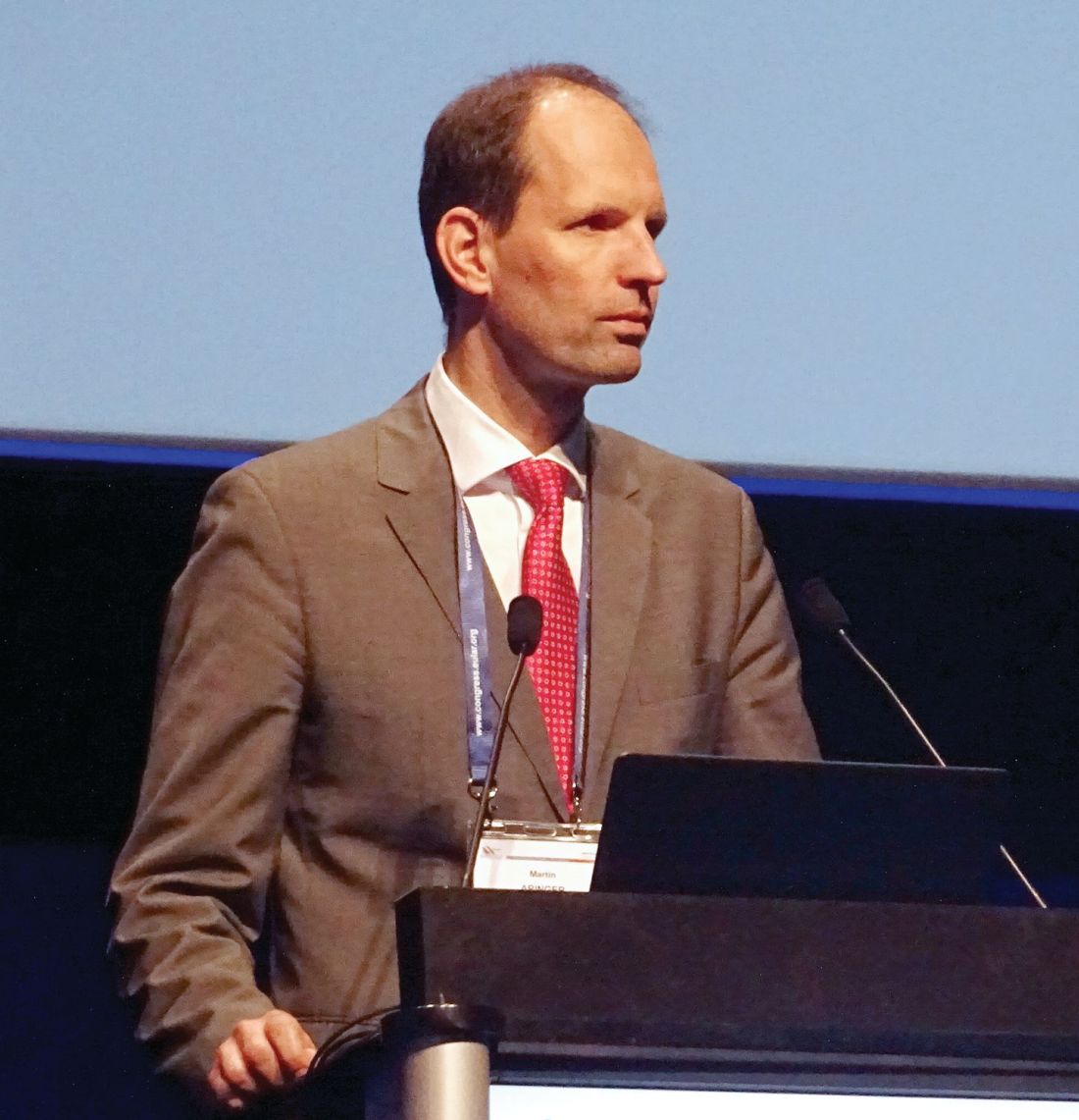
That’s because “85% of our experts said that they would feel confident classifying a patient as having lupus based only on a renal biopsy” and ANA positivity, said Dr. Johnson, who served as the ACR-appointed cochair of the criteria-writing panel along with a cochair selected by EULAR, Martin Aringer, MD, PhD, of the Technical University of Dresden (Germany). She cautioned that other levels of lupus nephritis, class II or V, confer only 8 points to the classification and so by themselves are not enough to label a person as having lupus.
During her presentation, Dr. Johnson cited the high levels of sensitivity and specificity that the new classification criteria demonstrated in a validation cohort of more than 1,000 cases and controls. In the validation analysis, the new criteria had a sensitivity of 96.12% and specificity of 94.43% for classifying SLE, giving the new criteria a better result on both these measures than either the 1997 ACR criteria (Arthritis Rheum. 1997 Sept;40[9]:1725) or the 2012 Systemic Lupus International Collaborating Clinics criteria (Arthritis Rheum. 2012 Aug;64[8]:2677-86).
The 22 criteria cluster into seven separate clinical domains and three different immunologic domains. The point values assigned to each criterion range from 2 to 10 points.
Dr. Johnson had no disclosures.
AMSTERDAM – The new systemic lupus erythematosus classification criteria of the American College of Rheumatology and the European League Against Rheumatism are based on a point system that will produce a “paradigm shift” in how the disease gets studied going forward, said Sindhu Johnson, MD, while presenting the latest version of the newly revised classification scheme at the European Congress of Rheumatology.
Until now, classification of systemic lupus erythematosus (SLE) was a yes-or-no decision, based on whether the patient had a minimum number of characteristic signs or symptoms. The new criteria, which are on track for formal endorsement before the end of 2018 by the two medical societies that sponsored the revision, instead use a point system that gives varying weight to each of the 22 criteria. A patient needs to score at least 10 points from these criteria, and all patients classified with SLE also must have an antinuclear antibody (ANA) titer of at least 1:80 on HEp-2 cells or an equivalent positive test. This means that the criteria also can define patients who just miss classification with SLE by meeting the ANA standard and by tallying 8 or 9 points, and the criteria also identify patients who far exceed the classification threshold by having the requisite ANA plus racking up as many as, perhaps, 20 or 30 points.
“This is a real research opportunity,” to follow patients who fall just short with 8 or 9 points to assess their longer-term prognosis, as well as to study whether “higher scores mean a higher risk for developing a bad outcome,” said Dr. Johnson, a rheumatologist at the University of Toronto and director of the Toronto Scleroderma Program. Other areas for future research with the new criteria include seeing how they work in various SLE subgroups, such as patients with renal-predominant disease or skin-predominant disease, and also seeing how they work in various ethnic populations.
“Diagnosis of lupus still falls within the realm of the treating physician,” but the classification criteria “inform our concept of the disease,” Dr. Johnson said in a video interview. “The new criteria allow for a shift in the way we think of the disease.”
For example, for the first time, the new criteria includes fever as a classification criterion, which receives 2 points if an infectious or other non-SLE cause can be discounted. Fever has recently been identified as a marker of early-stage SLE in at least some patients, and its addition to the classification criteria “adds a new dimension to how we think about the disease and allows us to distinguish early disease from mimicking diseases,” she explained. At the other end of the classification spectrum, a finding of class III or IV lupus nephritis on renal biopsy receives 10 points, and hence, this one finding plus having a high enough level of ANA leads to SLE classification regardless of whether the patient has any other signs or symptoms of the disease.
That’s because “85% of our experts said that they would feel confident classifying a patient as having lupus based only on a renal biopsy” and ANA positivity, said Dr. Johnson, who served as the ACR-appointed cochair of the criteria-writing panel along with a cochair selected by EULAR, Martin Aringer, MD, PhD, of the Technical University of Dresden (Germany). She cautioned that other levels of lupus nephritis, class II or V, confer only 8 points to the classification and so by themselves are not enough to label a person as having lupus.
During her presentation, Dr. Johnson cited the high levels of sensitivity and specificity that the new classification criteria demonstrated in a validation cohort of more than 1,000 cases and controls. In the validation analysis, the new criteria had a sensitivity of 96.12% and specificity of 94.43% for classifying SLE, giving the new criteria a better result on both these measures than either the 1997 ACR criteria (Arthritis Rheum. 1997 Sept;40[9]:1725) or the 2012 Systemic Lupus International Collaborating Clinics criteria (Arthritis Rheum. 2012 Aug;64[8]:2677-86).
The 22 criteria cluster into seven separate clinical domains and three different immunologic domains. The point values assigned to each criterion range from 2 to 10 points.
Dr. Johnson had no disclosures.
REPORTING FROM THE EULAR 2018 CONGRESS
SLE classification criteria perform well in validation study
AMSTERDAM – The first European League Against Rheumatism and American College of Rheumatology joint criteria for classifying systemic lupus erythematosus have a sensitivity and a specificity of more than 90%.
This is important because they improve upon the existing ACR and Systemic Lupus International Collaborating Clinics (SLICC) criteria, said Martin Aringer, MD, PhD, who cochaired the Steering Committee that produced the new classification criteria.
The video associated with this article is no longer available on this site. Please view all of our videos on the MDedge YouTube channel
Most clinicians working with lupus are familiar with the 1997 ACR criteria for the classification of systemic lupus erythematosus (SLE), which “had a relatively simple structure,” Dr. Aringer said during the opening plenary abstract session at the European Congress of Rheumatology. These considered items such as the presence of malar or discoid rash, photosensitivity, oral ulcers and arthritis, among others. These had a high specificity but a lower sensitivity. The development of the SLICC criteria in 2012 improved upon the sensitivity of the ACR criteria (92%-99% vs. 77%-91%), but at a loss in specificity (74%–88% vs. 91%-96%).
The SLICC criteria introduced two novel ideas, said Dr. Aringer, professor of medicine and chief of the division of rheumatology at the Technical University of Dresden (Germany). The first was that there had to be at least one immunologic criterion met, and the second was that biopsy-proven lupus nephritis had to be present with antinuclear antibodies (ANA) and anti-DNA antibodies detected.
One of the goals in developing the joint EULAR/ACR criteria therefore was to try to maintain the respective sensitivity and specificity achieved with the SLICC and ACR criteria. One of the key things that the new criteria looked at was to see if ANA could be used as an entry criterion. Investigations involving more than 13,000 patients with SLE showed that it could, with a antibody titer threshold of 1:80, exhibit a sensitivity of 98% (Arthritis Care Res. 2018;70[3]:428-38). Another goal was to see if histology-proven nephritis was a stronger predictor of SLE than clinical factors, such as oral ulcers, and to identify items that would only be included if there was no other more likely explanation (Lupus. 2016;25[8]:805-11).
Draft SLE classification criteria were developed based on an expert Delphi process and included ANA as an entry criterion and weighted items according to the likelihood of being associated with lupus. Items considered included the presence and severity of lupus nephritis, serology and other antibody tests, skin and central nervous system involvement, and hematologic and immunologic criteria such as the presence of thrombocytopenia and low complement (C3 and/or C4).
The final, simplified draft SLE classification criteria include 22 items in addition to the presence of ANA. A cut-off score of 10 or more is required for a classification of SLE. For example, a patient with an ANA of 1:80 or higher plus class III/IV nephritis (scoring 10) would be classified as having SLE. A patient with class II/V nephritis (scoring 8) would need another factor to be classified as having lupus, such as the presence of arthritis (scoring 6).
“Performance characteristics find sensitivity similar to the SLICC criteria while maintaining the specificity of the ACR 1997 criteria,” Dr. Aringer said, adding that these criteria will now be formally submitted to and reviewed by EULAR and ACR.
The sensitivity and specificity of the new criteria were 98% and 96% in the derivation cohort and 96% and 93% in the validation cohort.
“I was really very pleased and very happy to see that the revised or the new ACR/EULAR classification criteria had sensitivity and specificity of above 90%,” Thomas Dörner, MD, PhD, said in an interview at the congress. Dr. Dörner was a codeveloper of these criteria.
Over the past 10-15 years there have been several therapies that have failed to live up to their early promise as a potential treatment for lupus, said Dr. Dörner, professor of medicine at Charité–Universitätsmedizin Berlin. He noted that the failed treatment trials had led investigators to try to determine ways in which lupus might be best treated, such as by a “treat-to-target” approach to attain remission and low-disease activity. It also led to the reevaluation of how lupus is classified to see if that might be affecting the population of patients recruited into clinical trials.
“We had the feeling, and this is now confirmed by the new classification criteria, that a number of patients studied in earlier trials may have not fulfilled what we think is the classical lupus profile, so-called lupus or SLE mimickers,” Dr. Dörner said. This could have affected the chances of a treatment approach being successful versus placebo.
The new classification criteria are similar to those in other rheumatic diseases in that they give different weight to the effects on different organ systems, Dr. Dörner said. The stipulation that there must be a positive ANA test is also an important step, “really to make sure that we are looking at an autoimmune disease and nothing else,” he observed.
For patients who do not have a positive ANA test, they can of course still be treated, Dr. Dörner reassured, but for the classification criteria and entering patients into clinical trials, it’s really important to have strict classification criteria so that the results may be compared.
Dr. Aringer and Dr. Dörner had no relevant disclosures besides their involvement in developing the new classification criteria.
SOURCE: Aringer M et al. Ann Rheum Dis. 2018;77(Suppl 2):60. Abstract OP0020.
REPORTING FROM THE EULAR 2018 CONGRESS
Key clinical point: New classification criteria for systemic lupus erythematosus (SLE) achieve both high sensitivity and specificity.
Major finding: The sensitivity and specificity of the new criteria were 98% and 96% in the derivation cohort and 96% and 93% in the validation cohort.
Study details: An international cohort of 1,160 SLE patients and 1,058 non-SLE patients in whom the new criteria were tested and validated.
Disclosures: Dr. Aringer and Dr. Dörner had no relevant disclosures besides their involvement in developing the new classification criteria.
Source: Aringer M et al. Ann Rheum Dis. 2018;77(Suppl 2):60. Abstract OP0020.
Low vitamin D linked with DVT in lupus patients
AMSTERDAM – Low blood levels of vitamin D were linked with a roughly doubled risk for deep vein thrombosis in a review of nearly 1,400 patients with systemic lupus erythematosus at one U.S. center.
Based on these findings, patients with systemic lupus erythematosus (SLE) should have their blood vitamin D monitored regularly, and if it’s less than 40 ng/mL – the level that was linked with this thrombotic risk – they should receive a vitamin D supplement, Michelle A. Petri, MD, said while presenting a poster at the European Congress of Rheumatology.
She recommended supplementation that provides 50,000 IU of vitamin D weekly, a treatment that appears safe to add to two other routine treatments she recommends for SLE patients – aspirin and hydroxychloroquine.
SLE patients should also have their vitamin D level rechecked on a regular basis, perhaps annually, to confirm that their level remains above 40 ng/mL, said Dr. Petri, professor of medicine and director of the Lupus Center at Johns Hopkins Medicine in Baltimore. She acknowledged that this level is above the target level often applied to the general population, but remains safe.
“It looks like vitamin D may be a useful treatment to add to aspirin and hydroxychloroquine in patients with SLE. It looks very simple and important, but this finding should be repeated and validated by other groups,” commented John D. Isaacs, MD, professor of clinical rheumatology at Newcastle University in Newcastle upon Tyne, England.
Dr. Petri and her associates reviewed records for 1,392 SLE patients enrolled in a Johns Hopkins registry. The patients averaged about 43 years old, 92% were women, and 27% had a history of a thrombotic event, either prior to or after their enrollment. The most common thrombotic event was deep vein thrombosis (DVT), in 14%, and also included stroke in 7%, myocardial infarction in 4%, and a smaller number with other types of arterial or venous thromboses. All these patients also had their blood vitamin D level checked at least once, at the time of enrollment, and 77% had a level below 40 ng/mL.
The first analysis looked at the link between any thrombotic event and vitamin D levels, and included adjustment for age, race, sex, and level of lupus anticoagulant. This showed a statistically significant 2.3-fold increased risk for DVT among patients with a vitamin D level of less than 40 ng/mL, compared with those with a higher level. The researchers did not find a significant association between low vitamin D levels and the rates of total thrombotic events or any arterial thrombotic event.
A second analysis censored out thrombotic events that occurred prior to enrollment and focused on incident thromboses after enrollment into the registry. This analysis showed a statistically significant 75% increased rate of new onset DVT episodes among patients with low vitamin D at entry after adjustment for age, race, and sex. The researchers found no significant associations between low vitamin D and the incidence of any other type of incident thrombosis.
Dr. Petri and Dr. Isaacs reported having no relevant financial disclosures.
SOURCE: Petri MA et al. Ann Rheum Dis. 2018;77(Suppl 2):388, Abstract THU0341.
REPORTING FROM THE EULAR 2018 CONGRESS
Key clinical point:
Major finding: Lupus patients with a vitamin D level below 40 ng/mL had 2.3-fold more DVT events, compared with those with higher levels.
Study details: A review of 1,392 lupus patients at one U.S. center.
Disclosures: Dr. Petri and Dr. Isaacs reported having no relevant financial disclosures.
Source: Petri MA et al. Ann Rheum Dis. 2018;77(Suppl 2):388. Abstract THU0341.
FDA database reveals many rheumatic and musculoskeletal adverse events on immunotherapies
AMSTERDAM – Mining of the Food and Drug Administration adverse events database revealed a more substantial risk of rheumatic and musculoskeletal events on checkpoint inhibitor therapy than has been previously reported, according to Xerxes N. Pundole, PhD, an instructor in the research faculty at the University of Texas MD Anderson Cancer Center, Houston.
In a video interview, Dr. Pundole summarized data he presented at the European Congress of Rheumatology.
The video associated with this article is no longer available on this site. Please view all of our videos on the MDedge YouTube channel
So far, according to Dr. Pundole, there have been a relatively limited number of reports in the medical literature of inflammatory rheumatic or musculoskeletal events from checkpoint inhibitors. However, other inflammatory conditions, such as colitis and pneumonitis, are known to occur commonly with these agents. The FDA adverse event database provided an opportunity to evaluate how often rheumatic and musculoskeletal events are reported in the real world.
In this interview, Dr. Pundole explained that rheumatic and musculoskeletal events do occur at higher rates than would be expected in patients not treated with a checkpoint inhibitor. With data from more than 30,000 unique patients, the relative risks of some of these adverse events, such as polymyositis, were more than doubled, although the event rates were not evenly distributed.
Specifically, rheumatic and musculoskeletal adverse events were far less common with the cytotoxic T-lymphocyte antigen 4 checkpoint inhibitor ipilimumab (Yervoy) relative to programmed cell death protein 1 inhibitors, particularly nivolumab (Opdivo).
In another notable finding, a demographic stratification of the FDA database found elderly men to be overrepresented among patients developing adverse events related to musculoskeletal inflammation.
Overall, his data do support a relationship between checkpoint inhibitors and a greater risk of rheumatic and musculoskeletal adverse events than has been previously reported, but he noted that these data provide no specific guidance for those who already have RA or another inflammatory condition.
“Can you identify these adverse events early on to keep the patients on immune checkpoint inhibitor therapy and not have to stop their cancer treatment? That’s a question,” Dr. Pundole said. However, he suggested that the FDA data support clinician awareness of the problem and the studies that will establish strategies for preserving the benefit-to-risk ratio of checkpoint inhibitors in patients who are at greater risk of adverse events relative to immune function because of a preexisting inflammatory condition.
SOURCE: Pundole XN et al. Ann Rheum Dis. 2018;77(Suppl 2):147-148. Abstract OP0197.
REPORTING FROM THE EULAR 2018 CONGRESS

LLDAS shows potential as routine lupus treatment target
AMSTERDAM – The Lupus Low Disease Activity State measure of treatment response offers clinicians an attainable target for patients with systemic lupus erythematosus that correlates with a substantially reduced rate of organ damage, based on a retrospective assessment of data collected from more than 2,000 lupus patients at a single U.S. center.
The analysis showed that when patients with systemic lupus erythematosus (SLE) met the Lupus Low Disease Activity State (LLDAS) criteria at least half the time while on treatment, their overall rate of organ damage was reduced by 52%, compared with patients who never achieved LLDAS, Michelle A. Petri, MD, said at the European Congress of Rheumatology.
“LLDAS is something that anyone can use in practice,” and has the advantage of including a low steroid dose – no more than 7.5 mg prednisolone/day or an equivalent steroid – as one of its criteria, “a major bad actor” for SLE patients, Dr. Petri said in an interview. LLDAS “is absolutely ready for routine use,” although until now few clinicians have used it to monitor SLE patients, she noted.
“The LLDAS can be a useful target,” commented Ian N. Bruce, MD, professor of rheumatology at the University of Manchester (England), adding that the steroid dosage an SLE patient receives “is an important parameter to measure when assessing an SLE patient.
“It’s not far from being ready for routine use, but I’d like to see more evidence” that it’s a meaningful measure of an SLE patient’s disease status, he said in an interview.
To examine the clinical relevance of the LLDAS criteria, a five-point assessment for SLE first introduced in a 2016 report (Ann Rheum Dis. 2016 Sept;75[9]:1615-21), Dr. Petri and her associates applied it retrospectively to their records for 2,026 SLE patients in a Johns Hopkins registry. Clinicians at Johns Hopkins routinely assessed their SLE patients every 3 months and followed the patients for a median of about 10 years, and so had data from more than 81,000 patient encounters. The researchers used the longitudinal follow-up records to calculate an area under the curve for each patient that tracked their LLDAS state over time. This showed a clear dose-response relationship: The more time an SLE patient spent in LLDAS, the less organ damage they had. Patients who remained in LLDAS at least 75% of the time had a 60% reduction in cumulative organ damage, compared with patients who never achieved LLDAS, Dr. Petri said. The analysis also showed that LLDAS was substantially easier for patients to achieve than the Definitions of Remission in SLE (Ann Rheum Dis. 2017 March;76[3]:554-61). The Johns Hopkins cohort met the LLDAS definition about three times more often than they met the Definitions of Remission in SLE criteria, Dr. Petri said.
The new analysis also showed that LLDAS was especially effective in correlating with statistically significant reductions in future strokes, MI, and end-stage renal disease, though it did not significantly correlate with subsequent reductions in the incidence of cognitive impairment, deep vein thrombosis, malignancy, pulmonary fibrosis, pulmonary hypertension, or cataract development. But the strong correlation of time in LLDAS and the future rate of stroke, MI, or end-stage renal disease was very meaningful because those are the most important types of damage associated with SLE, Dr. Petri said. “LLDAS is a good treatment target as a surrogate” for future risk of SLE complications.
The study had no commercial funding, and Dr. Petri had no disclosures to report. Dr. Bruce has been a consultant to and speaker for GlaxoSmithKline, MedImmune, Pfizer, Roche, and UCB, and he has received research support from Genzyme, GlaxoSmithKline, Human Genome Sciences, Roche, and UCB.
SOURCE: Petri MA et al. Ann Rheum Dis. 2018;77(Suppl 2):111. Abstract OP0122.
REPORTING FROM THE EULAR 2018 CONGRESS
Key clinical point: The Lupus Low Disease Activity State is a good treatment target for systemic lupus erythematosus patients.
Major finding: Patients who achieved LLDAS at least half the time had 52% less organ damage than patients who never achieved LLDAS.
Study details: A review of case records from 2,026 SLE patients followed regularly at one U.S. center.
Disclosures: The study had no commercial funding, and Dr. Petri had no disclosures to report. Dr. Bruce has been a consultant to and speaker for GlaxoSmithKline, MedImmune, Pfizer, Roche, and UCB, and he has received research support from Genzyme, GlaxoSmithKline, Human Genome Sciences, Roche, and UCB.
Source: Petri MA et al. Ann Rheum Dis. 2018;77(Suppl 2):111. Abstract OP0122.
Baricitinib shows potential as lupus treatment
AMSTERDAM – A significantly higher proportion of patients with lupus experienced improvements in joint and skin symptoms if they were treated with baricitinib (Olumiant) than if they received placebo in a phase 2 trial.
The primary endpoint of arthritis or rash resolution as measured by the Systemic Lupus Erythematosus (SLE) Disease Activity Index 2000 (SLEDAI-2K) was met by approximately 67% of patients who were treated with 4 mg baricitinib once daily and by around 53% of patients given a matching placebo (P less than .05).
With no new safety concerns, these findings suggest that baricitinib could be of benefit in patients with SLE and further study is warranted in a phase 3 trial, said the presenting study investigator Daniel J. Wallace, MD, at the European Congress of Rheumatology. Dr. Wallace is the associate director of the Rheumatology Fellowship Program at Cedars-Sinai Medical Center, Los Angeles.
Baricitinib is already approved for use as a treatment for RA in more than 40 countries. On June 1, Eli Lilly announced that the Food and Drug Administration had given the green light for its use in RA in the United States, but only at a dose of 2 mg once daily, whereas a 2-mg and 4-mg once-daily dose is approved in most other countries.
Data from the phase 2 trial presented by Dr. Wallace did include a 2-mg dose arm, but the difference in treatment response rates versus placebo was not statistically significant.

“I think the placebo response is mainly inflated by the use of corticosteroids,” said Dr. Dörner, professor of medicine at Charité–Universitätsmedizin Berlin. “If one would have applied a steroid tapering regimen, I would have expected a larger effect size, and possibly also the 2-mg [dose] be more effective as compared to placebo.” This is something to consider when moving into a phase 3 trial, he suggested.
For inclusion in the phase 2 trial, patients had to meet the following criteria: Be positive for antinuclear antibodies and/or a positive anti-dsDNA test, have a SLEDAI-2K clinical score of 4 or more, and have active SLEDAI arthritis and/or rash. Patients with severe active lupus nephritis or CNS involvement were excluded.
The mean age of patients was around 44 years, and as might be expected, the study population was predominantly female (99%). Around two-thirds of patients were white, 19% were of Asian descent, and the rest were designated as “other”. The average time to SLE onset was 9.7 years in the placebo group and just over 11 years in the baricitinib arms, with similar SLEDAI-2K scores of about 8-9, about 7-8 tender joints, and about five swollen joints at baseline.
A number of other secondary endpoints were also met by the 4 mg baricitinib group, Dr. Wallace reported. This included the relatively new Lupus Low Disease Activity State, he said, which was met by 38% (n = 27) of patients treated with 4 mg baricitinib, 33% (n = 35) treated with 2 mg baricitinib, and 26% (n = 27) of those given placebo (P less than .05 for the 4-mg dose vs. placebo). There were also numerically fewer SLE flares, including fewer severe flares.
“Some of the other outcomes demonstrated statistical significance: Physician Global Assessment, tender joint count, worst joint pain, and worst pain on a numeric rating scale,” Dr. Wallace said. A trend towards improvement was seen in the swollen joint count, with modest improvement in fatigue.
Treatment-emergent adverse events were seen in around 71%-73% of patients given baricitinib and 65% of patients given placebo. Most were mild or moderate in nature, but serious adverse events did occur in approximately 10% of patients who received baricitinib and in 4% of those who received placebo.
What’s noteworthy, Dr. Dörner said during a press briefing, is the very low rate of venous thromboembolism seen in the trial. “We’d have expected to see more deep vein thrombosis,” he said. Only one case occurred, in a patent taking the 4-mg dose, but this patient had preexisting antiphospholipid antibodies.
Additionally, although the percentage of patients with serious infections was slightly higher in the 2 and 4 mg baricitinib arms than for placebo (1.9% and 5.8% vs. 1%, respectively) “this is what we expect for lupus patients,” Dr. Dörner said. Furthermore, herpes zoster infection, which is very often reactivated in lupus because of the disease or its treatment, was only reported in one patient in the placebo group and in one patient in the 4 mg group.
“I think there is a very promising outlook, at least for the 4-mg dose of baricitinib,” Dr. Dörner said. “There have been no new safety or tolerability issues when compared to the RA population, and we’re looking forward to seeing subsequent studies in this [SLE] patient population where we have a need for more efficacious therapies.”
The study was funded by Eli Lilly. Dr. Dörner was part of the trial’s steering committee and has acted as a consultant for Eli Lilly. He has also received grant or research support from Roche/Chugai, Janssen, and Sanofi-Aventis; consulted for AbbVie, Celgene, Roche, UCB, Merck Sharp & Dohme, Pfizer/Hospira, and Novartis; and he is part of the speakers bureaus for Amgen, Celgene, and Biogen. Dr. Wallace has acted as a consultant for Eli Lilly, as well as EMD Serono, Pfizer, and GlaxoSmithKline.
The video associated with this article is no longer available on this site. Please view all of our videos on the MDedge YouTube channel
SOURCE: Wallace DJ et al. Ann Rheum Dis. 2018;77(Suppl 2):59. Abstract OP0019.
AMSTERDAM – A significantly higher proportion of patients with lupus experienced improvements in joint and skin symptoms if they were treated with baricitinib (Olumiant) than if they received placebo in a phase 2 trial.
The primary endpoint of arthritis or rash resolution as measured by the Systemic Lupus Erythematosus (SLE) Disease Activity Index 2000 (SLEDAI-2K) was met by approximately 67% of patients who were treated with 4 mg baricitinib once daily and by around 53% of patients given a matching placebo (P less than .05).
With no new safety concerns, these findings suggest that baricitinib could be of benefit in patients with SLE and further study is warranted in a phase 3 trial, said the presenting study investigator Daniel J. Wallace, MD, at the European Congress of Rheumatology. Dr. Wallace is the associate director of the Rheumatology Fellowship Program at Cedars-Sinai Medical Center, Los Angeles.
Baricitinib is already approved for use as a treatment for RA in more than 40 countries. On June 1, Eli Lilly announced that the Food and Drug Administration had given the green light for its use in RA in the United States, but only at a dose of 2 mg once daily, whereas a 2-mg and 4-mg once-daily dose is approved in most other countries.
Data from the phase 2 trial presented by Dr. Wallace did include a 2-mg dose arm, but the difference in treatment response rates versus placebo was not statistically significant.
“I think the placebo response is mainly inflated by the use of corticosteroids,” said Dr. Dörner, professor of medicine at Charité–Universitätsmedizin Berlin. “If one would have applied a steroid tapering regimen, I would have expected a larger effect size, and possibly also the 2-mg [dose] be more effective as compared to placebo.” This is something to consider when moving into a phase 3 trial, he suggested.
For inclusion in the phase 2 trial, patients had to meet the following criteria: Be positive for antinuclear antibodies and/or a positive anti-dsDNA test, have a SLEDAI-2K clinical score of 4 or more, and have active SLEDAI arthritis and/or rash. Patients with severe active lupus nephritis or CNS involvement were excluded.
The mean age of patients was around 44 years, and as might be expected, the study population was predominantly female (99%). Around two-thirds of patients were white, 19% were of Asian descent, and the rest were designated as “other”. The average time to SLE onset was 9.7 years in the placebo group and just over 11 years in the baricitinib arms, with similar SLEDAI-2K scores of about 8-9, about 7-8 tender joints, and about five swollen joints at baseline.
A number of other secondary endpoints were also met by the 4 mg baricitinib group, Dr. Wallace reported. This included the relatively new Lupus Low Disease Activity State, he said, which was met by 38% (n = 27) of patients treated with 4 mg baricitinib, 33% (n = 35) treated with 2 mg baricitinib, and 26% (n = 27) of those given placebo (P less than .05 for the 4-mg dose vs. placebo). There were also numerically fewer SLE flares, including fewer severe flares.
“Some of the other outcomes demonstrated statistical significance: Physician Global Assessment, tender joint count, worst joint pain, and worst pain on a numeric rating scale,” Dr. Wallace said. A trend towards improvement was seen in the swollen joint count, with modest improvement in fatigue.
Treatment-emergent adverse events were seen in around 71%-73% of patients given baricitinib and 65% of patients given placebo. Most were mild or moderate in nature, but serious adverse events did occur in approximately 10% of patients who received baricitinib and in 4% of those who received placebo.
What’s noteworthy, Dr. Dörner said during a press briefing, is the very low rate of venous thromboembolism seen in the trial. “We’d have expected to see more deep vein thrombosis,” he said. Only one case occurred, in a patent taking the 4-mg dose, but this patient had preexisting antiphospholipid antibodies.
Additionally, although the percentage of patients with serious infections was slightly higher in the 2 and 4 mg baricitinib arms than for placebo (1.9% and 5.8% vs. 1%, respectively) “this is what we expect for lupus patients,” Dr. Dörner said. Furthermore, herpes zoster infection, which is very often reactivated in lupus because of the disease or its treatment, was only reported in one patient in the placebo group and in one patient in the 4 mg group.
“I think there is a very promising outlook, at least for the 4-mg dose of baricitinib,” Dr. Dörner said. “There have been no new safety or tolerability issues when compared to the RA population, and we’re looking forward to seeing subsequent studies in this [SLE] patient population where we have a need for more efficacious therapies.”
The study was funded by Eli Lilly. Dr. Dörner was part of the trial’s steering committee and has acted as a consultant for Eli Lilly. He has also received grant or research support from Roche/Chugai, Janssen, and Sanofi-Aventis; consulted for AbbVie, Celgene, Roche, UCB, Merck Sharp & Dohme, Pfizer/Hospira, and Novartis; and he is part of the speakers bureaus for Amgen, Celgene, and Biogen. Dr. Wallace has acted as a consultant for Eli Lilly, as well as EMD Serono, Pfizer, and GlaxoSmithKline.
The video associated with this article is no longer available on this site. Please view all of our videos on the MDedge YouTube channel
SOURCE: Wallace DJ et al. Ann Rheum Dis. 2018;77(Suppl 2):59. Abstract OP0019.
REPORTING FROM THE EULAR 2018 CONGRESS
Key clinical point: Baricitinib at 4 mg was associated with significant clinical improvements versus placebo and had an acceptable safety and tolerability profile.
Major finding: A higher percentage of patients receiving 4 mg of baricitinib than those receiving placebo achieved the primary endpoint of arthritis and/or rash remission as defined by the Systemic Lupus Erythematosus Disease Activity Index 2000 at week 24 (P less than .05).
Study details: A phase 2, multinational, double-blind, placebo-controlled, parallel group study of once-daily, oral baricitinib (2 mg and 4 mg) in 314 patients with SLE receiving standard therapy.
Disclosures: The study was funded by Eli Lilly. Dr. Dörner was part of the trial’s steering committee and has acted as a consultant for Eli Lilly. He has also received grant or research support from Roche/Chugai, Janssen, and Sanofi-Aventis; consulted for AbbVie, Celgene, Roche, UCB, Merck Sharp & Dohme, Pfizer/Hospira, and Novartis; and he is part of the speakers bureaus for Amgen, Celgene, and Biogen. Dr. Wallace has acted as a consultant for Eli Lilly, as well as EMD Serono, Pfizer, and GlaxoSmithKline.
Source: Wallace DJ et al. Ann Rheum Dis. 2018;77(Suppl 2):59. Abstract OP0019.