User login
EULAR scientific program highlights spectrum of translational research
EULAR 2018’s scientific program in Amsterdam is packed with lectures, clinical and basic science symposia, workshops, and special interest sessions covering the full spectrum of rheumatic diseases, said Dr. Robert Landewé, chair of the Scientific Program Committee.
“More than 5,000 scientific abstracts were submitted, which is an absolute, all-time record,” Dr. Landewé said. Four experts scored each abstract, and only the top 7% were invited for oral presentation during abstract sessions or symposia, he explained in an interview.

Wednesday, June 13
A high point of the 2018 scientific program is Wednesday’s opening plenary session, which will feature abstracts that were handpicked by Dr. Landewé and Dr. Thomas Dörner, professor of rheumatology at Charite Universitätsmedizin, Berlin. “This session includes highly scored abstracts, including late-breakers, on current advances in therapeutics and disease classification,” said Dr. Dörner, who chaired this year’s Abstract Selection Committee.
The plenary abstract session will cover new findings on gout and cardiovascular disease from CANTOS (Canakinumab Anti-inflammatory Thrombosis Outcome Study), long-term mortality in patients with early RA from the COBRA (Combinatietherapie Bij Reumatoide Artritis) study, the use of zoledronic acid to treat knee osteoarthritis with bone lesions, and the relationship between bisphosphonate drug holidays and hip fracture risk. Researchers also will discuss baricitinib in systemic lupus erythematosus (SLE), the value of MRI when treating remitted RA to target, the validation of SLE classification criteria, and draft classification criteria for ANCA-associated vasculitides.
A notable clinical science session on Wednesday will cover cancer and inflammation, Dr. Landewé said. “This is a topic of increasing interest because cancer and inflammation share mutual pathways.”
Novel cancer therapies such as immune checkpoint inhibitors have improved outcomes across a range of tumor types, but also can induce rheumatic disease, he added. Accordingly, presenters will discuss inflammation as “friend” versus “foe” in cancer treatment, the role of tumor necrosis factor in cancer, and risk of malignancy among patients with RA.
Also on Wednesday, a session will tackle the relationship between psychological distress and pain in immune-mediated disease. “Pain is the major symptom of rheumatic diseases, and the role of the psyche remains poorly understood,” Dr. Landewé said. “But we know one thing for sure: There is an association, and speakers from outside the field of rheumatology will help explain.”
Attendees at this bench-to-bedside session will learn how distress appears to exacerbate arthritis pain and how managing psychological stress can help optimize outcomes in arthritis pain. Experts also will describe research on integrated brain pathways in pain and distress, as well as risk factors for cognitive impairment in RA.
Thursday, June 14

Topics in this session will include the use of estrogens and other hormonal therapies in patients with rheumatic disease, registry studies of rheumatologic conditions during pregnancy, and how clinicians can best discuss sexual concerns with their rheumatology patients.
Another clinical science session scheduled for Thursday afternoon will delve into structural damage progression in patients with axial spondyloarthritis, Dr. Landewé said. “Can we inhibit this structural progression? Can we show it? Does it make sense? And which drug company will win the battle to have the precedent?”
He hopes that Dr. Désirée van der Heijde of the Netherlands and Dr. Xenofon Baraliakos of Germany will help answer these questions when they discuss the latest evidence on identifying and treating clinically relevant structural progression. Also in this session, researchers will describe the combined effects of tumor necrosis factor inhibitors and NSAIDs on radiographic progression in ankylosing spondylitis, and MRI evidence supporting treating early axial spondyloarthritis to target with the goal of achieving sustained remission of inflammation.
Also on Thursday afternoon, a case-based session will take a deep dive into giant cell arteritis (GCA), Dr. Landewé noted. Attendees will learn about diagnosing and managing vision loss and stroke and the latest on corticosteroid therapy in GCA. The session also will cover biologics. “Giant cell arteritis has entered the field of biologicals!” said Dr. Landewé. “This has major implications for this disease and the clinical choices to be made.”
The past 5 decades have seen marked progress in the diagnosis and treatment of SLE, with corresponding improvements in survival and quality of life. “Still, lupus is awfully difficult,” Dr. Landewé said. “Therefore, we have planned a classical bench-to-bedside symposium to provide an all-inclusive look at current thinking and future developments.”
Talks during this Thursday afternoon session will cover the latest findings on the pathogenesis of SLE, the clinical significance of autoantibodies, distinguishing early SLE from mimics, and the role of blood-brain barrier permeability and neuropsychiatric manifestations of SLE and progressive systemic sclerosis.
Friday, June 15
For the first time, the scientific program also will include a clinical science session held jointly with the European Society of Musculoskeletal Radiology (ESSR). Dr. Joachim Sieper of Germany and ESSR President Dr. Monique Reijnierse of the Netherlands will cochair the Friday afternoon session on the role of MRI in rheumatology. Attendees from both organizations will learn when to use MRI in early and established RA and spondyloarthritis, and how to interpret the results, with abundant time built in for questions and answers. Dr. Landewé called the joint session “a test case” for exciting web-based interactions between EULAR and ESSR.
Another clinical science session on Friday afternoon will dive into the diagnosis of spondyloarthritis, which Dr. Landewé called “a matter of recognizing patterns, not ticking boxes on a list of criteria. This symposium leads you through the art of pattern recognition.”
Later on Friday afternoon, a session will explore advances in biologic therapy of small-vessel vasculitis, he added. “Biologic disease-modifying antirheumatic drugs [bDMARDs] are becoming more and more important in this area of expanding interest.” Experts will address complement inhibition in ANCA-associated vasculitis (AAV), the use of induction and maintenance rituximab in AAV, the evolving role of mepolizumab in eosinophilic granulomatosis with polyangiitis, survival in AAV, and the use of rituximab for treating children with granulomatosis with polyangiitis and microscopic polyangiitis.
Saturday, June 16
On Saturday, a bench-to-bedside session will cover gout and kidney function. “This is an area with important new insights,” Dr. Dörner said. Presenters will discuss the genetics of hyperuricemia, renal urate transporters, and the pros and cons of using xanthine oxidase inhibitors to treat chronic kidney disease. Researchers will also cover studies of impaired neutrophil chemotaxis in patients with chronic kidney disease and hyperuricemia, and the relationship between renal medullar hyperechogenicity and gout severity.
Also on Saturday, a clinical science session titled, “Rheumatoid arthritis: Is it all in your head?” will explore emerging data on the relationship between inflammation and depression. Patients with RA often face both clinical depression and social isolation, and these complex psychosocial conditions can worsen one another. “In addition to proper drug choice, treating RA effectively depends on how concomitant problems, such as nonspecific pain, depression, and social isolation, are coped with in a broad context,” Dr. Landewé said. “When it comes to optimal management, rheumatologists need to communicate and prescribe, not just prescribe.”
Christian Apfelbacher, PhD, of Germany will discuss prevention and treatment strategies and Dr. Jonathan Cavanagh of the United Kingdom will cover neuroimaging in RA. Researchers also will discuss new findings on pain, depression, and anxiety in patients recently diagnosed with RA.
Also on Saturday, a special session will cover EULAR’s initiatives to improve clinical approaches (ESSCA), Dr. Dörner noted. This effort has produced new or updated recommendations on topics such as vaccination, Sjögren’s syndrome, glucocorticoid therapy, and management of hand osteoarthritis, he said. “These recommendations follow a number of others and are expected to impact clinical science as well as clinical practice.”
EULAR 2018’s scientific program in Amsterdam is packed with lectures, clinical and basic science symposia, workshops, and special interest sessions covering the full spectrum of rheumatic diseases, said Dr. Robert Landewé, chair of the Scientific Program Committee.
“More than 5,000 scientific abstracts were submitted, which is an absolute, all-time record,” Dr. Landewé said. Four experts scored each abstract, and only the top 7% were invited for oral presentation during abstract sessions or symposia, he explained in an interview.
Wednesday, June 13
A high point of the 2018 scientific program is Wednesday’s opening plenary session, which will feature abstracts that were handpicked by Dr. Landewé and Dr. Thomas Dörner, professor of rheumatology at Charite Universitätsmedizin, Berlin. “This session includes highly scored abstracts, including late-breakers, on current advances in therapeutics and disease classification,” said Dr. Dörner, who chaired this year’s Abstract Selection Committee.
The plenary abstract session will cover new findings on gout and cardiovascular disease from CANTOS (Canakinumab Anti-inflammatory Thrombosis Outcome Study), long-term mortality in patients with early RA from the COBRA (Combinatietherapie Bij Reumatoide Artritis) study, the use of zoledronic acid to treat knee osteoarthritis with bone lesions, and the relationship between bisphosphonate drug holidays and hip fracture risk. Researchers also will discuss baricitinib in systemic lupus erythematosus (SLE), the value of MRI when treating remitted RA to target, the validation of SLE classification criteria, and draft classification criteria for ANCA-associated vasculitides.
A notable clinical science session on Wednesday will cover cancer and inflammation, Dr. Landewé said. “This is a topic of increasing interest because cancer and inflammation share mutual pathways.”
Novel cancer therapies such as immune checkpoint inhibitors have improved outcomes across a range of tumor types, but also can induce rheumatic disease, he added. Accordingly, presenters will discuss inflammation as “friend” versus “foe” in cancer treatment, the role of tumor necrosis factor in cancer, and risk of malignancy among patients with RA.
Also on Wednesday, a session will tackle the relationship between psychological distress and pain in immune-mediated disease. “Pain is the major symptom of rheumatic diseases, and the role of the psyche remains poorly understood,” Dr. Landewé said. “But we know one thing for sure: There is an association, and speakers from outside the field of rheumatology will help explain.”
Attendees at this bench-to-bedside session will learn how distress appears to exacerbate arthritis pain and how managing psychological stress can help optimize outcomes in arthritis pain. Experts also will describe research on integrated brain pathways in pain and distress, as well as risk factors for cognitive impairment in RA.
Thursday, June 14
Topics in this session will include the use of estrogens and other hormonal therapies in patients with rheumatic disease, registry studies of rheumatologic conditions during pregnancy, and how clinicians can best discuss sexual concerns with their rheumatology patients.
Another clinical science session scheduled for Thursday afternoon will delve into structural damage progression in patients with axial spondyloarthritis, Dr. Landewé said. “Can we inhibit this structural progression? Can we show it? Does it make sense? And which drug company will win the battle to have the precedent?”
He hopes that Dr. Désirée van der Heijde of the Netherlands and Dr. Xenofon Baraliakos of Germany will help answer these questions when they discuss the latest evidence on identifying and treating clinically relevant structural progression. Also in this session, researchers will describe the combined effects of tumor necrosis factor inhibitors and NSAIDs on radiographic progression in ankylosing spondylitis, and MRI evidence supporting treating early axial spondyloarthritis to target with the goal of achieving sustained remission of inflammation.
Also on Thursday afternoon, a case-based session will take a deep dive into giant cell arteritis (GCA), Dr. Landewé noted. Attendees will learn about diagnosing and managing vision loss and stroke and the latest on corticosteroid therapy in GCA. The session also will cover biologics. “Giant cell arteritis has entered the field of biologicals!” said Dr. Landewé. “This has major implications for this disease and the clinical choices to be made.”
The past 5 decades have seen marked progress in the diagnosis and treatment of SLE, with corresponding improvements in survival and quality of life. “Still, lupus is awfully difficult,” Dr. Landewé said. “Therefore, we have planned a classical bench-to-bedside symposium to provide an all-inclusive look at current thinking and future developments.”
Talks during this Thursday afternoon session will cover the latest findings on the pathogenesis of SLE, the clinical significance of autoantibodies, distinguishing early SLE from mimics, and the role of blood-brain barrier permeability and neuropsychiatric manifestations of SLE and progressive systemic sclerosis.
Friday, June 15
For the first time, the scientific program also will include a clinical science session held jointly with the European Society of Musculoskeletal Radiology (ESSR). Dr. Joachim Sieper of Germany and ESSR President Dr. Monique Reijnierse of the Netherlands will cochair the Friday afternoon session on the role of MRI in rheumatology. Attendees from both organizations will learn when to use MRI in early and established RA and spondyloarthritis, and how to interpret the results, with abundant time built in for questions and answers. Dr. Landewé called the joint session “a test case” for exciting web-based interactions between EULAR and ESSR.
Another clinical science session on Friday afternoon will dive into the diagnosis of spondyloarthritis, which Dr. Landewé called “a matter of recognizing patterns, not ticking boxes on a list of criteria. This symposium leads you through the art of pattern recognition.”
Later on Friday afternoon, a session will explore advances in biologic therapy of small-vessel vasculitis, he added. “Biologic disease-modifying antirheumatic drugs [bDMARDs] are becoming more and more important in this area of expanding interest.” Experts will address complement inhibition in ANCA-associated vasculitis (AAV), the use of induction and maintenance rituximab in AAV, the evolving role of mepolizumab in eosinophilic granulomatosis with polyangiitis, survival in AAV, and the use of rituximab for treating children with granulomatosis with polyangiitis and microscopic polyangiitis.
Saturday, June 16
On Saturday, a bench-to-bedside session will cover gout and kidney function. “This is an area with important new insights,” Dr. Dörner said. Presenters will discuss the genetics of hyperuricemia, renal urate transporters, and the pros and cons of using xanthine oxidase inhibitors to treat chronic kidney disease. Researchers will also cover studies of impaired neutrophil chemotaxis in patients with chronic kidney disease and hyperuricemia, and the relationship between renal medullar hyperechogenicity and gout severity.
Also on Saturday, a clinical science session titled, “Rheumatoid arthritis: Is it all in your head?” will explore emerging data on the relationship between inflammation and depression. Patients with RA often face both clinical depression and social isolation, and these complex psychosocial conditions can worsen one another. “In addition to proper drug choice, treating RA effectively depends on how concomitant problems, such as nonspecific pain, depression, and social isolation, are coped with in a broad context,” Dr. Landewé said. “When it comes to optimal management, rheumatologists need to communicate and prescribe, not just prescribe.”
Christian Apfelbacher, PhD, of Germany will discuss prevention and treatment strategies and Dr. Jonathan Cavanagh of the United Kingdom will cover neuroimaging in RA. Researchers also will discuss new findings on pain, depression, and anxiety in patients recently diagnosed with RA.
Also on Saturday, a special session will cover EULAR’s initiatives to improve clinical approaches (ESSCA), Dr. Dörner noted. This effort has produced new or updated recommendations on topics such as vaccination, Sjögren’s syndrome, glucocorticoid therapy, and management of hand osteoarthritis, he said. “These recommendations follow a number of others and are expected to impact clinical science as well as clinical practice.”
Pemphigus remission rate tops 80% with rituximab
ORLANDO – , and could be approved for the indication soon.
With approval pending, “rituximab is quickly emerging as frontline therapy” for pemphigus, so “we should begin to prepare to answer our patients’ questions. It’s likely they will be interested in its use,” said Carolyn Kushner, a medical student and dermatology research fellow at the University of Pennsylvania, Philadelphia. Rituximab manufacturer Genentech announced the priority review for this indication in a Feb. 2018 press release.
“We get a lot of questions in the clinic,” she said at the International Investigative Dermatology meeting. Patients with pemphigus want to know how well rituximab will work, and if they’ll be able to go off other medications. They wonder if it’s safe, and when they’ll need to be retreated. The goal of the study was to provide information for both clinicians and patients regarding what to expect from the treatment.
Overall, 54 patients (48%) achieved a complete response off therapy (CROT) after their first treatment cycle, meaning they had no new lesions for at least 2 months off of all systemic and topical treatments. The median time to a complete response was 7.4 months, and the median time to relapse was 20.9 months after the first infusion. An additional 15 patients (13%) had a complete remission with minimal therapy after one cycle.
In short, “61% of patients achieved complete healing of their skin after one cycle,” Ms. Kushner said. The number rose to 82% (93 patients) when those who had more than one cycle were included. The maximum in the study was seven. Among all patients, the median time from the first to second rituximab dose was 25.1 months.
When age, sex, and disease duration were controlled for, patients who received lymphoma dosing – 375 mg/m2 weekly for 4 weeks – were 2.7 times more likely to achieve CROT than those on the rheumatoid arthritis dosing, two 1,000 mg IV infusions 2 weeks apart (P = .037). “We almost never use RA dosing now,” she said.
The odds of success also increased with age, with patients 45 years and older 3.5 to almost 7 times more likely to achieve CROT than younger patients, also a statistically significant finding.
There were four serious adverse events across 155 cycles of the lymphoma regimen, and one with 90 cycles of arthritis dosing, all infectious and none fatal. Ms. Kushner cautioned the true rate was probably higher, since their review data might have missed some cases.
Race, sex, and disease duration had no significant effect on response rates. About 60% of the patients were women.
Rituximab, approved in 1997, is a CD20-directed cytolytic antibody.
There was no industry funding for the work, and Ms. Kushner didn’t have any disclosures.
SOURCE: Kushner CJ et al. IID 2018, Abstract 552.
REPORTING FROM IID 2018
Key clinical point: Rituximab puts the majority of pemphigus patients in remission, with a median time between doses of about 2 years.
Major finding: Sixty-one percent of patients achieved complete healing of their skin after one cycle, increasing to 82% when those who had more than one cycle were included.
Study details: A single-center review of 113 patients
Disclosures: There was no industry funding, and the lead investigator had no disclosures.
Source: Kushner CJ et al. IID 2018, Abstract 552.
Long-term follow-up most important for hydroxychloroquine retinal screening
LIVERPOOL, ENGLAND – , but long-term follow-up is much more important, according to data presented at the British Society for Rheumatology annual conference.
In just one specialist rheumatology center in England, which treats more than 8,000 patients annually, the cost of the first year’s optical coherence tomography (OCT) assessment would be more than $60,000. Additional costs would be incurred to screen those who had been on the drug for more than 5 years ,who were known to be at greater risk of hydroxychloroquine-induced retinopathy. This is within the National Health Service in England where the cost of a single OCT scan is around $70; in the private health sector, the cost of one test can be as high as $400.
Indeed, of 887 hydroxychloroquine users identified, 44% had at least one risk factor for hydroxychloroquine-induced retinopathy. These included being older than 60 years of age (30% of all users), having renal (10%) or hepatic (2%) impairment, retinal disease at baseline (8%), or using high (more than 6.5 mg/kg) doses of the drug based on their actual (9%) or ideal (4%) body weight.
“The retinal toxicity of hydroxychloroquine is a bit of a hot topic at the moment,” Dr. Yates said at the conference. While the drug has been around for years and used successfully to treat many patients with rheumatoid arthritis and systemic lupus erythematosus (SLE), a known side effect is retinal toxicity.
Traditionally, retinopathy has been quoted as being a relatively rare side effect, affecting around 0.5%-2% of the treated population. Recent data (JAMA Ophthalmol. 2014;132[12]:1453-60) suggest, however, that is probably a vast underestimate, with 7.5% of patients taking hydroxychloroquine for more than 5 years likely to be affected, as are up to 20% of those taking the drug for up to 20 years of treatment.
Dr. Yates and associates wanted to assess the burden of hydroxychloroquine use at their center and look at the risk factors and impact of the recent screening guidelines issued by the British Society for Rheumatology (Rheumatology [Oxford]. 2017;56[6]:865-8) in 2017 and by the Royal College of Ophthalmologists in 2018. These state that patients should have a formal baseline ophthalmic examination, ideally including OCT, within 6-12 months of starting therapy and an annual eye assessment with repeat OCT thereafter for the following 5 years; the ophthalmology guidelines recommending annual screening for the duration of therapy.
One criticism of increased screening for retinal toxicity in routine practice is consultants saying that they see only a handful of cases during their career, Dr. Yates observed. However, if you consider that in an average rheumatology department there are five consultants and 900 patients on hydroxychloroquine, 500 patients take the drug for 5 years or longer, 2% are picked up with non-OCT screening, that amounts to around two cases per year over a 5- to 10-year period. “So that fits with the narrative of only having seen a handful of cases pre-OCT,” Dr. Yates reasoned.
“I believe that this is a real problem, but I’m afraid this is the tip of the iceberg,” commented Caroline Gordon, MD, after her presentation. “We’ve been screening our patients in Birmingham now for about 5 years and we are definitely finding a significant number of patients with hydroxychloroquine toxicity who can be picked up with OCT and visual fields screening.”
Dr. Gordon, professor of rheumatology at the University of Birmingham (England) and a consultant rheumatologist for the University Hospitals NHS Foundation Trust and the Sandwell & West Birmingham Hospitals NHS Trust, helps look after one of the largest cohorts of patients with SLE in the United Kingdom.
A baseline eye examination has always been recommended, Dr. Gordon said, but she suggested that this could remain in the realm of the opticians with further assessment and referral as needed.
“I’m not convinced, from the work we’ve done, that there is any value in the baseline OCT,” Dr. Gordon said, “because we never find anything on the baseline OCT that we didn’t already expect from the opticians’ assessment.”
It is the long-term (longer than10 years) follow-up that needs to be the focus, rather than the initial period, she stressed, as the highest risk appears to be in patients who have been taking the drug for 15 years or longer. Prior to this, different types of retinopathy can occur that are actually attributable to the underlying disease and are not related hydroxychloroquine. Of course, patients on higher doses of hydroxychloroquine may need closer monitoring early on, “as they are at risk,” she acknowledged.
Dr. Gordon suggested that the guidelines as they currently stand may not be that useful for real-life practice. Following them could result in a large amount of money being spent on early tests that are perhaps not necessary.
“What we do need to do is focus on the patients who’ve been on treatment long term,” she said.
SOURCE: Yates M et al. Rheumatology. 2018;57(Suppl. 3):key075.188.
REPORTING FROM BSR 2018
Key clinical point: Long-term follow up is important for assessing hydroxychloroquine toxicity.
Major finding: 44% of patients had at least one risk factor for hydroxychloroquine-induced retinopathy after more than 5 years of treatment.
Study details: Electronic record review of 887 patients treated with hydroxychloroquine for about 5 years in a large tertiary rheumatology service.
Disclosures: Dr. Yates had nothing to disclose.
Source: Yates M et al. Rheumatology. 2018;57(Suppl. 3):key075.312.
Cutaneous lupus: Switching antimalarials can delay immunosuppressive therapy
ORLANDO – , according to Anthony Fernandez, MD, PhD, director of medical and inpatient dermatology at the Cleveland Clinic.
A switch to chloroquine, or adding quinacrine, might do the trick, saving at least some patients from having to move on to immunosuppressive therapy, Dr. Fernandez said at the International Conference on Cutaneous Lupus Erythematosus.
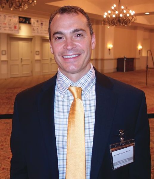
“What we are learning from the literature is that we can switch from one antimalarial to another. We need to think about this in our algorithms before reaching for potentially more toxic immunosuppressives,” Dr. Fernandez said.
As for quinacrine, about two-thirds of patients who fail hydroxychloroquine or chloroquine will have a positive response to quinacrine if added (Br J Dermatol. 2017 Jul;177[1]:188-96). “It’s important to remember that we are not adding any ocular toxicity” with quinacrine, he said.
Quinacrine does come with a major concern of its own: the risk of aplastic anemia. However, this seems to occur with doses higher than 100 mg/day, which are no longer recommended; there have been no reports of aplastic anemia in patients on 100 mg/day or less.
Quinacrine “is an underutilized antimalarial. I think a lot of people don’t know about it or know how to get it. It can be compounded into capsules for patients,” and for a reasonable price, at about $20 for a month supply at some pharmacies, Dr. Fernandez said.
Meanwhile, because of the risk of retinal toxicity with hydroxychloroquine, there’s been a shift in recent years from dosing up to 6.5 mg/kg per day of ideal body weight to a ceiling of 5 mg/kg per day of actual body weight (JAMA Ophthalmol. 2014 Dec;132[12]:1453-60), and a ceiling of 2.3 mg/kg per day actual body weight for chloroquine.
The idea was to prevent overdosing in people who are under their ideal body weight, but there have been concerns about the efficacy of the new dosing regimen in other patients. Dr. Fernandez has not seen evidence of this. “We do adhere to the new dosing recommendations” at the Cleveland Clinic, and “personally, I think we are seeing similar efficacy,” he said.
The most important risk factor for retinal toxicity is cumulative dose. The risk seems to be extremely low in the first 5 years, but increases afterwards. Most patients who develop retinal toxicity have taken a cumulative hydroxychloroquine dose of 1,000 g, equal to about 400 mg/d for 7 years. “The longer you are on the medicine, the higher your risk of developing retinal toxicity,” he noted.
Regardless of weight, it’s recommended to limit hydroxychloroquine to 400 mg daily and chloroquine to 250 mg daily, with a baseline ocular exam, and – barring any intervening problems – annual screening after 5 years.
In patients with highly active skin disease at baseline, Dr. Fernandez said he will sometimes start hydroxychloroquine higher than 400 mg initially but will bring patients back down to 400 mg after a few months.
Dr. Fernandez had no relevant disclosures.
ORLANDO – , according to Anthony Fernandez, MD, PhD, director of medical and inpatient dermatology at the Cleveland Clinic.
A switch to chloroquine, or adding quinacrine, might do the trick, saving at least some patients from having to move on to immunosuppressive therapy, Dr. Fernandez said at the International Conference on Cutaneous Lupus Erythematosus.
“What we are learning from the literature is that we can switch from one antimalarial to another. We need to think about this in our algorithms before reaching for potentially more toxic immunosuppressives,” Dr. Fernandez said.
As for quinacrine, about two-thirds of patients who fail hydroxychloroquine or chloroquine will have a positive response to quinacrine if added (Br J Dermatol. 2017 Jul;177[1]:188-96). “It’s important to remember that we are not adding any ocular toxicity” with quinacrine, he said.
Quinacrine does come with a major concern of its own: the risk of aplastic anemia. However, this seems to occur with doses higher than 100 mg/day, which are no longer recommended; there have been no reports of aplastic anemia in patients on 100 mg/day or less.
Quinacrine “is an underutilized antimalarial. I think a lot of people don’t know about it or know how to get it. It can be compounded into capsules for patients,” and for a reasonable price, at about $20 for a month supply at some pharmacies, Dr. Fernandez said.
Meanwhile, because of the risk of retinal toxicity with hydroxychloroquine, there’s been a shift in recent years from dosing up to 6.5 mg/kg per day of ideal body weight to a ceiling of 5 mg/kg per day of actual body weight (JAMA Ophthalmol. 2014 Dec;132[12]:1453-60), and a ceiling of 2.3 mg/kg per day actual body weight for chloroquine.
The idea was to prevent overdosing in people who are under their ideal body weight, but there have been concerns about the efficacy of the new dosing regimen in other patients. Dr. Fernandez has not seen evidence of this. “We do adhere to the new dosing recommendations” at the Cleveland Clinic, and “personally, I think we are seeing similar efficacy,” he said.
The most important risk factor for retinal toxicity is cumulative dose. The risk seems to be extremely low in the first 5 years, but increases afterwards. Most patients who develop retinal toxicity have taken a cumulative hydroxychloroquine dose of 1,000 g, equal to about 400 mg/d for 7 years. “The longer you are on the medicine, the higher your risk of developing retinal toxicity,” he noted.
Regardless of weight, it’s recommended to limit hydroxychloroquine to 400 mg daily and chloroquine to 250 mg daily, with a baseline ocular exam, and – barring any intervening problems – annual screening after 5 years.
In patients with highly active skin disease at baseline, Dr. Fernandez said he will sometimes start hydroxychloroquine higher than 400 mg initially but will bring patients back down to 400 mg after a few months.
Dr. Fernandez had no relevant disclosures.
EXPERT ANALYSIS FROM ICCLE 2018
Idiopathic pulmonary fibrosis a ‘robust diagnosis’
LIVERPOOL, ENGLAND – Very few patients with idiopathic pulmonary fibrosis have connective tissue disease antibodies, suggesting that IPF is a “robust diagnosis” when made on the basis of standard diagnostic tests, it was reported at the British Society for Rheumatology annual conference.
“The results were perhaps not what we’d expected,” said Caroline V. Cotton, PhD, of the Institute of Ageing and Chronic Disease at the University of Liverpool, England.
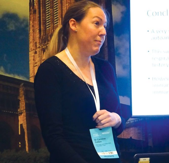
This means that chest physicians are getting the diagnosis right in the majority of cases, based on currently available methods, such as patients’ clinical history and examination, the results of high resolution–computed tomography, and widely available serology. “Which is good news,” Dr. Cotton observed.
Interstitial lung disease (ILD) comprises a huge spectrum of disorders. The main groups of ILDs are idiopathic, granulomatous, connective tissue disease–associated environmental, or medication exposure–associated; and the rare causes of ILD, each of which contain multiple subgroups of which IPF is one.
Sometimes it is obvious to respiratory physicians what the cause is, such as environmental exposure to asbestos or sarcoidosis for the granulomatous ILD, Dr. Cotton noted. Identifying connective tissue disease (CTD)–associated ILD can be more diagnostically challenging, however, and there are a large number of rheumatic conditions associated with CTD-associated ILD, including rheumatoid arthritis, systemic sclerosis, and Sjögren’s syndrome, to name a few.
One of the problems is that signs and symptoms of CTD may be absent at the time ILD starts to manifest and, even if signs are present, they may too subtle to be picked up in a general chest clinic. There also is a large number of antibodies for CTDs, but not all are widely available.
Dr. Cotton and her associates, therefore, wondered if there was a chance that patients being diagnosed with IPF actually could have covert CTD-associated ILD; this is an important distinction to make because the treatment differs for the two conditions. While ILD associated with CTD has a strong inflammatory component and is treated with corticosteroids and immunosuppressants, steroids can be harmful and increase mortality in IPF-ILD. The latter is treated with antifibrotic medications, such as pirfenidone and nintedanib.
For the study, serum samples from 250 patients with a definite diagnosis of IPF who were participating in the UK-BILD study were obtained and screened for known CTD antibodies using immunoprecipitation. Antibodies could be detected in just five (2%) patients – these included one patient each with anti-KS and anti-OJ antibodies, which are antisynthetase antibodies that are associated with myositis. Anti-Ku, another myositis-associated antibody, was identified in another patient, and one patient had an anti-RNA polymerase II antibody, which is associated with systemic sclerosis. Antimitochondrial autoantibodies were observed in one patient, and these are linked to primary biliary cirrhosis, which the patient was known to have.
There was nothing remarkable between the patients who did and did not have CTD antibodies in terms of their demographics, 76% and 80% were male, the mean ages were 73 and 70 years, respectively, and all were white.
However, 40% of patients did have unknown strong bands on immunoprecipitation, Dr. Cotton reported. This could suggest that there is an underlying immunological component to IPF, she added, but they had no recognized antibodies.
“A very small number of patients with IPF actually have the presence of autoantibodies strongly associated with CTDs. This suggests IPF is a very robust diagnosis; chest physicians are diagnosing it correctly most of the time, and they are really good at good at weeding out those who have got IPF and those who have potentially got connective tissue disease.” Dr. Cotton concluded.
Dr. Cotton had no conflicts of interest.
SOURCE: Cotton CV et al. Rheumatology. 2018;57[Suppl. 3]:key075.206.
LIVERPOOL, ENGLAND – Very few patients with idiopathic pulmonary fibrosis have connective tissue disease antibodies, suggesting that IPF is a “robust diagnosis” when made on the basis of standard diagnostic tests, it was reported at the British Society for Rheumatology annual conference.
“The results were perhaps not what we’d expected,” said Caroline V. Cotton, PhD, of the Institute of Ageing and Chronic Disease at the University of Liverpool, England.
This means that chest physicians are getting the diagnosis right in the majority of cases, based on currently available methods, such as patients’ clinical history and examination, the results of high resolution–computed tomography, and widely available serology. “Which is good news,” Dr. Cotton observed.
Interstitial lung disease (ILD) comprises a huge spectrum of disorders. The main groups of ILDs are idiopathic, granulomatous, connective tissue disease–associated environmental, or medication exposure–associated; and the rare causes of ILD, each of which contain multiple subgroups of which IPF is one.
Sometimes it is obvious to respiratory physicians what the cause is, such as environmental exposure to asbestos or sarcoidosis for the granulomatous ILD, Dr. Cotton noted. Identifying connective tissue disease (CTD)–associated ILD can be more diagnostically challenging, however, and there are a large number of rheumatic conditions associated with CTD-associated ILD, including rheumatoid arthritis, systemic sclerosis, and Sjögren’s syndrome, to name a few.
One of the problems is that signs and symptoms of CTD may be absent at the time ILD starts to manifest and, even if signs are present, they may too subtle to be picked up in a general chest clinic. There also is a large number of antibodies for CTDs, but not all are widely available.
Dr. Cotton and her associates, therefore, wondered if there was a chance that patients being diagnosed with IPF actually could have covert CTD-associated ILD; this is an important distinction to make because the treatment differs for the two conditions. While ILD associated with CTD has a strong inflammatory component and is treated with corticosteroids and immunosuppressants, steroids can be harmful and increase mortality in IPF-ILD. The latter is treated with antifibrotic medications, such as pirfenidone and nintedanib.
For the study, serum samples from 250 patients with a definite diagnosis of IPF who were participating in the UK-BILD study were obtained and screened for known CTD antibodies using immunoprecipitation. Antibodies could be detected in just five (2%) patients – these included one patient each with anti-KS and anti-OJ antibodies, which are antisynthetase antibodies that are associated with myositis. Anti-Ku, another myositis-associated antibody, was identified in another patient, and one patient had an anti-RNA polymerase II antibody, which is associated with systemic sclerosis. Antimitochondrial autoantibodies were observed in one patient, and these are linked to primary biliary cirrhosis, which the patient was known to have.
There was nothing remarkable between the patients who did and did not have CTD antibodies in terms of their demographics, 76% and 80% were male, the mean ages were 73 and 70 years, respectively, and all were white.
However, 40% of patients did have unknown strong bands on immunoprecipitation, Dr. Cotton reported. This could suggest that there is an underlying immunological component to IPF, she added, but they had no recognized antibodies.
“A very small number of patients with IPF actually have the presence of autoantibodies strongly associated with CTDs. This suggests IPF is a very robust diagnosis; chest physicians are diagnosing it correctly most of the time, and they are really good at good at weeding out those who have got IPF and those who have potentially got connective tissue disease.” Dr. Cotton concluded.
Dr. Cotton had no conflicts of interest.
SOURCE: Cotton CV et al. Rheumatology. 2018;57[Suppl. 3]:key075.206.
REPORTING FROM RHEUMATOLOGY 2018
Key clinical point: Few patients diagnosed as having idiopathic pulmonary fibrosis (IPF) are likely to have connective tissue disorders (CTD).
Major finding: Only 2% of patients had a recognized CTD antibody present.
Study details: 250 patients with IPF participating in the UK-BILD multicenter study.
Disclosures: Dr. Cotton stated she had no conflicts of interest.
Source: Cotton CV et al. Rheumatology. 2018;57(Suppl. 3):key075.206.
Hydroxychloroquine throws off Quantiferon-TB Gold results, study finds
ORLANDO – according to investigators from the University of Pennsylvania, Philadelphia.
Among 46 patients with lupus, dermatomyositis, or blistering diseases who had been on hydroxychloroquine within a year of testing, QuantiFERON-TB Gold (QFT-G) – the go-to TB test in many places – yielded indeterminate results in 37%. Meanwhile, just 9.6% of tests were indeterminate among 73 patients with those diseases who had not been on hydroxychloroquine (P less than .001). The findings could not be explained by concomitant use of prednisone and other immunosuppressives; there were no statistically significant differences between the groups. “This was shocking to us. We need to come up with a better screening test in this patient population,” said lead investigator Rebecca Gaffney, a research fellow at the University of Pennsylvania, and a medical student at Robert Wood Johnson Medical School, New Brunswick, NJ.*
It’s widely known that immunosuppressives interfere with QFT-G results, but antimalarials are considered immunomodulators, not immunosuppressives. The new study is probably the first to investigate the issue. The team is now pitting QFT-G against another TB blood test, the T-SPOT, in 100 patients to see if it’s a better option, in a trial that they expect to complete in 2018.
The investigators have a hunch that the T-SPOT might be better because, while QFT-G measures interferon-gamma concentrations in response to TB antigens, the T-SPOT “counts cells first to make sure you have a standard amount of cells, then looks at how many cells are releasing interferon-gamma,” Ms. Gaffney said, adding that “it seems like a more sensitive test,” especially for lymphocytopenic autoimmune patients. “We are really excited to see if there’s a better test for our patients, given all the clinical trials we do. We want to see what’s best, so there’s no barrier to receiving therapy.”
Subjects were around 50 years old on average, and the majority were women. Most were white, and about 20% were black.
There was no industry funding for the work, and Ms. Gaffney reported no disclosures.
*This article was updated on June 13. 2018.
REPORTING FROM ICCLE 2018
Timely dermatomyositis diagnosis, treatment remain elusive
ORLANDO – There was a median 1-year delay between the onset of symptoms and diagnosis of classic dermatomyositis, and a 17-month delay before diagnosis of amyopathic dermatomyositis, based on a review of 232 dermatomyositis patients seen at the University of Pennsylvania, Philadelphia.
Just 103 (44.4%) patients were diagnosed with dermatomyositis (DM) right out of the gate. Among the other 129, 48 (37.2%) were diagnosed with lupus, 38 (29.5%) with undifferentiated connective tissue disease, 10 (7.8%) went undiagnosed, and 33 (25.5%) were diagnosed with rosacea, psoriasis, rheumatoid arthritis, fibromyalgia, lichen planus, and a number of other conditions. By the time the DM diagnosis was finally confirmed, almost every patient had Gottron’s papules or sign.
Misdiagnosis of dermatomyositis (DM) is nothing new, but the study brings home just how common the problem is, even at a major academic medical institution.
One of the take homes is that , and remain vigilant for erythema on the lateral thighs or nasolabial fold, Gottron’s papules, and other diagnostic giveaways, the researchers said.

Interface dermatitis, in particular, can’t be relied on to differentiate the conditions. A better option is checking for lupus bands and membrane attack complexes on direct immunofluorescence.
There’s also just not enough awareness that dermatomyositis can present without the classic muscle symptoms and findings, i.e. clinically amyopathic DM. While 49 of 120 patients with classic dermatomyositis (40.8%) were misdiagnosed or undiagnosed in the study, the number rose to 80 of 112 (71.4%) among amyopathic patients.
“We saw that there was a much higher rate of misdiagnosis in patients who didn’t have any muscle disease. We have to raise awareness that amyopathic dermatomyositis is a very prevalent condition,” Dr. Patel said at the International Conference on Cutaneous Lupus Erythematosus.
“There might be some level of subclinical muscle activity where, if you did an MRI, you might see inflammation, but the patient doesn’t report any symptoms. There are also patients that don’t have any muscle findings on MRI, or elevated muscle enzymes, but still have the skin findings,” he said.
Perhaps the markedly increased risk of cancer in DM, especially within a year or 2 of symptom onset, is the strongest argument for earlier diagnosis. “There’s also a risk of interstitial lung disease, so making sure that you’re getting pulmonary function tests and age-appropriate malignancy screening in a timely fashion is very important,” Dr. Patel said.
Also, although many of the initial treatments for DM – sun protection and topical steroids and calcineurin inhibitors, for instance – are the same as for cutaneous lupus, medications like mycophenolate mofetil and methotrexate are used more readily. The sooner DM is recognized for what it is, the sooner patients can get relief, he said.
Almost all the patients were white women. The majority were 40-80 years old.
There was no industry funding for the work, and Dr. Patel didn’t have any disclosures.
SOURCE: da Silva DM et al. Presented at the 2018 International Conference on Cutaneous Lupus Erythematosus
ORLANDO – There was a median 1-year delay between the onset of symptoms and diagnosis of classic dermatomyositis, and a 17-month delay before diagnosis of amyopathic dermatomyositis, based on a review of 232 dermatomyositis patients seen at the University of Pennsylvania, Philadelphia.
Just 103 (44.4%) patients were diagnosed with dermatomyositis (DM) right out of the gate. Among the other 129, 48 (37.2%) were diagnosed with lupus, 38 (29.5%) with undifferentiated connective tissue disease, 10 (7.8%) went undiagnosed, and 33 (25.5%) were diagnosed with rosacea, psoriasis, rheumatoid arthritis, fibromyalgia, lichen planus, and a number of other conditions. By the time the DM diagnosis was finally confirmed, almost every patient had Gottron’s papules or sign.
Misdiagnosis of dermatomyositis (DM) is nothing new, but the study brings home just how common the problem is, even at a major academic medical institution.
One of the take homes is that , and remain vigilant for erythema on the lateral thighs or nasolabial fold, Gottron’s papules, and other diagnostic giveaways, the researchers said.
Interface dermatitis, in particular, can’t be relied on to differentiate the conditions. A better option is checking for lupus bands and membrane attack complexes on direct immunofluorescence.
There’s also just not enough awareness that dermatomyositis can present without the classic muscle symptoms and findings, i.e. clinically amyopathic DM. While 49 of 120 patients with classic dermatomyositis (40.8%) were misdiagnosed or undiagnosed in the study, the number rose to 80 of 112 (71.4%) among amyopathic patients.
“We saw that there was a much higher rate of misdiagnosis in patients who didn’t have any muscle disease. We have to raise awareness that amyopathic dermatomyositis is a very prevalent condition,” Dr. Patel said at the International Conference on Cutaneous Lupus Erythematosus.
“There might be some level of subclinical muscle activity where, if you did an MRI, you might see inflammation, but the patient doesn’t report any symptoms. There are also patients that don’t have any muscle findings on MRI, or elevated muscle enzymes, but still have the skin findings,” he said.
Perhaps the markedly increased risk of cancer in DM, especially within a year or 2 of symptom onset, is the strongest argument for earlier diagnosis. “There’s also a risk of interstitial lung disease, so making sure that you’re getting pulmonary function tests and age-appropriate malignancy screening in a timely fashion is very important,” Dr. Patel said.
Also, although many of the initial treatments for DM – sun protection and topical steroids and calcineurin inhibitors, for instance – are the same as for cutaneous lupus, medications like mycophenolate mofetil and methotrexate are used more readily. The sooner DM is recognized for what it is, the sooner patients can get relief, he said.
Almost all the patients were white women. The majority were 40-80 years old.
There was no industry funding for the work, and Dr. Patel didn’t have any disclosures.
SOURCE: da Silva DM et al. Presented at the 2018 International Conference on Cutaneous Lupus Erythematosus
REPORTING FROM ICCLE 2018
Key clinical point: Clinicians need to do a better job of catching DM early.
Major finding: It took a median of 12.2 months after the start of symptoms to diagnose classic dermatomyositis, and 17.1 months to diagnose amyopathic dermatomyositis.
Study details: Review of 232 patients
Disclosures: There was no industry funding, and the presenter didn’t have any disclosures.
Source: da Silva DM et al. Presented at the 2018 International Conference on Cutaneous Lupus Erythematosus
Median time to progression from discoid to systemic lupus may be less than a year
ORLANDO – Progression from cutaneous to systemic lupus happens far more quickly than is commonly reported in the literature, according to an investigation from Brigham and Women’s Hospital, Boston.
Most studies have emphasized mean time to progression, which is generally said to be about 8 years. The problem is that the range is broad, anywhere from a few months to 30 or more years, so “the outliers influence the mean, and therefore, may not provide a fully accurate representation of the interval in which [most] patients progress” from discoid lupus erythematosus (DLE) to systemic lupus erythematosus (SLE), lead investigator Scott Elman, MD, said at the International Conference on Cutaneous Lupus Erythematosus.
He and his team thought it would be more useful to instead look at median time to progression, the point at which half of patients develop systemic disease. They found that , with the first quartile progressing by 303 days. The mean time to progression, meanwhile, was over 4 years, because of outliers who progressed at anywhere from 27 days to 30 years.
“The median progression of DLE to SLE occurs much sooner than previously reported” with mean progression times, said Dr. Elman, an internal medicine and dermatology resident at Brigham and Women’s. “We believe the use of median [time] is [more] clinically useful for both providers and patients in their understanding of what a diagnosis of DLE means for the risk of developing systemic disease.
“As clinicians seeing DLE patients, the primary question we are asking ourselves is how frequently we should be monitoring for signs and symptoms of systemic lupus,” he said. Relying on mean time instead of median time might give a false sense of security – and even delay checking for arthritis, nephritis, and other manifestations. There’s no current standard of practice about when monitoring should occur, but “I certainly think that closer monitoring of new patients with a diagnosis of DLE, especially in their first couple of years,” is a good idea, he noted, adding that for most patients, “it seems that if it’s going to happen, it’s going to happen sooner” rather than later. At Brigham and Women’s, newly diagnosed patients are generally brought back every 3-6 months for lab tests and a thorough review of symptoms, Dr. Elman said.
The next step is to identify risk factors for early progression. The study did not find any significant differences in lab values, medication exposures, or disease manifestations between early and late progressors, perhaps because of the small sample size. The work continues.
There was no external funding for the work. Dr. Elman had no disclosures.
REPORTING FROM ICCLE 2018
Key clinical point: Progression from cutaneous to systemic lupus happens far more quickly than is commonly reported in the literature.
Major finding: The median time to progression was 398 days, among a group of patients diagnosed with discoid lupus erythematosus.
Study details: A review of 32 patients who converted to systemic disease.
Disclosures: There was no external funding for the work, and the lead investigator had no disclosures.
Don’t trust interface dermatitis to diagnose dermatomyositis
ORLANDO – (DM), according to David Fiorentino, MD, PhD, professor of dermatology, rheumatology, and immunology at Stanford (Calif.) University.
The video associated with this article is no longer available on this site. Please view all of our videos on the MDedge YouTube channel
Interface dermatitis on skin biopsy is “felt to be almost required by many people to make the diagnosis,” but he and his associates found that it was not present in about a quarter of a cohort of patients with DM. “We don’t want a clinician” to rule out the diagnosis based on its absence on a biopsy, “when its actually quite possible that the patient could have disease,” Dr. Fiorentino said at the International Conference on Cutaneous Lupus Erythematosus.
In general, skin biopsies in DM are tricky. “All of us take them, but we don’t really know how to interpret the information that comes back ... because we don’t really know how often many of [the associated] findings are seen” in DM patients, he noted.
One of the main concerns is to rule out lupus, but interface dermatitis is found in many of its cutaneous forms, as well as in graft-versus-host disease and other diseases.
So what’s a clinician to do? Fortunately, direct immunofluorescence can help. A positive lupus band test helps rule out DM, and the membrane attack complex helps rule it in, both with a good degree of certainty. In a video interview, Dr. Fiorentino explained these tests and how to use them.
Dr. Fiorentino had no relevant disclosures.
[email protected]
REPORTING FROM ICCLE 2018

Caution crucial for stem cell transplant in scleroderma, despite potential interest
SANDESTIN, FLA. – An expert called for restraint with the use of stem cell transplantation in scleroderma at the annual Congress of Clinical Rheumatology, emphasizing that the early mortality risk shows that the patient group that should be considered for the treatment is very narrow.
Results published earlier this year found a long-term benefit for myeloablative autologous stem cell transplantation, compared with cyclophosphamide, and were highly encouraging – but only in patients with severe disease (N Engl J Med. 2018;378:35-47). This might prompt patients to express interest in the treatment, many of whom are not suitable, said Janet Pope, MD, chair of rheumatology at St. Joseph’s Health Care in London, Ont.

Because of mortality seen in the first year in the stem cell transplant group, the benefit over cyclophosphamide was seen only in subsequent years, Dr. Pope stressed.
Consideration of the approach is only appropriate for patients with early diffuse scleroderma who have a 50% mortality risk over 5 years, Dr. Pope said. Patients in the SCOT trial had a modified Rodnan Skin Score (mRSS) of 30, with an average forced vital capacity of 74%, and 73 of the 75 patients in the trial had lung involvement. Essentially, Dr. Pope said, these are patients with some organ involvement that could be lethal, but not such severe organ involvement that it requires a transplant.
“These are our sick patients,” she said.
The results published this year are even more definitive, Dr. Pope said, considering that they largely meshed with results out of Europe, published in 2014, that showed that the stem cell transplant benefit was not seen after the first year because of early mortality (JAMA. 2014 Jun 25;311[24]:2490-8). In that trial, a 10% mortality risk was seen in the first year in the transplant group, and then a benefit to transplant in the long term over cyclophosphamide.
“These are very complementary positive trials,” Dr. Pope said. “We don’t want to kill 10% of our patients who don’t have perceived high chance of mortality in the next 5 years. ... These patients are highly selected, but I think it gives our patients hope and, for some of my patients, this will be a treatment for them.”
The long-term efficacy is also encouraging in that new but less potent treatments might yield good long-term results without the early deaths, she said. “It gives an idea and hope, and it helps us to understand that maybe with immune modulation, without the nuclear blast of stem cell transplant, maybe we can do better for our patients.”
Dr. Pope reported relevant financial relationships with Actelion, Bayer, Merck, Bristol-Myers Squibb, and Roche.
SANDESTIN, FLA. – An expert called for restraint with the use of stem cell transplantation in scleroderma at the annual Congress of Clinical Rheumatology, emphasizing that the early mortality risk shows that the patient group that should be considered for the treatment is very narrow.
Results published earlier this year found a long-term benefit for myeloablative autologous stem cell transplantation, compared with cyclophosphamide, and were highly encouraging – but only in patients with severe disease (N Engl J Med. 2018;378:35-47). This might prompt patients to express interest in the treatment, many of whom are not suitable, said Janet Pope, MD, chair of rheumatology at St. Joseph’s Health Care in London, Ont.
Because of mortality seen in the first year in the stem cell transplant group, the benefit over cyclophosphamide was seen only in subsequent years, Dr. Pope stressed.
Consideration of the approach is only appropriate for patients with early diffuse scleroderma who have a 50% mortality risk over 5 years, Dr. Pope said. Patients in the SCOT trial had a modified Rodnan Skin Score (mRSS) of 30, with an average forced vital capacity of 74%, and 73 of the 75 patients in the trial had lung involvement. Essentially, Dr. Pope said, these are patients with some organ involvement that could be lethal, but not such severe organ involvement that it requires a transplant.
“These are our sick patients,” she said.
The results published this year are even more definitive, Dr. Pope said, considering that they largely meshed with results out of Europe, published in 2014, that showed that the stem cell transplant benefit was not seen after the first year because of early mortality (JAMA. 2014 Jun 25;311[24]:2490-8). In that trial, a 10% mortality risk was seen in the first year in the transplant group, and then a benefit to transplant in the long term over cyclophosphamide.
“These are very complementary positive trials,” Dr. Pope said. “We don’t want to kill 10% of our patients who don’t have perceived high chance of mortality in the next 5 years. ... These patients are highly selected, but I think it gives our patients hope and, for some of my patients, this will be a treatment for them.”
The long-term efficacy is also encouraging in that new but less potent treatments might yield good long-term results without the early deaths, she said. “It gives an idea and hope, and it helps us to understand that maybe with immune modulation, without the nuclear blast of stem cell transplant, maybe we can do better for our patients.”
Dr. Pope reported relevant financial relationships with Actelion, Bayer, Merck, Bristol-Myers Squibb, and Roche.
EXPERT ANALYSIS FROM CCR 18