User login
NICE Expands Chemotherapy Options with Rituximab
The National Institute for Health and Clinical Excellence says that it will recommend rituximab in combination with several chemotherapy regimens as first-line treatments for people with advanced follicular lymphoma.
Current NICE guidance recommends rituximab in combination with cyclophosphamide, vincristine and prednisolone (CVP) for this patient group.
In final draft guidance issued Dec. 1, the agency, which makes cost- and clinical-effectiveness decisions for England and Wales, said that rituximab (MabThera, Roche) could also be used in combination with chlorambucil or the following chemotherapy regimens:
– Cyclophosphamide, doxorubicin, vincristine, and prednisolone (CHOP).
– Mitoxantrone, chlorambucil, and prednisolone (MCP).
– Cyclophosphamide, doxorubicin, etoposide, prednisolone, and interferon-alpha (CHVPi).
All recommended regimens are authorized in Europe, are commonly used in the United Kingdom, and have been evaluated with and without rituximab in open-label clinical trials in patients with stage III and IV follicular lymphoma. For all, addition of rituximab was shown to correspond with a significant survival benefit compared with the chemotherapy-alone groups.
Having a range of chemotherapy options available is important, NICE said, due to differences in patients’ fitness as they age. Chlorambucil is seen as an option mainly for older patients, or patients with a lower performance status.
Rituximab is a genetically engineered chimeric monoclonal antibody that targets cells bearing the CD20 surface marker. For follicular lymphoma, dosage is 375 mg/m2 body surface area for up to eight cycles, administered on day 1 of the chemotherapy cycle. Each 10-mL (100-mg) vial costs £174.63, or £873.15 for 500 mL.
Current NICE guidance also recommends rituximab monotherapy as a maintenance treatment immediately following first-line treatment with rituximab-containing chemotherapy regimens. While most patients presenting with advanced follicular lymphoma are treatment-naive, rituximab plus chemotherapy is also recommended by NICE for relapsed or refractory advanced follicular lymphoma.
The NICE reviewers found all of the rituximab-plus-chemotherapy regimens to be well within NICE’s cost-effectiveness parameters, with an estimated incremental cost effectiveness ratio of £7,720 per quality-adjusted life year for rituximab plus CVP; £10,800 per QALY gained for rituximab plus CHOP; and £9,320 per QALY gained for rituximab plus MCP. For CHVPi, the cost-effectiveness estimates remained uncertain. However, the agency felt it was unlikely that estimates would exceed its "threshold range" of between £20,000 and £30,000 per QALY.
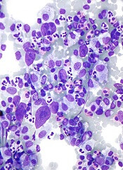
Severe Comorbidity Doubles Death Risk in Multiple Myeloma
PARIS – Elderly patients with multiple myeloma and severe comorbid disease are more than twice as likely to die as were those with no comorbidities, data from a single-center, retrospective study show.
Mild or moderate comorbidities did not appear to influence overall survival significantly in the 179-patient study. The hazard ratio (HR) for death in patients with severe comorbidity vs. none was 2.36 (P = .01), which was associated with a median overall survival of 15.1 months.
Median overall survival was 43.1 months for those with no comorbidities and 31.5 and 35 months, respectively, in those with mild (HR, 1.38; P = .26) or moderate (HR, 1.5; P = .19) comorbidities.
"The severity of comorbidities is associated with poorer survival in older adults with multiple myeloma," said lead author Dr. Tanya M. Wildes of Washington University in St. Louis.
Nevertheless, comorbidities are not currently incorporated into any staging systems for the disease, Dr. Wildes observed in an interview at the annual meeting of the International Society of Geriatric Oncology.
The research is part of a wider project that is looking at the value of performing a geriatric assessment to help predict which elderly patients with hematological malignancies may be able to undergo standard cancer treatment, or require additional monitoring for adverse events, or more supportive care.
"The severity of comorbidities is associated with poorer survival in older adults with multiple myeloma."
In the current study, Dr. Wildes and her colleagues identified all patients who were diagnosed and treated for multiple myeloma at Barnes-Jewish Hospital, St. Louis, between January 2000 and March 2010. Demographic, clinical, and survival data were obtained, with concomitant conditions graded using the Adult Comorbidity Evaluation (ACE) 27 index as none, mild, moderate, or severe.
The primary end point of the study was overall survival, the duration of which was calculated from the date of diagnosis until the time of last follow-up.
The median age of patients at baseline was 69 years (range, 65-91 years). There was a similar percentage of men (48.4%) and women (51.4%), and 75% of the population was white. Most of the remainder were black (23.5%).
According to the ACE-27 index, 41.3% of patients had mild, 24.6% had moderate, and 15.6% had severe comorbidities. The remaining 18.5% had no comorbidities.
"The challenge with multiple myeloma is that some of the comorbidities may be disease related as opposed to patient’s underlying comorbidities," Dr. Wildes noted. That would require reviewing the patients’ medical records, which was not done in the current evaluation of this data set but is something that the researchers plan on looking at next.
"These are hypothesis-generating data at the moment," Dr. Wildes said. Further study, to evaluate the impact of comorbidities on survival in multiple myeloma and their influence on patients’ tolerance of therapy and treatment decisions, is needed.
"On average, three comorbidities can be expected in a patient [aged] 65 years and older," said Dr. Lazzaro Repetto of the Istituto Nazionale di Riposo e Cura per Anziani at the Istituto di Ricovero e Cura a Carattere Scientifico in Rome.
Speaking at separate session during the meeting, Dr. Repetto said common comorbidities in elderly cancer patients included cardiovascular disease, renal insufficiency, diabetes, dementia, depression, anemia, osteoporosis, arthritis and arthrosis, and chronic obstructive pulmonary disease. All of these may have an impact on survival.
Indeed, other research presented by a Danish team showed that colorectal and lung cancers in particular were associated with a high number of comorbidities when compared with the general elderly population. A high comorbidity burden was also linked to reduced overall survival, but only in those with lung cancer, reported Dr. Trine Lembrecht Jørgensen of Odense (Denmark) University Hospital and associates.
The presence of comorbidities can alter treatment decisions, influencing the type of treatment offered, said Dr. Repetto. However, although assessing comorbid disease is important, it should always be part of a wider geriatric assessment, he advised. This should include measures of cognition, emotional and physical functioning, medication use, socioeconomic and social support factors, and the patient’s wishes.
"Using the geriatric assessment we can personalize treatment, and optimize the balance between benefit and risk of our decisions," Dr. Repetto suggested.
Dr. Wildes’ research was supported by a grant from the U.S. National Cancer Institute. Dr. Wildes and Dr. Repetto had no conflicts of interest.
FROM THE ANNUAL MEETING OF THE INTERNATIONAL SOCIETY OF GERIATRIC ONCOLOGY
Major Finding: Median overall survival in patients with severe comorbidity was 15.1 months vs. 43.1 months in patients with no comorbidity (hazard ratio for death, 2.36; P less than .01).
Data Source: Retrospective, single center study of 179 patients with multiple myeloma aged 65 years or older.
Disclosures: Dr. Wildes’ research was supported by a grant from the US National Cancer Institute. Neither Dr. Wildes nor Dr. Repetto reported any conflicts of interest.
Novel Therapies Put Multiple Myeloma 'On the Ropes'
SAN FRANCISCO – A sweep of new agents are poised to deliver what could be a knock-out blow to multiple myeloma, according to the director of the myeloma program at the University of California, San Francisco.
Some are second- or third-generation agents in a mainstay class that appear to have less toxicity than and/or overcome resistance to their predecessors, Dr. Jeffrey L. Wolf said at the annual Oncology Congress here. Others come from classes not previously used in this disease.

"There is a rush to develop new drugs in myeloma," Dr. Wolf told attendees. "We [understand] some mechanisms that the disease seems to favor, so we can interfere with those."
The prospects, in turn, are excellent: "We have made such tremendous headway in myeloma, except for those exceptional cases with 17p deletions and some other adverse prognostic features," he said. "As a disease, it seems to be on the ropes."
A Less-Toxic Proteasome Inhibitor
The first-generation proteasome inhibitor bortezomib (Velcade) improves myeloma outcomes, but maximizing its benefit will require addressing the peripheral neuropathy that limits its use. Three strategies may lessen this toxicity without compromising efficacy, Dr. Wolf suggested: modestly reducing the standard dose, adjusting the schedule from twice to once weekly, and altering the route of administration from intravenous to subcutaneous.
For example, in patients with pretreated myeloma, giving bortezomib subcutaneously instead of intravenously results in an identical overall response rate of 52% (Lancet Oncol. 2011;12:431-40). But there are significant reductions in rates of peripheral neuropathy of any grade (38% vs. 53%) and grade 3 or higher (6% vs. 16%).
"Practically everybody that we see now at UCSF gets subcutaneous [bortezomib]," Dr. Wolf said. It’s a great way to go back and treat patients who maybe otherwise have stopped their therapy because of their neuropathy."
Carfilzomib, an investigational next-generation proteasome inhibitor in phase III trials, is showing good antimyeloma activity and a low rate of peripheral neuropathy. Among patients with pretreated, relapsed, or refractory disease, carfilzomib monotherapy achieved overall response rates of 42% to 53% in a bortezomib-naive group (ASCO 2011 annual meeting. Abstract 8026) and 21% in a bortezomib-exposed group (Haematologica 2010;95:452 Abstract 1099). Median time to progression was at least 8 months for both.
Dr. Wolf said that unpublished data suggest that the response rate was still 17% specifically among patients who had had progression on bortezomib, "so there appears to be some activity in patients who are already refractory to a prior proteasome inhibitor."
Meanwhile, the rates of grade 1/2 and grade 3 peripheral neuropathy were 6% and 1%, respectively. And only a single patient of the 155 had to stop treatment because of this adverse effect.
When carfilzomib is combined with lenalidomide-dexamethasone (the so-called CRd regimen), the overall response rate in patients with pretreated, relapsed, and refractory disease is 78%, and the rate of complete or near complete response is 24% (ASCO 2011 meeting. Abstract 8025).
And, in newly diagnosed myeloma, among 18 patients receiving eight cycles of CRd, the overall response rate was 100%, and the rate of stringent-complete, complete, or near-complete response was 61% (2011 International Myeloma Workshop. Poster P-253). "This is very, very exciting—I don’t think we’ve seen this in any other combination," Dr. Wolf commented. "But these are small numbers of patients; we still need to increase the numbers of patients studied with this combination."
Bortezomib and carfilzomib may soon have company from several investigational proteasome inhibitors available in oral formulations that have shown promise in preclinical testing or have advanced to clinical trials: CEP-18770 (Cephalon), ONX-0912 (Onyx), and MLN-9708 (Millenium).
A Third-Generation IMiD in Trials
Pomalidomide, a third-generation immunomodulatory drug (IMiD), coming after thalidomide (Thalomid), and lenalidomide (Revlimid), is also showing good antimyeloma activity in clinical trials, according to Dr. Wolf.
Among patients with pretreated myeloma, the rate of partial or better response when pomalidomide is combined with dexamethasone has ranged from 25% to 42%, depending on the trial and patient population. Adverse events are primarily hematologic.
And in patients who have previously received lenalidomide, the response rate is similar, at 35% (ASCO 2011 annual meeting. Abstract 8067). "So, as with carfilzomib, where there appear to be responses in patients who have prior resistance to bortezomib, it appears that pomalidomide can give us responses in patients who have already had resistance to lenalidomide," he said.
HDAC Inhibitors Show Activity
The histone deacetylase (HDAC) inhibitor vorinostat (Zolinza) is approved for treatment of lymphoma. But it is being tested in clinical trials for myeloma, in combination therapy, with promising results, according to Dr. Wolf. Overall response rates have ranged from 40% to 94%, depending on the patient population and combination.
Similarly, the HDAC inhibitor romidepsin (Istodax) is approved for lymphoma treatment but is also being studied for antimyeloma activity. And panobinostat, an investigational member of this drug class, is being evaluated as a component of combination therapy in phase II and III myeloma trials.
Monoclonal Antibodies Tested
Elotuzumab is an investigational monoclonal antibody directed against CS1, a glycoprotein that is highly expressed on the surface of plasma cells and implicated in myeloma pathogenesis.
In a phase I trial among patients with relapsed or refractory myeloma, the combination of elotuzumab with lenalidomide and low-dose dexamethasone yielded an overall response rate of 82% (ASCO 2011 meeting. Abstract 8076). The rate was 83% among the subset of patients whose disease was refractory to the most recent therapy and 95% among the subset of lenalidomide-naive patients.
The combination of elotuzumab with bortezomib has also been tested in patients with relapsed or refractory myeloma. But the overall response rate with this combination was less impressive, at 48% (ASH 2010 meeting. Abstract 3023).
Other Agents and Pathways
Several other agents are being eyed for roles in myeloma therapy as well. They include bendamustine (Treanda), an old drug initially revived for lymphoma treatment; aurora kinase inhibitors (for example, MLN-8237); and inhibitors of the mammalian target of rapamycin, or mTOR (for example, INK-128).
Additionally, there is considerable interest in agents that target fibroblast growth factor receptor 3 (FGFR3) for one subgroup. "In patients with the 4;14 translocation, FGFR3 is overexpressed," Dr. Wolf explained. "Finding an inhibitor for that or a direct antibody ... may be quite effective in those patients."
Investigators are also assessing the impact of targeting certain signaling pathways, such as the Jak/Stat pathway and the AKT pathway. For instance, a phase III trial is testing perifosine, an investigational AKT inhibitor, in combination therapy among patients with relapsed or refractory myeloma (NCT01002248).
The Oncology Congress is presented by Reed Medical Education. Reed Medical Education and this news organization are owned by Reed Elsevier Inc.
Dr. Wolf disclosed that he is on the speakers bureaus of Millenium, Celgene, and Ortho-Biotech, and is a consultant to and speaker for Onyx.
SAN FRANCISCO – A sweep of new agents are poised to deliver what could be a knock-out blow to multiple myeloma, according to the director of the myeloma program at the University of California, San Francisco.
Some are second- or third-generation agents in a mainstay class that appear to have less toxicity than and/or overcome resistance to their predecessors, Dr. Jeffrey L. Wolf said at the annual Oncology Congress here. Others come from classes not previously used in this disease.
"There is a rush to develop new drugs in myeloma," Dr. Wolf told attendees. "We [understand] some mechanisms that the disease seems to favor, so we can interfere with those."
The prospects, in turn, are excellent: "We have made such tremendous headway in myeloma, except for those exceptional cases with 17p deletions and some other adverse prognostic features," he said. "As a disease, it seems to be on the ropes."
A Less-Toxic Proteasome Inhibitor
The first-generation proteasome inhibitor bortezomib (Velcade) improves myeloma outcomes, but maximizing its benefit will require addressing the peripheral neuropathy that limits its use. Three strategies may lessen this toxicity without compromising efficacy, Dr. Wolf suggested: modestly reducing the standard dose, adjusting the schedule from twice to once weekly, and altering the route of administration from intravenous to subcutaneous.
For example, in patients with pretreated myeloma, giving bortezomib subcutaneously instead of intravenously results in an identical overall response rate of 52% (Lancet Oncol. 2011;12:431-40). But there are significant reductions in rates of peripheral neuropathy of any grade (38% vs. 53%) and grade 3 or higher (6% vs. 16%).
"Practically everybody that we see now at UCSF gets subcutaneous [bortezomib]," Dr. Wolf said. It’s a great way to go back and treat patients who maybe otherwise have stopped their therapy because of their neuropathy."
Carfilzomib, an investigational next-generation proteasome inhibitor in phase III trials, is showing good antimyeloma activity and a low rate of peripheral neuropathy. Among patients with pretreated, relapsed, or refractory disease, carfilzomib monotherapy achieved overall response rates of 42% to 53% in a bortezomib-naive group (ASCO 2011 annual meeting. Abstract 8026) and 21% in a bortezomib-exposed group (Haematologica 2010;95:452 Abstract 1099). Median time to progression was at least 8 months for both.
Dr. Wolf said that unpublished data suggest that the response rate was still 17% specifically among patients who had had progression on bortezomib, "so there appears to be some activity in patients who are already refractory to a prior proteasome inhibitor."
Meanwhile, the rates of grade 1/2 and grade 3 peripheral neuropathy were 6% and 1%, respectively. And only a single patient of the 155 had to stop treatment because of this adverse effect.
When carfilzomib is combined with lenalidomide-dexamethasone (the so-called CRd regimen), the overall response rate in patients with pretreated, relapsed, and refractory disease is 78%, and the rate of complete or near complete response is 24% (ASCO 2011 meeting. Abstract 8025).
And, in newly diagnosed myeloma, among 18 patients receiving eight cycles of CRd, the overall response rate was 100%, and the rate of stringent-complete, complete, or near-complete response was 61% (2011 International Myeloma Workshop. Poster P-253). "This is very, very exciting—I don’t think we’ve seen this in any other combination," Dr. Wolf commented. "But these are small numbers of patients; we still need to increase the numbers of patients studied with this combination."
Bortezomib and carfilzomib may soon have company from several investigational proteasome inhibitors available in oral formulations that have shown promise in preclinical testing or have advanced to clinical trials: CEP-18770 (Cephalon), ONX-0912 (Onyx), and MLN-9708 (Millenium).
A Third-Generation IMiD in Trials
Pomalidomide, a third-generation immunomodulatory drug (IMiD), coming after thalidomide (Thalomid), and lenalidomide (Revlimid), is also showing good antimyeloma activity in clinical trials, according to Dr. Wolf.
Among patients with pretreated myeloma, the rate of partial or better response when pomalidomide is combined with dexamethasone has ranged from 25% to 42%, depending on the trial and patient population. Adverse events are primarily hematologic.
And in patients who have previously received lenalidomide, the response rate is similar, at 35% (ASCO 2011 annual meeting. Abstract 8067). "So, as with carfilzomib, where there appear to be responses in patients who have prior resistance to bortezomib, it appears that pomalidomide can give us responses in patients who have already had resistance to lenalidomide," he said.
HDAC Inhibitors Show Activity
The histone deacetylase (HDAC) inhibitor vorinostat (Zolinza) is approved for treatment of lymphoma. But it is being tested in clinical trials for myeloma, in combination therapy, with promising results, according to Dr. Wolf. Overall response rates have ranged from 40% to 94%, depending on the patient population and combination.
Similarly, the HDAC inhibitor romidepsin (Istodax) is approved for lymphoma treatment but is also being studied for antimyeloma activity. And panobinostat, an investigational member of this drug class, is being evaluated as a component of combination therapy in phase II and III myeloma trials.
Monoclonal Antibodies Tested
Elotuzumab is an investigational monoclonal antibody directed against CS1, a glycoprotein that is highly expressed on the surface of plasma cells and implicated in myeloma pathogenesis.
In a phase I trial among patients with relapsed or refractory myeloma, the combination of elotuzumab with lenalidomide and low-dose dexamethasone yielded an overall response rate of 82% (ASCO 2011 meeting. Abstract 8076). The rate was 83% among the subset of patients whose disease was refractory to the most recent therapy and 95% among the subset of lenalidomide-naive patients.
The combination of elotuzumab with bortezomib has also been tested in patients with relapsed or refractory myeloma. But the overall response rate with this combination was less impressive, at 48% (ASH 2010 meeting. Abstract 3023).
Other Agents and Pathways
Several other agents are being eyed for roles in myeloma therapy as well. They include bendamustine (Treanda), an old drug initially revived for lymphoma treatment; aurora kinase inhibitors (for example, MLN-8237); and inhibitors of the mammalian target of rapamycin, or mTOR (for example, INK-128).
Additionally, there is considerable interest in agents that target fibroblast growth factor receptor 3 (FGFR3) for one subgroup. "In patients with the 4;14 translocation, FGFR3 is overexpressed," Dr. Wolf explained. "Finding an inhibitor for that or a direct antibody ... may be quite effective in those patients."
Investigators are also assessing the impact of targeting certain signaling pathways, such as the Jak/Stat pathway and the AKT pathway. For instance, a phase III trial is testing perifosine, an investigational AKT inhibitor, in combination therapy among patients with relapsed or refractory myeloma (NCT01002248).
The Oncology Congress is presented by Reed Medical Education. Reed Medical Education and this news organization are owned by Reed Elsevier Inc.
Dr. Wolf disclosed that he is on the speakers bureaus of Millenium, Celgene, and Ortho-Biotech, and is a consultant to and speaker for Onyx.
EXPERT ANALYSIS FROM THE ANNUAL ONCOLOGY CONGRESS
Transplant Protocol Benefits Elderly With Hematologic Cancers
An analysis of long-term outcomes for elderly patients with advanced hematologic malignancies suggests they do as well as younger patients when treated with allogeneic hematopoietic cell transplantation following nonmyeloablative conditioning.
In patients aged 60-75 years, the protocol yielded a 5-year overall survival rate of 35% and a progression-free survival rate of 32%, according to a report in the Nov. 2 issue of JAMA. The overall 5-year survival rate was as high as 69% among the patients who had the lowest comorbidity scores and lowest disease risk.
Half of these older patients never required hospitalization either during or after treatment, and two-thirds of the survivors returned to normal or near-normal physical functioning, said Dr. Mohamed L. Sorror of the transplantation biology program at the Fred Hutchinson Cancer Research Center, Seattle, and his associates.
"These results are encouraging given the poor outcomes with nontransplantation treatments, especially for patients with high-risk acute myeloid leukemia, fludarabine-refractory chronic lymphocytic leukemia, or progressive lymphoma," the investigators noted. They assessed outcomes in 372 patients aged 60-75 years who were enrolled in prospective clinical trials of the therapy at 18 medical centers in 1998-2008. The study subjects were being treated for hematologic malignancies including leukemia, myelodysplastic syndromes, myeloproliferative diseases, multiple myeloma, and lymphoma.
Since older patients are not eligible for the intense cytotoxic conditioning regimens that precede high-dose allogeneic hematopoietic cell transplantation, these patients instead underwent nonmyeloablative conditioning that relies on graft-versus-tumor effects to cure the cancer. This included fludarabine and a low dose of total-body irradiation before transplantation and a course of immunosuppression with mycophenolate mofetil and a calcineurin inhibitor (cyclosporine or tacrolimus) afterward.
"These findings should help allay reluctance in entering older patients with hematologic cancers in non- myeloablative [transplant] protocols."
After a median follow-up of 55 months (range, 12-133 months), 133 patients were still alive. Overall 5-year survival was 35%, and 5-year progression-free survival was 32%.
When the data were analyzed by patient age, 5-year overall survival was 38% for those aged 60-64 years, 33% for those aged 65-69, and 25% for those aged 70 and older. "Regardless of age, 5-year survivals ranged from 23% in patients with high comorbidity scores and high disease risk to 69% in patients with low comorbidity scores and low disease risk," Dr. Sorror and his colleagues said (JAMA 2011;306:1874-83).
Approximately two-thirds of the survivors at 5 years had complete resolution of their graft-versus-host disease (GVHD) symptoms and were able to discontinue immunosuppressive medications after a median of 2.5 years. Both the incidence and the resolution of GVHD in these older study subjects were comparable to those reported in the literature for younger patients treated with high-dose hematopoietic cell transplantation.
"These findings, together with the normal to near-normal performance status of surviving patients, should help allay reluctance in entering older patients with hematologic cancers in nonmyeloablative [transplant] protocols," the researchers noted.
Disease progression and relapse accounted for most (135) of the 239 deaths. Relapse rates were 33% at 1 year and 41% at 5 years. Most nonrelapse deaths were attributed to multiple organ failure, GVHD, and infections.
Dr. Sorror and his associates noted that hematologic malignancies are predominantly diseases of the elderly, and the incidence is expected to increase up to 77% during the next 20 years, due in part to the aging of the general population. Yet the latest figures show that only 12% of patients treated with hematopoietic cell transplantation in recent years were older than 60 years.
"This clearly highlights the reluctance of physicians to offer allogeneic hematopoietic cell transplantation to elderly patients," they said.
The investigators are now starting a multicenter longitudinal study to follow such patients from diagnosis onward, in the hope of elucidating "the reasons behind the low rate of referral of older patients to transplantation, [as well as] how nonmyeloablative [transplantation] outcomes compare with those after conventional therapies."
This study was supported by the National Institutes of Health and the Leukemia & Lymphoma Society. Dr. Sorror’s associates reported numerous ties to industry sources.
The promising findings reported by Dr. Sorror and colleagues may have substantial implications for clinical decision making and for health care policies, particularly in view of the aging of the U.S. population, said Dr. Shin Mineishi.
Overall survival, progression-free survival, and other outcomes now appear almost comparable in older patients to those in younger patients. Yet physicians are still reluctant to refer older patients for the procedure. "Without a significant effort to promote the use of allogeneic HSCT [hematopoietic stem cell transplantation] in older patients, only a small fraction will receive the benefit of this new treatment option," he said.
Randomized studies comparing nonmyeloablative vs. reduced intensity allogeneic HSCT are needed, he said. In addition, older patients have different problems from younger patients; among these is that while older patients will require more resources to recover, Medicare provides insufficient coverage for many patients.
"Although age alone should no longer be considered a limiting factor for allogeneic HSCT, more questions have been raised, and more problems need to be resolved for achieving optimal outcomes for older patients receiving allogeneic HSCT."
Dr. Mineishi is in the blood and marrow transplant program at the University of Michigan, Ann Arbor. He reported ties to Genzyme. These remarks were adapted from his editorial accompanying Dr. Sorror’s report (JAMA 2011;306:1918-9).
An analysis of long-term outcomes for elderly patients with advanced hematologic malignancies suggests they do as well as younger patients when treated with allogeneic hematopoietic cell transplantation following nonmyeloablative conditioning.
In patients aged 60-75 years, the protocol yielded a 5-year overall survival rate of 35% and a progression-free survival rate of 32%, according to a report in the Nov. 2 issue of JAMA. The overall 5-year survival rate was as high as 69% among the patients who had the lowest comorbidity scores and lowest disease risk.
Half of these older patients never required hospitalization either during or after treatment, and two-thirds of the survivors returned to normal or near-normal physical functioning, said Dr. Mohamed L. Sorror of the transplantation biology program at the Fred Hutchinson Cancer Research Center, Seattle, and his associates.
"These results are encouraging given the poor outcomes with nontransplantation treatments, especially for patients with high-risk acute myeloid leukemia, fludarabine-refractory chronic lymphocytic leukemia, or progressive lymphoma," the investigators noted. They assessed outcomes in 372 patients aged 60-75 years who were enrolled in prospective clinical trials of the therapy at 18 medical centers in 1998-2008. The study subjects were being treated for hematologic malignancies including leukemia, myelodysplastic syndromes, myeloproliferative diseases, multiple myeloma, and lymphoma.
Since older patients are not eligible for the intense cytotoxic conditioning regimens that precede high-dose allogeneic hematopoietic cell transplantation, these patients instead underwent nonmyeloablative conditioning that relies on graft-versus-tumor effects to cure the cancer. This included fludarabine and a low dose of total-body irradiation before transplantation and a course of immunosuppression with mycophenolate mofetil and a calcineurin inhibitor (cyclosporine or tacrolimus) afterward.
"These findings should help allay reluctance in entering older patients with hematologic cancers in non- myeloablative [transplant] protocols."
After a median follow-up of 55 months (range, 12-133 months), 133 patients were still alive. Overall 5-year survival was 35%, and 5-year progression-free survival was 32%.
When the data were analyzed by patient age, 5-year overall survival was 38% for those aged 60-64 years, 33% for those aged 65-69, and 25% for those aged 70 and older. "Regardless of age, 5-year survivals ranged from 23% in patients with high comorbidity scores and high disease risk to 69% in patients with low comorbidity scores and low disease risk," Dr. Sorror and his colleagues said (JAMA 2011;306:1874-83).
Approximately two-thirds of the survivors at 5 years had complete resolution of their graft-versus-host disease (GVHD) symptoms and were able to discontinue immunosuppressive medications after a median of 2.5 years. Both the incidence and the resolution of GVHD in these older study subjects were comparable to those reported in the literature for younger patients treated with high-dose hematopoietic cell transplantation.
"These findings, together with the normal to near-normal performance status of surviving patients, should help allay reluctance in entering older patients with hematologic cancers in nonmyeloablative [transplant] protocols," the researchers noted.
Disease progression and relapse accounted for most (135) of the 239 deaths. Relapse rates were 33% at 1 year and 41% at 5 years. Most nonrelapse deaths were attributed to multiple organ failure, GVHD, and infections.
Dr. Sorror and his associates noted that hematologic malignancies are predominantly diseases of the elderly, and the incidence is expected to increase up to 77% during the next 20 years, due in part to the aging of the general population. Yet the latest figures show that only 12% of patients treated with hematopoietic cell transplantation in recent years were older than 60 years.
"This clearly highlights the reluctance of physicians to offer allogeneic hematopoietic cell transplantation to elderly patients," they said.
The investigators are now starting a multicenter longitudinal study to follow such patients from diagnosis onward, in the hope of elucidating "the reasons behind the low rate of referral of older patients to transplantation, [as well as] how nonmyeloablative [transplantation] outcomes compare with those after conventional therapies."
This study was supported by the National Institutes of Health and the Leukemia & Lymphoma Society. Dr. Sorror’s associates reported numerous ties to industry sources.
FROM JAMA
Major Finding: The 5-year survival was 35% overall and as high as 69% in the lowest-risk patients among those aged 60-75 years whose advanced hematologic malignancies were treated using hematopoietic cell transplantation following nonmyeloablative conditioning.
Data Source: An analysis of outcomes among 372 older patients enrolled in prospective clinical trials of transplant therapy at 18 medical centers in 1998-2008.
Disclosures: This study was supported by the National Institutes of Health and the Leukemia & Lymphoma Society. Dr. Sorror's associates reported numerous ties to industry sources.
PET Scans Key to Less Radiation for Hodgkin's Lymphoma
MIAMI BEACH – Patients with Hodgkin’s lymphoma may be spared additional radiotherapy following chemotherapy if they have a negative positron-emission tomography result, investigators from the German Hodgkin Study Group reported.
The negative predictive value for FDG (18fluorodeoxyglucose)–PET at 1 year was 94%, said Dr. Rolf P. Mueller of the University of Cologne (Germany). Among patients who had residual tumors measuring 2.5 cm or greater in diameter following chemotherapy, only 4% of those who were negative for residual disease on FDG-PET scans relapsed or required additional radiotherapy, compared with 11% of FDG-PET–positive patients.

"Thus, only those advanced-stage Hodgkin lymphoma patients with residual disease who are PET-positive patients might need additional radiotherapy," Dr. Mueller said at the annual meeting of the American Society of Radiation Oncology (ASTRO).
The investigators also found a significant difference in time-to-progression favoring PET-negative patients (P =.008) with Hodgkin’s lymphoma, also known as Hodgkin’s disease.
The percentage of patients who received radiation in this clinical trial, designated GHSG (German Hodgkin Study Group) HD-15, was 11%, compared with 70% of patients in the group’s GHSG-9 trial, Mueller noted. GHSG-15 studied the role of FDG-PET for evaluating residual disease and relapse risk among patients with advanced-stage Hodgkin’s lymphoma who had undergone six to eight cycles of chemotherapy with the BEACOPP regimen (bleomycin, etoposide, doxorubicin, cyclophosphamide, vincristine, procarbazine, and prednisone) (J. Clin. Oncol. 2003;21:1734-9).
Early results were published in 2008 (Blood 2008;112: 3989-94). In the current report, Mueller presented data on a larger cohort.
All patients with a partial response or better and a residual mass measuring 2.5 cm or greater received FDG-PET scans. Of the 728 patients with residual disease following BEACOPP, 540 (74.2%) were PET negative, and 188 were PET positive. Mueller presented data on 701 patients who had at least 1 year of follow-up.
At 1 year, 96% (522) of PET-negative patients had neither progression nor relapse, compared with 11% of those who were PET positive. Of the PET-negative patients, 23 experienced disease progression (eight in the residual mass, six with new disease outside of the mass, and nine with progression/relapse in both areas). An additional eight PET-negative patients required additional radiotherapy.
The study was funded by the member centers of the GSHG. Dr. Mueller had no conflict of interest disclosures.
MIAMI BEACH – Patients with Hodgkin’s lymphoma may be spared additional radiotherapy following chemotherapy if they have a negative positron-emission tomography result, investigators from the German Hodgkin Study Group reported.
The negative predictive value for FDG (18fluorodeoxyglucose)–PET at 1 year was 94%, said Dr. Rolf P. Mueller of the University of Cologne (Germany). Among patients who had residual tumors measuring 2.5 cm or greater in diameter following chemotherapy, only 4% of those who were negative for residual disease on FDG-PET scans relapsed or required additional radiotherapy, compared with 11% of FDG-PET–positive patients.
"Thus, only those advanced-stage Hodgkin lymphoma patients with residual disease who are PET-positive patients might need additional radiotherapy," Dr. Mueller said at the annual meeting of the American Society of Radiation Oncology (ASTRO).
The investigators also found a significant difference in time-to-progression favoring PET-negative patients (P =.008) with Hodgkin’s lymphoma, also known as Hodgkin’s disease.
The percentage of patients who received radiation in this clinical trial, designated GHSG (German Hodgkin Study Group) HD-15, was 11%, compared with 70% of patients in the group’s GHSG-9 trial, Mueller noted. GHSG-15 studied the role of FDG-PET for evaluating residual disease and relapse risk among patients with advanced-stage Hodgkin’s lymphoma who had undergone six to eight cycles of chemotherapy with the BEACOPP regimen (bleomycin, etoposide, doxorubicin, cyclophosphamide, vincristine, procarbazine, and prednisone) (J. Clin. Oncol. 2003;21:1734-9).
Early results were published in 2008 (Blood 2008;112: 3989-94). In the current report, Mueller presented data on a larger cohort.
All patients with a partial response or better and a residual mass measuring 2.5 cm or greater received FDG-PET scans. Of the 728 patients with residual disease following BEACOPP, 540 (74.2%) were PET negative, and 188 were PET positive. Mueller presented data on 701 patients who had at least 1 year of follow-up.
At 1 year, 96% (522) of PET-negative patients had neither progression nor relapse, compared with 11% of those who were PET positive. Of the PET-negative patients, 23 experienced disease progression (eight in the residual mass, six with new disease outside of the mass, and nine with progression/relapse in both areas). An additional eight PET-negative patients required additional radiotherapy.
The study was funded by the member centers of the GSHG. Dr. Mueller had no conflict of interest disclosures.
FROM THE ANNUAL MEETING OF THE AMERICAN SOCIETY FOR RADIATION ONCOLOGY
Major Finding: FDG-PET scans following chemotherapy in patients with advanced-stage Hodgkin’s lymphoma have a negative predictive value of 94%.
Data Source: The prospective GHSG HD-15 trial involving 701 patients.
Disclosures: The study was funded by the GSHG. Dr. Mueller had no conflict of interest disclosures.
FDA grants accelerated approval for brentuximab vedotin
The US Food and Drug Administration (FDA) has granted accelerated approval for brentuximab vedotin (Adcetris) to treat patients with Hodgkin lymphoma (HL) and systemic anaplastic large-cell lymphoma (sALCL).
The drug can now be used to treat HL patients after the failure of autologous stem cell transplant (ASCT) or, in patients who are not ASCT candidates, after they fail at least 2 prior multiagent chemotherapy regimens.
Brentuximab vedotin can also be used to treat patients with sALCL after the failure of at least 1 prior multiagent chemotherapy regimen.
About accelerated approval
The FDA instituted its accelerated approval program to allow for earlier approval of drugs that treat serious conditions and fill an unmet medical need. The approval is based on a surrogate endpoint that is not a measure of clinical benefit.
Drug companies are still required to conduct studies to confirm the anticipated clinical benefit. If these confirmatory trials suggest the drug actually provides a clinical benefit, the FDA grants traditional approval for the drug. If the confirmatory trial does not show a clinical benefit, FDA has regulatory procedures in place that could lead to removing the drug from the market.
Brentuximab vedotin in HL
Accelerated approval for the HL indication was based on a single-arm, multicenter trial to evaluate the objective response rate of brentuximab vedotin as a single agent. The 102 patients enrolled in the trial had CD30-positive HL that relapsed following ASCT.
Brentuximab vedotin prompted an overall response rate in these patients of 73%, with a median response duration of 6.7 months.
Thirty-two percent of patients achieved a complete remission, with a median duration of 20.5 months. Forty percent of patients achieved a partial remission, with a median duration of 3.5 months.
Brentuximab vedotin in sALCL
Accelerated approval for the treatment of sALCL was based on a single-arm, multicenter trial as well. The study included 58 patients with CD30-positive sALCL who had previously received frontline, multiagent chemotherapy treatment.
The primary efficacy endpoint of overall response rate was achieved in 86% of sALCL patients, with a median duration of 12.6 months.
Fifty-seven percent of patients achieved a complete remission, with a median duration of 13.2 months. Twenty-nine percent of patients achieved a partial response, with a median duration of 2.1 months.
The recommended dose and schedule for both indications is 1.8 mg/kg given intravenously over 30 minutes every 3 weeks. Treatment may last a total of 16 cycles, or until disease progresses or toxicity becomes unacceptable.
The agent, manufactured by Seattle Genetics under the trade name Adcetris, is the first new treatment for HL to be approved by the FDA since 1977 and the first to be indicated for sALCL. ![]()
FDA Approves Brentuximab for Two Lymphomas
The Food and Drug Administration on Aug. 19 approved brentuximab, a CD30-directed antibody drug-conjugate, for the treatment of Hodgkin’s lymphoma and systemic anaplastic large-cell lymphoma, after other treatments have failed.
Specifically, brentuximab was approved for the treatment of patients with Hodgkin’s lymphoma (HL) after failure of autologous stem cell transplant (ASCT) or after failure of at least two prior multiagent chemotherapy regimens in patients who are not ASCT candidates. It is the first new treatment approved for HL by the FDA since 1977, according to the FDA statement. The indications are based on response rates, and there are no available data that show "improvement in patient reported outcomes or survival" with treatment, according to the prescribing information.
The FDA also approved brentuximab for patients with systemic anaplastic large-cell lymphoma (ALCL), a rare form of lymphoma, after failure of at least one prior multiagent chemotherapy regimen. It is the first treatment approved for this rare lymphoma; approximately 2,000 new cases are diagnosed yearly in the United States.
Both types of lymphomas express the CD30 antigen. Brentuximab is a combination of an anti-CD30 antibody and a drug, monomethyl auristatin E (MMAE), a microtubule disrupting agent. The antibody directs the drug to C30-expressing tumor cells, where it is released.
Brentuximab is administered intravenously every 3 weeks; it will be marketed in the United States as Adcetris by Seattle Genetics. It will be available the week of Aug. 22, according to a company spokesperson.
The product was approved under the FDA’s accelerated approval program, which allows the agency to approve a drug for a serious disease based on data showing the drug is effective on a surrogate end point that is "reasonably likely" to predict clinical benefit. Accelerated approvals provide patients with access to promising treatments, but companies are required to provide confirmatory clinical data in order for the drug to be converted to a full approval and remain on the market.
"Early clinical data suggest that patients who received Adcetris for Hodgkin’s lymphoma and systemic anaplastic lymphoma experienced a significant response to the therapy," Dr. Richard Pazdur, director of the Office of Oncology Drug Products in the FDA’s Center for Drug Evaluation and Research, said in the statement.
In a study of 102 patients with HL treated with brentuximab, 73% of the patients had either a complete or partial response to treatment. In a similarly designed study of 58 patients with ALCL, 86% achieved a partial or complete response to treatment. Neutropenia, peripheral sensory neuropathy, fatigue, nausea, anemia, upper-respiratory infection, diarrhea, fever, cough, vomiting, and thrombocytopenia were among the most common adverse effects associated with treatment, according to the FDA.
At a meeting in July, an FDA advisory panel recommended the accelerated approval of both indications.
Brentuximab is under review for the same indications in Europe; a decision on approval is expected in the first half of 2012, according to a spokesperson for Millennium: the Takeda Oncology Co., which has rights to commercialize brentuximab outside the United States and Canada.
FROM THE FOOD AND DRUG ADMINISTRATION
CD19-redirected T cells induce remission in CLL patients
Gene therapy with a lentiviral vector expressing a chimeric antigen receptor with specificity for CD19 (CART19) has induced complete remission in 3 patients with chronic lymphocytic leukemia (CLL), according to research published simultaneously in the August 10 issues of The New England Journal of Medicine and Science Translational Medicine.
The research team, from the University of Pennsylvania, reported that the reinfused, modified T cells expanded to more than 1000 times the initial engraftment level. The patients’ remission was ongoing at 10 months after treatment.
The investigators believe the big difference between this genetically modified T cell and previous ones that had disappointing clinical activity is the addition of the CD137 (4-1BB) costimulatory signaling domain that significantly increases antitumor activity.
The team, led by Carl June, MD, described in the NEJM article the T-cell treatment of one of the patients with advanced, p53-deficient CLL.
A half year prior to enrolling in the trial, the 64-year-old patient’s T cells were collected and frozen. Before reinfusing the T cells into the patient, the investigators thawed the cells and transduced them with lentivirus expressing CD19-specific chimeric antigen receptor.
Four days prior to reinfusion, the patient received chemotherapy with pentostatin and cyclophosphamide to deplete his lymphocytes. After 3 days of chemotherapy, his bone marrow was hypercellular with approximately 40% involvement by CLL.
After 4 days of chemotherapy, the patient received an infusion of T cells, of which 5% were transduced, totaling 1.42 x 10⁷ transduced cells, split into 3 consecutive daily infusions.
Two weeks after the infusion, the patient experienced chills, fever, and fatigue, which intensified over the subsequent days. He was diagnosed with tumor lysis syndrome on day 22 after infusion. On day 23 after the CART19-cell infusion, the patient had no evidence of CLL in the bone marrow, and by day 28, his adenopathy was not palpable.
In addition to tumor lysis syndrome, the only other grade 3/4 toxicity observed was lymphopenia.
The investigators did not expect that such a low dose of chimeric antigen receptor T cells would result in a clinically evident antitumor response. The dose was several orders of magnitude lower than that used in previous studies of modified T cells.
They speculated that the course of chemotherapy administered to the patient prior to the CART19-cell infusion may have been responsible for the increased engraftment and for “potentiating the ability of chimeric antigen receptor T cells to kill stressed tumor cells that would otherwise survive the chemotherapy.”
The researchers conclude that continued study of CD19-redirected T cells is warranted and plan to test the approach in other CD19-positive tumors, including non-Hodgkin lymphoma and acute lymphocytic leukemia. ![]()
Paraneoplastic Autoimmune Multiorgan Syndrome Proves Rapidly Fatal
The skin may hold the key to differentiating classic pemphigus from the heterogenous autoimmune syndrome known as paraneoplastic autoimmune multiorgan syndrome.
It is a distinction of critical prognostic importance, because paraneoplastic autoimmune multiorgan syndrome (PAMS) typically is rapidly fatal, according to Dr. Sergei A. Grando, professor of dermatology and biologic chemistry at the University of California, Irvine. "The vast majority of patients die within several months of diagnosis, usually due to infections or respiratory failure, often taking the form of multiorgan system failure."

Two-thirds of patients with PAMS have a known internal malignancy at the time of their first mucocutaneous eruption. The most common of these neoplasms are non-Hodgkin’s lymphoma, which is present in more than 40% of PAMS patients; chronic lymphocytic leukemia, present in 30%; Castleman disease, present in 10%; and thymoma, present in 6%.
When a patient meets the diagnostic criteria for PAMS without having a known cancer, it is appropriate to launch a search for hidden malignancy, said Dr. Grando.
A key distinction between the skin lesions of PAMS and classic pemphigus is that PAMS involves inflammatory macules, papules, plaques, and blisters occurring on an inflammatory background over the trunk and extremities, including the palms and soles, but sparing the scalp.
In contrast, the generally more vesicular blisters and crusted erosions of pemphigus vulgaris display little erythema and usually occur on a noninflammatory background on the scalp, trunk, and extremities (but sparing the palms and soles). The most common location for skin lesions in PAMS is the palms; in pemphigus vulgaris, it is the scalp.
Also, Nikolsky's sign is positive in pemphigus vulgaris, but negative in PAMS, added Dr. Grando, who was among the investigators who first described PAMS a decade ago (Arch. Dermatol. 2001;137:193-206).
PAMS is characterized by severe and diffuse oral mucous membrane involvement, with persistent painful stomatitis because of blisters and erosions, and frequent involvement of other mucous membranes, including the eyes and genitalia. Cicatrizing conjunctivitis is particularly common in PAMS. In contrast, oral mucous membrane involvement in pemphigus is more discrete, with the eyes or other nonoral mucosa rarely involved.

Another key in the differential diagnosis: PAMS is associated with the HLA-DRB1*03 allele, whereas pemphigus vulgaris and foliaceous are strongly associated with the -04 and -14 alleles, respectively.
A hallmark of PAMS is respiratory involvement, with the sloughing of bronchial epithelial cells contributing to small airway occlusion and bronchiolitis obliterans. Classic pemphigus is free of respiratory involvement.
Both the esophagus and colon may be involved in PAMS, whereas only the esophagus is affected in pemphigus vulgaris.
In a published series of 28 PAMS patients, painful and generalized oral stomatitis was present in all 28, respiratory involvement was in 26, death from respiratory failure occurred in 22, and lichenoid skin involvement was present in 19. Only seven patients had no skin lesions (Br. J. Dermatol. 2003;149:1143-51).
At least five different subtypes of PAMS can be distinguished on the basis of the skin disease manifestations they most resemble. These subtypes of pemphiguslike PAMS include paraneoplastic pemphigus, bullous pemphigoid–like, erythema multiforme–like, lichen planus–like, and graft-vs.-host-disease–like versions of PAMS.
Dr. Grando eschews the term "paraneoplastic pemphigus" as too restrictive. After all, true pemphigus doesn’t usually affect the lungs, he noted.
Whereas most patients with classic pemphigus respond well to high-dose corticosteroids and recalcitrant disease can be effectively treated with cytotoxic agents, cyclosporine, intravenous gamma globulin, and other second-line agents, PAMS is resistant to all conventional forms of therapy.
"What can we offer? Really not much," Dr. Grando said during his presentation at the World Congress of Dermatology in Seoul, South Korea.
The preferred treatment regimen is a combination of prednisone and cyclosporine, with or without cyclophosphamide. Monthly courses of IVIG can buy patients a few extra months, he added.
Dr. Grando declared having no relevant financial interests.
The skin may hold the key to differentiating classic pemphigus from the heterogenous autoimmune syndrome known as paraneoplastic autoimmune multiorgan syndrome.
It is a distinction of critical prognostic importance, because paraneoplastic autoimmune multiorgan syndrome (PAMS) typically is rapidly fatal, according to Dr. Sergei A. Grando, professor of dermatology and biologic chemistry at the University of California, Irvine. "The vast majority of patients die within several months of diagnosis, usually due to infections or respiratory failure, often taking the form of multiorgan system failure."
Two-thirds of patients with PAMS have a known internal malignancy at the time of their first mucocutaneous eruption. The most common of these neoplasms are non-Hodgkin’s lymphoma, which is present in more than 40% of PAMS patients; chronic lymphocytic leukemia, present in 30%; Castleman disease, present in 10%; and thymoma, present in 6%.
When a patient meets the diagnostic criteria for PAMS without having a known cancer, it is appropriate to launch a search for hidden malignancy, said Dr. Grando.
A key distinction between the skin lesions of PAMS and classic pemphigus is that PAMS involves inflammatory macules, papules, plaques, and blisters occurring on an inflammatory background over the trunk and extremities, including the palms and soles, but sparing the scalp.
In contrast, the generally more vesicular blisters and crusted erosions of pemphigus vulgaris display little erythema and usually occur on a noninflammatory background on the scalp, trunk, and extremities (but sparing the palms and soles). The most common location for skin lesions in PAMS is the palms; in pemphigus vulgaris, it is the scalp.
Also, Nikolsky's sign is positive in pemphigus vulgaris, but negative in PAMS, added Dr. Grando, who was among the investigators who first described PAMS a decade ago (Arch. Dermatol. 2001;137:193-206).
PAMS is characterized by severe and diffuse oral mucous membrane involvement, with persistent painful stomatitis because of blisters and erosions, and frequent involvement of other mucous membranes, including the eyes and genitalia. Cicatrizing conjunctivitis is particularly common in PAMS. In contrast, oral mucous membrane involvement in pemphigus is more discrete, with the eyes or other nonoral mucosa rarely involved.
Another key in the differential diagnosis: PAMS is associated with the HLA-DRB1*03 allele, whereas pemphigus vulgaris and foliaceous are strongly associated with the -04 and -14 alleles, respectively.
A hallmark of PAMS is respiratory involvement, with the sloughing of bronchial epithelial cells contributing to small airway occlusion and bronchiolitis obliterans. Classic pemphigus is free of respiratory involvement.
Both the esophagus and colon may be involved in PAMS, whereas only the esophagus is affected in pemphigus vulgaris.
In a published series of 28 PAMS patients, painful and generalized oral stomatitis was present in all 28, respiratory involvement was in 26, death from respiratory failure occurred in 22, and lichenoid skin involvement was present in 19. Only seven patients had no skin lesions (Br. J. Dermatol. 2003;149:1143-51).
At least five different subtypes of PAMS can be distinguished on the basis of the skin disease manifestations they most resemble. These subtypes of pemphiguslike PAMS include paraneoplastic pemphigus, bullous pemphigoid–like, erythema multiforme–like, lichen planus–like, and graft-vs.-host-disease–like versions of PAMS.
Dr. Grando eschews the term "paraneoplastic pemphigus" as too restrictive. After all, true pemphigus doesn’t usually affect the lungs, he noted.
Whereas most patients with classic pemphigus respond well to high-dose corticosteroids and recalcitrant disease can be effectively treated with cytotoxic agents, cyclosporine, intravenous gamma globulin, and other second-line agents, PAMS is resistant to all conventional forms of therapy.
"What can we offer? Really not much," Dr. Grando said during his presentation at the World Congress of Dermatology in Seoul, South Korea.
The preferred treatment regimen is a combination of prednisone and cyclosporine, with or without cyclophosphamide. Monthly courses of IVIG can buy patients a few extra months, he added.
Dr. Grando declared having no relevant financial interests.
Multifocal bone infarctions in both knees: An unusual presentation of multiple myeloma
Ying-Kei Hui, MD,1 Thomas Slattery, MD,2 Dale M. Frank, MD,3 Carol Dolinskas, MD,4 and David Henry, MD, FACP5
Departments of 1Internal Medicine, Pennsylvania Hospital; 2Radiology, Pennsylvania Hospital; 3Pathology and Laboratory Medicine, Hospital of the University of Pennsylvania; 4Nuclear Medicine, Diagnostic Radiology, Pennsylvania Hospital; and 5Medicine, Pennsylvania Hospital, Philadelphia, PA
Multiple myeloma (MM) is a neoplastic proliferation of monoclonal plasma cells within the bone marrow, which overproduces immunoglobulin. This disorder accounts for approximately 1% of all reported neoplasms and 12%– 15% of all hematologic malignancies.1 It is the second most common hematologic malignancy diagnosed.2 The etiology is still not fully understood. MM typically affects older patients, ranging from 50–78 years (median, 61 years).3 Common clinical presentations include fatigue, anemia, renal failure, hypercalcemia, bone pain, and pathologic fractures.
Bone involvement in MM may vary at presentation. Most commonly, radiographic findings include multiple small, sharply defined, lytic, “punched-out” lesions without reactive bone formation, arising in the medullary cavity at sites of preserved hematopoiesis in adults (the axial skeleton). The pathophysiology of the bone findings is uncertain, though presumed to be resultant of either inhibition of osteoblastic activity and/ or activation of osteoclastic activity. Involvement of the cortex results in endosteal scalloping, with invasion of the periosteum and occasionally extraosseous extension. Lesions are most commonly seen in the vertebrae, ribs, skull, pelvic bones, and femur, in descending order of prevalence. Distal bone involvement is less common, though cases with predominant involvement in peripheral bones have been described. Uncommon radiographic presentations include diffuse skeletal osteopenia without focal lesions or sclerotic lesions. 4,5 To our knowledge, multiple bone infarcts as a complication of MM have not been reported in the medical literature.
Case presentation
A 47-year-old man with no significant medical history presented after the recent onset of painless hematuria, which spontaneously resolved after 2 days. He complained of left knee pain, which he noted after doing yard work.
Routine laboratory examination showed normocytic normochromic anemia, with a hemoglobin level of 11.5 g/dL and a mildly elevated alkaline phosphatase (ALP) level at 124 units/L. An MRI of the left knee showed increased red bone marrow within the distal femur and proximal tibia/fibula, initially thought to be compatible with anemia from an unexplained inflammatory process. Further urologic and gastroenterologic workup was negative.
Several months later, our patient was noted to have progressive fatigue, with a decrease in hemoglobin level to 10.6 g/dL and a mildly elevated erythrocyte sedimentation rate. Physical examination was otherwise unremarkable. A repeat MRI of both knees showed an extensive marrow infiltrative process, with multiple presumed secondary bone infarcts in the distal femora and proximal tibias, proximal fibulae, and patellae (Figure 1). A tandem skeletal survey showed mild diffuse osteopenia and several small, rounded, lytic foci in the skull (Figure 2), which were suspicious for MM. No focal radiographic lesions were seen (Figure 3).
Bone marrow biopsy from the left posterior iliac crest revealed hypocellular marrow (20%). An expansion of plasmacytoid cells with eccentrically placed, round nuclei, clumped chromatin, occasional nucleoli, and moderate amounts of eosinophilic cytoplasm accounted for 75% of the marrow cellularity. An aspirate smear demonstrated scattered mature plasma cells, accounting for roughly 15% of the total cellularity. Flow cytometry showed no overt evidence of marrow involvement by a lymphocytic clone. Bone biopsy was not performed in the areas of the knees seen to be abnormal on MRI examination.
Serum protein electrophoresis (SPEP) showed a band in the betagamma region. Immunofixation confirmed the presence of a monoclonal paraprotein, consisting predominantly of immunoglobulin A (IgA) heavy chain and kappa light chain. A bone marrow biopsy and aspirate showed replacement of most hematopoietic elements by sheets of mature plasma cells, accounting for 75% of the total marrow cellularity (Figure 4). Confirmatory immunostains were positive for CD138, CD117, and kappa light chain (Figure 5) and negative for CD79a and lambda light chain (Figure 6). A diagnosis of MM was made based on the finding of M protein in the urine, the presence of greater than 10% clonal plasma cells in bone marrow, and related clinical symptomatology (including anemia and hypercalcemia).
The patient was started on immunomodulating therapy with lenalidomide (Revlimid), bortezomib (Velcade), and dexamethasone. Autologous stem cell transplantation may be considered after appropriate treatment.
Discussion
We offer the case of a patient with MM who presented with bilateral knee infarcts. Synonyms of bone infarct include osteonecrosis, bone necrosis, avascular necrosis, aseptic necrosis, ischemic bone necrosis, and bone death.6 MRI is the most sensitive imaging modality for evaluation of the bone marrow. It can detect early osteonecrotic changes in bones well before they are visible on radiography7; this fact was exemplified in our case, in which the patient had only mild osteopenia on knee radiographs but extensive osteonecrotic changes on MRI examination.
Bone infarct more commonly involves the hips than the knees.7 Knee involvement can be differentiated into two main categories: primary and secondary. Primary, spontaneous, or idiopathic involvement tends to be unilateral and usually is seen in the elderly, although the recent literature suggests that many of these socalled spontaneous cases are actually secondary to subcortical microfractures, which are then complicated by osteonecrosis.5 Secondary causes tend to present at a younger age, with bilateral and multifocal involvement. Examples of secondary causes include steroid therapy, alcoholism, decompression syndrome, hemoglobinopathies (sickle cell disease), autoimmune disease (lupus and antiphospholipid disease), infections (human immunodeficiency virus), radiation, and trauma.8–15 Other causes, such as chemotherapy toxicity in pediatric leukemia16 and Gaucher disease, have been reported.7 Osteonecrosis of the jaw is a known treatment complication of bisphosphonate therapy in patients with MM17; however, there have been no previous reports describing the presentation of multifocal bone infarcts in both knees in patients with MM.
Although the pathogenesis of bone infarction is unclear, it is thought to be caused by the combined effects of systemic and local factors affecting the blood supply, vascular damage, increased intraosseous pressure, and mechanical stresses. These processes lead to compromise of the bone vasculature, resulting in the death of bone and marrow cells.9 In our case, MRI of both knees revealed an extensive marrow infiltrative process, which may have caused local vasculature damage and diminished blood supply resulting in bone infarctions.
Conclusion
Bone infarction of any joint is not a well-established complication of MM. Physicians should be aware of this potential presentation. Although there is no cure for MM, early recognition of MM can lead to more effective treatment, thus slowing disease progression and improving overall clinical outcomes.
Disclosures
The authors have no conflicts of interest to disclose.
References
1. Phekoo KJ, Schey SA, Richards MA, et al. A population study to define the incidence and survival of multiple myeloma in a National Health Service Region in UK. Br J Haematol 2004;127:299–304.
2. Esteve FR, Roodman GD. Pathophysiology of myeloma bone disease. Best Pract Res Clin Haematol 2007;20:613–624.
3. Jain M, Ascensao J, Schechter GP. Familial myeloma and monoclonal gammopathy: a report of eight African American families. Am J Hematol 2009;84:34–38.
4. Winterbottom AP, Shaw AS. Imaging patients with myeloma. Clin Radiol 2009;64:1–11.
5. Resnick D. Diagnosis of Bone and Joint Disorders. Philadelphia, PA: WB Saunders; 2002:2188–2233.
6. Stoller D, Tirman P, Bredella M, Branstetter R. Diagnostic Imaging: Orthopaedics. Philadelphia, PA: WB Saunders; 2003:82.
7. Assouline-Dayan Y, Chang C, Greenspan A, Shoenfeld Y, Gershwin ME. Pathogenesis and natural history of osteonecrosis. Semin Arthritis Rheum 2002;32:94–124.
8. Patel DV, Breazeale NM, Behr CT, Warren RF, Wickiewicz TL, O’Brien SJ. Osteonecrosis of the knee: current clinical concepts. Knee Surg Sports Traumatol Arthrosc 1998;6:2–11.
9. Chang CC, Greenspan A, Gershwin ME. Osteonecrosis: current perspectives on pathogenesis and treatment. Semin Arthritis Rheum 1993;23:47–69.
10. Jones LC, Mont MA, Le TB, et al. Procoagulants and osteonecrosis. J Rheumatol 2003; 30:783–791.
11. Saito N, Nadgir RN, Flower EN, Sakai O. Clinical and radiologic manifestations of sickle cell disease in the head and neck. Radiographics 2010;30:1021–1034.
12. Miller KD, Masur H, Jones EC, et al. High prevalence of osteonecrosis of the femoral head in HIV-infected adults. Ann Intern Med 2002;137:17–25.
13. Mendenhall WM. Mandibular: osteoradionecrosis. J Clin Oncol 2004;22:4867–4868.
14. Mok MY, Farewell VT, Isenberg DA. Risk factors for avascular necrosis of bone in patients with systemic lupus erythematosus: is there a role for antiphospholipid antibodies? Ann Rheum Dis 2000;59:462–467.
15. Kelman GJ, Williams GW, Colwell CW Jr, Walker RH. Steroid-related osteonecrosis of the knee: two case reports and a literature review. Clin Orthop Relat Res 1990;257:171–176.
16. Karimova EJ, Wozniak A, Wu J, Neel MD, Kaste SC. How does osteonecrosis about the knee progress in young patients with leukemia? A 2- to 7-year study. Clin Orthop Relat Res 2010;468:2454–2459.
17. Cafro AM, Barbarano L, Nosari AM, et al. Osteonecrosis of the jaw in patients with multiple myeloma treated with bisphosphonates: definition and management of the risk related to zoledronic acid. Clin Lymphoma Myeloma 2008;8:111–116.