User login
Galinpepimut-S receives orphan designation for MM
The US Food and Drug Administration (FDA) has granted orphan drug designation to galinpepimut-S (GPS) as a treatment for multiple myeloma (MM).
GPS is an immunotherapeutic that targets malignancies characterized by overexpression of the Wilms tumor 1 (WT1) antigen.
GPS consists of 4 peptide chains, 2 of which are modified chains that induce an innate immune response (CD4+/CD8+) against the WT1 antigen and access a range of HLA types.
When GPS is administered to a patient, the induced immune response has the potential to recognize and destroy cancer cells and provide ongoing support to the immune system so it can continue to target and destroy residual cancer cells.
GPS also has orphan designation from the FDA for the treatment of acute myeloid leukemia and malignant plural mesothelioma.
Phase 2 trial
GPS has been investigated in a phase 2 trial of MM patients. Results from this trial were recently presented at the 44th Annual Meeting of the EBMT.
Researchers evaluated GPS in combination with lenalidomide as maintenance therapy in MM patients who received an autologous stem cell transplant (ASCT).
The study enrolled 19 patients who began receiving GPS within 22 days of ASCT. They received 6 doses every 2 weeks. (Injection sites were pre-stimulated with granulocyte-macrophage colony-stimulating factor.)
Patients received 6 additional monthly doses of GPS as well as lenalidomide maintenance (10 mg daily) starting on day 100 post-ASCT.
Twelve patients received all 12 doses of GPS. Eleven patients achieved a complete response or very good partial response. All of these patients had CD4 immune responses, and 9 of them had CD8 immune responses.
The progression-free survival was 81% at 12 months and 62% at 18 months. The median progression-free survival was 23.6 months (range, 15.2 to not reached).
The overall survival was 88% at 18 months, and the median overall survival was not reached.
About orphan designation
The FDA grants orphan designation to products intended to treat, diagnose, or prevent diseases/disorders that affect fewer than 200,000 people in the US.
The designation provides incentives for sponsors to develop products for rare diseases. This may include tax credits toward the cost of clinical trials, prescription drug user fee waivers, and 7 years of market exclusivity if the product is approved.
Daratumumab approved for newly diagnosed MM
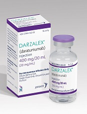
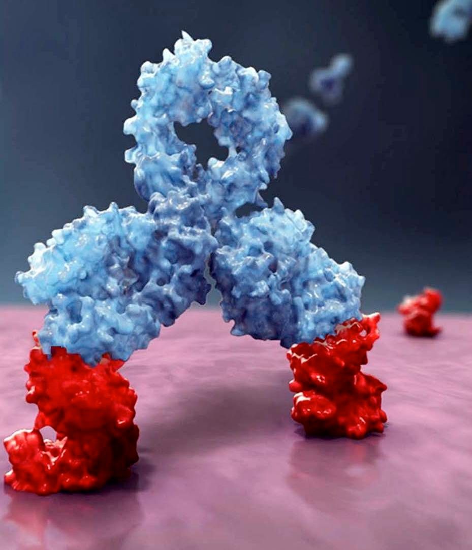
The US Food and Drug Administration (FDA) has granted another approval for the CD38-directed antibody daratumumab (Darzalex®).
Daratumumab is now approved for use in combination with bortezomib, melphalan, and prednisone (VMP) to treat patients with newly diagnosed multiple myeloma (MM) who are ineligible for autologous stem cell transplant.
Daratumumab was first approved by the FDA in 2015 as a monotherapy for MM patients who have received at least 3 prior lines of therapy, including a proteasome inhibitor (PI) and an immunomodulatory agent, or who are double-refractory to a PI and an immunomodulatory agent.
In 2016, daratumumab was approved for use in combination with lenalidomide and dexamethasone, or bortezomib and dexamethasone, to treat MM patients who have received at least 1 prior therapy.
In 2017, daratumumab was approved for use in combination with pomalidomide and dexamethasone to treat MM patients who have received at least 2 prior therapies, including lenalidomide and a PI.
For full prescribing information, visit www.darzalex.com.
Phase 3 trial
The FDA’s approval of daratumumab in combination with VMP is supported by data from the phase 3 ALCYONE (MMY3007) study. Results from this study were presented at the 2017 ASH Annual Meeting and simultaneously published in NEJM.
ALCYONE enrolled 706 patients with newly diagnosed MM who were not eligible for high-dose chemotherapy with autologous stem cell transplant. Patients were randomized to receive VMP or daratumumab plus VMP (D-VMP).
The overall response rates were 91% in the D-VMP arm and 74% in the VMP arm (P<0.0001). Rates of complete response were 43% and 24%, respectively. And rates of minimal residual disease negativity were 22% and 6%, respectively.
The median progression-free survival (PFS) was not reached in the D-VMP arm and was 18.1 months in the VMP arm. The 12-month PFS was 87% and 76%, respectively. The 18-month PFS was 72% and 50%, respectively.
The most common treatment-emergent adverse events (TEAEs; in the D-VMP and VMP arms, respectively) were neutropenia (50% and 53%), thrombocytopenia (49% and 54%), anemia (28% and 38%), peripheral sensory neuropathy (28% and 34%), upper respiratory tract infection (26% and 14%), diarrhea (24% and 25%), pyrexia (23% and 21%), and nausea (21% and 22%).
Infusion-related reactions occurred in 28% of patients in the D-VMP arm and 0% of those in the VMP arm.
The rate of grade 3/4 infections was higher in the D-VMP arm than the VMP arm—23% and 15%, respectively. In both arms, most infections resolved.
The most common grade 3/4 TEAEs (in the D-VMP and VMP arms, respectively) were neutropenia (40% and 39%), thrombocytopenia (34% and 38%), and anemia (16% and 20%).
The rate of discontinuation due to AEs was 5% in the D-VMP arm and 9% in the VMP arm.
FDA approves anti-CD38 with VMP in myeloma
The who are ineligible for autologous stem cell transplant (ASCT).
The drug is approved in combination with a standard VMP regimen – bortezomib (Velcade), melphalan, and prednisone. The FDA had granted priority review to the drug application in January 2018 based on the results of the phase 3 ALCYONE study (NCT02195479).
Daratumumab, an anti-CD38 monoclonal antibody, reduced the risk of disease progression or death by 50%, compared with VMP alone in the ALCYONE study. The median progression-free survival had not yet been reached in the daratumumab arm; the median progression-free survival was 18.1 months in the VMP-only arm (N Engl J Med. 2018;378:518-28).
Daratumumab is marketed by Janssen Biotech as Darzalex.
Drug may alleviate CIPN in MM patients
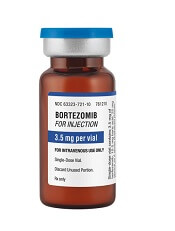
Researchers say they’ve discovered why multiple myeloma (MM) patients may experience chemotherapy-induced peripheral neuropathy (CIPN) when treated with bortezomib.
The group’s study also suggests fingolimod—a drug approved to treat multiple sclerosis—could mitigate CIPN without compromising the efficacy of bortezomib.
Daniela Salvemini, PhD, of the Saint Louis University School of Medicine in St. Louis, Missouri, and her colleagues reported these findings in the Journal of Experimental Medicine.
The researchers said bortezomib causes CIPN in more than 40% of patients, but the reasons for this are unclear.
With their study, Dr Salvemini and her colleagues found that bortezomib accelerates the production of sphingolipids, which have been linked to neuropathic pain.
Rats treated with bortezomib began to accumulate 2 sphingolipid metabolites—sphingosine 1-phosphate and dihydrosphingosine 1-phosphate—in their spinal cords at the time they began to show signs of neuropathic pain.
Blocking the production of these molecules prevented the animals from developing CIPN in response to bortezomib.
Sphingosine 1-phosphate and dihydrosphingosine 1-phosphate can activate a cell surface receptor protein called S1PR1. Dr Salvemini and her colleagues determined that the 2 metabolites cause CIPN by activating S1PR1 on the surface of astrocytes, resulting in neuroinflammation and enhanced release of the excitatory neurotransmitter glutamate.
Drugs that inhibit S1PR1 prevented rats from developing CIPN in response to bortezomib. One such inhibitor was fingolimod, a drug approved by the US Food and Drug Administration (FDA) to treat multiple sclerosis.
In addition to preventing CIPN, fingolimod did not inhibit bortezomib’s ability to kill MM cells. In fact, fingolimod has demonstrated anticancer activity in past studies.
“Because fingolimod shows promising anticancer potential and is already FDA-approved, we think that our findings in rats can be rapidly translated to the clinic to prevent and treat bortezomib-induced neuropathic pain,” Dr Salvemini said.
Key to MGUS and myeloma may lie in Iceland
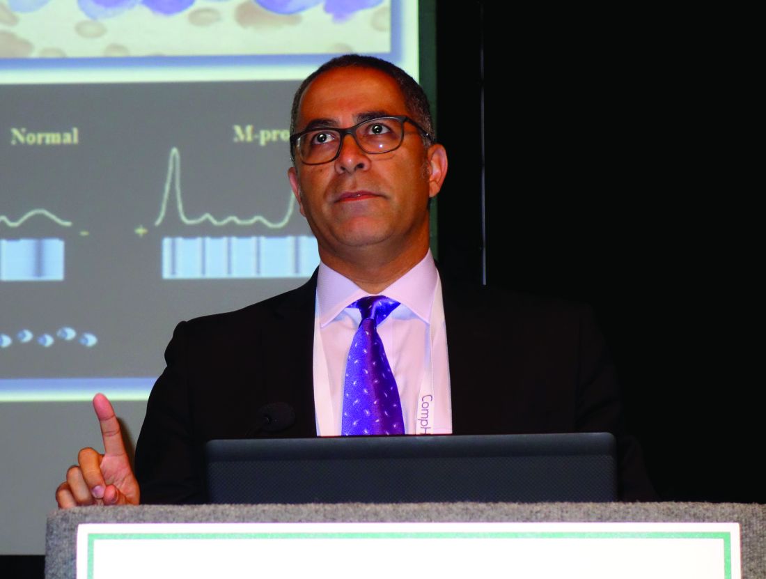
NEW ORLEANS – Everyone aged 40 years and older on the island nation of Iceland is being screened for monoclonal gammopathy of undetermined significance, smoldering myeloma, and full-blown multiple myeloma in an unprecedented project to identify the malignancy’s genetic roots, Joseph Mikhael, MD, said at the annual meeting of the American College of Physicians.
This effort to decode the underlying genetics of multiple myeloma is enormously facilitated by the fact that the DNA sequencing of the entire Icelandic population is already known, and everyone’s blood samples are stored in the national health care system.
The International Myeloma Foundation is funding the Icelandic project, called iStopMM (Iceland Screens Treats or Prevents Multiple Myeloma).
The results of iStopMM could be far reaching, in part because the findings will show whether screening an asymptomatic general population for MGUS – as for example, all American adults – is worthwhile.
In the interim, it’s important for primary care physicians to recognize when it is and isn’t appropriate to order a serum protein electrophoresis (SPE) study, on which the diagnosis of MGUS hinges. It’s also essential to recognize the difference between smoldering and full-blown multiple myeloma, because the distinction has implications for patient monitoring and treatment, added Dr. Mikhael, a hematologist at the City of Hope Cancer Center in Duarte, Calif.
Multiple myeloma accounts for 1% of all cancers and 10% of hematologic malignancies. MGUS is an obligate precursor of multiple myeloma. But MGUS is common, and therein lies a challenge for physicians – as well as a major source of anxiety for many MGUS-positive patients.
In a 12-year-old study, MGUS had a 3% prevalence in the general U.S. population older than age 50 years, and a greater than 5% prevalence after age 70. However, Dr. Mikhael thinks that more refined testing will show the true figure to be close to 10%. Thus, the great majority of patients with MGUS will never develop multiple myeloma.
Pending the potentially practice-changing outcome of iStopMM, Dr. Mikhael said SPE shouldn’t be ordered routinely in an asymptomatic patient, even one with a positive family history for multiple myeloma. The annual cost of monitoring the roughly 540,000 U.S. patients who now carry a diagnosis of MGUS – typically established as an incidental finding by primary care physicians while doing a work-up for another reason – is at least $110 million. And there’s no point in adding to that burden until the benefit of mass screening has been established.
An SPE is appropriate, however, in an older patient with unexplained anemia, known low immunoglobulin levels, unexplained renal insufficiency or neuropathy, or osteopenia or osteoporosis inconsistent with the patient’s age or gender – provided the patient doesn’t have a coexisting plasma cell dyscrasia or B-cell lymphoproliferative disorder, which would throw off the prognostic value of the test results, he continued.
When ordering an SPE to rule in/out MGUS, it’s essential to also order serum free light-chain testing, because it provides key prognostic information.
A landmark study led by investigators at the Mayo Clinic demonstrated that the risk of progression of MGUS to multiple myeloma or a related disorder is independently predicted by three key factors: a high serum M-protein spike of 15 g/L or more on the SPE; the presence of non-IgG MGUS; and an abnormal serum free light-chain ratio of less than 0.26 or more than 1.65. In this study, the 20-year risk of malignant transformation of MGUS ranged from 58% if all three risk factors were present, to just 5% if none were (Blood. 2005 Aug 1;106[3]:812-7).
CRAB vs. SLiM CRAB
Myeloma, smoldering myeloma, and MGUS were redefined a few years ago to reflect differences in prognosis. MGUS still requires the presence of a serum monoclonal protein in a concentration of 3 g/dL or less, less than 10% plasmacytosis in the bone marrow, asymptomatic status, and absence of end-organ damage as traditionally defined in the acronym CRAB (calcium elevation, renal insufficiency, anemia, or bony disease).
If CRAB is present in a patient with at least 10% plasma cells in bone marrow, that is by definition multiple myeloma warranting treatment. Smoldering myeloma requires at least 10% plasmacytosis in bone marrow and absence of the CRAB criteria. However, in a significant change, ultra–high-risk smoldering melanoma, defined by the acronym SLiM CRAB, is now considered active myeloma and should be treated (Lancet Oncol. 2014 Nov;15[12]:e538-48).
“Traditionally, we waited until CRAB [to define myeloma],” Dr. Mikhael explained. “But if you’re running toward a cliff, I don’t have to wait until you’re falling off to know you’re in trouble.”
The SLiM half of SLiM CRAB consists of 60% or more plasmacytosis in bone marrow, light chains in a kappa-to-lambda or lambda-to-kappa ratio of greater than 100, and MRI showing one or more focal lesions. If a patient is SLiM, with or without CRAB, that is now considered active myeloma warranting treatment.
Not all MGUS needs a bone marrow biopsy
A bone marrow biopsy and skeletal survey via whole-body CT or conventional radiographs can be deferred in patients with low-risk MGUS and no bony symptoms. Using the Mayo Clinic risk stratification model, low risk is defined as a serum M protein of 1.5 g/dL or less on SPE, an IgG isotype, and a normal free light-chain ratio.
The lifetime risk of progression in patients with MGUS who meet all three criteria is only about 2%. They can be followed at 6 months with an SPE, free light-chain testing, a CBC, and serum calcium and creatinine, then annually thereafter.
“For those who aren’t in this low-risk category, we actually do need to do a bone marrow test,” according to Dr. Mikhael. “Then, based on that, if they have malignancy, send them to a myeloma geek like me or to another hematologist. And if they don’t have a malignancy, they can be followed at 6 months and then subsequently at least every year.”
Dr. Mikhael has received research grants from AbbVie, Celgene, and Sanofi.
EXPERT ANALYSIS FROM ACP INTERNAL MEDICINE
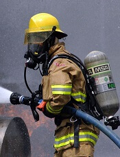
9/11 firefighters may have higher risk of MGUS, early MM
Exposure to the 9/11 World Trade Center (WTC) disaster site may increase the risk of monoclonal gammopathy of undetermined significance (MGUS) and early development of multiple myeloma (MM).
Researchers found that firefighters exposed to the site had a nearly 2-fold higher risk of MGUS than a control population.
And those firefighters who developed MM were diagnosed 12 years earlier than MM patients in the general US population.
These findings were published in JAMA Oncology.
Previous studies suggested that MGUS and MM tend to develop after exposure to toxic chemicals, and individuals exposed to the WTC disaster site have an increased risk of developing cancers.
The aerosolized dust from the collapsed WTC towers exposed firefighters and other first responders to high levels of polychlorinated biphenyls, polycyclic aromatic hydrocarbons, dioxins, asbestos, and other potential carcinogens, as well as diesel smoke from heavy machinery used in the 10-month rescue and recovery effort.
“With our 2011 study in The Lancet, we were the first to show that first responders were more likely to get many different types of cancer,” said David J. Prezant, MD, of Albert Einstein College of Medicine in Bronx, New York.
“We carried out this new study to do more than just treat cancer. We wanted to find early, predictive signs of cancer that would allow us to screen people and monitor those found to be at risk. By detecting MGUS, which predicts the development of multiple myeloma, we are able to do that.”
For statistical reasons, the study population was limited to 781 white, male WTC-exposed firefighters ages 50 to 79.
The researchers assess the prevalence of MGUS in these firefighters and compared it to the prevalence of MGUS in a non-exposed comparison group—men living in Olmsted County, Minnesota.
The prevalence of MGUS was 1.8-fold higher in the firefighters. There were 7.63 cases of MGUS per 100 firefighters and 4.34 cases per 100 non-exposed men.
The researchers also found the prevalence of light-chain MGUS was 3.1-fold higher in the firefighters than in the controls. The prevalence rate of light-chain MGUS was 3.08 and 0.98 per 100 persons, respectively.
“We saw a significantly higher incidence of MGUS in these first responders, and they’re developing it at a young age,” said study author Amit Verma, MBBS, of Albert Einstein College of Medicine.
The subjects’ early development of MGUS suggests they may face an increased risk for early onset MM as well.
In a separate analysis, the researchers examined 16 cases of MM among white, male WTC-exposed firefighters diagnosed between September 12, 2001, and July 1, 2017.
Their average age at diagnosis was 57, or 12 years younger than the average age for MM diagnosis in the US.
The researchers also found that 7 of 14 evaluable cases were light-chain MM. They said this is more than double the rate observed in other populations—50% vs 20%.
In the population of firefighters screened for MGUS, 38% (18/47) of those who had MGUS were found to have the light-chain subtype.
The researchers said these findings are of interest because previous research revealed associations between light-chain MM/MGUS and exposure to toxins and chronic immune stimulation.
The team also assessed CD20 expression in a subset of 7 MM patients. Five of these patients (71%) had CD20-positive cells, which is associated with poor prognosis.
Based on these findings, the researchers recommend that physicians screen first responders exposed to the WTC disaster site for MGUS.
“Screening for multiple myeloma risk by testing for MGUS is something we can offer these first responders, which is why this study is important,” Dr Prezant said.
Coming soon: CAR T-cell approvals in multiple myeloma
, and will “completely transform oncology,” according to Carl June, MD.
That approval is anticipated sometime in 2019.
“Myeloma is the most common blood cancer in adults, and there’s never been a curative therapy, but now there is a subset of patients who look like they’re cured with CAR T cells,” Dr. June, the Richard W. Vague Professor in Immunotherapy and a pioneer in CAR T-cell research at the University of Pennsylvania, Philadelphia, said in an interview.
The first treated patient in a trial of a novel anti-B-cell maturation antigen (BCMA)-specific CAR T-cell therapy (CART-BCMA) developed by University of Pennsylvania researchers in collaboration with Novartis is part of that subset.
Woodring Wright, MD, a professor of cell biology and medicine at the University of Texas Southwestern Medical Center (UT Southwestern) in Dallas recently outed himself as that first patient, announcing in a Feb. 14, 2018, UT Southwestern press report that CART-BCMA saved his life.
Dr. Wright, who holds the Southland Financial Corporation Distinguished Chair in Geriatrics at UT Southwestern, was diagnosed with multiple myeloma about 12 years ago and failed 11 prior chemotherapies before he was enrolled in the CART-BCMA trial.
“Now he considers himself cured,” Dr. June said.
More than 2 years after receiving CART-BCMA he remains cancer free, and is now conducting CAR T-cell-related research in his lab at UT Southwestern in an effort to broaden the effectiveness of current CAR T-cell therapies. Specifically, he is looking at whether the small percentage of patients in whom CAR T-cell therapy does not work might benefit from telomerase to lengthen telomeres, as most patients who fail CAR T-cell therapy are elderly patients who might have terminally short telomeres, UT Southwestern reported.
The ongoing University of Pennsylvania trial led by Adam D. Cohen, MD, director of myeloma immunotherapy at the Abramson Cancer Center, has an overall response rate of 64%; initial phase 1 efficacy and safety results were reported at the American Society of Hematology (ASH) annual meeting in 2016, and multiple companies are currently pursuing registration trials for CAR T therapies in myeloma, Dr. June said.
Among them are bluebird bio and Celgene, which together are developing an anti-BCMA CAR T-cell therapy known as bb2121. That product was granted breakthrough therapy designation by the Food and Drug Administration in November 2017, and will thus receive expedited review. It has also been fast-tracked in Europe.
The decision to fast-track bb2121 in the United States was based on preliminary results from the CRB-410 trial. Updated findings from that trial were presented in December 2017 at ASH and showed an overall response rate of 94% in 21 patients, with 17 of 18 patients who received doses above 50 x 106 CAR+ T cells having an overall response, and 10 of the 18 achieving complete remission. The progression-free survival rates were 81% at 6 months, and 71% at 9 months, with responses deepening over time. The complete response rates were 27% and 56% in May and October of 2017, respectively.
Responses were durable, lasting more than 1 year in several patients, the investigators reported. Phase 2 of the trial – the global pivotal KarMMA trial – is currently enrolling and will dose patients at between 150 and 350 x 106 CAR+ T cells.
Janssen Biotech Inc. (a Johnson & Johnson company) and Legend Biotech USA Inc./Legend Biotech Ireland Limited (of Genscript Biotech Corporation) have also joined forces to develop an anti-BCMA CAR T-cell product for multiple myeloma, Dr. June said.
The companies announced in December that they had entered into “a worldwide collaboration and license agreement” to develop the CAR T-cell drug candidate.
LCAR-B38M is currently accepted for review by the China Food and Drug Administration and is in the planning phase of clinical studies in the United States for multiple myeloma, according to that announcement.
The “race between companies” for a CAR T myeloma approval will lead to a welcome addition to the treatment armamentarium, because while myeloma represents only about 2% of all cancers, it is responsible for 7% of cancer costs, Dr. June said.
Since many patients live with their disease for a long time, that can mean huge “financial toxicity” associated with treatment and patients still usually have “an awful outcome involving a long death,” he said.
“So CAR T-cell therapy for myeloma will bring a huge change to the practice of oncology,” he added, explaining that the first CAR T-cell therapy approved (tisagenlecleucel, in August 2017) was for pediatric acute lymphoblastic leukemia that had relapsed at least twice. “That’s only about 600 kids a year in the U.S., so it’s an ultra-orphan market,” he said.
With the subsequent approval of axicabtagene ciloleucel (in October 2017) and the anticipated myeloma approval, CAR T-cell therapy will move away from orphan status.
“There are a lot of difficulties whenever you change to something new,” he said, comparing the CAR T-cell therapy evolution to that of bone marrow transplantation in the 1980s.
Early on, there were only two places in the country where a patient could get a bone marrow transplant – Fred Hutchinson Cancer Center in Seattle and Johns Hopkins University in Baltimore. “Everyone said ‘you’ll never be able to do it routinely, it’s only at these two referral centers,’ because of the skill needed and the intensity of it,” he said. “But over the years, millions of transplants have now been done; they’re done at many community centers. And it’s the same thing with CARs; Novartis now has 30 centers and people have to be trained. It’s a new skill set, and it will take time,” he said.
That can be particularly frustrating because there are many patients with diseases that “might benefit in a major way” from CAR T-cell therapy, but who can’t get on a clinical trial, Dr. June noted.
“There’s more demand than availability, and it’s going to take awhile ... it’s like liver transplants – there aren’t enough donors to go around, so people die, they get on lists, and it’s really hard to see that, but eventually it will get solved,” he said, adding that the solution will most likely involve the complementary use of off-the-shelf CAR T cells in certain patients to induce remission and perhaps provide a bridge to some other definitive therapy, and ultra-personalized CAR T therapy in others, as well as combinations that include CAR T cells and targeted agents or checkpoint inhibitors.
CRISPR-Cas9 gene editing is also being looked at as a tool for engineering multiple myeloma cellular immunotherapy (and other cancer treatments), as in the Parker Institute–funded NYCE study, Dr. June said.
“We’re actually removing the [programmed death-1] gene and the T-cell receptors ... it shows enormous potential for gene editing. CRISPR is going to be used for a lot of things, but the first use is with T-cell therapies, so we’re really excited about that trial,” he said. “We just opened and we’re screening patients now.”
Dr. June reported royalties and research funding from Novartis and an ownership interest in Tmunity Therapeutics.
World Trade Center responders face greater cancer burden, including greater risk of multiple myeloma
Rescue and recovery workers who were involved in the aftermath of the World Trade Center disaster may face a greater cancer burden than the general population, according to two studies published in JAMA Oncology.
In particular, they may be at risk of developing multiple myeloma at an earlier age.
The first study was a closed-cohort study of 14,474 employees of the Fire Department of the City of New York (FDNY) who were exposed to the World Trade Center disaster but were cancer-free as of Jan. 1, 2012. The aim was to project cancer incidence from 2012 through 2031, based on data from the FDNY World Trade Center Health Program, and compare those rates with age-, race-, and sex-specific New York cancer rates from the general population.
The modeling projected a “modestly” higher number of cancer cases in the white male subgroup of rescue and recovery workers exposed to the World Trade Center (2,714 vs. 2,596 for the general population of New York; P less than .001). Specifically, the investigators projected significantly higher case counts of prostate cancer (1,437 vs. 863), thyroid cancer (73 vs. 57), and melanoma (201 vs. 131), compared with the general population in New York, but fewer lung (237 vs. 373), colorectal (172 vs. 267), and kidney cancers (66 vs. 132) (P less than .001 for all).
“Our findings suggest that the FDNY WTC-exposed cohort may experience a greater burden of cancer than would be expected from a population with similar demographic characteristics,” wrote Rachel Zeig-Owens, DrPH, from the Montefiore Medical Center and Albert Einstein College of Medicine, both in New York, and coauthors, highlighting prostate cancer as a particular concern.
However, they also acknowledged that the elevated rates observed in people exposed to the World Trade Center disaster could be a result of increased surveillance, even though they did attempt to correct for that, and that firefighters in general might face higher risks.
“It is possible that firefighters have a higher risk of cancer than the general population owing to exposures associated with the occupation,” they wrote. However occupation could also have the opposite effect, as rescue and recovery workers tend to have lower smoking rates, which may explain the relatively low rates of certain cancers such as lung cancer, they said.
A second study examined the effect of the World Trade Center disaster on the risk of multiple myeloma and monoclonal gammopathies in exposed firefighters.
The seroprevalence study of monoclonal gammopathies of undetermined significance (MGUS) in 781 exposed firefighters revealed that the age-standardized prevalence of these was 76% higher in this population than it was in a white male reference population living in Minnesota.
In particular, the age-standardized prevalence of light-chain MGUS was more than threefold higher in exposed firefighters, compared with the reference population.
Researchers also analyzed a case series of 16 exposed white male firefighters who received a diagnosis of multiple myeloma after Sept. 11, 2001. Of the 14 patients for whom data on the monoclonal protein isotype was available, half had light-chain multiple myeloma.
“These findings are of interest due to previously observed associations between light-chain multiple myeloma and light-chain MGUS and exposure to toxins, and chronic immune stimulation,” wrote Ola Landgren, MD, PhD, from the Memorial Sloan Kettering Cancer Center and his coauthors.
Seven patients were also assessed for CD20 expression – a marker of poorer prognosis – and 71% were found to be CD20-positive, a prevalence around 3.5-fold higher than that seen in the general population.
The cohort with multiple myeloma was diagnosed on average 12 years younger than those in the general population. The authors commented that this was unlikely to be caused by lead-time bias because the time from first symptoms to clinical manifestation of the disease is usually around 1 year.
“Taken together, our results show that environmental exposure due to the WTC attacks is associated with myeloma precursor disease (MGUS and light-chain MGUS) and may be a risk factor for the development of multiple myeloma at an earlier age, particularly the light-chain subtype,” the authors wrote.
The first study was supported by the National Institute of Occupational Safety and Health; no conflicts of interest were declared.
The second study was supported by the V Foundation for Cancer Research, the Byrne Fund for the benefit of Memorial Sloan-Kettering Cancer Center, the National Cancer Institute, the Albert Einstein Cancer Center, and the National Institute for Occupational Safety and Health; no conflicts of interest were declared.
SOURCE: Zeig-Owens R et al. JAMA Oncology. 2018 April 26. doi: 10.1001/jamaoncol.2018.0504. Landgren O et al. JAMA Oncology. 2018 April 16. doi: 10.1001/jamaoncol.2018.0509.
When the heroes of the World Trade Center are diagnosed with even a common cancer, there is a natural tendency to assume that the diagnosis is the result of their service during the disaster. However, it is important to appreciate that the firefighting profession is known to be associated with higher risks of monoclonal gammopathy of undetermined significance and multiple myelomas, compared with the general population.
Given that, it would have been preferable to compare the World Trade Center–exposed populations with an equally intensively screened, age-matched cohort of firefighters from another major city.
If we apply Sir Richard Doll’s rule that a single epidemiologic study cannot be persuasive until the lower bound of the 95% confidence interval is greater than three, the relative risks in the study by Landgren and colleagues are too small to be persuasive.
The predicted increases in cancers of the prostate, thyroid, and myeloma are interesting, but these have also been previously reported in firefighters from other cities.
Despite this, we owe it to these men and women to find the truth and determine the illnesses that are associated with their service.
Otis W. Brawley, MD, is chief medical and scientific officer and executive vice president of the American Cancer Society and a professor at Emory University, Atlanta. These comments are taken from an accompanying editorial (JAMA Oncology. 2018 April 26. doi: 10.1001/jamaoncol.2018.0498.) No conflicts of interest were declared.
Rescue and recovery workers who were involved in the aftermath of the World Trade Center disaster may face a greater cancer burden than the general population, according to two studies published in JAMA Oncology.
In particular, they may be at risk of developing multiple myeloma at an earlier age.
The first study was a closed-cohort study of 14,474 employees of the Fire Department of the City of New York (FDNY) who were exposed to the World Trade Center disaster but were cancer-free as of Jan. 1, 2012. The aim was to project cancer incidence from 2012 through 2031, based on data from the FDNY World Trade Center Health Program, and compare those rates with age-, race-, and sex-specific New York cancer rates from the general population.
The modeling projected a “modestly” higher number of cancer cases in the white male subgroup of rescue and recovery workers exposed to the World Trade Center (2,714 vs. 2,596 for the general population of New York; P less than .001). Specifically, the investigators projected significantly higher case counts of prostate cancer (1,437 vs. 863), thyroid cancer (73 vs. 57), and melanoma (201 vs. 131), compared with the general population in New York, but fewer lung (237 vs. 373), colorectal (172 vs. 267), and kidney cancers (66 vs. 132) (P less than .001 for all).
“Our findings suggest that the FDNY WTC-exposed cohort may experience a greater burden of cancer than would be expected from a population with similar demographic characteristics,” wrote Rachel Zeig-Owens, DrPH, from the Montefiore Medical Center and Albert Einstein College of Medicine, both in New York, and coauthors, highlighting prostate cancer as a particular concern.
However, they also acknowledged that the elevated rates observed in people exposed to the World Trade Center disaster could be a result of increased surveillance, even though they did attempt to correct for that, and that firefighters in general might face higher risks.
“It is possible that firefighters have a higher risk of cancer than the general population owing to exposures associated with the occupation,” they wrote. However occupation could also have the opposite effect, as rescue and recovery workers tend to have lower smoking rates, which may explain the relatively low rates of certain cancers such as lung cancer, they said.
A second study examined the effect of the World Trade Center disaster on the risk of multiple myeloma and monoclonal gammopathies in exposed firefighters.
The seroprevalence study of monoclonal gammopathies of undetermined significance (MGUS) in 781 exposed firefighters revealed that the age-standardized prevalence of these was 76% higher in this population than it was in a white male reference population living in Minnesota.
In particular, the age-standardized prevalence of light-chain MGUS was more than threefold higher in exposed firefighters, compared with the reference population.
Researchers also analyzed a case series of 16 exposed white male firefighters who received a diagnosis of multiple myeloma after Sept. 11, 2001. Of the 14 patients for whom data on the monoclonal protein isotype was available, half had light-chain multiple myeloma.
“These findings are of interest due to previously observed associations between light-chain multiple myeloma and light-chain MGUS and exposure to toxins, and chronic immune stimulation,” wrote Ola Landgren, MD, PhD, from the Memorial Sloan Kettering Cancer Center and his coauthors.
Seven patients were also assessed for CD20 expression – a marker of poorer prognosis – and 71% were found to be CD20-positive, a prevalence around 3.5-fold higher than that seen in the general population.
The cohort with multiple myeloma was diagnosed on average 12 years younger than those in the general population. The authors commented that this was unlikely to be caused by lead-time bias because the time from first symptoms to clinical manifestation of the disease is usually around 1 year.
“Taken together, our results show that environmental exposure due to the WTC attacks is associated with myeloma precursor disease (MGUS and light-chain MGUS) and may be a risk factor for the development of multiple myeloma at an earlier age, particularly the light-chain subtype,” the authors wrote.
The first study was supported by the National Institute of Occupational Safety and Health; no conflicts of interest were declared.
The second study was supported by the V Foundation for Cancer Research, the Byrne Fund for the benefit of Memorial Sloan-Kettering Cancer Center, the National Cancer Institute, the Albert Einstein Cancer Center, and the National Institute for Occupational Safety and Health; no conflicts of interest were declared.
SOURCE: Zeig-Owens R et al. JAMA Oncology. 2018 April 26. doi: 10.1001/jamaoncol.2018.0504. Landgren O et al. JAMA Oncology. 2018 April 16. doi: 10.1001/jamaoncol.2018.0509.
FROM JAMA ONCOLOGY
Key clinical point: Monoclonal gammopathies and multiple myeloma may occur more often and earlier in World Trade Center rescue workers.
Major finding: Prevalence of light-chain monoclonal gammopathies is threefold higher in exposed firefighters than in a reference population of white males.
Study details: A cohort study in 14,474 employees of the Fire Department of the City of New York exposed to the Sept. 11, 2001, World Trade Center disaster, a case series of 16 exposed white male firefighters diagnosed with multiple myeloma, and a seroprevalence study of monoclonal gammopathies of undetermined significance in 781 exposed firefighters.
Disclosures: The first study was supported by the National Institute of Occupational Safety and Health; no conflicts of interest were declared. The second study was supported by the V Foundation for Cancer Research, the Byrne Fund for the benefit of Memorial Sloan-Kettering Cancer Center, the National Cancer Institute, the Albert Einstein Cancer Center, and the National Institute for Occupational Safety and Health; no conflicts of interest were declared.
Source: Zeig-Owens R et al. JAMA Oncology 2018, Apr 26. doi: 10.1001/jamaoncol.2018.0504. Landgren O et al. JAMA Oncology 2018, Apr 26. doi: 10.1001/jamaoncol.2018.0509.
Exercise linked to risk of death in cancer patients

CHICAGO—Researchers have identified a link between habitual physical activity (PA) and mortality among cancer patients.
Engaging in regular PA, both pre- and post-diagnosis, was associated with a significantly lower risk of death for the entire population studied and for patients with 8 specific types of cancer.
However, the association was not significant for patients with other cancer types, including hematologic malignancies.
Rikki Cannioto, PhD, of Roswell Park Comprehensive Cancer Center in Buffalo, New York, and her colleagues presented these findings at the AACR Annual Meeting 2018 (abstract 5254*).
The researchers examined the association between habitual PA and outcomes in 5807 cancer patients enrolled in the Data Bank and BioRepository at Roswell Park between 2003 and 2016.
The population was 54.8% female and 93% white. The average age at diagnosis was 60.6 years.
The researchers looked at patterns of PA over time, from the decade before the cancer was diagnosed and continuing for up to 1 year after diagnosis.
Patients who engaged in regular, moderate- to vigorous-intensity PA (such as walking, running, aerobics, or other cardiovascular exercise) both before and after their diagnosis were considered habitually active, whereas those who did not exercise regularly were considered habitually inactive.
Overall, 52% of patients reported habitual activity, and 19% reported habitual inactivity. Twenty-three percent of patients said their activity level decreased after diagnosis, and 6% said their activity level increased.
Patients were followed through January 31, 2018. The median time to follow-up was 53 months, and 33.7% of patients (n=1956) died during the follow-up period.
Results
The researchers found that patients who were active before and after diagnosis were 40% more likely to survive than those who were habitually inactive (P<0.001). Habitually inactive patients had a 66% increased risk of mortality compared to active patients.
The habitually active patients had a 37-month mean survival advantage over the inactive patients.
In addition, patients whose activity level increased after diagnosis had a 25% lower risk of death than patients who remained inactive after diagnosis.
The researchers observed a significant (P<0.05) association between habitual PA and decreased mortality in patients with breast, colon, prostate, bladder, endometrial, ovarian, esophageal, and skin cancers.
However, the association between PA and mortality was not significant for patients with hematologic malignancies (P=0.59) or kidney, liver, lung, pancreas, stomach, or “other” cancers.
The researchers said the associations between habitual PA and decreased mortality remained consistent regardless of a patient’s sex, tumor stage, smoking status, or body mass index.
“[W]hen it comes to exercise, something is better than nothing, but regular, weekly exercise seems to really make a difference,” Dr Cannioto said.
“In fact, patients who were physically active 3 or 4 days a week experienced an even greater benefit than those who exercised daily, and patients who had only 1 or 2 days of regular activity per week did nearly as well. This is particularly encouraging, as cancer patients and survivors can be overwhelmed by current physical activity recommendations.”
*Information in the abstract differs from the presentation.
Company stops development of drug for AL amyloidosis
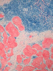
Prothena Corporation plc is discontinuing development of NEOD001, an investigational antibody intended for the treatment of AL amyloidosis.
The company’s decision was based on results from the phase 2b PRONTO study and a futility analysis of the phase 3 VITAL study.
NEOD001 did not meet the primary or secondary endpoints of the PRONTO study, so Prothena asked an independent data monitoring committee to review a futility analysis of the ongoing VITAL study.
The committee recommended discontinuation of the VITAL study, so Prothena decided to discontinue all development of NEOD001, including the VITAL study and open-label extension studies.
“We are deeply disappointed by this outcome, particularly for patients suffering from this devastating disease,” said Gene Kinney, PhD, president and chief executive officer of Prothena.
“We are surprised by the results from these 2 placebo-controlled studies and will continue to analyze the resulting data to share insights with our collaborators in the scientific, medical, and advocacy communities.”
Phase 3 VITAL study
The VITAL study was a multicenter, randomized, double-blind, placebo-controlled study of NEOD001 in treatment-naïve patients with AL amyloidosis and cardiac dysfunction.
The study enrolled 260 patients who were randomized on a 1:1 basis to receive 24 mg/kg of NEOD001 or placebo via intravenous infusion every 28 days. Patients in both arms received concurrent standard of care therapy.
The composite primary endpoint was event-based, with all-cause mortality or cardiac hospitalizations counted as events.
The futility analysis, based on 103 adjudicated events of the 156 events specified to complete the study, was not statistically significant. The hazard ratio was 0.84 favoring NEOD001 versus the control arm.
Phase 2b PRONTO study
The PRONTO study was a multicenter, randomized, double-blind, placebo-controlled study of NEOD001 in previously treated patients with AL amyloidosis and persistent cardiac dysfunction.
The study enrolled 129 patients who were randomized on a 1:1 basis to receive 24 mg/kg of NEOD001 (n=66) or placebo (n=63) via intravenous infusion every 28 days.
There was no significant difference between the treatment arms for any of the study’s endpoints.
The primary endpoint was cardiac best response, as assessed by N-terminal pro B-type natriuretic peptide (NT-proBNP) through 12 months of treatment. This endpoint was achieved by 39.4% of patients in the NEOD001 arm and 47.6% in the placebo arm.
The NT-proBNP rate of change (slope) through 12 months of treatment was 9.80 in the NEOD001 arm and 81.42 in the placebo arm.
The mean change in Short-form 36 Physical Component Summary Score after 12 months of treatment was 0.19 and 0.97, respectively.
The median change in 6-Minute Walk Test distance after 12 months of treatment was 19.25 m and 8.00 m, respectively.
And the mean change in Neuropathy Impairment Score of the Lower Limb after 12 months of treatment was -1.20 and -0.60, respectively.
The rate of renal best response (as assessed by proteinuria and estimated glomerular filtration rate) through 12 months of treatment was 53.8% in the NEOD001 arm and 33.3% in the placebo arm.
The rate of all-cause mortality was 4.5% (n=3) and 9.5% (n=6), respectively.
Prothena said NEOD001 was generally safe and well tolerated in this trial.