User login
The American Journal of Orthopedics is an Index Medicus publication that is valued by orthopedic surgeons for its peer-reviewed, practice-oriented clinical information. Most articles are written by specialists at leading teaching institutions and help incorporate the latest technology into everyday practice.
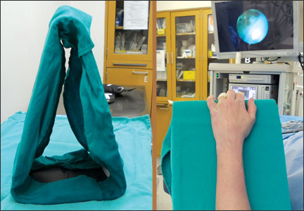
Anterior Pelvic External Fixation: Is There an Optimal Placement for the Supra-Acetabular Pin?
Commentary to "100 Most Cited Articles in Fracture Surgery"
With their current report of “100 Most Cited Articles in Fracture Surgery,” which appeared in the December 2013 issue of The American Journal of Orthopedics, Baldwin and colleagues expand upon “classic citation” reviews that have been reported in recent years in orthopedic surgery and other fields including clinical care medicine and anesthesia.
I so enjoyed reviewing these top 100 articles which brought back fond (mostly!) memories of my orthopedic training in the early 1980s and my first reading of such classics as Neer’s 1970 article on displaced proximal humerus fractures, and Gustilo’s 1976 paper on open fractures, two of the most frequently cited authors in this list.
Several aspects of Baldwin’s paper are noteworthy. The first is how technology has fundamentally altered our access to information. Only with the aid of tools such as a computer could the vast amount of publications spanning nearly 60 years have been reviewed to formulate this top 100 list. The PubMed and Google Scholar search engines of today have transformed the formally tedious—and often incomplete—literature reviews of my residency days.
Second, nearly two-thirds of the clinical papers were evidence level IV case series reporting on the outcomes of patients treated with one method with neither comparison groups nor sensitivity analysis. It is truly astounding that so much of the foundation of orthopedic surgery is based on studies with so little scientific rigor. Of course, this is not to diminish enormous contributions of the early leaders of modern orthopedic surgery, whose keen powers of observation and vast clinical experience established principles of clinical practice still valuable today.
Lastly, the authors rightly state that the exercise of reviewing these most cited fracture articles offers "insight into the past and current trends … and provides the foundation for future investigations." Rigorous future scientific studies using modern techniques will either confirm or refute the work of our classic teachers. With continued research, we will discover "the truth" and determine which treatments really benefit our patients. In the end, that is what really matters.
Baldwin K, Namdari S, Donegan D, Kavatch K, Ahn J, Mehta S. 100 Most Cited Articles in Fracture Surgery. Am J Orthop. 2013;42(12):547-552.
Total Calcium and Vitamin D Intake Linked to Bone Health
Total calcium and vitamin D intake are positively associated with bone health, according to results from the Canadian Multicentre osteoporosis study (CaMos) published in the December 2013 issue of the Journal of Musculoskeletal and Neuronal Interactions. This was observed even though total intake often fell below recommendations and despite an increase over time in supplement intake, the authors noted.
“Calcium and vitamin D are essential nutrients for maximizing peak bone mass, retaining required bone mass, preventing bone loss in later life, and thus potentially reducing the risk of osteoporosis, which is a growing health and economic problem in Canada and other countries,” W. Zhou, McGill University, Montreal, and colleagues explained.
The investigators followed over 9000 men and women 25 years and older for 10 years, and 899 between the age of 16 and 24 years for 2 years. Overall, they found that calcium and vitamin D supplementation increased over time in adults, but decrease in women aged 16 to 18 years. In addition, Zhou and colleagues found that at baseline, higher calcium and vitamin D intakes were associated with higher total hip and femoral neck BMD in young men and cumulatively high levels of calcium and vitamin D intake over time contributed to better BMD maintenance at lumbar spine and hip sites in adult women.
“This is the first longitudinal assessment of total calcium and vitamin D intakes among Canadians and the potential impact of cumulative intake on an important bone health parameter, [bone mineral density],” the authors concluded.
Zhou W, Langsetmo L, Berger C, et al; CaMos Research Group. Longitudinal changes in calcium and vitamin D intakes and relationship to bone mineral density in a prospective population-based study: the Canadian Multicentre Osteoporosis Study (CaMos). J Musculoskelet Neuronal Interact. 2013 Dec;13(4):470-479.
Data Suggest Non-NOF Fragility Fractures Should Be Treated First
Treating patients with non–neck-of-femur (NOF) fragility fractures from osteoporosis before NOF fractures may improve quality-of-life and reduce burden to hospital services and funding, according to a study published the December 2013 Journal of Orthopaedic Surgery and Research.
“Despite the known impact of fragility fractures, osteoporosis still remains unrecognized and untreated in over 50% of patients who present with fragility fractures,” according to authors Tamer Mettyas, Orthopaedic Department of, Royal Brisbane and Women’s Hospital, Australia, and Clare Carpenter, Orthopaedic Department, University Hospital of Wales, Heath Park, Cardiff, United Kingdoms. “Orthopaedic surgeons are often the first to encounter these patients who present with fragility fractures.”
The investigator sought to evaluate current practice of the National Institute for Health and Clinical Excellence (NICE) and British Orthopaedic Association (BOA) guidelines of the secondary prevention of osteoporosis. Patients 50 years and older who were admitted as in-patients with non-NOF fragility fractures between March and September 2008 were included in the study. Mettyas and Carpenter retrospectively evaluated data from March 2008, including risk factors and whether patients were treated; September data was prospectively evaluated after the new trauma admission sheet was introduced. Finally, the authors performed a cross-sectional study assessing services for NOF and non-NOF fragility fractures in September.
Overall, they found that 29% of fragility fractures were non-NOF (mean age, 70 years), and 71% were NOF fractures (mean age, 80). “There is a great difference in the care provided to these patients,” the authors observed. In particular, non-NOF fragility fractures received less attention for osteoporosis assessment (25%) and less interest for investigation by the medical staff (11%), as well as less attention for the treatment of osteoporosis (35%), compared with NOF fracture (35%, 47%, and 71%, respectively). Furthermore, 25% of NOF patients had previous fragility fractures, compared with 6% of non-NOF patients.
“We believe that treating patients with non-NOF fractures from osteoporosis before proceeding to NOF fractures would improve their quality and reduce the burden on hospital services and funding,” the authors concluded.
Mettyas T, Carpenter C. Secondary prevention of osteoporosis in non-neck of femur fractures: is it value for money? A retrospective, prospective and cross-sectional cohort study. J Orthop Surg Res. 2013;8(1):44.
miRNAs Play a Role in Bone Health in HIV Patients
In this review, Fabiola E. Del Carpio-Cano and colleagues, from Temple University School of Medicine, Philadelphia, discuss micro-ribonucleic acid (miRNA) in human immunodeficiency virus (HIV)-induced osteoporosis, including basic concepts of bone biology and current research on the role of miRNAs in bone development.
“Increased life expectancy and the need for long-term antiretroviral therapy have brought new challenges to the clinical management of HIV-infected individuals,” the authors explained in a study published online ahead of print October 27, 2013 in the Journal of Osteoporosis. “The prevalence of osteoporosis and fractures is increased in HIV-infected patients; thus optimal strategies for risk management and treatment in this group of patients need to be redefined.”
Del Carpio-Cano and colleagues discuss bone biology, including the 2 distinct phases of bone formation, homeostatic bone remodeling, the role of transforming growth factor-β and bone morphogenic proteins (BMPs) in bone formation, and Runx2. In addition, the authors review pathophysiology of osteoporosis, biology and function of miRNA, the regulatory role of miRNA in bone biology, as well as HIV and bone biology.
“Prevention of bone loss is an important component of HIV care as the HIV population grows older,” the authors concluded. “Understanding the mechanisms by which HIV infection affects bone biology leading to osteoporosis is crucial to delineate potential adjuvant treatment.”
Del Carpio-Cano FE, Dela Cadena RA, Sawaya BE. HIV and Bone Disease: A Perspective of the Role of microRNAs in Bone Biology upon HIV Infection. J Osteoporos. 2013. [Epub ahead of print]