User login
Doug Brunk is a San Diego-based award-winning reporter who began covering health care in 1991. Before joining the company, he wrote for the health sciences division of Columbia University and was an associate editor at Contemporary Long Term Care magazine when it won a Jesse H. Neal Award. His work has been syndicated by the Los Angeles Times and he is the author of two books related to the University of Kentucky Wildcats men's basketball program. Doug has a master’s degree in magazine journalism from the S.I. Newhouse School of Public Communications at Syracuse University. Follow him on Twitter @dougbrunk.
More Evidence Cryolipolysis Freezes Away Fat
DANA POINT, CALIF. – A growing body of evidence-based research supports the efficacy of cryolipolysis for noninvasive fat reduction in the abdomen and flanks.
At the Summit in Aesthetic Medicine sponsored by Skin Disease Education Foundation (SDEF), Dr. Lawrence S. Bass highlighted results from several recent studies that support its use.
In the first study, presented by Dr. Christine C. Dierickx at the 2012 American Society for Laser Medicine and Surgery (ASLMS) meeting, researchers cataloged side effects in a phone review conducted with patients 1 month following cryolipolysis. All interviewed patients reported erythema that lasted about 1 hour and paresthesia that lasted for up to 1 month after treatment. Other reported side effects were ecchymosis (4%), vasovagal reactions from treatments on the abdomen (2.1%), induration/edema (0.9% and lasting 2-5 weeks), and severe pain (0.6% and lasting for a maximum of 1 month).
A separate study, presented by Dr. Gerald Boey at the 2012 ASLMS meeting, found an amplified fat reduction when concomitant massage was used. For the trial, 10 patients underwent 2 minutes of massage to one side only after cryolipolysis treatment. Ultrasound evaluation revealed that an additional 68% reduction in fat was achieved on the massaged side.
A safety and efficacy trial, carried out at 16 centers and involving 314 patients, was presented by Dr. A. Jay Burns at the 2010 ASLMS meeting. The study found no serious side effects and no skin damage or pigment change from the procedure. Minor side effects included transient bruising, minor pain, reduced sensation, erythema, and edema – all of which resolved spontaneously.
Of 41 patients in the study who underwent ultrasound measurement, the average reduction in fat was 21% after a single procedure. "That’s a consistent number you see in many studies," said Dr. Bass, of the plastic surgery department at New York University School of Medicine.
In a separate study also presented at the 2010 ASLMS meeting, researchers led by Dr. Ivan A. Rosales-Berber investigated the effect of consecutive cryolipolysis treatments separated by 2-3 months. The second treatment resulted in a similar proportion of fat reduction as the first treatment (14.6% vs. 16.5%, respectively).
Three months after the second procedure, the fat layer reduction exceeded 25%. This "means that there is repeatability [with cryolipolysis]," Dr. Bass said. "That’s important, because unfortunately a lot of patients have more than one body contouring emergency in their lives."
An earlier published study determined that cryolipolysis associated with modest reversible short-term changes in peripheral nerve function (Aesthetic Plast. Surg. 2009;33:482-8). Ten patients underwent a thorough neurological evaluation at baseline and weekly after the procedure. All patients returned to baseline peripheral nerve function in an average of 3.6 weeks. The study also found that mean fat reduction by ultrasound was 20.4% at 2 months and 25.5% at 6 months. "Even though we think it takes about 90 days for this whole apoptotic process to resolve, there may be something ongoing, or some tissue consolidation taking place for a longer period of time, allowing patients to continue to improve," Dr. Bass noted.
He concluded his remarks by explaining how he stratifies candidates for liposuction or cryolipolysis. "The patient who wants the maximum improvement or who wants it in one step is definitely going to line up on the liposuction side," he said. "The patient who either needs a small change that’s very localized or prioritizes a quick recovery over the extent of the result is going to line up on the cryolipolysis side."
Dr. Bass disclosed that he is a member of the advisory board for Allergan, Kythera, and Merz Aesthetics. He is also an investigator for Kythera and a consultant to Palomar and Sanuwave.
SDEF and this news organization are owned by Elsevier.
EXPERT ANALYSIS FROM THE SDEF SUMMIT IN AESTHETIC MEDICINE
H. Pylori Infection, Colonic Neoplasms Linked
SAN DIEGO – Patients with Helicobacter pylori infection face a significantly increased risk for developing colonic neoplasms, according to what is believed to be the largest investigation of the association.
Several studies have suggested that H. pylori infection is a risk factor for colonic neoplasms, but all of them involved relatively small case populations, Dr. Amnon Sonnenberg explained at the annual Digestive Disease Week.

Dr. Sonnenberg, a gastroenterologist with the Portland (Ore.) VA Medical Center, investigated the relationship between H. pylori infection and the presence of colonic neoplasms in 156,269 patients who had undergone both a colonoscopy and an esophagogastroduodenoscopy. Surgical pathology samples from all the patients were stored in an electronic database at the Miraca Research Institute, a specialized gastrointestinal lab that serves private outpatient endoscopy centers throughout the United States. More than 1,500 gastroenterologists contributed to the database between January 2008 and December 2011.
Patients were an average age of 58 years, and 59% were female. Among the total, 16,759 (11%) had H. pylori gastritis on immunochemistry. The prevalence of H. pylori gastritis was 9% in patients without polyps, 11% in patients with hyperplastic polyps, 12% in patients with adenoma, 14% in patients with advanced adenoma, 15% in patients with villous adenoma or polyps with high-grade dysplasia, and 18% in patients with colonic adenocarcinoma.
There was a slight trend for the prevalence of H. pylori to rise with the increasing number, as well as the size, of adenomatous polyps. The prevalence of H. pylori was similar for all colon sites.
Significant associations also were noted between the development of colonic adenoma and the following other types of gastric histopathology: intestinal metaplasia, gastric adenoma, gastric cancer, and gastric lymphoma. Similar but even more significant associations were seen between advanced adenoma and these types of gastric histopathology.

Multivariate logistic regression confirmed that the development of H. pylori gastritis was associated with advanced age, male gender, hyperplastic polyps, adenoma, villous adenoma or high-grade dysplasia, and adenocarcinoma.
"H. pylori gastritis confers an increased risk for colonic neoplasm," concluded Dr. Sonnenberg, who is also professor of medicine in the division of gastroenterology and hepatology at Oregon Health and Science University.
"The risk applies to all types of colonic neoplasms and appears to increase with advancing stage of the neoplasm from hyperplastic and adenomatous polyps to tubulovillous adenoma, adenoma with high-grade dysplasia, and adenocarcinoma. Such risk is not limited to chronic active gastritis but is found in other types of gastric histopathology related to H. pylori, such as gastric intestinal metaplasia, gastric adenoma, gastric lymphoma, and gastric cancer," he said.
Long-term infection with H. pylori "may be related to elevated gastrin levels that may act as a growth factor," he speculated.
Dr. Sonnenberg disclosed having received a research grant from Takeda Pharmaceutical Company. No support was received for this study.
SAN DIEGO – Patients with Helicobacter pylori infection face a significantly increased risk for developing colonic neoplasms, according to what is believed to be the largest investigation of the association.
Several studies have suggested that H. pylori infection is a risk factor for colonic neoplasms, but all of them involved relatively small case populations, Dr. Amnon Sonnenberg explained at the annual Digestive Disease Week.
Dr. Sonnenberg, a gastroenterologist with the Portland (Ore.) VA Medical Center, investigated the relationship between H. pylori infection and the presence of colonic neoplasms in 156,269 patients who had undergone both a colonoscopy and an esophagogastroduodenoscopy. Surgical pathology samples from all the patients were stored in an electronic database at the Miraca Research Institute, a specialized gastrointestinal lab that serves private outpatient endoscopy centers throughout the United States. More than 1,500 gastroenterologists contributed to the database between January 2008 and December 2011.
Patients were an average age of 58 years, and 59% were female. Among the total, 16,759 (11%) had H. pylori gastritis on immunochemistry. The prevalence of H. pylori gastritis was 9% in patients without polyps, 11% in patients with hyperplastic polyps, 12% in patients with adenoma, 14% in patients with advanced adenoma, 15% in patients with villous adenoma or polyps with high-grade dysplasia, and 18% in patients with colonic adenocarcinoma.
There was a slight trend for the prevalence of H. pylori to rise with the increasing number, as well as the size, of adenomatous polyps. The prevalence of H. pylori was similar for all colon sites.
Significant associations also were noted between the development of colonic adenoma and the following other types of gastric histopathology: intestinal metaplasia, gastric adenoma, gastric cancer, and gastric lymphoma. Similar but even more significant associations were seen between advanced adenoma and these types of gastric histopathology.
Multivariate logistic regression confirmed that the development of H. pylori gastritis was associated with advanced age, male gender, hyperplastic polyps, adenoma, villous adenoma or high-grade dysplasia, and adenocarcinoma.
"H. pylori gastritis confers an increased risk for colonic neoplasm," concluded Dr. Sonnenberg, who is also professor of medicine in the division of gastroenterology and hepatology at Oregon Health and Science University.
"The risk applies to all types of colonic neoplasms and appears to increase with advancing stage of the neoplasm from hyperplastic and adenomatous polyps to tubulovillous adenoma, adenoma with high-grade dysplasia, and adenocarcinoma. Such risk is not limited to chronic active gastritis but is found in other types of gastric histopathology related to H. pylori, such as gastric intestinal metaplasia, gastric adenoma, gastric lymphoma, and gastric cancer," he said.
Long-term infection with H. pylori "may be related to elevated gastrin levels that may act as a growth factor," he speculated.
Dr. Sonnenberg disclosed having received a research grant from Takeda Pharmaceutical Company. No support was received for this study.
FROM THE ANNUAL DIGESTIVE DISEASE WEEK
Major Finding: The prevalence of H. pylori gastritis was 9% in patients without polyps, 11% in patients with hyperplastic polyps, 12% in patients with adenoma, 14% in patients with advanced adenoma, 15% in patients with villous adenoma or polyps with high-grade dysplasia, and 18% in patients with colonic adenocarcinoma.
Data Source: Findings are based on an analysis of pathology data from 156,269 patients who had undergone both a colonoscopy and an esophagogastroduodenoscopy.
Disclosures: Dr. Sonnenberg disclosed having received a research grant from Takeda Pharmaceutical Company. No support was received for this study.
Fertility Preservation No Longer Experimental for Cancer Patients
Fertility preservation for young women facing cancer therapy has garnered endorsements from leading medical groups in oncology and reproductive medicine, and it isn’t in the experimental stages anymore, interviews with experts suggest.
"There is more literature coming out showing that egg freezing does work," said Dr. Clarisa R. Gracia, director of fertility preservation at the University of Pennsylvania Health System, Philadelphia. "We have more data that we can show patients to say, ‘This is not as experimental as it used to be. It is improving, and it is a real option to offer.’ "

For example, several randomized controlled trials have shown that using frozen eggs is equivalent to using fresh eggs (Hum. Reprod. 2010;25:66-73; Hum. Reprod. 2010;25:2239-46).
The two most common methods of fertility preservation are embryo freezing and egg freezing, according to Dr. Nicole Noyes, a New York City–based reproductive endocrinologist who specializes in infertility, in vitro fertilization (IVF), and egg freezing.
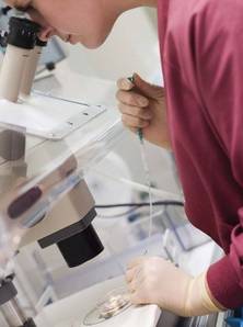
Both procedures involve the use of follicle-stimulating hormone, an injectable medication that causes maturation of multiple eggs in the woman’s ovaries as opposed to a single egg that is naturally ovulated. The stimulated eggs are then gently aspirated from the ovary and placed in a Petri dish.
From this point, either the eggs can be frozen in the unfertilized state or, alternatively, sperm is added, allowing for fertilization and, thus, the creation of embryos prior to freezing. Using either method, eggs or embryos remain cryopreserved until a pregnancy is desired, at which time they are removed from liquid nitrogen for usage. In the case of eggs, fertilization is required post thaw before transfer back to the uterus.
Single girls and women who don’t have a male partner and, thus, don’t have a source of male gamete – other than donor sperm from a bank or "friend" – most often choose egg freezing, whereas women in a committed relationship more often freeze embryos.
"In my patient population, even patients in a committed relationship often choose to freeze some of their gametes unfertilized to increase reproductive autonomy and to lower the relationship pressure – especially given all the stress," she said.
Another fertility preservation option is ovarian tissue freezing, an experimental procedure in which clinicians remove a piece of the ovary via laparoscopy and freeze it. The stored tissue can then be thawed and transplanted back into the pelvis at a later time, in the hope of achieving ovarian function with ovulation and subsequent pregnancy.
"Ovarian tissue freezing is also sometimes offered to prepubertal girls with hopes that it will later function as a source of eggs for that person. To date, there have been no pregnancies or births from tissue transplanted back after being removed at a prepubertal age," Dr. Noyes said.
The advantage of ovarian tissue freezing is that it can be performed in a few hours. The disadvantages are that it requires surgery (general anesthesia and laparoscopy or laparotomy), and there are relatively few successes at this time.
"There have been 17 live births reported worldwide from ovarian tissue freezing and transplantation" in patients who had cancer, Dr. Gracia said "While that’s a limited number, it shows that it can work, and it gives ammunition to keep referring patients to reproductive endocrinologists who are involved in fertility preservation and who are aware of what’s available."
Timing and Cancer Type Are Critical
Young women with breast cancer, gynecologic cancer, or a hematologic malignancy are the patients most often seeking fertility preservation, said Dr. Noyes. It takes an average of 17 days from the day of consult with a reproductive endocrinologist to the day of oocyte retrieval, and this can be an obstacle.
"For some malignancies, that’s too long to wait, such as for hematologic malignancies including non-Hodgkin’s lymphoma – the really sick patients who have a full tumor load," she said.
"In breast cancer cases, we generally do oocyte retrieval between surgery and chemotherapy. There’s usually a 4-6 week window there, so that usually is not an issue. The problem is, sometimes the oncologist doesn’t call you until they’re 5 weeks after their surgery to ask if you can do the oocyte retrieval in 7 days."
Bone marrow transplant patients face almost a 100% chance of ovarian failure from the pretreatment for the transplant. "Those patients are great candidates for fertility preservation," Dr. Noyes said. "Any regimen that has an alkylating agent such as cyclophosphamide (Cytoxan), those patients are at high risk. Almost all breast cancer patients get Cytoxan."
Not all female cancer patients opt for fertility preservation, however. For example, the initial treatment for Hodgkin’s lymphoma, ABVD (doxorubicin hydrochloride [Adriamycin], bleomycin, vinblastine, and dacarbazine), is a chemotherapeutic regimen with low toxicity that usually does not cause infertility.
"Many of those patients do not end up doing fertility preservation, because ultimately 90% of women with Hodgkin’s lymphoma get cured and will be able to get pregnant without a problem, as long as they try before the age of 35," Dr. Gracia said.
The problem is that "10% of women who relapse will then require a bone marrow transplant as the subsequent therapy," she continued, and no one knows who they will be. "That treatment causes infertility in over 95% of patients."
Many oncologists do not refer Hodgkin’s lymphoma patients on the assumption that all will be fine after ABVD therapy. "But if the chemotherapy regimen fails and [the patients] come back to see me, at that point the options are more limited in terms of what you can offer them. ... Their ovaries may not respond as well to treatment in order to stimulate the ovaries with injections of gonadotropin, and then retrieve eggs in order to freeze eggs or embryos."
Providing fertility preservation for patients with leukemia can be challenging, Dr. Gracia added, because they sometimes are so sick "that it’s not really safe to put them through procedures like ovarian stimulation, or egg or embryo freezing. Those are patients we are often not able to help."

Cancer prognosis also can be a factor in deciding whether a patient is a good candidate for fertility preservation, although Dr. Noyes urged caution on that issue. "Even if a patient has stage III cancer and the survival rate is 20%, I don’t think it’s my place to judge that. ... If she is in the 20% who survive, she will not be happy if you say, ‘I didn’t do oocyte retrieval because I didn’t figure you would make it,’ " she explained.
In a survey answered by 516 oncologists, slightly less than half of respondents took "a neutral stance" on the advisability of poor-prognosis patients pursuing fertility preservation. While 23% agreed that these patients should not pursue fertility preservation, 32% disagreed; data were missing for 1%. Similarly, 51% had no opinion on posthumous parenting, only 16% expressed support, and 32% were opposed. Again, data were missing for 1%.
"Oncologists’ personal attitudes regarding posthumous reproduction were related to referral of patients with a poor prognosis," the authors wrote, noting that religious beliefs can influence physician attitudes (J. Support. Oncol. 2011 [doi: 10.1016/j.suponc.2011.09.006]).
How Old, How Young, and With Whom?
Age cutoffs for fertility preservation vary from practice to practice, but most physicians won’t perform the practice in patients older than age 46. When providing counseling about fertility preservation options, Dr. Noyes makes it a point to ask the patient if she is in a committed relationship with someone with whom she can rear a child.
"Oftentimes rather than just freezing all embryos, I’ll freeze half as unfertilized eggs and half as embryos, even if the woman is in a committed relationship," Dr. Noyes said. "This creates less pressure on the couple, and there is some reproductive autonomy if things don’t work out. The woman could still use those eggs with someone else."
Dr. Gracia once had a 49-year-old cancer patient seek her assistance with fertility preservation. "I told her, ‘You’re close to menopause and unfortunately, fertility preservation technologies will not work at your age.’ Even in situations where a woman’s fertility is already compromised, a cancer diagnosis often makes individuals even more aware of their future fertility, since treatments may take away their ability to have children."
With IVF, the efficiency is about a 6%-7% live birth rate per egg. With egg freezing, the efficiency stands at about 5% per egg, "so it’s similar to IVF, which is good," Dr. Noyes said. "We know from years and years of IVF that every egg isn’t meant to be a baby. Otherwise, the pregnancy success rate would have gone from about 30%, which it was in 1990, to almost 100% now. It improved from about 30% in 1990 to 50%-60% now."
To date, Dr. Noyes has performed fertility preservation in about 125 cancer patients, and 6 of them have returned to use the frozen eggs or embryos. "On average, they are using their preserved eggs or embryos about 3 years after preserving them," she said. "It depends on their age when they freeze the eggs or embryos. The very young girls aren’t going to be back for a decade or more."
The best candidates for ovarian tissue freezing are children, "because it’s not possible to stimulate the ovaries of a girl who hasn’t gone through puberty yet," Dr. Gracia said. "That’s really the only option they have. We also offer it to patients who are at extremely high risk for going through ovarian failure or infertility, like patients who need to get transplants or those who receive high doses of alkylating agents."
Fears of Recurrence
Some breast cancer patients ask Dr. Karine Chung, director of the Fertility Preservation Program at the University of Southern California, Los Angeles, if fertility treatments or becoming pregnant will increase the risk that the cancer may return. She tells them that "while theoretically it is a concern that becoming pregnant after breast cancer could increase the rate of recurrence, there have been a number of very well-designed, large studies that have shown that that is not the case."
"As far as whether going through IVF or ovarian stimulation increases the risk of recurrence, we don’t think so. There is very limited data looking at that, but if you compare what happens during an IVF cycle and what happens during pregnancy, an IVF cycle is a very short period of time in which a woman is exposed to high levels of estrogen, whereas pregnancy is a relatively long time in which a woman is exposed to high levels of estrogen. If pregnancy has not turned out increased recurrence, we don’t think that IVF will, either," she said.
Dr. Alison W. Loren, fellowship program director in the division of hematology/oncology at the University of Pennsylvania, Philadelphia, tells her hematology patients that there is "really no increased risk of recurrence of blood cancers, if they choose to become pregnant after their cancer treatment."
On the other hand, women who have hormone receptor–positive cancers "are generally counseled to wait at least 5 years after their cancer treatment is complete to become pregnant, but that’s not evidence based. There are concerns in cases where the fertility window is shortened. For example, if you advise a 32-year-old woman to wait 5 years after her last cancer treatment to get pregnant, you could be jeopardizing her ability to get pregnant."
Increased recurrence rates for certain types of cancer following fertility preservation were reported at the March 2102 annual meeting of the Society of Gynecologic Oncology in Austin, Tex. A study from Japan reported a recurrence rate of 24% after use of high-dose medroxyprogesterone acetate in young patients with endometrial cancer. The total number of pregnancies was 42; delivery outcomes were 19 normal deliveries, 11 cesarean sections, 2 premature deliveries, 5 spontaneous abortions, 2 induced abortions, 1 unknown, and 2 ongoing pregnancies (Gynecol. Oncol. 2012;125;S5 [doi: 10.1016/j.ygyno.2011.12.006]). A separate, multicenter study in the United States found that the risk of recurrence in patients with serous borderline ovarian tumors was 35% among patients who underwent fertility-sparing surgery, compared with 15% in other patients; at least 7 healthy infants were delivered (Gynecol. Oncol. 2012;125;S5 [doi: 10.1016/j.ygyno.2011.12.007]).
The data have not yet been published in a medical journal except as meeting abstracts, however, and Dr. Loren was reluctant to comment until more information becomes available.
Cost Can Be an Obstacle
Insurance rarely covers the cost of fertility preservation for cancer patients, and the costs can reach $20,000. Fertile Hope’s Sharing Hope Program for Women, an initiative of the Lance Armstrong Foundation, invites cancer patients to apply for discounted services and donated medications (www.fertilehope.org). Eligibility requirements include being a U.S. citizen or permanent resident with an annual household income of less than $75,000 (single) or $100,000 (married), and having not yet started fertility-damaging cancer treatments.
Other resources for patients include Fertile Action (http://fertileaction.org), which was founded by a breast cancer survivor, and MyOncofertility.org (http://myoncofertility.org), an education resource provided by the Oncofertility Consortium.
Who Has Expertise in Fertility Preservation?
New guidelines from the National Comprehensive Cancer Network establish young adult and adolescent cancer patients as a distinct group whom oncologists should routinely send for fertility counseling. Will they?
Oncologists are not necessarily knowledgeable about the options or in the habit of referring patients to reproductive endocrinologists or other clinicians who specialize in fertility, according to Dr. Chung.
"Because [the oncologists’] knowledge of fertility and ovarian function in general is somewhat limited, to have a meaningful discussion about fertility preservation options with these patients is difficult," she said.
The American Society of Clinical Oncology (J. Clin. Oncol 2006:24:2917-31). and the American Society for Reproductive Medicine (Fertil. Steril. 2005;83:1622-8) have previously recommended that all women be offered counseling and an opportunity to preserve their fertility before cancer therapy, but implementation appears to be mixed.
According to a 2009 survey of 249 oncologists who practice at academic medical centers, 95% reported routinely discussing a cancer treatment’s impact on fertility (Fertil. Steril. 2010;94:1652-6). Only 39% said they routinely referred female cancer patients to a reproductive endocrinologist or ob.gyn. who specializes in fertility, however, while 18% said they never refer their patients. Reasons most commonly cited for not referring included lack of patient interest in fertility preservation (38%), lack of time because of emergent need to start therapy (28%), and poor prognosis for future fertility (6%).
Three key factors contribute to a lack of referrals by oncologists, according to Dr. Loren: a lack of time, a lack of access to reproductive endocrinologists in the oncologist’s network/institution, and a lack of baseline knowledge about fertility preservation options.
"Many oncologists don’t know a lot about this; so they worry if they bring this up, patients are going to start asking, ‘What are my options?’ " Dr. Loren said.
"We have studied this issue specifically among clinicians who provide bone marrow transplantation to cancer patients. Lack of knowledge and lack of access to a provider are the biggest reasons that people don’t refer patients for fertility preservation," she added.
Dr. Loren advises gynecology specialists to make networking inroads with oncologists by offering to be their referral source for patients with questions about fertility preservation. This may include speaking at grand rounds and at occasional meetings of oncology faculty and staff – anything to increase awareness of options for patients. "A key to this is good communication," she said.
In a year-long pilot study carried out at the Tampa, Fla.–based Lee Moffitt Cancer Center to develop a referral system for fertility preservation in patients with newly diagnosed cancer, measures that included placement of informational brochures in the waiting room and establishment of a dedicated telephone line for referrals led to a ninefold increase in the number of calls received (J. Natl. Compr. Canc. Netw. 2011;9:1219-25). The researchers concluded that such a referral system "allows oncologists to fulfill their obligation and make informed decisions about fertility preservation, thereby improving the full cancer care continuum."
Dr. Chung, Dr. Loren, Dr. Noyes and Dr. Gracia said that they had no relevant financial disclosures.
Fertility preservation for young women facing cancer therapy has garnered endorsements from leading medical groups in oncology and reproductive medicine, and it isn’t in the experimental stages anymore, interviews with experts suggest.
"There is more literature coming out showing that egg freezing does work," said Dr. Clarisa R. Gracia, director of fertility preservation at the University of Pennsylvania Health System, Philadelphia. "We have more data that we can show patients to say, ‘This is not as experimental as it used to be. It is improving, and it is a real option to offer.’ "
For example, several randomized controlled trials have shown that using frozen eggs is equivalent to using fresh eggs (Hum. Reprod. 2010;25:66-73; Hum. Reprod. 2010;25:2239-46).
The two most common methods of fertility preservation are embryo freezing and egg freezing, according to Dr. Nicole Noyes, a New York City–based reproductive endocrinologist who specializes in infertility, in vitro fertilization (IVF), and egg freezing.
Both procedures involve the use of follicle-stimulating hormone, an injectable medication that causes maturation of multiple eggs in the woman’s ovaries as opposed to a single egg that is naturally ovulated. The stimulated eggs are then gently aspirated from the ovary and placed in a Petri dish.
From this point, either the eggs can be frozen in the unfertilized state or, alternatively, sperm is added, allowing for fertilization and, thus, the creation of embryos prior to freezing. Using either method, eggs or embryos remain cryopreserved until a pregnancy is desired, at which time they are removed from liquid nitrogen for usage. In the case of eggs, fertilization is required post thaw before transfer back to the uterus.
Single girls and women who don’t have a male partner and, thus, don’t have a source of male gamete – other than donor sperm from a bank or "friend" – most often choose egg freezing, whereas women in a committed relationship more often freeze embryos.
"In my patient population, even patients in a committed relationship often choose to freeze some of their gametes unfertilized to increase reproductive autonomy and to lower the relationship pressure – especially given all the stress," she said.
Another fertility preservation option is ovarian tissue freezing, an experimental procedure in which clinicians remove a piece of the ovary via laparoscopy and freeze it. The stored tissue can then be thawed and transplanted back into the pelvis at a later time, in the hope of achieving ovarian function with ovulation and subsequent pregnancy.
"Ovarian tissue freezing is also sometimes offered to prepubertal girls with hopes that it will later function as a source of eggs for that person. To date, there have been no pregnancies or births from tissue transplanted back after being removed at a prepubertal age," Dr. Noyes said.
The advantage of ovarian tissue freezing is that it can be performed in a few hours. The disadvantages are that it requires surgery (general anesthesia and laparoscopy or laparotomy), and there are relatively few successes at this time.
"There have been 17 live births reported worldwide from ovarian tissue freezing and transplantation" in patients who had cancer, Dr. Gracia said "While that’s a limited number, it shows that it can work, and it gives ammunition to keep referring patients to reproductive endocrinologists who are involved in fertility preservation and who are aware of what’s available."
Timing and Cancer Type Are Critical
Young women with breast cancer, gynecologic cancer, or a hematologic malignancy are the patients most often seeking fertility preservation, said Dr. Noyes. It takes an average of 17 days from the day of consult with a reproductive endocrinologist to the day of oocyte retrieval, and this can be an obstacle.
"For some malignancies, that’s too long to wait, such as for hematologic malignancies including non-Hodgkin’s lymphoma – the really sick patients who have a full tumor load," she said.
"In breast cancer cases, we generally do oocyte retrieval between surgery and chemotherapy. There’s usually a 4-6 week window there, so that usually is not an issue. The problem is, sometimes the oncologist doesn’t call you until they’re 5 weeks after their surgery to ask if you can do the oocyte retrieval in 7 days."
Bone marrow transplant patients face almost a 100% chance of ovarian failure from the pretreatment for the transplant. "Those patients are great candidates for fertility preservation," Dr. Noyes said. "Any regimen that has an alkylating agent such as cyclophosphamide (Cytoxan), those patients are at high risk. Almost all breast cancer patients get Cytoxan."
Not all female cancer patients opt for fertility preservation, however. For example, the initial treatment for Hodgkin’s lymphoma, ABVD (doxorubicin hydrochloride [Adriamycin], bleomycin, vinblastine, and dacarbazine), is a chemotherapeutic regimen with low toxicity that usually does not cause infertility.
"Many of those patients do not end up doing fertility preservation, because ultimately 90% of women with Hodgkin’s lymphoma get cured and will be able to get pregnant without a problem, as long as they try before the age of 35," Dr. Gracia said.
The problem is that "10% of women who relapse will then require a bone marrow transplant as the subsequent therapy," she continued, and no one knows who they will be. "That treatment causes infertility in over 95% of patients."
Many oncologists do not refer Hodgkin’s lymphoma patients on the assumption that all will be fine after ABVD therapy. "But if the chemotherapy regimen fails and [the patients] come back to see me, at that point the options are more limited in terms of what you can offer them. ... Their ovaries may not respond as well to treatment in order to stimulate the ovaries with injections of gonadotropin, and then retrieve eggs in order to freeze eggs or embryos."
Providing fertility preservation for patients with leukemia can be challenging, Dr. Gracia added, because they sometimes are so sick "that it’s not really safe to put them through procedures like ovarian stimulation, or egg or embryo freezing. Those are patients we are often not able to help."
Cancer prognosis also can be a factor in deciding whether a patient is a good candidate for fertility preservation, although Dr. Noyes urged caution on that issue. "Even if a patient has stage III cancer and the survival rate is 20%, I don’t think it’s my place to judge that. ... If she is in the 20% who survive, she will not be happy if you say, ‘I didn’t do oocyte retrieval because I didn’t figure you would make it,’ " she explained.
In a survey answered by 516 oncologists, slightly less than half of respondents took "a neutral stance" on the advisability of poor-prognosis patients pursuing fertility preservation. While 23% agreed that these patients should not pursue fertility preservation, 32% disagreed; data were missing for 1%. Similarly, 51% had no opinion on posthumous parenting, only 16% expressed support, and 32% were opposed. Again, data were missing for 1%.
"Oncologists’ personal attitudes regarding posthumous reproduction were related to referral of patients with a poor prognosis," the authors wrote, noting that religious beliefs can influence physician attitudes (J. Support. Oncol. 2011 [doi: 10.1016/j.suponc.2011.09.006]).
How Old, How Young, and With Whom?
Age cutoffs for fertility preservation vary from practice to practice, but most physicians won’t perform the practice in patients older than age 46. When providing counseling about fertility preservation options, Dr. Noyes makes it a point to ask the patient if she is in a committed relationship with someone with whom she can rear a child.
"Oftentimes rather than just freezing all embryos, I’ll freeze half as unfertilized eggs and half as embryos, even if the woman is in a committed relationship," Dr. Noyes said. "This creates less pressure on the couple, and there is some reproductive autonomy if things don’t work out. The woman could still use those eggs with someone else."
Dr. Gracia once had a 49-year-old cancer patient seek her assistance with fertility preservation. "I told her, ‘You’re close to menopause and unfortunately, fertility preservation technologies will not work at your age.’ Even in situations where a woman’s fertility is already compromised, a cancer diagnosis often makes individuals even more aware of their future fertility, since treatments may take away their ability to have children."
With IVF, the efficiency is about a 6%-7% live birth rate per egg. With egg freezing, the efficiency stands at about 5% per egg, "so it’s similar to IVF, which is good," Dr. Noyes said. "We know from years and years of IVF that every egg isn’t meant to be a baby. Otherwise, the pregnancy success rate would have gone from about 30%, which it was in 1990, to almost 100% now. It improved from about 30% in 1990 to 50%-60% now."
To date, Dr. Noyes has performed fertility preservation in about 125 cancer patients, and 6 of them have returned to use the frozen eggs or embryos. "On average, they are using their preserved eggs or embryos about 3 years after preserving them," she said. "It depends on their age when they freeze the eggs or embryos. The very young girls aren’t going to be back for a decade or more."
The best candidates for ovarian tissue freezing are children, "because it’s not possible to stimulate the ovaries of a girl who hasn’t gone through puberty yet," Dr. Gracia said. "That’s really the only option they have. We also offer it to patients who are at extremely high risk for going through ovarian failure or infertility, like patients who need to get transplants or those who receive high doses of alkylating agents."
Fears of Recurrence
Some breast cancer patients ask Dr. Karine Chung, director of the Fertility Preservation Program at the University of Southern California, Los Angeles, if fertility treatments or becoming pregnant will increase the risk that the cancer may return. She tells them that "while theoretically it is a concern that becoming pregnant after breast cancer could increase the rate of recurrence, there have been a number of very well-designed, large studies that have shown that that is not the case."
"As far as whether going through IVF or ovarian stimulation increases the risk of recurrence, we don’t think so. There is very limited data looking at that, but if you compare what happens during an IVF cycle and what happens during pregnancy, an IVF cycle is a very short period of time in which a woman is exposed to high levels of estrogen, whereas pregnancy is a relatively long time in which a woman is exposed to high levels of estrogen. If pregnancy has not turned out increased recurrence, we don’t think that IVF will, either," she said.
Dr. Alison W. Loren, fellowship program director in the division of hematology/oncology at the University of Pennsylvania, Philadelphia, tells her hematology patients that there is "really no increased risk of recurrence of blood cancers, if they choose to become pregnant after their cancer treatment."
On the other hand, women who have hormone receptor–positive cancers "are generally counseled to wait at least 5 years after their cancer treatment is complete to become pregnant, but that’s not evidence based. There are concerns in cases where the fertility window is shortened. For example, if you advise a 32-year-old woman to wait 5 years after her last cancer treatment to get pregnant, you could be jeopardizing her ability to get pregnant."
Increased recurrence rates for certain types of cancer following fertility preservation were reported at the March 2102 annual meeting of the Society of Gynecologic Oncology in Austin, Tex. A study from Japan reported a recurrence rate of 24% after use of high-dose medroxyprogesterone acetate in young patients with endometrial cancer. The total number of pregnancies was 42; delivery outcomes were 19 normal deliveries, 11 cesarean sections, 2 premature deliveries, 5 spontaneous abortions, 2 induced abortions, 1 unknown, and 2 ongoing pregnancies (Gynecol. Oncol. 2012;125;S5 [doi: 10.1016/j.ygyno.2011.12.006]). A separate, multicenter study in the United States found that the risk of recurrence in patients with serous borderline ovarian tumors was 35% among patients who underwent fertility-sparing surgery, compared with 15% in other patients; at least 7 healthy infants were delivered (Gynecol. Oncol. 2012;125;S5 [doi: 10.1016/j.ygyno.2011.12.007]).
The data have not yet been published in a medical journal except as meeting abstracts, however, and Dr. Loren was reluctant to comment until more information becomes available.
Cost Can Be an Obstacle
Insurance rarely covers the cost of fertility preservation for cancer patients, and the costs can reach $20,000. Fertile Hope’s Sharing Hope Program for Women, an initiative of the Lance Armstrong Foundation, invites cancer patients to apply for discounted services and donated medications (www.fertilehope.org). Eligibility requirements include being a U.S. citizen or permanent resident with an annual household income of less than $75,000 (single) or $100,000 (married), and having not yet started fertility-damaging cancer treatments.
Other resources for patients include Fertile Action (http://fertileaction.org), which was founded by a breast cancer survivor, and MyOncofertility.org (http://myoncofertility.org), an education resource provided by the Oncofertility Consortium.
Who Has Expertise in Fertility Preservation?
New guidelines from the National Comprehensive Cancer Network establish young adult and adolescent cancer patients as a distinct group whom oncologists should routinely send for fertility counseling. Will they?
Oncologists are not necessarily knowledgeable about the options or in the habit of referring patients to reproductive endocrinologists or other clinicians who specialize in fertility, according to Dr. Chung.
"Because [the oncologists’] knowledge of fertility and ovarian function in general is somewhat limited, to have a meaningful discussion about fertility preservation options with these patients is difficult," she said.
The American Society of Clinical Oncology (J. Clin. Oncol 2006:24:2917-31). and the American Society for Reproductive Medicine (Fertil. Steril. 2005;83:1622-8) have previously recommended that all women be offered counseling and an opportunity to preserve their fertility before cancer therapy, but implementation appears to be mixed.
According to a 2009 survey of 249 oncologists who practice at academic medical centers, 95% reported routinely discussing a cancer treatment’s impact on fertility (Fertil. Steril. 2010;94:1652-6). Only 39% said they routinely referred female cancer patients to a reproductive endocrinologist or ob.gyn. who specializes in fertility, however, while 18% said they never refer their patients. Reasons most commonly cited for not referring included lack of patient interest in fertility preservation (38%), lack of time because of emergent need to start therapy (28%), and poor prognosis for future fertility (6%).
Three key factors contribute to a lack of referrals by oncologists, according to Dr. Loren: a lack of time, a lack of access to reproductive endocrinologists in the oncologist’s network/institution, and a lack of baseline knowledge about fertility preservation options.
"Many oncologists don’t know a lot about this; so they worry if they bring this up, patients are going to start asking, ‘What are my options?’ " Dr. Loren said.
"We have studied this issue specifically among clinicians who provide bone marrow transplantation to cancer patients. Lack of knowledge and lack of access to a provider are the biggest reasons that people don’t refer patients for fertility preservation," she added.
Dr. Loren advises gynecology specialists to make networking inroads with oncologists by offering to be their referral source for patients with questions about fertility preservation. This may include speaking at grand rounds and at occasional meetings of oncology faculty and staff – anything to increase awareness of options for patients. "A key to this is good communication," she said.
In a year-long pilot study carried out at the Tampa, Fla.–based Lee Moffitt Cancer Center to develop a referral system for fertility preservation in patients with newly diagnosed cancer, measures that included placement of informational brochures in the waiting room and establishment of a dedicated telephone line for referrals led to a ninefold increase in the number of calls received (J. Natl. Compr. Canc. Netw. 2011;9:1219-25). The researchers concluded that such a referral system "allows oncologists to fulfill their obligation and make informed decisions about fertility preservation, thereby improving the full cancer care continuum."
Dr. Chung, Dr. Loren, Dr. Noyes and Dr. Gracia said that they had no relevant financial disclosures.
Fundoplication Doesn't Diminish Efficacy of Ablation in Barrett's Patients
SAN DIEGO – Radiofrequency ablation was found to be safe and effective in treating Barrett’s esophagus patients, whether or not they had undergone a prior fundoplication, based on results from a multicenter registry study.
"Gastroesophageal reflux causes esophageal mucosal injury and inflammation that may impair healing and squamous re-epithelialization after treatment of Barrett’s esophagus [BE] with radiofrequency ablation [RFA]," Dr. Nicholas J. Shaheen said at the annual Digestive Disease Week.

"It is unclear if fundoplication surgery, as a mechanical barrier to all reflux, improves treatment outcomes of RFA for BE. Similarly, some have postulated that change in the conformation of the hiatus with surgery might make ablation problematic."
Dr. Shaheen, director of the Center for Esophageal Diseases and Swallowing at the University of North Carolina, Chapel Hill, and his associates conducted the largest study of its kind, he said. They evaluated records from the RFA Patient Registry to assess the relationship between prior fundoplication and both efficacy and safety outcomes in patients with BE who were treated with RFA.
The registry comprises 113 community-based and 35 academic-affiliated medical institutions in the United States. "About 75% of these institutions are private practices, so this is an interesting snapshot of RFA as it’s practiced outside of tertiary care centers," Dr. Shaheen said.
All study patients had BE confirmed by endoscopy and histology, and had undergone treatment with RFA. Enrollment commenced in July 2007 and ended in July 2011. Data were gleaned from standard case report forms and included demographics; relevant medical history; histologic grade prior to treatment; endoscopic findings; dates and total number of RFA treatment sessions; and ablation outcomes and complications.
"The vast amount of data is prospective, but less than 15% is retrospective," Dr. Shaheen said. "This is a registry study, so we could not mandate treatment, but a treatment protocol was suggested." Twice-daily use of proton pump inhibitors (PPIs) was recommended to everyone in the study, including those post fundoplication.
The mean age of patients was 61 years, and the majority were white. The mean pretreatment length of BE was about 4 cm, and patients received an average of 2.5 treatments with RFA. "Importantly, in this registry, about half of the patients had nondysplastic disease," Dr. Shaheen said. "Also, roughly 20% in each group had low- and high-grade dysplasia."
Dr. Shaheen reported results from the safety cohort, which included 5,537 patients who received RFA, and results from the efficacy cohort, which included 2,466 patients with biopsies performed 12 months after enrollment. Safety outcomes included perforation, stricture that required dilation, bleeding that required hospitalization or transfusion, and hospitalization. Efficacy outcomes included complete eradication of intestinal metaplasia and dysplasia.
Among patients in the safety cohort, 301 had a prior fundoplication, whereas 5,236 had received medical therapy only. Patients in the fundoplication group had a somewhat lower mean age than those in the medical therapy group (59 vs. 62 years, P = .0002), and were slightly less likely to be African American (0.3% vs. 1.6%, P = .03) or Hispanic (0.7% vs. 2.6%, P = .03). In the fundoplication group, the length of BE was greater (5.0 cm vs. 4.1 cm, P less than .0001), and the prevalence of nondysplastic BE was higher (53% vs. 49%, P = .05).
In addition, fewer patients in the fundoplication group were taking twice-daily PPIs compared with those in the medical therapy group (82% vs. 95%, P less than .001). There were no significant between-group differences in the rates of bleeding, hospitalization, or stricture.
Among patients in the efficacy cohort, 136 had a prior fundoplication and 2,330 had received medical therapy only. The fundoplication patients were slightly younger than the medical therapy patients (59 vs. 62 years, P = .004), had a somewhat greater length of BE (5.0 cm vs. 4.3 cm, P = .009), and were less likely to be taking twice-daily PPIs (75% vs. 93%, P less than .001).
Rates of complete eradication of intestinal metaplasia were 71% in the fundoplication group vs. 73% in the medical therapy group, a nonsignificant difference, while the rate of complete eradication of dysplasia was identical in both groups at 87%.
Among patients who had nondysplastic BE, 75% in the fundoplication group achieved complete eradication of intestinal metaplasia, compared with 81% in the medical therapy group, a nonsignificant difference.
Regarding combined efficacy for dysplastic BE and intramucosal carcinoma, 67% of patients in the fundoplication group achieved complete eradication of intestinal metaplasia, compared with 66% in the medical therapy group, and both groups had a rate of 87% for complete eradication of dysplasia; none of these differences were significant.
Dr. Shaheen acknowledged certain limitations of the study, including the fact that the functional status of the fundoplications was not known. "All we know is that some patients had them," he said. "These are observational data and subject to selection bias. Also note that this is hardly a pure comparison, because the people who got fundoplication were also getting PPIs. However, one might expect that to bias the study in favor of fundoplication, to the degree that the addition of PPIs helps."
The study was funded by Covidien, which provided the RFA equipment used in the study, and by grants from the National Institutes of Health. Dr. Shaheen disclosed that he has consulting relationships with numerous pharmaceutical and medical device companies, but none with Covidien.
SAN DIEGO – Radiofrequency ablation was found to be safe and effective in treating Barrett’s esophagus patients, whether or not they had undergone a prior fundoplication, based on results from a multicenter registry study.
"Gastroesophageal reflux causes esophageal mucosal injury and inflammation that may impair healing and squamous re-epithelialization after treatment of Barrett’s esophagus [BE] with radiofrequency ablation [RFA]," Dr. Nicholas J. Shaheen said at the annual Digestive Disease Week.
"It is unclear if fundoplication surgery, as a mechanical barrier to all reflux, improves treatment outcomes of RFA for BE. Similarly, some have postulated that change in the conformation of the hiatus with surgery might make ablation problematic."
Dr. Shaheen, director of the Center for Esophageal Diseases and Swallowing at the University of North Carolina, Chapel Hill, and his associates conducted the largest study of its kind, he said. They evaluated records from the RFA Patient Registry to assess the relationship between prior fundoplication and both efficacy and safety outcomes in patients with BE who were treated with RFA.
The registry comprises 113 community-based and 35 academic-affiliated medical institutions in the United States. "About 75% of these institutions are private practices, so this is an interesting snapshot of RFA as it’s practiced outside of tertiary care centers," Dr. Shaheen said.
All study patients had BE confirmed by endoscopy and histology, and had undergone treatment with RFA. Enrollment commenced in July 2007 and ended in July 2011. Data were gleaned from standard case report forms and included demographics; relevant medical history; histologic grade prior to treatment; endoscopic findings; dates and total number of RFA treatment sessions; and ablation outcomes and complications.
"The vast amount of data is prospective, but less than 15% is retrospective," Dr. Shaheen said. "This is a registry study, so we could not mandate treatment, but a treatment protocol was suggested." Twice-daily use of proton pump inhibitors (PPIs) was recommended to everyone in the study, including those post fundoplication.
The mean age of patients was 61 years, and the majority were white. The mean pretreatment length of BE was about 4 cm, and patients received an average of 2.5 treatments with RFA. "Importantly, in this registry, about half of the patients had nondysplastic disease," Dr. Shaheen said. "Also, roughly 20% in each group had low- and high-grade dysplasia."
Dr. Shaheen reported results from the safety cohort, which included 5,537 patients who received RFA, and results from the efficacy cohort, which included 2,466 patients with biopsies performed 12 months after enrollment. Safety outcomes included perforation, stricture that required dilation, bleeding that required hospitalization or transfusion, and hospitalization. Efficacy outcomes included complete eradication of intestinal metaplasia and dysplasia.
Among patients in the safety cohort, 301 had a prior fundoplication, whereas 5,236 had received medical therapy only. Patients in the fundoplication group had a somewhat lower mean age than those in the medical therapy group (59 vs. 62 years, P = .0002), and were slightly less likely to be African American (0.3% vs. 1.6%, P = .03) or Hispanic (0.7% vs. 2.6%, P = .03). In the fundoplication group, the length of BE was greater (5.0 cm vs. 4.1 cm, P less than .0001), and the prevalence of nondysplastic BE was higher (53% vs. 49%, P = .05).
In addition, fewer patients in the fundoplication group were taking twice-daily PPIs compared with those in the medical therapy group (82% vs. 95%, P less than .001). There were no significant between-group differences in the rates of bleeding, hospitalization, or stricture.
Among patients in the efficacy cohort, 136 had a prior fundoplication and 2,330 had received medical therapy only. The fundoplication patients were slightly younger than the medical therapy patients (59 vs. 62 years, P = .004), had a somewhat greater length of BE (5.0 cm vs. 4.3 cm, P = .009), and were less likely to be taking twice-daily PPIs (75% vs. 93%, P less than .001).
Rates of complete eradication of intestinal metaplasia were 71% in the fundoplication group vs. 73% in the medical therapy group, a nonsignificant difference, while the rate of complete eradication of dysplasia was identical in both groups at 87%.
Among patients who had nondysplastic BE, 75% in the fundoplication group achieved complete eradication of intestinal metaplasia, compared with 81% in the medical therapy group, a nonsignificant difference.
Regarding combined efficacy for dysplastic BE and intramucosal carcinoma, 67% of patients in the fundoplication group achieved complete eradication of intestinal metaplasia, compared with 66% in the medical therapy group, and both groups had a rate of 87% for complete eradication of dysplasia; none of these differences were significant.
Dr. Shaheen acknowledged certain limitations of the study, including the fact that the functional status of the fundoplications was not known. "All we know is that some patients had them," he said. "These are observational data and subject to selection bias. Also note that this is hardly a pure comparison, because the people who got fundoplication were also getting PPIs. However, one might expect that to bias the study in favor of fundoplication, to the degree that the addition of PPIs helps."
The study was funded by Covidien, which provided the RFA equipment used in the study, and by grants from the National Institutes of Health. Dr. Shaheen disclosed that he has consulting relationships with numerous pharmaceutical and medical device companies, but none with Covidien.
FROM THE ANNUAL DIGESTIVE DISEASE WEEK
Antireflux Procedures Increasingly Performed at Low-Volume Centers
SAN DIEGO – Since the late 1990s, when the annual number of antireflux procedures performed in the United States peaked, the overall rate of complications has increased, operations have shifted to more complex procedures in sicker patients, and the largest gains in caseload have occurred at urban nonteaching hospitals.
Those are three key findings from a large analysis of National Inpatient Sample (NIS) data reported by Dr. Paul D. Colavita at the annual Digestive Disease Week.
"Around the same time that antireflux surgery surged in the late 1990s, the Leapfrog Group – a large consortium of public and private health care purchasers – emerged as leaders in the national call for regionalization of complex surgical procedures in 2000," said Dr. Colavita, of the general surgery department at Carolinas Medical Center, Charlotte, N.C. "The objective of our study was to measure the extent of regionalization from its peak in 1998-1999 with data from 2008-2009."
He and his associates examined NIS data from 1998-1999 and 2008-2009 for antireflux procedures performed on patients with a diagnosis of gastroesophageal reflux, esophagitis, esophageal ulcer, and hiatal hernia. They excluded cases of achalasia, gangrenous/incarcerated diaphragmatic hernia, and patients younger than 18 years.
To measure the effect of regionalization, the researchers used 1998-1999 annual hospital volume of antireflux operations stratified into one of three baseline terciles: low-volume centers, defined as 1-14 annual procedures; medium-volume centers (15-37 annual procedures); and high-volume centers (38 or more annual procedures). The researchers also compared complications and outcomes that occurred in 1998-99 (time period 1) with those that occurred in 2008-09 (time period 2).
Dr. Colavita reported that 11,803 procedures were performed in time period 1, compared with 8,855 in time period 2. The proportion of procedures performed at low-volume centers increased between time periods 1 and 2 (from 33.3% to 40.4%) and declined at high-volume centers (from 33.4% to 25.3%).
As has been found with other surgical procedures, outcomes improved in high-volume centers, he said. The researchers attribute the lack of regionalization, in part, to the increased number of surgeons trained to perform these procedures. "In the past decade, surgical residents have been trained to perform antireflux procedures," Dr. Colavita said. "These former residents are now general surgeons who are performing these procedures in a variety of hospitals in different locations, both urban and rural."
Certain baseline characteristics differed significantly between time periods 1 and 2, including age (a mean of 50 vs. 57 years, respectively), percentage of women (57% vs. 69%), percentage of white patients (91% vs. 85%), and those with a Charlson comorbidity index score of more than 1 (5% vs. 10%). All differences had a P value of less than .0001.
There was no difference in inpatient mortality between time periods 1 and 2 (0.41% vs. 0.34%, respectively; P = .433), but there were significant increases in the rate of complications (4% vs. 6%) and total charges ($16,000 vs. $40,000), while the frequency of routine discharge decreased (95% vs. 92%). All differences had a P value of less than .0001. The average length of stay increased by 0.15 days (P = .001).
Multivariate regression analysis revealed that between time periods 1 and 2, low-volume centers remained independently associated with increased complications (odds ratio 1.8 vs. 1.9, respectively), nonroutine discharge (OR 2.0 vs. 1.8), length of stay (0.82 vs. 0.94 days), and higher total charges ($2,700 vs. $3,200).
"Taking these findings into consideration, we as health care providers must decide whether or not to call for increased referrals to high-volume centers," Dr. Colavita said.
Multivariate regression revealed two significant predictors of antireflux procedures performed at low-volume centers during time periods 1 and 2: emergent admission (OR 3.7 vs. 3.0, respectively) and lack of private insurance (OR 1.3 in both). Nonwhite race was a significant predictor in time period 1 only (OR 1.8), while significant predictors in time period 2 only included residing in the poorest vs. wealthiest zip code (OR 1.2), decreasing age (OR 1.1), and female gender (OR 1.2).
Dr. Colavita also reported that rural hospitals had decreased surgical volume between time periods 1 and 2 (19% to 10%), while the volume increased at urban nonteaching hospitals (from 33% to 39%) and at urban teaching hospitals (from 48% to 51%).
"Urban teaching hospitals had the highest proportion of high-volume cases in both eras, while urban nonteaching hospitals had the highest proportion of low-volume cases in both eras," he said.
Dr. Colavita acknowledged certain limitations of the study, including the lack of opportunity for follow-up in the NIS database and the fact that there are no codes for redo fundoplications. Also, "higher volumes have a shorter length of stay," he said. "This is a smaller window to detect death, complications, and overall cost. We also lack operative details such as operative time."
Dr. Colavita said he had no relevant financial conflicts to disclose.
FROM THE ANNUAL DIGESTIVE DISEASE WEEK
Experimental Traser Device Could Be Laser's Replacement
DANA POINT, CALIF. – A new therapeutic light source being developed in Israel may ultimately replace many of the aesthetic laser devices currently used.
The device, known as Traser (Total Reflection Amplification of Spontaneous Emission of Radiation), "is not a laser and it’s not an intense pulsed light," Dr. Christopher Zachary said at the SDEF Summit in Aesthetic Medicine sponsored by Skin Disease Education Foundation (SDEF).
"No photons come from the flash lamps. It’s one device with many wavelengths. It’s tunable, has a high peak power, and has variable pulse duration from 0.45 to 100 milliseconds," he said.
The Traser contains a "dye cell" (an internally reflecting body that hosts a fluorescent dye), one rear mirror, flashlamps above and below, an output waveguide, and a reflector cavity that houses the device. The Traser is the brainchild of Morgan Gustavsson, a pioneer of intense pulsed-light technology. It was first described in the peer-reviewed online journal PLoS ONE (2012;7:e35899 [doi: 10.1371/journal.pone.0035899]).
"It’s one device with many wavelengths. It’s tunable, has a high peak power, and has variable pulse duration from 0.45 to 100 milliseconds," Dr. Christopher Zachary said.
Unlike a laser, the Traser has no optical resonator, no output coupler, no stimulated emission, no filters, and no filter technology. "Photons from the flash lamps excite fluorescent dye material and spontaneously emit a different narrow spectrum of light," explained Dr. Zachary, professor and chair of the dermatology department at the University of California, Irvine, who plans to begin studying the device this summer.
"This traps 45%-61% of the light that’s generated, and the photons propagate axially along the length of the dye cell in both directions. The mirror redirects the light forward, and light is passively coupled out at the distal end of the cell," he said.
The dyes used to date are water soluble, "and do not require hazardous solvents or additives," wrote Dr. Zachary and Mr. Gustavsson, who is with Rockport Consulting Services in Newport Beach, Calif., in the PLoS One article. "When required, [the dyes] are reclaimed by a filter loop, which in less than a minute can completely eliminate the dye and purify the circulating water. This water is not only reconstituted with new dyes within the Traser circulation cavity, but is also used to cool the device."
Changing the dye enables the user to produce UVA, blue, green, orange, red, and near-infrared wavelengths, which allows for wide variability in a single device. The fluorescent dyes used and described in the PLoS ONE article include pyrromethene 556, rhodamine 590, and sulforhodamine 640 chloride. "The wavelength that comes out is dependent on the dye that you use and its concentration," Dr. Zachary said at the meeting. "It’s a very manipulable yet sensitive system."
In theory, the Traser "could replace the KTP laser and could be very good for individual blood vessels, for instance, or a rosaceous blush. It could replace the ruby laser as a hair-removal device, or the pulsed dye laser for port wine stains – all from a single device. All you do is change the dye. I love the science of this: It’s simple but has all the characteristics of a device that is going to be very effective. I’d like to see some studies conducted in animals and in humans."
He concluded his remarks by predicting that the TRASER will probably be less costly than a laser, and "highly tunable and with multiple pulse durations, and one that could replace three to five lasers in your office."
Dr. Zachary said that he had no relevant financial conflicts to disclose. SDEF and this news organization are owned by Elsevier.
EXPERT ANALYSIS FROM THE SDEF SUMMIT IN AESTHETIC MEDICINE
Liposonix Slimmed Waists by Almost 3 cm
DANA POINT, CALIF. – Of the 152 patients who have been treated with the Liposonix body sculpting system in controlled clinical trials to date, the average reduction in waist circumference reached 2.8 cm.
The system, cleared by the Food and Drug Administration in September 2011 for noninvasive fat removal, "has a good safety profile, no discernible effect on internal organs or adjacent structures, and minimal adverse events," Dr. Michael A.C. Kane said at the Summit in Aesthetic Medicine sponsored by Skin Disease Education Foundation (SDEF), where he reviewed the evidence on the device.
Solta Medical's Liposonix system uses high-intensity, focused ultrasound to destroy anterior abdominal subcutaneous adipose tissue in a single treatment without harming skin or surrounding tissue. The pattern generator of the device "allows for uniform delivery of ultrasonic energy, analogous to pattern generators used with aesthetic lasers," said Dr. Kane, a plastic surgeon in private practice in New York. The treatment area measures 21 cm2.
Liposonix's mechanism of action is necrosis by a thermal process. "An ultrasound beam that can focus at a specific depth beneath the dermis, combined with proprietary application techniques, results in the intended adipose tissue disruption," Dr. Kane explained. "The pattern generator moves the focus of the ultrasound to successive parts of the adipose tissue. The automated movement of the transducer creates a matrix of lesions produced by highly focused, high-intensity ultrasound."
Macrophage cells, he continued, "are attracted to the treated area to engulf and transport lipids and cellular debris. As the treated area is cleared, the lesion resolves naturally and results in an overall reduction in local adipose tissue volume."
According to data on the 152 patients treated with Liposonix, the most common post-treatment side effects were temporary erythema, mild ecchymosis, discomfort, and edema. The total energy dose ranged from 118 to 148 J/cm2, the treatment depth was 13 mm beneath the skin line, and the average change in waist circumference was a reduction of 2.8 cm, generally within 8-12 weeks post treatment. No significant changes in blood lipid levels, liver function tests, or other blood parameters were observed.
Patients expressed a high degree of satisfaction, Dr. Kane said, with 91.3% saying they were satisfied with the results of treatment and 87% saying they were likely or very likely to have a second treatment.
The bottom line, he concluded, "is that ... ultrasonic energy can be accurately used to kill fat cells."
Dr. Kane disclosed that he received compensation from Medicis, which originally developed the Liposonix system, as a consultant and research investigator.
SDEF and this news organization are owned by Elsevier.
EXPERT ANALYSIS FROM THE SDEF SUMMIT IN AESTHETIC MEDICINE
Frozen Fat Returns Volume to Aging Face
DANA POINT, CALIF. – Fat transfer for facial volume restoration holds certain advantages over off-the-shelf fillers, according to Dr. Lisa M. Donofrio. Chief among them is that the procedure involves adding a biologically identical substance to the targeted treatment area.
Fat transfer "is also capable of dramatic changes [and] has the potential for permanence; it's nonreactive, and it may promote stem cell growth," Dr. Donofrio said at the Summit in Aesthetic Medicine sponsored by Skin Disease Education Foundation (SDEF).

In the upper third of the face, the goal of fat transfer is to achieve a smooth, convex forehead, forehead to brow continuity, full temples, and a "railroad tracking" of the upper lid, "so that the upper lid margin and the sulcus are parallel," said Dr. Donofrio of the department of dermatology at Yale University, New Haven, Conn. Advantages of using fat in the upper face, she said, "are that the results can be dramatic; it’s opaque; it’s structural and can be put in all layers of the skin; and it’s very long lasting."
In the middle third of the face, the goal of fat transfer is to achieve a smooth, convex cheek, lid to cheek continuity, full buccal fat, and a broad cheekbone, said Dr. Donofrio, who is also with the department of dermatology at Tulane University in New Orleans. She characterized fat transfer in the middle third of the face as "the longest lasting and [most economical] large-volume filler. It’s able to achieve three-dimensional volumization and it has very predictable longevity."
In the lower third of the face, the goal of fat transfer is to achieve cheek to prementum continuity, bucca to chin continuity, a continuous jawline sweep, and a full submandibular area. "The strength of fat in this area is its ability to be structural," she explained. "When combined with microsuction, it can redistribute fat to a youthful contour."
Dr. Donofrio routinely freezes fat prior to transfer "because it works," she said. "Why does it work? I have no idea, but maybe there is some microenvironment that is creating growth factors."
She uses the centrifuge minimally, "just to remove some of the tumescent fluid," and leaves triglycerides in the syringe. She places the fat into a freezer at –20° C for up to 12 hours, and then moves it to a plasma freezer at –30° C. She defrosts the fat rapidly prior to transfer.
Of the more than 6,000 fat transfers she has performed, about three-quarters of them have involved frozen fat. When patients return to her for additional volumization, "they will not entertain the idea of doing only a fresh fat transfer, because they think the frozen fat transfer did so well," she noted.
Dr. Donofrio disclosed that she is a member of the scientific advisory board of, a consultant to, and investigator for Allergan and Medicis. She is also an investigator for Cynosure, Kythera, Mentor, and Merz.
SDEF and this news organization are owned by Elsevier.
DANA POINT, CALIF. – Fat transfer for facial volume restoration holds certain advantages over off-the-shelf fillers, according to Dr. Lisa M. Donofrio. Chief among them is that the procedure involves adding a biologically identical substance to the targeted treatment area.
Fat transfer "is also capable of dramatic changes [and] has the potential for permanence; it's nonreactive, and it may promote stem cell growth," Dr. Donofrio said at the Summit in Aesthetic Medicine sponsored by Skin Disease Education Foundation (SDEF).
In the upper third of the face, the goal of fat transfer is to achieve a smooth, convex forehead, forehead to brow continuity, full temples, and a "railroad tracking" of the upper lid, "so that the upper lid margin and the sulcus are parallel," said Dr. Donofrio of the department of dermatology at Yale University, New Haven, Conn. Advantages of using fat in the upper face, she said, "are that the results can be dramatic; it’s opaque; it’s structural and can be put in all layers of the skin; and it’s very long lasting."
In the middle third of the face, the goal of fat transfer is to achieve a smooth, convex cheek, lid to cheek continuity, full buccal fat, and a broad cheekbone, said Dr. Donofrio, who is also with the department of dermatology at Tulane University in New Orleans. She characterized fat transfer in the middle third of the face as "the longest lasting and [most economical] large-volume filler. It’s able to achieve three-dimensional volumization and it has very predictable longevity."
In the lower third of the face, the goal of fat transfer is to achieve cheek to prementum continuity, bucca to chin continuity, a continuous jawline sweep, and a full submandibular area. "The strength of fat in this area is its ability to be structural," she explained. "When combined with microsuction, it can redistribute fat to a youthful contour."
Dr. Donofrio routinely freezes fat prior to transfer "because it works," she said. "Why does it work? I have no idea, but maybe there is some microenvironment that is creating growth factors."
She uses the centrifuge minimally, "just to remove some of the tumescent fluid," and leaves triglycerides in the syringe. She places the fat into a freezer at –20° C for up to 12 hours, and then moves it to a plasma freezer at –30° C. She defrosts the fat rapidly prior to transfer.
Of the more than 6,000 fat transfers she has performed, about three-quarters of them have involved frozen fat. When patients return to her for additional volumization, "they will not entertain the idea of doing only a fresh fat transfer, because they think the frozen fat transfer did so well," she noted.
Dr. Donofrio disclosed that she is a member of the scientific advisory board of, a consultant to, and investigator for Allergan and Medicis. She is also an investigator for Cynosure, Kythera, Mentor, and Merz.
SDEF and this news organization are owned by Elsevier.
EXPERT ANALYSIS FROM THE SDEF SUMMIT IN AESTHETIC MEDICINE
PMA Process Taking Longer and Longer
In the medical device arena, pre-market approval (PMA) submissions to the FDA keep taking longer and longer to process, according to Bob Rhatigan, senior vice president of facial aesthetics for Allergan.
"A decade ago we used to see a PMA approval time of 9-12 months," Mr. Rhatigan told attendees of the Summit in Aesthetic Medicine sponsored by Skin Disease Education Foundation (SDEF). "In the middle of the last decade, that increased to 16 months, and as recently as 2010, that time frame is over 2 years. It's something that we are anxiously watching. We don't see any signs of that abating."
The implications are gloomy for clinicians engaged in research of medical devices who wish to continue working in the United States. The current regulatory environment, Mr. Rhatigan said, "is working to push clinical research and studies outside of the U.S. market. It is not inconceivable, looking forward, to think about the bulk if not all of clinical research moving offshore as a result of companies like Allergan needing to get products approved more quickly. We are a bit pessimistic right now but [are] attempting to influence [legislators], as we want to make sure innovation in this industry continues to be ripe in the U.S. market."
SDEF and this news organization are owned by Elsevier.
-- Doug Brunk
Laparoscopy Tops Open Resection for Most Gastric Tumors
SAN DIEGO – Patients who underwent laparoscopic resection of gastric submucosal neoplasms had shorter operative times, less blood loss, lower postoperative morbidity, and shorter hospital lengths of stay compared with those who underwent open resection, according to results from a single-center study.
"Numerous reports not only have assessed the feasibility, but confirmed the long-term oncologic efficacy of laparoscopic resection for gastrointestinal stromal tumors [GISTs]," Dr. Sabha Ganai said at the annual Digestive Disease Week.

"However, in 2004, the European Society of Medical Oncology published a consensus report suggesting that laparoscopic surgery may result in a higher risk of tumor rupture and peritoneal seeding, and suggested that laparoscopic resection may be acceptable in cases of small intramural tumors – those 2 cm or smaller," she added (Ann. Oncol. 2005;16:566-78).
Dr. Ganai, a fellow in surgical oncology and clinical medical ethics at the University of Chicago, noted that concerns exist "regarding the ability to generalize laparoscopic techniques to the spectra of gastric submucosal neoplasms, specifically related to tumor size and location, particularly the GE junction/cardia, the antrum/pylorus, and posteriorly based lesions."
To evaluate patient selection for a minimally invasive approach to resection, Dr. Ganai and her associates compared laparoscopic and open techniques used in the resection of gastric submucosal neoplasms. "We hypothesized that there are predictors of unsuccessful laparoscopic resection, with failures defined by conversions, complications, and recurrences," she said.
The retrospective study involved 106 patients with gastric submucosal neoplasms who underwent operative management at the university from October 2002 to March 2012. There were 79 patients in the laparoscopic group and 27 in the open group. The mean age was 63 years, the mean body mass index was 29 kg/m2, and 57% were male.
There was less preoperative use of ultrasound in the open group vs. the laparoscopic group (67% vs. 87%, respectively), and greater pretreatment lesion size (9.5 cm vs. 3.9 cm). In addition, the open group had a 6-month greater interval from diagnosis to surgery (10.1 months vs. 4.4 months), and more neoadjuvant imatinib was used in the open group (26% vs. 5%).
Most tumors (76%) were GISTs; the rest were leiomyomas (9%), schwannomas (6%), carcinoids (3%), and other types (6%).
There were no significant differences between the groups in terms of tumor location; most were found in the greater curvature (41% open vs. 32% laparoscopic). "There were slightly more posterior lesions in the open group, but this was not statistically significant," she said. "On presentation, patients in the open group had a significantly greater presentation with abdominal pain, while those in the laparoscopic group were more likely to present with GI bleed."
Most patients underwent sleeve or wedge resection, with 11% of patients undergoing transgastric wedge resections. A higher proportion of the open group required a gastroenteric anastomosis (37% vs. 6%) and a multivisceral resection (41% vs. 1%). Overall, operative time was significantly greater in the open group (a mean of 230 minutes vs. 132 minutes), as was the amount of estimated blood loss (a mean of 364 mL vs. 35 mL).
Most GISTs in the laparoscopic group (64%) ranged in size from 2 cm to 5 cm, while the majority in the open group (58%) were greater than 5 cm. In addition, the open group had a higher mitotic index (44% vs. 20%).
Postoperatively, hospital length of stay was significantly shorter in the laparoscopic group (a mean of 3.3 days) than in the open group (a mean of 8.4 days). The laparoscopic group also had significantly fewer surgical site infections (1% vs. 22%), anastomotic leaks (0% vs. 7%), and postoperative arrhythmias (0% vs. 15%).
Overall complications, evaluated on the basis of the Accordion Severity Grading System of surgical complications, were less severe in the laparoscopic group. "However, there was one perioperative death in the laparoscopic group related to a massive myocardial infarction, as well as two reoperations, one related to a port site hernia and one related to a gastric outlet obstruction in an antral lesion," Dr. Ganai said.
On multivariate analysis, conversion was predicted by tumor size greater than 8 cm (odds ratio, 18.48), while recurrence was predicted by having a mitotic rate of greater than 5 mitoses per 50 high-power fields (OR, 4.68). Laparoscopic resection resulted in better perioperative outcomes, with less morbidity, shorter operative times, less blood loss, and shorter lengths of stay (P less than .05). No significant difference was seen in survival; 90% and 81% of patients were alive 3 years after laparoscopic and open resection, respectively (hazard ratio, 0.4; P = .13). "Tumor biology and imatinib may play a greater role in oncologic outcome than technical considerations," Dr. Ganai suggested.
She acknowledged certain limitations of the study, including its retrospective design, the potential for selection bias, and limited follow-up, "especially in the lower-risk tumors."
Dr. Ganai said that she had no relevant financial conflicts to disclose.
SAN DIEGO – Patients who underwent laparoscopic resection of gastric submucosal neoplasms had shorter operative times, less blood loss, lower postoperative morbidity, and shorter hospital lengths of stay compared with those who underwent open resection, according to results from a single-center study.
"Numerous reports not only have assessed the feasibility, but confirmed the long-term oncologic efficacy of laparoscopic resection for gastrointestinal stromal tumors [GISTs]," Dr. Sabha Ganai said at the annual Digestive Disease Week.
"However, in 2004, the European Society of Medical Oncology published a consensus report suggesting that laparoscopic surgery may result in a higher risk of tumor rupture and peritoneal seeding, and suggested that laparoscopic resection may be acceptable in cases of small intramural tumors – those 2 cm or smaller," she added (Ann. Oncol. 2005;16:566-78).
Dr. Ganai, a fellow in surgical oncology and clinical medical ethics at the University of Chicago, noted that concerns exist "regarding the ability to generalize laparoscopic techniques to the spectra of gastric submucosal neoplasms, specifically related to tumor size and location, particularly the GE junction/cardia, the antrum/pylorus, and posteriorly based lesions."
To evaluate patient selection for a minimally invasive approach to resection, Dr. Ganai and her associates compared laparoscopic and open techniques used in the resection of gastric submucosal neoplasms. "We hypothesized that there are predictors of unsuccessful laparoscopic resection, with failures defined by conversions, complications, and recurrences," she said.
The retrospective study involved 106 patients with gastric submucosal neoplasms who underwent operative management at the university from October 2002 to March 2012. There were 79 patients in the laparoscopic group and 27 in the open group. The mean age was 63 years, the mean body mass index was 29 kg/m2, and 57% were male.
There was less preoperative use of ultrasound in the open group vs. the laparoscopic group (67% vs. 87%, respectively), and greater pretreatment lesion size (9.5 cm vs. 3.9 cm). In addition, the open group had a 6-month greater interval from diagnosis to surgery (10.1 months vs. 4.4 months), and more neoadjuvant imatinib was used in the open group (26% vs. 5%).
Most tumors (76%) were GISTs; the rest were leiomyomas (9%), schwannomas (6%), carcinoids (3%), and other types (6%).
There were no significant differences between the groups in terms of tumor location; most were found in the greater curvature (41% open vs. 32% laparoscopic). "There were slightly more posterior lesions in the open group, but this was not statistically significant," she said. "On presentation, patients in the open group had a significantly greater presentation with abdominal pain, while those in the laparoscopic group were more likely to present with GI bleed."
Most patients underwent sleeve or wedge resection, with 11% of patients undergoing transgastric wedge resections. A higher proportion of the open group required a gastroenteric anastomosis (37% vs. 6%) and a multivisceral resection (41% vs. 1%). Overall, operative time was significantly greater in the open group (a mean of 230 minutes vs. 132 minutes), as was the amount of estimated blood loss (a mean of 364 mL vs. 35 mL).
Most GISTs in the laparoscopic group (64%) ranged in size from 2 cm to 5 cm, while the majority in the open group (58%) were greater than 5 cm. In addition, the open group had a higher mitotic index (44% vs. 20%).
Postoperatively, hospital length of stay was significantly shorter in the laparoscopic group (a mean of 3.3 days) than in the open group (a mean of 8.4 days). The laparoscopic group also had significantly fewer surgical site infections (1% vs. 22%), anastomotic leaks (0% vs. 7%), and postoperative arrhythmias (0% vs. 15%).
Overall complications, evaluated on the basis of the Accordion Severity Grading System of surgical complications, were less severe in the laparoscopic group. "However, there was one perioperative death in the laparoscopic group related to a massive myocardial infarction, as well as two reoperations, one related to a port site hernia and one related to a gastric outlet obstruction in an antral lesion," Dr. Ganai said.
On multivariate analysis, conversion was predicted by tumor size greater than 8 cm (odds ratio, 18.48), while recurrence was predicted by having a mitotic rate of greater than 5 mitoses per 50 high-power fields (OR, 4.68). Laparoscopic resection resulted in better perioperative outcomes, with less morbidity, shorter operative times, less blood loss, and shorter lengths of stay (P less than .05). No significant difference was seen in survival; 90% and 81% of patients were alive 3 years after laparoscopic and open resection, respectively (hazard ratio, 0.4; P = .13). "Tumor biology and imatinib may play a greater role in oncologic outcome than technical considerations," Dr. Ganai suggested.
She acknowledged certain limitations of the study, including its retrospective design, the potential for selection bias, and limited follow-up, "especially in the lower-risk tumors."
Dr. Ganai said that she had no relevant financial conflicts to disclose.
FROM THE ANNUAL DIGESTIVE DISEASE WEEK