User login
M. Alexander Otto began his reporting career early in 1999 covering the pharmaceutical industry for a national pharmacists' magazine and freelancing for the Washington Post and other newspapers. He then joined BNA, now part of Bloomberg News, covering health law and the protection of people and animals in medical research. Alex next worked for the McClatchy Company. Based on his work, Alex won a year-long Knight Science Journalism Fellowship to MIT in 2008-2009. He joined the company shortly thereafter. Alex has a newspaper journalism degree from Syracuse (N.Y.) University and a master's degree in medical science -- a physician assistant degree -- from George Washington University. Alex is based in Seattle.
Pediatric hypertension linked to troubling MRI changes
CHICAGO – There’s another reason to worry about hypertension in children: cognitive decline later in life.
The video associated with this article is no longer available on this site. Please view all of our videos on the MDedge YouTube channel
In a pilot study, Marc Lande, MD, a professor of pediatric nephrology at the University of Rochester (N.Y.), and his colleagues found similar to what’s found in adults with cognitive impairment from hypertension.
The work is ongoing, but it helps explain the subtle deficits on cognitive testing that have been previously demonstrated in children with hypertension.
“The fact that we are finding anything at this very early stage of disease is striking and somewhat bothersome. The hope is that, by improving blood pressure in children, you can improve subsequent cognition and maybe even delay the onset of dementia further down the road,” Dr. Lande said.
For now, the findings underscore the need to diagnose and manage hypertension in children, but there might be additional treatment implications in the future, especially if blood pressure targets are found that ameliorate the problem.
Dr. Lande explained the issues and the emerging evidence in a video interview at the joint scientific sessions of the American Heart Association Council on Hypertension, AHA Council on Kidney in Cardiovascular Disease, and American Society of Hypertension.
REPORTING FROM JOINT HYPERTENSION 2018
Years after ALLHAT, alpha-blocker use still common, risky
CHICAGO – The risk of cardiovascular and hypotension-related events is higher with alpha-blockers than with other hypertension drugs, but almost 20 years after the pivotal ALLHAT trial first raised safety concerns, they are still widely used, according to investigators from the University of Ottawa.
ALLHAT (Antihypertensive and Lipid-Lowering Treatment to Prevent Heart Attack Trial) linked the alpha-blocker doxazosin (Cardura) to an increased risk of heart failure and stroke, which led to the early cessation of the doxazosin arm. Guidelines no longer include alpha-blockers among the primary options for hypertension (JAMA. 2000;283[15]:1967-75).
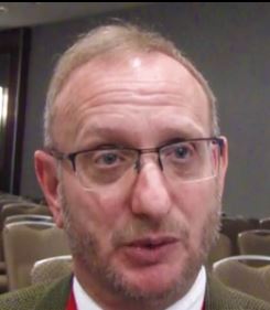
However, there’s been some doubt about ALLHAT; doxazosin patients had diuretics withdrawn as part of the study, which might have contributed to the increased risks. “A lot of arguments have been made that perhaps alpha-blockers aren’t that bad and maybe should still be used, so we took a second look,” said lead investigator and nephrologist Swapnil Hiremath, MD, an assistant professor of medicine at the University of Ottawa.
What he and his team found “confirmed and expanded on the findings of ALLHAT.” Apart from a few specific situations, “don’t prescribe alpha-blockers. If a patient is on an alpha-blocker, consider prescribing an alternative,” Dr. Hiremath said at the joint scientific sessions of the American Heart Association Council on Hypertension, AHA Council on Kidney in Cardiovascular Disease, and American Society of Hypertension.
The drugs are still widely used, according to the team’s review of Ontario health data. From 1995 to 2015, nearly 81,000 patients were prescribed alpha-blockers for hypertension, sometimes as monotherapy, with no real downward trend in prescriptions over time.
There are some selected indications for alpha-blockers, including intolerance of other antihypertensives, pheochromocytoma management, and resistant hypertension. “So I thought maybe there would be 5,000 or 10,000. The fact that we found almost 81,000 was an eye-opener. I’m pretty sure 81,000 patients in Ontario don’t have resistant hypertension,” Dr. Hiremath said.
Patients with benign prostatic hypertrophy, another indication, were excluded from the study.
Before ALLHAT, alpha-blockers were considered first-line drugs, so maybe prescribers are just “sticking with something they know and are familiar with,” he said.
To assess the risks of continued use, the investigators used propensity scoring to match 69,092 patients prescribed alpha-blockers to 69,092 who were prescribed other antihypertensives, based on age, comorbidities, date of treatment, and a slew of other potential confounders. Patients were considered to be on an alpha-blocker only when they were filling prescriptions for the drugs. If they were not filling prescriptions they flipped into the unexposed arm.
The incident rates of ED visits and hospitalizations for hypotension and related complications – syncope, falls, and fractures – were markedly higher among alpha-blocker users. After adjusting for the total number of antihypertensives patients were on, those on alpha-blockers were 34% more likely to go to the ED or be hospitalized for hypotension, 49% more likely for syncope, 27% more likely for falls, and 41% more likely for fractures. First-dose effects don’t explain the findings; patients were often on alpha-blockers for years beforehand.
Alpha-blocker patients were also 26% more likely to have a major cardiovascular event, including heart failure and MI. The risks were greatest in those older than age 85 years. The results were all statistically significant.
About 9,000 alpha-blocker patients had a fracture versus 3,351 matched patients on other antihypertensives; “9,000 patients out of 70,000 is a huge number. These drugs are useful in some situations, but be careful,” Dr. Hiremath said.
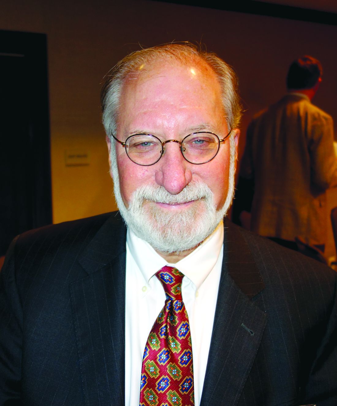
“This is observational data, but it’s consistent with ALLHAT, and the outcomes are even worse. We didn’t in [subsequent] guidelines say that you should [never] use an alpha-blocker in hypertension. Maybe we should have,” said ALLHAT investigator William Cushman, MD, a professor of medicine and physiology at the University of Tennessee, Memphis.
There was no industry funding for the work, and the investigators reported having no financial disclosures. Dr. Cushman wasn’t involved in the work.
SOURCE: Hiremath S et al. Joint Hypertension 2018, Abstract P375.
CHICAGO – The risk of cardiovascular and hypotension-related events is higher with alpha-blockers than with other hypertension drugs, but almost 20 years after the pivotal ALLHAT trial first raised safety concerns, they are still widely used, according to investigators from the University of Ottawa.
ALLHAT (Antihypertensive and Lipid-Lowering Treatment to Prevent Heart Attack Trial) linked the alpha-blocker doxazosin (Cardura) to an increased risk of heart failure and stroke, which led to the early cessation of the doxazosin arm. Guidelines no longer include alpha-blockers among the primary options for hypertension (JAMA. 2000;283[15]:1967-75).
However, there’s been some doubt about ALLHAT; doxazosin patients had diuretics withdrawn as part of the study, which might have contributed to the increased risks. “A lot of arguments have been made that perhaps alpha-blockers aren’t that bad and maybe should still be used, so we took a second look,” said lead investigator and nephrologist Swapnil Hiremath, MD, an assistant professor of medicine at the University of Ottawa.
What he and his team found “confirmed and expanded on the findings of ALLHAT.” Apart from a few specific situations, “don’t prescribe alpha-blockers. If a patient is on an alpha-blocker, consider prescribing an alternative,” Dr. Hiremath said at the joint scientific sessions of the American Heart Association Council on Hypertension, AHA Council on Kidney in Cardiovascular Disease, and American Society of Hypertension.
The drugs are still widely used, according to the team’s review of Ontario health data. From 1995 to 2015, nearly 81,000 patients were prescribed alpha-blockers for hypertension, sometimes as monotherapy, with no real downward trend in prescriptions over time.
There are some selected indications for alpha-blockers, including intolerance of other antihypertensives, pheochromocytoma management, and resistant hypertension. “So I thought maybe there would be 5,000 or 10,000. The fact that we found almost 81,000 was an eye-opener. I’m pretty sure 81,000 patients in Ontario don’t have resistant hypertension,” Dr. Hiremath said.
Patients with benign prostatic hypertrophy, another indication, were excluded from the study.
Before ALLHAT, alpha-blockers were considered first-line drugs, so maybe prescribers are just “sticking with something they know and are familiar with,” he said.
To assess the risks of continued use, the investigators used propensity scoring to match 69,092 patients prescribed alpha-blockers to 69,092 who were prescribed other antihypertensives, based on age, comorbidities, date of treatment, and a slew of other potential confounders. Patients were considered to be on an alpha-blocker only when they were filling prescriptions for the drugs. If they were not filling prescriptions they flipped into the unexposed arm.
The incident rates of ED visits and hospitalizations for hypotension and related complications – syncope, falls, and fractures – were markedly higher among alpha-blocker users. After adjusting for the total number of antihypertensives patients were on, those on alpha-blockers were 34% more likely to go to the ED or be hospitalized for hypotension, 49% more likely for syncope, 27% more likely for falls, and 41% more likely for fractures. First-dose effects don’t explain the findings; patients were often on alpha-blockers for years beforehand.
Alpha-blocker patients were also 26% more likely to have a major cardiovascular event, including heart failure and MI. The risks were greatest in those older than age 85 years. The results were all statistically significant.
About 9,000 alpha-blocker patients had a fracture versus 3,351 matched patients on other antihypertensives; “9,000 patients out of 70,000 is a huge number. These drugs are useful in some situations, but be careful,” Dr. Hiremath said.
“This is observational data, but it’s consistent with ALLHAT, and the outcomes are even worse. We didn’t in [subsequent] guidelines say that you should [never] use an alpha-blocker in hypertension. Maybe we should have,” said ALLHAT investigator William Cushman, MD, a professor of medicine and physiology at the University of Tennessee, Memphis.
There was no industry funding for the work, and the investigators reported having no financial disclosures. Dr. Cushman wasn’t involved in the work.
SOURCE: Hiremath S et al. Joint Hypertension 2018, Abstract P375.
REPORTING FROM JOINT HYPERTENSION 2018
Key clinical point:
Major finding: Patients on alpha-blockers, versus other antihypertensives, were 26% more likely to have a major cardiovascular event, including heart failure and MI.
Study details: A propensity score matching study involving about 140,000 patients.
Disclosures: There was no industry funding for the work, and the investigators reported having no financial disclosures.
Source: Hiremath S et al. Joint Hypertension 2018, Abstract P375.
How to handle anorexia in community hospitals
Food is nonnegotiable
ATLANTA – Everyone has to be on the same page when it comes to anorexia nervosa in a community hospital, according to pediatric hospitalists at Moses H. Cone Memorial Hospital in Greensboro, N.C.

Anorexia cases used to be rare there. When one came in, “everyone was anxious because we just didn’t know quite what to do,” said Suresh Nagappan, MD, a pediatrician and member of the teaching faculty at the hospital. Parents would hear one thing from one provider, something else from the next, and leave angry and confused. “Basically, it was a mess. We needed to standardize it,” he added.
So Dr. Nagappan and his colleagues created guidelines for treating patients with eating disorders about 3 years ago. “It was meeting after meeting for months, but well worth it,” he said at Pediatric Hospital Medicine.
Word of the hospital’s newfound expertise in anorexia has spread since then, and now it’s not unusual for Moses H. Cone to handle a few cases a week.
The pediatric hospitalist team has come to realize that, first and foremost, patients and families need to know why they are there; it’s about medical stabilization, not treating the eating disorder. That comes after discharge. Families need help sometimes to understand that it’s not a quick fix.
To make things clear, there’s strict criteria now for admission, based on American Academy of Pediatrics guidance. The main trigger is being under 75% of ideal body weight, but patients must also have systolic blood pressure below 90 mm Hg and other worrisome signs. “Sometimes, it feels like we’re splitting hairs” on who gets admitted, “but if we don’t have strict criteria on admission, we don’t have an end goal for discharge,” said pediatrician Maggie S. Hall, MD, also on the Moses H. Cone teaching faculty.
As for treatment, “food is medicine, and it’s not negotiable. We make that clear to everyone on day 1. If patients don’t eat their actual meal, they have 20 minutes to drink a supplement. If they can’t do that, they get a nasogastric tube,” Dr. Nagappan said. The tube is pulled after each meal, so that it remains an incentive to eat.
The team start patients with 1,600 calories a day and increase the intake by 200-250 calories a day. The goal is for a patient to gain 100-200 grams per day. Patients pick out what they want to eat with the help of a dietitian. When meals set off overwhelming anxiety, the Moses H. Cone team has learned that benzodiazepines can help.
Ironically, the initiation of regular meals is the most dangerous time for patients. As anorexic bodies switch from catabolic to anabolic metabolism, electrolytes can drop to dangerously low levels, causing arrhythmias, heart failure, and death. In general, “the reason these kids die is cardiac,” Dr. Nagappan said at the meeting, sponsored by the Society of Hospital Medicine, the AAP, and the Academic Pediatric Association.
Refeeding syndrome, as it’s known, is clinically significant in perhaps 6% of patients. The risk goes up if they are below 70% of their ideal body weight; have a prolonged QTc interval; or begin treatment with low phosphorous, magnesium, or potassium.
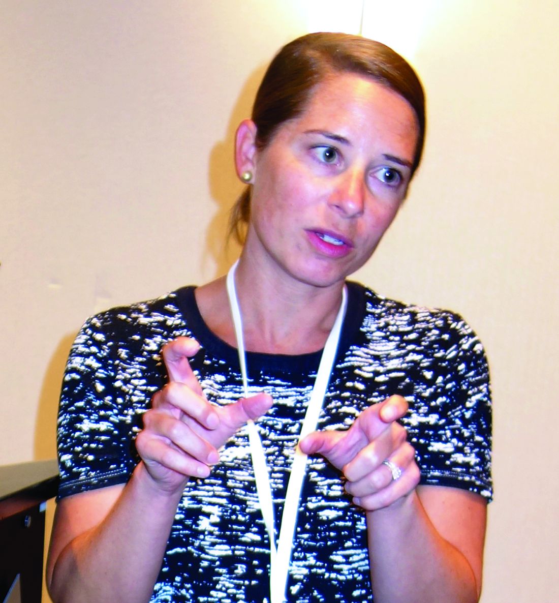
To counter the threat, electrolytes are measured twice a day at Moses H. Cone during the first week of treatment, and ECGs are taken daily for the first few days. “One thing to be really careful about is when you notice their heart rate beginning to creep up during rest. That can be a sign of developing cardiomyopathy; it’s an indication for us to get echocardiograms,” Dr. Hall said.
The Moses H. Cone team like to include families in meal times – it’s been shown to help – but family members need to be coached beforehand. They can’t be punitive. Mealtime talk has to be positive, and can’t focus on eating. Parents often need help handling their own anger and guilt before trying to eat with their child. Progress has to be monitored, but Dr. Nagappan cautioned that “you have to be really careful about how you get weights”; it should always be in the morning after the first void. Urine needs to be checked to make sure patients aren’t water loading.
Staff should be neutral about weight results, and keep them to themselves. Even something as benign as “good job” can be a problem. “You don’t want these patients focused on their weight. You want them focused on getting better and eating and taking it step by step,” he said.
The presenters had no disclosures to report.
Food is nonnegotiable
Food is nonnegotiable
ATLANTA – Everyone has to be on the same page when it comes to anorexia nervosa in a community hospital, according to pediatric hospitalists at Moses H. Cone Memorial Hospital in Greensboro, N.C.
Anorexia cases used to be rare there. When one came in, “everyone was anxious because we just didn’t know quite what to do,” said Suresh Nagappan, MD, a pediatrician and member of the teaching faculty at the hospital. Parents would hear one thing from one provider, something else from the next, and leave angry and confused. “Basically, it was a mess. We needed to standardize it,” he added.
So Dr. Nagappan and his colleagues created guidelines for treating patients with eating disorders about 3 years ago. “It was meeting after meeting for months, but well worth it,” he said at Pediatric Hospital Medicine.
Word of the hospital’s newfound expertise in anorexia has spread since then, and now it’s not unusual for Moses H. Cone to handle a few cases a week.
The pediatric hospitalist team has come to realize that, first and foremost, patients and families need to know why they are there; it’s about medical stabilization, not treating the eating disorder. That comes after discharge. Families need help sometimes to understand that it’s not a quick fix.
To make things clear, there’s strict criteria now for admission, based on American Academy of Pediatrics guidance. The main trigger is being under 75% of ideal body weight, but patients must also have systolic blood pressure below 90 mm Hg and other worrisome signs. “Sometimes, it feels like we’re splitting hairs” on who gets admitted, “but if we don’t have strict criteria on admission, we don’t have an end goal for discharge,” said pediatrician Maggie S. Hall, MD, also on the Moses H. Cone teaching faculty.
As for treatment, “food is medicine, and it’s not negotiable. We make that clear to everyone on day 1. If patients don’t eat their actual meal, they have 20 minutes to drink a supplement. If they can’t do that, they get a nasogastric tube,” Dr. Nagappan said. The tube is pulled after each meal, so that it remains an incentive to eat.
The team start patients with 1,600 calories a day and increase the intake by 200-250 calories a day. The goal is for a patient to gain 100-200 grams per day. Patients pick out what they want to eat with the help of a dietitian. When meals set off overwhelming anxiety, the Moses H. Cone team has learned that benzodiazepines can help.
Ironically, the initiation of regular meals is the most dangerous time for patients. As anorexic bodies switch from catabolic to anabolic metabolism, electrolytes can drop to dangerously low levels, causing arrhythmias, heart failure, and death. In general, “the reason these kids die is cardiac,” Dr. Nagappan said at the meeting, sponsored by the Society of Hospital Medicine, the AAP, and the Academic Pediatric Association.
Refeeding syndrome, as it’s known, is clinically significant in perhaps 6% of patients. The risk goes up if they are below 70% of their ideal body weight; have a prolonged QTc interval; or begin treatment with low phosphorous, magnesium, or potassium.
To counter the threat, electrolytes are measured twice a day at Moses H. Cone during the first week of treatment, and ECGs are taken daily for the first few days. “One thing to be really careful about is when you notice their heart rate beginning to creep up during rest. That can be a sign of developing cardiomyopathy; it’s an indication for us to get echocardiograms,” Dr. Hall said.
The Moses H. Cone team like to include families in meal times – it’s been shown to help – but family members need to be coached beforehand. They can’t be punitive. Mealtime talk has to be positive, and can’t focus on eating. Parents often need help handling their own anger and guilt before trying to eat with their child. Progress has to be monitored, but Dr. Nagappan cautioned that “you have to be really careful about how you get weights”; it should always be in the morning after the first void. Urine needs to be checked to make sure patients aren’t water loading.
Staff should be neutral about weight results, and keep them to themselves. Even something as benign as “good job” can be a problem. “You don’t want these patients focused on their weight. You want them focused on getting better and eating and taking it step by step,” he said.
The presenters had no disclosures to report.
EXPERT ANALYSIS FROM PHM 2018
The dextrose-sulfonylurea challenge: a screen for monogenetic diabetes?
ORLANDO – When investigators at Washington University, St. Louis, gave 42 patients with type 1 diabetes mellitus a dose of the sulfonylurea glipizide (Glucotrol), a curious thing happened.
C-peptide levels rose in 13 patients (31%), which means they secreted their own insulin.
The finding was unexpected; people with type 1 diabetes mellitus (T1DM) aren’t supposed to be able to produce endogenous insulin because they don’t have beta cells. They shouldn’t have had any response to glipizide, a beta-cell stimulator.
It wasn’t that the 13 subjects were in the honeymoon phase of T1DM, meaning that they still had a few beta cells left. Like the other patients, their glucose levels rose when they were given dextrose, but their C-peptide levels did not. Also, all patients, including the 13 who secreted insulin, had been diagnosed with T1DM for a mean of 6 years and were insulin dependent. They ranged in age up to 33 years, and their hemoglobin A1c was about 8.6%.
In short, the 13 patients didn’t have classic type 1 diabetes. They had something wrong with their beta cells, explained Colin G. Nichols, PhD, a professor of cell biology and physiology at Washington University.
They likely had monogenetic diabetes, a genetic mutation that caused potassium channels in their beta cells to be permanently hyperpolarized. Blocking the channels with a sulfonylurea allowed the cells to depolarize and secrete insulin.
Monogenetic diabetes is a known but underrecognized entity. If it’s not picked up in infancy, most patients are misdiagnosed with classic T1DM and inappropriately treated with insulin. If they finally try a sulfonylurea, “they don’t need insulin injections anymore. This has been a very magical story for this group of patients,” Dr. Nichols said at the annual scientific sessions of the American Diabetes Association.
All they need is a sulfonylurea pill once a day.
Monogenetic patients are missed because there are no easy, widely-available screening tests for the condition. Genetic testing works, but it’s expensive and often not done. Dr. Nichols and his team hope their dextrose-sulfonylurea challenge will solve the problem.
They are currently working to recruit more subjects and perform confirmatory genetic testing. They want to know how much insulin secretion is possible for their glipizide responders and how long their responses last. If funding comes through, they hope to do a screening and treatment trial in patients with T1DM.
The promise is that the dextrose-sulfonylurea challenge will shorten the time to a correct diagnosis and proper treatment, and save people from decades of insulin shots. It might also pick up nongenetic, metabolic causes of beta-cell potassium channel dysfunction that would respond to sulfonylureas. The challenge could even be used to prescreen for genetic testing, to increase its yield and shift the cost-benefit ratio more into the black.
As for the specifics of the study, the 42 patients were given an intravenous dextrose bolus of 0.5 g/kg, followed 20 minutes later by a single dose of glipizide, 0.3 mg/kg. Blood glucose and C-peptide were measured at baseline and at regular intervals during the 4-hour challenge. The challenge was safe; there were no serious side effects.
Among the 13 responders – meaning flat insulin secretion with dextrose but insulin secretion with glipizide – the peak change in C-peptide was around 0.41 ng/mL ± 0.45 an hour or so after the glipizide dose, and was maintained for about an hour from a baseline of about 0.6 ng/mL.
To make sure that the responders didn’t have just a delayed insulin response to dextrose, they were given another dextrose challenge 6 months later, without the glipizide. Again, their C-peptide levels were flat.
The investigators had no disclosures to report, and there was no industry funding.
SOURCE: Nichols CG et al. ADA 2018, Abstract 310-LB.
REPORTING FROM ADA 2018
Key clinical point: Some patients with type 1 diabetes mellitus might need just a daily dose of a sulfonylurea instead of multiple insulin injections; a test to find them is being developed.
Major finding: Nearly one-third of patients with long-standing, insulin-dependent type 1 diabetes secreted their own insulin when given the beta-cell stimulator glipizide.
Study details: An observational study of 42 people with type 1 diabetes mellitus.
Disclosures: The investigators had no disclosures to report, and there was no industry funding.
Source: Nichols CG et al. ADA 2018, Abstract 310-LB.
Plan now for outpatient diabetes tech in the hospital
ORLANDO –
A third or more of patients with type 1 diabetes mellitus and growing numbers of patients with insulin-dependent type 2 diabetes mellitus patients are using pumps and sensor technology. The American Diabetes Association advocates allowing patients who are physically and mentally able to continue to use their pumps when hospitalized, and there’s general consensus that continuous glucose monitors (CGM) can be used in the hospital.
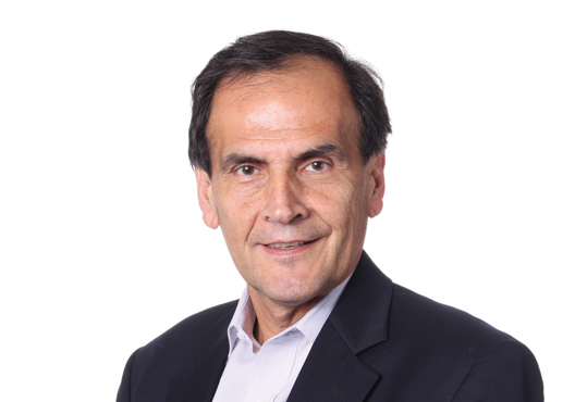
All in all, it’s a good thing, according to Guillermo E. Umpierrez, MD, professor of endocrinology at Emory University and chief of endocrinology at Grady Memorial Hospital, both in Atlanta.
In a talk at the annual scientific sessions of the American Diabetes Association, Dr. Umpierrez reviewed a number of studies showing that glycemic control with the new technology is no worse in the hospital – and sometimes even better – than with traditional point-of-care glucose testing and insulin administration. There is a lack of randomized, controlled trials to prove the point definitively, but what evidence does exist is promising.
“This technology is rapidly advancing, and I am very optimistic that we are going to see more and more of these devices in the hospital. If patients can manage themselves, allow them to use CGM, allow them to use their pumps,” he said.
As for closed loop systems – automated glucose sensing and insulin administration – emerging evidence suggests they “allow you to have very good glucose control and less glycemic variability,” both inside and outside of the ICU, he said. “I am very hopeful before I retire that there will be management of a significant number of patients with closed loop systems.”
To keep up, training for hospital providers on the new technology is now “mandatory at all levels,” Dr. Umpierrez said, and if they haven’t done so already, hospitals need to put policies and procedures in place for when, and when not, to allow patients to use their diabetes equipment, and how to integrate it into care.
Among many things to consider, patients must be well enough to use their pumps and monitors, be able to demonstrate their functions, and also want to participate in their own care.
Contraindications to inpatient pump use include impaired consciousness, critical illness, and hyperglycemic crises because insulin requirements change too rapidly and dramatically for pumps. Lack of trained providers and supplies is another hurdle. Pumps also need to come off for MRIs.
CGM, meanwhile, has been shown to improve glycemic control, detecting both hyperglycemia and hypoglycemia more readily than point-of-care testing. It’s good at picking up trends in glucose levels, and Dr. Umpierrez anticipates a time when readings will be transmitted to nurses’ stations automatically to track blood glucose trends. “I think that’s the future,” he said.
But, as with insulin pumps, there are caveats. Among them, it’s unclear how well CGM works during hypoxia, hypothermia, and hypotension. Thrombus formation and infections have been reported with intravascular monitors, and a number of agents can throw off some CGM devices, including acetaminophen, heparin, and dopamine.
Dr. Umpierrez disclosed relationships with AstraZeneca, Merck, Novo Nordisk, Sanofi, and other companies.
ORLANDO –
A third or more of patients with type 1 diabetes mellitus and growing numbers of patients with insulin-dependent type 2 diabetes mellitus patients are using pumps and sensor technology. The American Diabetes Association advocates allowing patients who are physically and mentally able to continue to use their pumps when hospitalized, and there’s general consensus that continuous glucose monitors (CGM) can be used in the hospital.
All in all, it’s a good thing, according to Guillermo E. Umpierrez, MD, professor of endocrinology at Emory University and chief of endocrinology at Grady Memorial Hospital, both in Atlanta.
In a talk at the annual scientific sessions of the American Diabetes Association, Dr. Umpierrez reviewed a number of studies showing that glycemic control with the new technology is no worse in the hospital – and sometimes even better – than with traditional point-of-care glucose testing and insulin administration. There is a lack of randomized, controlled trials to prove the point definitively, but what evidence does exist is promising.
“This technology is rapidly advancing, and I am very optimistic that we are going to see more and more of these devices in the hospital. If patients can manage themselves, allow them to use CGM, allow them to use their pumps,” he said.
As for closed loop systems – automated glucose sensing and insulin administration – emerging evidence suggests they “allow you to have very good glucose control and less glycemic variability,” both inside and outside of the ICU, he said. “I am very hopeful before I retire that there will be management of a significant number of patients with closed loop systems.”
To keep up, training for hospital providers on the new technology is now “mandatory at all levels,” Dr. Umpierrez said, and if they haven’t done so already, hospitals need to put policies and procedures in place for when, and when not, to allow patients to use their diabetes equipment, and how to integrate it into care.
Among many things to consider, patients must be well enough to use their pumps and monitors, be able to demonstrate their functions, and also want to participate in their own care.
Contraindications to inpatient pump use include impaired consciousness, critical illness, and hyperglycemic crises because insulin requirements change too rapidly and dramatically for pumps. Lack of trained providers and supplies is another hurdle. Pumps also need to come off for MRIs.
CGM, meanwhile, has been shown to improve glycemic control, detecting both hyperglycemia and hypoglycemia more readily than point-of-care testing. It’s good at picking up trends in glucose levels, and Dr. Umpierrez anticipates a time when readings will be transmitted to nurses’ stations automatically to track blood glucose trends. “I think that’s the future,” he said.
But, as with insulin pumps, there are caveats. Among them, it’s unclear how well CGM works during hypoxia, hypothermia, and hypotension. Thrombus formation and infections have been reported with intravascular monitors, and a number of agents can throw off some CGM devices, including acetaminophen, heparin, and dopamine.
Dr. Umpierrez disclosed relationships with AstraZeneca, Merck, Novo Nordisk, Sanofi, and other companies.
EXPERT ANALYSIS FROM ADA 2018
Eat/sleep/console approach almost eliminates morphine for NAS
ATLANTA – In just 7 months, the University of North Carolina Children’s Hospital, Chapel Hill, dropped the length of stay for neonatal abstinence syndrome from about 11 days to 5 days by moving from scheduled to PRN morphine dosing and abandoning the Finnegan score, according to a report at the Pediatric Hospital Medicine meeting.

The use of morphine fell from 93% of infants transferred to the hospital’s inpatient floors for neonatal abstinence syndrome (NAS) to just 12%, with no downsides for infants or moms.
“Our results have been incredibly encouraging,” said lead investigator and pediatrics resident Thomas Blount, MD. The take-home message is to treat the infant, rather than relying on the Finnegan score.
UNC Children’s, which treats about 50 infants a year for NAS on its inpatient floors, had been using the traditional approach: babies were automatically scheduled for morphine and Finnegan scoring – a withdrawal symptom checklist – every 4 hours, regardless of need. Sometimes infants weren’t even assessed to see if they actually needed morphine before the next dose was given.
“Waking babies up every 4 hours just seemed crazy; of course, they were going to have heightened neurologic signs and symptoms.” Meanwhile, families and providers were frustrated that infants who were otherwise doing well were held for an extra week or more to wean them off morphine, Dr. Blount said at the meeting.
In Nov. 2017, the hospital switched to the eat/sleep/console (ESC) model for NAS on its inpatient floors. The model emphasizes what’s been shown to work in recent years: keeping the infant with the mother; encouraging breast feeding, skin-on-skin contact, and other comfort measures; and supplementing feeds to help with weight gain. Morphine is reserved for when those measures fail and given only with a needs assessment (Hosp Pediatr. 2018 Jan;8(1):1-6).
The hospital ditched Finnegan scoring on its inpatient floors. Nurses were asked instead to check if infants were feeding adequately, sleeping at least an hour between feedings, and able to be consoled within 10 minutes when upset. If the infants met all three of those ESC criteria, providers moved on. They left the baby swaddled, relied on ambient white noise of ocean waves, and checked back on them later. “They didn’t mess with them,” Dr. Blount said at the meeting, sponsored by the Society of Hospital Medicine, the American Academy of Pediatrics, and the Academic Pediatric Association.
Finnegan scoring “was causing so much anxiety. Staff and families became hypervigilant,” set off by every little twitch and yawn the baby made. It was a good thing when it was abandoned; everyone relaxed, he said.
The changes made a huge difference. The average number of morphine doses dropped from 39 per infant to just 7 total doses among 23 infants in the first 7 months of the ESC initiative. Currently, morphine is used in only about 1 of 10 cases. “We estimate that we’ve given over 900 fewer doses” since ESC was put in place, Dr. Blount said.

There’s been no change in 30-day readmission rates – just one since the changes were made, for bronchiolitis – and no change in weight loss among infants with NAS. Babies are meeting all the ESC criteria to thrive.
“We had a lot of pushback initially, mostly from nursing staff and residents wondering how this was going to work. It quickly went away,” Dr. Blount said.
His team is now considering rolling ESC out to the newborn nursery and NICU.
There was no industry funding for the work, and Dr. Blount didn’t have any disclosures.
ATLANTA – In just 7 months, the University of North Carolina Children’s Hospital, Chapel Hill, dropped the length of stay for neonatal abstinence syndrome from about 11 days to 5 days by moving from scheduled to PRN morphine dosing and abandoning the Finnegan score, according to a report at the Pediatric Hospital Medicine meeting.
The use of morphine fell from 93% of infants transferred to the hospital’s inpatient floors for neonatal abstinence syndrome (NAS) to just 12%, with no downsides for infants or moms.
“Our results have been incredibly encouraging,” said lead investigator and pediatrics resident Thomas Blount, MD. The take-home message is to treat the infant, rather than relying on the Finnegan score.
UNC Children’s, which treats about 50 infants a year for NAS on its inpatient floors, had been using the traditional approach: babies were automatically scheduled for morphine and Finnegan scoring – a withdrawal symptom checklist – every 4 hours, regardless of need. Sometimes infants weren’t even assessed to see if they actually needed morphine before the next dose was given.
“Waking babies up every 4 hours just seemed crazy; of course, they were going to have heightened neurologic signs and symptoms.” Meanwhile, families and providers were frustrated that infants who were otherwise doing well were held for an extra week or more to wean them off morphine, Dr. Blount said at the meeting.
In Nov. 2017, the hospital switched to the eat/sleep/console (ESC) model for NAS on its inpatient floors. The model emphasizes what’s been shown to work in recent years: keeping the infant with the mother; encouraging breast feeding, skin-on-skin contact, and other comfort measures; and supplementing feeds to help with weight gain. Morphine is reserved for when those measures fail and given only with a needs assessment (Hosp Pediatr. 2018 Jan;8(1):1-6).
The hospital ditched Finnegan scoring on its inpatient floors. Nurses were asked instead to check if infants were feeding adequately, sleeping at least an hour between feedings, and able to be consoled within 10 minutes when upset. If the infants met all three of those ESC criteria, providers moved on. They left the baby swaddled, relied on ambient white noise of ocean waves, and checked back on them later. “They didn’t mess with them,” Dr. Blount said at the meeting, sponsored by the Society of Hospital Medicine, the American Academy of Pediatrics, and the Academic Pediatric Association.
Finnegan scoring “was causing so much anxiety. Staff and families became hypervigilant,” set off by every little twitch and yawn the baby made. It was a good thing when it was abandoned; everyone relaxed, he said.
The changes made a huge difference. The average number of morphine doses dropped from 39 per infant to just 7 total doses among 23 infants in the first 7 months of the ESC initiative. Currently, morphine is used in only about 1 of 10 cases. “We estimate that we’ve given over 900 fewer doses” since ESC was put in place, Dr. Blount said.
There’s been no change in 30-day readmission rates – just one since the changes were made, for bronchiolitis – and no change in weight loss among infants with NAS. Babies are meeting all the ESC criteria to thrive.
“We had a lot of pushback initially, mostly from nursing staff and residents wondering how this was going to work. It quickly went away,” Dr. Blount said.
His team is now considering rolling ESC out to the newborn nursery and NICU.
There was no industry funding for the work, and Dr. Blount didn’t have any disclosures.
REPORTING FROM PHM 2018
Key clinical point: When it comes to neonatal abstinence syndrome, treat the infant, not the Finnegan score.
Major finding: The University of North Carolina Children’s Hospital dropped the length of stay for neonatal abstinence syndrome from about 11 to 5 days by moving from scheduled to PRN morphine and abandoning Finnegan scoring. Morphine use fell more than 80%.
Study details: Review of a 7-month quality improvement project
Disclosures: There was no industry funding for the work. The lead investigator didn’t have any disclosures.
Restrictive transfusion will likely be standard, doc says
MUNICH—A restrictive transfusion strategy during cardiovascular surgery will likely become the standard of care, according to an investigator from the TRICS III trial.
The study showed that a restrictive approach to transfusion did not increase the risk of poor outcomes at 6 months after cardiac surgery.
David Mazer, MD, of St. Michael’s Hospital, University of Toronto in Ontario, Canada, presented these findings at the 2018 ESC Congress. The results were published simultaneously in NEJM.
Dr. Mazer and his colleagues previously reported results from the TRICS III trial showing that 28-day outcomes were similar whether patients undergoing cardiac surgery were treated with a restrictive or a liberal transfusion strategy.
However, the team wanted to look into 6-month results to rule out latent problems, such as sequelae from perioperative organ hypoxia.
“Our research question was, ‘At what point does the risk of anemia, or the risk of a lower hemoglobin, outweigh the risk of transfusion?’” Dr. Mazer said. “We wanted to know whether it is safe to let your hemoglobin go to a lower level before you transfuse. The answer is yes. It’ll save blood, make blood more available, reduce costs of transfusion, and result in similar or better outcomes.”
Patients
The trial included 2317 patients who were randomized to a restrictive transfusion strategy, which meant they received red cell transfusions if their hemoglobin concentrations fell below 7.5 g/dL intraoperatively or postoperatively.
Another 2347 patients were randomized to the liberal approach, which meant they received transfusions if their hemoglobin fell below 9.5 g/dL in the operating room and intensive care unit (ICU) and below 8.5 g/dL outside the ICU.
Baseline characteristics were well balanced between the arms. Patients were a mean of 72 years old, and 35% were female. The majority of patients in both arms underwent coronary artery bypass surgery, valve surgery, or both. Heart transplants were excluded.
Results
At 6 months, 17.4% of patients in the restrictive arm and 17.1% in the liberal arm met the primary composite outcome of death from any cause, myocardial infarction, stroke, or new-onset renal failure with dialysis (P=0.006 for noninferiority).
Unexpectedly, patients age 75 and older had a lower risk of the primary outcome with the restrictive strategy, while the liberal strategy was associated with lower risk in younger patients.
For all age groups, there were no significant differences between the treatment arms for the individual components of the primary composite outcome or for secondary outcomes.
The mortality rate was 6.2% in the restrictive arm and 6.4% in the liberal arm. The rate of myocardial infarction was 7.3% in both arms.
The rate of stroke was 4% in the restrictive arm and 3.3% in the liberal arm. The incidence of new-onset renal failure with dialysis was 3.9% and 4.2%, respectively.
The secondary composite outcome included the components of the primary outcome plus hospital readmissions, emergency department visits, and coronary revascularization. This outcome occurred in 43.8% of patients in the restrictive arm and 42.8% in the liberal arm.
The incidence of hospital readmissions/emergency visits was 35.5% in the restrictive arm and 33.6% in the liberal arm. The incidence of coronary revascularization was 0.7% and 0.9%, respectively.
The investigators also found the restrictive transfusion strategy effectively saved blood. Just over half of patients (52.3%) in the restrictive arm and almost three-quarters (72.6%) of those in the liberal arm were transfused after randomization.
“This research has already started to change transfusion practice around the world,” Dr. Mazer said. “With this data at six months, we’ve proven the longer-term safety of restrictive therapy. This approach has already been adopted into guidelines and will likely become the standard of care worldwide.”
This study was funded by the Canadian Institutes of Health Research, Canadian Blood Services, the National Health and Medical Research Council in Australia, and the Health Research Council of New Zealand. Dr. Mazer had no relevant disclosures.
Restrictive transfusions do not increase long-term CV surgery risk
MUNICH – in a large, randomized trial presented at the annual congress of the European Society of Cardiology, and published simultaneously in the New England Journal of Medicine.
The investigators previously reported that 28 day outcomes were non-inferior with the restrictive approach, but they wanted to look into 6 month results to rule out latent problems, such as sequelae from perioperative organ hypoxia.
The new outcomes from the study, dubbed the Transfusion Requirements in Cardiac Surgery (TRICS) III trial, offer some reassurance at a time when cardiac surgeons are shifting towards more restrictive transfusion policies (N Engl J Med. 2018 Aug 26.doi: 10.1056/NEJMoa1808561).
The trial randomized 2,317 patients to red cell transfusions if their hemoglobin concentrations fell below 7.5 g/dL intraoperatively or postoperatively. Another 2,347 were randomized to the liberal approach, with transfusions below 9.5 g/dL in the operating room and ICU, and below 8.5 g/dL outside of the ICU. The arms were well matched, with a mean score of 8 in both groups on the 47-point European System for Cardiac Operative Risk Evaluation I score.
At 6 months, 17.4% of patients in the restrictive arm, versus 17.1% in the liberal-threshold group, met the primary composite outcome of death from any cause, myocardial infarction, stroke, or new onset renal failure with dialysis (P = .006 for noninferiority with the restrictive threshold); 6.2% of patients in the restrictive-threshold group, versus 6.4% in the liberal-arm, had died at that point, a statistically insignificant difference.
Also at 6 months, 43.8% of patients in the restrictive-threshold group, versus 42.8% in the liberal arm, met the study’s secondary composite outcome, which included the components of the primary outcome plus hospital readmissions, ED visits, and coronary revascularization. The difference was again statistically insignificant.
The restrictive strategy saved a lot of blood. Just over half of the patients, versus almost three-quarters in the liberal arm, were transfused during their index admissions. When transfused, patients in the restrictive arm received a median of 2 units of red cells; liberal-arm patients received a median of 3 units.
Unexpectedly, patients 75 years and older had a lower risk of poor outcomes with the restrictive strategy, while the liberal strategy was associated with lower risk in younger subjects. The findings “appear to contradict” current practice, “in which a liberal transfusion strategy is used in older patients undergoing cardiac or noncardiac surgery,” said investigators led by C. David Mazer, MD, a professor in the department of anesthesia at the University of Toronto.
“One could hypothesize that older patients may have unacceptable adverse effects related to transfusion (e.g., volume overload and inflammatory and infectious complications) or that there may be age-related differences in the adverse-effect profile of transfusion or anemia. ... Whether transfusion thresholds should differ according to age” needs to be determined, they said.
The participants were a mean of 72 years old, and 35% were women. The majority in both arms underwent coronary artery bypass surgery, valve surgery, or both. Heart transplants were excluded. The trial was conducted in 19 countries, including China and India; results were consistent across study sites.
The work was funded by the Canadian Institutes of Health Research, among others. Dr. Mazer had no relevant disclosures.
SOURCE: Mazer CD et al. N Engl J Med. 2018 Aug 26. doi:10.1056/NEJMoa1808561
REPORTING FROM THE ESC CONGRESS 2018
Key clinical point: A restrictive transfusion strategy during cardiovascular surgery, versus a more traditional liberal approach, did not increase the risk of poor outcomes at 6 months.
Major finding: At 6 months, 17.4% of patients in the restrictive arm, versus 17.1% in the liberal-threshold group, met the primary composite outcome of death from any cause, myocardial infarction, stroke, or new onset renal failure with dialysis (P = .006 for noninferiority with the restrictive approach).
Study details: Randomized, multicenter trial with over 5,000 surgery patients
Disclosures: The work was funded by the Canadian Institutes of Health Research, among others. The lead investigator had no relevant disclosures.
Source: Mazer CD et al. N Engl J Med. 2018 Aug 26. doi:10.1056/NEJMoa1808561
Restrictions on EMT glucagon administration should be lifted
ORLANDO – In 42 states, emergency medical technicians aren’t allowed to administer glucagon to patients with severe hypoglycemia, and that’s a problem, according to investigators from the Joslin Diabetes Center, Boston.
Paramedics can administer the drug, but they’re only about a quarter of medical first-responders, so in many parts of the country, ambulance crews show up with only EMTs [emergency medical technicians] on board, and patients have to wait for the ED to get glucagon.
The delay increases the risk of coma, brain damage, and death. Just like in stroke, time is key with severe hypoglycemia. “The minutes really add up,” said lead investigator Nicole Wagner, a one-time researcher at Joslin and now a medical student at Thomas Jefferson Medical College, Philadelphia.
“Increasing the availability of glucagon in the prehospital setting will likely result in reduced cost burden and adverse consequences of severe hypoglycemia. All emergency persons should have access to glucagon along with the training to administer it,” she and her team concluded at the 2018 American Diabetes Association scientific sessions meeting.
The situation is puzzling because glucagon has a good safety profile – the main concern is nausea – and families of diabetics, at least at Joslin, are taught all the time how to mix and inject it.
One of the investigators, a former EMT, ran into the problem when he was working in New York, so the team wanted to find out how widespread it is. They called emergency medical services offices in all 50 states and asked about their glucagon protocols. “We were surprised [by what we found]. We didn’t expect it,” Ms. Wagner said.
In some states, EMTs aren’t even allowed to check blood glucose.
In the eight states that allow EMTs to administer glucagon – Alaska, Montana, Minnesota, Wisconsin, Illinois, Kansas, Virginia, and Rhode Island – it seemed that someone at some point got fired up and lobbied for change. In the other states seemed to have fallen through the cracks. “When we pressed the offices a little bit, we” were told about bureaucratic red tape, “and that maybe it’s something that would be considered down the line,” Ms. Wagner said.
The Joslin team wants to get proactive. Joslin is one of the nation’s leading diabetes institutions, and it has worked on advocacy before. EMT glucagon might be its next campaign. “It’s something we feel should be addressed. We could work with the EMT community to push this through,” she said.
Meanwhile, the glucagon autoinjectors and nasal sprays companies are working on might alleviate the problem. Time will tell.
The team also looked at the 89,263 cases in the National Emergency Management Information System from 2013-2015 in which glucagon was administered; it’s likely the number would have been far higher if EMTs were allowed to give the drug.
Ambulances showed up an average of 15.34 minutes after the first call. Meanwhile, there were 3,944 adverse events with glucagon, mostly nausea.
Less than half of the cases were dispatched correctly as “diabetic problems,” so it’s likely that EMTs who couldn’t give glucagon handled the call.
There was no industry funding for the work. Ms. Wagner had no disclosures.
SOURCE: Wagner NE et al. 2018 American Diabetes Association scientific sessions abstract 387-P
REPORTING FROM ADA 2018
Key clinical point: States need to allow emergency medical technicians to administer glucagon.
Major finding: Only eight do; elsewhere, delays mean that patients face coma, brain damage, and death.
Study details: Query of emergency medical headquarters in all 50 states
Disclosures: There was no industry funding for the work. The presenter didn’t have any disclosures.
Source: Wagner NE et al. 2018 American Diabetes Association scientific sessions abstract 387-P
Childhood obesity linked to severe dental infections
ATLANTA – Childhood obesity increases the risk of severe dental infections, according to a review presented at the Pediatric Hospital Medicine meeting.
Among 171 children admitted to Rady Children’s Hospital, San Diego, for infected cavities, obese children were almost four times more likely than others to require surgery, and five times more likely to have a tooth pulled.
The average cost for obese children was $13,000/day, versus $10,000/day for nonobese children, probably because of the greater need for surgery. Average length of stay was the same between the two groups, just under 2 days. The findings were statistically significant.
Obesity turns out to be “an important risk factor for invasive interventions for pediatric dental infections,” concluded study leader Michelle Edmunds, MD, a pediatric hospital medicine fellow at Rady.
Childhood obesity has been associated with cavities before, but it’s new information that it increases the severity of dental infections. The finding is something for pediatricians to be aware of and to use to encourage regular dental care. “Even if you are obese, if you are getting routine care, you should be able to have cavities fixed” before they get out of hand. “Unfortunately, a lot of the kids we see don’t get routine care,” Dr. Edmunds said.
The investigators couldn’t assess the role of diet because there wasn’t enough information about it in the medical records. It certainly must play a role, because soda and other junk foods increase the risk of both obesity and cavities.
Other factors also are likely at play. Obesity might affect the oral flora, and perhaps the balance of pathogens. It also seems to reduce healing and infection-fighting ability, so “there might be some immunocompromise that’s playing a role here,” Dr. Edmunds said.
The team compared 25 children up to 18 years old who were at or above the 95th percentile for body mass index – the study definition of obesity – to 146 children who were below that mark. They had all been admitted through the ED between July 2011 and June 2016 with dental abscesses, facial cellulitis, or other dental-associated infections. Eighty percent of the children were on Medicaid, which has, itself, been associated with less frequent visits to the dentist.
About 50% of the children were discharged home without a dental procedure. Among the rest, a quarter had incision and drainage, and a quarter had tooth extractions. Overall 72% (18) of obese children had surgery, usually extractions, versus 43% (62) of nonobese children.
There’s perhaps around 200,000 pediatric ED visits a year in the United States for dental problems. “We’ve had toddlers and kids who have never seen a dentist before; all of their teeth were rotten and had to be pulled out, every single tooth,” Dr. Edmunds said at the meeting sponsored by the Society of Hospital Medicine, the American Academy of Pediatrics, and the Academic Pediatric Association.
The mean age in the study was about 8 years. Nearly 60% of the subjects were boys, and a bit over 60% Hispanic. There were no statistical difference in demographics, prior antibiotic use, or cavity history between obese and nonobese children. Obese children were more likely to be on Medicaid, but not significantly so.
There was no industry funding for the work, and Dr. Edmunds didn’t have any disclosures.
REPORTING FROM PHM 2018
Key clinical point: Childhood obesity increases the risk of severe dental infections.
Major finding:
Study details: Review of 171 children admitted to Rady Children’s Hospital, San Diego, for infected cavities
Disclosures: There was no industry funding, and the presenter had no disclosures.
