User login
New device could noninvasively detect osteoarthritis using sound and motion
LIVERPOOL, ENGLAND – The measurement of acoustic emissions and kinetic instability of the knee could become a promising noninvasive way to detect osteoarthritis (OA), according to a recent study.
Researchers at the University of Oulu (Finland) have developed a prototype device that allows multimodal assessment of the sound and motion of the knee.
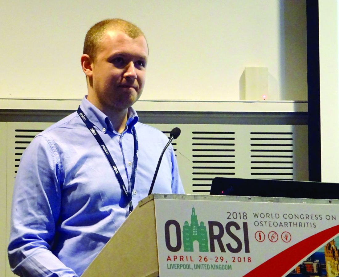
“The OA diagnostic chain has many problems, some modalities are very expensive, and some have a low sensitivity as well,” said study investigator Aleksei Tiulpin, MSc, at the World Congress on Osteoarthritis, referring to magnetic resonance imaging, x-ray imaging, and symptomatic assessment, respectively.

The investigational device that the research team has developed is worn like a brace around the knee and has microphones embedded within it to capture sound coming from the right and left sides of the knee. The device also uses two accelerometers, one placed on the thigh and one on the lower limb to measure movement simultaneously.
“We developed all the software and all the hardware ourselves for this project,” Mr. Tiulpin noted at the congress, sponsored by the Osteoarthritis Research Society International. “The acoustic and kinematic information was measured simultaneously.”
To see whether the prototype device was able to aid the diagnosis of OA, Mr. Tiulpin and his associates recruited 66 women aged 44-67 years old, roughly half of whom (51.5%) had radiographically confirmed knee OA of Kellgren-Lawrence grade 2 or higher.
The participants were asked to perform three exercise tests while wearing the device. First, they had to stand from a sitting position 10 times. Second, they were asked to extend the leg from the knee while sitting down (flexion-extension) 10 times. Third, and finally, they were asked to perform a one leg stand on the right leg twice.
For the acoustic data analysis, data from the standing phase of the sit-to-stand test and the extension phase of the flexion-extension tests were used. Mr. Tuilpin explained that the acoustic signals underwent processing to segment and filter them into candidate locations. The average sound patterns seen in the candidate locations were then analyzed, then a consistency analysis was undertaken. With this approach, inconsistent patterns of knee crepitation could be captured, Mr. Tiulpin explained.
Kinematic signals received from the movement sensors were used to determine the degree of knee instability. Higher signal magnitudes could potentially indicate stability problems, which can be quantified using signal power, Mr. Tiulpin’s slides stated.
A variety of statistical calculations were made to see how well the device might predict OA changes, and a model combining body mass index and age had an area under the curve of 84%, which suggested that it might be possible to improve OA detection with the addition of the device versus BMI and age alone.
“Our results indicated highly promising applications of the method,” Mr. Tiulpin suggested.
These findings are “very interesting,” commented one of the moderators of the session, Erwin van Spil, MD, of University Medical Center Utrecht (The Netherlands) as he opened up the floor to questions.
“I’ve had this question for years … what causes the clicks?” one delegate asked Mr. Tiulpin during discussion. He responded that it could be down to many things, one of which is cartilage components grinding against each other.
Dr. Spil, who was not involved in the study, commented in an interview that using acoustics in the detection of knee OA was still quite a novel concept. “It’s noninvasive, which is quite unique in our general approach, and it might enable an early diagnosis of OA, which is what we are aiming for.”
Although the study did have control subjects, it’s not clear at this point whether very early OA was being assessed, Dr. Spil suggested. “We did not have the opportunity to ask about the OA characteristics. I think they had radiographic OA, but we don’t know the grade and we were not informed about the clinical situation, so we don’t know if anyone had pain, for example.”
Mr. Tiulpin did not have any conflicts of interest to disclose.
SOURCE: Tiulpin A et al. Osteoarthritis Cartilage 2018;26(1):S40–S41.
LIVERPOOL, ENGLAND – The measurement of acoustic emissions and kinetic instability of the knee could become a promising noninvasive way to detect osteoarthritis (OA), according to a recent study.
Researchers at the University of Oulu (Finland) have developed a prototype device that allows multimodal assessment of the sound and motion of the knee.
“The OA diagnostic chain has many problems, some modalities are very expensive, and some have a low sensitivity as well,” said study investigator Aleksei Tiulpin, MSc, at the World Congress on Osteoarthritis, referring to magnetic resonance imaging, x-ray imaging, and symptomatic assessment, respectively.
The investigational device that the research team has developed is worn like a brace around the knee and has microphones embedded within it to capture sound coming from the right and left sides of the knee. The device also uses two accelerometers, one placed on the thigh and one on the lower limb to measure movement simultaneously.
“We developed all the software and all the hardware ourselves for this project,” Mr. Tiulpin noted at the congress, sponsored by the Osteoarthritis Research Society International. “The acoustic and kinematic information was measured simultaneously.”
To see whether the prototype device was able to aid the diagnosis of OA, Mr. Tiulpin and his associates recruited 66 women aged 44-67 years old, roughly half of whom (51.5%) had radiographically confirmed knee OA of Kellgren-Lawrence grade 2 or higher.
The participants were asked to perform three exercise tests while wearing the device. First, they had to stand from a sitting position 10 times. Second, they were asked to extend the leg from the knee while sitting down (flexion-extension) 10 times. Third, and finally, they were asked to perform a one leg stand on the right leg twice.
For the acoustic data analysis, data from the standing phase of the sit-to-stand test and the extension phase of the flexion-extension tests were used. Mr. Tuilpin explained that the acoustic signals underwent processing to segment and filter them into candidate locations. The average sound patterns seen in the candidate locations were then analyzed, then a consistency analysis was undertaken. With this approach, inconsistent patterns of knee crepitation could be captured, Mr. Tiulpin explained.
Kinematic signals received from the movement sensors were used to determine the degree of knee instability. Higher signal magnitudes could potentially indicate stability problems, which can be quantified using signal power, Mr. Tiulpin’s slides stated.
A variety of statistical calculations were made to see how well the device might predict OA changes, and a model combining body mass index and age had an area under the curve of 84%, which suggested that it might be possible to improve OA detection with the addition of the device versus BMI and age alone.
“Our results indicated highly promising applications of the method,” Mr. Tiulpin suggested.
These findings are “very interesting,” commented one of the moderators of the session, Erwin van Spil, MD, of University Medical Center Utrecht (The Netherlands) as he opened up the floor to questions.
“I’ve had this question for years … what causes the clicks?” one delegate asked Mr. Tiulpin during discussion. He responded that it could be down to many things, one of which is cartilage components grinding against each other.
Dr. Spil, who was not involved in the study, commented in an interview that using acoustics in the detection of knee OA was still quite a novel concept. “It’s noninvasive, which is quite unique in our general approach, and it might enable an early diagnosis of OA, which is what we are aiming for.”
Although the study did have control subjects, it’s not clear at this point whether very early OA was being assessed, Dr. Spil suggested. “We did not have the opportunity to ask about the OA characteristics. I think they had radiographic OA, but we don’t know the grade and we were not informed about the clinical situation, so we don’t know if anyone had pain, for example.”
Mr. Tiulpin did not have any conflicts of interest to disclose.
SOURCE: Tiulpin A et al. Osteoarthritis Cartilage 2018;26(1):S40–S41.
REPORTING FROM OARSI 2018
Key clinical point: A prototype device showed a promising application as a noninvasive method to detect osteoarthritis.
Major finding: In a model that combined it with BMI and age, the device had an area under the curve of 84%, which suggested that this device might be able to improve OA detection.
Study details: Single-center study of 66 women aged 44-67 years old; 51.5% had radiographically confirmed OA of Kellgren-Lawrence grade 2 or higher.
Disclosures: Mr. Tiulpin did not have any conflicts of interest to disclose.
Source: Tiulpin A et al. Osteoarthritis Cartilage. 2018;26(1):S40–S41.
Novel x-ray score distinguishes psoriatic arthritis from osteoarthritis of the hand
LIVERPOOL, ENGLAND – A novel radiologic scoring system differentiated psoriatic arthritis (PsA) from nodal osteoarthritis (OA) of the hand in a pilot study.
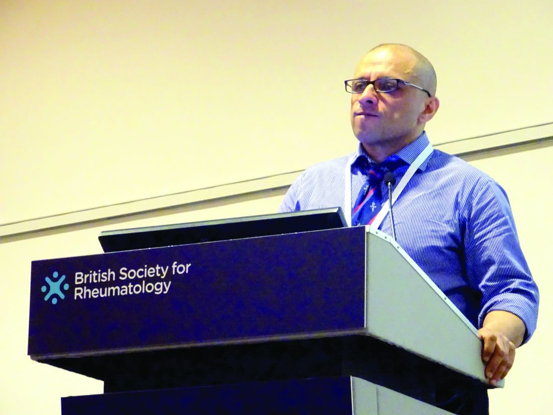
“It’s a dilemma that’s faced, perhaps every couple of weeks, by most [rheumatologists]: Is it osteoarthritis or is it early psoriatic arthritis?” said Sardar Bahadur, MD, at the British Society for Rheumatology annual conference.
Both conditions are seen in daily practice, although the prevalence of hand OA is less frequent than knee OA. Approximately one in five of all adults in the United Kingdom have OA and 1%-2% have psoriasis. Of these, the prevalence of hand OA is about 11% and 0.1%-0.3% have psoriatic arthritis.
Being able to differentiate between the two conditions has important consequences for treatment, Dr. Bahadur said.
“Getting the diagnosis wrong could have major implications,” he said. “If you miss psoriatic arthritis, then potentially you are going to find irreversible joint damage causing pain and disability, and the opposite is also true, with misdiagnosis of osteoarthritis, with overuse of immunosuppression and all the cost implications as well as medicolegal consequences.”
Dr. Bahadur of the department of rehabilitation medicine and rheumatology at the Defence Medical Rehabilitation Centre Headley Court, in Epsom, England, added: “So early diagnosis is very important, it means early treatment, it means better care, potentially preventing serious and irreversible damage.”
Together with researchers at Guy’s and St Thomas’ NHS Trust, London, Dr. Bahadur hypothesized that changes in hand x-rays were distinct and could be reliably used to differentiate between the two conditions. They developed a scoring system for hand radiographs that looked at the differences in the interphalangeal joints, soft tissue, and bone features of patients with known OA or PsA.
Dr. Bahadur noted that the aim was to focus on plain film radiographs of the hands because these were inexpensive, universally accessible, did not rely on radiologists’ interpretation, and changes in the hands were known to occur in both OA and PsA.
A total of 99 sets of hand x-rays taken between 2008 and 2016 from 50 patients with OA and 49 patients with PsA were obtained. These were anonymized and then analyzed by a musculoskeletal radiologist using the scoring system the team had developed. The radiologist was unaware of the patients’ clinical status. The results were then compared to the clinical diagnosis.
The novel method of scoring each x-ray was then taught to two rheumatology and one radiology trainee during a 1-hour training session and were then asked to score the same radiographs.
Dr. Bahadur reported that the radiologist reported normal hand radiographs in five patients and, of the remaining 94 sets of left- and right-hand radiographs, the scoring system correctly allocated 100% of images to either PsA, OA, or rheumatoid arthritis (RA).
Of note was that the radiologist correctly identified two patients with nodal hand OA who later developed PsA several years later, and one patient with RA who was initially thought to have PsA.
“The system could be successfully used by nonradiologists,” Dr. Bahadur proposed. There was good agreement between the scoring system results and the clinical diagnosis then used by the trainees, with 88% and 67% of the radiographs correctly matched to the clinical diagnosis by the rheumatology trainees, and 70% for the radiology trainee.
Dr. Bahadur noted that the features that were consistently identified as being different between hand OA and PsA patients were soft tissue changes, such as dactylitis, as well as erosions, new bone formation, and other features such as subchondral surface changes and cysts.
The results of this single-center study show that the novel radiologic scoring system of hand radiographs was effective at differentiating patients with PsA from nodal OA.
“The ambition is to make this usable by nonradiologists,” Dr. Bahadur said. A multicenter trial would be the next step to look at the use of the scoring system.
Dr. Bahadur had no conflicts of interest to disclose.
SOURCE: Bahadur S et al. Rheumatology. 2018;57[Suppl. 3]:key075.184
LIVERPOOL, ENGLAND – A novel radiologic scoring system differentiated psoriatic arthritis (PsA) from nodal osteoarthritis (OA) of the hand in a pilot study.
“It’s a dilemma that’s faced, perhaps every couple of weeks, by most [rheumatologists]: Is it osteoarthritis or is it early psoriatic arthritis?” said Sardar Bahadur, MD, at the British Society for Rheumatology annual conference.
Both conditions are seen in daily practice, although the prevalence of hand OA is less frequent than knee OA. Approximately one in five of all adults in the United Kingdom have OA and 1%-2% have psoriasis. Of these, the prevalence of hand OA is about 11% and 0.1%-0.3% have psoriatic arthritis.
Being able to differentiate between the two conditions has important consequences for treatment, Dr. Bahadur said.
“Getting the diagnosis wrong could have major implications,” he said. “If you miss psoriatic arthritis, then potentially you are going to find irreversible joint damage causing pain and disability, and the opposite is also true, with misdiagnosis of osteoarthritis, with overuse of immunosuppression and all the cost implications as well as medicolegal consequences.”
Dr. Bahadur of the department of rehabilitation medicine and rheumatology at the Defence Medical Rehabilitation Centre Headley Court, in Epsom, England, added: “So early diagnosis is very important, it means early treatment, it means better care, potentially preventing serious and irreversible damage.”
Together with researchers at Guy’s and St Thomas’ NHS Trust, London, Dr. Bahadur hypothesized that changes in hand x-rays were distinct and could be reliably used to differentiate between the two conditions. They developed a scoring system for hand radiographs that looked at the differences in the interphalangeal joints, soft tissue, and bone features of patients with known OA or PsA.
Dr. Bahadur noted that the aim was to focus on plain film radiographs of the hands because these were inexpensive, universally accessible, did not rely on radiologists’ interpretation, and changes in the hands were known to occur in both OA and PsA.
A total of 99 sets of hand x-rays taken between 2008 and 2016 from 50 patients with OA and 49 patients with PsA were obtained. These were anonymized and then analyzed by a musculoskeletal radiologist using the scoring system the team had developed. The radiologist was unaware of the patients’ clinical status. The results were then compared to the clinical diagnosis.
The novel method of scoring each x-ray was then taught to two rheumatology and one radiology trainee during a 1-hour training session and were then asked to score the same radiographs.
Dr. Bahadur reported that the radiologist reported normal hand radiographs in five patients and, of the remaining 94 sets of left- and right-hand radiographs, the scoring system correctly allocated 100% of images to either PsA, OA, or rheumatoid arthritis (RA).
Of note was that the radiologist correctly identified two patients with nodal hand OA who later developed PsA several years later, and one patient with RA who was initially thought to have PsA.
“The system could be successfully used by nonradiologists,” Dr. Bahadur proposed. There was good agreement between the scoring system results and the clinical diagnosis then used by the trainees, with 88% and 67% of the radiographs correctly matched to the clinical diagnosis by the rheumatology trainees, and 70% for the radiology trainee.
Dr. Bahadur noted that the features that were consistently identified as being different between hand OA and PsA patients were soft tissue changes, such as dactylitis, as well as erosions, new bone formation, and other features such as subchondral surface changes and cysts.
The results of this single-center study show that the novel radiologic scoring system of hand radiographs was effective at differentiating patients with PsA from nodal OA.
“The ambition is to make this usable by nonradiologists,” Dr. Bahadur said. A multicenter trial would be the next step to look at the use of the scoring system.
Dr. Bahadur had no conflicts of interest to disclose.
SOURCE: Bahadur S et al. Rheumatology. 2018;57[Suppl. 3]:key075.184
REPORTING FROM RHEUMATOLOGY 2018
Key clinical point:
Major finding: Using the scoring system, 100% of images were correctly allocated to PsA, OA, or RA.
Study details: Single center pilot study assessing 99 x-rays of both hands taken between 2008 and 2016 of patients with OA (n = 50) or PsA (n = 49).
Disclosures: Dr. Bahadur had no conflicts of interest to disclose.
Source: Bahadur S et al., Rheumatology. 2018;57[Suppl. 3]:key075.184
Cannabidiol gel for osteoarthritis knee pain gives lukewarm results
LIVERPOOL, ENGLAND – There was no significant reduction in pain from knee osteoarthritis (OA) with the use of investigational cannabidiol (CBD) gel ZYN002 in a phase 2a trial presented at the World Congress on Osteoarthritis.
The mean reductions in baseline knee pain scores from study entry to a 12-week assessment were –2.4 for placebo and –2.6 (P = .5) and –2.8 (P = .25), respectively, for a 250-mg and a 500-mg formulation of the gel.

While there was a trend for benefit, it was “neither statistically or clinically significant,” reported David Hunter, MBBS, PhD.
However, he observed that a significantly (P = .016) greater number of patients who received the 250-mg dose (52.7%) were “composite responders,” compared with patients who received placebo (34.1%). A composite response was defined as at least a 30% reduction in pain, and a 20% decrease in WOMAC physical function subscale score at the last observation.
Although the percentage of composite responders was also higher than placebo with the 500-mg dose, the difference wasn’t significant (45.1% vs. 34.1%; P = .0169).
Post-hoc analyses also suggested that perhaps some patients may benefit more than others, reported Dr. Hunter, professor of medicine at the University of Sydney and the Royal North Shore Hospital, Sydney.
For example, patients with baseline pain scores or 7 or more had greater mean reduction in pain at 12 weeks with both doses of the gel combined than placebo at week 4 (–2.2 vs. –1.6; P = .029), although the difference was not significant at week 8 (–3.0 vs. –2.2; P = .05) or 12 (–3.3 vs. –2.5; P = .086).
Women also exhibited a greater placebo response than did men, and “patients with less variability in baseline pain scores may have had greater separation between placebo and the treatment,” Dr. Hunter said. Indeed, 50%-52% of patients with less than 33% variation in baseline scores had a composite response to the gel, versus 27% for the placebo arm.
Evidence from preclinical models suggest that cannabinoids have antinociceptive and antihyperalgesic effects, Dr. Hunter explained at the congress, sponsored by the Osteoarthritis Research Society International. CBD has also been shown to have broad anti-inflammatory effects, and it may even promote osteoclast cell function and decrease bone resorption.
ZYN002 is a synthetic CBD formulated for transdermal delivery using a patented method to enhance its permeation through the skin. According to the manufacturer, Zynerba, it was developed for neuropsychiatric disorders, including fragile X syndrome, adult refractory epilepsy, and developmental and epileptic encephalopathies.
The primary aim of the phase 2 trial reported by Dr. Hunter was to assess ZYN002’s efficacy in managing osteoarthritis knee pain. Secondary objectives were to assess the gel’s safety and tolerability.
The STOP 1 (Synthetic Transdermal Cannabidiol for the Treatment of Knee Pain Due to Osteoarthritis) trial was a double-blind, placebo-controlled trial. For inclusion in the study, patients had to be between age 40 and 75 years and have had knee pain for at least 12 months because of primary OA, based on clinical and x-ray data as per American College of Rheumatology criteria. Anyone with a history of fibromyalgia or epilepsy was excluded.
A total of 320 patients with painful knee OA, with a mean age of 62 years, were randomized and underwent a 1-week washout period in which all their analgesic medications being used for osteoarthritis knee pain, except acetaminophen, were stopped. That was followed by a 7- to 10-day period when baseline daily worst pain levels were captured using a 0-10 numeric rating scale. Patients then underwent 12 weeks of treatment with either a high (500 mg) or a low (250 mg) dose of the gel, or placebo, given in twice-daily doses.
Just over a third (34%) of patients in the placebo arm discontinued the study, compared with 22% and 24% of those in the high- and low-dose gel arms. The main reason for discontinuation was withdrawn consent because of lack of efficacy in the placebo arm, with 8%, 8%, and 4% of patients, respectively, discontinuing because of adverse effects.
“Treatment-emergent adverse effects were roughly equally distributed across the three groups,” Dr. Hunter reported. The adverse events of more interest, he noted, were application site dryness, reaction, or pain. There was “a slight predisposition” to each of these in the 250-mg gel arm (5%, 3%, and 3% of patients affected) versus the 500-mg gel (3%, 0%, and 0%) and placebo (1%, 1%, 0%) arms.
SOURCE: Hunter D et al. Osteoarthritis Cartilage 2018:26(1):S26. Abstract 30.
LIVERPOOL, ENGLAND – There was no significant reduction in pain from knee osteoarthritis (OA) with the use of investigational cannabidiol (CBD) gel ZYN002 in a phase 2a trial presented at the World Congress on Osteoarthritis.
The mean reductions in baseline knee pain scores from study entry to a 12-week assessment were –2.4 for placebo and –2.6 (P = .5) and –2.8 (P = .25), respectively, for a 250-mg and a 500-mg formulation of the gel.
While there was a trend for benefit, it was “neither statistically or clinically significant,” reported David Hunter, MBBS, PhD.
However, he observed that a significantly (P = .016) greater number of patients who received the 250-mg dose (52.7%) were “composite responders,” compared with patients who received placebo (34.1%). A composite response was defined as at least a 30% reduction in pain, and a 20% decrease in WOMAC physical function subscale score at the last observation.
Although the percentage of composite responders was also higher than placebo with the 500-mg dose, the difference wasn’t significant (45.1% vs. 34.1%; P = .0169).
Post-hoc analyses also suggested that perhaps some patients may benefit more than others, reported Dr. Hunter, professor of medicine at the University of Sydney and the Royal North Shore Hospital, Sydney.
For example, patients with baseline pain scores or 7 or more had greater mean reduction in pain at 12 weeks with both doses of the gel combined than placebo at week 4 (–2.2 vs. –1.6; P = .029), although the difference was not significant at week 8 (–3.0 vs. –2.2; P = .05) or 12 (–3.3 vs. –2.5; P = .086).
Women also exhibited a greater placebo response than did men, and “patients with less variability in baseline pain scores may have had greater separation between placebo and the treatment,” Dr. Hunter said. Indeed, 50%-52% of patients with less than 33% variation in baseline scores had a composite response to the gel, versus 27% for the placebo arm.
Evidence from preclinical models suggest that cannabinoids have antinociceptive and antihyperalgesic effects, Dr. Hunter explained at the congress, sponsored by the Osteoarthritis Research Society International. CBD has also been shown to have broad anti-inflammatory effects, and it may even promote osteoclast cell function and decrease bone resorption.
ZYN002 is a synthetic CBD formulated for transdermal delivery using a patented method to enhance its permeation through the skin. According to the manufacturer, Zynerba, it was developed for neuropsychiatric disorders, including fragile X syndrome, adult refractory epilepsy, and developmental and epileptic encephalopathies.
The primary aim of the phase 2 trial reported by Dr. Hunter was to assess ZYN002’s efficacy in managing osteoarthritis knee pain. Secondary objectives were to assess the gel’s safety and tolerability.
The STOP 1 (Synthetic Transdermal Cannabidiol for the Treatment of Knee Pain Due to Osteoarthritis) trial was a double-blind, placebo-controlled trial. For inclusion in the study, patients had to be between age 40 and 75 years and have had knee pain for at least 12 months because of primary OA, based on clinical and x-ray data as per American College of Rheumatology criteria. Anyone with a history of fibromyalgia or epilepsy was excluded.
A total of 320 patients with painful knee OA, with a mean age of 62 years, were randomized and underwent a 1-week washout period in which all their analgesic medications being used for osteoarthritis knee pain, except acetaminophen, were stopped. That was followed by a 7- to 10-day period when baseline daily worst pain levels were captured using a 0-10 numeric rating scale. Patients then underwent 12 weeks of treatment with either a high (500 mg) or a low (250 mg) dose of the gel, or placebo, given in twice-daily doses.
Just over a third (34%) of patients in the placebo arm discontinued the study, compared with 22% and 24% of those in the high- and low-dose gel arms. The main reason for discontinuation was withdrawn consent because of lack of efficacy in the placebo arm, with 8%, 8%, and 4% of patients, respectively, discontinuing because of adverse effects.
“Treatment-emergent adverse effects were roughly equally distributed across the three groups,” Dr. Hunter reported. The adverse events of more interest, he noted, were application site dryness, reaction, or pain. There was “a slight predisposition” to each of these in the 250-mg gel arm (5%, 3%, and 3% of patients affected) versus the 500-mg gel (3%, 0%, and 0%) and placebo (1%, 1%, 0%) arms.
SOURCE: Hunter D et al. Osteoarthritis Cartilage 2018:26(1):S26. Abstract 30.
REPORTING FROM OARSI 2018
Key clinical point:
Major finding: Mean knee pain scores at 12 weeks fell by –2.4 for placebo, and –2.6 (P = .5) and –2.8 (P = .25) for a 250-mg and a 500-mg formulation of the gel.
Study details: A 12-week, randomized, double-blind, placebo-controlled, phase 2, multidose study involving 320 patients with osteoarthritis knee pain for at least 12 months.
Disclosures: Dr. Hunter has consulted for Flexion, Merck Serono, TissueGene, and Zynerba, and has received royalties from DJO for a patellofemoral brace.
Source: Hunter D et al. Osteoarthritis Cartilage 2018:26(1):S26. Abstract 30.
Surgery may be best option for hip impingement syndrome
LIVERPOOL, ENGLAND – Hip arthroscopic surgery produced better long-term results than did personalized hip physiotherapy for femoroacetabular impingement syndrome in a randomized trial conduced across multiple U.K. centers.
At 12 months, respective International Hip Outcome Tool-33 (iHOT-33) scores were 58.8 and 49.7, a difference of 9.1 points before and 6.8 points after adjustment for potential confounding factors (P = .0093).

“This trial shows that hip arthroscopic surgery and personalized hip therapy both improved hip-related quality of life for patients with FAI [femoroacetabular impingement] syndrome, but that the surgery did indeed produce a greater improvement at our primary time point of 12 months,” she added. Dr. Foster is professor of musculoskeletal health in primary care at Keele University, Newcastle-under-Lyme, England, one of the 23 centers involved in the FASHIoN study in England, Wales, and Scotland.
“FAI is a very common cause of hip and groin pain in young adults, and it’s associated with the development of hip osteoarthritis,” Dr. Foster noted.
There are three types of FAI – pincer, cam, and combined. The pincer type of FAI is where there is “prominence or overcoverage of the rim of the acetabulum,” and the cam type is where there is a “bony prominence of the femoral head-neck junction,” she explained at the meeting sponsored by the Osteoarthritis Research Society International.
The link to OA comes when the femur and acetabulum prematurely connect, usually during activity, causing damage to the labrum and articular cartilage in the long term. Thus, treating FAI is important, not just for relieving patient’s pain and joint stiffness.
Hip arthroscopic surgery has become an established way of treating FAI syndrome – more than 2,400 operations were performed in 2013 in the United Kingdom alone, Dr. Foster observed. The aim of surgery is to try to reshape the hip joint to prevent impingement, and resect, repair, or reconstruct any intra-articular damage that may be present.
“Physiotherapy aims to improve hip muscle control and strength, and to correct the abnormal movement patterns that we see in these patients,” Dr. Foster said. “Through that, we hope to prevent the premature contact that occurs in FAI syndrome and thereby improve symptoms, allowing patients to return to activities and prevent recurrence.”
Working with physiotherapists, physicians, and surgeons, Dr. Foster and her associates have previously developed a “best conservative care” intervention that they call personalized hip therapy (PHT), which involves the delivery and supervision of an individualized exercise program by experienced physiotherapists over a 3- to 6-month period, and which patients repeat at home (PM R. 2013 May;5[5]:418-26).
The aim of the UK FASHIoN trial was to compare the clinical and cost-effectiveness of hip arthroscopy and PHT for FAI syndrome, as there was no robust clinical trial evidence to demonstrate a benefit of one over the other.
A total of 351 adults with hip and groin pain were randomized to either arthroscopic surgery (n = 173) or PHT (n = 178). The mean age of participants was 35 years, with no significant differences between the two treatment groups in terms of baseline demographics or type or duration of hip impingement.
While surgery was better in terms of patient outcomes, the study didn’t demonstrate its cost-effectiveness within the first 12 months, Dr. Foster observed. Cost-effectiveness, together with various other quality-of-life measurements, was a secondary endpoint of the study.
“Longer-term outcomes are required to establish whether improvement is sustained, and whether surgery is cost-effective at the longer time points for our health service,” she said.
Responding to a question about whether any of the patients in the study had radiographic evidence of osteoarthritis, Dr. Foster said that such patients had been excluded from the study.
“One of the hopes of the trial’s team is that, with the long-term follow-up, we might be able to get data at 5 and 10 years on things like hip osteoarthritis in these patients,” she said.
The study was funded by the National Institute for Health Research. Dr. Foster had no financial relationships or commercial interests to disclose.
SOURCE: Griffin DR et al. Osteoarthritis Cartilage. 2018:26(1):S24-25. Abstract 28
LIVERPOOL, ENGLAND – Hip arthroscopic surgery produced better long-term results than did personalized hip physiotherapy for femoroacetabular impingement syndrome in a randomized trial conduced across multiple U.K. centers.
At 12 months, respective International Hip Outcome Tool-33 (iHOT-33) scores were 58.8 and 49.7, a difference of 9.1 points before and 6.8 points after adjustment for potential confounding factors (P = .0093).
“This trial shows that hip arthroscopic surgery and personalized hip therapy both improved hip-related quality of life for patients with FAI [femoroacetabular impingement] syndrome, but that the surgery did indeed produce a greater improvement at our primary time point of 12 months,” she added. Dr. Foster is professor of musculoskeletal health in primary care at Keele University, Newcastle-under-Lyme, England, one of the 23 centers involved in the FASHIoN study in England, Wales, and Scotland.
“FAI is a very common cause of hip and groin pain in young adults, and it’s associated with the development of hip osteoarthritis,” Dr. Foster noted.
There are three types of FAI – pincer, cam, and combined. The pincer type of FAI is where there is “prominence or overcoverage of the rim of the acetabulum,” and the cam type is where there is a “bony prominence of the femoral head-neck junction,” she explained at the meeting sponsored by the Osteoarthritis Research Society International.
The link to OA comes when the femur and acetabulum prematurely connect, usually during activity, causing damage to the labrum and articular cartilage in the long term. Thus, treating FAI is important, not just for relieving patient’s pain and joint stiffness.
Hip arthroscopic surgery has become an established way of treating FAI syndrome – more than 2,400 operations were performed in 2013 in the United Kingdom alone, Dr. Foster observed. The aim of surgery is to try to reshape the hip joint to prevent impingement, and resect, repair, or reconstruct any intra-articular damage that may be present.
“Physiotherapy aims to improve hip muscle control and strength, and to correct the abnormal movement patterns that we see in these patients,” Dr. Foster said. “Through that, we hope to prevent the premature contact that occurs in FAI syndrome and thereby improve symptoms, allowing patients to return to activities and prevent recurrence.”
Working with physiotherapists, physicians, and surgeons, Dr. Foster and her associates have previously developed a “best conservative care” intervention that they call personalized hip therapy (PHT), which involves the delivery and supervision of an individualized exercise program by experienced physiotherapists over a 3- to 6-month period, and which patients repeat at home (PM R. 2013 May;5[5]:418-26).
The aim of the UK FASHIoN trial was to compare the clinical and cost-effectiveness of hip arthroscopy and PHT for FAI syndrome, as there was no robust clinical trial evidence to demonstrate a benefit of one over the other.
A total of 351 adults with hip and groin pain were randomized to either arthroscopic surgery (n = 173) or PHT (n = 178). The mean age of participants was 35 years, with no significant differences between the two treatment groups in terms of baseline demographics or type or duration of hip impingement.
While surgery was better in terms of patient outcomes, the study didn’t demonstrate its cost-effectiveness within the first 12 months, Dr. Foster observed. Cost-effectiveness, together with various other quality-of-life measurements, was a secondary endpoint of the study.
“Longer-term outcomes are required to establish whether improvement is sustained, and whether surgery is cost-effective at the longer time points for our health service,” she said.
Responding to a question about whether any of the patients in the study had radiographic evidence of osteoarthritis, Dr. Foster said that such patients had been excluded from the study.
“One of the hopes of the trial’s team is that, with the long-term follow-up, we might be able to get data at 5 and 10 years on things like hip osteoarthritis in these patients,” she said.
The study was funded by the National Institute for Health Research. Dr. Foster had no financial relationships or commercial interests to disclose.
SOURCE: Griffin DR et al. Osteoarthritis Cartilage. 2018:26(1):S24-25. Abstract 28
REPORTING FROM OARSI 2018
Key clinical point: Hip arthroscopy produced better results at 12 months than did the best conservative care.
Major finding: iHOT-33 scores at 12 months were 58.3 for surgery and 49.7 for personalized hip therapy (P = .0093)
Study details: Multicenter, randomized controlled UK FASHIoN trial of 351 adults with hip and groin pain.
Disclosures: The study was funded by the National Institute for Health Research. Dr. Foster had nothing to disclose.
Source: Griffin DR et al. Osteoarthritis Cartilage. 2018:26(1):S24-25. Abstract 28.
Sprifermin moves FORWARD with sustained effects in osteoarthritis
LIVERPOOL, ENGLAND – At 3 years of follow-up, the cartilage-building effects of sprifermin appear to be sustained, according to further data to be released from the phase 2 FORWARD trial.
The difference from placebo in mean cartilage thickness at the tibiofemoral joint (TFJ) at 3 years was 0.05 mm for a 100-mcg dose of sprifermin given every 6 months (P less than .0001), 0.02 mm for a 100-mcg dose given every 12 months (P = .193), 0.01 mm for a 50-mcg dose given every 6 months (P = .530), and –0.02 mm for a 50-mcg dose given every 12 months (P = .160).
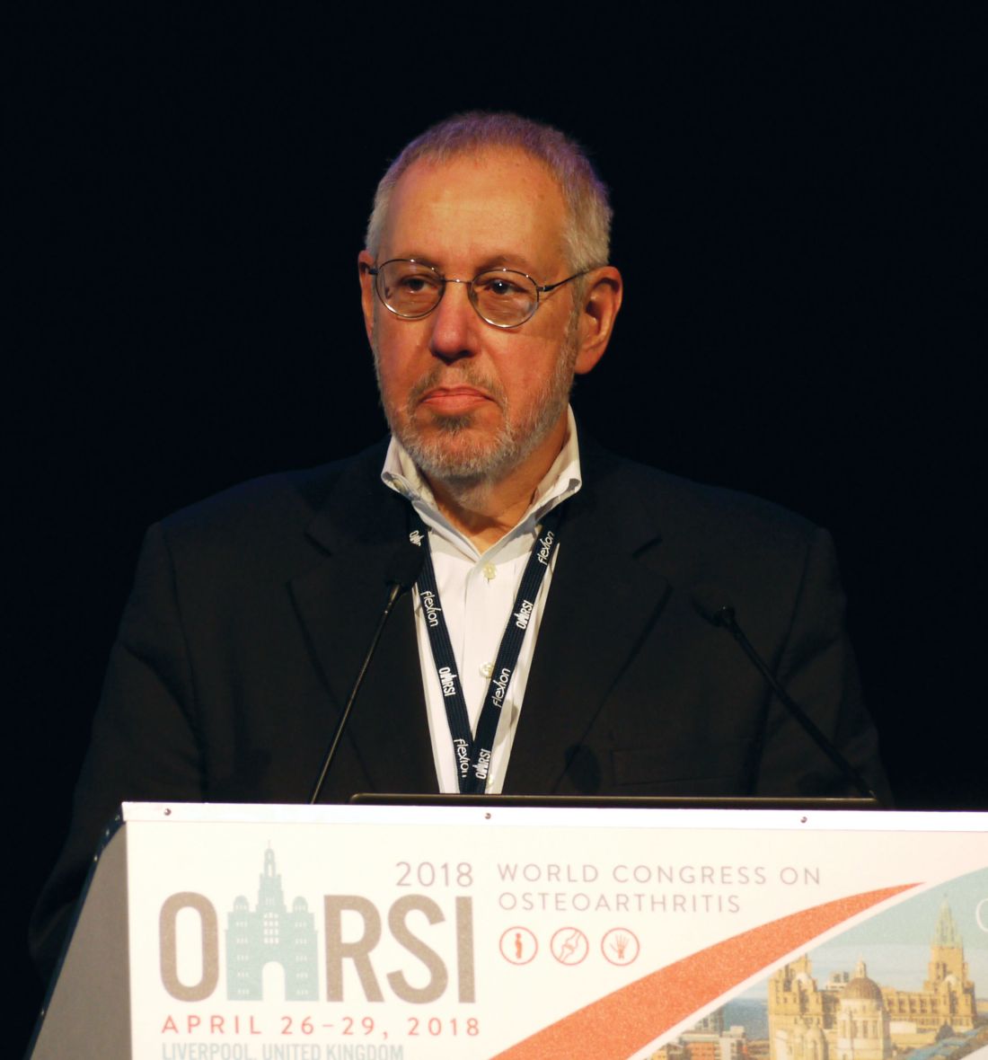
“These data, 18 months after the last active injection, extend the results which we previously presented and assessed at 6 months after the last injection,” study investigator Marc Hochberg, MD, said at the World Congress on Osteoarthritis.
The prior 2-year findings, which were reported at the annual meeting of the American College of Rheumatology in November 2017, showed statistically significant dose-dependent structural modification in TFJ cartilage. Furthermore, the increase in cartilage thickness was seen in both medial and lateral compartments of the TFJ, and in the central medial subregion of the TFJ.
Sprifermin is one of several drugs currently being investigated as a potential disease-modifying osteoarthritis drug, none of which are currently licensed for use. It is a novel human recombinant version of fibroblast growth factor 18 that has been shown to increase chondrocyte proliferation that results in overall extracellular matrix production and subsequent hyaline-line cartilage formation.
The study comprised 549 patients with symptomatic radiographic primary femorotibial knee OA who were aged 40-85 years. For inclusion, patients had to have Kellgren-Lawrence Grade 2 or 3, with a medial minimum joint space width of 2.5 mm or more. In addition, patients had to have a history of OA pain for at least 6 months and either symptoms requiring pain medication or a pain score of 4-9 on the 10-point question 1 of the Western Ontario and McMaster Universities Osteoarthritis Index (WOMAC).
Dr. Hochberg, professor of medicine, epidemiology, and public health and head of the division of rheumatology and clinical immunology at the University of Maryland, Baltimore, noted that the current analysis at 3 years’ follow up had been prespecified and that the plan was to continue follow-up out to 5 years.
There was a statistically significant treatment effect and dose response effect seen in TFJ cartilage thickness.
“Although cartilage thickness declined in all treatment groups between years 2 and 3, the difference in cartilage thickness observed in year 2 with sprifermin at the highest dose [100 mcg every 6 months] versus placebo persisted through year 3,” Dr. Hochberg said at the congress, which was sponsored by the Osteoarthritis Research Society International.
_MARYLAND_web.jpg)
As for secondary endpoints of thickness in the medial and lateral tibiofemoral cartilage, “there are significant differences between the higher-dose sprifermin group and the placebo group,” he said.
“Based on imaging, sprifermin appears to be effective at modifying structural changes in articular cartilage in a dose-dependent manner in patients with knee osteoarthritis, with an acceptable safety profile.”
Dr. Hochberg added that there was “marked symptomatic improvement” as shown by changes in WOMAC scores in all treatment groups including placebo. The improvement in total WOMAC scores by approximately 50% in all treatment groups by the second year was continued to the third year.
Adverse effects occurred with a similar frequency in the active treatment groups and the placebo group. They were also of a similar nature. The most commonly reported side effects involved the musculoskeletal system or were connective tissue disorders (e.g. arthralgia). Importantly, there was no difference in the frequency, severity, or nature of serious adverse events, treatment-related adverse events, or discontinuation due to adverse events with active versus placebo therapy, Dr. Hochberg said.
The percentages of patients completing the study to the second and third years were a respective 87.8% and 81.6% in the sprifermin groups and 80.6% and 75.9% in the placebo group.
Merck KGaA and EMD Serono Research Institute funded the study. Dr. Hochberg has received consulting fees from EMD Serono and multiple other companies developing treatments for OA.
SOURCE: Hochberg M et al. Osteoarthritis Cartilage. 2018:26(1):S26-27. Abstract 32
LIVERPOOL, ENGLAND – At 3 years of follow-up, the cartilage-building effects of sprifermin appear to be sustained, according to further data to be released from the phase 2 FORWARD trial.
The difference from placebo in mean cartilage thickness at the tibiofemoral joint (TFJ) at 3 years was 0.05 mm for a 100-mcg dose of sprifermin given every 6 months (P less than .0001), 0.02 mm for a 100-mcg dose given every 12 months (P = .193), 0.01 mm for a 50-mcg dose given every 6 months (P = .530), and –0.02 mm for a 50-mcg dose given every 12 months (P = .160).
“These data, 18 months after the last active injection, extend the results which we previously presented and assessed at 6 months after the last injection,” study investigator Marc Hochberg, MD, said at the World Congress on Osteoarthritis.
The prior 2-year findings, which were reported at the annual meeting of the American College of Rheumatology in November 2017, showed statistically significant dose-dependent structural modification in TFJ cartilage. Furthermore, the increase in cartilage thickness was seen in both medial and lateral compartments of the TFJ, and in the central medial subregion of the TFJ.
Sprifermin is one of several drugs currently being investigated as a potential disease-modifying osteoarthritis drug, none of which are currently licensed for use. It is a novel human recombinant version of fibroblast growth factor 18 that has been shown to increase chondrocyte proliferation that results in overall extracellular matrix production and subsequent hyaline-line cartilage formation.
The study comprised 549 patients with symptomatic radiographic primary femorotibial knee OA who were aged 40-85 years. For inclusion, patients had to have Kellgren-Lawrence Grade 2 or 3, with a medial minimum joint space width of 2.5 mm or more. In addition, patients had to have a history of OA pain for at least 6 months and either symptoms requiring pain medication or a pain score of 4-9 on the 10-point question 1 of the Western Ontario and McMaster Universities Osteoarthritis Index (WOMAC).
Dr. Hochberg, professor of medicine, epidemiology, and public health and head of the division of rheumatology and clinical immunology at the University of Maryland, Baltimore, noted that the current analysis at 3 years’ follow up had been prespecified and that the plan was to continue follow-up out to 5 years.
There was a statistically significant treatment effect and dose response effect seen in TFJ cartilage thickness.
“Although cartilage thickness declined in all treatment groups between years 2 and 3, the difference in cartilage thickness observed in year 2 with sprifermin at the highest dose [100 mcg every 6 months] versus placebo persisted through year 3,” Dr. Hochberg said at the congress, which was sponsored by the Osteoarthritis Research Society International.
As for secondary endpoints of thickness in the medial and lateral tibiofemoral cartilage, “there are significant differences between the higher-dose sprifermin group and the placebo group,” he said.
“Based on imaging, sprifermin appears to be effective at modifying structural changes in articular cartilage in a dose-dependent manner in patients with knee osteoarthritis, with an acceptable safety profile.”
Dr. Hochberg added that there was “marked symptomatic improvement” as shown by changes in WOMAC scores in all treatment groups including placebo. The improvement in total WOMAC scores by approximately 50% in all treatment groups by the second year was continued to the third year.
Adverse effects occurred with a similar frequency in the active treatment groups and the placebo group. They were also of a similar nature. The most commonly reported side effects involved the musculoskeletal system or were connective tissue disorders (e.g. arthralgia). Importantly, there was no difference in the frequency, severity, or nature of serious adverse events, treatment-related adverse events, or discontinuation due to adverse events with active versus placebo therapy, Dr. Hochberg said.
The percentages of patients completing the study to the second and third years were a respective 87.8% and 81.6% in the sprifermin groups and 80.6% and 75.9% in the placebo group.
Merck KGaA and EMD Serono Research Institute funded the study. Dr. Hochberg has received consulting fees from EMD Serono and multiple other companies developing treatments for OA.
SOURCE: Hochberg M et al. Osteoarthritis Cartilage. 2018:26(1):S26-27. Abstract 32
REPORTING FROM OARSI 2018
Key clinical point:
Major finding: The difference from placebo in mean cartilage thickness at the tibiofemoral joint at 3 years was 0.05 mm for the 100-mcg dose of sprifermin given every 6 months (P less than .0001).
Study details: Phase 2 study of 549 patients with knee osteoarthritis treated with one of four intra-articular doses of sprifermin or placebo.
Disclosures: Merck KGaA and EMD Serono Research Institute funded the study. Dr. Hochberg has received consulting fees from EMD Serono and multiple other companies developing treatments for OA.
Source: Hochberg M et al. Osteoarthritis Cartilage. 2018:26(1):S26-27. Abstract 32.
‘Bright future’ for growth factor therapy in osteoarthritis
LIVERPOOL, ENGLAND – (OA), according to David Hunter, MBBS, PhD.
Dr. Hunter, who is the Florance and Cope Chair of Rheumatology and professor of medicine at the University of Sydney and the Royal North Shore Hospital, Sydney, Australia, outlined some of the recent research with these agents in OA.

In 2013, a U.S. study (Arthritis Care Res. 2013;65[5]:703-11) showed that the 2007-2008 incidence of symptomatic knee OA was highest between the ages of 54 and 64 years, with the estimated median age at diagnosis of 55.8 years. Patients were substantially younger than were those who had been diagnosed at a median age of 68.5 years more than a decade earlier in 1991-1992.
“So, we’re developing [OA] at a much earlier age, but we’re obviously living longer and living with that morbidity for a substantial period of time,” Dr. Hunter said at the World Congress on Osteoarthritis, sponsored by the Osteoarthritis Research Society International.
“It’s within that context that we really need to think about both the individual and societal burden of osteoarthritis, and why we’re developing agents such as growth factors to help retard symptoms related to disease,” continued Dr. Hunter, chair of the Institute of Bone and Joint Research, deputy dean of the Northern Clinical School, and consultant rheumatologist at North Sydney Orthopaedic and Sports Medicine Centre, Sydney.
OA is characterized by articular cartilage damage, low-grade synovial inflammation, and hypertrophic bone changes that lead to pain and functional deterioration (Expert Opin Emerg Drugs. 2015;20[3]:361-78). Attempts to modify the disease process have thus focused on these three areas, aiming to regulate cartilage catabolism and anabolism, control inflammation, and remodel subchondral bone (Rheumatology [Oxford]. 2018;57[suppl. 4]:iv108-iv123).
Growth factors fall under the category of regulating cartilage catabolism and anabolism, playing a “significant role in musculoskeletal tissues’ homeostasis and repair,” Dr. Hunter explained. “They influence chemotaxis, differentiation, proliferation, and synthetic activity of cartilage and bone cells, and can regulate physiological remodeling and cartilage healing.”
Examples of growth factors of interest in OA include transforming growth factor-beta superfamily and bone morphogenic proteins 2 and 7, fibroblast growth factors 2 and 18, and insulin-like growth factor-1.
Two currently unproven and somewhat controversial ways of regulating cartilage catabolism and anabolism is the use of mesenchymal stem cells (MSCs) and platelet-rich plasma (PRP). Neither are ready for widespread use, Dr. Hunter observed, and more data on these approaches still need to be gathered.
The rationale behind the use of MSCs is that all joint tissues contain these cells, and they can potentially differentiate into cartilage, bone, or other tissues. There are changes in the quantity, phenotype, and differentiation potential of these cells in patients with OA, however, leading researchers to see if intra-articular injection of MSCs could be a possible therapeutic strategy. MSCs can be isolated from a number of different tissues and differentiate into multiple cells types. Animal model data suggest that their transplantation into an affected joint can stimulate articular repair.
“From a clinical perspective, there have been a vast array of clinical trials looking at different types of MSCs and different types of administration,” Dr. Hunter noted. Pooled data suggest that stem cell therapy does help to improve pain and function. However, the quality of trials has been called into question, with a positive publication bias suggested. There’s no clear evidence of any structural benefits, Dr. Hunter stated. “There’s a lot of stem cells being used for clinical purposes without the evidence to support their efficacy,” he cautioned. “We need much better regulation.”
There are several PRP agents, which deliver growth factors, cytokines, adhesive proteins, and other plasma proteins, such as clotting factors. It’s unknown how PRP therapy works in OA, whether it is just pain relieving or could affect the progression of joint damage. So far, studies have been done only in knee OA, so data in other joints need to be obtained.
“Whilst they have great potential, the literature on the use of PRP is conflicting,” Dr. Hunter said. “There are a lot of positive trials out there, but their quality in general is quite low.”
Better data are perhaps available for the recombinant human fibroblast growth factor–18 sprifermin, with recent 3-year data from an ongoing 5-year randomized, placebo-controlled, phase 2 study showing increased total cartilage thickness in the tibiofemoral joint relative to placebo. “There does appear to be a structure benefit,” Dr. Hunter said of sprifermin’s effects in the study versus intra-articular saline.
Although improvement in total Western Ontario and McMaster Universities Osteoarthritis Index scores were seen in the patients with knee OA, there was no real differentiation by dose or by placebo. The rationale for having a longer trial follow-up – the last dose of sprifermin was given at 18 months – is to see if there is a later effect.
In the later stages of development is Invossa TissueGene-C; this is drug that consists of patients’ normal and genetically-modified chondrocyte cells that express transforming growth factor-beta 1. Phase 2 findings have shown that it can improve pain and function scores in patients with moderate to advanced knee OA (BMC Musculoskelet Disord. 2017;18[1]:461). Based on those findings, two phase 3 trials are currently underway, with some further findings presented elsewhere at the OARSI meeting.
Even though there is still no disease-modifying osteoarthritis drug currently licensed for use, “I’m really positive about all of what we’ve learned, particularly with the application to future trials and ongoing trials in this space,” Dr. Hunter observed.
“Future strategies should differentiate between those agents aiming to reduce pain, and those targeting structural evolution, incorporating a combination of anabolic and anti-catabolic therapies,” Dr. Hunter suggested. One of the challenges, however, is to address the placebo effect, which can be exaggerated by the context of administration or surgery.
Furthermore, he proposed that new treatments should “fit into the natural progression of OA” by focusing on early disease where changes might still be reversible. “We’re getting more nuanced about where to intervene,” Dr. Hunter said.
Emerging treatments should also take the location of the disease and its heterogenic nature into account.
Dr. Hunter has consulted for Zynerba, TLC, Kolon TissueGene, and Merck Serono, and he disclosed receiving royalties from DJO for a patellofemoral brace patent.
SOURCE: Lohmander S et al. Osteoarthritis Cartilage. 2018:26(1):S6. Abstract I-18
LIVERPOOL, ENGLAND – (OA), according to David Hunter, MBBS, PhD.
Dr. Hunter, who is the Florance and Cope Chair of Rheumatology and professor of medicine at the University of Sydney and the Royal North Shore Hospital, Sydney, Australia, outlined some of the recent research with these agents in OA.
In 2013, a U.S. study (Arthritis Care Res. 2013;65[5]:703-11) showed that the 2007-2008 incidence of symptomatic knee OA was highest between the ages of 54 and 64 years, with the estimated median age at diagnosis of 55.8 years. Patients were substantially younger than were those who had been diagnosed at a median age of 68.5 years more than a decade earlier in 1991-1992.
“So, we’re developing [OA] at a much earlier age, but we’re obviously living longer and living with that morbidity for a substantial period of time,” Dr. Hunter said at the World Congress on Osteoarthritis, sponsored by the Osteoarthritis Research Society International.
“It’s within that context that we really need to think about both the individual and societal burden of osteoarthritis, and why we’re developing agents such as growth factors to help retard symptoms related to disease,” continued Dr. Hunter, chair of the Institute of Bone and Joint Research, deputy dean of the Northern Clinical School, and consultant rheumatologist at North Sydney Orthopaedic and Sports Medicine Centre, Sydney.
OA is characterized by articular cartilage damage, low-grade synovial inflammation, and hypertrophic bone changes that lead to pain and functional deterioration (Expert Opin Emerg Drugs. 2015;20[3]:361-78). Attempts to modify the disease process have thus focused on these three areas, aiming to regulate cartilage catabolism and anabolism, control inflammation, and remodel subchondral bone (Rheumatology [Oxford]. 2018;57[suppl. 4]:iv108-iv123).
Growth factors fall under the category of regulating cartilage catabolism and anabolism, playing a “significant role in musculoskeletal tissues’ homeostasis and repair,” Dr. Hunter explained. “They influence chemotaxis, differentiation, proliferation, and synthetic activity of cartilage and bone cells, and can regulate physiological remodeling and cartilage healing.”
Examples of growth factors of interest in OA include transforming growth factor-beta superfamily and bone morphogenic proteins 2 and 7, fibroblast growth factors 2 and 18, and insulin-like growth factor-1.
Two currently unproven and somewhat controversial ways of regulating cartilage catabolism and anabolism is the use of mesenchymal stem cells (MSCs) and platelet-rich plasma (PRP). Neither are ready for widespread use, Dr. Hunter observed, and more data on these approaches still need to be gathered.
The rationale behind the use of MSCs is that all joint tissues contain these cells, and they can potentially differentiate into cartilage, bone, or other tissues. There are changes in the quantity, phenotype, and differentiation potential of these cells in patients with OA, however, leading researchers to see if intra-articular injection of MSCs could be a possible therapeutic strategy. MSCs can be isolated from a number of different tissues and differentiate into multiple cells types. Animal model data suggest that their transplantation into an affected joint can stimulate articular repair.
“From a clinical perspective, there have been a vast array of clinical trials looking at different types of MSCs and different types of administration,” Dr. Hunter noted. Pooled data suggest that stem cell therapy does help to improve pain and function. However, the quality of trials has been called into question, with a positive publication bias suggested. There’s no clear evidence of any structural benefits, Dr. Hunter stated. “There’s a lot of stem cells being used for clinical purposes without the evidence to support their efficacy,” he cautioned. “We need much better regulation.”
There are several PRP agents, which deliver growth factors, cytokines, adhesive proteins, and other plasma proteins, such as clotting factors. It’s unknown how PRP therapy works in OA, whether it is just pain relieving or could affect the progression of joint damage. So far, studies have been done only in knee OA, so data in other joints need to be obtained.
“Whilst they have great potential, the literature on the use of PRP is conflicting,” Dr. Hunter said. “There are a lot of positive trials out there, but their quality in general is quite low.”
Better data are perhaps available for the recombinant human fibroblast growth factor–18 sprifermin, with recent 3-year data from an ongoing 5-year randomized, placebo-controlled, phase 2 study showing increased total cartilage thickness in the tibiofemoral joint relative to placebo. “There does appear to be a structure benefit,” Dr. Hunter said of sprifermin’s effects in the study versus intra-articular saline.
Although improvement in total Western Ontario and McMaster Universities Osteoarthritis Index scores were seen in the patients with knee OA, there was no real differentiation by dose or by placebo. The rationale for having a longer trial follow-up – the last dose of sprifermin was given at 18 months – is to see if there is a later effect.
In the later stages of development is Invossa TissueGene-C; this is drug that consists of patients’ normal and genetically-modified chondrocyte cells that express transforming growth factor-beta 1. Phase 2 findings have shown that it can improve pain and function scores in patients with moderate to advanced knee OA (BMC Musculoskelet Disord. 2017;18[1]:461). Based on those findings, two phase 3 trials are currently underway, with some further findings presented elsewhere at the OARSI meeting.
Even though there is still no disease-modifying osteoarthritis drug currently licensed for use, “I’m really positive about all of what we’ve learned, particularly with the application to future trials and ongoing trials in this space,” Dr. Hunter observed.
“Future strategies should differentiate between those agents aiming to reduce pain, and those targeting structural evolution, incorporating a combination of anabolic and anti-catabolic therapies,” Dr. Hunter suggested. One of the challenges, however, is to address the placebo effect, which can be exaggerated by the context of administration or surgery.
Furthermore, he proposed that new treatments should “fit into the natural progression of OA” by focusing on early disease where changes might still be reversible. “We’re getting more nuanced about where to intervene,” Dr. Hunter said.
Emerging treatments should also take the location of the disease and its heterogenic nature into account.
Dr. Hunter has consulted for Zynerba, TLC, Kolon TissueGene, and Merck Serono, and he disclosed receiving royalties from DJO for a patellofemoral brace patent.
SOURCE: Lohmander S et al. Osteoarthritis Cartilage. 2018:26(1):S6. Abstract I-18
EXPERT ANALYSIS FROM OARSI 2018
Methotrexate-induced pulmonary fibrosis risk examined in 10-year study
LIVERPOOL, ENGLAND – A 10-year follow up of patients with inflammatory arthritis has shown that methotrexate does not appear to increase the risk of pulmonary fibrosis.
“As rheumatologists, it’s a really important message that methotrexate does not cause chronic pulmonary fibrosis and it should not be stopped because of pulmonary fibrosis,” Julie Dawson, MD, said in an interview at the British Society for Rheumatology annual conference. “It’s the rheumatoid arthritis. It’s not the methotrexate.”
Dr. Dawson, of St. Helens and Knowsley Teaching Hospitals NHS Trust, St. Helens, England, added that the current findings were consistent with her team’s prior research looking at earlier time periods. There was also no correlation between the duration or dose of methotrexate used and the development of the lung disease, she said.
“If anything, the suggestion is you’d be more symptomatic if you delay using methotrexate,” Dr. Dawson observed. If patients are not doing well on methotrexate, then perhaps adjusting therapy or changing to another drug would of course be the next step, but if patients are well controlled then “stopping it is the worst thing to do” for their arthritis, she said.
“This is of great clinical interest, and we can be reassured now about this, I think. This is really good, long-term data,” said Devesh Mewar, MD, of Royal Liverpool and Broadgreen University Hospitals NHS Trust, Liverpool, England, who was not involved in the research.
“We know that methotrexate is associated with a pneumonitis reaction, but there is no high-quality evidence that methotrexate is associated with a chronic pulmonary fibrosis” Dr. Dawson said, explaining the rationale for the current study she presented during a poster session. Previous studies considered data for up to 5 years, she added, so the aim of the current study, therefore, was to look at the longer-term effect of methotrexate use on the incidence of pulmonary fibrosis.
Data on 129 patients who had started treatment with methotrexate from 2004 to 2007 were analyzed, of whom 63 (49%) had stayed on methotrexate for 10 or more years. Most (82%) had been given methotrexate to treat rheumatoid arthritis (RA), with other indications including inflammatory arthritis (5.4%) and psoriatic arthritis (4.7%).
“Practice was different 10 years ago, so just 56% of patients commenced methotrexate within the first year of the diagnosis of rheumatoid arthritis,” Dr. Dawson reported.

Only four cases of symptomatic pulmonary fibrosis were seen, all in the RA patients, and three of these were in patients who had started methotrexate over 1 year after their diagnosis. The incidence of 3.8% seen in the study matches the expected incidence of pulmonary fibrosis in RA and was actually “at the lower end of the expected incidence,” Dr. Dawson said. Previous studies have suggested an incidence rate of RA-associated interstitial lung disease of about 3%-7%.
All of the pulmonary fibrosis cases had occurred in men and 75% were seropositive for rheumatoid factor. The mean duration of RA at the time of onset of pulmonary fibrosis was 7.8 years and the usual interstitial pattern of fibrosis was seen. The 125 patients without pulmonary fibrosis had taken methotrexate for a mean of 8 years at a mean final weekly dose of 16.3 mg, compared with a mean of 6 years at a mean dose of 18.1 mg per week in the 4 patients with pulmonary fibrosis.
One of the next steps is to look at cases where methotrexate has been stopped and the effects of that on pulmonary fibrosis and disease activity. In Dr. Dawson’s experience, stopping methotrexate just affects the management of the arthritis and had no difference to the progression of pulmonary fibrosis.
If patients start to experience any lung symptoms while continuing methotrexate, such as shortness of breath, then they would need to be assessed and undergo lung function tests to monitor their condition. Treating the fibrosis using an antifibrotic drug, such as pirfenidone, is something that might be possible in the future, but this needs investigation in inflammatory arthritis as the drug is currently only licensed for use in idiopathic cases.
This is something the British Rheumatoid Interstitial Lung network plans to investigate in a placebo-controlled study of RA patients with fibrotic lung disease. “We’re looking to see if antifibrotic agents are going to slow the disease as it does in idiopathic pulmonary fibrosis, which is obviously quite exciting when it’s such a hard condition to treat,” said Dr. Dawson, who will be one of the study’s investigators.
Dr. Dawson had no conflicts of interest to disclose. Dr. Mewar was not involved in the study and had nothing to disclose.
SOURCE: Dawson J et al. Rheumatology. 2018;57[Suppl. 3]:key075.470.
The subject of this retrospective study is of great interest. The authors point out that pulmonary fibrosis (as opposed to acute allergic reaction, which is extremely rare) is also extremely uncommon in patients using methotrexate over the long haul. Over 10 years, their data points to a 3.1% incidence of symptomatic pulmonary fibrosis.
The issue here is its generalizability. There were 63 patients who used methotrexate for 10 years or more and 88 who used it for 5 years or more, according to the poster. This must represent a highly selected population. For example, what percent of the total RA/psoriatic arthritis/”inflammatory arthritis” population do these patients represent, i.e., what is the denominator here? The authors stated that the 63 patients who stayed on methotrexate for 10 or more years represent 49% of the 129 patients on methotrexate overall in the study. This is a highly unusual datum, as most of the literature indicates that only 40% or less of patients stay on methotrexate for even 5 years. And this completely ignores the issue of adherence over this long a period; these patients must represent a truly minuscule percentage of the total if they actually stayed on methotrexate with even moderate adherence for 10 years.
Importantly, the authors point out that they had only four cases of symptomatic pulmonary fibrosis. Once more, this points to the highly selective group of patients seen, as this study does not examine patients with asymptomatic pulmonary fibrosis, including those with fibrosis on high-resolution CT of the lungs or chest film or evidence of abnormalities on pulmonary function tests, but who do not have sufficient symptoms ascribed to methotrexate to bring them to medical attention.

Daniel E. Furst, MD, is professor of rheumatology at the University of Washington, Seattle, who also is affiliated with the University of California, Los Angeles, and the University of Florence, Italy. He was not involved with the study.
The subject of this retrospective study is of great interest. The authors point out that pulmonary fibrosis (as opposed to acute allergic reaction, which is extremely rare) is also extremely uncommon in patients using methotrexate over the long haul. Over 10 years, their data points to a 3.1% incidence of symptomatic pulmonary fibrosis.
The issue here is its generalizability. There were 63 patients who used methotrexate for 10 years or more and 88 who used it for 5 years or more, according to the poster. This must represent a highly selected population. For example, what percent of the total RA/psoriatic arthritis/”inflammatory arthritis” population do these patients represent, i.e., what is the denominator here? The authors stated that the 63 patients who stayed on methotrexate for 10 or more years represent 49% of the 129 patients on methotrexate overall in the study. This is a highly unusual datum, as most of the literature indicates that only 40% or less of patients stay on methotrexate for even 5 years. And this completely ignores the issue of adherence over this long a period; these patients must represent a truly minuscule percentage of the total if they actually stayed on methotrexate with even moderate adherence for 10 years.
Importantly, the authors point out that they had only four cases of symptomatic pulmonary fibrosis. Once more, this points to the highly selective group of patients seen, as this study does not examine patients with asymptomatic pulmonary fibrosis, including those with fibrosis on high-resolution CT of the lungs or chest film or evidence of abnormalities on pulmonary function tests, but who do not have sufficient symptoms ascribed to methotrexate to bring them to medical attention.
Daniel E. Furst, MD, is professor of rheumatology at the University of Washington, Seattle, who also is affiliated with the University of California, Los Angeles, and the University of Florence, Italy. He was not involved with the study.
LIVERPOOL, ENGLAND – A 10-year follow up of patients with inflammatory arthritis has shown that methotrexate does not appear to increase the risk of pulmonary fibrosis.
“As rheumatologists, it’s a really important message that methotrexate does not cause chronic pulmonary fibrosis and it should not be stopped because of pulmonary fibrosis,” Julie Dawson, MD, said in an interview at the British Society for Rheumatology annual conference. “It’s the rheumatoid arthritis. It’s not the methotrexate.”
Dr. Dawson, of St. Helens and Knowsley Teaching Hospitals NHS Trust, St. Helens, England, added that the current findings were consistent with her team’s prior research looking at earlier time periods. There was also no correlation between the duration or dose of methotrexate used and the development of the lung disease, she said.
“If anything, the suggestion is you’d be more symptomatic if you delay using methotrexate,” Dr. Dawson observed. If patients are not doing well on methotrexate, then perhaps adjusting therapy or changing to another drug would of course be the next step, but if patients are well controlled then “stopping it is the worst thing to do” for their arthritis, she said.
“This is of great clinical interest, and we can be reassured now about this, I think. This is really good, long-term data,” said Devesh Mewar, MD, of Royal Liverpool and Broadgreen University Hospitals NHS Trust, Liverpool, England, who was not involved in the research.
“We know that methotrexate is associated with a pneumonitis reaction, but there is no high-quality evidence that methotrexate is associated with a chronic pulmonary fibrosis” Dr. Dawson said, explaining the rationale for the current study she presented during a poster session. Previous studies considered data for up to 5 years, she added, so the aim of the current study, therefore, was to look at the longer-term effect of methotrexate use on the incidence of pulmonary fibrosis.
Data on 129 patients who had started treatment with methotrexate from 2004 to 2007 were analyzed, of whom 63 (49%) had stayed on methotrexate for 10 or more years. Most (82%) had been given methotrexate to treat rheumatoid arthritis (RA), with other indications including inflammatory arthritis (5.4%) and psoriatic arthritis (4.7%).
“Practice was different 10 years ago, so just 56% of patients commenced methotrexate within the first year of the diagnosis of rheumatoid arthritis,” Dr. Dawson reported.
Only four cases of symptomatic pulmonary fibrosis were seen, all in the RA patients, and three of these were in patients who had started methotrexate over 1 year after their diagnosis. The incidence of 3.8% seen in the study matches the expected incidence of pulmonary fibrosis in RA and was actually “at the lower end of the expected incidence,” Dr. Dawson said. Previous studies have suggested an incidence rate of RA-associated interstitial lung disease of about 3%-7%.
All of the pulmonary fibrosis cases had occurred in men and 75% were seropositive for rheumatoid factor. The mean duration of RA at the time of onset of pulmonary fibrosis was 7.8 years and the usual interstitial pattern of fibrosis was seen. The 125 patients without pulmonary fibrosis had taken methotrexate for a mean of 8 years at a mean final weekly dose of 16.3 mg, compared with a mean of 6 years at a mean dose of 18.1 mg per week in the 4 patients with pulmonary fibrosis.
One of the next steps is to look at cases where methotrexate has been stopped and the effects of that on pulmonary fibrosis and disease activity. In Dr. Dawson’s experience, stopping methotrexate just affects the management of the arthritis and had no difference to the progression of pulmonary fibrosis.
If patients start to experience any lung symptoms while continuing methotrexate, such as shortness of breath, then they would need to be assessed and undergo lung function tests to monitor their condition. Treating the fibrosis using an antifibrotic drug, such as pirfenidone, is something that might be possible in the future, but this needs investigation in inflammatory arthritis as the drug is currently only licensed for use in idiopathic cases.
This is something the British Rheumatoid Interstitial Lung network plans to investigate in a placebo-controlled study of RA patients with fibrotic lung disease. “We’re looking to see if antifibrotic agents are going to slow the disease as it does in idiopathic pulmonary fibrosis, which is obviously quite exciting when it’s such a hard condition to treat,” said Dr. Dawson, who will be one of the study’s investigators.
Dr. Dawson had no conflicts of interest to disclose. Dr. Mewar was not involved in the study and had nothing to disclose.
SOURCE: Dawson J et al. Rheumatology. 2018;57[Suppl. 3]:key075.470.
REPORTING FROM RHEUMATOLOGY 2018
Key clinical point:
Major finding: At 10 years’ follow-up, four patients (3.1%) developed pulmonary fibrosis.
Study details: Retrospective analysis of 129 patients with inflammatory arthritis treated with methotrexate for up to 10 years.
Disclosures: Dr. Dawson had no conflicts of interest to disclose. Dr. Mewar was not involved in the study and had nothing to disclose.
Source: Dawson J et al. Rheumatology. 2018;57[Suppl. 3]:key075.470.
TissueGene-C effects on knee OA seen at 3 years
LIVERPOOL, ENGLAND – TissueGene-C continues to show promise as a potential disease-modifying osteoarthritis drug, according to the long-term follow-up data of a phase 3 trial.
Within 2-3 years of receiving a single injection of the novel cell-gene therapy, patients with moderate knee OA were still experiencing significant improvement in the coprimary endpoints of knee symptoms, function, sports activity, and knee pain versus baseline values.
Differences in International Knee Documentation Committee (IKDC) scores from baseline to 2 and 3 years were a respective 15.3 and 14.8 points (both P less than .05 vs. baseline). Pain, assessed on a visual analog scale, was also significantly improved from baseline to 2 and 3 years (score changes –23.5 and –23.3; P less than .05 vs. baseline).
There were also significant improvements in the secondary endpoint of pain, stiffness, and physical function measured by the Western Ontario and McMaster Universities Osteoarthritis Index (WOMAC), with changes in scores of –19.8 and –17.4 versus baseline at 2 and 3 years, respectively (both P less than .05 vs. baseline).
“INVOISSA-K [TissueGene-C] is the first-in-class cell and gene therapy for the treatment of knee OA,” Dr. Lee reminded delegates at the congress, which is sponsored by the Osteoarthritis Research Society International. The novel treatment was made by collecting chondrocytes from a subject with polydactyl hands and then culturing these to create two subpopulations of cells, one with allogenic chondrocytes and the other with genetically modified chondrocytes that overexpress transforming growth factor-beta1. These subpopulations are then mixed in the ratio of 3:1 and delivered in a single intra-articular injection.
Dr. Lee, who is the president and chief executive officer of Kolon TissueGene, Rockville, Md., reported new findings of a phase 3 trial conducted exclusively in Korea at 12 study centers, the results of which were first presented at OARSI 2016.
At that time only the 12-month primary endpoint data were available, which showed that TissueGene-C improved IKDC scores from baseline by a significantly greater amount than a saline placebo, with changes in scores of 15.1 and 5 points, respectively, versus baseline values. Improvements in IKDC scores were seen as early as 3 months but only became significantly better than placebo at 6 months.
Similarly, visual analog scale pain scores had improved from baseline as early as 3 months but were only significantly different from placebo at 6 months (–23.4 change from baseline for TissueGene-C vs. –14.6 for placebo) and out to 12 months (–24.5 vs. –10.3). WOMAC scores with TissueGene-C were only significantly different from placebo at 12 months (–13.5 vs. –6.2 from baseline, respectively).
The 12-month OMERACT-OARSI responder rate was 84% for TissueGene-C and 45% for placebo. The most common adverse events seen with TissueGene-C were related to the injection site, with peripheral edema (9%), arthralgia (8%), joint swelling (6%), and injection-site pain (5%) reported.
A total of 163 patients were recruited into the study, all had Kellgren-Lawrence grade 3 knee OA, which was also graded as 3 or 4 on the International Cartilage Regeneration & Joint Preservation Society scale. Major lesions had be 6 cm2 or smaller. Active treatment was given to 78 patients, and 81 received placebo. The primary assessment period was at 12 months, but patients continued to be followed out to 5 years.
At OARSI 2018, Dr. Lee presented the findings of 12-month structural modification analyses for the first time. These showed that while the osteophyte score on MRI was not significantly different from baseline in the active-treatment arm, there was a significantly increased osteophyte score and total cartilage defect in the placebo-treated patients versus baseline values.
Subchondral bone changes at 12 months showed a trend for less bone area change with TissueGene-C than placebo and a trend for increased cartilage thickness. Greater reductions in serum CTX-1 and urine CTX-II were seen with active treatment than placebo, relative to screening values.
X-ray evaluation of joint-space narrowing showed that nonprogression was more likely in patients treated with TissueGene-C than with placebo (77% vs. 57%), although this was not significant. In addition, fewer patients treated with TissueGene-C than placebo who needed a total knee replacement at 2 years (0% and 7.5%) and at 3 years (2% and 14%).
TissueGene-C “has great potential” for being the first disease-modifying osteoarthritis drug to get to market, Dr. Lee suggested.
The clinical development program for TissueGene-C is further advanced in Korea than in the United States, where a phase 3 trial is about to start recruitment soon.
“We started our clinical trials in 2005 in Korea and in the U.S. simultaneously,” Dr. Lee said, noting that a biologics license application submitted in Korea in 2016 had been accepted by the Ministry of Food and Drug Safety on July 12, 2017, while only phase 1 and 2 trials have been completed in the United States
The study was funded by Kolon Life Science and TissueGene. Dr. Lee is an employee of TissueGene.
SOURCE: Lee B et al. Osteoarthritis Cartilage. 2018 Apr;26(1):S43-4.
REPORTING FROM OARSI 2018
Key clinical point: A single intra-articular injection of TissueGene C produced significant and long-term relief of knee osteoarthritis.
Major finding: Changes in baseline International Knee Documentation Committee and visual analog scale pain scores from baseline to 2 and 3 years were 15.3 and 14.8 points (P less than .05), and –23.5 and –23.3 (P less than .05), respectively.
Study details: A multicenter, phase 3, randomized, double-blind, placebo-controlled trial of 163 patients with knee osteoarthritis.
Disclosures: The study was funded by Kolon Life Science and TissueGene. Dr. Lee is an employee of TissueGene.
Source: Lee B et al. Osteoarthritis Cartilage. 2018 Apr;26(1):S43-4.
Heart disease in GPA exacts high toll in year 2 and beyond
LIVERPOOL, ENGLAND – Cardiovascular disease was the predominant cause of death of patients with granulomatosis with polyangiitis 1-5 years after a diagnosis in a study by U.K. researchers, suggesting that this could be a target for future intervention.
While active disease was the No. 1 cause of death within the first year of diagnosis in 40% of patients with granulomatosis with polyangiitis (GPA), it was overtaken by cardiovascular disease (CVD) as the main cause of death in 37.5% of patients in the next 4 years from diagnosis.

“The idea for this study came from patients with vasculitis who were polled by Vasculitis UK,” Fiona A. Pearce, MBBS, explained at the British Society for Rheumatology annual conference.
“Further research into mortality was one of their top priorities as patients want to know the honest truth about what is going to happen to them,” added Dr. Pearce, of the division of epidemiology and public health at the University of Nottingham (England).
GPA is a rare type of vasculitis that is estimated to occur in around 1,350 people every year in the United Kingdom. Mortality is known to be high, with around 11%-14% of patients dying in the first year of diagnosis, but there are few data on what happens over a longer term.
The aim of the study was therefore to examine patient survival in the long term – what were the mortality rates several years post diagnosis? Did the risk of death remain high throughout this time and did the causes of death change?
“Of course, the important clinical question was, “Can we then improve things?’ ” Dr. Pearce said.
Two U.K. databases – Clinical Practice Research Datalink and Hospital Episode Statistics – were used to identify patients with GPA diagnosed between 1998 and 2014 and match each case to 10 controls based on age, gender, and family practice. Data on the cause and date of any deaths were then obtained from the Office of National Statistics.
Overall, 465 cases of GPA were matched to 4,610 controls. The median age of participants was 61 years and 57% were male. There were 50 cases with more than 10 years of follow-up data available and data on 139 deaths could be analyzed.
A survival analysis showed that there was a significant reduction in cases versus controls, “but it’s not a constant,” Dr. Pearce noted.
“In the time immediately after diagnosis, the risk of death in people with GPA is very high, and over the first 6 months it tails off.” Then the mortalities are very similar, albeit much lower, to those of controls, but with another dip around 8 years.
The number at risk of death in the second year was 310 for GPA vs. 3,543 for controls and 230 vs. 2,622 at 4 years, 138 vs. 1,704 at 6 years, 93 vs. 1,136 at 8 years, and 49 vs. 658 at 10 years.
Looking at the data another way, the hazard ratios for death comparing GPA cases to controls was 24.5 in the first month, 14.6 in months 1-2, 7.5 by 2-3 months, 4.3 at 3-6 months, 1.6 at 6 months to 8 years, and 3.2 at 8-10 years.
Mortality seems to have improved with time. Splitting the cohort into two time periods based on their diagnosis, those diagnosed between 2008 and 2014 had a lower risk of death than did those diagnosed between 1998 and 2007, although it was still more than four times higher than the background population.
The leading cause of death in patients with GPA 5-10 years after diagnosis was cancer (30.3% of cases), but when this was compared against the general population, the risk was no greater (hazard ratio, 1.0).
GPA cases were also 2.9 times more likely than controls to die as a result of an infection, suggesting that this together with CVD could be a target for mortality reduction strategies.
“We can’t get away from the fact that although this is a large study, there are still small numbers of patients because this is rare disease,” Dr. Pearce observed. “We also don’t have detailed clinical information on each patient, so we can’t look for associations or clinical phenotypes at diagnosis, and there are no biomarkers.”
From a clinical perspective, she noted, it is important to remember that deaths in the first year are mainly from active disease and to continue to try to diagnose and treat the condition as early as possible.
Arthritis Research UK funded the trial. Dr. Pearce had nothing to disclose.
SOURCE: Pearce F et al. Rheumatology. 2018;57(Suppl. 3):key075.204.
LIVERPOOL, ENGLAND – Cardiovascular disease was the predominant cause of death of patients with granulomatosis with polyangiitis 1-5 years after a diagnosis in a study by U.K. researchers, suggesting that this could be a target for future intervention.
While active disease was the No. 1 cause of death within the first year of diagnosis in 40% of patients with granulomatosis with polyangiitis (GPA), it was overtaken by cardiovascular disease (CVD) as the main cause of death in 37.5% of patients in the next 4 years from diagnosis.
“The idea for this study came from patients with vasculitis who were polled by Vasculitis UK,” Fiona A. Pearce, MBBS, explained at the British Society for Rheumatology annual conference.
“Further research into mortality was one of their top priorities as patients want to know the honest truth about what is going to happen to them,” added Dr. Pearce, of the division of epidemiology and public health at the University of Nottingham (England).
GPA is a rare type of vasculitis that is estimated to occur in around 1,350 people every year in the United Kingdom. Mortality is known to be high, with around 11%-14% of patients dying in the first year of diagnosis, but there are few data on what happens over a longer term.
The aim of the study was therefore to examine patient survival in the long term – what were the mortality rates several years post diagnosis? Did the risk of death remain high throughout this time and did the causes of death change?
“Of course, the important clinical question was, “Can we then improve things?’ ” Dr. Pearce said.
Two U.K. databases – Clinical Practice Research Datalink and Hospital Episode Statistics – were used to identify patients with GPA diagnosed between 1998 and 2014 and match each case to 10 controls based on age, gender, and family practice. Data on the cause and date of any deaths were then obtained from the Office of National Statistics.
Overall, 465 cases of GPA were matched to 4,610 controls. The median age of participants was 61 years and 57% were male. There were 50 cases with more than 10 years of follow-up data available and data on 139 deaths could be analyzed.
A survival analysis showed that there was a significant reduction in cases versus controls, “but it’s not a constant,” Dr. Pearce noted.
“In the time immediately after diagnosis, the risk of death in people with GPA is very high, and over the first 6 months it tails off.” Then the mortalities are very similar, albeit much lower, to those of controls, but with another dip around 8 years.
The number at risk of death in the second year was 310 for GPA vs. 3,543 for controls and 230 vs. 2,622 at 4 years, 138 vs. 1,704 at 6 years, 93 vs. 1,136 at 8 years, and 49 vs. 658 at 10 years.
Looking at the data another way, the hazard ratios for death comparing GPA cases to controls was 24.5 in the first month, 14.6 in months 1-2, 7.5 by 2-3 months, 4.3 at 3-6 months, 1.6 at 6 months to 8 years, and 3.2 at 8-10 years.
Mortality seems to have improved with time. Splitting the cohort into two time periods based on their diagnosis, those diagnosed between 2008 and 2014 had a lower risk of death than did those diagnosed between 1998 and 2007, although it was still more than four times higher than the background population.
The leading cause of death in patients with GPA 5-10 years after diagnosis was cancer (30.3% of cases), but when this was compared against the general population, the risk was no greater (hazard ratio, 1.0).
GPA cases were also 2.9 times more likely than controls to die as a result of an infection, suggesting that this together with CVD could be a target for mortality reduction strategies.
“We can’t get away from the fact that although this is a large study, there are still small numbers of patients because this is rare disease,” Dr. Pearce observed. “We also don’t have detailed clinical information on each patient, so we can’t look for associations or clinical phenotypes at diagnosis, and there are no biomarkers.”
From a clinical perspective, she noted, it is important to remember that deaths in the first year are mainly from active disease and to continue to try to diagnose and treat the condition as early as possible.
Arthritis Research UK funded the trial. Dr. Pearce had nothing to disclose.
SOURCE: Pearce F et al. Rheumatology. 2018;57(Suppl. 3):key075.204.
REPORTING FROM RHEUMATOLOGY 2018
Key clinical point:
Major finding: The main cause of death changed from active disease (40% of all cases) within the first year to cardiovascular disease at years 1-5 (37.5%).
Study details: Retrospective cohort study of 465 individuals newly diagnosed with granulomatosis with polyangiitis matched to 4,610 controls from the general U.K. primary care population.
Disclosures: Arthritis Research UK funded the trial. Dr. Pearce had nothing to disclose.
Source: Pearce F et al. Rheumatology. 2018;57(Suppl. 3):key075.204.
Aim for remission, not low disease activity, in rheumatoid arthritis
LIVERPOOL, ENGLAND – , according to the conclusion of a study presented at the British Society for Rheumatology annual conference.
The study showed clear differences in functional and quality of life outcomes over time when comparing patients who achieved remission with those who achieved low disease activity.

Indeed, from baseline assessments to 12 months follow-up, HAQ scores fell from an average of about 0.8 for those in remission and 0.9 for those with a low disease activity index to approximately 0.4 and 0.6, respectively.
The physical component score of the SF-36 also improved from around 35 and 30 at baseline in the remission and low disease activity groups to just above 40 and just under 35, respectively, at 12 months.
Baseline SF-36 mental component scores were around 51 and 49 in each group, respectively, at baseline but improved to around 55 with remission and remained steady in the low disease activity group at 12 months.
“This is something you often don’t see,” observed Sam Norton, PhD, who presented the findings on behalf of the lead author Elena Nikiphorou, MD. Dr. Norton is a senior lecturer in the department of health psychology at King’s College London whose research interests lie in studying the psychological well-being and illness outcomes in rheumatoid arthritis and other chronic physical illnesses.
“Of course, there could be a bit of reverse causality with people with good mental health being more likely to hit remission, which is why you can see there is a gap at baseline as well,” Dr. Norton suggested.
The researchers used data on 2,701 patients who were enrolled in the Early Rheumatoid Arthritis Network (ERAN) and Early Rheumatoid Arthritis Study (ERAS) cohorts. The research question was whether achieving low disease activity was an acceptable target in rheumatoid arthritis, and if disease outcomes made a difference to those who achieved remission.
The mean age of participants was 55 years in ERAS and 57 years in ERAN, with a similar percentage of female participants (67%), and slightly better baseline HAQ and Disease Activity Scale scores in the ERAN cohort, which is to be expected, Dr. Norton said, as this was a later-recruited population of patients (2002-2013 vs. 1986-2000 for ERAS).
Disease activity was categorized in three ways: firstly, remission or low disease activity over 1-5 years were defined as mean DAS28 score of less than 2.6 and a score of 2.6-3.2, respectively. Secondly, sustained low disease activity or remission was considered over 1-2 years, and thirdly, Boolean remission over 1-2 years, which are strict criteria of remission to meet.
Overall, 23.4% of patients achieved remission and 13.7% achieved low disease activity, and a respective 10.3% and 13.7% met criteria for sustained remission or sustained low disease activity. Just 3.4% met Boolean criteria for remission.
“The key messages are: There is a really important difference between remission and low disease activity score categories and that treating people to a remission target means they will do better in terms of quality of life outcomes over time compared to just stopping at a low disease activity,” Dr. Norton noted.
There was an important caveat to stating that remission should be the primary treat-to-target goal, in that there will likely be a relatively small proportion of patients that will achieve the strictest definition of remission, he added. Perhaps different targets need to be set for those with comorbidities or who are older.
“So, while remission should be a primary target there should be other targets considered alongside that,” he proposed.
Dr. Norton and his coauthors had nothing to disclose.
SOURCE: Nikiphorou E et al. Rheumatology. 2018;57[Suppl. 3]:key075.189.
LIVERPOOL, ENGLAND – , according to the conclusion of a study presented at the British Society for Rheumatology annual conference.
The study showed clear differences in functional and quality of life outcomes over time when comparing patients who achieved remission with those who achieved low disease activity.
Indeed, from baseline assessments to 12 months follow-up, HAQ scores fell from an average of about 0.8 for those in remission and 0.9 for those with a low disease activity index to approximately 0.4 and 0.6, respectively.
The physical component score of the SF-36 also improved from around 35 and 30 at baseline in the remission and low disease activity groups to just above 40 and just under 35, respectively, at 12 months.
Baseline SF-36 mental component scores were around 51 and 49 in each group, respectively, at baseline but improved to around 55 with remission and remained steady in the low disease activity group at 12 months.
“This is something you often don’t see,” observed Sam Norton, PhD, who presented the findings on behalf of the lead author Elena Nikiphorou, MD. Dr. Norton is a senior lecturer in the department of health psychology at King’s College London whose research interests lie in studying the psychological well-being and illness outcomes in rheumatoid arthritis and other chronic physical illnesses.
“Of course, there could be a bit of reverse causality with people with good mental health being more likely to hit remission, which is why you can see there is a gap at baseline as well,” Dr. Norton suggested.
The researchers used data on 2,701 patients who were enrolled in the Early Rheumatoid Arthritis Network (ERAN) and Early Rheumatoid Arthritis Study (ERAS) cohorts. The research question was whether achieving low disease activity was an acceptable target in rheumatoid arthritis, and if disease outcomes made a difference to those who achieved remission.
The mean age of participants was 55 years in ERAS and 57 years in ERAN, with a similar percentage of female participants (67%), and slightly better baseline HAQ and Disease Activity Scale scores in the ERAN cohort, which is to be expected, Dr. Norton said, as this was a later-recruited population of patients (2002-2013 vs. 1986-2000 for ERAS).
Disease activity was categorized in three ways: firstly, remission or low disease activity over 1-5 years were defined as mean DAS28 score of less than 2.6 and a score of 2.6-3.2, respectively. Secondly, sustained low disease activity or remission was considered over 1-2 years, and thirdly, Boolean remission over 1-2 years, which are strict criteria of remission to meet.
Overall, 23.4% of patients achieved remission and 13.7% achieved low disease activity, and a respective 10.3% and 13.7% met criteria for sustained remission or sustained low disease activity. Just 3.4% met Boolean criteria for remission.
“The key messages are: There is a really important difference between remission and low disease activity score categories and that treating people to a remission target means they will do better in terms of quality of life outcomes over time compared to just stopping at a low disease activity,” Dr. Norton noted.
There was an important caveat to stating that remission should be the primary treat-to-target goal, in that there will likely be a relatively small proportion of patients that will achieve the strictest definition of remission, he added. Perhaps different targets need to be set for those with comorbidities or who are older.
“So, while remission should be a primary target there should be other targets considered alongside that,” he proposed.
Dr. Norton and his coauthors had nothing to disclose.
SOURCE: Nikiphorou E et al. Rheumatology. 2018;57[Suppl. 3]:key075.189.
REPORTING FROM RHEUMATOLOGY 2018
Key clinical point: Better outcomes were achieved if patients with rheumatoid arthritis met criteria for remission rather than low disease activity.
Major finding: The study showed clear differences in functional and quality of life outcomes over time when comparing patients who achieved remission with those who achieved low disease activity.
Study details: A prospective study of 2,701 patients enrolled in two early rheumatoid arthritis cohorts.
Disclosures: Dr. Norton and his coauthors had nothing to disclose.
Source: Nikiphorou E et al. Rheumatology. 2018;57[Suppl. 3]:key075.189.