User login
Long-term follow-up most important for hydroxychloroquine retinal screening
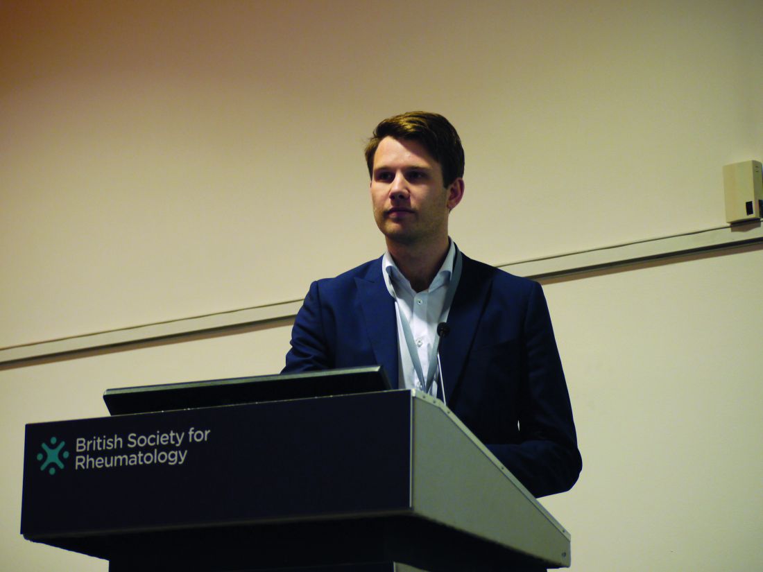
LIVERPOOL, ENGLAND – , but long-term follow-up is much more important, according to data presented at the British Society for Rheumatology annual conference.
In just one specialist rheumatology center in England, which treats more than 8,000 patients annually, the cost of the first year’s optical coherence tomography (OCT) assessment would be more than $60,000. Additional costs would be incurred to screen those who had been on the drug for more than 5 years ,who were known to be at greater risk of hydroxychloroquine-induced retinopathy. This is within the National Health Service in England where the cost of a single OCT scan is around $70; in the private health sector, the cost of one test can be as high as $400.
Indeed, of 887 hydroxychloroquine users identified, 44% had at least one risk factor for hydroxychloroquine-induced retinopathy. These included being older than 60 years of age (30% of all users), having renal (10%) or hepatic (2%) impairment, retinal disease at baseline (8%), or using high (more than 6.5 mg/kg) doses of the drug based on their actual (9%) or ideal (4%) body weight.
“The retinal toxicity of hydroxychloroquine is a bit of a hot topic at the moment,” Dr. Yates said at the conference. While the drug has been around for years and used successfully to treat many patients with rheumatoid arthritis and systemic lupus erythematosus (SLE), a known side effect is retinal toxicity.
Traditionally, retinopathy has been quoted as being a relatively rare side effect, affecting around 0.5%-2% of the treated population. Recent data (JAMA Ophthalmol. 2014;132[12]:1453-60) suggest, however, that is probably a vast underestimate, with 7.5% of patients taking hydroxychloroquine for more than 5 years likely to be affected, as are up to 20% of those taking the drug for up to 20 years of treatment.
Dr. Yates and associates wanted to assess the burden of hydroxychloroquine use at their center and look at the risk factors and impact of the recent screening guidelines issued by the British Society for Rheumatology (Rheumatology [Oxford]. 2017;56[6]:865-8) in 2017 and by the Royal College of Ophthalmologists in 2018. These state that patients should have a formal baseline ophthalmic examination, ideally including OCT, within 6-12 months of starting therapy and an annual eye assessment with repeat OCT thereafter for the following 5 years; the ophthalmology guidelines recommending annual screening for the duration of therapy.
One criticism of increased screening for retinal toxicity in routine practice is consultants saying that they see only a handful of cases during their career, Dr. Yates observed. However, if you consider that in an average rheumatology department there are five consultants and 900 patients on hydroxychloroquine, 500 patients take the drug for 5 years or longer, 2% are picked up with non-OCT screening, that amounts to around two cases per year over a 5- to 10-year period. “So that fits with the narrative of only having seen a handful of cases pre-OCT,” Dr. Yates reasoned.
“I believe that this is a real problem, but I’m afraid this is the tip of the iceberg,” commented Caroline Gordon, MD, after her presentation. “We’ve been screening our patients in Birmingham now for about 5 years and we are definitely finding a significant number of patients with hydroxychloroquine toxicity who can be picked up with OCT and visual fields screening.”
Dr. Gordon, professor of rheumatology at the University of Birmingham (England) and a consultant rheumatologist for the University Hospitals NHS Foundation Trust and the Sandwell & West Birmingham Hospitals NHS Trust, helps look after one of the largest cohorts of patients with SLE in the United Kingdom.
A baseline eye examination has always been recommended, Dr. Gordon said, but she suggested that this could remain in the realm of the opticians with further assessment and referral as needed.
“I’m not convinced, from the work we’ve done, that there is any value in the baseline OCT,” Dr. Gordon said, “because we never find anything on the baseline OCT that we didn’t already expect from the opticians’ assessment.”
It is the long-term (longer than10 years) follow-up that needs to be the focus, rather than the initial period, she stressed, as the highest risk appears to be in patients who have been taking the drug for 15 years or longer. Prior to this, different types of retinopathy can occur that are actually attributable to the underlying disease and are not related hydroxychloroquine. Of course, patients on higher doses of hydroxychloroquine may need closer monitoring early on, “as they are at risk,” she acknowledged.
Dr. Gordon suggested that the guidelines as they currently stand may not be that useful for real-life practice. Following them could result in a large amount of money being spent on early tests that are perhaps not necessary.
“What we do need to do is focus on the patients who’ve been on treatment long term,” she said.
SOURCE: Yates M et al. Rheumatology. 2018;57(Suppl. 3):key075.188.
REPORTING FROM BSR 2018
Key clinical point: Long-term follow up is important for assessing hydroxychloroquine toxicity.
Major finding: 44% of patients had at least one risk factor for hydroxychloroquine-induced retinopathy after more than 5 years of treatment.
Study details: Electronic record review of 887 patients treated with hydroxychloroquine for about 5 years in a large tertiary rheumatology service.
Disclosures: Dr. Yates had nothing to disclose.
Source: Yates M et al. Rheumatology. 2018;57(Suppl. 3):key075.312.
Raised LDL cholesterol, hsCRP tied to polymyalgia rheumatica, GCA
LIVERPOOL, ENGLAND – The presence of traditional cardiovascular risk factors may precede the development of polymyalgia rheumatica and giant cell arteritis.
Data from the EPIC-Norfolk study, reported at the British Society for Rheumatology annual conference, showed that raised LDL cholesterol was associated with the onset of polymyalgia rheumatica (PMR) and that high sensitivity C-reactive protein (hsCRP) was associated with giant cell arteritis (GCA).

“There’s been an association between vascular disease and PMR and GCA reported, but the way cardiovascular disease has been defined has been based on rather late endpoints, such as angina, myocardial infarction, peripheral vascular disease, and ischemia,” said Max Yates, MBBS, MRCP, in an interview.
“So, what we wanted to do was look at underlying risk factors for those diseases and see how they play in, in terms of the timing of the diagnosis of PMR and GCA,” he explained. Dr. Yates, who is a National Institute for Health Research clinical lecturer in rheumatology at the University of East Anglia, Norwich, England, noted that this was probably the first prospective study to look at clinical and laboratory parameters for vascular disease prior to the onset of these diseases.
Previously, French researchers suggested that there might be a link between hypertension and subsequent PMR, but that was a descriptive study published over 30 years ago, Dr. Yates said. “There was another case-control study from the Mayo Clinic where they said that smoking was associated with incidence GCA,” he added. “So most of the work has been retrospective, case-control studies.”
The EPIC (European Prospective Investigation of Cancer)-Norfolk study is a large, prospective, community-based cohort study that, as its name might suggest, was originally set up to look at risk factors for cancer. Since then it has broadened to enable the study of risk factors for a whole host of other conditions.
More than 30,000 people aged 40-70 years were recruited into the study during 1993-1997, and 25,600 people (440,237 at-risk person-years) who had the necessary baseline and follow-up data were included in the current analysis performed by Dr. Yates and associates.
A total of 395 cases of PMR and 118 cases of GCA were identified using current classification criteria. Those with PMR were diagnosed at a mean age of 73.6 years and those with GCA at a mean age of 74.1 years. For both conditions, about three-quarters of patients were women.
The investigators then looked back at the patients’ original recruitment data in terms of their cardiovascular risk factors, which included their blood pressure readings; body mass index; smoking status; presence of diabetes; hsCRP; and LDL cholesterol, triglycerides, and HDL cholesterol levels.
“Ultimately, these traditional cardiovascular risk factors are present early on, prior to PMR and GCA,” Dr. Yates said.
What this means is that perhaps clinicians need to be more aware of managing these risk factors aggressively, he suggested, but therein lies a problem. “It’s obviously very difficult, early on, before anyone’s developed any disease, to target these risk factors, and you have to balance the risk and benefit for individuals.”
GCA is a “pretty rare” disease whereas PMR is “quite common,” Dr. Yates said, “but we probably need to target these risk factors as soon as people are diagnosed with these conditions, to try to prevent the cardiovascular morbidity that is seen.”
These data might also help explain the underlying etiology and why there is an increased risk of vascular disease seen in populations of patients with inflammatory arthritides.
Dr. Yates had no conflicts of interest to disclose.
SOURCE: Yates M et al. Rheumatology. 2018 Apr;57[Suppl. 3]:key075.312.
LIVERPOOL, ENGLAND – The presence of traditional cardiovascular risk factors may precede the development of polymyalgia rheumatica and giant cell arteritis.
Data from the EPIC-Norfolk study, reported at the British Society for Rheumatology annual conference, showed that raised LDL cholesterol was associated with the onset of polymyalgia rheumatica (PMR) and that high sensitivity C-reactive protein (hsCRP) was associated with giant cell arteritis (GCA).
“There’s been an association between vascular disease and PMR and GCA reported, but the way cardiovascular disease has been defined has been based on rather late endpoints, such as angina, myocardial infarction, peripheral vascular disease, and ischemia,” said Max Yates, MBBS, MRCP, in an interview.
“So, what we wanted to do was look at underlying risk factors for those diseases and see how they play in, in terms of the timing of the diagnosis of PMR and GCA,” he explained. Dr. Yates, who is a National Institute for Health Research clinical lecturer in rheumatology at the University of East Anglia, Norwich, England, noted that this was probably the first prospective study to look at clinical and laboratory parameters for vascular disease prior to the onset of these diseases.
Previously, French researchers suggested that there might be a link between hypertension and subsequent PMR, but that was a descriptive study published over 30 years ago, Dr. Yates said. “There was another case-control study from the Mayo Clinic where they said that smoking was associated with incidence GCA,” he added. “So most of the work has been retrospective, case-control studies.”
The EPIC (European Prospective Investigation of Cancer)-Norfolk study is a large, prospective, community-based cohort study that, as its name might suggest, was originally set up to look at risk factors for cancer. Since then it has broadened to enable the study of risk factors for a whole host of other conditions.
More than 30,000 people aged 40-70 years were recruited into the study during 1993-1997, and 25,600 people (440,237 at-risk person-years) who had the necessary baseline and follow-up data were included in the current analysis performed by Dr. Yates and associates.
A total of 395 cases of PMR and 118 cases of GCA were identified using current classification criteria. Those with PMR were diagnosed at a mean age of 73.6 years and those with GCA at a mean age of 74.1 years. For both conditions, about three-quarters of patients were women.
The investigators then looked back at the patients’ original recruitment data in terms of their cardiovascular risk factors, which included their blood pressure readings; body mass index; smoking status; presence of diabetes; hsCRP; and LDL cholesterol, triglycerides, and HDL cholesterol levels.
“Ultimately, these traditional cardiovascular risk factors are present early on, prior to PMR and GCA,” Dr. Yates said.
What this means is that perhaps clinicians need to be more aware of managing these risk factors aggressively, he suggested, but therein lies a problem. “It’s obviously very difficult, early on, before anyone’s developed any disease, to target these risk factors, and you have to balance the risk and benefit for individuals.”
GCA is a “pretty rare” disease whereas PMR is “quite common,” Dr. Yates said, “but we probably need to target these risk factors as soon as people are diagnosed with these conditions, to try to prevent the cardiovascular morbidity that is seen.”
These data might also help explain the underlying etiology and why there is an increased risk of vascular disease seen in populations of patients with inflammatory arthritides.
Dr. Yates had no conflicts of interest to disclose.
SOURCE: Yates M et al. Rheumatology. 2018 Apr;57[Suppl. 3]:key075.312.
REPORTING FROM BSR 2018
Key clinical point: The presence of traditional cardiovascular risk factors may precede the development of polymyalgia rheumatica (PMR) and giant cell arteritis (GCA).
Major finding: Raised LDL cholesterol was linked with the onset of PMR (subhazard ratio, 1.29) and raised hsCRP was associated with GCA (SHR, 1.85).
Study details: Data from the EPIC-Norfolk study: 385 cases of PMR and 118 cases of GCA identified from a population of more than 25,000 subjects.
Disclosures: Dr. Yates had no conflicts of interest to disclose.
Source: Yates M et al. Rheumatology. 2018 Apr;57[Suppl. 3]:key075.312.
Idiopathic pulmonary fibrosis a ‘robust diagnosis’
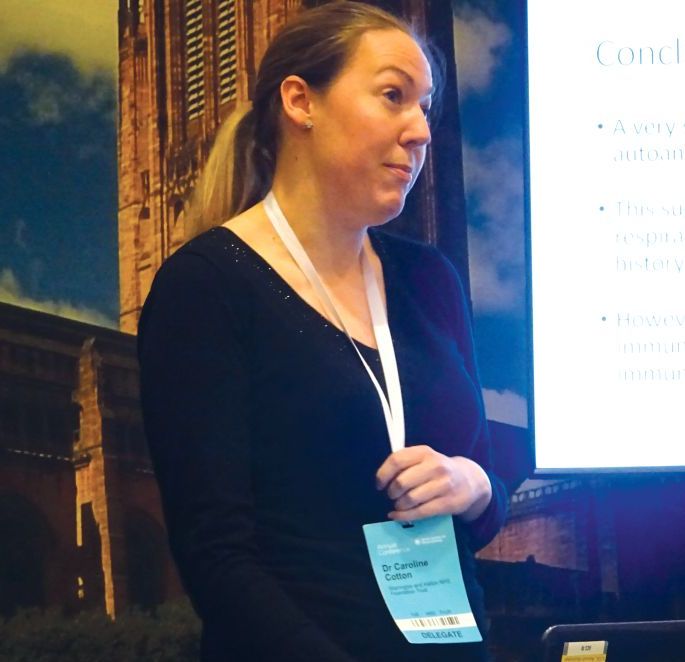
LIVERPOOL, ENGLAND – Very few patients with idiopathic pulmonary fibrosis have connective tissue disease antibodies, suggesting that IPF is a “robust diagnosis” when made on the basis of standard diagnostic tests, it was reported at the British Society for Rheumatology annual conference.
“The results were perhaps not what we’d expected,” said Caroline V. Cotton, PhD, of the Institute of Ageing and Chronic Disease at the University of Liverpool, England.
This means that chest physicians are getting the diagnosis right in the majority of cases, based on currently available methods, such as patients’ clinical history and examination, the results of high resolution–computed tomography, and widely available serology. “Which is good news,” Dr. Cotton observed.
Interstitial lung disease (ILD) comprises a huge spectrum of disorders. The main groups of ILDs are idiopathic, granulomatous, connective tissue disease–associated environmental, or medication exposure–associated; and the rare causes of ILD, each of which contain multiple subgroups of which IPF is one.
Sometimes it is obvious to respiratory physicians what the cause is, such as environmental exposure to asbestos or sarcoidosis for the granulomatous ILD, Dr. Cotton noted. Identifying connective tissue disease (CTD)–associated ILD can be more diagnostically challenging, however, and there are a large number of rheumatic conditions associated with CTD-associated ILD, including rheumatoid arthritis, systemic sclerosis, and Sjögren’s syndrome, to name a few.
One of the problems is that signs and symptoms of CTD may be absent at the time ILD starts to manifest and, even if signs are present, they may too subtle to be picked up in a general chest clinic. There also is a large number of antibodies for CTDs, but not all are widely available.
Dr. Cotton and her associates, therefore, wondered if there was a chance that patients being diagnosed with IPF actually could have covert CTD-associated ILD; this is an important distinction to make because the treatment differs for the two conditions. While ILD associated with CTD has a strong inflammatory component and is treated with corticosteroids and immunosuppressants, steroids can be harmful and increase mortality in IPF-ILD. The latter is treated with antifibrotic medications, such as pirfenidone and nintedanib.
For the study, serum samples from 250 patients with a definite diagnosis of IPF who were participating in the UK-BILD study were obtained and screened for known CTD antibodies using immunoprecipitation. Antibodies could be detected in just five (2%) patients – these included one patient each with anti-KS and anti-OJ antibodies, which are antisynthetase antibodies that are associated with myositis. Anti-Ku, another myositis-associated antibody, was identified in another patient, and one patient had an anti-RNA polymerase II antibody, which is associated with systemic sclerosis. Antimitochondrial autoantibodies were observed in one patient, and these are linked to primary biliary cirrhosis, which the patient was known to have.
There was nothing remarkable between the patients who did and did not have CTD antibodies in terms of their demographics, 76% and 80% were male, the mean ages were 73 and 70 years, respectively, and all were white.
However, 40% of patients did have unknown strong bands on immunoprecipitation, Dr. Cotton reported. This could suggest that there is an underlying immunological component to IPF, she added, but they had no recognized antibodies.
“A very small number of patients with IPF actually have the presence of autoantibodies strongly associated with CTDs. This suggests IPF is a very robust diagnosis; chest physicians are diagnosing it correctly most of the time, and they are really good at good at weeding out those who have got IPF and those who have potentially got connective tissue disease.” Dr. Cotton concluded.
Dr. Cotton had no conflicts of interest.
SOURCE: Cotton CV et al. Rheumatology. 2018;57[Suppl. 3]:key075.206.
REPORTING FROM RHEUMATOLOGY 2018
Key clinical point: Few patients diagnosed as having idiopathic pulmonary fibrosis (IPF) are likely to have connective tissue disorders (CTD).
Major finding: Only 2% of patients had a recognized CTD antibody present.
Study details: 250 patients with IPF participating in the UK-BILD multicenter study.
Disclosures: Dr. Cotton stated she had no conflicts of interest.
Source: Cotton CV et al. Rheumatology. 2018;57(Suppl. 3):key075.206.
Hip pain predicts OA mortality beyond comorbidities
LIVERPOOL, ENGLAND – Hip pain increases all-cause mortality in people with OA by a third, according to data obtained from a large, community-based study.
The hazard ratio for all-cause mortality was 1.33 (95% confidence interval, 1.17-1.51) in people who self-reported hip pain over the course of up to 25 years’ follow-up. The presence of hip pain also increased the risk of cardiovascular mortality (HR, 1.22; 95% CI, 0.99-1.50).
The hazard ratios for all-cause and cardiovascular mortality in patients with radiographic hip pain were 1.04 (95% CI, 0.91-1.17) and 1.01(95% CI, 0.82-1.24), and the all-cause and cardiovascular mortality hazard ratios in patients with both hip pain and radiographic OA were 1.01 (95% CI, 0.87-1.18) and 1.01 (95% CI, 0.80-1.28).
These data support hip pain as a predictor of mortality, observed Dr. Cleveland, of the University of North Carolina at Chapel Hill. “While there have been a number of studies that have looked at osteoarthritis as a risk factor for mortality, a lot of these studies have looked at arthritis in general or have looked at specifically knee osteoarthritis.
“There have been only a handful of studies that have looked at hip osteoarthritis as a risk factor for mortality,” she added, and considered all together, the results have been equivocal.
The aim of the current study she presented at the congress sponsored by the Osteoarthritis Research Society International was to explore whether or not hip OA was associated with all-cause and cardiovascular disease–specific mortality, independent of any comorbidities. The comorbidities considered were cancer, liver disease, hypertension, type 2 diabetes mellitus, and cardiovascular disease.
Data on 3,919 people with and without hip OA were obtained from the Johnston County Osteoarthritis Project, a longitudinal cohort of white and African American residents aged 45 years or older. Enrollment was carried out in two waves, with 3,185 people recruited between 1990 and 1998 and 1,015 people recruited between 2003 and 2004. Only those with baseline and follow-up assessment data, including at least one hip radiograph, were included in the present analysis.
The mean age at recruitment was 62 years, 61% were women, and two-thirds were white. Around 17% (n = 655) had radiographic hip OA and hip pain (symptomatic OA) at baseline, 10% (n = 787) had radiographic OA alone, and 27% (n = 1,156) had hip pain alone. The remaining 45% (n = 1,321) had neither hip pain nor radiographic damage.
Over the course of up to 25 years’ follow-up, there were 1,762 deaths from any cause – 311 occurred in the group with symptomatic OA at baseline, 382 in the group with radiographic OA alone, 509 in those with hip pain alone, and 560 in those with neither hip pain nor radiographic OA.
Median survival was lowest in individuals with symptomatic OA, at 16.1 years, and highest in individuals without either, at 22.8 years. Median survival was similar for those with hip pain only (18.6 years) and with radiographic hip OA only (18.5 years).
Stratified by race, all-cause mortality was increased with hip pain alone, and was more pronounced in white patients (HR, 1.39) than in African American patients (HR, 1.24). Interestingly, the risk of all-cause death was lower in African American patients who had symptomatic hip OA than their white counterparts (HR, 0.78 and 1.16, respectively).
Furthermore, Dr. Cleveland reported that hip pain was strongly associated with all-cause mortality in those younger than 65 years (HR, 1.56) when compared with those who were 65 years and up (HR, 1.18). Of note, the risk of death was higher in younger patients with symptomatic hip pain than in older patients (HR, 1.33 and 0.88, respectively).
A 41% increased death risk was also observed in patients with hip pain who had a body mass index of 30 kg/m2 (HR, 1.41 vs. HR, 1.29 for those with hip pain and a body mass index of less than 30 kg/m2).
“Our results are independent of comorbidities and sociodemographic measures,” Dr. Cleveland said. “This suggests there are mechanisms beyond comorbidities in the link between radiographic hip OA and mortality risk.”
The Johnston County Osteoarthritis Project is funded by the Centers for Disease Control and Prevention and the National Institutes of Arthritis and Musculoskeletal and Skin Diseases. Dr. Cleveland had no conflicts of interest to report.
SOURCE: Cleveland RJ et al. Osteoarthritis Cartilage. 2018:26(1):S10-1. Abstract 1.
REPORTING FROM OARSI 2018
Key clinical point: Hip pain is a predictor of mortality in patients with OA and is independent of any comorbidities.
Major finding: The hazard ratios for all-cause and cardiovascular mortality in patients with hip pain were 1.33 and 1.22.
Study details: A longitudinal cohort study of almost 4,000 people, with up to 25 years’ follow-up.
Disclosures: The Johnston County Osteoarthritis Project is funded by the Centers for Disease Control and Prevention and the National Institutes of Arthritis and Musculoskeletal and Skin Diseases. Dr. Cleveland had no conflicts of interest to report.
Source: Cleveland RJ et al. Osteoarthritis Cartilage. 2018 Apr:26(1):S10-1. Abstract 1
Post-traumatic osteoarthritis needs to be prevention focus
, Jackie Whittaker, PT, PhD, said at the World Congress of Osteoarthritis.
“We all know that the burden of this disease is enormous and it’s expanding at an alarming rate,” she said at the congress, sponsored by the Osteoarthritis Research Society International.
“The only way that we have at this moment to try to reduce the burden of this disease is to shift our approach to management upstream and focus on prevention,” said Dr. Whittaker, who is an associate professor and research director at the University of Alberta, Edmonton, Canada.
According to the World Health Organization, OA is expected to become the fourth leading cause of disability worldwide by 2020. Furthermore, there will be a projected rise in prevalence from 12% to 25% in North America by 2030.
Dr. Whittaker suggested that it was time to try to identify those at risk of developing OA, such as after a joint injury, and ran through some suggestions on how posttraumatic OA (PTOA) might be preventable.
Secondary prevention of posttraumatic osteoarthritis
The prevention of PTOA can be split into primary, secondary and tertiary prevention, with primary prevention trying to prevent injuries from occurring in the first place.
“Strategies aimed at identifying and slowing down the onset of symptomatic osteoarthritis in preclinical populations would be referred to as secondary prevention,” Dr. Whittaker explained, adding that tertiary prevention would then be strategies aimed at improving function and reducing disability in those who already have symptomatic PTOA.
While there are programs that address primary and tertiary prevention – such as Footy First, an exercise training program adopted by the Australian Football League to reduce the risk of common leg injuries and the Good Life with osteoArthritis in Denmark (GLA:D®) education and supervised exercise program for those with symptomatic OA – there is more of a gap for secondary prevention.
Some of the first steps to developing a secondary prevention model would be to determine the extent of PTOA after joint injury and then identify risk factors or causal mechanisms. Then, prevention strategies could be developed and tested before implementation and effectiveness studies are performed.
Performing the necessary prospective cohort studies, however, is when things get challenging – PTOA can take 10–15 years to manifest and studies would potentially need to run for long periods of time, which comes at a cost. Other challenges are that there is no commonly agree definition. PTOA is multifactorial, and because people may be in their 20s, there could be other contributing factors to the disease course.
Identifying modifiable risk factors
There are a “fair number” of prospective and retrospective studies that have been done to try to identify patients at risk for OA after joint injury. Several have looked at unmodifiable risk factors, such as age and sex, and the type of injury. Others have looked at potentially modifiable factors such as the treatment approach and avoiding re-injury, joint mechanics and strength, body composition and aerobic fitness and behavioral characteristics such as physical activity and return to sport.
Data from the Alberta Youth PrE-OA Study, an ongoing longitudinal cohort study, have shown that structural changes consistent with OA are not unique to tears in the anterior cruciate ligament or to meniscal tears, which are known to up the risk of PTOA. Furthermore, the odds of having MRI-defined OA 3–10 years after a knee injury varies by the injury history, type, and surgery.
Other findings from the Alberta Youth PrE-OA Study data have shown that previously injured subjects have a 30% risk for re-injury, with weaker knee extensors and flexors and poorer dynamic balance than uninjured study participants. The results have also shown that there is reduced physical activity and avoidance in those who have been injured.
Who is the population at risk?
“Although the supporting evidence and the level of evidence for some of the risk factors is not as thorough as we would like it, there are some common themes across the literature, that are consistent to what we see in clinical practice, and what we know from primary and tertiary prevention,” Dr. Whittaker said. “Based upon that, we can start to hypothesize who’s at greatest risk of developing posttraumatic osteoarthritis and what we might start doing about it right now.”
Dr. Whittaker proposed the following risk factors could be used to “build a profile” of someone at risk of PTOA:
- Having sustained an anterior cruciate ligament (ACL) tear with or without a damaged meniscus
- Elevated body mass index (BMI) or adiposity, and low grade systemic inflammation
- Weak knee muscles, and poor dynamic balance
- Reduced or disengaged from physical activity
- Insufficient rehabilitation, pain or stiffness, or fear of movement
There is also an argument for nutrition being involved, Dr. Whittaker said, with levels of micronutrients such as vitamins D and K and calcium playing an integral role in bone health.
What could secondary prevention look like?
“So, with this risk profile, what can be done?” Dr. Whittaker asked rhetorically. Exercise therapy is key, and increasing the muscle strength of the knee and lower extremity are an important component of this strategy. Strength alone may not be enough, so exercise programs will need to increase the neuromuscular control of functional movements. Tackling people’s fear of movement will also be a priority. “I think it’s very important that we promote, if not implement, physical activity guidelines.”
Education is also going to be an important part of any secondary prevention program for PTOA, ensuring that people have realistic expectations about re-injury and their risk of developing osteoarthritis and the importance of remaining physically active. Balancing physical activity against their likelihood of flare-ups and learning how to avoid re-injury if at all possible. Weight control and diet will also be a component to include.
One final component is how clinicians work with patients to minimize their risk of PTOA. “We need to co-manage these patients,” Dr. Whittaker said. “We need to have difficult conversations with them and really balance giving them a picture of reality without over medicalizing the situation.”
Dr. Whitaker stated that she had no financial relationships or commercial interests to disclose.
SOURCE: Whittaker J, et al. Osteoarthritis Cartilage 2018:26(1):S7-8. Abstract I-22.
REPORTING FROM OARSI 2018
Prior knee injury osteoarthritis ‘distinct subgroup’
LIVERPOOL, ENGLAND – according to data from the Good Life with osteoArthritis in Denmark (GLA:D®) Registry.
Clear differences in the clinical presentation can be observed between patients with knee OA who have and have not had a previous knee injury, researcher Paetur Mikal Holm reported at the World Congress on Osteoarthritis.
“These differences include being younger, being a male, having a lower BMI [body mass index], and being more physically active; also, we see reduced quality of life, longer symptom duration, and widespread pain” said Mr. Holm, a PhD fellow in the department of sports science and clinical biomechanics, musculoskeletal function, and physiotherapy at the University of Southern Denmark in Odense. “The last two characteristics in particular – widespread pain and symptom duration – may have an important impact on treatment,” he added.
“People with a prior knee injury have a 25% higher probability of having widespread pain compared to people without a prior knee injury,” Mr. Holm and his associates said in their abstract. This could mean that specialized interventions are needed to address pain in these individuals, especially when there is a long duration of symptoms.
Disease characteristics and trajectories in knee osteoarthritis are highly variable, Mr. Holm observed, and there are multiple phenotypes already reported. Posttraumatic knee OA is one of these phenotypes, but, until now, their clinical characteristics were poorly understood.
A cross-sectional study was performed using baseline data on 5,477 individuals enrolled in the GLA:D® Registry, 91% of whom had radiographic knee OA. These patients were divided into two groups based on whether they did (n = 2,440) or did not (n = 3,037) self-report a prior knee injury. This is one of the limitations of the study, Mr. Holm acknowledged. There was no specific investigation of what the prior knee injury actually was.
However, results clearly showed a significant (P less than .05) difference between the two groups. For example, the mean age of those with a prior knee injury was 64 years, whereas those without were about 65 years old (odds ratio, 0.99; 95% confidence interval, 0.98-0.99). About 29% of those with a previous knee injury were men, versus roughly 25% of those without (OR, 1.28; 95% CI, 1.12-1.46). Widespread pain was present in about 45% and 38%, respectively, for those with and without a prior knee injury (OR, 1.25; 95% CI, 1.11-1.40), and the mean duration of symptoms was 60 versus 35 months (OR, 1.05; 95% CI, 1.04-1.06). Those who reported exercising 2-4 days a week had an OR of 1.30 (95% CI, 1.05-1.59).
“Being a clinician, to me it is a worry to see that these people are young and have had symptoms for a long time,” coauthor Ewa M. Roos, PT, PhD, said at the congress, which was sponsored by the Osteoarthritis Research Society International.
“Here is a group of people that we don’t necessarily think about as athletes, these are people with osteoarthritis. They are seen in primary care, but maybe we don’t think about them as osteoarthritis patients at a younger age, but this is a group that we need to be very careful to recognize,” added Dr. Roos, also of University of Southern Denmark. She cochaired the session where the findings were presented.
The GLA:D® Registry is funded by various sources including the Danish Rheumatism Association, Naestved-Slagelse-Ringsted Hospitals, Region Zealand, and the Danish Physiotherapy Association. Dr. Holm had no conflicts of interest.
SOURCE: Holm PM et al. Osteoarthr Cartil. 2018:26(1):S56.OARSI 2018 Abstract 84.
REPORTING FROM OARSI 2018
Key clinical point: People with posttraumatic knee osteoarthritis are a distinct subgroup of patients that might be underrecognized.
Major finding: Widespread pain was present in about 45% and 38%, respectively, of those with and without a prior knee injury (OR, 1.25, 95% CI, 1.11-1.40).
Study details: Cross-sectional study of 5,477 patients enrolled in the Good Life with osteoArthritis in Denmark (GLA:D®) Registry.
Disclosures: The GLA:D® Registry is funded by various sources including the Danish Rheumatism Association, Naestved-Slagelse-Ringsted Hospitals, Region Zealand, and the Danish Physiotherapy Association. The presenting author, Dr. Paetur Mikal Holm, had no conflicts of interest.
Source: Holm PM et al. Osteoarthr Cartil. 2018:26(1):S56. Abstract 84.
Emergency gout admission increase is ‘call to arms’
LIVERPOOL, ENGLAND – The rate of emergency hospital admissions for gout in England has seen a 59% increase over the past decade, while that of rheumatoid arthritis has halved, it was reported at the British Society for Rheumatology annual conference.
Over an approximate 10-year period (2006-2017), the incidence rate of unplanned gout admissions increased from 7.9 to 12.5 admissions per 100,000 of the population. This represented an increase from 0.023% to 0.032% of all hospital admissions during the period.
To put that into perspective, unplanned admissions for rheumatoid arthritis (RA), decreased from 8.6 to 4.3 admissions per 100,000 of the population, said Mark D. Russell, MD, a rheumatology registrar at Guy’s and St Thomas’ Hospital in London.
Furthermore, primary care prescriptions for common gout medications have seen a dramatic increase over the same time period in England; allopurinol prescriptions are up 72%, there’s been a 166% increase in colchicine prescriptions, and a 20-fold increase in febuxostat (Uloric) prescriptions since data became available for its in 2010.
“Gout’s very much a treatable condition,” Dr. Russell said, but with 82% of all gout admissions being unplanned, “there’s clearly more to do; this should be a call to arms for rheumatologists to help reduce the in-patient burden of this condition.”
The mean length of hospital stay was estimated at 6.6 days, with the median being 3.2 days. Gout accounted for just under 350,000 hospital bed days from 2006 to 2017, and with the cost of a single gout admission being anything from £850 up to £5,600, it constitutes a significant burden for the country’s National Health Service.
But what can be done? Would a “door-to-needle time campaign” help? So that when patients attend the emergency department they are assessed rapidly and treated accordingly? Dr. Russell queried.
Education could be the key, was the consensus during the discussion following his presentation. Education, and not just of those affected by the condition, but also of the family physicians who seem to have a “knee-jerk reaction” to prescribe medications, and not always appropriately. Even hospital staff may need help in differentiating gout from other emergency presentations, with some admitting patients suspecting infection or wrongly discharging them.
Pharmacist-led gout clinics
Another approach to try to avoid emergency hospital visits could be better management and perhaps setting up specialist gout clinics. Such clinics have already been piloted, and rheumatology pharmacist Jane Whiteman shared her experience of setting up a monthly, pharmacist-led gout clinic in a separate presentation.
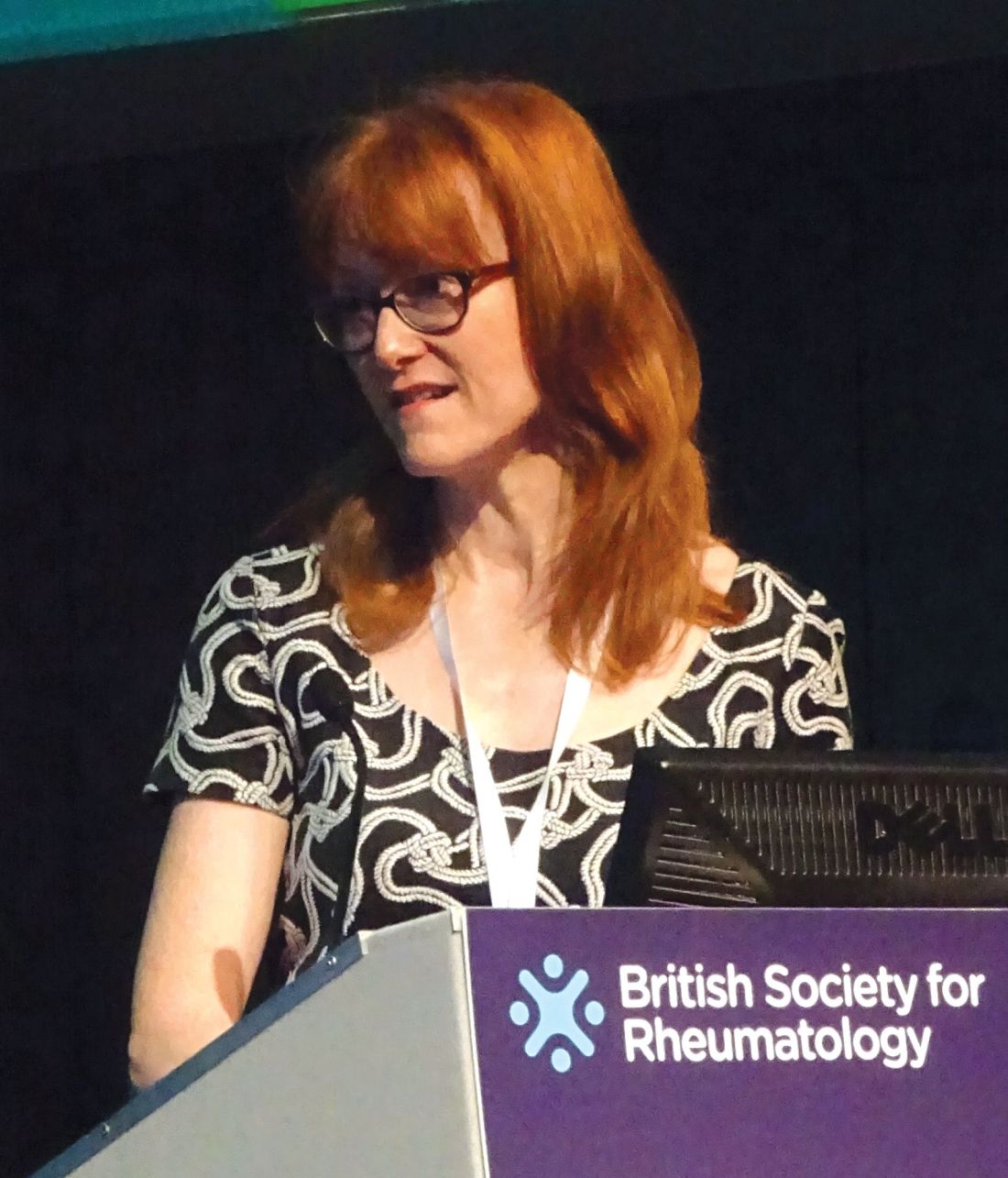
Dr. Whiteman, who works at Royal Victoria Hospital, part of the Belfast Health and Social Care Trust in Ireland, presented data on 52 patients who were seen at the clinic between June 2015 and May 2017. The total number of patient visits was 87, with an average of 1.7 visits per patient.
Of 38 patients who were discharged from the clinic, 29 (76%) had met target levels of serum uric acid, which guidelines from the British Society for Rheumatology set at less than 0.3 mmol/L (5 mg/dL) and those from the European League of Rheumatism set at less than 0.36 mmol/L (6 mg/dL).
It is important to reduce serum uric acid levels, Dr. Whiteman explained. “Gout occurs when serum uric acid rises and urate crystals reach their saturation point in the serum and start to crystallize out into the joints and into the tissues,” she said. “It’s a progressive disease that usually starts in one joint, but if the serum uric acid isn’t controlled then it can go on to affect a number of joints,” and cause chronic arthritis, among other potentially serious conditions, and is an independent risk factor for cardiovascular and renal disease.
“Studies across the world have shown that gout is not well looked after; patient adherence to treatment is very poor,” Dr. Whiteman said. “Among all the chronic diseases it has the lowest adherence to treatment,” she added, noting that one study showed just one-fifth of patients remained on gout medication at 1 year.
Patient education is an important part of the clinic’s services, as when people are asymptomatic and between bouts of gout, they perhaps do not realize that they still need to take their medication. Education thus needs to include talking about their diet and lifestyle, providing information on medication, and why it is important to keep their serum uric acid levels in check.
After patients’ initial referral to the clinic, they are followed up by the clinic every month until their serum urate levels are below the 0.3 mmol/L target. They can then be discharged and monitored by their family physician.
Postdischarge, Dr. Whiteman and her colleagues found that 22 (76%) of the 29 patients who had met their target levels of serum uric acid later had serum uric acid tested at least once, showing an average level of 0.29 mmol/L at follow-up. This reassuring result occurred perhaps because of a majority of patients (79%) who were still being prescribed, and presumably taking, urate-lowering therapy. Seven patients did not undergo follow-up serum uric acid testing because they had died (one), were in the hospital (one), were not taking medication because they had made diet or lifestyle changes (three), or had their treatment on hold (two).
“I think the gout clinic has been a success; it has addressed barriers to optimal management through education of the patient,” Dr. Whiteman said.
Both presenters had nothing to disclose.
SOURCE: Russel M et al. Rheumatology. 2018;57[Suppl. 3]:key075.186; Whiteman J, et al. Rheumatology. 2018;57[Suppl. 3]:key075.215.
LIVERPOOL, ENGLAND – The rate of emergency hospital admissions for gout in England has seen a 59% increase over the past decade, while that of rheumatoid arthritis has halved, it was reported at the British Society for Rheumatology annual conference.
Over an approximate 10-year period (2006-2017), the incidence rate of unplanned gout admissions increased from 7.9 to 12.5 admissions per 100,000 of the population. This represented an increase from 0.023% to 0.032% of all hospital admissions during the period.
To put that into perspective, unplanned admissions for rheumatoid arthritis (RA), decreased from 8.6 to 4.3 admissions per 100,000 of the population, said Mark D. Russell, MD, a rheumatology registrar at Guy’s and St Thomas’ Hospital in London.
Furthermore, primary care prescriptions for common gout medications have seen a dramatic increase over the same time period in England; allopurinol prescriptions are up 72%, there’s been a 166% increase in colchicine prescriptions, and a 20-fold increase in febuxostat (Uloric) prescriptions since data became available for its in 2010.
“Gout’s very much a treatable condition,” Dr. Russell said, but with 82% of all gout admissions being unplanned, “there’s clearly more to do; this should be a call to arms for rheumatologists to help reduce the in-patient burden of this condition.”
The mean length of hospital stay was estimated at 6.6 days, with the median being 3.2 days. Gout accounted for just under 350,000 hospital bed days from 2006 to 2017, and with the cost of a single gout admission being anything from £850 up to £5,600, it constitutes a significant burden for the country’s National Health Service.
But what can be done? Would a “door-to-needle time campaign” help? So that when patients attend the emergency department they are assessed rapidly and treated accordingly? Dr. Russell queried.
Education could be the key, was the consensus during the discussion following his presentation. Education, and not just of those affected by the condition, but also of the family physicians who seem to have a “knee-jerk reaction” to prescribe medications, and not always appropriately. Even hospital staff may need help in differentiating gout from other emergency presentations, with some admitting patients suspecting infection or wrongly discharging them.
Pharmacist-led gout clinics
Another approach to try to avoid emergency hospital visits could be better management and perhaps setting up specialist gout clinics. Such clinics have already been piloted, and rheumatology pharmacist Jane Whiteman shared her experience of setting up a monthly, pharmacist-led gout clinic in a separate presentation.
Dr. Whiteman, who works at Royal Victoria Hospital, part of the Belfast Health and Social Care Trust in Ireland, presented data on 52 patients who were seen at the clinic between June 2015 and May 2017. The total number of patient visits was 87, with an average of 1.7 visits per patient.
Of 38 patients who were discharged from the clinic, 29 (76%) had met target levels of serum uric acid, which guidelines from the British Society for Rheumatology set at less than 0.3 mmol/L (5 mg/dL) and those from the European League of Rheumatism set at less than 0.36 mmol/L (6 mg/dL).
It is important to reduce serum uric acid levels, Dr. Whiteman explained. “Gout occurs when serum uric acid rises and urate crystals reach their saturation point in the serum and start to crystallize out into the joints and into the tissues,” she said. “It’s a progressive disease that usually starts in one joint, but if the serum uric acid isn’t controlled then it can go on to affect a number of joints,” and cause chronic arthritis, among other potentially serious conditions, and is an independent risk factor for cardiovascular and renal disease.
“Studies across the world have shown that gout is not well looked after; patient adherence to treatment is very poor,” Dr. Whiteman said. “Among all the chronic diseases it has the lowest adherence to treatment,” she added, noting that one study showed just one-fifth of patients remained on gout medication at 1 year.
Patient education is an important part of the clinic’s services, as when people are asymptomatic and between bouts of gout, they perhaps do not realize that they still need to take their medication. Education thus needs to include talking about their diet and lifestyle, providing information on medication, and why it is important to keep their serum uric acid levels in check.
After patients’ initial referral to the clinic, they are followed up by the clinic every month until their serum urate levels are below the 0.3 mmol/L target. They can then be discharged and monitored by their family physician.
Postdischarge, Dr. Whiteman and her colleagues found that 22 (76%) of the 29 patients who had met their target levels of serum uric acid later had serum uric acid tested at least once, showing an average level of 0.29 mmol/L at follow-up. This reassuring result occurred perhaps because of a majority of patients (79%) who were still being prescribed, and presumably taking, urate-lowering therapy. Seven patients did not undergo follow-up serum uric acid testing because they had died (one), were in the hospital (one), were not taking medication because they had made diet or lifestyle changes (three), or had their treatment on hold (two).
“I think the gout clinic has been a success; it has addressed barriers to optimal management through education of the patient,” Dr. Whiteman said.
Both presenters had nothing to disclose.
SOURCE: Russel M et al. Rheumatology. 2018;57[Suppl. 3]:key075.186; Whiteman J, et al. Rheumatology. 2018;57[Suppl. 3]:key075.215.
AT RHEUMATOLOGY 2018
Key clinical point:
Major finding: The rate of emergency hospital admissions for gout in England increased by 59%, from 7.9 to 12.5 admissions per 100,000 of the population; 76% of patients attending a pharmacist-led gout clinic achieved target serum uric acid levels.
Study details: An analysis of National Health Service data from April 2006 to March 2017 on hospital admissions for gout and primary care prescription data and a separate study of 52 patients attending a pharmacist-led gout clinic.
Disclosures: Both presenters had nothing to disclose.
Sources: Russell M et al. Rheumatology. 2018;57[Suppl. 3]:key075.186; Whiteman J et al. Rheumatology. 2018;57[Suppl. 3]:key075.215.
Ixekizumab improves psoriatic arthritis patient-reported outcomes
LIVERPOOL, ENGLAND – In biologic-experienced patients with psoriatic arthritis, the interleukin-17 inhibitor ixekizumab not only met the primary efficacy endpoint of a pivotal phase 3 trial, but also improved multiple patient-reported outcomes in doing so.
Newly-released results from the Study of Ixekizumab in Participants With Active Psoriatic Arthritis (SPIRIT-P2) showed that patients who received active treatment exhibited significantly better changes in physical function, quality of life, itch score, and work productivity compared with those given placebo.
Patients treated with ixekizumab 80 mg every 2 or 4 weeks more often achieved the MCID by week 24, reaching 40% for 80 mg every 2 weeks and 43% for every 4 weeks, compared with 17% for placebo.
A total of 363 patients who met CASPAR (Classification Criteria for Psoriatic Arthritis) criteria were randomized into the SPIRIT-P2 trial. Patients could be included only if they had at least three tender and three swollen joints, active skin lesions, or a documented history of skin psoriasis, and had received prior treatment with a tumor necrosis factor inhibitor (TNFi).
“The population of patients studied is representative of the patients we see in our clinics,” said Dr. Marzo-Ortega, a consultant rheumatologist at Leeds Teaching Hospitals NHS Trust, England. The mean age was 52 years, a similar percentage of men and women were seen, and the majority (53%-58%) were inadequate responders to one TNFi. One-third had not responded to two TNFis, and 8%-10% had an intolerance.
The primary endpoint results, which have been previously presented and published (Lancet. 2017;389[10086]:2317-27), showed that a significantly (P less than .0001) higher percentage of patients treated with either of the two regimens of ixekizumab achieved a 20% response level on American College of Rheumatology criteria (ACR20) at 24 weeks. Indeed, 48% of 123 patients given 80 mg of ixekizumab every 2 weeks and 53% of 122 given 80 mg every 4 weeks achieved an ACR20 versus 20% of 118 placebo-treated patients. Also, on two key secondary endpoints at 24 weeks, an ACR50 response was achieved by a respective 33%, 35%, and 5% of patients, and an ACR70 by 12%, 22%, and 0%, she said.
Dr. Marzo-Ortega noted that the study had only been powered to show a difference between the active treatment and placebo, and not between the two doses, and that, looking at the speed of response, a difference from placebo was already being seen by week 2 “and certainly by week 4,” indicating an early effect. Data from the trial at 52 weeks are being analyzed and should be available soon, she said.
Another efficacy measure used was the percentage of patients achieving minimum disease activity at week 24. “These are stringent criteria to achieve: Five of seven criteria need to be met,” Dr. Marzo-Ortega said. “This was achieved by almost 30% of patients on the 4-weekly dose,” and by 24% on the 2-weekly dose, but by just 3% of those given placebo.
“One of the remarkable things is that nearly a third [of patients] achieved PASI [Psoriasis Area Severity Index] 100 by week 24, which is complete resolution of skin psoriasis,” she said. This is one of the first times this type of skin response has been seen in a psoriasis trial, she noted.
However, there was little difference between the active treatment and placebo arms in terms of the percentage of patients seeing a response on enthesitis, and only the dose taken every 4 weeks had a significant benefit over placebo in terms of improving dactylitis.
It is not clear why these modest results were seen in the joints, perhaps there were too few patients. While this is surprising, Dr. Marzo-Ortega noted that she “wouldn’t put too much weight on” the lack of an enthesis response; these are “fantastic drugs for the skin,” she said. “There is no doubt about it.”
Other findings from the trial included a significant improvement in itch with both ixekizumab regimens versus placebo, as shown by a greater reduction in numerical rating scale scores from baseline to week 24 (–3.4 and –3.5 vs. –1.2; P less than .001).
Significant improvements with ixekizumab versus placebo in patients’ mental and physical health were also seen when data from the Short Form–36 and EQ-5D instruments were analyzed.
There was also evidence that treatment with ixekizumab significantly improved patients’ presenteeism, work productivity, and activity impairment at work when compared against placebo. There was no difference in absenteeism, as measured by changes in Work Productivity and Activity Impairment Questionnaire–Specific Health Problem scores from baseline to week 24.
SPIRIT-P2 is one of two pivotal trials conducted with ixekizumab in patients with psoriatic arthritis; the other is SPIRIT-P1, which was conducted in biologic-naive patients (Ann Rheum Dis. 2017;76[1]:79-87). Investigators recently reported 1-year data from it (J Rheumatol. 2018;45[3]:367-77.).
Eli Lilly, which markets ixekizumab as Taltz, sponsored the study. Dr. Marzo-Ortega disclosed receiving honoraria from AbbVie, Celgene, Eli Lilly, Novartis, and UCB, and honoraria and research funding from Janssen and Pfizer. Several other authors reported disclosures with many manufacturers of biologics for psoriatic arthritis, including Eli Lilly.
SOURCE: Marzo-Ortega H et al. Rheumatology. 2018;57[Suppl. 3]:key075.185.
REPORTING FROM RHEUMATOLOGY 2018
Key clinical point: Ixekizumab has multiple beneficial effects in patients with psoriatic arthritis previously treated with biologics.
Major finding: Mean HAQ-DI score changes (baseline to week 24) were –0.4 and –0.6 with ixekizumab (80 mg every 2 or 4 weeks) and –0.2 for placebo (P less than or equal to .001).
Study details: SPIRIT-P2: A randomized, double-blind, placebo-controlled phase 3 trial of ixekizumab in 363 biologic-experienced patients.
Disclosures: Eli Lilly, which markets ixekizumab as Taltz, sponsored the study. Dr. Marzo-Ortega disclosed receiving honoraria from AbbVie, Celgene, Eli Lilly, Novartis, and UCB, and honoraria and research funding from Janssen and Pfizer. Several other authors reported disclosures with many manufacturers of biologics for psoriatic arthritis, including Eli Lilly.
Source: Marzo-Ortega H et al. Rheumatology. 2018;57[Suppl. 3]:key075.185.
Comorbid spondyloarthritis and fibromyalgia lower response to TNF inhibitors
LIVERPOOL, ENGLAND – Individuals with axial spondyloarthritis (axSpA) who have comorbid fibromyalgia do respond to biologic therapy but at a seemingly lower rate if they have a high symptom score during early assessments.
Data from the British Society for Rheumatology Biologics Register in Ankylosing Spondylitis (BSRBR-AS) showed that participants with axSpA who were starting biologic therapy with a tumor necrosis factor inhibitor (TNFi) for the first time also had worse disease activity at baseline if they met fibromyalgia research criteria than if they did not.
“The issue of fibromyalgia as a comorbidity in axial SpA has been the subject of current considerable interest,” said Dr. Macfarlane, who is Clinical Chair in Epidemiology at the University of Aberdeen (Scotland), where the BSRBR-AS is run. This is for several reasons: “One is the challenge of identifying patients with axial SpA who have comorbid fibromyalgia.
“The second is the concern that comorbid fibromyalgia may have an effect on disease indices such as BASDAI [Bath Ankylosing Spondylitis Disease Activity Index],” Dr. Macfarlane observed.
This leads to the third issue: “Are patients with comorbid fibromyalgia inappropriately receiving biological therapy, and how do they respond if they do receive it?”
Previous data from the BSRBR-AS have shown that approximately one in five patients with axSpA meet research criteria for fibromyalgia (Arthritis Rheumatol. 2017;69:2144-50) and that the prevalence of the comorbidity was higher in those who met Assessment of SpondyloArthritis International Society imaging criteria and lower in those who only met clinical criteria (25% vs. 10%).
The current aims of the study presented by Dr. Macfarlane were to first quantify the extent to which meeting fibromyalgia criteria was associated with higher measures of disease activity and impact, and then to see if meeting these criteria was associated with a poorer response to first use of a TNFi.
In order to find out, Dr. Macfarlane and his associates examined data from the prospective BSRBR-AS, which has been running since 2012. The BSRBR-AS recruits patients with ASAS-confirmed axSpA who are newly starting biologics from 83 U.K. centers.
At recruitment and at 3 months, patients starting biologic treatment undergo several assessments, which since 2015 has included research criteria for fibromyalgia (J Rheumatol. 2011;38:1113–22). The latter incorporate a Widespread Pain Index rated 0-19 and a Symptom Severity Scale rated 0-12 and cover items such as fatigue and waking up unrefreshed, and the presence of lower abdominal pain or headaches.
To date, around 1,750 participants in the BSRBR-AS have completed the fibromyalgia criteria, Dr. Macfarlane said.
Across the board, baseline measures of disease activity were lower in patients who met the fibromyalgia criteria versus those who did not. This included the BASDAI and the Bath Ankylosing Spondylitis Functional Index, where scores were approximately 6.5 and 4.5 for each measure.
“Quality of life was significantly lower in those who were fibromyalgia positive,” Dr. Macfarlane said. Indeed, whatever the measure used, from the disease-specific Ankylosing Spondylitis Quality of Life (ASQoL) Scale to the general EuroQoL Quality of Life Scale, there were significant differences between those who did and did not meet fibromyalgia criteria. There were also higher scores for depression, anxiety, poorer-quality sleep, and higher levels of fatigue.
Patients treated with TNFi therapy showed improvement in both BASDAI and ASQoL scores regardless of whether they met fibromyalgia criteria, but crucially, the responses were still lower and significantly different from those without fibromyalgia.
An ASAS20 response to TNFi therapy was met by “slightly fewer” patients who met the fibromyalgia criteria than by those who did not at all follow-up points: at 3 months (about 35% vs. 45%), 6 months (about 58% vs. 61%), and 12 months (about 60% vs. 62%).
A high score on the Symptom Severity Scale but not the Widespread Pain Index of the fibromyalgia criteria was associated with a lower response to TNFi therapy at 3 months. “Such patients may benefit from the use of TNFi and nonpharmacological therapy,” Dr. Macfarlane said.
The BSRBR-AS is funded by the British Society for Rheumatology, which in turn receives funding from AbbVie, Pfizer, and UCB. Dr. Macfarlane did not provide any disclosures but has previously acknowledged receiving an honorarium from Pfizer and research funding from AbbVie and Pfizer for the Scotland Registry for Ankylosing Spondylitis study.
SOURCE: Macfarlane GJ et al. Rheumatology. 2018;57(Suppl. 3):key075.183.
REPORTING FROM RHEUMATOLOGY 2018
Key clinical point: Comorbid axial spondyloarthritis and fibromyalgia are associated with a lower response to biologic therapy.
Major finding: About 35% vs. 45% of patients who did and did not meet research criteria for fibromyalgia had an ASAS20 response at 3 months.
Study details: Data on 1,750 participants of the British Society for Rheumatology Biologics Register in Ankylosing Spondylitis (BSRBR-AS).
Disclosures: The BSRBR-AS is funded by the British Society for Rheumatology, which in turn receives funding from AbbVie, Pfizer, and UCB. Dr. Macfarlane did not provide any disclosures but has previously acknowledged receiving an honorarium from Pfizer and research funding from AbbVie and Pfizer for the Scotland Registry for Ankylosing Spondylitis study.
Source: Macfarlane GJ et al. Rheumatology. 2018;57(Suppl. 3):key075.183.
Disease burden higher in osteoarthritis than rheumatoid arthritis
LIVERPOOL, ENGLAND – Osteoarthritis is associated with a “considerably higher disease burden” than rheumatoid arthritis 6 months after initial presentation, according to one expert’s analysis at the World Congress on Osteoarthritis.
This may partly be because of the improved treatments now available for rheumatoid arthritis, whereas there remain few treatments, and no disease-modifying therapy as yet, for osteoarthritis, Theodore Pincus, MD, suggested at the congress sponsored by the Osteoarthritis Research Society International.

“The ‘conventional’ wisdom is that ‘osteoarthritis is the most common type of arthritis,’ and ‘rheumatoid arthritis is recognized as the most crippling or disabling type of arthritis,’ ” he said, citing text from a health website and a report of the World Health Organization.
“We all know there is a lot of information on the Internet that may not be as accurate as we would like,” he observed. “We characterize this as ‘eminence-based medicine,’ ” Dr. Pincus joked, “which is defined as making the same mistakes with increasing confidence over an impressive number of years!” The alternative is, of course, evidence-based medicine, which is “the best approach,” requiring data from both clinical observations and clinical trials.
Even seemingly credible sources of health information can relay incorrect, or out-of-date, messages, such as RA being associated with worse functional status than OA. Recent observational data (RMD Open. 2017;3[1]:e000391), suggest that actually the reverse may be true, and that the disease burden seen with OA in routine care is as great as, if not greater than, RA.
Indeed, patients with OA who completed the Multi-Dimensional Health Assessment Questionnaire (MDHAQ)/Routine Assessment of Patient Index Data (RAPID3) at diagnosis at four different sites were found to have similar or worse scores for physical function, pain, and patient global assessment when compared with RA.
The MDHAQ/RAPID3 is a simple assessment tool that consists of two pages and asks patients to rate items such as their physical function in activities of daily living and levels of pain in the past week. It also asks about levels of anxiety, depression, and quality of sleep, and it includes a self-reported joint count and a patient global assessment. Scores on RAPID3 range from 0 to 30, and comprise three 0-10 scores for physical function, pain, and patient global assessment subscales in which higher scores indicate greater disease burden.
“Using this tool, we’ve been able to obtain data on patients with OA and RA for at least 30 years,” Dr. Pincus said.
One of the issues with comparing the burden of the two diseases, he noted, is that there are few places that have used the same assessment tool.
Dr. Pincus and his associates at Rush University have also shown that the disease burden in OA remains high 6 months after first visit, while greater improvement is seen in RA over this period (Osteoarthritis Cartilage. 2018;26[1]:S260. Abstract 491).
In a study of 151 patients with OA and 202 with RA, they found the composite RAPID3 scores were equally high in patients with OA and RA at their first visit (16.0 vs. 15.5, respectively) but higher in OA patients at the 6-month reassessment (14.3 vs. 11.9; P less than .004).
“We can now say that at presentation, OA and RA are similar in MDHAQ/RAPID3 scores, which were adjusted for age and BMI,” Dr. Pincus said. “Both the OA and RA patients improved, but considerably greater improvement in RA versus OA resulted in significantly poorer status for OA versus RA at 6 months.”
However, that’s not to say that OA is a worse disease than RA in every patient, Dr. Pincus was keen to point out. “Some patients with each disease have mild, moderate, or severe disease,” he stated. RA is used as benchmark for a severe disease, so these data highlight that “OA is a severe disease as well.”
This sentiment was the focus of a 2016 white paper produced by OARSI and submitted to the Food and Drug Administration, which states the case for the need to take OA more seriously and for regulatory restrictions to be removed to enable new treatments to be developed.
The prevalence of OA is at least 10-20 times higher than RA, and it’s likely that a large percentage of OA patients never get to see a rheumatologist, Dr. Pincus said. Yet the resources that go into managing RA are far greater if one excludes joint replacement.
Dr. Pincus noted that RA was not always regarded as a severe disease: 30 years ago the textbooks were stating that it had a good prognosis in the majority of cases and that patients could, by and large, use conservative regimens to manage their disease. However, real-world evidence showed that RA was associated with severe declines in function, high levels of work disability, and increased mortality, Dr. Pincus observed.
“Is osteoarthritis in 2018 where rheumatoid arthritis was in 1988, 30 years ago?” he asked rhetorically.
“The risk of long-term mortality in RA, OA, and most rheumatic disease is similar to, or greater than, hypertension, diabetes, as well as many cardiovascular and neoplastic diseases,” Dr. Pincus continued. Whereas mechanisms exist to try to log all cancer cases and compile data on the number of deaths, a rheumatic disease often is not listed anywhere on the death certificate, even as contributing to mortality, as rheumatic diseases generally are not the acute cause of death.
Functional disability and socioeconomic status are more important predictors of work disability and mortality than “any biomarker or imaging data, except x-ray.” Perhaps, Dr. Pincus said, these could also be important indicators of poor prognosis in OA and all chronic diseases?
“Physical function is a big deal,” he said. Data from a study looking at adults over the age of 50 years in the general Finnish population showed 5-year survival was significantly reduced by poorer functional capacity and less frequent physical exercise, at levels higher than smoking. Perhaps, the musculoskeletal system is more important than the other organs of the body for maintaining health, Dr. Pincus suggested.
Assessing functional status with tools such as the MDHAQ/RAPID3 is “really useful” in daily practice, Dr. Pincus said. He concluded with the words of Rudolph Virchow, who observed more than 100 years ago, that “the improvement of medicine would eventually prolong human life, but improvement of social conditions could achieve this result now and more rapidly and successfully.”
Dr. Pincus is the president of Medical History Services, which receives royalties and license fees from copyright and trademark of MDHAQ, RAPID3, or both, all of which are used to support further development of quantitative clinical measurement by both patients and physicians. He holds stock in the company and has received research funding from the company. Dr. Pincus also disclosed having a consulting agreement with Lilly.
SOURCE: Pincus T et al. Osteoarthritis Cartilage. 2018:26(1):S4. Abstract I-11.
*This story was updated 5/24/2018.
LIVERPOOL, ENGLAND – Osteoarthritis is associated with a “considerably higher disease burden” than rheumatoid arthritis 6 months after initial presentation, according to one expert’s analysis at the World Congress on Osteoarthritis.
This may partly be because of the improved treatments now available for rheumatoid arthritis, whereas there remain few treatments, and no disease-modifying therapy as yet, for osteoarthritis, Theodore Pincus, MD, suggested at the congress sponsored by the Osteoarthritis Research Society International.
“The ‘conventional’ wisdom is that ‘osteoarthritis is the most common type of arthritis,’ and ‘rheumatoid arthritis is recognized as the most crippling or disabling type of arthritis,’ ” he said, citing text from a health website and a report of the World Health Organization.
“We all know there is a lot of information on the Internet that may not be as accurate as we would like,” he observed. “We characterize this as ‘eminence-based medicine,’ ” Dr. Pincus joked, “which is defined as making the same mistakes with increasing confidence over an impressive number of years!” The alternative is, of course, evidence-based medicine, which is “the best approach,” requiring data from both clinical observations and clinical trials.
Even seemingly credible sources of health information can relay incorrect, or out-of-date, messages, such as RA being associated with worse functional status than OA. Recent observational data (RMD Open. 2017;3[1]:e000391), suggest that actually the reverse may be true, and that the disease burden seen with OA in routine care is as great as, if not greater than, RA.
Indeed, patients with OA who completed the Multi-Dimensional Health Assessment Questionnaire (MDHAQ)/Routine Assessment of Patient Index Data (RAPID3) at diagnosis at four different sites were found to have similar or worse scores for physical function, pain, and patient global assessment when compared with RA.
The MDHAQ/RAPID3 is a simple assessment tool that consists of two pages and asks patients to rate items such as their physical function in activities of daily living and levels of pain in the past week. It also asks about levels of anxiety, depression, and quality of sleep, and it includes a self-reported joint count and a patient global assessment. Scores on RAPID3 range from 0 to 30, and comprise three 0-10 scores for physical function, pain, and patient global assessment subscales in which higher scores indicate greater disease burden.
“Using this tool, we’ve been able to obtain data on patients with OA and RA for at least 30 years,” Dr. Pincus said.
One of the issues with comparing the burden of the two diseases, he noted, is that there are few places that have used the same assessment tool.
Dr. Pincus and his associates at Rush University have also shown that the disease burden in OA remains high 6 months after first visit, while greater improvement is seen in RA over this period (Osteoarthritis Cartilage. 2018;26[1]:S260. Abstract 491).
In a study of 151 patients with OA and 202 with RA, they found the composite RAPID3 scores were equally high in patients with OA and RA at their first visit (16.0 vs. 15.5, respectively) but higher in OA patients at the 6-month reassessment (14.3 vs. 11.9; P less than .004).
“We can now say that at presentation, OA and RA are similar in MDHAQ/RAPID3 scores, which were adjusted for age and BMI,” Dr. Pincus said. “Both the OA and RA patients improved, but considerably greater improvement in RA versus OA resulted in significantly poorer status for OA versus RA at 6 months.”
However, that’s not to say that OA is a worse disease than RA in every patient, Dr. Pincus was keen to point out. “Some patients with each disease have mild, moderate, or severe disease,” he stated. RA is used as benchmark for a severe disease, so these data highlight that “OA is a severe disease as well.”
This sentiment was the focus of a 2016 white paper produced by OARSI and submitted to the Food and Drug Administration, which states the case for the need to take OA more seriously and for regulatory restrictions to be removed to enable new treatments to be developed.
The prevalence of OA is at least 10-20 times higher than RA, and it’s likely that a large percentage of OA patients never get to see a rheumatologist, Dr. Pincus said. Yet the resources that go into managing RA are far greater if one excludes joint replacement.
Dr. Pincus noted that RA was not always regarded as a severe disease: 30 years ago the textbooks were stating that it had a good prognosis in the majority of cases and that patients could, by and large, use conservative regimens to manage their disease. However, real-world evidence showed that RA was associated with severe declines in function, high levels of work disability, and increased mortality, Dr. Pincus observed.
“Is osteoarthritis in 2018 where rheumatoid arthritis was in 1988, 30 years ago?” he asked rhetorically.
“The risk of long-term mortality in RA, OA, and most rheumatic disease is similar to, or greater than, hypertension, diabetes, as well as many cardiovascular and neoplastic diseases,” Dr. Pincus continued. Whereas mechanisms exist to try to log all cancer cases and compile data on the number of deaths, a rheumatic disease often is not listed anywhere on the death certificate, even as contributing to mortality, as rheumatic diseases generally are not the acute cause of death.
Functional disability and socioeconomic status are more important predictors of work disability and mortality than “any biomarker or imaging data, except x-ray.” Perhaps, Dr. Pincus said, these could also be important indicators of poor prognosis in OA and all chronic diseases?
“Physical function is a big deal,” he said. Data from a study looking at adults over the age of 50 years in the general Finnish population showed 5-year survival was significantly reduced by poorer functional capacity and less frequent physical exercise, at levels higher than smoking. Perhaps, the musculoskeletal system is more important than the other organs of the body for maintaining health, Dr. Pincus suggested.
Assessing functional status with tools such as the MDHAQ/RAPID3 is “really useful” in daily practice, Dr. Pincus said. He concluded with the words of Rudolph Virchow, who observed more than 100 years ago, that “the improvement of medicine would eventually prolong human life, but improvement of social conditions could achieve this result now and more rapidly and successfully.”
Dr. Pincus is the president of Medical History Services, which receives royalties and license fees from copyright and trademark of MDHAQ, RAPID3, or both, all of which are used to support further development of quantitative clinical measurement by both patients and physicians. He holds stock in the company and has received research funding from the company. Dr. Pincus also disclosed having a consulting agreement with Lilly.
SOURCE: Pincus T et al. Osteoarthritis Cartilage. 2018:26(1):S4. Abstract I-11.
*This story was updated 5/24/2018.
REPORTING FROM OARSI 2018