User login
CCJM delivers practical clinical articles relevant to internists, cardiologists, endocrinologists, and other specialists, all written by known experts.
Copyright © 2019 Cleveland Clinic. All rights reserved. The information provided is for educational purposes only. Use of this website is subject to the disclaimer and privacy policy.
gambling
compulsive behaviors
ammunition
assault rifle
black jack
Boko Haram
bondage
child abuse
cocaine
Daech
drug paraphernalia
explosion
gun
human trafficking
ISIL
ISIS
Islamic caliphate
Islamic state
mixed martial arts
MMA
molestation
national rifle association
NRA
nsfw
pedophile
pedophilia
poker
porn
pornography
psychedelic drug
recreational drug
sex slave rings
slot machine
terrorism
terrorist
Texas hold 'em
UFC
substance abuse
abuseed
abuseer
abusees
abuseing
abusely
abuses
aeolus
aeolused
aeoluser
aeoluses
aeolusing
aeolusly
aeoluss
ahole
aholeed
aholeer
aholees
aholeing
aholely
aholes
alcohol
alcoholed
alcoholer
alcoholes
alcoholing
alcoholly
alcohols
allman
allmaned
allmaner
allmanes
allmaning
allmanly
allmans
alted
altes
alting
altly
alts
analed
analer
anales
analing
anally
analprobe
analprobeed
analprobeer
analprobees
analprobeing
analprobely
analprobes
anals
anilingus
anilingused
anilinguser
anilinguses
anilingusing
anilingusly
anilinguss
anus
anused
anuser
anuses
anusing
anusly
anuss
areola
areolaed
areolaer
areolaes
areolaing
areolaly
areolas
areole
areoleed
areoleer
areolees
areoleing
areolely
areoles
arian
arianed
arianer
arianes
arianing
arianly
arians
aryan
aryaned
aryaner
aryanes
aryaning
aryanly
aryans
asiaed
asiaer
asiaes
asiaing
asialy
asias
ass
ass hole
ass lick
ass licked
ass licker
ass lickes
ass licking
ass lickly
ass licks
assbang
assbanged
assbangeded
assbangeder
assbangedes
assbangeding
assbangedly
assbangeds
assbanger
assbanges
assbanging
assbangly
assbangs
assbangsed
assbangser
assbangses
assbangsing
assbangsly
assbangss
assed
asser
asses
assesed
asseser
asseses
assesing
assesly
assess
assfuck
assfucked
assfucker
assfuckered
assfuckerer
assfuckeres
assfuckering
assfuckerly
assfuckers
assfuckes
assfucking
assfuckly
assfucks
asshat
asshated
asshater
asshates
asshating
asshatly
asshats
assholeed
assholeer
assholees
assholeing
assholely
assholes
assholesed
assholeser
assholeses
assholesing
assholesly
assholess
assing
assly
assmaster
assmastered
assmasterer
assmasteres
assmastering
assmasterly
assmasters
assmunch
assmunched
assmuncher
assmunches
assmunching
assmunchly
assmunchs
asss
asswipe
asswipeed
asswipeer
asswipees
asswipeing
asswipely
asswipes
asswipesed
asswipeser
asswipeses
asswipesing
asswipesly
asswipess
azz
azzed
azzer
azzes
azzing
azzly
azzs
babeed
babeer
babees
babeing
babely
babes
babesed
babeser
babeses
babesing
babesly
babess
ballsac
ballsaced
ballsacer
ballsaces
ballsacing
ballsack
ballsacked
ballsacker
ballsackes
ballsacking
ballsackly
ballsacks
ballsacly
ballsacs
ballsed
ballser
ballses
ballsing
ballsly
ballss
barf
barfed
barfer
barfes
barfing
barfly
barfs
bastard
bastarded
bastarder
bastardes
bastarding
bastardly
bastards
bastardsed
bastardser
bastardses
bastardsing
bastardsly
bastardss
bawdy
bawdyed
bawdyer
bawdyes
bawdying
bawdyly
bawdys
beaner
beanered
beanerer
beaneres
beanering
beanerly
beaners
beardedclam
beardedclamed
beardedclamer
beardedclames
beardedclaming
beardedclamly
beardedclams
beastiality
beastialityed
beastialityer
beastialityes
beastialitying
beastialityly
beastialitys
beatch
beatched
beatcher
beatches
beatching
beatchly
beatchs
beater
beatered
beaterer
beateres
beatering
beaterly
beaters
beered
beerer
beeres
beering
beerly
beeyotch
beeyotched
beeyotcher
beeyotches
beeyotching
beeyotchly
beeyotchs
beotch
beotched
beotcher
beotches
beotching
beotchly
beotchs
biatch
biatched
biatcher
biatches
biatching
biatchly
biatchs
big tits
big titsed
big titser
big titses
big titsing
big titsly
big titss
bigtits
bigtitsed
bigtitser
bigtitses
bigtitsing
bigtitsly
bigtitss
bimbo
bimboed
bimboer
bimboes
bimboing
bimboly
bimbos
bisexualed
bisexualer
bisexuales
bisexualing
bisexually
bisexuals
bitch
bitched
bitcheded
bitcheder
bitchedes
bitcheding
bitchedly
bitcheds
bitcher
bitches
bitchesed
bitcheser
bitcheses
bitchesing
bitchesly
bitchess
bitching
bitchly
bitchs
bitchy
bitchyed
bitchyer
bitchyes
bitchying
bitchyly
bitchys
bleached
bleacher
bleaches
bleaching
bleachly
bleachs
blow job
blow jobed
blow jober
blow jobes
blow jobing
blow jobly
blow jobs
blowed
blower
blowes
blowing
blowjob
blowjobed
blowjober
blowjobes
blowjobing
blowjobly
blowjobs
blowjobsed
blowjobser
blowjobses
blowjobsing
blowjobsly
blowjobss
blowly
blows
boink
boinked
boinker
boinkes
boinking
boinkly
boinks
bollock
bollocked
bollocker
bollockes
bollocking
bollockly
bollocks
bollocksed
bollockser
bollockses
bollocksing
bollocksly
bollockss
bollok
bolloked
bolloker
bollokes
bolloking
bollokly
bolloks
boner
bonered
bonerer
boneres
bonering
bonerly
boners
bonersed
bonerser
bonerses
bonersing
bonersly
bonerss
bong
bonged
bonger
bonges
bonging
bongly
bongs
boob
boobed
boober
boobes
boobies
boobiesed
boobieser
boobieses
boobiesing
boobiesly
boobiess
boobing
boobly
boobs
boobsed
boobser
boobses
boobsing
boobsly
boobss
booby
boobyed
boobyer
boobyes
boobying
boobyly
boobys
booger
boogered
boogerer
boogeres
boogering
boogerly
boogers
bookie
bookieed
bookieer
bookiees
bookieing
bookiely
bookies
bootee
booteeed
booteeer
booteees
booteeing
booteely
bootees
bootie
bootieed
bootieer
bootiees
bootieing
bootiely
booties
booty
bootyed
bootyer
bootyes
bootying
bootyly
bootys
boozeed
boozeer
boozees
boozeing
boozely
boozer
boozered
boozerer
boozeres
boozering
boozerly
boozers
boozes
boozy
boozyed
boozyer
boozyes
boozying
boozyly
boozys
bosomed
bosomer
bosomes
bosoming
bosomly
bosoms
bosomy
bosomyed
bosomyer
bosomyes
bosomying
bosomyly
bosomys
bugger
buggered
buggerer
buggeres
buggering
buggerly
buggers
bukkake
bukkakeed
bukkakeer
bukkakees
bukkakeing
bukkakely
bukkakes
bull shit
bull shited
bull shiter
bull shites
bull shiting
bull shitly
bull shits
bullshit
bullshited
bullshiter
bullshites
bullshiting
bullshitly
bullshits
bullshitsed
bullshitser
bullshitses
bullshitsing
bullshitsly
bullshitss
bullshitted
bullshitteded
bullshitteder
bullshittedes
bullshitteding
bullshittedly
bullshitteds
bullturds
bullturdsed
bullturdser
bullturdses
bullturdsing
bullturdsly
bullturdss
bung
bunged
bunger
bunges
bunging
bungly
bungs
busty
bustyed
bustyer
bustyes
bustying
bustyly
bustys
butt
butt fuck
butt fucked
butt fucker
butt fuckes
butt fucking
butt fuckly
butt fucks
butted
buttes
buttfuck
buttfucked
buttfucker
buttfuckered
buttfuckerer
buttfuckeres
buttfuckering
buttfuckerly
buttfuckers
buttfuckes
buttfucking
buttfuckly
buttfucks
butting
buttly
buttplug
buttpluged
buttpluger
buttpluges
buttpluging
buttplugly
buttplugs
butts
caca
cacaed
cacaer
cacaes
cacaing
cacaly
cacas
cahone
cahoneed
cahoneer
cahonees
cahoneing
cahonely
cahones
cameltoe
cameltoeed
cameltoeer
cameltoees
cameltoeing
cameltoely
cameltoes
carpetmuncher
carpetmunchered
carpetmuncherer
carpetmuncheres
carpetmunchering
carpetmuncherly
carpetmunchers
cawk
cawked
cawker
cawkes
cawking
cawkly
cawks
chinc
chinced
chincer
chinces
chincing
chincly
chincs
chincsed
chincser
chincses
chincsing
chincsly
chincss
chink
chinked
chinker
chinkes
chinking
chinkly
chinks
chode
chodeed
chodeer
chodees
chodeing
chodely
chodes
chodesed
chodeser
chodeses
chodesing
chodesly
chodess
clit
clited
cliter
clites
cliting
clitly
clitoris
clitorised
clitoriser
clitorises
clitorising
clitorisly
clitoriss
clitorus
clitorused
clitoruser
clitoruses
clitorusing
clitorusly
clitoruss
clits
clitsed
clitser
clitses
clitsing
clitsly
clitss
clitty
clittyed
clittyer
clittyes
clittying
clittyly
clittys
cocain
cocaine
cocained
cocaineed
cocaineer
cocainees
cocaineing
cocainely
cocainer
cocaines
cocaining
cocainly
cocains
cock
cock sucker
cock suckered
cock suckerer
cock suckeres
cock suckering
cock suckerly
cock suckers
cockblock
cockblocked
cockblocker
cockblockes
cockblocking
cockblockly
cockblocks
cocked
cocker
cockes
cockholster
cockholstered
cockholsterer
cockholsteres
cockholstering
cockholsterly
cockholsters
cocking
cockknocker
cockknockered
cockknockerer
cockknockeres
cockknockering
cockknockerly
cockknockers
cockly
cocks
cocksed
cockser
cockses
cocksing
cocksly
cocksmoker
cocksmokered
cocksmokerer
cocksmokeres
cocksmokering
cocksmokerly
cocksmokers
cockss
cocksucker
cocksuckered
cocksuckerer
cocksuckeres
cocksuckering
cocksuckerly
cocksuckers
coital
coitaled
coitaler
coitales
coitaling
coitally
coitals
commie
commieed
commieer
commiees
commieing
commiely
commies
condomed
condomer
condomes
condoming
condomly
condoms
coon
cooned
cooner
coones
cooning
coonly
coons
coonsed
coonser
coonses
coonsing
coonsly
coonss
corksucker
corksuckered
corksuckerer
corksuckeres
corksuckering
corksuckerly
corksuckers
cracked
crackwhore
crackwhoreed
crackwhoreer
crackwhorees
crackwhoreing
crackwhorely
crackwhores
crap
craped
craper
crapes
craping
craply
crappy
crappyed
crappyer
crappyes
crappying
crappyly
crappys
cum
cumed
cumer
cumes
cuming
cumly
cummin
cummined
cumminer
cummines
cumming
cumminged
cumminger
cumminges
cumminging
cummingly
cummings
cummining
cumminly
cummins
cums
cumshot
cumshoted
cumshoter
cumshotes
cumshoting
cumshotly
cumshots
cumshotsed
cumshotser
cumshotses
cumshotsing
cumshotsly
cumshotss
cumslut
cumsluted
cumsluter
cumslutes
cumsluting
cumslutly
cumsluts
cumstain
cumstained
cumstainer
cumstaines
cumstaining
cumstainly
cumstains
cunilingus
cunilingused
cunilinguser
cunilinguses
cunilingusing
cunilingusly
cunilinguss
cunnilingus
cunnilingused
cunnilinguser
cunnilinguses
cunnilingusing
cunnilingusly
cunnilinguss
cunny
cunnyed
cunnyer
cunnyes
cunnying
cunnyly
cunnys
cunt
cunted
cunter
cuntes
cuntface
cuntfaceed
cuntfaceer
cuntfacees
cuntfaceing
cuntfacely
cuntfaces
cunthunter
cunthuntered
cunthunterer
cunthunteres
cunthuntering
cunthunterly
cunthunters
cunting
cuntlick
cuntlicked
cuntlicker
cuntlickered
cuntlickerer
cuntlickeres
cuntlickering
cuntlickerly
cuntlickers
cuntlickes
cuntlicking
cuntlickly
cuntlicks
cuntly
cunts
cuntsed
cuntser
cuntses
cuntsing
cuntsly
cuntss
dago
dagoed
dagoer
dagoes
dagoing
dagoly
dagos
dagosed
dagoser
dagoses
dagosing
dagosly
dagoss
dammit
dammited
dammiter
dammites
dammiting
dammitly
dammits
damn
damned
damneded
damneder
damnedes
damneding
damnedly
damneds
damner
damnes
damning
damnit
damnited
damniter
damnites
damniting
damnitly
damnits
damnly
damns
dick
dickbag
dickbaged
dickbager
dickbages
dickbaging
dickbagly
dickbags
dickdipper
dickdippered
dickdipperer
dickdipperes
dickdippering
dickdipperly
dickdippers
dicked
dicker
dickes
dickface
dickfaceed
dickfaceer
dickfacees
dickfaceing
dickfacely
dickfaces
dickflipper
dickflippered
dickflipperer
dickflipperes
dickflippering
dickflipperly
dickflippers
dickhead
dickheaded
dickheader
dickheades
dickheading
dickheadly
dickheads
dickheadsed
dickheadser
dickheadses
dickheadsing
dickheadsly
dickheadss
dicking
dickish
dickished
dickisher
dickishes
dickishing
dickishly
dickishs
dickly
dickripper
dickrippered
dickripperer
dickripperes
dickrippering
dickripperly
dickrippers
dicks
dicksipper
dicksippered
dicksipperer
dicksipperes
dicksippering
dicksipperly
dicksippers
dickweed
dickweeded
dickweeder
dickweedes
dickweeding
dickweedly
dickweeds
dickwhipper
dickwhippered
dickwhipperer
dickwhipperes
dickwhippering
dickwhipperly
dickwhippers
dickzipper
dickzippered
dickzipperer
dickzipperes
dickzippering
dickzipperly
dickzippers
diddle
diddleed
diddleer
diddlees
diddleing
diddlely
diddles
dike
dikeed
dikeer
dikees
dikeing
dikely
dikes
dildo
dildoed
dildoer
dildoes
dildoing
dildoly
dildos
dildosed
dildoser
dildoses
dildosing
dildosly
dildoss
diligaf
diligafed
diligafer
diligafes
diligafing
diligafly
diligafs
dillweed
dillweeded
dillweeder
dillweedes
dillweeding
dillweedly
dillweeds
dimwit
dimwited
dimwiter
dimwites
dimwiting
dimwitly
dimwits
dingle
dingleed
dingleer
dinglees
dingleing
dinglely
dingles
dipship
dipshiped
dipshiper
dipshipes
dipshiping
dipshiply
dipships
dizzyed
dizzyer
dizzyes
dizzying
dizzyly
dizzys
doggiestyleed
doggiestyleer
doggiestylees
doggiestyleing
doggiestylely
doggiestyles
doggystyleed
doggystyleer
doggystylees
doggystyleing
doggystylely
doggystyles
dong
donged
donger
donges
donging
dongly
dongs
doofus
doofused
doofuser
doofuses
doofusing
doofusly
doofuss
doosh
dooshed
doosher
dooshes
dooshing
dooshly
dooshs
dopeyed
dopeyer
dopeyes
dopeying
dopeyly
dopeys
douchebag
douchebaged
douchebager
douchebages
douchebaging
douchebagly
douchebags
douchebagsed
douchebagser
douchebagses
douchebagsing
douchebagsly
douchebagss
doucheed
doucheer
douchees
doucheing
douchely
douches
douchey
doucheyed
doucheyer
doucheyes
doucheying
doucheyly
doucheys
drunk
drunked
drunker
drunkes
drunking
drunkly
drunks
dumass
dumassed
dumasser
dumasses
dumassing
dumassly
dumasss
dumbass
dumbassed
dumbasser
dumbasses
dumbassesed
dumbasseser
dumbasseses
dumbassesing
dumbassesly
dumbassess
dumbassing
dumbassly
dumbasss
dummy
dummyed
dummyer
dummyes
dummying
dummyly
dummys
dyke
dykeed
dykeer
dykees
dykeing
dykely
dykes
dykesed
dykeser
dykeses
dykesing
dykesly
dykess
erotic
eroticed
eroticer
erotices
eroticing
eroticly
erotics
extacy
extacyed
extacyer
extacyes
extacying
extacyly
extacys
extasy
extasyed
extasyer
extasyes
extasying
extasyly
extasys
fack
facked
facker
fackes
facking
fackly
facks
fag
faged
fager
fages
fagg
fagged
faggeded
faggeder
faggedes
faggeding
faggedly
faggeds
fagger
fagges
fagging
faggit
faggited
faggiter
faggites
faggiting
faggitly
faggits
faggly
faggot
faggoted
faggoter
faggotes
faggoting
faggotly
faggots
faggs
faging
fagly
fagot
fagoted
fagoter
fagotes
fagoting
fagotly
fagots
fags
fagsed
fagser
fagses
fagsing
fagsly
fagss
faig
faiged
faiger
faiges
faiging
faigly
faigs
faigt
faigted
faigter
faigtes
faigting
faigtly
faigts
fannybandit
fannybandited
fannybanditer
fannybandites
fannybanditing
fannybanditly
fannybandits
farted
farter
fartes
farting
fartknocker
fartknockered
fartknockerer
fartknockeres
fartknockering
fartknockerly
fartknockers
fartly
farts
felch
felched
felcher
felchered
felcherer
felcheres
felchering
felcherly
felchers
felches
felching
felchinged
felchinger
felchinges
felchinging
felchingly
felchings
felchly
felchs
fellate
fellateed
fellateer
fellatees
fellateing
fellately
fellates
fellatio
fellatioed
fellatioer
fellatioes
fellatioing
fellatioly
fellatios
feltch
feltched
feltcher
feltchered
feltcherer
feltcheres
feltchering
feltcherly
feltchers
feltches
feltching
feltchly
feltchs
feom
feomed
feomer
feomes
feoming
feomly
feoms
fisted
fisteded
fisteder
fistedes
fisteding
fistedly
fisteds
fisting
fistinged
fistinger
fistinges
fistinging
fistingly
fistings
fisty
fistyed
fistyer
fistyes
fistying
fistyly
fistys
floozy
floozyed
floozyer
floozyes
floozying
floozyly
floozys
foad
foaded
foader
foades
foading
foadly
foads
fondleed
fondleer
fondlees
fondleing
fondlely
fondles
foobar
foobared
foobarer
foobares
foobaring
foobarly
foobars
freex
freexed
freexer
freexes
freexing
freexly
freexs
frigg
frigga
friggaed
friggaer
friggaes
friggaing
friggaly
friggas
frigged
frigger
frigges
frigging
friggly
friggs
fubar
fubared
fubarer
fubares
fubaring
fubarly
fubars
fuck
fuckass
fuckassed
fuckasser
fuckasses
fuckassing
fuckassly
fuckasss
fucked
fuckeded
fuckeder
fuckedes
fuckeding
fuckedly
fuckeds
fucker
fuckered
fuckerer
fuckeres
fuckering
fuckerly
fuckers
fuckes
fuckface
fuckfaceed
fuckfaceer
fuckfacees
fuckfaceing
fuckfacely
fuckfaces
fuckin
fuckined
fuckiner
fuckines
fucking
fuckinged
fuckinger
fuckinges
fuckinging
fuckingly
fuckings
fuckining
fuckinly
fuckins
fuckly
fucknugget
fucknuggeted
fucknuggeter
fucknuggetes
fucknuggeting
fucknuggetly
fucknuggets
fucknut
fucknuted
fucknuter
fucknutes
fucknuting
fucknutly
fucknuts
fuckoff
fuckoffed
fuckoffer
fuckoffes
fuckoffing
fuckoffly
fuckoffs
fucks
fucksed
fuckser
fuckses
fucksing
fucksly
fuckss
fucktard
fucktarded
fucktarder
fucktardes
fucktarding
fucktardly
fucktards
fuckup
fuckuped
fuckuper
fuckupes
fuckuping
fuckuply
fuckups
fuckwad
fuckwaded
fuckwader
fuckwades
fuckwading
fuckwadly
fuckwads
fuckwit
fuckwited
fuckwiter
fuckwites
fuckwiting
fuckwitly
fuckwits
fudgepacker
fudgepackered
fudgepackerer
fudgepackeres
fudgepackering
fudgepackerly
fudgepackers
fuk
fuked
fuker
fukes
fuking
fukly
fuks
fvck
fvcked
fvcker
fvckes
fvcking
fvckly
fvcks
fxck
fxcked
fxcker
fxckes
fxcking
fxckly
fxcks
gae
gaeed
gaeer
gaees
gaeing
gaely
gaes
gai
gaied
gaier
gaies
gaiing
gaily
gais
ganja
ganjaed
ganjaer
ganjaes
ganjaing
ganjaly
ganjas
gayed
gayer
gayes
gaying
gayly
gays
gaysed
gayser
gayses
gaysing
gaysly
gayss
gey
geyed
geyer
geyes
geying
geyly
geys
gfc
gfced
gfcer
gfces
gfcing
gfcly
gfcs
gfy
gfyed
gfyer
gfyes
gfying
gfyly
gfys
ghay
ghayed
ghayer
ghayes
ghaying
ghayly
ghays
ghey
gheyed
gheyer
gheyes
gheying
gheyly
gheys
gigolo
gigoloed
gigoloer
gigoloes
gigoloing
gigololy
gigolos
goatse
goatseed
goatseer
goatsees
goatseing
goatsely
goatses
godamn
godamned
godamner
godamnes
godamning
godamnit
godamnited
godamniter
godamnites
godamniting
godamnitly
godamnits
godamnly
godamns
goddam
goddamed
goddamer
goddames
goddaming
goddamly
goddammit
goddammited
goddammiter
goddammites
goddammiting
goddammitly
goddammits
goddamn
goddamned
goddamner
goddamnes
goddamning
goddamnly
goddamns
goddams
goldenshower
goldenshowered
goldenshowerer
goldenshoweres
goldenshowering
goldenshowerly
goldenshowers
gonad
gonaded
gonader
gonades
gonading
gonadly
gonads
gonadsed
gonadser
gonadses
gonadsing
gonadsly
gonadss
gook
gooked
gooker
gookes
gooking
gookly
gooks
gooksed
gookser
gookses
gooksing
gooksly
gookss
gringo
gringoed
gringoer
gringoes
gringoing
gringoly
gringos
gspot
gspoted
gspoter
gspotes
gspoting
gspotly
gspots
gtfo
gtfoed
gtfoer
gtfoes
gtfoing
gtfoly
gtfos
guido
guidoed
guidoer
guidoes
guidoing
guidoly
guidos
handjob
handjobed
handjober
handjobes
handjobing
handjobly
handjobs
hard on
hard oned
hard oner
hard ones
hard oning
hard only
hard ons
hardknight
hardknighted
hardknighter
hardknightes
hardknighting
hardknightly
hardknights
hebe
hebeed
hebeer
hebees
hebeing
hebely
hebes
heeb
heebed
heeber
heebes
heebing
heebly
heebs
hell
helled
heller
helles
helling
hellly
hells
hemp
hemped
hemper
hempes
hemping
hemply
hemps
heroined
heroiner
heroines
heroining
heroinly
heroins
herp
herped
herper
herpes
herpesed
herpeser
herpeses
herpesing
herpesly
herpess
herping
herply
herps
herpy
herpyed
herpyer
herpyes
herpying
herpyly
herpys
hitler
hitlered
hitlerer
hitleres
hitlering
hitlerly
hitlers
hived
hiver
hives
hiving
hivly
hivs
hobag
hobaged
hobager
hobages
hobaging
hobagly
hobags
homey
homeyed
homeyer
homeyes
homeying
homeyly
homeys
homo
homoed
homoer
homoes
homoey
homoeyed
homoeyer
homoeyes
homoeying
homoeyly
homoeys
homoing
homoly
homos
honky
honkyed
honkyer
honkyes
honkying
honkyly
honkys
hooch
hooched
hoocher
hooches
hooching
hoochly
hoochs
hookah
hookahed
hookaher
hookahes
hookahing
hookahly
hookahs
hooker
hookered
hookerer
hookeres
hookering
hookerly
hookers
hoor
hoored
hoorer
hoores
hooring
hoorly
hoors
hootch
hootched
hootcher
hootches
hootching
hootchly
hootchs
hooter
hootered
hooterer
hooteres
hootering
hooterly
hooters
hootersed
hooterser
hooterses
hootersing
hootersly
hooterss
horny
hornyed
hornyer
hornyes
hornying
hornyly
hornys
houstoned
houstoner
houstones
houstoning
houstonly
houstons
hump
humped
humpeded
humpeder
humpedes
humpeding
humpedly
humpeds
humper
humpes
humping
humpinged
humpinger
humpinges
humpinging
humpingly
humpings
humply
humps
husbanded
husbander
husbandes
husbanding
husbandly
husbands
hussy
hussyed
hussyer
hussyes
hussying
hussyly
hussys
hymened
hymener
hymenes
hymening
hymenly
hymens
inbred
inbreded
inbreder
inbredes
inbreding
inbredly
inbreds
incest
incested
incester
incestes
incesting
incestly
incests
injun
injuned
injuner
injunes
injuning
injunly
injuns
jackass
jackassed
jackasser
jackasses
jackassing
jackassly
jackasss
jackhole
jackholeed
jackholeer
jackholees
jackholeing
jackholely
jackholes
jackoff
jackoffed
jackoffer
jackoffes
jackoffing
jackoffly
jackoffs
jap
japed
japer
japes
japing
japly
japs
japsed
japser
japses
japsing
japsly
japss
jerkoff
jerkoffed
jerkoffer
jerkoffes
jerkoffing
jerkoffly
jerkoffs
jerks
jism
jismed
jismer
jismes
jisming
jismly
jisms
jiz
jized
jizer
jizes
jizing
jizly
jizm
jizmed
jizmer
jizmes
jizming
jizmly
jizms
jizs
jizz
jizzed
jizzeded
jizzeder
jizzedes
jizzeding
jizzedly
jizzeds
jizzer
jizzes
jizzing
jizzly
jizzs
junkie
junkieed
junkieer
junkiees
junkieing
junkiely
junkies
junky
junkyed
junkyer
junkyes
junkying
junkyly
junkys
kike
kikeed
kikeer
kikees
kikeing
kikely
kikes
kikesed
kikeser
kikeses
kikesing
kikesly
kikess
killed
killer
killes
killing
killly
kills
kinky
kinkyed
kinkyer
kinkyes
kinkying
kinkyly
kinkys
kkk
kkked
kkker
kkkes
kkking
kkkly
kkks
klan
klaned
klaner
klanes
klaning
klanly
klans
knobend
knobended
knobender
knobendes
knobending
knobendly
knobends
kooch
kooched
koocher
kooches
koochesed
koocheser
koocheses
koochesing
koochesly
koochess
kooching
koochly
koochs
kootch
kootched
kootcher
kootches
kootching
kootchly
kootchs
kraut
krauted
krauter
krautes
krauting
krautly
krauts
kyke
kykeed
kykeer
kykees
kykeing
kykely
kykes
lech
leched
lecher
leches
leching
lechly
lechs
leper
lepered
leperer
leperes
lepering
leperly
lepers
lesbiansed
lesbianser
lesbianses
lesbiansing
lesbiansly
lesbianss
lesbo
lesboed
lesboer
lesboes
lesboing
lesboly
lesbos
lesbosed
lesboser
lesboses
lesbosing
lesbosly
lesboss
lez
lezbianed
lezbianer
lezbianes
lezbianing
lezbianly
lezbians
lezbiansed
lezbianser
lezbianses
lezbiansing
lezbiansly
lezbianss
lezbo
lezboed
lezboer
lezboes
lezboing
lezboly
lezbos
lezbosed
lezboser
lezboses
lezbosing
lezbosly
lezboss
lezed
lezer
lezes
lezing
lezly
lezs
lezzie
lezzieed
lezzieer
lezziees
lezzieing
lezziely
lezzies
lezziesed
lezzieser
lezzieses
lezziesing
lezziesly
lezziess
lezzy
lezzyed
lezzyer
lezzyes
lezzying
lezzyly
lezzys
lmaoed
lmaoer
lmaoes
lmaoing
lmaoly
lmaos
lmfao
lmfaoed
lmfaoer
lmfaoes
lmfaoing
lmfaoly
lmfaos
loined
loiner
loines
loining
loinly
loins
loinsed
loinser
loinses
loinsing
loinsly
loinss
lubeed
lubeer
lubees
lubeing
lubely
lubes
lusty
lustyed
lustyer
lustyes
lustying
lustyly
lustys
massa
massaed
massaer
massaes
massaing
massaly
massas
masterbate
masterbateed
masterbateer
masterbatees
masterbateing
masterbately
masterbates
masterbating
masterbatinged
masterbatinger
masterbatinges
masterbatinging
masterbatingly
masterbatings
masterbation
masterbationed
masterbationer
masterbationes
masterbationing
masterbationly
masterbations
masturbate
masturbateed
masturbateer
masturbatees
masturbateing
masturbately
masturbates
masturbating
masturbatinged
masturbatinger
masturbatinges
masturbatinging
masturbatingly
masturbatings
masturbation
masturbationed
masturbationer
masturbationes
masturbationing
masturbationly
masturbations
methed
mether
methes
mething
methly
meths
militaryed
militaryer
militaryes
militarying
militaryly
militarys
mofo
mofoed
mofoer
mofoes
mofoing
mofoly
mofos
molest
molested
molester
molestes
molesting
molestly
molests
moolie
moolieed
moolieer
mooliees
moolieing
mooliely
moolies
moron
moroned
moroner
morones
moroning
moronly
morons
motherfucka
motherfuckaed
motherfuckaer
motherfuckaes
motherfuckaing
motherfuckaly
motherfuckas
motherfucker
motherfuckered
motherfuckerer
motherfuckeres
motherfuckering
motherfuckerly
motherfuckers
motherfucking
motherfuckinged
motherfuckinger
motherfuckinges
motherfuckinging
motherfuckingly
motherfuckings
mtherfucker
mtherfuckered
mtherfuckerer
mtherfuckeres
mtherfuckering
mtherfuckerly
mtherfuckers
mthrfucker
mthrfuckered
mthrfuckerer
mthrfuckeres
mthrfuckering
mthrfuckerly
mthrfuckers
mthrfucking
mthrfuckinged
mthrfuckinger
mthrfuckinges
mthrfuckinging
mthrfuckingly
mthrfuckings
muff
muffdiver
muffdivered
muffdiverer
muffdiveres
muffdivering
muffdiverly
muffdivers
muffed
muffer
muffes
muffing
muffly
muffs
murdered
murderer
murderes
murdering
murderly
murders
muthafuckaz
muthafuckazed
muthafuckazer
muthafuckazes
muthafuckazing
muthafuckazly
muthafuckazs
muthafucker
muthafuckered
muthafuckerer
muthafuckeres
muthafuckering
muthafuckerly
muthafuckers
mutherfucker
mutherfuckered
mutherfuckerer
mutherfuckeres
mutherfuckering
mutherfuckerly
mutherfuckers
mutherfucking
mutherfuckinged
mutherfuckinger
mutherfuckinges
mutherfuckinging
mutherfuckingly
mutherfuckings
muthrfucking
muthrfuckinged
muthrfuckinger
muthrfuckinges
muthrfuckinging
muthrfuckingly
muthrfuckings
nad
naded
nader
nades
nading
nadly
nads
nadsed
nadser
nadses
nadsing
nadsly
nadss
nakeded
nakeder
nakedes
nakeding
nakedly
nakeds
napalm
napalmed
napalmer
napalmes
napalming
napalmly
napalms
nappy
nappyed
nappyer
nappyes
nappying
nappyly
nappys
nazi
nazied
nazier
nazies
naziing
nazily
nazis
nazism
nazismed
nazismer
nazismes
nazisming
nazismly
nazisms
negro
negroed
negroer
negroes
negroing
negroly
negros
nigga
niggaed
niggaer
niggaes
niggah
niggahed
niggaher
niggahes
niggahing
niggahly
niggahs
niggaing
niggaly
niggas
niggased
niggaser
niggases
niggasing
niggasly
niggass
niggaz
niggazed
niggazer
niggazes
niggazing
niggazly
niggazs
nigger
niggered
niggerer
niggeres
niggering
niggerly
niggers
niggersed
niggerser
niggerses
niggersing
niggersly
niggerss
niggle
niggleed
niggleer
nigglees
niggleing
nigglely
niggles
niglet
nigleted
nigleter
nigletes
nigleting
nigletly
niglets
nimrod
nimroded
nimroder
nimrodes
nimroding
nimrodly
nimrods
ninny
ninnyed
ninnyer
ninnyes
ninnying
ninnyly
ninnys
nooky
nookyed
nookyer
nookyes
nookying
nookyly
nookys
nuccitelli
nuccitellied
nuccitellier
nuccitellies
nuccitelliing
nuccitellily
nuccitellis
nympho
nymphoed
nymphoer
nymphoes
nymphoing
nympholy
nymphos
opium
opiumed
opiumer
opiumes
opiuming
opiumly
opiums
orgies
orgiesed
orgieser
orgieses
orgiesing
orgiesly
orgiess
orgy
orgyed
orgyer
orgyes
orgying
orgyly
orgys
paddy
paddyed
paddyer
paddyes
paddying
paddyly
paddys
paki
pakied
pakier
pakies
pakiing
pakily
pakis
pantie
pantieed
pantieer
pantiees
pantieing
pantiely
panties
pantiesed
pantieser
pantieses
pantiesing
pantiesly
pantiess
panty
pantyed
pantyer
pantyes
pantying
pantyly
pantys
pastie
pastieed
pastieer
pastiees
pastieing
pastiely
pasties
pasty
pastyed
pastyer
pastyes
pastying
pastyly
pastys
pecker
peckered
peckerer
peckeres
peckering
peckerly
peckers
pedo
pedoed
pedoer
pedoes
pedoing
pedoly
pedophile
pedophileed
pedophileer
pedophilees
pedophileing
pedophilely
pedophiles
pedophilia
pedophiliac
pedophiliaced
pedophiliacer
pedophiliaces
pedophiliacing
pedophiliacly
pedophiliacs
pedophiliaed
pedophiliaer
pedophiliaes
pedophiliaing
pedophilialy
pedophilias
pedos
penial
penialed
penialer
peniales
penialing
penially
penials
penile
penileed
penileer
penilees
penileing
penilely
peniles
penis
penised
peniser
penises
penising
penisly
peniss
perversion
perversioned
perversioner
perversiones
perversioning
perversionly
perversions
peyote
peyoteed
peyoteer
peyotees
peyoteing
peyotely
peyotes
phuck
phucked
phucker
phuckes
phucking
phuckly
phucks
pillowbiter
pillowbitered
pillowbiterer
pillowbiteres
pillowbitering
pillowbiterly
pillowbiters
pimp
pimped
pimper
pimpes
pimping
pimply
pimps
pinko
pinkoed
pinkoer
pinkoes
pinkoing
pinkoly
pinkos
pissed
pisseded
pisseder
pissedes
pisseding
pissedly
pisseds
pisser
pisses
pissing
pissly
pissoff
pissoffed
pissoffer
pissoffes
pissoffing
pissoffly
pissoffs
pisss
polack
polacked
polacker
polackes
polacking
polackly
polacks
pollock
pollocked
pollocker
pollockes
pollocking
pollockly
pollocks
poon
pooned
pooner
poones
pooning
poonly
poons
poontang
poontanged
poontanger
poontanges
poontanging
poontangly
poontangs
porn
porned
porner
pornes
porning
pornly
porno
pornoed
pornoer
pornoes
pornography
pornographyed
pornographyer
pornographyes
pornographying
pornographyly
pornographys
pornoing
pornoly
pornos
porns
prick
pricked
pricker
prickes
pricking
prickly
pricks
prig
priged
priger
priges
priging
prigly
prigs
prostitute
prostituteed
prostituteer
prostitutees
prostituteing
prostitutely
prostitutes
prude
prudeed
prudeer
prudees
prudeing
prudely
prudes
punkass
punkassed
punkasser
punkasses
punkassing
punkassly
punkasss
punky
punkyed
punkyer
punkyes
punkying
punkyly
punkys
puss
pussed
pusser
pusses
pussies
pussiesed
pussieser
pussieses
pussiesing
pussiesly
pussiess
pussing
pussly
pusss
pussy
pussyed
pussyer
pussyes
pussying
pussyly
pussypounder
pussypoundered
pussypounderer
pussypounderes
pussypoundering
pussypounderly
pussypounders
pussys
puto
putoed
putoer
putoes
putoing
putoly
putos
queaf
queafed
queafer
queafes
queafing
queafly
queafs
queef
queefed
queefer
queefes
queefing
queefly
queefs
queer
queered
queerer
queeres
queering
queerly
queero
queeroed
queeroer
queeroes
queeroing
queeroly
queeros
queers
queersed
queerser
queerses
queersing
queersly
queerss
quicky
quickyed
quickyer
quickyes
quickying
quickyly
quickys
quim
quimed
quimer
quimes
quiming
quimly
quims
racy
racyed
racyer
racyes
racying
racyly
racys
rape
raped
rapeded
rapeder
rapedes
rapeding
rapedly
rapeds
rapeed
rapeer
rapees
rapeing
rapely
raper
rapered
raperer
raperes
rapering
raperly
rapers
rapes
rapist
rapisted
rapister
rapistes
rapisting
rapistly
rapists
raunch
raunched
rauncher
raunches
raunching
raunchly
raunchs
rectus
rectused
rectuser
rectuses
rectusing
rectusly
rectuss
reefer
reefered
reeferer
reeferes
reefering
reeferly
reefers
reetard
reetarded
reetarder
reetardes
reetarding
reetardly
reetards
reich
reiched
reicher
reiches
reiching
reichly
reichs
retard
retarded
retardeded
retardeder
retardedes
retardeding
retardedly
retardeds
retarder
retardes
retarding
retardly
retards
rimjob
rimjobed
rimjober
rimjobes
rimjobing
rimjobly
rimjobs
ritard
ritarded
ritarder
ritardes
ritarding
ritardly
ritards
rtard
rtarded
rtarder
rtardes
rtarding
rtardly
rtards
rum
rumed
rumer
rumes
ruming
rumly
rump
rumped
rumper
rumpes
rumping
rumply
rumprammer
rumprammered
rumprammerer
rumprammeres
rumprammering
rumprammerly
rumprammers
rumps
rums
ruski
ruskied
ruskier
ruskies
ruskiing
ruskily
ruskis
sadism
sadismed
sadismer
sadismes
sadisming
sadismly
sadisms
sadist
sadisted
sadister
sadistes
sadisting
sadistly
sadists
scag
scaged
scager
scages
scaging
scagly
scags
scantily
scantilyed
scantilyer
scantilyes
scantilying
scantilyly
scantilys
schlong
schlonged
schlonger
schlonges
schlonging
schlongly
schlongs
scrog
scroged
scroger
scroges
scroging
scrogly
scrogs
scrot
scrote
scroted
scroteed
scroteer
scrotees
scroteing
scrotely
scroter
scrotes
scroting
scrotly
scrots
scrotum
scrotumed
scrotumer
scrotumes
scrotuming
scrotumly
scrotums
scrud
scruded
scruder
scrudes
scruding
scrudly
scruds
scum
scumed
scumer
scumes
scuming
scumly
scums
seaman
seamaned
seamaner
seamanes
seamaning
seamanly
seamans
seamen
seamened
seamener
seamenes
seamening
seamenly
seamens
seduceed
seduceer
seducees
seduceing
seducely
seduces
semen
semened
semener
semenes
semening
semenly
semens
shamedame
shamedameed
shamedameer
shamedamees
shamedameing
shamedamely
shamedames
shit
shite
shiteater
shiteatered
shiteaterer
shiteateres
shiteatering
shiteaterly
shiteaters
shited
shiteed
shiteer
shitees
shiteing
shitely
shiter
shites
shitface
shitfaceed
shitfaceer
shitfacees
shitfaceing
shitfacely
shitfaces
shithead
shitheaded
shitheader
shitheades
shitheading
shitheadly
shitheads
shithole
shitholeed
shitholeer
shitholees
shitholeing
shitholely
shitholes
shithouse
shithouseed
shithouseer
shithousees
shithouseing
shithousely
shithouses
shiting
shitly
shits
shitsed
shitser
shitses
shitsing
shitsly
shitss
shitt
shitted
shitteded
shitteder
shittedes
shitteding
shittedly
shitteds
shitter
shittered
shitterer
shitteres
shittering
shitterly
shitters
shittes
shitting
shittly
shitts
shitty
shittyed
shittyer
shittyes
shittying
shittyly
shittys
shiz
shized
shizer
shizes
shizing
shizly
shizs
shooted
shooter
shootes
shooting
shootly
shoots
sissy
sissyed
sissyer
sissyes
sissying
sissyly
sissys
skag
skaged
skager
skages
skaging
skagly
skags
skank
skanked
skanker
skankes
skanking
skankly
skanks
slave
slaveed
slaveer
slavees
slaveing
slavely
slaves
sleaze
sleazeed
sleazeer
sleazees
sleazeing
sleazely
sleazes
sleazy
sleazyed
sleazyer
sleazyes
sleazying
sleazyly
sleazys
slut
slutdumper
slutdumpered
slutdumperer
slutdumperes
slutdumpering
slutdumperly
slutdumpers
sluted
sluter
slutes
sluting
slutkiss
slutkissed
slutkisser
slutkisses
slutkissing
slutkissly
slutkisss
slutly
sluts
slutsed
slutser
slutses
slutsing
slutsly
slutss
smegma
smegmaed
smegmaer
smegmaes
smegmaing
smegmaly
smegmas
smut
smuted
smuter
smutes
smuting
smutly
smuts
smutty
smuttyed
smuttyer
smuttyes
smuttying
smuttyly
smuttys
snatch
snatched
snatcher
snatches
snatching
snatchly
snatchs
sniper
snipered
sniperer
sniperes
snipering
sniperly
snipers
snort
snorted
snorter
snortes
snorting
snortly
snorts
snuff
snuffed
snuffer
snuffes
snuffing
snuffly
snuffs
sodom
sodomed
sodomer
sodomes
sodoming
sodomly
sodoms
spic
spiced
spicer
spices
spicing
spick
spicked
spicker
spickes
spicking
spickly
spicks
spicly
spics
spik
spoof
spoofed
spoofer
spoofes
spoofing
spoofly
spoofs
spooge
spoogeed
spoogeer
spoogees
spoogeing
spoogely
spooges
spunk
spunked
spunker
spunkes
spunking
spunkly
spunks
steamyed
steamyer
steamyes
steamying
steamyly
steamys
stfu
stfued
stfuer
stfues
stfuing
stfuly
stfus
stiffy
stiffyed
stiffyer
stiffyes
stiffying
stiffyly
stiffys
stoneded
stoneder
stonedes
stoneding
stonedly
stoneds
stupided
stupider
stupides
stupiding
stupidly
stupids
suckeded
suckeder
suckedes
suckeding
suckedly
suckeds
sucker
suckes
sucking
suckinged
suckinger
suckinges
suckinging
suckingly
suckings
suckly
sucks
sumofabiatch
sumofabiatched
sumofabiatcher
sumofabiatches
sumofabiatching
sumofabiatchly
sumofabiatchs
tard
tarded
tarder
tardes
tarding
tardly
tards
tawdry
tawdryed
tawdryer
tawdryes
tawdrying
tawdryly
tawdrys
teabagging
teabagginged
teabagginger
teabagginges
teabagginging
teabaggingly
teabaggings
terd
terded
terder
terdes
terding
terdly
terds
teste
testee
testeed
testeeed
testeeer
testeees
testeeing
testeely
testeer
testees
testeing
testely
testes
testesed
testeser
testeses
testesing
testesly
testess
testicle
testicleed
testicleer
testiclees
testicleing
testiclely
testicles
testis
testised
testiser
testises
testising
testisly
testiss
thrusted
thruster
thrustes
thrusting
thrustly
thrusts
thug
thuged
thuger
thuges
thuging
thugly
thugs
tinkle
tinkleed
tinkleer
tinklees
tinkleing
tinklely
tinkles
tit
tited
titer
tites
titfuck
titfucked
titfucker
titfuckes
titfucking
titfuckly
titfucks
titi
titied
titier
tities
titiing
titily
titing
titis
titly
tits
titsed
titser
titses
titsing
titsly
titss
tittiefucker
tittiefuckered
tittiefuckerer
tittiefuckeres
tittiefuckering
tittiefuckerly
tittiefuckers
titties
tittiesed
tittieser
tittieses
tittiesing
tittiesly
tittiess
titty
tittyed
tittyer
tittyes
tittyfuck
tittyfucked
tittyfucker
tittyfuckered
tittyfuckerer
tittyfuckeres
tittyfuckering
tittyfuckerly
tittyfuckers
tittyfuckes
tittyfucking
tittyfuckly
tittyfucks
tittying
tittyly
tittys
toke
tokeed
tokeer
tokees
tokeing
tokely
tokes
toots
tootsed
tootser
tootses
tootsing
tootsly
tootss
tramp
tramped
tramper
trampes
tramping
tramply
tramps
transsexualed
transsexualer
transsexuales
transsexualing
transsexually
transsexuals
trashy
trashyed
trashyer
trashyes
trashying
trashyly
trashys
tubgirl
tubgirled
tubgirler
tubgirles
tubgirling
tubgirlly
tubgirls
turd
turded
turder
turdes
turding
turdly
turds
tush
tushed
tusher
tushes
tushing
tushly
tushs
twat
twated
twater
twates
twating
twatly
twats
twatsed
twatser
twatses
twatsing
twatsly
twatss
undies
undiesed
undieser
undieses
undiesing
undiesly
undiess
unweded
unweder
unwedes
unweding
unwedly
unweds
uzi
uzied
uzier
uzies
uziing
uzily
uzis
vag
vaged
vager
vages
vaging
vagly
vags
valium
valiumed
valiumer
valiumes
valiuming
valiumly
valiums
venous
virgined
virginer
virgines
virgining
virginly
virgins
vixen
vixened
vixener
vixenes
vixening
vixenly
vixens
vodkaed
vodkaer
vodkaes
vodkaing
vodkaly
vodkas
voyeur
voyeured
voyeurer
voyeures
voyeuring
voyeurly
voyeurs
vulgar
vulgared
vulgarer
vulgares
vulgaring
vulgarly
vulgars
wang
wanged
wanger
wanges
wanging
wangly
wangs
wank
wanked
wanker
wankered
wankerer
wankeres
wankering
wankerly
wankers
wankes
wanking
wankly
wanks
wazoo
wazooed
wazooer
wazooes
wazooing
wazooly
wazoos
wedgie
wedgieed
wedgieer
wedgiees
wedgieing
wedgiely
wedgies
weeded
weeder
weedes
weeding
weedly
weeds
weenie
weenieed
weenieer
weeniees
weenieing
weeniely
weenies
weewee
weeweeed
weeweeer
weeweees
weeweeing
weeweely
weewees
weiner
weinered
weinerer
weineres
weinering
weinerly
weiners
weirdo
weirdoed
weirdoer
weirdoes
weirdoing
weirdoly
weirdos
wench
wenched
wencher
wenches
wenching
wenchly
wenchs
wetback
wetbacked
wetbacker
wetbackes
wetbacking
wetbackly
wetbacks
whitey
whiteyed
whiteyer
whiteyes
whiteying
whiteyly
whiteys
whiz
whized
whizer
whizes
whizing
whizly
whizs
whoralicious
whoralicioused
whoraliciouser
whoraliciouses
whoraliciousing
whoraliciously
whoraliciouss
whore
whorealicious
whorealicioused
whorealiciouser
whorealiciouses
whorealiciousing
whorealiciously
whorealiciouss
whored
whoreded
whoreder
whoredes
whoreding
whoredly
whoreds
whoreed
whoreer
whorees
whoreface
whorefaceed
whorefaceer
whorefacees
whorefaceing
whorefacely
whorefaces
whorehopper
whorehoppered
whorehopperer
whorehopperes
whorehoppering
whorehopperly
whorehoppers
whorehouse
whorehouseed
whorehouseer
whorehousees
whorehouseing
whorehousely
whorehouses
whoreing
whorely
whores
whoresed
whoreser
whoreses
whoresing
whoresly
whoress
whoring
whoringed
whoringer
whoringes
whoringing
whoringly
whorings
wigger
wiggered
wiggerer
wiggeres
wiggering
wiggerly
wiggers
woody
woodyed
woodyer
woodyes
woodying
woodyly
woodys
wop
woped
woper
wopes
woping
woply
wops
wtf
wtfed
wtfer
wtfes
wtfing
wtfly
wtfs
xxx
xxxed
xxxer
xxxes
xxxing
xxxly
xxxs
yeasty
yeastyed
yeastyer
yeastyes
yeastying
yeastyly
yeastys
yobbo
yobboed
yobboer
yobboes
yobboing
yobboly
yobbos
zoophile
zoophileed
zoophileer
zoophilees
zoophileing
zoophilely
zoophiles
anal
ass
ass lick
balls
ballsac
bisexual
bleach
causas
cheap
cost of miracles
cunt
display network stats
fart
fda and death
fda AND warn
fda AND warning
fda AND warns
feom
fuck
gfc
humira AND expensive
illegal
madvocate
masturbation
nuccitelli
overdose
porn
shit
snort
texarkana
direct\-acting antivirals
assistance
ombitasvir
support path
harvoni
abbvie
direct-acting antivirals
paritaprevir
advocacy
ledipasvir
vpak
ritonavir with dasabuvir
program
gilead
greedy
financial
needy
fake-ovir
viekira pak
v pak
sofosbuvir
support
oasis
discount
dasabuvir
protest
ritonavir
section[contains(@class, 'nav-hidden')]
footer[@id='footer']
div[contains(@class, 'pane-pub-article-cleveland-clinic')]
div[contains(@class, 'pane-pub-home-cleveland-clinic')]
div[contains(@class, 'pane-pub-topic-cleveland-clinic')]
div[contains(@class, 'panel-panel-inner')]
div[contains(@class, 'pane-node-field-article-topics')]
section[contains(@class, 'footer-nav-section-wrapper')]
Perioperative management of diabetes: Translating evidence into practice
Diabetes confers an increased risk of perioperative morbidity and mortality, mostly from infection and cardiovascular events. It is not unusual for surgical patients with diabetes to have a number of comorbidities or underlying chronic vascular complications that put them at risk for cardiovascular events or an infectious complication. Silent ischemia, coronary artery disease, and autonomic neuropathy are common among patients with diabetes, and each can contribute to perioperative morbidity and mortality. These are important considerations since nearly one-fifth of surgical patients have diabetes and since a person with diabetes has a 50% risk of undergoing surgery at some point in his or her lifetime.1
This article reviews the preoperative evaluation of patients with diabetes, discusses the relation between glycemic control and perioperative outcomes, and examines targets and strategies for glycemic control in patients with type 1 and type 2 diabetes throughout the perioperative period.
PREOPERATIVE EVALUATION
The preoperative evaluation must consider first and foremost the status of the patient’s diabetes and his or her surgical risk factors. Also important are the characteristics of the procedure to be performed, the method of anesthesia to be used, and select laboratory values.
Diabetes status
The type of diabetes and its treatment must be considered. Type 1 diabetes requires continuous insulin therapy to prevent ketoacidosis; patients with type 2 diabetes are usually treated with oral medications with or without insulin. Baseline control of blood glucose is a predictor of morbidity following surgery. Hypoglycemia is associated with increased morbidity in the inpatient setting, so a history of severe hypoglycemic events or of difficulty recognizing hypoglycemia (hypoglycemia unawareness) should be elicited in the preoperative evaluation. Complications of diabetes and other comorbidities also must be evaluated, along with their treatments.
Surgical risk factors
Patients with diabetes have surgical risk factors specific to their health—namely, cardiovascular risk factors that may or may not have been previously diagnosed. Patients with diabetes may have silent ischemia, atypical manifestations of coronary ischemia, or underlying cardiomyopathy. Many patients with type 2 diabetes have hypertension, which may complicate perioperative management. Other common surgical risk factors in this population include obesity, chronic kidney disease, and undiagnosed autonomic dysfunction, which may compromise hemodynamic stability in the perioperative period. Additionally, patients with long-standing diabetes experience reductions in pulmonary function (eg, forced expiratory volume, peak expiratory flow, and diffusion capacity for carbon monoxide) related to disease duration and vascular injury,2 which may complicate weaning from ventilatory support.
Characteristics of the procedure and anesthetic
Both surgery and anesthesia may induce an increase in levels of stress hormones (epinephrine, cortisol, growth hormone) and inflammatory cytokines (interleukin-6 and tumor necrosis factor–alpha), resulting in insulin resistance and impaired insulin secretion (even among patients who present with adequate insulin secretion). These in turn contribute to lipolysis and protein catabolism, leading to hyperglycemia and, if a patient is severely insulin deficient, ketoacidosis. Other factors that particularly affect insulin resistance and secretion include cardiovascular bypass surgery, sepsis, the need for total parenteral nutrition, and steroid therapy.
The characteristics of the surgical procedure, including the type of surgery as well as its urgency, duration, and timing (morning vs later in the day), are important in planning for perioperative glycemic management. For example, a short, minor procedure may require only observation, whereas more extensive procedures warrant periodic monitoring and active glycemic management with insulin infusions.
The type of anesthesia should also be considered. Compared with epidural anesthesia, general anesthesia is associated with greater stimulation of the sympathetic nervous system and increased catecholamine levels, resulting in more pronounced hyperglycemia.3
Preoperative tests
Preoperative testing and laboratory evaluation should include, at minimum, an electrocardiogram, a basic metabolic panel to assess renal function, electrolyte levels, and hemoglobin A1c measurement. For low-risk procedures in patients with adequate exercise tolerance, no diagnostic tests might be needed. In any case, knowledge of the hemoglobin A1c level may help not only to classify perioperative risk but also to determine postoperative care, including the choice of antiglycemic medications at discharge.
IMPORTANCE OF GLYCEMIC CONTROL
Preoperative glycemic control has a significant impact on the risk of infectious complications—including pneumonia, wound infection, urinary tract infection, and sepsis—in patients with diabetes across a variety of surgical procedures.4 Similarly, postoperative glycemic control—to a mean blood glucose level less than 200 mg/dL in the immediate postoperative period—significantly reduces the incidence of deep sternal wound infection after open heart surgery.5
Among patients undergoing cardiothoracic surgery, both cardiac-related and overall mortality are greater with increasing postoperative blood glucose levels, although a cause-and-effect relationship has not been established.6
Glycemic control matters regardless of diabetes status
Hyperglycemia affects mortality regardless of diabetes status. In a study of 779 consecutive patients admitted for acute myocardial infarction, mortality at 180 days was highly associated with hyperglycemia on admission independent of a history of diabetes; the highest mortality was among hyperglycemic patients without previously known diabetes.7 Similarly, a large study of glycemic control in intensive care unit (ICU) patients receiving insulin found that mortality in nondiabetic patients increased with median glucose level and was higher than mortality in diabetic patients.8 These findings suggest a need for vigilance in the perioperative and critical care management of all patients with hyperglycemia, regardless of preadmission diabetes diagnosis, as they carry significant morbidity and mortality risk.
GLYCEMIC CONTROL IN THE CRITICALLY ILL: SOME SUPPORT FOR A MODIFIED TARGET, BUT VIGILANCE FOR HYPOGLYCEMIA NEEDED
The landmark study by Van den Berghe et al of intensive insulin therapy in surgical ICU patients demonstrated significant reductions in morbidity and mortality when glucose levels were controlled aggressively (80 to 110 mg/dL; average, 103 mg/dL) compared with conventional control (180 to 200 mg/dL).9 The benefit of intensive glycemic control was evident on outcomes such as the occurrence of sepsis, need for dialysis, need for blood transfusion, and development of acute polyneuropathy. Intensive insulin therapy was also associated with cost savings compared with conventional insulin therapy in mechanically ventilated patients.10
However, a number of subsequent studies have clearly shown that as blood glucose levels approach normoglycemia, the risks of hypoglycemia, especially severe hypoglycemia, can offset the benefits of tight blood glucose control.
A follow-up study by Ven den Berghe et al in a medical ICU failed to show a mortality benefit from tight glycemic control, though patients in the intensive control arm experienced less renal injury, faster weaning from ventilation, and earlier discharge from the ICU and hospital.11
The recent NICE-SUGAR study of aggressive glucose control in the ICU randomized patients to a target blood glucose of 81 to 108 mg/dL (intensive group) or 180 mg/dL or less (control group).12 At study’s end, the groups’ mean blood glucose levels were 115 mg/dL and 144 mg/dL, respectively, while rates of severe hypoglycemia (blood glucose < 40 mg/dL) were 6.8% and 0.5%, respectively. Mortality rates were higher in the intensive therapy group (27.5%) than in the control group (24.9%), driven by severe hypoglycemic events. Notably, blood glucose monitoring in this and other studies was conducted at a frequency of anywhere between 1 and 4 hours.
The conclusions of the available data would support, for the time being, a modified glycemic target in critically ill patients, with strict avoidance of severe hypoglycemia. The recent consensus statement from the American Association of Clinical Endocrinologists and the American Diabetes Association recommends using insulin therapy if blood glucose levels exceed 180 mg/dL, with target glucose levels less than 180 mg/dL in critically ill patients and less than 140 mg/dL in non–critically ill patients.13 Development and implementation of safer insulin infusion algorithms and more frequent and accurate blood glucose monitoring in this setting should enable us to achieve better glycemic targets with lower risk.
ELEMENTS OF PHYSIOLOGIC INSULIN REPLACEMENT
In hospitalized patients with hyperglycemia, three different components of insulin replacement require management1:
Basal insulin replacement consists of a long-acting insulin preparation administered regardless of the patient’s oral intake status, with the premise of matching hepatic (endogenous) glucose production
Prandial insulin replacement requires a rapid-acting insulin preparation given to cover nutritional needs
Supplemental (or correction) insulin replacement requires a rapid-acting preparation (usually the same insulin type as for prandial coverage) to correct blood glucose values that exceed predetermined glycemic targets.
For most patients, basal insulin replacement might be appropriate preoperatively to control fasting glucose, whereas during surgery, especially if prolonged or high risk, an intravenous (IV) insulin drip is the most effective means of glucose control. The postoperative transition from the IV insulin drip usually involves basal insulin replacement plus supplemental rapid-acting insulin. Prandial or nutritional insulin should be started once the patient begins to receive nutrition (oral, enteral, or hyperalimentation).
GOALS OF PERIOPERATIVE GLYCEMIC CONTROL
Perioperative glycemic management has several key objectives:
- Avoidance of clinically significant hyper- or hypoglycemia
- Maintenance of electrolyte and fluid balance
- Prevention of ketoacidosis, which is imperative in patients with type 1 diabetes, who require insulin at all times
- Achievement of specific glycemic targets, as discussed above—ie, less than 180 mg/dL in critically ill patients and less than 140 mg/dL in stable patients.13
Strategies differ across the perioperative timeline
Strategies for perioperative glycemic control differ before, during, and after surgery, as summarized immediately below and detailed in the following sections.
Preoperatively, glycemia should be stabilized, typically with subcutaneous insulin, if there is enough time to do so. For patients who have not previously been on insulin, placing them on an insulin supplemental scale to correct glycemia to desired targets might be a first step. In the setting of hyperglycemia, these patients may also be started on a low dose of basal insulin, with preference given to basal insulin analogs, given their consistent and relatively peakless action profile and lower risk of hypoglycemia. A starting dose of 0.2 to 0.4 U/kg is appropriate and carries a low risk of hypoglycemia. For patients already using insulin on an outpatient basis, continuing their basal insulin dose, possibly at a reduced dosage (25% less), together with supplemental-scale insulin coverage, should stabilize blood glucose levels. For patients on combination insulin or premixed insulin types, the basal insulin dose for preoperative management can be estimated by taking the patient’s usual total daily dose and delivering 40% to 50% of that dose as a basal insulin analog injection. Clearly, a supplemental scale should be implemented along with basal insulin replacement.
Intraoperatively, switching to IV insulin may be appropriate for stabilizing glycemia, depending on the type of surgery. A number of IV insulin protocols have been proposed, although no consistent comparisons of efficacy or safety among these protocols have been published.
Postoperatively, patients eventually should be transitioned from IV to subcutaneous insulin when glycemic control stabilizes. This transition may be complicated for many reasons. Oral intake may be inconsistent. The surgery and surrounding environment can induce stressors, promote susceptibility to infection, and increase insulin resistance. Additionally, some patients may be on hyperalimentation. Specific instructions for the transition from IV to subcutaneous insulin are covered later in this article.
PREOPERATIVE GLYCEMIC MANAGEMENT
In patients with type 2 diabetes, oral agents pose certain safety risks and should be discontinued prior to surgery.
Sulfonylureas may induce hypoglycemia in patients who are placed on NPO (“nothing by mouth”) orders and should be held in patients who are fasting.
Metformin can induce lactic acidosis if kidney function declines and should be withheld 1 to 2 days before planned surgery if a need for IV contrast is anticipated or the procedure could potentially lead to hemodynamic instability and reduced renal perfusion.
Thiazolidinediones may cause fluid retention that can complicate the postoperative period; they can be discontinued several days prior to a planned surgery.
GLP-1 agonists, such as exenatide, can slow gastric motility and potentially delay gastrointestinal recovery after major surgery; they should be held the day of surgery.
DPP-4 inhibitors (incretin enhancers), such as sitagliptin, do not have significant side effects and, if need be, can be continued. Because incretin therapies act via a glucose-dependent mechanism, they are unlikely to cause hypoglycemia, even in a patient whose oral intake is held or delayed. On the other hand, since their effect is mostly in reducing postprandial glycemia, there may be little need to use them in a patient who is NPO.
Patients with type 1 diabetes must continue basal insulin replacement preoperatively (0.2 to 0.3 U/kg/day of a long-acting insulin). Patients with type 2 diabetes may benefit from basal insulin replacement, as previously noted.
Supplemental insulin scales are used to correct hyperglycemia regardless of a patient’s oral intake status. They can be individualized based on the estimated total daily insulin dose and require glycemic targets to be established. Fingerstick glucose monitoring should be done every 4 to 6 hours in a patient who is NPO, and supplemental-scale insulin should be used to correct glucose values that exceed target. For supplemental-scale coverage, rapid-acting insulin analogs have a shorter duration of action than human regular insulin and may be given subcutaneously every 4 to 6 hours, whereas regular insulin should not be given more often than every 6 hours to correct hyperglycemia. These differences in action duration should be kept in mind to minimize the potential for insulin stacking.
INTRAOPERATIVE GLYCEMIC MANAGEMENT
Procedure length is an important determinant
Strategies for intraoperative glucose management vary according to the length of the procedure.
For minor, short procedures, the preoperative glucose management orders may be continued.
For longer, more complex procedures, a switch to an IV insulin drip is safe and allows rapid adjustments in dosing and plasma glucose levels. Ideally, IV insulin is started prior to the procedure so that the glucose level is stable once the patient arrives in the operating room. Given the logistics of IV insulin management, including the need for frequent monitoring (hourly) and dose adjustments, this type of treatment should be reserved for environments with adequate numbers of trained staff.
IV regular insulin is therapy of choice
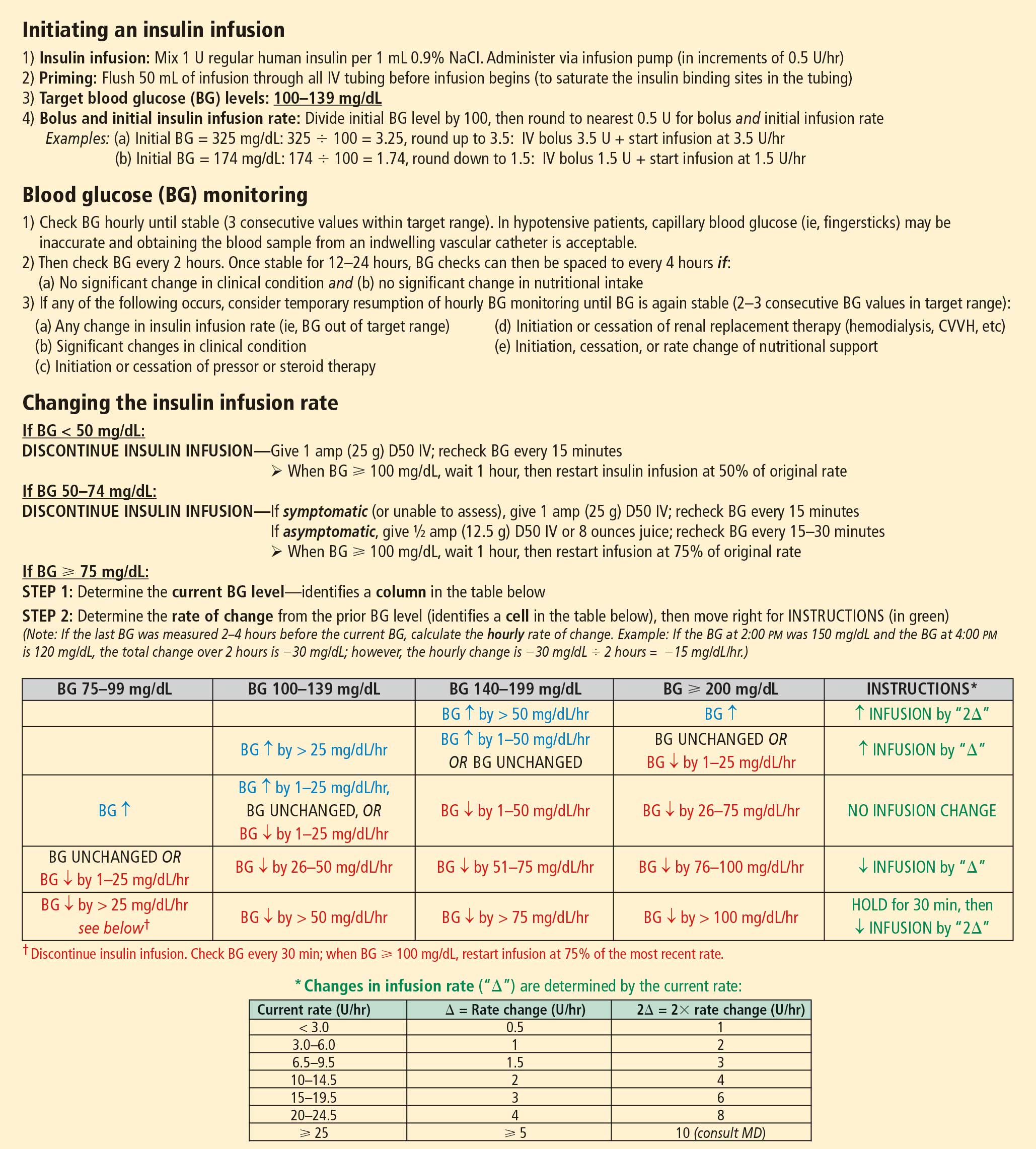
Several different algorithms for IV regular insulin therapy are in use. Some are static, such as those of Markovitz et al14 and Stockton et al,15 while others are dynamic (ie, doses are self-adjusted based on changes in blood glucose level), such as the “Yale protocol” of Goldberg et al (Figure 1).16
POSTOPERATIVE GLYCEMIC MANAGEMENT
Start subcutaneous transition before stopping IV drip
Transitioning from IV to subcutaneous insulin is often complicated. Nonoral nutrition options (ie, parenteral nutrition or enteral supplementation) must be considered. As noted, insulin must be replaced according to physiologic needs, which requires that a long-acting basal insulin be used regardless of oral intake status, a rapid-acting insulin be given to cover prandial or nutritional needs, and supplemental rapid-acting insulin be used to correct hyperglycemia.
In the transition from IV insulin, basal insulin replacement can begin at any time. I recommend starting the transition from IV to subcutaneous insulin about 12 to 24 hours before discontinuing the insulin drip. In type 1 diabetes, this transition ensures basal insulin coverage and minimizes the risk of developing ketones and ketoacidosis. In type 2 diabetes, it can ensure a more stable transition and better glycemic control.
Determining the basal insulin dose
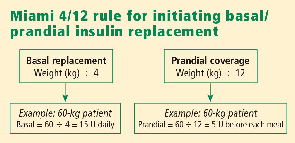
Switching to subcutaneous supplemental insulin
Instructions must be given for switching to subcutaneous supplemental doses of insulin. Glycemic targets, generally from less than 130 to 150 mg/dL, must be established, as must the frequency of fingerstick testing:
- If the patient is being fed enterally or parenterally, fingerstick testing is recommended every 4 to 6 hours if a rapid-acting insulin analog is used and every 6 hours if regular insulin is used.
- If the patient is eating, fingerstick testing should be performed before meals and at bedtime.
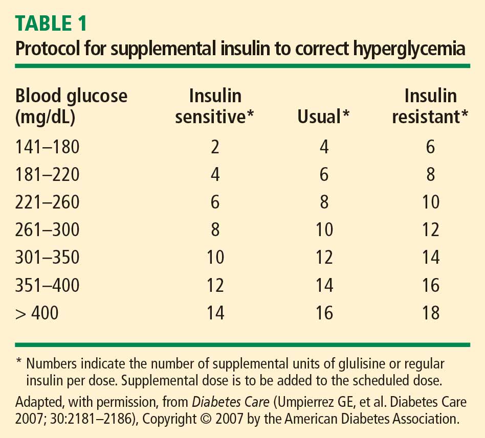
Covering nutritional requirements
Nutrition-related insulin needs depend on the type of caloric intake prescribed:
In patients receiving total parenteral nutrition (TPN), start 1 U of regular insulin (placed in the bag) for every 10 to 15 g of dextrose in the TPN mixture.
In patients receiving enteral nutrition, use regular insulin every 6 hours or a rapid-acting insulin analog every 4 hours. Start 1 U of insulin subcutaneously for every 10 to 15 g of delivered carbohydrates. For example, if a patient is receiving 10 g of carbohydrates per hour, a rapid-acting analog given at a dose of 4 U every 4 hours (1 U per 10 g of carbohydrates) should adequately cover enteral feedings. For any bolus feedings, give the injection as a full bolus 15 to 20 minutes in advance, based on the carbohydrate content of the feeding.
In patients who are eating, use regular insulin or a rapid-acting insulin analog before meals. Again, start 1 U of insulin subcutaneously for every 10 to 15 g of carbohydrates, or use the prandial portion of the Miami 4/12 rule (Figure 2). For example, in a 60-kg patient one would start with 5 U (60 ÷ 12) of a rapid-acting insulin before each meal.
Basal/bolus replacement outperforms supplemental-scale regular insulin
Use of a basal/bolus insulin regimen appears to be more beneficial than supplemental-scale regular insulin in hospitalized patients with type 2 diabetes, according to a recent randomized trial comparing the two approaches in 130 such patients with blood glucose levels greater than 140 mg/dL.17 In the group randomized to basal/bolus insulin, the starting total daily dose was 0.4 to 0.5 U/kg/day, with half the dose given as basal insulin (insulin glargine) once daily and half given as a rapid-acting insulin analog (glulisine) in fixed doses before every meal. A rapid-acting analog was used for supplemental insulin in the basal/bolus regimen. By study’s end, patients in the basal/bolus group were receiving a higher total daily insulin dose than those in the supplemental-scale group (mean of 42 U/day vs 13 U/day).
Mean daily blood glucose levels were 27 mg/dL lower, on average, in patients who received basal/bolus therapy compared with the supplemental-scale group, yet there was no difference between groups in the risk of hypoglycemia. More patients randomized to basal/bolus therapy achieved the glycemic goal of less than 140 mg/dL (66% vs 38%). Fourteen percent of patients assigned to supplemental-scale insulin had values persistently greater than 240 mg/dL and had to be switched to the basal/bolus regimen.17
SUMMARY
Perioperative glycemic control can reduce morbidity, particularly the incidence of infectious complications, in surgical patients, even in those without diagnosed diabetes. Optimal management of glycemia in the perioperative period involves applying principles of physiologic insulin replacement. Postoperatively, the transition from IV to subcutaneous insulin can be achieved through the use of basal insulin for coverage of fasting insulin needs, regardless of the patient’s feeding status, and the use of rapid-acting insulin to cover hyperglycemia and nutritional needs. Management of hospitalized patients exclusively with supplemental-scale regular insulin should be abandoned.
DISCUSSION
Question from the audience: As an attending physician in a preoperative clinic I’m never sure what to do with NPH insulin the morning of surgery. What guidance can you give?
Dr. Meneghini: NPH is a peaking basal insulin, and the peak can induce hypoglycemia in a patient who is NPO. If we have the opportunity, we try to switch patients previously receiving insulin therapy to a long-acting basal insulin analog, which has a much flatter action profile and is safer in the fasting state. If there is no opportunity for switching, we instruct the patient to take two-thirds of his or her usual morning dose of insulin and we initiate a D5 drip when the patient arrives at the hospital.
Question from the audience: How do you handle perioperative insulin in patients on insulin pumps?
Dr. Meneghini: The pumps provide a subcutaneous basal insulin infusion, which should, if set correctly, maintain stable blood glucose levels when the patient is NPO. Supplemental doses of insulin to correct hyperglycemia can be delivered via the usual subcutaneous practice with a syringe or insulin pen. If you are uncomfortable with pump function, or if the pump insertion site interferes with the surgery site, simply replace the 24-hour basal amount delivered via pump with an injection of glargine or detemir divided into twice-daily injections. Correct hyperglycemia with supplemental-scale insulin as per usual protocol.
Question from the audience: The manufacturer of insulin glargine makes no recommendations for its use the night before or morning of surgery. What do you recommend?
Dr. Meneghini: It depends on whether the glargine is dosed appropriately. Most patients with type 2 diabetes require 0.4 to 0.6 U/kg/day of a long-acting insulin. If they’re on much more, they may be overdosed, and I would cut the basal dose by about half. Otherwise, 75% to 100% of the usual basal amount is appropriate. In type 1 diabetes, the usual replacement dose of basal insulin is 0.2 to 0.3 U/kg/day. If a patient is in this range, the basal insulin can be continued. Patients who experience hypoglycemia, or a substantial fall in blood glucose if meals are skipped or delayed, may be getting too much basal insulin and might benefit from a dose reduction when placed on NPO status.
Question from the audience: Metformin has a black-box warning advising that it be stopped at least 48 hours before surgery, but patients often come to surgery having taken metformin within the prior 12 to 24 hours. How should we manage such patients coming for elective surgery?
Dr. Meneghini: Metformin is cleared exclusively by the kidneys; its accumulation as a result of impaired kidney function (eg, due to hemodynamic instability or radiology studies using IV iodine) can result in increased lactic acid production by the liver and lactic acidosis. A patient who has taken metformin within the prior 48 hours but doesn’t have a risk of hemodynamic dysfunction is at low risk of lactic acidosis if hydrated appropriately. There’s not much choice if a patient needs urgent surgery and has recently taken metformin; in that case, just ensure maintenance of adequate glomerular filtration via fluid repletion to clear the drug.
Question from the audience: What’s the evidence for tight glycemic control or any type of glycemic control in patients undergoing outpatient surgery or “same-day” patients who will be admitted to a regular surgical floor? Also, what would you consider maximal glucose values for a patient going into elective surgery?
Dr. Meneghini: I haven’t seen any guidelines for glycemic control in patients undergoing outpatient surgery. If a patient has poor glycemic control coming into surgery, even for a minor procedure, the risk of an infectious complication may be increased. Keeping blood glucose below 180 mg/dL and avoiding electrolyte imbalances is likely sufficient in such patients. On the second question, if it’s an elective procedure and can be delayed a few hours, you can certainly institute IV insulin therapy to correct hyperglycemia rapidly—just ensure adequate replacement of fluids since the patient may have had volume depletion or dehydration as a result of the preceding osmotic diuresis. Once glycemic control is improved (blood glucose < 180–200 mg/dL), the patient can proceed to surgery.
Question from the audience: What are your recommendations for resuming oral diabetes medications after surgery?
Dr. Meneghini: Once patients are tolerating their meals and being considered for discharge, you may want to resume their oral medications, assuming their admission hemoglobin A1c levels were near goal. If glycemic control was inadequate preoperatively, this may be a good opportunity to adjust their prior regimen to more appropriate therapy. In some cases, this might include some form of insulin, either basal therapy or basal and supplemental insulin.
- Clement S, Braithwaite SS, Magee MF, et al. Management of diabetes and hyperglycemia in hospitals. Diabetes Care 2004; 27:553–591.
- Kaparianos A, Argyropoulou E, Sampsonas F, et al. Pulmonary complications in diabetes mellitus. Chron Respir Dis 2008; 5:101–108.
- Grigoleit HG. Anesthesia and blood glucose. Acta Diabetologica 1973; 10:569–574.
- Dronge AS, Perkal MF, Kancir S, et al. Long-term glycemic control and postoperative infectious complications. Arch Surg 2006; 141:375–380.
- Zerr KJ, Furnary AP, Grunkemeier GL, Bookin S, Kanhere V, Starr A. Glucose control lowers the risk of wound infection in diabetics after open heart operations. Ann Thorac Surg 1997; 63:356–361.
- Furnary AP, Gao G, Grunkemeier GL, et al. Continuous insulin infusion reduces mortality in patients with diabetes undergoing coronary artery bypass grafting. J Thorac Cardiovasc Surg 2003; 125:1007–10021.
- Ainla T, Baburin A, Tessalu R, et al. The association between hyperglycaemia on admission and 180-day mortality in acute myocardial infarction patients with and without diabetes. Diabet Med 2005; 22:1321–1325.
- Rady MY, Johnson DJ, Patel BM, et al. Influence of individual characteristics on outcome of glycemic control in intensive care unit patients with or without diabetes mellitus. Mayo Clin Proc 2005; 80:1558–1567.
- Van den Berghe G, Wouters P, Weekers F, et al. Intensive insulin therapy in the critically ill patients. N Engl J Med 2001; 345:1359–1367.
- Van den Berghe G, Wouters PJ, Kesteloot K, Hilleman DE. Analysis of healthcare resource utilization with intensive insulin therapy in critically ill patients. Crit Care Med 2006; 34:612–616.
- Van den Berghe G, Wawilmer A, Hermans G, et al. Intensive insulin therapy in the medical ICU. New Engl J Med 2006; 354:449–461.
- NICE-SUGAR Study Investigators. Intensive vs conventional glucose control in critically ill patients. N Engl J Med 2009; 360:1283–1297.
- Moghissi ES, Korytowski MT, DiNardo M, et al. American Association of Clinical Endocrinologists and American Diabetes Association consensus statement on inpatient glycemic control. Endocr Pract 2009; 15:353–369.
- Markovitz LJ, Wiechmann RJ, Harris N, et al. Description and evaluation of a glycemic management protocol for patients with diabetes undergoing heart surgery. Endocr Pract 2002; 8:10–18.
- Stockton L, Baird M, Cook CB, et al. Development and implementation of evidence-based guidelines for IV insulin: a statewide collaborative approach. Insulin 2008; 3:67–77.
- Goldberg PA, Siegel MD, Sherwin RS, et al. Implementation of a safe and effective insulin infusion protocol in a medical intensive care unit. Diabetes Care 2004; 27:461–467.
- Umpierrez GE, Smiley D, Zisman A, et al. Randomized study of basal-bolus insulin therapy in the inpatient management of patients with type 2 diabetes (RABBIT 2 trial). Diabetes Care 2007; 30:2181–2186.
Diabetes confers an increased risk of perioperative morbidity and mortality, mostly from infection and cardiovascular events. It is not unusual for surgical patients with diabetes to have a number of comorbidities or underlying chronic vascular complications that put them at risk for cardiovascular events or an infectious complication. Silent ischemia, coronary artery disease, and autonomic neuropathy are common among patients with diabetes, and each can contribute to perioperative morbidity and mortality. These are important considerations since nearly one-fifth of surgical patients have diabetes and since a person with diabetes has a 50% risk of undergoing surgery at some point in his or her lifetime.1
This article reviews the preoperative evaluation of patients with diabetes, discusses the relation between glycemic control and perioperative outcomes, and examines targets and strategies for glycemic control in patients with type 1 and type 2 diabetes throughout the perioperative period.
PREOPERATIVE EVALUATION
The preoperative evaluation must consider first and foremost the status of the patient’s diabetes and his or her surgical risk factors. Also important are the characteristics of the procedure to be performed, the method of anesthesia to be used, and select laboratory values.
Diabetes status
The type of diabetes and its treatment must be considered. Type 1 diabetes requires continuous insulin therapy to prevent ketoacidosis; patients with type 2 diabetes are usually treated with oral medications with or without insulin. Baseline control of blood glucose is a predictor of morbidity following surgery. Hypoglycemia is associated with increased morbidity in the inpatient setting, so a history of severe hypoglycemic events or of difficulty recognizing hypoglycemia (hypoglycemia unawareness) should be elicited in the preoperative evaluation. Complications of diabetes and other comorbidities also must be evaluated, along with their treatments.
Surgical risk factors
Patients with diabetes have surgical risk factors specific to their health—namely, cardiovascular risk factors that may or may not have been previously diagnosed. Patients with diabetes may have silent ischemia, atypical manifestations of coronary ischemia, or underlying cardiomyopathy. Many patients with type 2 diabetes have hypertension, which may complicate perioperative management. Other common surgical risk factors in this population include obesity, chronic kidney disease, and undiagnosed autonomic dysfunction, which may compromise hemodynamic stability in the perioperative period. Additionally, patients with long-standing diabetes experience reductions in pulmonary function (eg, forced expiratory volume, peak expiratory flow, and diffusion capacity for carbon monoxide) related to disease duration and vascular injury,2 which may complicate weaning from ventilatory support.
Characteristics of the procedure and anesthetic
Both surgery and anesthesia may induce an increase in levels of stress hormones (epinephrine, cortisol, growth hormone) and inflammatory cytokines (interleukin-6 and tumor necrosis factor–alpha), resulting in insulin resistance and impaired insulin secretion (even among patients who present with adequate insulin secretion). These in turn contribute to lipolysis and protein catabolism, leading to hyperglycemia and, if a patient is severely insulin deficient, ketoacidosis. Other factors that particularly affect insulin resistance and secretion include cardiovascular bypass surgery, sepsis, the need for total parenteral nutrition, and steroid therapy.
The characteristics of the surgical procedure, including the type of surgery as well as its urgency, duration, and timing (morning vs later in the day), are important in planning for perioperative glycemic management. For example, a short, minor procedure may require only observation, whereas more extensive procedures warrant periodic monitoring and active glycemic management with insulin infusions.
The type of anesthesia should also be considered. Compared with epidural anesthesia, general anesthesia is associated with greater stimulation of the sympathetic nervous system and increased catecholamine levels, resulting in more pronounced hyperglycemia.3
Preoperative tests
Preoperative testing and laboratory evaluation should include, at minimum, an electrocardiogram, a basic metabolic panel to assess renal function, electrolyte levels, and hemoglobin A1c measurement. For low-risk procedures in patients with adequate exercise tolerance, no diagnostic tests might be needed. In any case, knowledge of the hemoglobin A1c level may help not only to classify perioperative risk but also to determine postoperative care, including the choice of antiglycemic medications at discharge.
IMPORTANCE OF GLYCEMIC CONTROL
Preoperative glycemic control has a significant impact on the risk of infectious complications—including pneumonia, wound infection, urinary tract infection, and sepsis—in patients with diabetes across a variety of surgical procedures.4 Similarly, postoperative glycemic control—to a mean blood glucose level less than 200 mg/dL in the immediate postoperative period—significantly reduces the incidence of deep sternal wound infection after open heart surgery.5
Among patients undergoing cardiothoracic surgery, both cardiac-related and overall mortality are greater with increasing postoperative blood glucose levels, although a cause-and-effect relationship has not been established.6
Glycemic control matters regardless of diabetes status
Hyperglycemia affects mortality regardless of diabetes status. In a study of 779 consecutive patients admitted for acute myocardial infarction, mortality at 180 days was highly associated with hyperglycemia on admission independent of a history of diabetes; the highest mortality was among hyperglycemic patients without previously known diabetes.7 Similarly, a large study of glycemic control in intensive care unit (ICU) patients receiving insulin found that mortality in nondiabetic patients increased with median glucose level and was higher than mortality in diabetic patients.8 These findings suggest a need for vigilance in the perioperative and critical care management of all patients with hyperglycemia, regardless of preadmission diabetes diagnosis, as they carry significant morbidity and mortality risk.
GLYCEMIC CONTROL IN THE CRITICALLY ILL: SOME SUPPORT FOR A MODIFIED TARGET, BUT VIGILANCE FOR HYPOGLYCEMIA NEEDED
The landmark study by Van den Berghe et al of intensive insulin therapy in surgical ICU patients demonstrated significant reductions in morbidity and mortality when glucose levels were controlled aggressively (80 to 110 mg/dL; average, 103 mg/dL) compared with conventional control (180 to 200 mg/dL).9 The benefit of intensive glycemic control was evident on outcomes such as the occurrence of sepsis, need for dialysis, need for blood transfusion, and development of acute polyneuropathy. Intensive insulin therapy was also associated with cost savings compared with conventional insulin therapy in mechanically ventilated patients.10
However, a number of subsequent studies have clearly shown that as blood glucose levels approach normoglycemia, the risks of hypoglycemia, especially severe hypoglycemia, can offset the benefits of tight blood glucose control.
A follow-up study by Ven den Berghe et al in a medical ICU failed to show a mortality benefit from tight glycemic control, though patients in the intensive control arm experienced less renal injury, faster weaning from ventilation, and earlier discharge from the ICU and hospital.11
The recent NICE-SUGAR study of aggressive glucose control in the ICU randomized patients to a target blood glucose of 81 to 108 mg/dL (intensive group) or 180 mg/dL or less (control group).12 At study’s end, the groups’ mean blood glucose levels were 115 mg/dL and 144 mg/dL, respectively, while rates of severe hypoglycemia (blood glucose < 40 mg/dL) were 6.8% and 0.5%, respectively. Mortality rates were higher in the intensive therapy group (27.5%) than in the control group (24.9%), driven by severe hypoglycemic events. Notably, blood glucose monitoring in this and other studies was conducted at a frequency of anywhere between 1 and 4 hours.
The conclusions of the available data would support, for the time being, a modified glycemic target in critically ill patients, with strict avoidance of severe hypoglycemia. The recent consensus statement from the American Association of Clinical Endocrinologists and the American Diabetes Association recommends using insulin therapy if blood glucose levels exceed 180 mg/dL, with target glucose levels less than 180 mg/dL in critically ill patients and less than 140 mg/dL in non–critically ill patients.13 Development and implementation of safer insulin infusion algorithms and more frequent and accurate blood glucose monitoring in this setting should enable us to achieve better glycemic targets with lower risk.
ELEMENTS OF PHYSIOLOGIC INSULIN REPLACEMENT
In hospitalized patients with hyperglycemia, three different components of insulin replacement require management1:
Basal insulin replacement consists of a long-acting insulin preparation administered regardless of the patient’s oral intake status, with the premise of matching hepatic (endogenous) glucose production
Prandial insulin replacement requires a rapid-acting insulin preparation given to cover nutritional needs
Supplemental (or correction) insulin replacement requires a rapid-acting preparation (usually the same insulin type as for prandial coverage) to correct blood glucose values that exceed predetermined glycemic targets.
For most patients, basal insulin replacement might be appropriate preoperatively to control fasting glucose, whereas during surgery, especially if prolonged or high risk, an intravenous (IV) insulin drip is the most effective means of glucose control. The postoperative transition from the IV insulin drip usually involves basal insulin replacement plus supplemental rapid-acting insulin. Prandial or nutritional insulin should be started once the patient begins to receive nutrition (oral, enteral, or hyperalimentation).
GOALS OF PERIOPERATIVE GLYCEMIC CONTROL
Perioperative glycemic management has several key objectives:
- Avoidance of clinically significant hyper- or hypoglycemia
- Maintenance of electrolyte and fluid balance
- Prevention of ketoacidosis, which is imperative in patients with type 1 diabetes, who require insulin at all times
- Achievement of specific glycemic targets, as discussed above—ie, less than 180 mg/dL in critically ill patients and less than 140 mg/dL in stable patients.13
Strategies differ across the perioperative timeline
Strategies for perioperative glycemic control differ before, during, and after surgery, as summarized immediately below and detailed in the following sections.
Preoperatively, glycemia should be stabilized, typically with subcutaneous insulin, if there is enough time to do so. For patients who have not previously been on insulin, placing them on an insulin supplemental scale to correct glycemia to desired targets might be a first step. In the setting of hyperglycemia, these patients may also be started on a low dose of basal insulin, with preference given to basal insulin analogs, given their consistent and relatively peakless action profile and lower risk of hypoglycemia. A starting dose of 0.2 to 0.4 U/kg is appropriate and carries a low risk of hypoglycemia. For patients already using insulin on an outpatient basis, continuing their basal insulin dose, possibly at a reduced dosage (25% less), together with supplemental-scale insulin coverage, should stabilize blood glucose levels. For patients on combination insulin or premixed insulin types, the basal insulin dose for preoperative management can be estimated by taking the patient’s usual total daily dose and delivering 40% to 50% of that dose as a basal insulin analog injection. Clearly, a supplemental scale should be implemented along with basal insulin replacement.
Intraoperatively, switching to IV insulin may be appropriate for stabilizing glycemia, depending on the type of surgery. A number of IV insulin protocols have been proposed, although no consistent comparisons of efficacy or safety among these protocols have been published.
Postoperatively, patients eventually should be transitioned from IV to subcutaneous insulin when glycemic control stabilizes. This transition may be complicated for many reasons. Oral intake may be inconsistent. The surgery and surrounding environment can induce stressors, promote susceptibility to infection, and increase insulin resistance. Additionally, some patients may be on hyperalimentation. Specific instructions for the transition from IV to subcutaneous insulin are covered later in this article.
PREOPERATIVE GLYCEMIC MANAGEMENT
In patients with type 2 diabetes, oral agents pose certain safety risks and should be discontinued prior to surgery.
Sulfonylureas may induce hypoglycemia in patients who are placed on NPO (“nothing by mouth”) orders and should be held in patients who are fasting.
Metformin can induce lactic acidosis if kidney function declines and should be withheld 1 to 2 days before planned surgery if a need for IV contrast is anticipated or the procedure could potentially lead to hemodynamic instability and reduced renal perfusion.
Thiazolidinediones may cause fluid retention that can complicate the postoperative period; they can be discontinued several days prior to a planned surgery.
GLP-1 agonists, such as exenatide, can slow gastric motility and potentially delay gastrointestinal recovery after major surgery; they should be held the day of surgery.
DPP-4 inhibitors (incretin enhancers), such as sitagliptin, do not have significant side effects and, if need be, can be continued. Because incretin therapies act via a glucose-dependent mechanism, they are unlikely to cause hypoglycemia, even in a patient whose oral intake is held or delayed. On the other hand, since their effect is mostly in reducing postprandial glycemia, there may be little need to use them in a patient who is NPO.
Patients with type 1 diabetes must continue basal insulin replacement preoperatively (0.2 to 0.3 U/kg/day of a long-acting insulin). Patients with type 2 diabetes may benefit from basal insulin replacement, as previously noted.
Supplemental insulin scales are used to correct hyperglycemia regardless of a patient’s oral intake status. They can be individualized based on the estimated total daily insulin dose and require glycemic targets to be established. Fingerstick glucose monitoring should be done every 4 to 6 hours in a patient who is NPO, and supplemental-scale insulin should be used to correct glucose values that exceed target. For supplemental-scale coverage, rapid-acting insulin analogs have a shorter duration of action than human regular insulin and may be given subcutaneously every 4 to 6 hours, whereas regular insulin should not be given more often than every 6 hours to correct hyperglycemia. These differences in action duration should be kept in mind to minimize the potential for insulin stacking.
INTRAOPERATIVE GLYCEMIC MANAGEMENT
Procedure length is an important determinant
Strategies for intraoperative glucose management vary according to the length of the procedure.
For minor, short procedures, the preoperative glucose management orders may be continued.
For longer, more complex procedures, a switch to an IV insulin drip is safe and allows rapid adjustments in dosing and plasma glucose levels. Ideally, IV insulin is started prior to the procedure so that the glucose level is stable once the patient arrives in the operating room. Given the logistics of IV insulin management, including the need for frequent monitoring (hourly) and dose adjustments, this type of treatment should be reserved for environments with adequate numbers of trained staff.
IV regular insulin is therapy of choice
Several different algorithms for IV regular insulin therapy are in use. Some are static, such as those of Markovitz et al14 and Stockton et al,15 while others are dynamic (ie, doses are self-adjusted based on changes in blood glucose level), such as the “Yale protocol” of Goldberg et al (Figure 1).16
POSTOPERATIVE GLYCEMIC MANAGEMENT
Start subcutaneous transition before stopping IV drip
Transitioning from IV to subcutaneous insulin is often complicated. Nonoral nutrition options (ie, parenteral nutrition or enteral supplementation) must be considered. As noted, insulin must be replaced according to physiologic needs, which requires that a long-acting basal insulin be used regardless of oral intake status, a rapid-acting insulin be given to cover prandial or nutritional needs, and supplemental rapid-acting insulin be used to correct hyperglycemia.
In the transition from IV insulin, basal insulin replacement can begin at any time. I recommend starting the transition from IV to subcutaneous insulin about 12 to 24 hours before discontinuing the insulin drip. In type 1 diabetes, this transition ensures basal insulin coverage and minimizes the risk of developing ketones and ketoacidosis. In type 2 diabetes, it can ensure a more stable transition and better glycemic control.
Determining the basal insulin dose
Switching to subcutaneous supplemental insulin
Instructions must be given for switching to subcutaneous supplemental doses of insulin. Glycemic targets, generally from less than 130 to 150 mg/dL, must be established, as must the frequency of fingerstick testing:
- If the patient is being fed enterally or parenterally, fingerstick testing is recommended every 4 to 6 hours if a rapid-acting insulin analog is used and every 6 hours if regular insulin is used.
- If the patient is eating, fingerstick testing should be performed before meals and at bedtime.
Covering nutritional requirements
Nutrition-related insulin needs depend on the type of caloric intake prescribed:
In patients receiving total parenteral nutrition (TPN), start 1 U of regular insulin (placed in the bag) for every 10 to 15 g of dextrose in the TPN mixture.
In patients receiving enteral nutrition, use regular insulin every 6 hours or a rapid-acting insulin analog every 4 hours. Start 1 U of insulin subcutaneously for every 10 to 15 g of delivered carbohydrates. For example, if a patient is receiving 10 g of carbohydrates per hour, a rapid-acting analog given at a dose of 4 U every 4 hours (1 U per 10 g of carbohydrates) should adequately cover enteral feedings. For any bolus feedings, give the injection as a full bolus 15 to 20 minutes in advance, based on the carbohydrate content of the feeding.
In patients who are eating, use regular insulin or a rapid-acting insulin analog before meals. Again, start 1 U of insulin subcutaneously for every 10 to 15 g of carbohydrates, or use the prandial portion of the Miami 4/12 rule (Figure 2). For example, in a 60-kg patient one would start with 5 U (60 ÷ 12) of a rapid-acting insulin before each meal.
Basal/bolus replacement outperforms supplemental-scale regular insulin
Use of a basal/bolus insulin regimen appears to be more beneficial than supplemental-scale regular insulin in hospitalized patients with type 2 diabetes, according to a recent randomized trial comparing the two approaches in 130 such patients with blood glucose levels greater than 140 mg/dL.17 In the group randomized to basal/bolus insulin, the starting total daily dose was 0.4 to 0.5 U/kg/day, with half the dose given as basal insulin (insulin glargine) once daily and half given as a rapid-acting insulin analog (glulisine) in fixed doses before every meal. A rapid-acting analog was used for supplemental insulin in the basal/bolus regimen. By study’s end, patients in the basal/bolus group were receiving a higher total daily insulin dose than those in the supplemental-scale group (mean of 42 U/day vs 13 U/day).
Mean daily blood glucose levels were 27 mg/dL lower, on average, in patients who received basal/bolus therapy compared with the supplemental-scale group, yet there was no difference between groups in the risk of hypoglycemia. More patients randomized to basal/bolus therapy achieved the glycemic goal of less than 140 mg/dL (66% vs 38%). Fourteen percent of patients assigned to supplemental-scale insulin had values persistently greater than 240 mg/dL and had to be switched to the basal/bolus regimen.17
SUMMARY
Perioperative glycemic control can reduce morbidity, particularly the incidence of infectious complications, in surgical patients, even in those without diagnosed diabetes. Optimal management of glycemia in the perioperative period involves applying principles of physiologic insulin replacement. Postoperatively, the transition from IV to subcutaneous insulin can be achieved through the use of basal insulin for coverage of fasting insulin needs, regardless of the patient’s feeding status, and the use of rapid-acting insulin to cover hyperglycemia and nutritional needs. Management of hospitalized patients exclusively with supplemental-scale regular insulin should be abandoned.
DISCUSSION
Question from the audience: As an attending physician in a preoperative clinic I’m never sure what to do with NPH insulin the morning of surgery. What guidance can you give?
Dr. Meneghini: NPH is a peaking basal insulin, and the peak can induce hypoglycemia in a patient who is NPO. If we have the opportunity, we try to switch patients previously receiving insulin therapy to a long-acting basal insulin analog, which has a much flatter action profile and is safer in the fasting state. If there is no opportunity for switching, we instruct the patient to take two-thirds of his or her usual morning dose of insulin and we initiate a D5 drip when the patient arrives at the hospital.
Question from the audience: How do you handle perioperative insulin in patients on insulin pumps?
Dr. Meneghini: The pumps provide a subcutaneous basal insulin infusion, which should, if set correctly, maintain stable blood glucose levels when the patient is NPO. Supplemental doses of insulin to correct hyperglycemia can be delivered via the usual subcutaneous practice with a syringe or insulin pen. If you are uncomfortable with pump function, or if the pump insertion site interferes with the surgery site, simply replace the 24-hour basal amount delivered via pump with an injection of glargine or detemir divided into twice-daily injections. Correct hyperglycemia with supplemental-scale insulin as per usual protocol.
Question from the audience: The manufacturer of insulin glargine makes no recommendations for its use the night before or morning of surgery. What do you recommend?
Dr. Meneghini: It depends on whether the glargine is dosed appropriately. Most patients with type 2 diabetes require 0.4 to 0.6 U/kg/day of a long-acting insulin. If they’re on much more, they may be overdosed, and I would cut the basal dose by about half. Otherwise, 75% to 100% of the usual basal amount is appropriate. In type 1 diabetes, the usual replacement dose of basal insulin is 0.2 to 0.3 U/kg/day. If a patient is in this range, the basal insulin can be continued. Patients who experience hypoglycemia, or a substantial fall in blood glucose if meals are skipped or delayed, may be getting too much basal insulin and might benefit from a dose reduction when placed on NPO status.
Question from the audience: Metformin has a black-box warning advising that it be stopped at least 48 hours before surgery, but patients often come to surgery having taken metformin within the prior 12 to 24 hours. How should we manage such patients coming for elective surgery?
Dr. Meneghini: Metformin is cleared exclusively by the kidneys; its accumulation as a result of impaired kidney function (eg, due to hemodynamic instability or radiology studies using IV iodine) can result in increased lactic acid production by the liver and lactic acidosis. A patient who has taken metformin within the prior 48 hours but doesn’t have a risk of hemodynamic dysfunction is at low risk of lactic acidosis if hydrated appropriately. There’s not much choice if a patient needs urgent surgery and has recently taken metformin; in that case, just ensure maintenance of adequate glomerular filtration via fluid repletion to clear the drug.
Question from the audience: What’s the evidence for tight glycemic control or any type of glycemic control in patients undergoing outpatient surgery or “same-day” patients who will be admitted to a regular surgical floor? Also, what would you consider maximal glucose values for a patient going into elective surgery?
Dr. Meneghini: I haven’t seen any guidelines for glycemic control in patients undergoing outpatient surgery. If a patient has poor glycemic control coming into surgery, even for a minor procedure, the risk of an infectious complication may be increased. Keeping blood glucose below 180 mg/dL and avoiding electrolyte imbalances is likely sufficient in such patients. On the second question, if it’s an elective procedure and can be delayed a few hours, you can certainly institute IV insulin therapy to correct hyperglycemia rapidly—just ensure adequate replacement of fluids since the patient may have had volume depletion or dehydration as a result of the preceding osmotic diuresis. Once glycemic control is improved (blood glucose < 180–200 mg/dL), the patient can proceed to surgery.
Question from the audience: What are your recommendations for resuming oral diabetes medications after surgery?
Dr. Meneghini: Once patients are tolerating their meals and being considered for discharge, you may want to resume their oral medications, assuming their admission hemoglobin A1c levels were near goal. If glycemic control was inadequate preoperatively, this may be a good opportunity to adjust their prior regimen to more appropriate therapy. In some cases, this might include some form of insulin, either basal therapy or basal and supplemental insulin.
- Clement S, Braithwaite SS, Magee MF, et al. Management of diabetes and hyperglycemia in hospitals. Diabetes Care 2004; 27:553–591.
- Kaparianos A, Argyropoulou E, Sampsonas F, et al. Pulmonary complications in diabetes mellitus. Chron Respir Dis 2008; 5:101–108.
- Grigoleit HG. Anesthesia and blood glucose. Acta Diabetologica 1973; 10:569–574.
- Dronge AS, Perkal MF, Kancir S, et al. Long-term glycemic control and postoperative infectious complications. Arch Surg 2006; 141:375–380.
- Zerr KJ, Furnary AP, Grunkemeier GL, Bookin S, Kanhere V, Starr A. Glucose control lowers the risk of wound infection in diabetics after open heart operations. Ann Thorac Surg 1997; 63:356–361.
- Furnary AP, Gao G, Grunkemeier GL, et al. Continuous insulin infusion reduces mortality in patients with diabetes undergoing coronary artery bypass grafting. J Thorac Cardiovasc Surg 2003; 125:1007–10021.
- Ainla T, Baburin A, Tessalu R, et al. The association between hyperglycaemia on admission and 180-day mortality in acute myocardial infarction patients with and without diabetes. Diabet Med 2005; 22:1321–1325.
- Rady MY, Johnson DJ, Patel BM, et al. Influence of individual characteristics on outcome of glycemic control in intensive care unit patients with or without diabetes mellitus. Mayo Clin Proc 2005; 80:1558–1567.
- Van den Berghe G, Wouters P, Weekers F, et al. Intensive insulin therapy in the critically ill patients. N Engl J Med 2001; 345:1359–1367.
- Van den Berghe G, Wouters PJ, Kesteloot K, Hilleman DE. Analysis of healthcare resource utilization with intensive insulin therapy in critically ill patients. Crit Care Med 2006; 34:612–616.
- Van den Berghe G, Wawilmer A, Hermans G, et al. Intensive insulin therapy in the medical ICU. New Engl J Med 2006; 354:449–461.
- NICE-SUGAR Study Investigators. Intensive vs conventional glucose control in critically ill patients. N Engl J Med 2009; 360:1283–1297.
- Moghissi ES, Korytowski MT, DiNardo M, et al. American Association of Clinical Endocrinologists and American Diabetes Association consensus statement on inpatient glycemic control. Endocr Pract 2009; 15:353–369.
- Markovitz LJ, Wiechmann RJ, Harris N, et al. Description and evaluation of a glycemic management protocol for patients with diabetes undergoing heart surgery. Endocr Pract 2002; 8:10–18.
- Stockton L, Baird M, Cook CB, et al. Development and implementation of evidence-based guidelines for IV insulin: a statewide collaborative approach. Insulin 2008; 3:67–77.
- Goldberg PA, Siegel MD, Sherwin RS, et al. Implementation of a safe and effective insulin infusion protocol in a medical intensive care unit. Diabetes Care 2004; 27:461–467.
- Umpierrez GE, Smiley D, Zisman A, et al. Randomized study of basal-bolus insulin therapy in the inpatient management of patients with type 2 diabetes (RABBIT 2 trial). Diabetes Care 2007; 30:2181–2186.
KEY POINTS
- Surgery and anesthesia can induce hormonal and inflammatory stressors that increase the risk of complications in patients with diabetes.
- Elevated blood glucose levels are associated with worse outcomes in surgical patients, even among those not diagnosed with diabetes.
- The perioperative glycemic target in critically ill patients is 140 to 180 mg/dL. Evidence for a target in patients who are not critically ill is less robust, though fasting levels less than 140 mg/dL and random levels less than 180 mg/dL are appropriate.
- Postoperative nutrition-related insulin needs vary by nutrition type (parenteral or enteral), but ideally all regimens should incorporate a basal/bolus approach to insulin replacement.
Postoperative pulmonary complications: An update on risk assessment and reduction
Although pulmonary complications are not as well studied as cardiac complications in the postoperative setting, they are just as common following noncardiac surgery and are even more costly. It is worthwhile to identify surgical patients most at risk of postoperative pulmonary complications and take measures known to mitigate risk. This paper discusses important risk factors to identify during a preoperative pulmonary evaluation and then focuses on recent advances in strategies for reducing postoperative pulmonary complications. Teaching questions are included throughout, along with the rationale behind their answers.
POSTOPERATIVE PULMONARY COMPLICATIONS: WHAT ARE WE TRYING TO PREVENT AND WHY?
The definition of postoperative pulmonary complications is more variable and less intuitive than that of cardiac complications. Cardiac complications—postoperative myocardial infarction, cardiac death, and pulmonary edema—are more consistently defined and measured in clinical trials. Studies of postoperative pulmonary complications often group together pneumonia, respiratory failure, atelectasis, bronchospasm, and exacerbation of chronic obstructive pulmonary disease (COPD), making it more difficult to individually evaluate risk factors for different outcomes.
There are several reasons why it is important to consider pulmonary risk when evaluating patients preoperatively:
Pulmonary complications are as common as cardiac complications following noncardiac surgery. For example, in a secondary analysis of the cohort of noncardiac surgical patients used to validate the Revised Cardiac Risk Index,1 Fleischmann et al found that the incidence of pulmonary complications (2.7%) was highly comparable to that of cardiac complications (2.5%).2
Respiratory failure is a marker of ill health and predicts further complications. Postoperative respiratory failure (often defined as the need for ventilation for more than 48 hours after surgery) is an extremely morbid event. Johnson et al compared the outcomes of patients with and without respiratory failure as a complication of surgery.3 Among patients with respiratory failure, 26% died within 30 days, 6% had a myocardial infarction, 35% developed pneumonia, 10% developed acute renal failure, and 3% developed a deep vein thrombosis or pulmonary embolism; in contrast, rates of each of these events were lower than 2% among patients without respiratory failure.
Pulmonary complications are expensive and require lengthy hospitalization. The National Surgical Quality Improvement Program (NSQIP) compared hospitalization costs and length of stay among patients with various postoperative complications.4 Among infectious, cardiovascular, venous thromboembolic, and pulmonary complications, pulmonary complications were by far the most costly and, along with venous thromboembolic complications, required the longest mean hospital stay.
For these reasons, identifying patients at risk for pulmonary complications and developing a strategy to reduce the risk is clearly worthwhile.
IDENTIFYING RISK FOR PULMONARY COMPLICATIONS
Question: Which of the following is the most important risk factor for postoperative pulmonary complications?
A. High-risk surgical site
B. General anesthesia
C. COPD
D. Obesity
The correct answer is A. Pulmonary complications differ from cardiac complications in an important way: procedure-related factors are more predictive of pulmonary complications than are patient-related factors. Even healthy patients undergoing high-risk surgery are at risk for pulmonary complications. As for the other answer choices, general anesthesia and COPD are risk factors but are not as important as surgical site, and obesity has not been shown to be a risk factor at all.
Take-home points from the 2006 ACP guideline
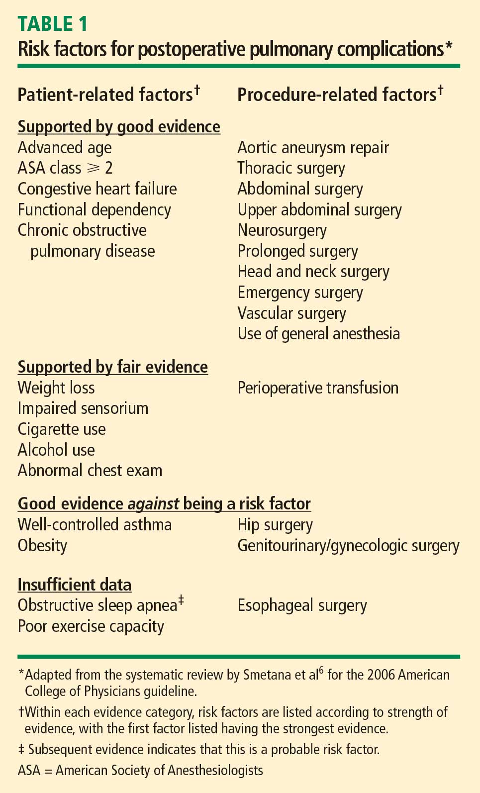
Patient-related risk factors. As noted in Table 1, the most important patient-related risk factors identified in the ACP guideline are increasing age and increasing American Society of Anesthesiologists (ASA) classification of comorbidity.
The effect of advanced age becomes particularly notable around age 60 years and escalates from there. This effect of age differs from that for cardiac complications, for which age drops out as a risk factor after adjustment for other diseases and risk factors. For pulmonary complications, in contrast, even older patients who are healthy are at increased risk.
The ASA classification is a general index of overall morbidity that ranges from class 1 (normal healthy patient) to class 5 (moribund patient who is not expected to survive without the operation).
Notably, COPD and smoking were only minor risk factors in the ACP analysis.
Procedure-related risk factors. Surgical site was found to be the most important of any of the patient- or procedure-related risk factors. The closer the incision is to the diaphragm, the greater the risk for pulmonary complications. Aortic, thoracic, and abdominal procedures carry the highest risk (Table 1), and among abdominal procedures, upper abdominal surgery (eg, cholecystectomy) is riskier than lower abdominal surgery (eg, gynecologic).
Other procedure-related risk factors identified were emergency surgery, surgery lasting more than 3 hours, use of general anesthesia, and multiple transfusions (Table 1).
Newly identified risk factors
Question: Which of the following has recently been identified as a risk factor for postoperative pulmonary complications?
A. Epidural anesthesia
B. Insulin-treated diabetes
C. Obstructive sleep apnea
D. Immobility
The correct answer is C. There is no evidence that epidural anesthesia or insulin-treated diabetes are risk factors. Immobility seems intuitively correct but has not emerged as a risk factor among high-quality studies in the literature.
Obstructive sleep apnea. The role of obstructive sleep apnea was unclear prior to publication of new data in the last couple of years. Hwang et al enrolled 172 patients who were soon to have elective surgery and had at least two of four clinical features of obstructive sleep apnea (snoring, daytime somnolence, witnessed apnea event, or crowded oropharynx).8 Patients underwent nocturnal oximetry before surgery and were divided into two groups based on number of desaturation episodes per hour. Patients with five or more desaturations had markedly higher rates of postoperative respiratory complications (8 complications among 98 patients) than did patients with fewer than five desaturations (1 complication among 74 patients). The presence of five or more desaturations was also associated with higher rates of cardiac, gastrointestinal, and bleeding complications. Though this was a small study, its results suggest a significant association between obstructive sleep apnea and pulmonary complications.
The issue of whether to screen patients for obstructive sleep apnea before major noncardiac surgery is still unresolved.
Pulmonary hypertension has also been identified as a risk factor in recent years with the publication of two studies that estimated its impact on morbidity and mortality after major noncardiac surgery.9,10 One of the studies, a retrospective database review, found a 28% incidence of respiratory failure among 145 surgical patients with pulmonary hypertension.9 In the other study, a prospective case-control trial, respiratory failure occurred in 21% of patients with pulmonary hypertension compared with only 3% of matched controls.10 In the case-control study, pulmonary hypertension was also associated with significantly elevated rates of heart failure and in-hospital death.
The results of these studies do not support preoperative screening for undiagnosed pulmonary hypertension, but they do underscore the need to recognize established pulmonary hypertension as an important risk factor for postoperative complications.
AN UPDATED INDEX FOR RESPIRATORY FAILURE
Several years ago, investigators from the Veterans Affairs Medical Centers developed a respiratory failure index using a design similar to those of well-established indices for cardiac risk.11 The same group also developed a separate risk index for pneumonia.12
This respiratory failure index was recently updated3 to reflect experience from private and academic hospitals, making the results more generally applicable. The researchers evaluated data from 180,000 patients undergoing major general or vascular surgery (defined according to the NSQIP) over a 3-year period. Respiratory failure was defined as requiring at least 48 hours of ventilation or unplanned reintubation.
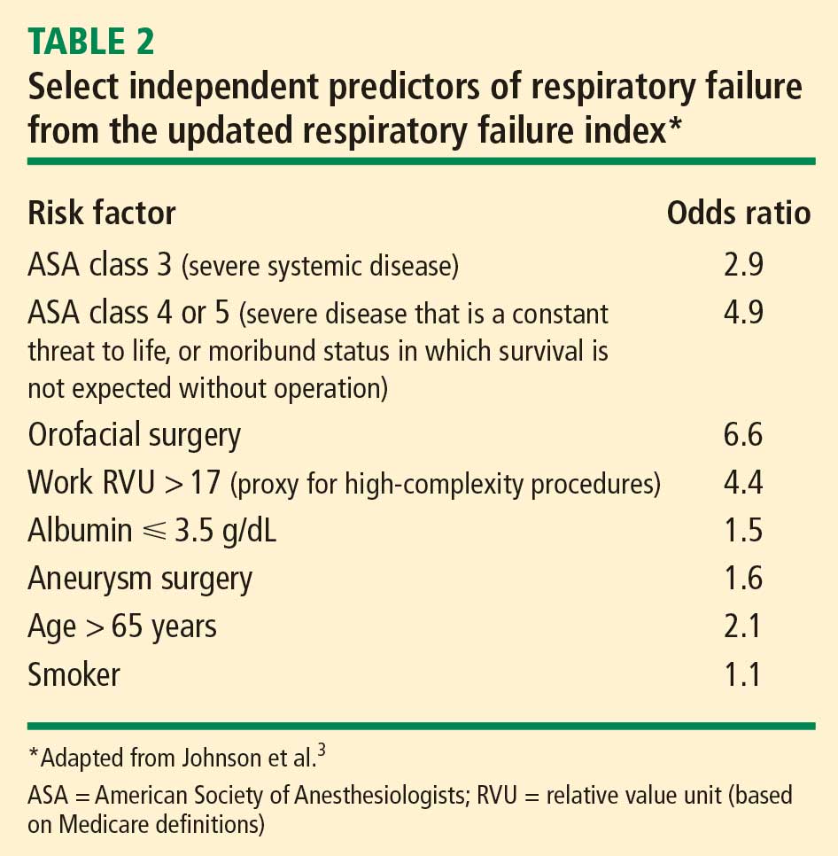
Comparison and contrast with the ACP guideline
Question: How does the updated respiratory failure index differ most significantly from the 2006 ACP guideline?
A. New index places greater emphasis on ASA class
B. New index offers a simplified weighted point scheme
C. New index ranks low albumin as a less important risk factor
D. New index attributes low risk to cigarette use
The correct answer is C: low albumin is a minor risk factor in the respiratory failure index, whereas it was one of the single most important predictors in the ACP guideline. As for the other answer choices, the new index places about the same emphasis on ASA class and cigarette use as does the ACP guideline, and it does not offer a simplified approach, as it incorporates 28 different factors.
Overall, most risk factors were similar in the updated respiratory failure index and the ACP guideline, but the index differs in several important ways:
- The index assigns less risk to low albumin, functional dependence, and congestive heart failure
- The index assigns greater risk to orofacial surgery
- The index identifies several new risk factors—high-complexity surgery, preoperative sepsis, ascites, and hypernatremia (serum sodium > 145 mmol/L).
STRATEGIES FOR RISK REDUCTION
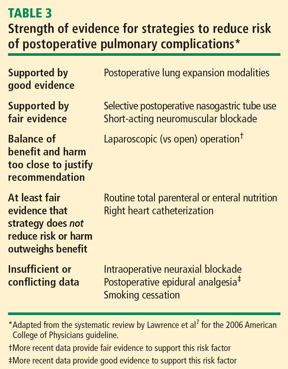
Postoperative CPAP: Good option when exercise ability is limited
Among the postoperative lung expansion modalities, continuous positive airway pressure (CPAP) is particularly useful for patients who are unable to perform deep breathing or incentive spirometry exercises. A recent systematic literature review identified nine randomized controlled trials of CPAP vs standard therapy in a total of 654 patients undergoing abdominal surgery.13 Meta-analysis of these studies showed that CPAP was associated with significant reductions in the risk of overall postoperative pulmonary complications (odds ratio [OR] = 0.66; 95% CI, 0.52–0.85), atelectasis (OR = 0.75; 95% CI, 0.58–0.97), and pneumonia (OR = 0.33; 95% CI, 0.14–0.75) relative to standard therapy.
Use nasogastric tubes selectively
Nasogastric tubes can be used either routinely following abdominal surgery or only in select patients—eg, those who have symptomatic abdominal distention or nausea. The difference is important since nasogastric tubes may potentially increase the risk of aspiration and thus lead to a pulmonary complication. Nelson et al conducted a meta-analysis of 24 studies that compared routine nasogastric tube use in abdominal surgery with selective use based on symptoms or abdominal distention.14 They found that routine use was associated with a significant increase in postoperative pulmonary complications (OR = 1.45; 95% CI, 1.08–1.93) relative to selective use, without achieving any of its intended goals.
Laparoscopic vs open surgery: Evidence begins to follow intuition
Intuitively, it seems that laparoscopic procedures should reduce risk for postoperative pulmonary complications compared with open surgical procedures, as they are associated with less postoperative pain, which should facilitate deep breathing and improve postoperative lung volumes. Nevertheless, evidence for whether laparoscopic surgery reduces the risk of pulmonary complications has been mixed until recently.
In 2008, however, Weller and Rosati published an analysis of a nationally representative database of 19,156 patients who underwent bariatric surgery in 2005.15 After adjusting for comorbidities, they found that the rate of postoperative pulmonary complications was nearly double if patients underwent open surgery as opposed to laparoscopic surgery (OR = 1.92; 95% CI, 1.54–2.38). Open surgery was also associated with significantly higher rates of sepsis, cardiovascular events, and reoperation compared with laparoscopic procedures. This study suggests that choosing laparoscopic procedures is another strategy that may reduce pulmonary complication rates, at least in the setting of bariatric surgery.
Postoperative thoracic epidural analgesia
Question: Thoracic epidural analgesia reduces rates of which of the following?
A. Pneumonia following abdominal aortic aneurysm repair
B. Pulmonary complications following coronary bypass surgery
C. Respiratory failure following abdominal surgery
D. All of the above
The correct answer is D. Thoracic epidural analgesia is another important strategy for reducing postoperative pulmonary complications, as demonstrated by a 2007 systematic literature review by Liu and Wu.16 Their analysis showed that rates of pneumonia, respiratory failure, and pulmonary complications overall were reduced by approximately one-third to more than one-half with the use of postoperative thoracic epidural analgesia in patients undergoing aortic aneurysm repair, coronary bypass surgery, and abdominal surgery.
Smoking cessation: The jury is still out
Whether preoperative cigarette cessation reduces pulmonary complication rates has been controversial over the past decade. Early reports showed that among patients who smoke, those who quit shortly before surgery actually had higher complication rates than patients who continued to smoke. The most reasonable explanation seems to be that many patients who stop smoking report increased coughing and sputum production for the first month or two. Selection bias also may have played a role in these findings.
More recently, two randomized trials studied the impact of perioperative smoking intervention programs involving counseling and nicotine replacement.17,18 Unfortunately, both studies primarily studied patients undergoing low-risk procedures and were insufficiently powered to show a difference in pulmonary complication rates. The question of whether smoking cessation is an effective strategy to reduce postoperative pulmonary risk remains unanswered.
Preoperative intensive lung expansion: A promising new intervention
While the effectiveness of postoperative lung expansion techniques is undisputed,7preoperative lung expansion—also known as inspiratory muscle training—has only recently been investigated. Hulzebos et al randomized 279 patients undergoing coronary artery bypass graft surgery who were at high risk for developing pulmonary complications to either usual care or inspiratory muscle training.19 The latter intervention involved 20 minutes per day of incentive spirometry, active breathing, and forced expiration techniques for at least 2 weeks prior to surgery. Rates of high-grade postoperative pulmonary complications were cut in half (OR = 0.52; 95% CI, 0.30–0.92) and rates of pneumonia were reduced by 60% (OR = 0.40; 95% CI, 0.19–0.84) in patients who received inspiratory muscle training relative to the usual-care group.
In clinical practice, preoperative inspiratory muscle training can be done in a chest physical therapy outpatient setting or a pulmonary rehabilitation clinic in the hospital.
SUMMARY
There have been a number of significant recent developments in the perioperative management of pulmonary complications:
- Obstructive sleep apnea has been confirmed as a risk factor, and pulmonary hypertension has emerged as a novel risk factor.
- An updated respiratory failure index has emerged as a useful research tool to identify high-risk patients and to ensure uniform risk stratification in future research.
- Evidence has mounted for the effectiveness of several risk-reduction strategies, including the use of laparoscopic procedures for bariatric surgery; selective use of nasogastric tubes; postoperative thoracic epidural analgesia; and intensive preoperative inspiratory muscle training.
DISCUSSION
Question from the audience: I do preoperative evaluations in an orthopedic ambulatory surgery center. Our surgeons often tell me, “Just order preoperative pulmonary function tests,” or, “Get a blood gas.” How should I respond?
Dr. Smetana: This is an area of some controversy, but in general, spirometry does not add much to a preoperative risk assessment that is based on a history and physical exam. Usually if the spirometry is abnormal, it will not be a surprise after careful clinical assessment. Arterial blood gases have no role in routine preoperative assessment.
Question from the audience: A chest x-ray is often requested preoperatively, but is it a necessary study?
Dr. Smetana: The data for preoperative chest x-rays are fairly poor and don’t allow us to assess whether they accurately predict complication rates. Most studies on chest x-rays have looked at how they affect preoperative management—eg, whether they change the anesthesia or even the surgery—and have shown that preoperative management changes in only about 1% to 2% of cases. So the chest x-ray is a fairly low-yield test in this setting.
One could argue that a preoperative chest x-ray might provide a baseline for postoperative comparison, but actually it is not usually helpful in this regard. Having a baseline does not make it easier to correctly diagnose pneumonia postoperatively, for example. Abnormal chest x-rays correlate with higher risk, but most patients with abnormal films would be suspected of being at higher risk anyway based on findings from the clinical assessment.
Question from the audience: Many primary care doctors in my hospital screen patients for pulmonary hypertension, but this raises the question of what to do with any information gained. What do you tell patients? Anesthesiologists?
Dr. Smetana: I don’t recommend preoperative screening for pulmonary hypertension unless there is some specific clinical reason to look for it. We don’t know if the perioperative risks that I described for patients with diagnosed or symptomatic pulmonary hypertension would also apply to patients with unrecognized, asymptomatic pulmonary hypertension that happened to be identified by screening.
Patients with pulmonary hypertension are at very high risk, especially for respiratory failure. But we don’t have any risk-reduction strategies specific to these patients, although I would recommend applying the general risk-reduction strategies that I discussed.
Question from the audience: I saw a man at my high-risk preoperative clinic who scored normally on a 6-minute walk test but then was found sound asleep when I was ready to see him a little while later. I suspected he had undiagnosed sleep apnea, and therefore had an increased risk of postoperative pulmonary complications, but what evidence would I have to delay his surgery to diagnose the sleep apnea and stabilize him on CPAP?
Dr. Smetana: For a patient with clinically suspected but undiagnosed sleep apnea, we have some evidence that the diagnosis should be pursued before surgery is performed.8 If the surgery were elective, it would be appropriate to have the patient evaluated and, if obstructive sleep apnea were diagnosed, treated in the customary way with CPAP. For patients who are hospitalized after surgery, CPAP can be continued as soon as possible in the hospital.
I would not have made this recommendation a few years ago, but now the evidence is more compelling. However, at this point I would not recommend routine preoperative screening of all patients for sleep apnea. Ongoing research is looking at this question.
Follow-up question: How long should surgery be delayed to optimize the patient on CPAP?
Dr. Smetana: Risk for postoperative respiratory failure is reduced very quickly after initiating CPAP therapy. A week would probably be sufficient, but there are no good data to specifically address that question.
Question from the audience: What about patients with asthma who are undergoing surgery—which ones benefit from stress-level steroids and preoperative nebulizer therapy?
Dr. Smetana: Surprisingly, asthma—if well controlled—is not a risk factor for postoperative pulmonary complications. Patients within 80% of their predicted or personal best peak flow appear to have a risk similar to that of patients without asthma. For patients with uncontrolled or poorly controlled asthma, the general rule is the same as for patients with COPD: treat them the same as if they weren’t having surgery. If a patient with asthma has a clinical indication for corticosteroids based on his or her condition, give corticosteroids whether or not surgery is planned. Corticosteroids are safe and do not raise the risk of postoperative wound complications. But we have no evidence to support routine use of steroids for all patients with asthma simply because elective surgery is planned.
Follow-up question: Do you optimize poorly controlled patients with oral prednisone for several days preoperatively, or do you use a stress protocol?
Dr. Smetana: For a patient whom you would normally treat with an outpatient course of prednisone, you should do just that. For a patient with an exacerbation severe enough to require admission for intravenous steroids and inhaled nebulizer therapy, then you should use that strategy. If the surgery is elective, it should be delayed until the patient is at his or her personal best.
- Lee TH, Marcantonio ER, Mangione CM, et al. Derivation and prospective validation of a simple index for prediction of cardiac risk of major noncardiac surgery. Circulation 1999; 100:1043–1049.
- Fleischmann KE, Goldman L, Young B, et al. Association between cardiac and noncardiac complications in patients undergoing noncardiac surgery: outcomes and effects on length of stay. Am J Med 2003; 115:515–520.
- Johnson RG, Arozullah AM, Neumayer L, et al. Multivariable predictors of postoperative respiratory failure after general and vascular surgery: results from the Patient Safety in Surgery Study. J Am Coll Surg 2007; 204:1188–1198.
- Dimick JB, Chen SL, Taheri PA, et al. Hospital costs associated with surgical complications: a report from the private-sector National Surgical Quality Improvement Program. J Am Coll Surg 2004; 199:531–537.
- Qaseem A, Snow V, Fitterman N, et al. Risk assessment for and strategies to reduce perioperative pulmonary complications for patients undergoing noncardiothoracic surgery: a guideline from the American College of Physicians. Ann Intern Med 2006; 144:575–580.
- Smetana GW, Lawrence VA, Cornell JE. Preoperative pulmonary risk stratification for noncardiothoracic surgery: systematic review for the American College of Physicians. Ann Intern Med 2006; 144:581–595.
- Lawrence VA, Cornell JE, Smetana GW; American College of Physicians. Strategies to reduce postoperative pulmonary complications after noncardiothoracic surgery: systematic review for the American College of Physicians. Ann Intern Med 2006; 144:596–608.
- Hwang D, Shakir N, Limann B, et al. Association of sleep-disordered breathing with postoperative complications. Chest 2008; 133:1128–1134.
- Ramakrishna G, Sprung J, Ravi BS, et al. Impact of pulmonary hypertension on the outcomes of noncardiac surgery: predictors of perioperative morbidity and mortality. J Am Coll Cardiol 2005; 45:1691–1699.
- Lai HC, Lai HC, Wang KY, Lee WL, Ting CT, Liu TJ. Severe pulmonary hypertension complicates postoperative outcome of noncardiac surgery. Br J Anaesth 2007; 99:184–190.
- Arozullah AM, Daley J, Henderson WG, Khuri SF. Multifactorial risk index for predicting postoperative respiratory failure in men after major noncardiac surgery. The National Veterans Administration Surgical Quality Improvement Program. Ann Surg 2000; 232:242–253.
- Arozullah AM, Khuri SF, Henderson WG, et al. Development and validation of a multifactorial risk index for predicting postoperative pneumonia after major noncardiac surgery. Ann Intern Med 2001; 135:847–857.
- Ferreyra GP, Baussano I, Squadrone V, et al. Continuous positive airway pressure for treatment of respiratory complications after abdominal surgery: a systematic review and meta-analysis. Ann Surg 2008; 247:617–626.
- Nelson R, Edwards S, Tse B. Prophylactic nasogastric decompression after abdominal surgery. Cochrane Database Syst Rev 2007; Jul 18 (3):CD004929.
- Weller WE, Rosati C. Comparing outcomes of laparoscopic versus open bariatric surgery. Ann Surg 2008; 248:10–15.
- Liu SS, Wu CL. Effect of postoperative analgesia on major postoperative complications: a systematic update of the evidence. Anesth Analg 2007; 104:689–702.
- Møller AM, Villebro N, Pedersen T, Tønnesen H. Effect of preoperative smoking intervention on postoperative complications: a randomised clinical trial. Lancet 2002; 359:114–117.
- Lindström D, Sadr Azodi O, Wladis A, et al. Effects of a perioperative smoking cessation intervention on postoperative complications: a randomized trial. Ann Surg 2008; 248:739–745.
- Hulzebos EH, Helders PJ, Favié NJ, et al. Preoperative intensive inspiratory muscle training to prevent postoperative pulmonary complications in high-risk patients undergoing CABG surgery: a randomized clinical trial. JAMA 2006; 296:1851–1857.
Although pulmonary complications are not as well studied as cardiac complications in the postoperative setting, they are just as common following noncardiac surgery and are even more costly. It is worthwhile to identify surgical patients most at risk of postoperative pulmonary complications and take measures known to mitigate risk. This paper discusses important risk factors to identify during a preoperative pulmonary evaluation and then focuses on recent advances in strategies for reducing postoperative pulmonary complications. Teaching questions are included throughout, along with the rationale behind their answers.
POSTOPERATIVE PULMONARY COMPLICATIONS: WHAT ARE WE TRYING TO PREVENT AND WHY?
The definition of postoperative pulmonary complications is more variable and less intuitive than that of cardiac complications. Cardiac complications—postoperative myocardial infarction, cardiac death, and pulmonary edema—are more consistently defined and measured in clinical trials. Studies of postoperative pulmonary complications often group together pneumonia, respiratory failure, atelectasis, bronchospasm, and exacerbation of chronic obstructive pulmonary disease (COPD), making it more difficult to individually evaluate risk factors for different outcomes.
There are several reasons why it is important to consider pulmonary risk when evaluating patients preoperatively:
Pulmonary complications are as common as cardiac complications following noncardiac surgery. For example, in a secondary analysis of the cohort of noncardiac surgical patients used to validate the Revised Cardiac Risk Index,1 Fleischmann et al found that the incidence of pulmonary complications (2.7%) was highly comparable to that of cardiac complications (2.5%).2
Respiratory failure is a marker of ill health and predicts further complications. Postoperative respiratory failure (often defined as the need for ventilation for more than 48 hours after surgery) is an extremely morbid event. Johnson et al compared the outcomes of patients with and without respiratory failure as a complication of surgery.3 Among patients with respiratory failure, 26% died within 30 days, 6% had a myocardial infarction, 35% developed pneumonia, 10% developed acute renal failure, and 3% developed a deep vein thrombosis or pulmonary embolism; in contrast, rates of each of these events were lower than 2% among patients without respiratory failure.
Pulmonary complications are expensive and require lengthy hospitalization. The National Surgical Quality Improvement Program (NSQIP) compared hospitalization costs and length of stay among patients with various postoperative complications.4 Among infectious, cardiovascular, venous thromboembolic, and pulmonary complications, pulmonary complications were by far the most costly and, along with venous thromboembolic complications, required the longest mean hospital stay.
For these reasons, identifying patients at risk for pulmonary complications and developing a strategy to reduce the risk is clearly worthwhile.
IDENTIFYING RISK FOR PULMONARY COMPLICATIONS
Question: Which of the following is the most important risk factor for postoperative pulmonary complications?
A. High-risk surgical site
B. General anesthesia
C. COPD
D. Obesity
The correct answer is A. Pulmonary complications differ from cardiac complications in an important way: procedure-related factors are more predictive of pulmonary complications than are patient-related factors. Even healthy patients undergoing high-risk surgery are at risk for pulmonary complications. As for the other answer choices, general anesthesia and COPD are risk factors but are not as important as surgical site, and obesity has not been shown to be a risk factor at all.
Take-home points from the 2006 ACP guideline
Patient-related risk factors. As noted in Table 1, the most important patient-related risk factors identified in the ACP guideline are increasing age and increasing American Society of Anesthesiologists (ASA) classification of comorbidity.
The effect of advanced age becomes particularly notable around age 60 years and escalates from there. This effect of age differs from that for cardiac complications, for which age drops out as a risk factor after adjustment for other diseases and risk factors. For pulmonary complications, in contrast, even older patients who are healthy are at increased risk.
The ASA classification is a general index of overall morbidity that ranges from class 1 (normal healthy patient) to class 5 (moribund patient who is not expected to survive without the operation).
Notably, COPD and smoking were only minor risk factors in the ACP analysis.
Procedure-related risk factors. Surgical site was found to be the most important of any of the patient- or procedure-related risk factors. The closer the incision is to the diaphragm, the greater the risk for pulmonary complications. Aortic, thoracic, and abdominal procedures carry the highest risk (Table 1), and among abdominal procedures, upper abdominal surgery (eg, cholecystectomy) is riskier than lower abdominal surgery (eg, gynecologic).
Other procedure-related risk factors identified were emergency surgery, surgery lasting more than 3 hours, use of general anesthesia, and multiple transfusions (Table 1).
Newly identified risk factors
Question: Which of the following has recently been identified as a risk factor for postoperative pulmonary complications?
A. Epidural anesthesia
B. Insulin-treated diabetes
C. Obstructive sleep apnea
D. Immobility
The correct answer is C. There is no evidence that epidural anesthesia or insulin-treated diabetes are risk factors. Immobility seems intuitively correct but has not emerged as a risk factor among high-quality studies in the literature.
Obstructive sleep apnea. The role of obstructive sleep apnea was unclear prior to publication of new data in the last couple of years. Hwang et al enrolled 172 patients who were soon to have elective surgery and had at least two of four clinical features of obstructive sleep apnea (snoring, daytime somnolence, witnessed apnea event, or crowded oropharynx).8 Patients underwent nocturnal oximetry before surgery and were divided into two groups based on number of desaturation episodes per hour. Patients with five or more desaturations had markedly higher rates of postoperative respiratory complications (8 complications among 98 patients) than did patients with fewer than five desaturations (1 complication among 74 patients). The presence of five or more desaturations was also associated with higher rates of cardiac, gastrointestinal, and bleeding complications. Though this was a small study, its results suggest a significant association between obstructive sleep apnea and pulmonary complications.
The issue of whether to screen patients for obstructive sleep apnea before major noncardiac surgery is still unresolved.
Pulmonary hypertension has also been identified as a risk factor in recent years with the publication of two studies that estimated its impact on morbidity and mortality after major noncardiac surgery.9,10 One of the studies, a retrospective database review, found a 28% incidence of respiratory failure among 145 surgical patients with pulmonary hypertension.9 In the other study, a prospective case-control trial, respiratory failure occurred in 21% of patients with pulmonary hypertension compared with only 3% of matched controls.10 In the case-control study, pulmonary hypertension was also associated with significantly elevated rates of heart failure and in-hospital death.
The results of these studies do not support preoperative screening for undiagnosed pulmonary hypertension, but they do underscore the need to recognize established pulmonary hypertension as an important risk factor for postoperative complications.
AN UPDATED INDEX FOR RESPIRATORY FAILURE
Several years ago, investigators from the Veterans Affairs Medical Centers developed a respiratory failure index using a design similar to those of well-established indices for cardiac risk.11 The same group also developed a separate risk index for pneumonia.12
This respiratory failure index was recently updated3 to reflect experience from private and academic hospitals, making the results more generally applicable. The researchers evaluated data from 180,000 patients undergoing major general or vascular surgery (defined according to the NSQIP) over a 3-year period. Respiratory failure was defined as requiring at least 48 hours of ventilation or unplanned reintubation.
Comparison and contrast with the ACP guideline
Question: How does the updated respiratory failure index differ most significantly from the 2006 ACP guideline?
A. New index places greater emphasis on ASA class
B. New index offers a simplified weighted point scheme
C. New index ranks low albumin as a less important risk factor
D. New index attributes low risk to cigarette use
The correct answer is C: low albumin is a minor risk factor in the respiratory failure index, whereas it was one of the single most important predictors in the ACP guideline. As for the other answer choices, the new index places about the same emphasis on ASA class and cigarette use as does the ACP guideline, and it does not offer a simplified approach, as it incorporates 28 different factors.
Overall, most risk factors were similar in the updated respiratory failure index and the ACP guideline, but the index differs in several important ways:
- The index assigns less risk to low albumin, functional dependence, and congestive heart failure
- The index assigns greater risk to orofacial surgery
- The index identifies several new risk factors—high-complexity surgery, preoperative sepsis, ascites, and hypernatremia (serum sodium > 145 mmol/L).
STRATEGIES FOR RISK REDUCTION
Postoperative CPAP: Good option when exercise ability is limited
Among the postoperative lung expansion modalities, continuous positive airway pressure (CPAP) is particularly useful for patients who are unable to perform deep breathing or incentive spirometry exercises. A recent systematic literature review identified nine randomized controlled trials of CPAP vs standard therapy in a total of 654 patients undergoing abdominal surgery.13 Meta-analysis of these studies showed that CPAP was associated with significant reductions in the risk of overall postoperative pulmonary complications (odds ratio [OR] = 0.66; 95% CI, 0.52–0.85), atelectasis (OR = 0.75; 95% CI, 0.58–0.97), and pneumonia (OR = 0.33; 95% CI, 0.14–0.75) relative to standard therapy.
Use nasogastric tubes selectively
Nasogastric tubes can be used either routinely following abdominal surgery or only in select patients—eg, those who have symptomatic abdominal distention or nausea. The difference is important since nasogastric tubes may potentially increase the risk of aspiration and thus lead to a pulmonary complication. Nelson et al conducted a meta-analysis of 24 studies that compared routine nasogastric tube use in abdominal surgery with selective use based on symptoms or abdominal distention.14 They found that routine use was associated with a significant increase in postoperative pulmonary complications (OR = 1.45; 95% CI, 1.08–1.93) relative to selective use, without achieving any of its intended goals.
Laparoscopic vs open surgery: Evidence begins to follow intuition
Intuitively, it seems that laparoscopic procedures should reduce risk for postoperative pulmonary complications compared with open surgical procedures, as they are associated with less postoperative pain, which should facilitate deep breathing and improve postoperative lung volumes. Nevertheless, evidence for whether laparoscopic surgery reduces the risk of pulmonary complications has been mixed until recently.
In 2008, however, Weller and Rosati published an analysis of a nationally representative database of 19,156 patients who underwent bariatric surgery in 2005.15 After adjusting for comorbidities, they found that the rate of postoperative pulmonary complications was nearly double if patients underwent open surgery as opposed to laparoscopic surgery (OR = 1.92; 95% CI, 1.54–2.38). Open surgery was also associated with significantly higher rates of sepsis, cardiovascular events, and reoperation compared with laparoscopic procedures. This study suggests that choosing laparoscopic procedures is another strategy that may reduce pulmonary complication rates, at least in the setting of bariatric surgery.
Postoperative thoracic epidural analgesia
Question: Thoracic epidural analgesia reduces rates of which of the following?
A. Pneumonia following abdominal aortic aneurysm repair
B. Pulmonary complications following coronary bypass surgery
C. Respiratory failure following abdominal surgery
D. All of the above
The correct answer is D. Thoracic epidural analgesia is another important strategy for reducing postoperative pulmonary complications, as demonstrated by a 2007 systematic literature review by Liu and Wu.16 Their analysis showed that rates of pneumonia, respiratory failure, and pulmonary complications overall were reduced by approximately one-third to more than one-half with the use of postoperative thoracic epidural analgesia in patients undergoing aortic aneurysm repair, coronary bypass surgery, and abdominal surgery.
Smoking cessation: The jury is still out
Whether preoperative cigarette cessation reduces pulmonary complication rates has been controversial over the past decade. Early reports showed that among patients who smoke, those who quit shortly before surgery actually had higher complication rates than patients who continued to smoke. The most reasonable explanation seems to be that many patients who stop smoking report increased coughing and sputum production for the first month or two. Selection bias also may have played a role in these findings.
More recently, two randomized trials studied the impact of perioperative smoking intervention programs involving counseling and nicotine replacement.17,18 Unfortunately, both studies primarily studied patients undergoing low-risk procedures and were insufficiently powered to show a difference in pulmonary complication rates. The question of whether smoking cessation is an effective strategy to reduce postoperative pulmonary risk remains unanswered.
Preoperative intensive lung expansion: A promising new intervention
While the effectiveness of postoperative lung expansion techniques is undisputed,7preoperative lung expansion—also known as inspiratory muscle training—has only recently been investigated. Hulzebos et al randomized 279 patients undergoing coronary artery bypass graft surgery who were at high risk for developing pulmonary complications to either usual care or inspiratory muscle training.19 The latter intervention involved 20 minutes per day of incentive spirometry, active breathing, and forced expiration techniques for at least 2 weeks prior to surgery. Rates of high-grade postoperative pulmonary complications were cut in half (OR = 0.52; 95% CI, 0.30–0.92) and rates of pneumonia were reduced by 60% (OR = 0.40; 95% CI, 0.19–0.84) in patients who received inspiratory muscle training relative to the usual-care group.
In clinical practice, preoperative inspiratory muscle training can be done in a chest physical therapy outpatient setting or a pulmonary rehabilitation clinic in the hospital.
SUMMARY
There have been a number of significant recent developments in the perioperative management of pulmonary complications:
- Obstructive sleep apnea has been confirmed as a risk factor, and pulmonary hypertension has emerged as a novel risk factor.
- An updated respiratory failure index has emerged as a useful research tool to identify high-risk patients and to ensure uniform risk stratification in future research.
- Evidence has mounted for the effectiveness of several risk-reduction strategies, including the use of laparoscopic procedures for bariatric surgery; selective use of nasogastric tubes; postoperative thoracic epidural analgesia; and intensive preoperative inspiratory muscle training.
DISCUSSION
Question from the audience: I do preoperative evaluations in an orthopedic ambulatory surgery center. Our surgeons often tell me, “Just order preoperative pulmonary function tests,” or, “Get a blood gas.” How should I respond?
Dr. Smetana: This is an area of some controversy, but in general, spirometry does not add much to a preoperative risk assessment that is based on a history and physical exam. Usually if the spirometry is abnormal, it will not be a surprise after careful clinical assessment. Arterial blood gases have no role in routine preoperative assessment.
Question from the audience: A chest x-ray is often requested preoperatively, but is it a necessary study?
Dr. Smetana: The data for preoperative chest x-rays are fairly poor and don’t allow us to assess whether they accurately predict complication rates. Most studies on chest x-rays have looked at how they affect preoperative management—eg, whether they change the anesthesia or even the surgery—and have shown that preoperative management changes in only about 1% to 2% of cases. So the chest x-ray is a fairly low-yield test in this setting.
One could argue that a preoperative chest x-ray might provide a baseline for postoperative comparison, but actually it is not usually helpful in this regard. Having a baseline does not make it easier to correctly diagnose pneumonia postoperatively, for example. Abnormal chest x-rays correlate with higher risk, but most patients with abnormal films would be suspected of being at higher risk anyway based on findings from the clinical assessment.
Question from the audience: Many primary care doctors in my hospital screen patients for pulmonary hypertension, but this raises the question of what to do with any information gained. What do you tell patients? Anesthesiologists?
Dr. Smetana: I don’t recommend preoperative screening for pulmonary hypertension unless there is some specific clinical reason to look for it. We don’t know if the perioperative risks that I described for patients with diagnosed or symptomatic pulmonary hypertension would also apply to patients with unrecognized, asymptomatic pulmonary hypertension that happened to be identified by screening.
Patients with pulmonary hypertension are at very high risk, especially for respiratory failure. But we don’t have any risk-reduction strategies specific to these patients, although I would recommend applying the general risk-reduction strategies that I discussed.
Question from the audience: I saw a man at my high-risk preoperative clinic who scored normally on a 6-minute walk test but then was found sound asleep when I was ready to see him a little while later. I suspected he had undiagnosed sleep apnea, and therefore had an increased risk of postoperative pulmonary complications, but what evidence would I have to delay his surgery to diagnose the sleep apnea and stabilize him on CPAP?
Dr. Smetana: For a patient with clinically suspected but undiagnosed sleep apnea, we have some evidence that the diagnosis should be pursued before surgery is performed.8 If the surgery were elective, it would be appropriate to have the patient evaluated and, if obstructive sleep apnea were diagnosed, treated in the customary way with CPAP. For patients who are hospitalized after surgery, CPAP can be continued as soon as possible in the hospital.
I would not have made this recommendation a few years ago, but now the evidence is more compelling. However, at this point I would not recommend routine preoperative screening of all patients for sleep apnea. Ongoing research is looking at this question.
Follow-up question: How long should surgery be delayed to optimize the patient on CPAP?
Dr. Smetana: Risk for postoperative respiratory failure is reduced very quickly after initiating CPAP therapy. A week would probably be sufficient, but there are no good data to specifically address that question.
Question from the audience: What about patients with asthma who are undergoing surgery—which ones benefit from stress-level steroids and preoperative nebulizer therapy?
Dr. Smetana: Surprisingly, asthma—if well controlled—is not a risk factor for postoperative pulmonary complications. Patients within 80% of their predicted or personal best peak flow appear to have a risk similar to that of patients without asthma. For patients with uncontrolled or poorly controlled asthma, the general rule is the same as for patients with COPD: treat them the same as if they weren’t having surgery. If a patient with asthma has a clinical indication for corticosteroids based on his or her condition, give corticosteroids whether or not surgery is planned. Corticosteroids are safe and do not raise the risk of postoperative wound complications. But we have no evidence to support routine use of steroids for all patients with asthma simply because elective surgery is planned.
Follow-up question: Do you optimize poorly controlled patients with oral prednisone for several days preoperatively, or do you use a stress protocol?
Dr. Smetana: For a patient whom you would normally treat with an outpatient course of prednisone, you should do just that. For a patient with an exacerbation severe enough to require admission for intravenous steroids and inhaled nebulizer therapy, then you should use that strategy. If the surgery is elective, it should be delayed until the patient is at his or her personal best.
- Lee TH, Marcantonio ER, Mangione CM, et al. Derivation and prospective validation of a simple index for prediction of cardiac risk of major noncardiac surgery. Circulation 1999; 100:1043–1049.
- Fleischmann KE, Goldman L, Young B, et al. Association between cardiac and noncardiac complications in patients undergoing noncardiac surgery: outcomes and effects on length of stay. Am J Med 2003; 115:515–520.
- Johnson RG, Arozullah AM, Neumayer L, et al. Multivariable predictors of postoperative respiratory failure after general and vascular surgery: results from the Patient Safety in Surgery Study. J Am Coll Surg 2007; 204:1188–1198.
- Dimick JB, Chen SL, Taheri PA, et al. Hospital costs associated with surgical complications: a report from the private-sector National Surgical Quality Improvement Program. J Am Coll Surg 2004; 199:531–537.
- Qaseem A, Snow V, Fitterman N, et al. Risk assessment for and strategies to reduce perioperative pulmonary complications for patients undergoing noncardiothoracic surgery: a guideline from the American College of Physicians. Ann Intern Med 2006; 144:575–580.
- Smetana GW, Lawrence VA, Cornell JE. Preoperative pulmonary risk stratification for noncardiothoracic surgery: systematic review for the American College of Physicians. Ann Intern Med 2006; 144:581–595.
- Lawrence VA, Cornell JE, Smetana GW; American College of Physicians. Strategies to reduce postoperative pulmonary complications after noncardiothoracic surgery: systematic review for the American College of Physicians. Ann Intern Med 2006; 144:596–608.
- Hwang D, Shakir N, Limann B, et al. Association of sleep-disordered breathing with postoperative complications. Chest 2008; 133:1128–1134.
- Ramakrishna G, Sprung J, Ravi BS, et al. Impact of pulmonary hypertension on the outcomes of noncardiac surgery: predictors of perioperative morbidity and mortality. J Am Coll Cardiol 2005; 45:1691–1699.
- Lai HC, Lai HC, Wang KY, Lee WL, Ting CT, Liu TJ. Severe pulmonary hypertension complicates postoperative outcome of noncardiac surgery. Br J Anaesth 2007; 99:184–190.
- Arozullah AM, Daley J, Henderson WG, Khuri SF. Multifactorial risk index for predicting postoperative respiratory failure in men after major noncardiac surgery. The National Veterans Administration Surgical Quality Improvement Program. Ann Surg 2000; 232:242–253.
- Arozullah AM, Khuri SF, Henderson WG, et al. Development and validation of a multifactorial risk index for predicting postoperative pneumonia after major noncardiac surgery. Ann Intern Med 2001; 135:847–857.
- Ferreyra GP, Baussano I, Squadrone V, et al. Continuous positive airway pressure for treatment of respiratory complications after abdominal surgery: a systematic review and meta-analysis. Ann Surg 2008; 247:617–626.
- Nelson R, Edwards S, Tse B. Prophylactic nasogastric decompression after abdominal surgery. Cochrane Database Syst Rev 2007; Jul 18 (3):CD004929.
- Weller WE, Rosati C. Comparing outcomes of laparoscopic versus open bariatric surgery. Ann Surg 2008; 248:10–15.
- Liu SS, Wu CL. Effect of postoperative analgesia on major postoperative complications: a systematic update of the evidence. Anesth Analg 2007; 104:689–702.
- Møller AM, Villebro N, Pedersen T, Tønnesen H. Effect of preoperative smoking intervention on postoperative complications: a randomised clinical trial. Lancet 2002; 359:114–117.
- Lindström D, Sadr Azodi O, Wladis A, et al. Effects of a perioperative smoking cessation intervention on postoperative complications: a randomized trial. Ann Surg 2008; 248:739–745.
- Hulzebos EH, Helders PJ, Favié NJ, et al. Preoperative intensive inspiratory muscle training to prevent postoperative pulmonary complications in high-risk patients undergoing CABG surgery: a randomized clinical trial. JAMA 2006; 296:1851–1857.
KEY POINTS
- Pulmonary complications are as common as cardiac complications following noncardiac surgery.
- Surgical site is the most important predictor of risk for postoperative pulmonary complications: aortic, thoracic, and upper abdominal surgeries are high-risk procedures, even in healthy patients.
- Obstructive sleep apnea and pulmonary hypertension have recently been identified as risk factors, but the limited available evidence does not support preoperative screening for these conditions in patients without symptoms.
- Postoperative continuous positive airway pressure therapy is effective for reducing pulmonary complications in patients who are unable to perform deep breathing or incentive spirometry exercises.
- The jury is out on whether smoking cessation shortly before surgery lowers risk for postoperative pulmonary complications.
Postoperative gastrointestinal tract dysfunction: An overview of causes and management strategies
Tolerance of an enteral diet is one of the fundamental components of postoperative wellness, along with the ability to mobilize freely without supplemental oxygen and a readiness to be discharged home as soon as possible. Accordingly, postoperative gastrointestinal (GI) tract dysfunction is best defined as intolerance of an enteral diet after having been tolerant of one preoperatively. I prefer the term postoperative GI tract dysfunction over postoperative ileus, as ileus is ill defined, covering a wide spectrum of clinical signs and having a range of published incidences so broad (5%–100%) that it defies useful discussion.
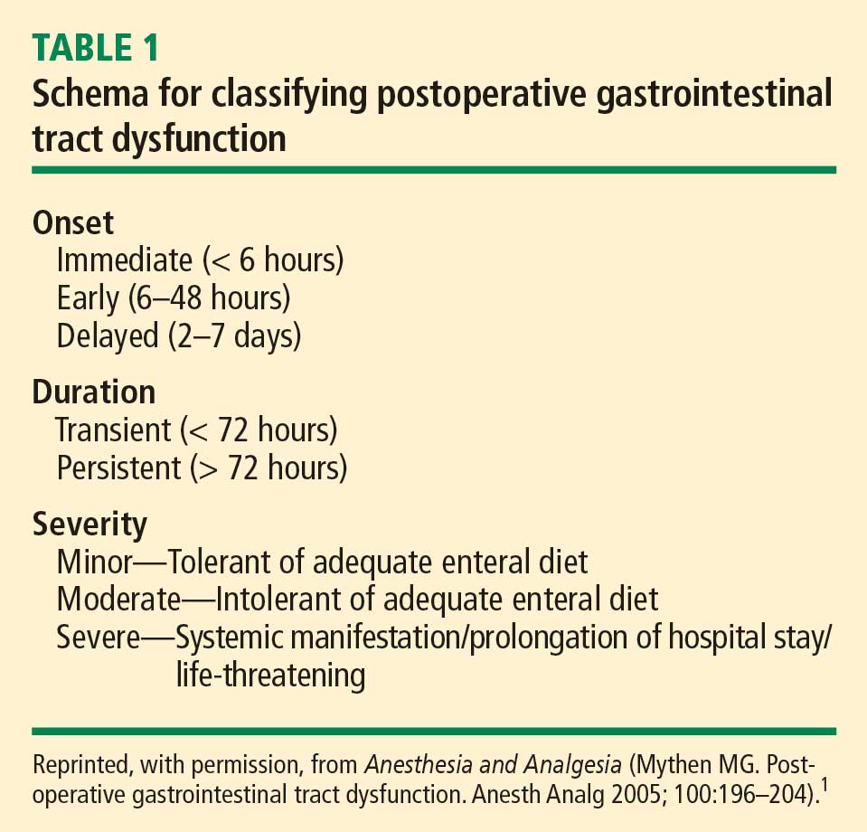
GI DYSFUNCTION: A COMMON POSTOPERATIVE MORBIDITY
Postoperative GI tract dysfunction is common, as illustrated by a large prospective cohort study at Duke University Medical Center3 that used the Postoperative Morbidity Survey (which has since been validated4) to document complications following major noncardiac surgery (ie, anticipated duration > 2 hours and anticipated blood loss > 500 mL). Hospital discharge was delayed in 27% of the study’s 438 patients as a result of a postoperative complication, and GI dysfunction was the most common type of complication overall and on postoperative days 5, 8, and 15. Episodes of GI dysfunction ranged from intolerance of an enteral diet to ischemic gut resulting in multiple organ failure.3
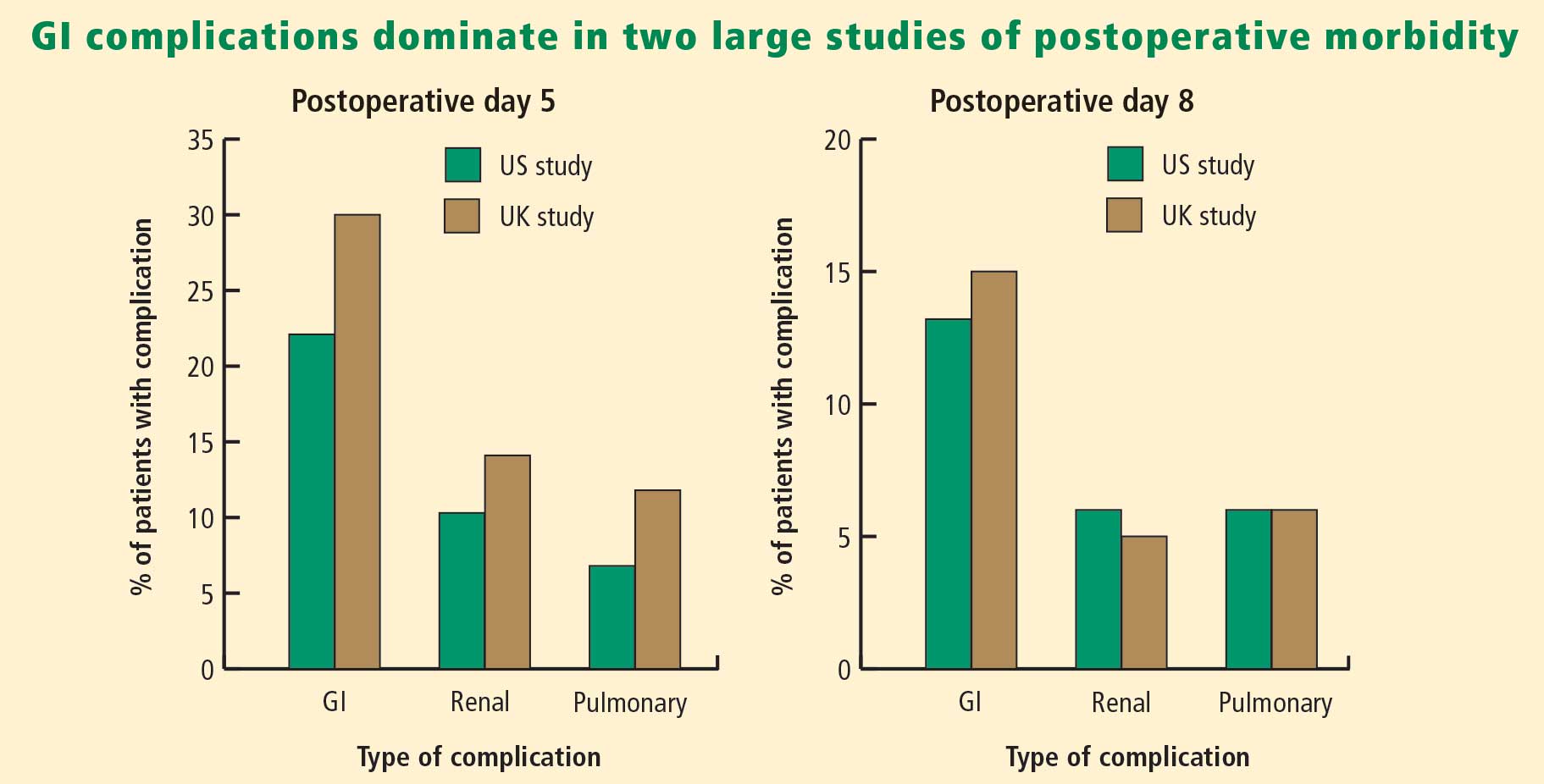
A MULTIFACTORIAL PATHOGENESIS
The pathophysiology of postoperative GI tract dysfunction can be ischemic, metabolic, toxic, neurogenic, myogenic, pharmacologic, or mechanical.
It is important to recognize that in many cases no single factor explains the whole story behind postsurgical GI tract dysfunction, and none of these factors is an ipso facto cause of such dysfunction. For instance, a “mechanical” pathogenesis refers to any manipulation of the gut that causes an inflammatory response in the gut’s various layers, resulting in injury.5,6 However, GI tract dysfunction commonly occurs after operations (including laparoscopic procedures) in which the gut was not handled at all. Similarly, in terms of a pharmacologic pathophysiology, while opioids can affect GI propulsion and cause constipation,7,8 avoidance of opioid use does not ensure prevention of GI tract dysfunction. Moreover, opioid abusers do not generally exhibit intolerance of enteral nutrition.
A common mechanism that is often ignored is perioperative gut ischemia resulting in low-grade injury. Low-grade hypovolemia can cause loss of perfusion to the tip of the microvillus, triggering apoptosis and potentially necrosis, which typically requires about 3 days for recovery. An experiment among 6 healthy volunteers who underwent elective hemorrhage (25% of blood volume removed) over 1 hour demonstrated that gastric tonometry was an earlier indicator of hypovolemia than were commonly measured hemodynamic variables such as invasive blood pressure, stroke volume, heart rate, and lactate and arterial blood gas measurements.9
FLUID LOADING AIDS GI RECOVERY
A targeted increase of intravascular volume and global blood flow perioperatively has been shown repeatedly to improve surgical outcome.10–24 In clinical trials, the most common intervention to achieve the predetermined hemodynamic goal has been fluid loading. Overall, targeted increases in perioperative global blood flow have been associated with reduced mortality,25 with the presumed mechanism being maintenance of end-organ perfusion.
The role of end-organ perfusion maintenance was confirmed in a controlled study of 60 patients undergoing cardiac surgery in which perioperative fluid loading (with colloid) maintained gut perfusion as measured by gastric tonometry, whereas a control group had a reproducible reduction in gut perfusion.15 Fluid loading was associated with a significant reduction in the incidence of gut mucosal hypoperfusion—from 56% to 7%—and significant reductions in the incidence of minor and major complications, mean days in the hospital, and mean days in the intensive care unit.
Fluid type matters
The type of intraoperative fluid loading is a factor in postoperative recovery.
Colloid vs crystalloid. Moretti et al found that colloid (6% hetastarch in saline or 6% hetastarch in balanced salt) was superior to crystalloid (lactated Ringer’s solution) in preventing nausea, severe pain, vomiting, periorbital edema, and double vision postoperatively (P < .05 for all) despite comparable hemodynamic profiles.26
Ringer’s vs normal saline. Williams et al compared intravenous lactated Ringer’s solution with normal saline (0.9% sodium chloride) in a randomized study of healthy volunteers.27 The group that received normal saline demonstrated central nervous system changes and a much higher incidence of abdominal discomfort, a finding consistent with the toxic properties of chlorine to the gut.
Balanced electrolyte solutions vs saline-based fluids. Wilkes et al compared crystalloid and colloid solutions with physiologically balanced electrolyte formulations (Hextend) against saline-based fluids (Hespan) in elderly surgical patients.28 They found that balanced electrolyte solutions were superior in improving gastric mucosal perfusion and preventing hyperchloremic metabolic acidosis. As a result of a reduction in GI tract perfusion, postoperative vomiting was more frequent in the group receiving saline-based fluids.
Evidence for Doppler-guided fluid management
Use of esophageal Doppler ultrasonography to guide fluid administration intraoperatively is fairly common in the United Kingdom and is based on randomized controlled trials showing that Doppler-guided colloid administration to maximize stroke volume reduces morbidity and length of hospital stay in surgical patients. In one government-supported study of 128 colorectal resection patients, Doppler-guided small boluses of colloid increased stroke volume, cardiac output, and oxygen delivery compared with conventional (central venous pressure–based) fluid management.29 Gut function improved significantly faster with Doppler-guided fluid management as evidenced by a more rapid return of flatus, opening of bowels, and achievement of a full diet, and by faster discharge from the hospital. The incidence of GI complications was reduced from 45.3% in the conventional management group to 14.1% in the Doppler group. The relative risk of GI tract dysfunction was 5.3 times higher with conventional management.
OTHER STRATEGIES TO REDUCE POSTOPERATIVE GI DYSFUNCTION
In addition to fluid loading, a number of other methods have been studied in an attempt to reduce the incidence of postoperative GI tract dysfunction.
Epidural neostigmine: Improvement in some measures
Epidural neostigmine was compared with saline control in a randomized study of 45 patients scheduled for abdominal aortic surgery.30 Time to first bowel sounds and time to first flatus were significantly shorter in the neostigmine group, but time to first defecation and the incidence of postoperative complications were similar between the groups.
Laxatives speed return of GI function
In a study of 53 women undergoing fast-track hysterectomy, recovery of GI tract function was faster in those randomized to receive laxatives (magnesium oxide and disodium phosphate) starting 6 hours postoperatively compared with those receiving placebo.31 Median time to first defecation was reduced from 69 hours in the placebo group to 45 hours in the laxative group (P < .0001), and postoperative hospitalization was shortened by a median of 1 day in the laxative group. There were no significant between-group differences in pain scores, postoperative nausea and vomiting, or the use of morphine or antiemetics.
Fentanyl reduces gastric myoelectrical activity
Intravenous administration of the opioid fentanyl significantly reduced gastric myoelectrical activity in an uncontrolled study of 20 patients undergoing elective surgery, but wide variation in effect was observed among patients.32 There was no correlation between the myoelectrical outcome and the presence of polymorphisms of the mu-opioid receptor gene.
Systemic lidocaine accelerates return of bowel function
Perioperative administration of systemic lidocaine, given as a 1.5-mg/kg bolus followed by continuous infusion at 2 mg/min, accelerated the return of bowel function and shortened the length of hospital stay compared with placebo in a randomized study of 60 colorectal surgery patients.33
Early oral feeding cuts length of stay
A recent meta-analysis of randomized trials found that early oral intake of fluids and food after major abdominal gynecologic surgery was associated with an increased risk of nausea but a reduced length of hospital stay.34 The authors recommended an individualized approach to early feeding, and called for cost-effectiveness and patient satisfaction studies.
Mosapride improves gastric emptying
Mosapride is a 5-HT4 agonist that has been shown to improve gastric emptying in a randomized controlled study of 40 patients undergoing laparoscopic colectomy.35 Time to first postoperative bowel movement, time to maximal gastric emptying rate, and postoperative hospital stay were all significantly shorter in patients receiving mosapride versus control. Mosapride is not currently approved for marketing in the United States.
Mu-opioid antagonists: Some show promise, others don’t
Mu-opioid receptor antagonists have been developed primarily to reverse opioid-induced bowel dysfunction. Commercially available drugs in this class include alvimopan, methylnaltrexone, nalbuphine, and naloxone. A recent meta-analysis of 23 randomized controlled studies of these agents for opioid-induced bowel dysfunction concluded that alvimopan and methylnaltrexone were superior to placebo but that evidence was insufficient for the safety or efficacy of naloxone and nalbuphine.36
Nasogastric decompression: Usually more harm than benefit
Prophylactic nasogastric decompression is an intervention devoid of evidence. A meta-analysis of 33 studies encompassing 5,240 patients randomized to routine nasogastric tube placement, selective nasogastric tube use, or no nasogastric tube placement after abdominal surgery found no advantage to routine nasogastric tube use.37 In fact, patients not receiving routine tube placement had a significantly earlier return of bowel function and a significant decrease in pulmonary complications. The incidence of anastomotic leak was not different among the groups. Routine tube use was associated with a lower incidence of vomiting but more patient discomfort. The clear conclusion is that, in most situations, elective placement of a nasogastric tube only causes harm.
Chewing gum: A simple intervention that works
In a recent meta-analysis of five randomized controlled trials, the simple intervention of gum chewing after colorectal surgery significantly accelerated the time to flatus and time to defecation, and was associated with a nonsignificant trend toward a shorter postoperative hospital stay.38
CONCLUSIONS ON MANAGEMENT
Traditional measures intended to reduce the incidence of postoperative GI tract dysfunction—administration of prokinetic drugs, placement of nasogastric tubes, avoidance of food and fluids—are not beneficial and are often harmful. Administration of targeted amounts of fluid to optimize ventricular filling and end-organ perfusion has repeatedly been demonstrated to improve outcomes, particularly those related to GI tract perfusion and function. Administration of larger volumes of colloid, to achieve predetermined increases in stroke volume, improves gut perfusion and reduces the incidence of GI tract dysfunction.
Many simple, inexpensive, and readily available strategies for preventing or reversing postoperative GI tract dysfunction have some degree of evidence-based support and should be considered. I would recommend a multimodal approach that includes a limited surgical incision, regional local anesthesia without use of opioids, immediate postoperative mobilization, early enteral feeding, and postoperative gum chewing.1 Such an approach promises to reduce GI tract dysfunction and other postoperative complications as well as to shorten hospital stay.
DISCUSSION
Question from the audience: You mentioned the selective use of nasogastric tubes. In which patients would you use them?
Dr. Mythen: For upper GI surgeries—esophagectomy, for example—a nasogastric tube is inevitable. Beyond that, the specific indications for tube placement are very limited. At our institution, we no longer place nasogastric tubes following the vast majority of GI tract operations, with esophagectomy being the exception.
Question from the audience: Would you comment on the selective contribution of thoracic epidural analgesia with respect to early feeding after abdominal or colon surgery?
Dr. Mythen: If you’re an enthusiast for thoracic epidurals, you can present the literature in a way that definitively demonstrates a huge advantage to thoracic epidurals. When they work well for the individual, they are fantastic, but you must have a very effective team and system to deliver success to the whole patient population. At our institution the failure rate 20 to 24 hours postoperatively is about 50%.
Question from the audience: I’m an internist and I’ve never heard of the esophageal Doppler-directed fluid bolus protocol—or of anyone using colloids at all. Is that something that is generally practiced in the United States?
Dr. Mythen: Some institutions are practicing goal-directed fluid management now. If you measure stroke volume and give small boluses of colloid, you need a lot less fluid to achieve a higher intravascular volume and goal. At our institution, we’ve repackaged it as “goal-directed fluid restriction” to gain acceptance among surgeons. Uptake has been slower in the United States, though studies here have reinforced the message and been supported by editorials. Guessing about fluids, which we’ve done historically, is not very smart. One thing that differentiates an anesthesiologist from an anesthetic technician is the ability to give goal-directed fluid therapy. The ability to act in a targeted fashion makes it possible to achieve an appropriate physiological goal, but it is more difficult.
Question from the audience: In terms of maintenance fluids and chloride toxicity, is there an alternative to D5 half-normal saline for maintenance fluid?
Dr. Mythen: We don’t have a very good postoperative maintenance fluid; D5 half-normal with some potassium is probably as good as it gets at present. I emphasize getting patients to drink as quickly as possible. If they’re not drinking (not using the GI tract), they need a very high level of physician input because fluid balance is rocket science. The GI tract is very clever. Once patients are drinking and eating, they’re fine, but if they still have an intravenous line in, close attention is required.
Question from the audience: Would you use lactated Ringer’s solution in a patient who is just not eating or drinking?
Dr. Mythen: I do, actually. I tend to mix it in with some D5 half-normal saline because lactated Ringer’s is a great solution. The body can use the lactate to make sugar if necessary. The brain is one of the few organs that will metabolize lactate.
Follow-up question: Would you use it at a lower rate to prevent volume overload?
Dr. Mythen: Yes, at 60 mL/hr. The important thing is that if intravenous fluids are still required, the patient needs to be in a fairly supervised, high-dependency environment. You must address the real issue: Why aren’t they drinking? If the patient is not drinking postoperatively, someone’s done a bad job or there is something that needs fixing.
Question from audience: In the operating room, do you have a preference between albumin and a high-molecular-weight hetastarch like Hextend?
Dr. Mythen: Europe is slightly different in its choice of colloids. We’ve pretty much abandoned the high-molecular-weight starches. We do not use albumin at our institution for cost reasons, and we can’t find any evidence to support its use. We would have to close one intensive care unit bed to be able to afford using albumin. We use low-molecular-weight hydroxyethyl starches, which I believe are now coming into the United States. They have no major coagulation effect.
- Mythen MG. Postoperative gastrointestinal tract dysfunction. Anesth Analg 2005; 100:196–204.
- Gan TJ, Meyer T, Apfel CC, et al. Consensus guidelines for managing postoperative nausea and vomiting. Anesth Analg 2003; 97:62–71.
- Bennett-Guerrero E, Welsby I, Dunn TJ, et al. The use of a postoperative morbidity survey to evaluate patients with prolonged hospitalization after routine, moderate-risk, elective surgery. Anesth Analg 1999; 89:514–519.
- Grocott MP, Browne JP, Van der Meulen J, et al. The Postoperative Morbidity Survey was validated and used to describe morbidity after major surgery. J Clin Epidemiol 2007; 60:919–928.
- Kalff JC, Schraut WH, Simmons RL, Bauer AJ. Surgical manipulation of the gut elicits an intestinal muscularis inflammatory response resulting in postsurgical ileus. Ann Surg 1998; 228:652–663.
- Schwarz NT, Beer-Stolz D, Simmons RL, Bauer AJ. Pathogenesis of paralytic ileus: intestinal manipulation opens a transient pathway between the intestinal lumen and the leukocytic infiltrate of the jejunal muscularis. Ann Surg 2002; 235:31–40.
- Manara L, Bianchetti A. The central and peripheral influences of opioids on gastrointestinal propulsion. Annu Rev Pharmacol Toxicol 1985; 25:249–273.
- Manara L, Bianchi G, Ferretti P, Tavani A. Inhibition of gastrointestinal transit by morphine in rats results primarily from direct drug action on gut opioid sites. J Pharmacol Exp Ther 1986; 237:945–949.
- Hamilton-Davies C, Mythen MG, Salmon JB, et al. Comparison of commonly used clinical indicators of hypovolaemia with gastrointestinal tonometry. Intensive Care Med 1997; 23:276–281.
- Bender JS, Smith-Meek MA, Jones CE. Routine pulmonary artery catheterization does not reduce morbidity and mortality of elective vascular surgery: results of a prospective, randomized trial. Ann Surg 1997; 226:229–237.
- Berlauk JF, Abrams JH, Gilmour IJ, et al. Preoperative optimization of cardiovascular hemodynamics improves outcome in peripheral vascular surgery: a prospective, randomized clinical trial. Ann Surg 1991; 214:289–299.
- Boyd O, Grounds RM, Bennett ED. A randomized clinical trial of the effect of deliberate perioperative increase of oxygen delivery on mortality in high-risk surgical patients. JAMA 1993; 270:2699–2707.
- Gan TJ, Soppitt A, Maroof M, et al. Goal-directed intraoperative fluid administration reduces length of hospital stay after major surgery. Anesthesiology 2002; 97:820–826.
- Lobo SM, Salgado PF, Castillo VG, et al. Effects of maximizing oxygen delivery on morbidity and mortality in high-risk surgical patients. Crit Care Med 2000; 28:3396–3404.
- Mythen MG, Webb AR. Perioperative plasma volume expansion reduces the incidence of gut mucosal hypoperfusion during cardiac surgery. Arch Surg 1995; 130:423–429.
- Pölönen P, Ruokonen E, Hippeläinen M, Pöyhönen M, Takala J. A prospective, randomized study of goal-oriented hemodynamic therapy in cardiac surgical patients. Anesth Analg 2000; 90:1052–1059.
- Sandham JD, Hull RD, Brant RF, et al. A randomized, controlled trial of the use of pulmonary-artery catheters in high-risk surgical patients. N Engl J Med 2003; 348:5–14.
- Shoemaker WC, Appel PL, Kram HB, Nathan RC, Thompson JL. Multicomponent noninvasive physiologic monitoring of circulatory function. Crit Care Med 1988; 16:482–490.
- Sinclair S, James S, Singer M. Intraoperative intravascular volume optimisation and length of hospital stay after repair of proximal femoral fracture: randomised controlled trial. BMJ 1997; 315:909–912.
- Ueno S, Tanabe G, Yamada H, et al. Response of patients with cirrhosis who have undergone partial hepatectomy to treatment aimed at achieving supranormal oxygen delivery and consumption. Surgery 1998; 123:278–286.
- Valentine RJ, Duke ML, Inman MH, et al. Effectiveness of pulmonary artery catheters in aortic surgery: a randomized trial. J Vasc Surg 1998; 27:203–212.
- Venn R, Steele A, Richardson P, Poloniecki J, Grounds M, Newman P. Randomized controlled trial to investigate influence of the fluid challenge on duration of hospital stay and perioperative morbidity in patients with hip fractures. Br J Anaesth 2002; 88:65–71.
- Wilson J, Woods I, Fawcett J, et al. Reducing the risk of major elective surgery: randomised controlled trial of preoperative optimisation of oxygen delivery. BMJ 1999; 318:1099–1103.
- Ziegler DW, Wright JG, Choban PS, Flancbaum L. A prospective randomized trial of preoperative “optimization” of cardiac function in patients undergoing elective peripheral vascular surgery. Surgery 1997; 122:584–592.
- Grocott MPW, Hamilton MA, Bennett ED, Harrison D, Rowan K. Perioperative increase in global blood flow to explicit defined goals and outcomes following surgery. Cochrane Database Syst Rev 2006; 2:CD004082. doi:10.1002/14651858.CD004082.pub4.
- Moretti EW, Robertson KM, El-Moalem H, Gan TJ. Intraoperative colloid administration reduces postoperative nausea and vomiting and improves postoperative outcomes compared with crystalloid administration. Anesth Analg 2003; 96:611–617.
- Williams EL, Hildebrand KL, McCormick SA, Bedel MJ. The effect of intravenous lactated Ringer’s solution versus 0.9% sodium chloride solution on serum osmolality in human volunteers. Anesth Analg 1999; 88:999–1003.
- Wilkes NJ, Woolf R, Mutch M, et al. The effects of balanced versus saline-based hetastarch and crystalloid solutions on acid-base and electrolyte status and gastric mucosal perfusion in elderly surgical patients. Anesth Analg 2001; 93:811–816.
- Wakeling HG, McFall MR, Jenkins CS, et al. Intraoperative oesophageal Doppler guided fluid management shortens postoperative hospital stay after major bowel surgery. Br J Anaesth 2005; 95:634–642.
- Caliskan E, Turkoz A, Sener M, et al. A prospective randomized double-blind study to determine the effect of thoracic epidural neostigmine on postoperative ileus after abdominal aortic surgery. Anesth Analg 2008; 106:959–964.
- Hansen CT, Sørensen M, Møller C, Ottesen B, Kehlet H. Effect of laxatives on gastrointestinal functional recovery in fast-track hysterectomy: a double-blind, placebo-controlled randomized study. Am J Obstet Gynecol 2007; 196:311.e1–311.e7.
- Walldén J, Lindberg G, Sandin M, Thörn SE, Wattwil M. Effects of fentanyl on gastric myoelectrical activity: a possible association with polymorphisms of the mu-opioid receptor gene? Acta Anaesthesiol Scand 2008; 52:708–715.
- Herroeder S, Pecher S, Schönherr ME, et al. Systemic lidocaine shortens length of hospital stay after colorectal surgery: a double-blinded, randomized, placebo-controlled trial. Ann Surg 2007; 246:192–200.
- Charoenkwan K, Phillipson G, Vutyavanich T. Early versus delayed oral fluids and food for reducing complications after major abdominal gynaecologic surgery. Cochrane Database Syst Rev 2007; 4:CD004508. doi:10.1002/14651858.CD004508.pub3.
- Narita K, Tsunoda A, Takenaka K, et al. Effect of mosapride on recovery of intestinal motility after hand-assisted laparoscopic colectomy for carcinoma. Dis Colon Rectum 2008; 51:1692–1695.
- McNicol ED, Boyce D, Schumann R, Carr DB. Mu-opioid antagonists for opioid-induced bowel dysfunction. Cochrane Database Syst Rev 2008; 2:CD006332. doi:10.1002/14651858.CD006332.pub2.
- Nelson RL, Edwards S, Tse B. Prophylactic nasogastric decompression after abdominal surgery. Cochrane Database Syst Rev 2007; 3:CD004929. doi:10.1002/14651858.CD004929.pub3.
- de Castro SM, van den Esschert JW, van Heek NT, et al. A systematic review of the efficacy of gum chewing for the amelioration of postoperative ileus. Dig Surg 2008; 25:39–45.
Tolerance of an enteral diet is one of the fundamental components of postoperative wellness, along with the ability to mobilize freely without supplemental oxygen and a readiness to be discharged home as soon as possible. Accordingly, postoperative gastrointestinal (GI) tract dysfunction is best defined as intolerance of an enteral diet after having been tolerant of one preoperatively. I prefer the term postoperative GI tract dysfunction over postoperative ileus, as ileus is ill defined, covering a wide spectrum of clinical signs and having a range of published incidences so broad (5%–100%) that it defies useful discussion.
GI DYSFUNCTION: A COMMON POSTOPERATIVE MORBIDITY
Postoperative GI tract dysfunction is common, as illustrated by a large prospective cohort study at Duke University Medical Center3 that used the Postoperative Morbidity Survey (which has since been validated4) to document complications following major noncardiac surgery (ie, anticipated duration > 2 hours and anticipated blood loss > 500 mL). Hospital discharge was delayed in 27% of the study’s 438 patients as a result of a postoperative complication, and GI dysfunction was the most common type of complication overall and on postoperative days 5, 8, and 15. Episodes of GI dysfunction ranged from intolerance of an enteral diet to ischemic gut resulting in multiple organ failure.3
A MULTIFACTORIAL PATHOGENESIS
The pathophysiology of postoperative GI tract dysfunction can be ischemic, metabolic, toxic, neurogenic, myogenic, pharmacologic, or mechanical.
It is important to recognize that in many cases no single factor explains the whole story behind postsurgical GI tract dysfunction, and none of these factors is an ipso facto cause of such dysfunction. For instance, a “mechanical” pathogenesis refers to any manipulation of the gut that causes an inflammatory response in the gut’s various layers, resulting in injury.5,6 However, GI tract dysfunction commonly occurs after operations (including laparoscopic procedures) in which the gut was not handled at all. Similarly, in terms of a pharmacologic pathophysiology, while opioids can affect GI propulsion and cause constipation,7,8 avoidance of opioid use does not ensure prevention of GI tract dysfunction. Moreover, opioid abusers do not generally exhibit intolerance of enteral nutrition.
A common mechanism that is often ignored is perioperative gut ischemia resulting in low-grade injury. Low-grade hypovolemia can cause loss of perfusion to the tip of the microvillus, triggering apoptosis and potentially necrosis, which typically requires about 3 days for recovery. An experiment among 6 healthy volunteers who underwent elective hemorrhage (25% of blood volume removed) over 1 hour demonstrated that gastric tonometry was an earlier indicator of hypovolemia than were commonly measured hemodynamic variables such as invasive blood pressure, stroke volume, heart rate, and lactate and arterial blood gas measurements.9
FLUID LOADING AIDS GI RECOVERY
A targeted increase of intravascular volume and global blood flow perioperatively has been shown repeatedly to improve surgical outcome.10–24 In clinical trials, the most common intervention to achieve the predetermined hemodynamic goal has been fluid loading. Overall, targeted increases in perioperative global blood flow have been associated with reduced mortality,25 with the presumed mechanism being maintenance of end-organ perfusion.
The role of end-organ perfusion maintenance was confirmed in a controlled study of 60 patients undergoing cardiac surgery in which perioperative fluid loading (with colloid) maintained gut perfusion as measured by gastric tonometry, whereas a control group had a reproducible reduction in gut perfusion.15 Fluid loading was associated with a significant reduction in the incidence of gut mucosal hypoperfusion—from 56% to 7%—and significant reductions in the incidence of minor and major complications, mean days in the hospital, and mean days in the intensive care unit.
Fluid type matters
The type of intraoperative fluid loading is a factor in postoperative recovery.
Colloid vs crystalloid. Moretti et al found that colloid (6% hetastarch in saline or 6% hetastarch in balanced salt) was superior to crystalloid (lactated Ringer’s solution) in preventing nausea, severe pain, vomiting, periorbital edema, and double vision postoperatively (P < .05 for all) despite comparable hemodynamic profiles.26
Ringer’s vs normal saline. Williams et al compared intravenous lactated Ringer’s solution with normal saline (0.9% sodium chloride) in a randomized study of healthy volunteers.27 The group that received normal saline demonstrated central nervous system changes and a much higher incidence of abdominal discomfort, a finding consistent with the toxic properties of chlorine to the gut.
Balanced electrolyte solutions vs saline-based fluids. Wilkes et al compared crystalloid and colloid solutions with physiologically balanced electrolyte formulations (Hextend) against saline-based fluids (Hespan) in elderly surgical patients.28 They found that balanced electrolyte solutions were superior in improving gastric mucosal perfusion and preventing hyperchloremic metabolic acidosis. As a result of a reduction in GI tract perfusion, postoperative vomiting was more frequent in the group receiving saline-based fluids.
Evidence for Doppler-guided fluid management
Use of esophageal Doppler ultrasonography to guide fluid administration intraoperatively is fairly common in the United Kingdom and is based on randomized controlled trials showing that Doppler-guided colloid administration to maximize stroke volume reduces morbidity and length of hospital stay in surgical patients. In one government-supported study of 128 colorectal resection patients, Doppler-guided small boluses of colloid increased stroke volume, cardiac output, and oxygen delivery compared with conventional (central venous pressure–based) fluid management.29 Gut function improved significantly faster with Doppler-guided fluid management as evidenced by a more rapid return of flatus, opening of bowels, and achievement of a full diet, and by faster discharge from the hospital. The incidence of GI complications was reduced from 45.3% in the conventional management group to 14.1% in the Doppler group. The relative risk of GI tract dysfunction was 5.3 times higher with conventional management.
OTHER STRATEGIES TO REDUCE POSTOPERATIVE GI DYSFUNCTION
In addition to fluid loading, a number of other methods have been studied in an attempt to reduce the incidence of postoperative GI tract dysfunction.
Epidural neostigmine: Improvement in some measures
Epidural neostigmine was compared with saline control in a randomized study of 45 patients scheduled for abdominal aortic surgery.30 Time to first bowel sounds and time to first flatus were significantly shorter in the neostigmine group, but time to first defecation and the incidence of postoperative complications were similar between the groups.
Laxatives speed return of GI function
In a study of 53 women undergoing fast-track hysterectomy, recovery of GI tract function was faster in those randomized to receive laxatives (magnesium oxide and disodium phosphate) starting 6 hours postoperatively compared with those receiving placebo.31 Median time to first defecation was reduced from 69 hours in the placebo group to 45 hours in the laxative group (P < .0001), and postoperative hospitalization was shortened by a median of 1 day in the laxative group. There were no significant between-group differences in pain scores, postoperative nausea and vomiting, or the use of morphine or antiemetics.
Fentanyl reduces gastric myoelectrical activity
Intravenous administration of the opioid fentanyl significantly reduced gastric myoelectrical activity in an uncontrolled study of 20 patients undergoing elective surgery, but wide variation in effect was observed among patients.32 There was no correlation between the myoelectrical outcome and the presence of polymorphisms of the mu-opioid receptor gene.
Systemic lidocaine accelerates return of bowel function
Perioperative administration of systemic lidocaine, given as a 1.5-mg/kg bolus followed by continuous infusion at 2 mg/min, accelerated the return of bowel function and shortened the length of hospital stay compared with placebo in a randomized study of 60 colorectal surgery patients.33
Early oral feeding cuts length of stay
A recent meta-analysis of randomized trials found that early oral intake of fluids and food after major abdominal gynecologic surgery was associated with an increased risk of nausea but a reduced length of hospital stay.34 The authors recommended an individualized approach to early feeding, and called for cost-effectiveness and patient satisfaction studies.
Mosapride improves gastric emptying
Mosapride is a 5-HT4 agonist that has been shown to improve gastric emptying in a randomized controlled study of 40 patients undergoing laparoscopic colectomy.35 Time to first postoperative bowel movement, time to maximal gastric emptying rate, and postoperative hospital stay were all significantly shorter in patients receiving mosapride versus control. Mosapride is not currently approved for marketing in the United States.
Mu-opioid antagonists: Some show promise, others don’t
Mu-opioid receptor antagonists have been developed primarily to reverse opioid-induced bowel dysfunction. Commercially available drugs in this class include alvimopan, methylnaltrexone, nalbuphine, and naloxone. A recent meta-analysis of 23 randomized controlled studies of these agents for opioid-induced bowel dysfunction concluded that alvimopan and methylnaltrexone were superior to placebo but that evidence was insufficient for the safety or efficacy of naloxone and nalbuphine.36
Nasogastric decompression: Usually more harm than benefit
Prophylactic nasogastric decompression is an intervention devoid of evidence. A meta-analysis of 33 studies encompassing 5,240 patients randomized to routine nasogastric tube placement, selective nasogastric tube use, or no nasogastric tube placement after abdominal surgery found no advantage to routine nasogastric tube use.37 In fact, patients not receiving routine tube placement had a significantly earlier return of bowel function and a significant decrease in pulmonary complications. The incidence of anastomotic leak was not different among the groups. Routine tube use was associated with a lower incidence of vomiting but more patient discomfort. The clear conclusion is that, in most situations, elective placement of a nasogastric tube only causes harm.
Chewing gum: A simple intervention that works
In a recent meta-analysis of five randomized controlled trials, the simple intervention of gum chewing after colorectal surgery significantly accelerated the time to flatus and time to defecation, and was associated with a nonsignificant trend toward a shorter postoperative hospital stay.38
CONCLUSIONS ON MANAGEMENT
Traditional measures intended to reduce the incidence of postoperative GI tract dysfunction—administration of prokinetic drugs, placement of nasogastric tubes, avoidance of food and fluids—are not beneficial and are often harmful. Administration of targeted amounts of fluid to optimize ventricular filling and end-organ perfusion has repeatedly been demonstrated to improve outcomes, particularly those related to GI tract perfusion and function. Administration of larger volumes of colloid, to achieve predetermined increases in stroke volume, improves gut perfusion and reduces the incidence of GI tract dysfunction.
Many simple, inexpensive, and readily available strategies for preventing or reversing postoperative GI tract dysfunction have some degree of evidence-based support and should be considered. I would recommend a multimodal approach that includes a limited surgical incision, regional local anesthesia without use of opioids, immediate postoperative mobilization, early enteral feeding, and postoperative gum chewing.1 Such an approach promises to reduce GI tract dysfunction and other postoperative complications as well as to shorten hospital stay.
DISCUSSION
Question from the audience: You mentioned the selective use of nasogastric tubes. In which patients would you use them?
Dr. Mythen: For upper GI surgeries—esophagectomy, for example—a nasogastric tube is inevitable. Beyond that, the specific indications for tube placement are very limited. At our institution, we no longer place nasogastric tubes following the vast majority of GI tract operations, with esophagectomy being the exception.
Question from the audience: Would you comment on the selective contribution of thoracic epidural analgesia with respect to early feeding after abdominal or colon surgery?
Dr. Mythen: If you’re an enthusiast for thoracic epidurals, you can present the literature in a way that definitively demonstrates a huge advantage to thoracic epidurals. When they work well for the individual, they are fantastic, but you must have a very effective team and system to deliver success to the whole patient population. At our institution the failure rate 20 to 24 hours postoperatively is about 50%.
Question from the audience: I’m an internist and I’ve never heard of the esophageal Doppler-directed fluid bolus protocol—or of anyone using colloids at all. Is that something that is generally practiced in the United States?
Dr. Mythen: Some institutions are practicing goal-directed fluid management now. If you measure stroke volume and give small boluses of colloid, you need a lot less fluid to achieve a higher intravascular volume and goal. At our institution, we’ve repackaged it as “goal-directed fluid restriction” to gain acceptance among surgeons. Uptake has been slower in the United States, though studies here have reinforced the message and been supported by editorials. Guessing about fluids, which we’ve done historically, is not very smart. One thing that differentiates an anesthesiologist from an anesthetic technician is the ability to give goal-directed fluid therapy. The ability to act in a targeted fashion makes it possible to achieve an appropriate physiological goal, but it is more difficult.
Question from the audience: In terms of maintenance fluids and chloride toxicity, is there an alternative to D5 half-normal saline for maintenance fluid?
Dr. Mythen: We don’t have a very good postoperative maintenance fluid; D5 half-normal with some potassium is probably as good as it gets at present. I emphasize getting patients to drink as quickly as possible. If they’re not drinking (not using the GI tract), they need a very high level of physician input because fluid balance is rocket science. The GI tract is very clever. Once patients are drinking and eating, they’re fine, but if they still have an intravenous line in, close attention is required.
Question from the audience: Would you use lactated Ringer’s solution in a patient who is just not eating or drinking?
Dr. Mythen: I do, actually. I tend to mix it in with some D5 half-normal saline because lactated Ringer’s is a great solution. The body can use the lactate to make sugar if necessary. The brain is one of the few organs that will metabolize lactate.
Follow-up question: Would you use it at a lower rate to prevent volume overload?
Dr. Mythen: Yes, at 60 mL/hr. The important thing is that if intravenous fluids are still required, the patient needs to be in a fairly supervised, high-dependency environment. You must address the real issue: Why aren’t they drinking? If the patient is not drinking postoperatively, someone’s done a bad job or there is something that needs fixing.
Question from audience: In the operating room, do you have a preference between albumin and a high-molecular-weight hetastarch like Hextend?
Dr. Mythen: Europe is slightly different in its choice of colloids. We’ve pretty much abandoned the high-molecular-weight starches. We do not use albumin at our institution for cost reasons, and we can’t find any evidence to support its use. We would have to close one intensive care unit bed to be able to afford using albumin. We use low-molecular-weight hydroxyethyl starches, which I believe are now coming into the United States. They have no major coagulation effect.
- Mythen MG. Postoperative gastrointestinal tract dysfunction. Anesth Analg 2005; 100:196–204.
- Gan TJ, Meyer T, Apfel CC, et al. Consensus guidelines for managing postoperative nausea and vomiting. Anesth Analg 2003; 97:62–71.
- Bennett-Guerrero E, Welsby I, Dunn TJ, et al. The use of a postoperative morbidity survey to evaluate patients with prolonged hospitalization after routine, moderate-risk, elective surgery. Anesth Analg 1999; 89:514–519.
- Grocott MP, Browne JP, Van der Meulen J, et al. The Postoperative Morbidity Survey was validated and used to describe morbidity after major surgery. J Clin Epidemiol 2007; 60:919–928.
- Kalff JC, Schraut WH, Simmons RL, Bauer AJ. Surgical manipulation of the gut elicits an intestinal muscularis inflammatory response resulting in postsurgical ileus. Ann Surg 1998; 228:652–663.
- Schwarz NT, Beer-Stolz D, Simmons RL, Bauer AJ. Pathogenesis of paralytic ileus: intestinal manipulation opens a transient pathway between the intestinal lumen and the leukocytic infiltrate of the jejunal muscularis. Ann Surg 2002; 235:31–40.
- Manara L, Bianchetti A. The central and peripheral influences of opioids on gastrointestinal propulsion. Annu Rev Pharmacol Toxicol 1985; 25:249–273.
- Manara L, Bianchi G, Ferretti P, Tavani A. Inhibition of gastrointestinal transit by morphine in rats results primarily from direct drug action on gut opioid sites. J Pharmacol Exp Ther 1986; 237:945–949.
- Hamilton-Davies C, Mythen MG, Salmon JB, et al. Comparison of commonly used clinical indicators of hypovolaemia with gastrointestinal tonometry. Intensive Care Med 1997; 23:276–281.
- Bender JS, Smith-Meek MA, Jones CE. Routine pulmonary artery catheterization does not reduce morbidity and mortality of elective vascular surgery: results of a prospective, randomized trial. Ann Surg 1997; 226:229–237.
- Berlauk JF, Abrams JH, Gilmour IJ, et al. Preoperative optimization of cardiovascular hemodynamics improves outcome in peripheral vascular surgery: a prospective, randomized clinical trial. Ann Surg 1991; 214:289–299.
- Boyd O, Grounds RM, Bennett ED. A randomized clinical trial of the effect of deliberate perioperative increase of oxygen delivery on mortality in high-risk surgical patients. JAMA 1993; 270:2699–2707.
- Gan TJ, Soppitt A, Maroof M, et al. Goal-directed intraoperative fluid administration reduces length of hospital stay after major surgery. Anesthesiology 2002; 97:820–826.
- Lobo SM, Salgado PF, Castillo VG, et al. Effects of maximizing oxygen delivery on morbidity and mortality in high-risk surgical patients. Crit Care Med 2000; 28:3396–3404.
- Mythen MG, Webb AR. Perioperative plasma volume expansion reduces the incidence of gut mucosal hypoperfusion during cardiac surgery. Arch Surg 1995; 130:423–429.
- Pölönen P, Ruokonen E, Hippeläinen M, Pöyhönen M, Takala J. A prospective, randomized study of goal-oriented hemodynamic therapy in cardiac surgical patients. Anesth Analg 2000; 90:1052–1059.
- Sandham JD, Hull RD, Brant RF, et al. A randomized, controlled trial of the use of pulmonary-artery catheters in high-risk surgical patients. N Engl J Med 2003; 348:5–14.
- Shoemaker WC, Appel PL, Kram HB, Nathan RC, Thompson JL. Multicomponent noninvasive physiologic monitoring of circulatory function. Crit Care Med 1988; 16:482–490.
- Sinclair S, James S, Singer M. Intraoperative intravascular volume optimisation and length of hospital stay after repair of proximal femoral fracture: randomised controlled trial. BMJ 1997; 315:909–912.
- Ueno S, Tanabe G, Yamada H, et al. Response of patients with cirrhosis who have undergone partial hepatectomy to treatment aimed at achieving supranormal oxygen delivery and consumption. Surgery 1998; 123:278–286.
- Valentine RJ, Duke ML, Inman MH, et al. Effectiveness of pulmonary artery catheters in aortic surgery: a randomized trial. J Vasc Surg 1998; 27:203–212.
- Venn R, Steele A, Richardson P, Poloniecki J, Grounds M, Newman P. Randomized controlled trial to investigate influence of the fluid challenge on duration of hospital stay and perioperative morbidity in patients with hip fractures. Br J Anaesth 2002; 88:65–71.
- Wilson J, Woods I, Fawcett J, et al. Reducing the risk of major elective surgery: randomised controlled trial of preoperative optimisation of oxygen delivery. BMJ 1999; 318:1099–1103.
- Ziegler DW, Wright JG, Choban PS, Flancbaum L. A prospective randomized trial of preoperative “optimization” of cardiac function in patients undergoing elective peripheral vascular surgery. Surgery 1997; 122:584–592.
- Grocott MPW, Hamilton MA, Bennett ED, Harrison D, Rowan K. Perioperative increase in global blood flow to explicit defined goals and outcomes following surgery. Cochrane Database Syst Rev 2006; 2:CD004082. doi:10.1002/14651858.CD004082.pub4.
- Moretti EW, Robertson KM, El-Moalem H, Gan TJ. Intraoperative colloid administration reduces postoperative nausea and vomiting and improves postoperative outcomes compared with crystalloid administration. Anesth Analg 2003; 96:611–617.
- Williams EL, Hildebrand KL, McCormick SA, Bedel MJ. The effect of intravenous lactated Ringer’s solution versus 0.9% sodium chloride solution on serum osmolality in human volunteers. Anesth Analg 1999; 88:999–1003.
- Wilkes NJ, Woolf R, Mutch M, et al. The effects of balanced versus saline-based hetastarch and crystalloid solutions on acid-base and electrolyte status and gastric mucosal perfusion in elderly surgical patients. Anesth Analg 2001; 93:811–816.
- Wakeling HG, McFall MR, Jenkins CS, et al. Intraoperative oesophageal Doppler guided fluid management shortens postoperative hospital stay after major bowel surgery. Br J Anaesth 2005; 95:634–642.
- Caliskan E, Turkoz A, Sener M, et al. A prospective randomized double-blind study to determine the effect of thoracic epidural neostigmine on postoperative ileus after abdominal aortic surgery. Anesth Analg 2008; 106:959–964.
- Hansen CT, Sørensen M, Møller C, Ottesen B, Kehlet H. Effect of laxatives on gastrointestinal functional recovery in fast-track hysterectomy: a double-blind, placebo-controlled randomized study. Am J Obstet Gynecol 2007; 196:311.e1–311.e7.
- Walldén J, Lindberg G, Sandin M, Thörn SE, Wattwil M. Effects of fentanyl on gastric myoelectrical activity: a possible association with polymorphisms of the mu-opioid receptor gene? Acta Anaesthesiol Scand 2008; 52:708–715.
- Herroeder S, Pecher S, Schönherr ME, et al. Systemic lidocaine shortens length of hospital stay after colorectal surgery: a double-blinded, randomized, placebo-controlled trial. Ann Surg 2007; 246:192–200.
- Charoenkwan K, Phillipson G, Vutyavanich T. Early versus delayed oral fluids and food for reducing complications after major abdominal gynaecologic surgery. Cochrane Database Syst Rev 2007; 4:CD004508. doi:10.1002/14651858.CD004508.pub3.
- Narita K, Tsunoda A, Takenaka K, et al. Effect of mosapride on recovery of intestinal motility after hand-assisted laparoscopic colectomy for carcinoma. Dis Colon Rectum 2008; 51:1692–1695.
- McNicol ED, Boyce D, Schumann R, Carr DB. Mu-opioid antagonists for opioid-induced bowel dysfunction. Cochrane Database Syst Rev 2008; 2:CD006332. doi:10.1002/14651858.CD006332.pub2.
- Nelson RL, Edwards S, Tse B. Prophylactic nasogastric decompression after abdominal surgery. Cochrane Database Syst Rev 2007; 3:CD004929. doi:10.1002/14651858.CD004929.pub3.
- de Castro SM, van den Esschert JW, van Heek NT, et al. A systematic review of the efficacy of gum chewing for the amelioration of postoperative ileus. Dig Surg 2008; 25:39–45.
KEY POINTS
- GI tract dysfunction is the most common type of postoperative morbidity and frequently delays hospital discharge.
- Low-grade hypovolemia leading to gut ischemia is a common but neglected mechanism of postoperative GI tract dysfunction.
- Administration of colloid to achieve target levels of cardiac output improves gut perfusion and lowers the incidence of GI tract dysfunction.
- Doppler-guided fluid management reduces GI morbidity and length of hospital stay in surgical patients.
Case studies in perioperative management: Challenges, controversies, and common ground
CASE 1: RADICAL PROSTATECTOMY IN A MAN WITH ACUTE DEEP VEIN THROMBOSIS
A 69-year-old man is seen in the preoperative clinic 1 week before a scheduled radical prostatectomy. He has been diagnosed with femoral deep vein thrombosis (DVT) following a complaint of calf soreness.
Question 1.1: How would you treat him for his DVT?
A. Intravenous (IV) unfractionated heparin (UFH)
B. Low-molecular-weight heparin (LMWH)
C. Inferior vena cava (IVC) filter
D. Combination of pharmacologic therapy and then an IVC filter
Dr. Steven L. Cohn: The latest edition of the American College of Chest Physicians (ACCP) evidence-based guidelines on antithrombotic therapy recommends the use of therapeutic-dose subcutaneous LMWH over IV UFH for initial treatment of acute DVT in the outpatient or inpatient setting.1 Additionally, indications for an IVC filter include the prevention of pulmonary embolism (PE) in a patient with DVT who requires full-dose anticoagulation but cannot receive it, as would be the case here if the patient proceeds with surgery as scheduled. So if surgery will be postponed, the best option is LMWH; if surgery will not be postponed, the best answer is a combination of pharmacologic therapy with low-dose LMWH and an IVC filter, preferably a retrievable one.2
Question 1.2: You recommend postponing surgery, but the patient is worried about metastatic disease. For how long should surgery be postponed?
A. 2 weeks
B. 1 month
C. 2 months
D. 3 months
E. 6 months
Dr. Cohn: In the absence of anticoagulation therapy, the risk of venous thromboembolism (VTE) is approximately 40% (~1% per day) during the first month following an acute VTE and then declines markedly, to approximately 10%, during the second and third months following the acute event.3 Therefore, I would suggest that the patient wait at least 1 month after an acute DVT before undergoing surgery.
Dr. BobbieJean Sweitzer: This patient is in a hypercoagulable state, and the surgery itself will induce excess hypercoagulability. With a femoral DVT already present, his risk of VTE or PE is likely to be greater than 1% per day during the first month. If he does develop a PE, it may potentially be fatal.
Question 1.3: According to the patient, the surgeon and the internist discussed options, but the surgeon “doesn’t believe in filters” and the patient doesn’t want to postpone the procedure, despite your recommendation. Two weeks later he shows up for surgery having stopped his LMWH 3 days before. What would you do?
A. Cancel the surgery and restart full-dose LMWH
B. Proceed with prophylactic-dose LMWH
C. Proceed after giving a full therapeutic dose
D. Insert a filter and give DVT prophylaxis
Dr. Cohn: A bridging protocol should have been discussed with the surgeon and anesthesiologist before the procedure. Therapeutic levels of LMWH persist as long as 18 hours after discontinuation; therefore, the ACCP recommends interrupting LMWH 24 hours before surgery.4
Dr. Sweitzer: The lack of a bridging protocol in this case created a problem. The patient was afraid to continue anticoagulation after hearing the internist and surgeon disagree about the plan, and thus stopped it entirely, and he did not want to delay surgery because he was fearful of metastasis. The surgeon was adamant that IVC filters don’t work. The internist was concerned that the patient was at high risk for a PE. Even though the documented risk of postponing radical prostatectomy for a short time is inconsequential, I was convinced that the patient would not believe this if metastasis were to develop in the future.
Question 1.4: How would you have managed his anticoagulation perioperatively?
A. Stop LMWH 12 hours before surgery and restart at full dose 12 to 24 hours after surgery
B. Stop LMWH 24 hours before surgery and restart at full dose 24 hours after surgery
C. Stop LMWH 24 hours before surgery and restart prophylactic dosing 12 to 24 hours after surgery, and then full-dose LMWH in 48 to 72 hours
D. Stop LMWH 24 hours before surgery and restart at full dose 72 hours after surgery
Dr. Cohn: The correct timing for stopping LMWH is 24 hours before surgery. As for how to resume anticoagulation in patients at high risk for VTE or those undergoing major surgery, the latest ACCP guidelines recommend the following4:
- Reinitiation of anticoagulation 12 to 24 hours postoperatively, assuming adequate hemostasis in patients not at high risk for bleeding
- Use of a prophylactic dose or no anticoagulation for up to 72 hours if the patient is at high risk for bleeding.
These recommendations are a departure from previous practice, in which we routinely restarted anticoagulation 6 to 12 hours postoperatively.
Dr. Sweitzer: According to guidelines from the American Society of Regional Anesthesia and Pain Medicine (ASRA),5 if twice-daily LMWH is stopped 24 hours ahead of time (as long as patients have normal renal function), it is safe to perform epidural or spinal anesthesia, if either is an option. If full-dose UFH is used, the partial thromboplastin time (PTT) is monitored and central neuraxial blockade may be done if the PTT is in the normal range, which typically is 2 to 6 hours after UFH is stopped.
Additionally, the platelet count should be checked every 3 days postoperatively while the patient is on UFH or LMWH. It may be just as important to monitor the platelet count preoperatively if the patient has been on UFH or LMWH for an extended duration, especially if a central neuraxial anesthetic technique is planned.
Dr. Cohn: The reason for monitoring the platelet count is the potential for heparin-induced thrombocytopenia in patients on UFH. I recently encountered a patient who developed postoperative heparin-induced thrombocytopenia with thrombosis while on LMWH, which is relatively uncommon compared with UFH.
Case resolution
After much discussion of the risk of a significant PE with the patient, family, urologist, and vascular surgeon, it is decided that a temporary IVC filter will be placed in the operating room immediately after induction of general anesthesia and before the prostatectomy. The operation is delayed about 1 hour to allow this option. The patient is successfully treated and has the IVC filter removed 1 month postoperatively.
CASE 2: RADICAL CYSTECTOMY IN ELDERLY MAN WITH CARDIAC RISK FACTORS
A 78-year-old obese Russian-speaking man is seen in the preoperative clinic prior to a scheduled radical cystectomy for highly invasive bladder cancer. He is a poor historian and argues with the several family members accompanying him, but it is determined that his medical history includes hypertension, diabetes mellitus, a myocardial infarction (MI) 5 years previously (in Russia), and stable angina that is determined to be class II.
He had no previous work-up and no electrocardiogram (ECG). His medications are aspirin, metoprolol, and metformin. His blood pressure is 190/100 mm Hg, heart rate 90 beats per minute, and body mass index 32. On examination, there is no murmur, S3 gallop, or rales. His blood glucose is 220 mg/dL, and his creatinine is slightly elevated (1.4 mg/dL). ECG verifies a prior MI.
Question 2.1: Which of the following additional tests should be ordered preoperatively?
A. Hemoglobin (Hb) A1c
B. Lipid profile
C. Both
D. Neither
Dr. Sweitzer: Because the surgery is not elective, no immediate benefit would be achieved by ordering either an HbA1c or a lipid profile. However, if you view the preoperative evaluation as an opportunity to manage risk factors over the long term, then it may be a good idea to order the lipid profile because this patient has rarely engaged the health care system. Likewise, the HbA1c can be ordered to set in place his long-term management. Sometimes we focus on the preoperative visit only in the context of the surgery, but if a test or intervention is appropriate and needed for long-term management, then it is appropriate to do now.
Dr. Cohn: There is no evidence to support using the preoperative HbA1c to alter management decisions. I would not postpone surgery based on the HbA1c value, as I would if his glucose level were 600 mg/dL. Most of the studies that have assessed postoperative complications based on preoperative HbA1c did not control for postoperative glucose levels. The incidence of complications varies based on the type of complication and the type of surgery.
Similarly, I would not use lipid values to guide management of this patient. Studies suggest that perioperative statin therapy may reduce postoperative morbidity and mortality in patients undergoing vascular surgery (see article by Poldermans on page S79 of this supplement), but our patient already has indications for a statin—a remote MI and diabetes—independent of what his lipid values are.
Question 2.2: How would you manage his elevated blood pressure (190/100 mm Hg)?
A. Discontinue metoprolol and start a different antihypertensive drug
B. Increase the metoprolol dose
C. Continue metoprolol and add a second drug
D. Observe him on his current regimen
Dr. Cohn: I would increase the dose of metoprolol and consider adding another drug, in view of his heart rate (90 beats per minute) and his cardiac status. Beta-blocker therapy should not be discontinued because doing so in the perioperative period is associated with an increased risk of adverse events such as cardiac death and MI.
Dr. Sweitzer: I would push up the metoprolol a bit to reduce the heart rate, knowing that beta-blockers are probably not the most efficacious antihypertensive agents. I would caution against starting an angiotensin-converting enzyme (ACE) inhibitor or an angiotensin receptor blocker (ARB) because he is scheduled to undergo a fairly significant procedure with expected blood loss and fluid shifts, and either of those agents in combination with a beta-blocker would be challenging to manage on the day of surgery.
Question 2.3: How would you manage his metformin perioperatively?
A. Discontinue it 48 hours preoperatively
B. Discontinue it 24 hours preoperatively
C. Withhold it on the morning of surgery
D. Continue it on the morning of surgery
Dr. Sweitzer: We routinely advise patients to hold all their oral diabetes medications the morning of surgery, primarily because many anesthesiologists are uncertain about the differing risks of hypoglycemia associated with the various oral agents.
Most of us will never see a patient who has lactic acidosis from metformin use. A systematic literature review and analysis found no increase in the risk of lactic acidosis with metformin compared with other oral hypoglycemics,6 so fear of lactic acidosis is not a valid reason to discontinue metformin. In fact, I think it is inappropriate to ever postpone or cancel surgery simply because the patient inadvertently took metformin on the morning of surgery. Some may argue that patients with renal insufficiency are at higher risk of lactic acidosis from metformin use on the morning of surgery, but keep in mind that renal insufficiency is a relative contraindication to metformin use in the first place. Unless the patient is scheduled for a bilateral nephrectomy, his or her renal function is not going to be acutely reduced enough to enable a morning dose of metformin to cause lactic acidosis.
Dr. Cohn: Additionally, in a recent study of patients undergoing coronary artery bypass graft surgery (CABG), there was no increased risk of in-hospital morbidity or mortality in patients who received metformin on the morning of surgery,7 although I typically stop it 24 hours before major surgery.
Question 2.4: With respect to statin therapy, which course would you choose preoperatively?
A. Start a statin at a low dose
B. Start a statin at an intermediate dose
C. Start a statin at a high dose
D. Do not start a statin
Dr. Cohn: The answer to this question is not clear cut. The reason not to start a prophylactic statin would be the lack of evidence of benefit in patients undergoing noncardiac, nonvascular surgery, although there is evidence of potential benefit in patients undergoing vascular surgery.* The arguments in favor of starting a statin are that this patient has independent indications for a statin and the planned surgery is a high-risk procedure.
(* Editor’s note: In the time since this summit, results of the DECREASE-IV trial were published [Dunkelgrun et al, Ann Surg 2009; 249:921–926], showing a statisically nonsignificant trend toward improved outcomes at 30 days with fluvastatin in intermediate-risk patients undergoing noncardiovascular surgery.)
In cohort studies, perioperative death rates have been lower in statin recipients than in those not taking a statin.8 In the Dutch Echographic Cardiac Risk Evaluation Applying Stress Echo III (DECREASE III), which randomized noncardiac vascular surgery patients to perioperative fluvastatin or placebo, rates of MI and the composite end point of nonfatal MI or cardiovascular death were significantly lower in the statin group than in the placebo group.9
Question 2.5: Which of the following cardiac tests would you order preoperatively?
A. Exercise ECG
B. Dobutamine stress echocardiogram
C. Dipyridamole nuclear imaging
D. Coronary angiography
E. No further cardiac testing
Dr. Cohn: I wouldn’t do any cardiac testing since this patient needs surgery for his malignancy and the results of any testing would be highly unlikely to change management, in terms of canceling the surgery. This approach is consistent with the 2007 guidelines on perioperative cardiovascular evaluation for noncardiac surgery issued by the American College of Cardiology (ACC) and the American Heart Association (AHA).10
Dr. Sweitzer: I would differ on this question. This patient has not been evaluated adequately for his coronary artery disease. He has poor functional capacity that complicates assessment of his symptoms. He also has diabetes, so he is more likely to have silent myocardial ischemia. At age 78, he is understandably concerned about his survival: radical cystectomy is a major operation associated with significant blood loss, fluid shifts, and a long-term recuperative state. In this case, a cardiac evaluation may change management, not in terms of considering coronary revascularization before the surgery, but in terms of affecting the assessment of his chance of surviving this major operation, his life span following the operation, and his quality of life. For example, a highly positive dobutamine stress echocardiogram or certain wall motion abnormalities would suggest that he might not be protected even by optimal perioperative medical management.
Question 2.6: Which of the following would you do preoperatively to assess pulmonary risk?
A. Obtain pulmonary function tests
B. Order a sleep study
C. Both
D. Neither
Dr. Sweitzer: There is no evidence supporting routine pulmonary function tests for patients undergoing procedures other than lung resection. If obstructive sleep apnea were suspected, I would order a sleep study only if I had access to one quickly to avoid delaying the surgery. Cancer surgery should never be delayed to get a sleep study. However, if this patient were seen in the primary care clinic, I would order a sleep study and, if indicated, put him on continuous positive airway pressure (CPAP). Whether or not preoperative CPAP makes a difference hasn’t been shown. No randomized controlled trials have been conducted, but there are some suggestions that the risks of ischemia and atrial arrhythmias in patients with known coronary artery disease can be reduced with CPAP. It is not always easy to initiate CPAP postoperatively because the number of CPAP machines is limited and titration by a respiratory technician is required, which is typically done in a sleep lab.
How the case was actually managed
Neither an HbA1c measurement nor a lipid profile was ordered preoperatively, for lack of supportive evidence. The patient was continued on his beta-blocker and the dosage was increased sufficiently to control his blood pressure and heart rate. Metformin was continued, and statin therapy was begun preoperatively in light of the patient’s independent indications for it and the high-risk nature of the procedure. Stress testing was not ordered, in light of the lack of indication, given the patient’s stable angina. The patient refused a sleep study. The operation was lengthy and involved significant blood loss. The patient had a complicated postoperative course and ultimately died from multiorgan failure.
CASE 3: OPERATIONS OF VARIABLE RISK IN ELDERLY MAN WITH ACTIVE CARDIAC CONDITION
Scenario A: A 75-year-old man with diabetes, class III angina, and Q waves in inferior leads on his ECG is scheduled for elective femoropopliteal bypass surgery. His medications include isosorbide mononitrate (120 mg), amlodipine (10 mg), metoprolol controlled release (100 mg), atorvastatin (80 mg), insulin, and aspirin (81 mg). His heart rate is 64 beats per minute, blood pressure is controlled at 120/80 mm Hg, low-density lipoprotein cholesterol is 80 mg/dL, and creatinine is 1.5 μmol/L.
Scenario B: Consider the same patient undergoing elective cholecystectomy instead of a femoropopliteal bypass.
Scenario C: Consider the same patient scheduled for a cystoscopy instead of the other procedures. He had one episode of gross hematuria 1 week ago that resolved. Work-up by his urologist included a urinalysis and culture that were normal, cytology that was negative for malignancy, and a sonogram and computed tomography scan that were both negative. He has had no further bleeding and is not anemic. The urologist wants to do the cystoscopy for the sake of completeness.
Question 3.1: What would be your preoperative course of action in the above scenarios?
A. Order a dobutamine stress echocardiogram
B. Order nuclear imaging with dipyridamole or adenosine
C. Order coronary angiography
D. Order a resting two-dimensional echocardiogram
E. Continue his current medications and send to surgery with no further testing
Dr. Cohn: This is a man with an active cardiac condition and class III angina, which is considered severe angina in the ACC/AHA 2007 guidelines on perioperative cardiac evaluation and care.10 The guidelines’ recommendation is to delay surgery for further evaluation and treatment. He is already on maximal medical therapy, which has failed to control his symptoms. He has poor exercise capacity. The only difference among the case scenarios is a variation in surgical risk.
This patient has independent indications for coronary angiography regardless of whether or not he’s undergoing surgery. He deserves evaluation for possible revascularization to improve his quality of life and symptoms.
I would send the patient to the catheterization lab in every one of these instances, with the possible exception of the cystoscopy scenario, where one could argue that revascularization with stenting would require antiplatelet therapy that might increase the bleeding risk, and also that the antiplatelet therapy would have to be interrupted for the cystoscopy, potentially increasing thrombotic risk.
Dr. Sweitzer: I disagree. The ACC/AHA 2007 guidelines do not recommend going directly to catheterization but rather recommend delaying surgery for further evaluation and treatment.10 We must ask whether this patient is truly receiving optimal medical management. After all, he is not on an ACE inhibitor or an ARB.
We must also consider whether the surgery is truly elective. In the first scenario, if he has peripheral vascular disease, he is likely to develop gangrene and have a further decrease in exercise capacity, which reduces his functional ability and increases his risk of comorbid conditions. He is at significant risk of developing worsening renal insufficiency or renal failure if he undergoes angiography. Coronary revascularization will delay treatment of his peripheral vascular disease. The Coronary Artery Revascularization Prophylaxis (CARP) trial showed no benefit of coronary revascularization relative to medical management in patients undergoing vascular surgery,11 as is planned for this patient. I believe one must balance two competing risks and have an in-depth discussion with the patient.
In the second scenario, not treating gallstones or preventing cholelithiasis poses more risk to the health of this diabetic patient than does elective surgery if he needs a cholecystectomy. Emergency surgery, especially for acute cholecystitis, also significantly increases the risk of a cardiac event.
In the third scenario, the cystoscopy may uncover bladder cancer, which may be adversely affected by a delay of surgery. Regardless, the patient had gross hematuria and would be at risk for further bleeding should he undergo stenting with the requisite antiplatelet therapy.
Catheterization is not normally recommended unless CABG or stenting is being considered, yet I have seen no data that either of these procedures prolongs life except in very limited circumstances such as left main disease treated with bypass grafting. Though it is true that CABG reduces the incidence and severity of angina, it does not modify the physiologic cause of angina but rather may result in symptom improvement by damaging somatic nerve fibers to the heart. Putting a stent in this patient would be like applying a bandage: his symptoms will likely recur if he does not receive optimal medical management.
In a 2007 science advisory, several major medical societies cautioned against percutaneous coronary intervention (PCI) with drug-eluting stent placement in patients expected to undergo noncardiac surgery that would require interruption of antiplatelet therapy in the following 12 months (and against PCI with bare metal stent placement in patients undergoing such surgery in the following 4 to 6 weeks).12 Therefore, I would not recommend catheterization for a patient whose noncardiac disease is likely to require surgery in the very near future, as is the case in each of the surgical scenarios above. One could consider noninvasive stress testing, which would be a safer approach and would almost certainly identify either significant stenosis of the left main coronary artery or three-vessel disease, which would be the only possible reasons to recommend CABG. I don’t believe there is any role for PCI for this patient.
Dr. Cohn: I argue for symptom relief even if it doesn’t prolong life. This patient cannot walk across the room without having symptoms despite taking multiple medications. I think he deserves a chance at revascularization if the angiogram shows he has a stenosis amenable to it, but I agree that a drug-eluting stent should not be placed if we know that he will undergo surgery within a few months.
CASE 4: VENTRAL HERNIA REPAIR IN A MIDDLE-AGED WOMAN
A 60-year-old woman is scheduled for ventral hernia repair. Her medical history is unremarkable, with the exception of hypertension. She denies any bleeding problems and had no complications after a laparoscopic cholecystectomy 10 years ago. She has no family history of bleeding disorders.
Question 4.1: Would you order a prothrombin time (PT)/partial thromboplastin time (PTT)?
A. Yes
B. No
Dr. Cohn: I would not.
Dr. Sweitzer: I agree.
Question 4.2: Although not requested, a PT/PTT was ordered anyway. The PT is normal (12.2 sec/12 sec) and the PTT is abnormal (40 sec/25 sec). What is the most likely cause of the PTT abnormality?
A. Laboratory error
B. Factor VII deficiency
C. Factor IX deficiency
D. Factor XI deficiency
E. Factor XII deficiency
Dr. Cohn: The most likely cause is a sample with insufficient blood in the tube. The test wasn’t indicated in the first place, but now it must be done again.
Question 4.3: The PTT is repeated and remains abnormal: 42 sec/25 sec. Mixing studies correct the abnormality to 29 sec/25 sec. Based on this information, what is the most likely cause of the PTT abnormality?
A. Laboratory error
B. Lupus anticoagulant
C. Prekallikrein factor deficiency
D. Factor XII deficiency
Dr. Cohn: This is not a case of lupus anticoagulant because the abnormal PTT was corrected by the mixing study. Causes of a prolonged PTT include deficiencies of factors XII, XI, and IX, so factor XII deficiency is the most likely explanation, though a deficiency higher up the coagulation cascade (ie, prekallikrein factor deficiency) is possible. In the absence of any personal or family bleeding history, it is unlikely to be a deficiency of factors VII or IX (the hemophiliac) or of factor XI, so a deficiency of factor XII or one of the prekallikrein factors is more likely.
Dr. Sweitzer: A mixing study is indeed the appropriate first step. It is ordered from the lab and involves mixing the patient’s blood with normal plasma and incubating the mixture. If the mixture corrects the PTT result, as was the case with this patient, it indicates a coagulation factor deficiency in the patient’s blood; if it doesn’t correct, that should prompt evaluation for lupus anticoagulant or the presence of some other protein or hormone that’s prolonging the PTT.
Question 4.4: How would you manage this patient perioperatively?
A. Fresh frozen plasma
B. Platelet transfusion
C. Cryoprecipitate
D. Factor VII
E. No treatment necessary
Dr. Cohn: No treatment is necessary. Factor XII deficiency does not cause bleeding, regardless of the PTT. Factor XI deficiency is associated with bleeding, but usually there is a family history or a personal history of bleeding with surgery.
Screening coagulation studies are not usually indicated in a patient without a personal or family history of bleeding, liver disease, alcohol or drug use, or current anticoagulant therapy. Such studies are usually normal in such patients, and when they are not, it’s usually because of a lab error or a disease (hypercoagulable state) or factor deficiency that does not cause bleeding
Dr. Sweitzer: However, if the PTT is prolonged, the cause should be identified, because if the patient is sent to the operating room without an explanation for the prolongation, the perioperative team might think the patient has a bleeding problem and use fresh frozen plasma too readily. Fresh frozen plasma is not appropriate for everyone and may actually make a potentially hypercoagulable state worse.
DISCUSSION
Question from the audience: It was said that use of ACE inhibitors and ARBs should be avoided around the time of surgery. I’ve done an extensive literature search and found minimal to no evidence to support this practice. To the contrary, I found fairly good evidence to indicate that heart failure can be exacerbated significantly and acutely, as early as within 24 hours, when patients are taken off their ACE inhibitor or ARB. I would like your viewpoint on this basic pathology in perioperative medicine.
Dr. Cohn: The literature on the use of ACE inhibitors or ARBs prior to noncardiac surgery consists of five studies with fewer than 500 patients in total, as recently reviewed by Rosenman et al.13 Although there was no excess of death or MI associated with taking these medications on the morning of surgery, they did increase the need for fluid and pressors.
Dr. Sweitzer: Patients with hypertension have bigger variations of blood pressure, both hypo- and hypertension, in the perioperative period. For this reason, it was standard of care 30 years ago to stop all antihypertensive drugs, including beta-blockers, preoperatively. We soon found that although this practice prevented many episodes of hypotension, it increased the occurrence of perioperative hypertension and the likelihood of cardiac events. It then became standard of care to always continue antihypertensive drugs on the morning of surgery. In the late 1980s and early 1990s, several studies showed that ACE inhibitors and ARBs were associated with a more profound drop in blood pressure upon induction of general anesthesia compared with other antihypertensives.
The usual ways we treat drops in blood pressure—with phenylephrine and ephedrine—are not very effective in treating hypotension associated with general anesthesia in patients taking ACE inhibitors or ARBs. Vasopressin is effective in treating refractory hypotension during surgery, but anesthesiologists don’t use it often. Reducing the doses of induction agents is another means of attenuating the hypotension induced by ACE inhibitors and ARBs.
We should not routinely stop ACE inhibitors and ARBs on the day of surgery, particularly in patients being treated for heart failure, angina, or a prior MI. My bias is to selectively hold ACE inhibitors and ARBs on the morning of surgery in patients who are undergoing a significant operation with a high likelihood of hypotension, have well-controlled preoperative blood pressure, are taking multiple antihypertensive agents, and do not have heart failure. Otherwise, patients should continue their ACE inhibitors and ARBs on the morning of surgery, and the anesthesiologist should be prepared for significant hypotension upon induction of anesthesia, alter anesthesia induction doses accordingly, have vasopressin handy, and avoid the temptation to treat hypotension with fluids or repeated doses of phenylephrine and ephedrine. The previous comment about concerns with ACE inhibitors and ARBs was in the context of initiating new therapies in the immediate preoperative period.
Question from the audience: Urinalysis is ordered for many patients undergoing orthopedic surgery, and invariably some bacteriuria is found. Can you comment on the value of urinalysis and subsequent treatment of abnormal results?
Dr. Cohn: I believe you should never order a urinalysis in an asymptomatic patient, with the exception of patients undergoing procedures that involve genitourinary or gynecologic instrumentation. Ordering a urinalysis before joint replacement has been promoted in the orthopedic literature on the theoretical grounds that bacteria might somehow seed and colonize the joint. Orthopedic surgeons like to do it, but I disregard their requests for it.
Dr. Sweitzer: One study showed that we’d need to spend $1.5 million on screening urinalysis for asymptomatic patients scheduled for joint replacement surgery in order to prevent one joint infection.14
Dr. Cohn: Also, patients are going to get their one dose of cephalosporin before surgery anyway, and that will probably knock out any bacteria that would be found on urinalysis.
Question from the audience: Can you clarify how the 2007 ACC/AHA perioperative guidelines define an active cardiac condition? The patient in your third case report had class III angina, or angina with less than usual activities, but nothing was presented to suggest that his symptoms were unstable. I would suggest that despite his class III symptoms, his angina was stable, and I would have continued down the algorithm rather than defining his cardiac condition as active and considering an intervention.
Dr. Cohn: An active cardiac condition is defined by the ACC as unstable coronary syndromes, which include acute (within the prior 7 days) or recent (within the prior 30 days) MI, unstable angina, and severe (class III or IV) angina.
- Kearon C, Kahn SR, Agnelli G, Goldhaber S, Raskob GE, Comerota AJ. Antithrombotic therapy for venous thromboembolic disease. American College of Chest Physicians Evidence-Based Clinical Practice Guidelines (8th edition). Chest 2008; 133(6 suppl):454S–545S.
- Geerts WH, Bergqvist D, Pineo GF, et al. Prevention of venous thromboembolism: American College of Chest Physicians Evidence-Based Clinical Practice Guidelines (8th edition). Chest 2008; 133(6 suppl):381S–453S.
- Kearon C, Hirsh J. Management of anticoagulation before and after elective surgery. N Engl J Med 1997; 336:1506–1511.
- Douketis JD, Berger PB, Dunn AS, et al. The perioperative management of antithrombotic therapy: American College of Chest Physicians Evidence-Based Clinical Practice Guidelines (8th edition). Chest 2008; 133(6 suppl):299S–339S.
- Horlocker TT, Wedel DJ, Benzon H, et al. Regional anesthesia in the anticoagulated patient: defining the risks (the second ASRA Consensus Conference on Neuraxial Anesthesia and Anticoagulation). Regional Anesthesia & Pain Medicine. 2003; 28:172–197. Available at: http://www.asra.com/consensus-statements/2.html. Accessed May 11, 2009.
- Salpeter S, Gryeber E, Pasternak G, Salpeter E. Risk of fatal and nonfatal lactic acidosis with metformin use in type 2 diabetes mellitus. Cochrane Syst Rev 2006; (1):CD002967.
- Duncan AI, Koch CG, Xu M, et al. Recent metformin ingestion does not increase in-hospital morbidity or mortality after cardiac surgery. Anesth Analg 2007; 104:42–50.
- Kapoor AS, Kanji H, Buckingham J, Devereaux PJ, McAlister FA. Strength of evidence for perioperative use of statins to reduce cardiovascular risk: systematic review of controlled studies. BMJ 2006; 333:1149.
- Poldermans D. Fluvastatin XL use is associated with improved cardiac outcome after major vascular surgery: results from a randomized placebo controlled trial. Presented at: European Society of Cardiology Congress 2008; September 1, 2008; Munich, Germany.
- Fleisher LA, Beckman JA, Brown KA, et al. ACC/AHA 2007 guidelines on perioperative cardiovascular evaluation and care for noncardiac surgery. J Am Coll Cardiol 2007; 50:e159–e242.
- McFalls EO, Ward HB, Moritz TE, et al. Coronary-artery revascularization before elective major vascular surgery. N Engl J Med 2004; 351:2795–2804.
- Grines CL, Bonow RO, Casey DE Jr, et al. Prevention of premature discontinuation of dual antiplatelet therapy in patients with coronary artery stents: a science advisory from the American Heart Association, American College of Cardiology, Society for Cardiovascular Angiography and Interventions, American College of Surgeons, and American Dental Association, with representation from the American College of Physicians. Circulation 2007; 115:813–818.
- Rosenman DJ, McDonald FS, Ebbert JO, Erwin PJ, LaBella M, Montori VM. Clinical consequences of withholding versus administering renin-agiotensin-aldosterone system antagonists in the preoperative period. J Hosp Med 2008; 3:319–325.
- Lawrence VA, Gafni A, Gross M. The unproven utility of the preoperative urinalysis: economic evaluation. J Clin Epidemiol 1989; 42:1185–1192.
CASE 1: RADICAL PROSTATECTOMY IN A MAN WITH ACUTE DEEP VEIN THROMBOSIS
A 69-year-old man is seen in the preoperative clinic 1 week before a scheduled radical prostatectomy. He has been diagnosed with femoral deep vein thrombosis (DVT) following a complaint of calf soreness.
Question 1.1: How would you treat him for his DVT?
A. Intravenous (IV) unfractionated heparin (UFH)
B. Low-molecular-weight heparin (LMWH)
C. Inferior vena cava (IVC) filter
D. Combination of pharmacologic therapy and then an IVC filter
Dr. Steven L. Cohn: The latest edition of the American College of Chest Physicians (ACCP) evidence-based guidelines on antithrombotic therapy recommends the use of therapeutic-dose subcutaneous LMWH over IV UFH for initial treatment of acute DVT in the outpatient or inpatient setting.1 Additionally, indications for an IVC filter include the prevention of pulmonary embolism (PE) in a patient with DVT who requires full-dose anticoagulation but cannot receive it, as would be the case here if the patient proceeds with surgery as scheduled. So if surgery will be postponed, the best option is LMWH; if surgery will not be postponed, the best answer is a combination of pharmacologic therapy with low-dose LMWH and an IVC filter, preferably a retrievable one.2
Question 1.2: You recommend postponing surgery, but the patient is worried about metastatic disease. For how long should surgery be postponed?
A. 2 weeks
B. 1 month
C. 2 months
D. 3 months
E. 6 months
Dr. Cohn: In the absence of anticoagulation therapy, the risk of venous thromboembolism (VTE) is approximately 40% (~1% per day) during the first month following an acute VTE and then declines markedly, to approximately 10%, during the second and third months following the acute event.3 Therefore, I would suggest that the patient wait at least 1 month after an acute DVT before undergoing surgery.
Dr. BobbieJean Sweitzer: This patient is in a hypercoagulable state, and the surgery itself will induce excess hypercoagulability. With a femoral DVT already present, his risk of VTE or PE is likely to be greater than 1% per day during the first month. If he does develop a PE, it may potentially be fatal.
Question 1.3: According to the patient, the surgeon and the internist discussed options, but the surgeon “doesn’t believe in filters” and the patient doesn’t want to postpone the procedure, despite your recommendation. Two weeks later he shows up for surgery having stopped his LMWH 3 days before. What would you do?
A. Cancel the surgery and restart full-dose LMWH
B. Proceed with prophylactic-dose LMWH
C. Proceed after giving a full therapeutic dose
D. Insert a filter and give DVT prophylaxis
Dr. Cohn: A bridging protocol should have been discussed with the surgeon and anesthesiologist before the procedure. Therapeutic levels of LMWH persist as long as 18 hours after discontinuation; therefore, the ACCP recommends interrupting LMWH 24 hours before surgery.4
Dr. Sweitzer: The lack of a bridging protocol in this case created a problem. The patient was afraid to continue anticoagulation after hearing the internist and surgeon disagree about the plan, and thus stopped it entirely, and he did not want to delay surgery because he was fearful of metastasis. The surgeon was adamant that IVC filters don’t work. The internist was concerned that the patient was at high risk for a PE. Even though the documented risk of postponing radical prostatectomy for a short time is inconsequential, I was convinced that the patient would not believe this if metastasis were to develop in the future.
Question 1.4: How would you have managed his anticoagulation perioperatively?
A. Stop LMWH 12 hours before surgery and restart at full dose 12 to 24 hours after surgery
B. Stop LMWH 24 hours before surgery and restart at full dose 24 hours after surgery
C. Stop LMWH 24 hours before surgery and restart prophylactic dosing 12 to 24 hours after surgery, and then full-dose LMWH in 48 to 72 hours
D. Stop LMWH 24 hours before surgery and restart at full dose 72 hours after surgery
Dr. Cohn: The correct timing for stopping LMWH is 24 hours before surgery. As for how to resume anticoagulation in patients at high risk for VTE or those undergoing major surgery, the latest ACCP guidelines recommend the following4:
- Reinitiation of anticoagulation 12 to 24 hours postoperatively, assuming adequate hemostasis in patients not at high risk for bleeding
- Use of a prophylactic dose or no anticoagulation for up to 72 hours if the patient is at high risk for bleeding.
These recommendations are a departure from previous practice, in which we routinely restarted anticoagulation 6 to 12 hours postoperatively.
Dr. Sweitzer: According to guidelines from the American Society of Regional Anesthesia and Pain Medicine (ASRA),5 if twice-daily LMWH is stopped 24 hours ahead of time (as long as patients have normal renal function), it is safe to perform epidural or spinal anesthesia, if either is an option. If full-dose UFH is used, the partial thromboplastin time (PTT) is monitored and central neuraxial blockade may be done if the PTT is in the normal range, which typically is 2 to 6 hours after UFH is stopped.
Additionally, the platelet count should be checked every 3 days postoperatively while the patient is on UFH or LMWH. It may be just as important to monitor the platelet count preoperatively if the patient has been on UFH or LMWH for an extended duration, especially if a central neuraxial anesthetic technique is planned.
Dr. Cohn: The reason for monitoring the platelet count is the potential for heparin-induced thrombocytopenia in patients on UFH. I recently encountered a patient who developed postoperative heparin-induced thrombocytopenia with thrombosis while on LMWH, which is relatively uncommon compared with UFH.
Case resolution
After much discussion of the risk of a significant PE with the patient, family, urologist, and vascular surgeon, it is decided that a temporary IVC filter will be placed in the operating room immediately after induction of general anesthesia and before the prostatectomy. The operation is delayed about 1 hour to allow this option. The patient is successfully treated and has the IVC filter removed 1 month postoperatively.
CASE 2: RADICAL CYSTECTOMY IN ELDERLY MAN WITH CARDIAC RISK FACTORS
A 78-year-old obese Russian-speaking man is seen in the preoperative clinic prior to a scheduled radical cystectomy for highly invasive bladder cancer. He is a poor historian and argues with the several family members accompanying him, but it is determined that his medical history includes hypertension, diabetes mellitus, a myocardial infarction (MI) 5 years previously (in Russia), and stable angina that is determined to be class II.
He had no previous work-up and no electrocardiogram (ECG). His medications are aspirin, metoprolol, and metformin. His blood pressure is 190/100 mm Hg, heart rate 90 beats per minute, and body mass index 32. On examination, there is no murmur, S3 gallop, or rales. His blood glucose is 220 mg/dL, and his creatinine is slightly elevated (1.4 mg/dL). ECG verifies a prior MI.
Question 2.1: Which of the following additional tests should be ordered preoperatively?
A. Hemoglobin (Hb) A1c
B. Lipid profile
C. Both
D. Neither
Dr. Sweitzer: Because the surgery is not elective, no immediate benefit would be achieved by ordering either an HbA1c or a lipid profile. However, if you view the preoperative evaluation as an opportunity to manage risk factors over the long term, then it may be a good idea to order the lipid profile because this patient has rarely engaged the health care system. Likewise, the HbA1c can be ordered to set in place his long-term management. Sometimes we focus on the preoperative visit only in the context of the surgery, but if a test or intervention is appropriate and needed for long-term management, then it is appropriate to do now.
Dr. Cohn: There is no evidence to support using the preoperative HbA1c to alter management decisions. I would not postpone surgery based on the HbA1c value, as I would if his glucose level were 600 mg/dL. Most of the studies that have assessed postoperative complications based on preoperative HbA1c did not control for postoperative glucose levels. The incidence of complications varies based on the type of complication and the type of surgery.
Similarly, I would not use lipid values to guide management of this patient. Studies suggest that perioperative statin therapy may reduce postoperative morbidity and mortality in patients undergoing vascular surgery (see article by Poldermans on page S79 of this supplement), but our patient already has indications for a statin—a remote MI and diabetes—independent of what his lipid values are.
Question 2.2: How would you manage his elevated blood pressure (190/100 mm Hg)?
A. Discontinue metoprolol and start a different antihypertensive drug
B. Increase the metoprolol dose
C. Continue metoprolol and add a second drug
D. Observe him on his current regimen
Dr. Cohn: I would increase the dose of metoprolol and consider adding another drug, in view of his heart rate (90 beats per minute) and his cardiac status. Beta-blocker therapy should not be discontinued because doing so in the perioperative period is associated with an increased risk of adverse events such as cardiac death and MI.
Dr. Sweitzer: I would push up the metoprolol a bit to reduce the heart rate, knowing that beta-blockers are probably not the most efficacious antihypertensive agents. I would caution against starting an angiotensin-converting enzyme (ACE) inhibitor or an angiotensin receptor blocker (ARB) because he is scheduled to undergo a fairly significant procedure with expected blood loss and fluid shifts, and either of those agents in combination with a beta-blocker would be challenging to manage on the day of surgery.
Question 2.3: How would you manage his metformin perioperatively?
A. Discontinue it 48 hours preoperatively
B. Discontinue it 24 hours preoperatively
C. Withhold it on the morning of surgery
D. Continue it on the morning of surgery
Dr. Sweitzer: We routinely advise patients to hold all their oral diabetes medications the morning of surgery, primarily because many anesthesiologists are uncertain about the differing risks of hypoglycemia associated with the various oral agents.
Most of us will never see a patient who has lactic acidosis from metformin use. A systematic literature review and analysis found no increase in the risk of lactic acidosis with metformin compared with other oral hypoglycemics,6 so fear of lactic acidosis is not a valid reason to discontinue metformin. In fact, I think it is inappropriate to ever postpone or cancel surgery simply because the patient inadvertently took metformin on the morning of surgery. Some may argue that patients with renal insufficiency are at higher risk of lactic acidosis from metformin use on the morning of surgery, but keep in mind that renal insufficiency is a relative contraindication to metformin use in the first place. Unless the patient is scheduled for a bilateral nephrectomy, his or her renal function is not going to be acutely reduced enough to enable a morning dose of metformin to cause lactic acidosis.
Dr. Cohn: Additionally, in a recent study of patients undergoing coronary artery bypass graft surgery (CABG), there was no increased risk of in-hospital morbidity or mortality in patients who received metformin on the morning of surgery,7 although I typically stop it 24 hours before major surgery.
Question 2.4: With respect to statin therapy, which course would you choose preoperatively?
A. Start a statin at a low dose
B. Start a statin at an intermediate dose
C. Start a statin at a high dose
D. Do not start a statin
Dr. Cohn: The answer to this question is not clear cut. The reason not to start a prophylactic statin would be the lack of evidence of benefit in patients undergoing noncardiac, nonvascular surgery, although there is evidence of potential benefit in patients undergoing vascular surgery.* The arguments in favor of starting a statin are that this patient has independent indications for a statin and the planned surgery is a high-risk procedure.
(* Editor’s note: In the time since this summit, results of the DECREASE-IV trial were published [Dunkelgrun et al, Ann Surg 2009; 249:921–926], showing a statisically nonsignificant trend toward improved outcomes at 30 days with fluvastatin in intermediate-risk patients undergoing noncardiovascular surgery.)
In cohort studies, perioperative death rates have been lower in statin recipients than in those not taking a statin.8 In the Dutch Echographic Cardiac Risk Evaluation Applying Stress Echo III (DECREASE III), which randomized noncardiac vascular surgery patients to perioperative fluvastatin or placebo, rates of MI and the composite end point of nonfatal MI or cardiovascular death were significantly lower in the statin group than in the placebo group.9
Question 2.5: Which of the following cardiac tests would you order preoperatively?
A. Exercise ECG
B. Dobutamine stress echocardiogram
C. Dipyridamole nuclear imaging
D. Coronary angiography
E. No further cardiac testing
Dr. Cohn: I wouldn’t do any cardiac testing since this patient needs surgery for his malignancy and the results of any testing would be highly unlikely to change management, in terms of canceling the surgery. This approach is consistent with the 2007 guidelines on perioperative cardiovascular evaluation for noncardiac surgery issued by the American College of Cardiology (ACC) and the American Heart Association (AHA).10
Dr. Sweitzer: I would differ on this question. This patient has not been evaluated adequately for his coronary artery disease. He has poor functional capacity that complicates assessment of his symptoms. He also has diabetes, so he is more likely to have silent myocardial ischemia. At age 78, he is understandably concerned about his survival: radical cystectomy is a major operation associated with significant blood loss, fluid shifts, and a long-term recuperative state. In this case, a cardiac evaluation may change management, not in terms of considering coronary revascularization before the surgery, but in terms of affecting the assessment of his chance of surviving this major operation, his life span following the operation, and his quality of life. For example, a highly positive dobutamine stress echocardiogram or certain wall motion abnormalities would suggest that he might not be protected even by optimal perioperative medical management.
Question 2.6: Which of the following would you do preoperatively to assess pulmonary risk?
A. Obtain pulmonary function tests
B. Order a sleep study
C. Both
D. Neither
Dr. Sweitzer: There is no evidence supporting routine pulmonary function tests for patients undergoing procedures other than lung resection. If obstructive sleep apnea were suspected, I would order a sleep study only if I had access to one quickly to avoid delaying the surgery. Cancer surgery should never be delayed to get a sleep study. However, if this patient were seen in the primary care clinic, I would order a sleep study and, if indicated, put him on continuous positive airway pressure (CPAP). Whether or not preoperative CPAP makes a difference hasn’t been shown. No randomized controlled trials have been conducted, but there are some suggestions that the risks of ischemia and atrial arrhythmias in patients with known coronary artery disease can be reduced with CPAP. It is not always easy to initiate CPAP postoperatively because the number of CPAP machines is limited and titration by a respiratory technician is required, which is typically done in a sleep lab.
How the case was actually managed
Neither an HbA1c measurement nor a lipid profile was ordered preoperatively, for lack of supportive evidence. The patient was continued on his beta-blocker and the dosage was increased sufficiently to control his blood pressure and heart rate. Metformin was continued, and statin therapy was begun preoperatively in light of the patient’s independent indications for it and the high-risk nature of the procedure. Stress testing was not ordered, in light of the lack of indication, given the patient’s stable angina. The patient refused a sleep study. The operation was lengthy and involved significant blood loss. The patient had a complicated postoperative course and ultimately died from multiorgan failure.
CASE 3: OPERATIONS OF VARIABLE RISK IN ELDERLY MAN WITH ACTIVE CARDIAC CONDITION
Scenario A: A 75-year-old man with diabetes, class III angina, and Q waves in inferior leads on his ECG is scheduled for elective femoropopliteal bypass surgery. His medications include isosorbide mononitrate (120 mg), amlodipine (10 mg), metoprolol controlled release (100 mg), atorvastatin (80 mg), insulin, and aspirin (81 mg). His heart rate is 64 beats per minute, blood pressure is controlled at 120/80 mm Hg, low-density lipoprotein cholesterol is 80 mg/dL, and creatinine is 1.5 μmol/L.
Scenario B: Consider the same patient undergoing elective cholecystectomy instead of a femoropopliteal bypass.
Scenario C: Consider the same patient scheduled for a cystoscopy instead of the other procedures. He had one episode of gross hematuria 1 week ago that resolved. Work-up by his urologist included a urinalysis and culture that were normal, cytology that was negative for malignancy, and a sonogram and computed tomography scan that were both negative. He has had no further bleeding and is not anemic. The urologist wants to do the cystoscopy for the sake of completeness.
Question 3.1: What would be your preoperative course of action in the above scenarios?
A. Order a dobutamine stress echocardiogram
B. Order nuclear imaging with dipyridamole or adenosine
C. Order coronary angiography
D. Order a resting two-dimensional echocardiogram
E. Continue his current medications and send to surgery with no further testing
Dr. Cohn: This is a man with an active cardiac condition and class III angina, which is considered severe angina in the ACC/AHA 2007 guidelines on perioperative cardiac evaluation and care.10 The guidelines’ recommendation is to delay surgery for further evaluation and treatment. He is already on maximal medical therapy, which has failed to control his symptoms. He has poor exercise capacity. The only difference among the case scenarios is a variation in surgical risk.
This patient has independent indications for coronary angiography regardless of whether or not he’s undergoing surgery. He deserves evaluation for possible revascularization to improve his quality of life and symptoms.
I would send the patient to the catheterization lab in every one of these instances, with the possible exception of the cystoscopy scenario, where one could argue that revascularization with stenting would require antiplatelet therapy that might increase the bleeding risk, and also that the antiplatelet therapy would have to be interrupted for the cystoscopy, potentially increasing thrombotic risk.
Dr. Sweitzer: I disagree. The ACC/AHA 2007 guidelines do not recommend going directly to catheterization but rather recommend delaying surgery for further evaluation and treatment.10 We must ask whether this patient is truly receiving optimal medical management. After all, he is not on an ACE inhibitor or an ARB.
We must also consider whether the surgery is truly elective. In the first scenario, if he has peripheral vascular disease, he is likely to develop gangrene and have a further decrease in exercise capacity, which reduces his functional ability and increases his risk of comorbid conditions. He is at significant risk of developing worsening renal insufficiency or renal failure if he undergoes angiography. Coronary revascularization will delay treatment of his peripheral vascular disease. The Coronary Artery Revascularization Prophylaxis (CARP) trial showed no benefit of coronary revascularization relative to medical management in patients undergoing vascular surgery,11 as is planned for this patient. I believe one must balance two competing risks and have an in-depth discussion with the patient.
In the second scenario, not treating gallstones or preventing cholelithiasis poses more risk to the health of this diabetic patient than does elective surgery if he needs a cholecystectomy. Emergency surgery, especially for acute cholecystitis, also significantly increases the risk of a cardiac event.
In the third scenario, the cystoscopy may uncover bladder cancer, which may be adversely affected by a delay of surgery. Regardless, the patient had gross hematuria and would be at risk for further bleeding should he undergo stenting with the requisite antiplatelet therapy.
Catheterization is not normally recommended unless CABG or stenting is being considered, yet I have seen no data that either of these procedures prolongs life except in very limited circumstances such as left main disease treated with bypass grafting. Though it is true that CABG reduces the incidence and severity of angina, it does not modify the physiologic cause of angina but rather may result in symptom improvement by damaging somatic nerve fibers to the heart. Putting a stent in this patient would be like applying a bandage: his symptoms will likely recur if he does not receive optimal medical management.
In a 2007 science advisory, several major medical societies cautioned against percutaneous coronary intervention (PCI) with drug-eluting stent placement in patients expected to undergo noncardiac surgery that would require interruption of antiplatelet therapy in the following 12 months (and against PCI with bare metal stent placement in patients undergoing such surgery in the following 4 to 6 weeks).12 Therefore, I would not recommend catheterization for a patient whose noncardiac disease is likely to require surgery in the very near future, as is the case in each of the surgical scenarios above. One could consider noninvasive stress testing, which would be a safer approach and would almost certainly identify either significant stenosis of the left main coronary artery or three-vessel disease, which would be the only possible reasons to recommend CABG. I don’t believe there is any role for PCI for this patient.
Dr. Cohn: I argue for symptom relief even if it doesn’t prolong life. This patient cannot walk across the room without having symptoms despite taking multiple medications. I think he deserves a chance at revascularization if the angiogram shows he has a stenosis amenable to it, but I agree that a drug-eluting stent should not be placed if we know that he will undergo surgery within a few months.
CASE 4: VENTRAL HERNIA REPAIR IN A MIDDLE-AGED WOMAN
A 60-year-old woman is scheduled for ventral hernia repair. Her medical history is unremarkable, with the exception of hypertension. She denies any bleeding problems and had no complications after a laparoscopic cholecystectomy 10 years ago. She has no family history of bleeding disorders.
Question 4.1: Would you order a prothrombin time (PT)/partial thromboplastin time (PTT)?
A. Yes
B. No
Dr. Cohn: I would not.
Dr. Sweitzer: I agree.
Question 4.2: Although not requested, a PT/PTT was ordered anyway. The PT is normal (12.2 sec/12 sec) and the PTT is abnormal (40 sec/25 sec). What is the most likely cause of the PTT abnormality?
A. Laboratory error
B. Factor VII deficiency
C. Factor IX deficiency
D. Factor XI deficiency
E. Factor XII deficiency
Dr. Cohn: The most likely cause is a sample with insufficient blood in the tube. The test wasn’t indicated in the first place, but now it must be done again.
Question 4.3: The PTT is repeated and remains abnormal: 42 sec/25 sec. Mixing studies correct the abnormality to 29 sec/25 sec. Based on this information, what is the most likely cause of the PTT abnormality?
A. Laboratory error
B. Lupus anticoagulant
C. Prekallikrein factor deficiency
D. Factor XII deficiency
Dr. Cohn: This is not a case of lupus anticoagulant because the abnormal PTT was corrected by the mixing study. Causes of a prolonged PTT include deficiencies of factors XII, XI, and IX, so factor XII deficiency is the most likely explanation, though a deficiency higher up the coagulation cascade (ie, prekallikrein factor deficiency) is possible. In the absence of any personal or family bleeding history, it is unlikely to be a deficiency of factors VII or IX (the hemophiliac) or of factor XI, so a deficiency of factor XII or one of the prekallikrein factors is more likely.
Dr. Sweitzer: A mixing study is indeed the appropriate first step. It is ordered from the lab and involves mixing the patient’s blood with normal plasma and incubating the mixture. If the mixture corrects the PTT result, as was the case with this patient, it indicates a coagulation factor deficiency in the patient’s blood; if it doesn’t correct, that should prompt evaluation for lupus anticoagulant or the presence of some other protein or hormone that’s prolonging the PTT.
Question 4.4: How would you manage this patient perioperatively?
A. Fresh frozen plasma
B. Platelet transfusion
C. Cryoprecipitate
D. Factor VII
E. No treatment necessary
Dr. Cohn: No treatment is necessary. Factor XII deficiency does not cause bleeding, regardless of the PTT. Factor XI deficiency is associated with bleeding, but usually there is a family history or a personal history of bleeding with surgery.
Screening coagulation studies are not usually indicated in a patient without a personal or family history of bleeding, liver disease, alcohol or drug use, or current anticoagulant therapy. Such studies are usually normal in such patients, and when they are not, it’s usually because of a lab error or a disease (hypercoagulable state) or factor deficiency that does not cause bleeding
Dr. Sweitzer: However, if the PTT is prolonged, the cause should be identified, because if the patient is sent to the operating room without an explanation for the prolongation, the perioperative team might think the patient has a bleeding problem and use fresh frozen plasma too readily. Fresh frozen plasma is not appropriate for everyone and may actually make a potentially hypercoagulable state worse.
DISCUSSION
Question from the audience: It was said that use of ACE inhibitors and ARBs should be avoided around the time of surgery. I’ve done an extensive literature search and found minimal to no evidence to support this practice. To the contrary, I found fairly good evidence to indicate that heart failure can be exacerbated significantly and acutely, as early as within 24 hours, when patients are taken off their ACE inhibitor or ARB. I would like your viewpoint on this basic pathology in perioperative medicine.
Dr. Cohn: The literature on the use of ACE inhibitors or ARBs prior to noncardiac surgery consists of five studies with fewer than 500 patients in total, as recently reviewed by Rosenman et al.13 Although there was no excess of death or MI associated with taking these medications on the morning of surgery, they did increase the need for fluid and pressors.
Dr. Sweitzer: Patients with hypertension have bigger variations of blood pressure, both hypo- and hypertension, in the perioperative period. For this reason, it was standard of care 30 years ago to stop all antihypertensive drugs, including beta-blockers, preoperatively. We soon found that although this practice prevented many episodes of hypotension, it increased the occurrence of perioperative hypertension and the likelihood of cardiac events. It then became standard of care to always continue antihypertensive drugs on the morning of surgery. In the late 1980s and early 1990s, several studies showed that ACE inhibitors and ARBs were associated with a more profound drop in blood pressure upon induction of general anesthesia compared with other antihypertensives.
The usual ways we treat drops in blood pressure—with phenylephrine and ephedrine—are not very effective in treating hypotension associated with general anesthesia in patients taking ACE inhibitors or ARBs. Vasopressin is effective in treating refractory hypotension during surgery, but anesthesiologists don’t use it often. Reducing the doses of induction agents is another means of attenuating the hypotension induced by ACE inhibitors and ARBs.
We should not routinely stop ACE inhibitors and ARBs on the day of surgery, particularly in patients being treated for heart failure, angina, or a prior MI. My bias is to selectively hold ACE inhibitors and ARBs on the morning of surgery in patients who are undergoing a significant operation with a high likelihood of hypotension, have well-controlled preoperative blood pressure, are taking multiple antihypertensive agents, and do not have heart failure. Otherwise, patients should continue their ACE inhibitors and ARBs on the morning of surgery, and the anesthesiologist should be prepared for significant hypotension upon induction of anesthesia, alter anesthesia induction doses accordingly, have vasopressin handy, and avoid the temptation to treat hypotension with fluids or repeated doses of phenylephrine and ephedrine. The previous comment about concerns with ACE inhibitors and ARBs was in the context of initiating new therapies in the immediate preoperative period.
Question from the audience: Urinalysis is ordered for many patients undergoing orthopedic surgery, and invariably some bacteriuria is found. Can you comment on the value of urinalysis and subsequent treatment of abnormal results?
Dr. Cohn: I believe you should never order a urinalysis in an asymptomatic patient, with the exception of patients undergoing procedures that involve genitourinary or gynecologic instrumentation. Ordering a urinalysis before joint replacement has been promoted in the orthopedic literature on the theoretical grounds that bacteria might somehow seed and colonize the joint. Orthopedic surgeons like to do it, but I disregard their requests for it.
Dr. Sweitzer: One study showed that we’d need to spend $1.5 million on screening urinalysis for asymptomatic patients scheduled for joint replacement surgery in order to prevent one joint infection.14
Dr. Cohn: Also, patients are going to get their one dose of cephalosporin before surgery anyway, and that will probably knock out any bacteria that would be found on urinalysis.
Question from the audience: Can you clarify how the 2007 ACC/AHA perioperative guidelines define an active cardiac condition? The patient in your third case report had class III angina, or angina with less than usual activities, but nothing was presented to suggest that his symptoms were unstable. I would suggest that despite his class III symptoms, his angina was stable, and I would have continued down the algorithm rather than defining his cardiac condition as active and considering an intervention.
Dr. Cohn: An active cardiac condition is defined by the ACC as unstable coronary syndromes, which include acute (within the prior 7 days) or recent (within the prior 30 days) MI, unstable angina, and severe (class III or IV) angina.
- Kearon C, Kahn SR, Agnelli G, Goldhaber S, Raskob GE, Comerota AJ. Antithrombotic therapy for venous thromboembolic disease. American College of Chest Physicians Evidence-Based Clinical Practice Guidelines (8th edition). Chest 2008; 133(6 suppl):454S–545S.
- Geerts WH, Bergqvist D, Pineo GF, et al. Prevention of venous thromboembolism: American College of Chest Physicians Evidence-Based Clinical Practice Guidelines (8th edition). Chest 2008; 133(6 suppl):381S–453S.
- Kearon C, Hirsh J. Management of anticoagulation before and after elective surgery. N Engl J Med 1997; 336:1506–1511.
- Douketis JD, Berger PB, Dunn AS, et al. The perioperative management of antithrombotic therapy: American College of Chest Physicians Evidence-Based Clinical Practice Guidelines (8th edition). Chest 2008; 133(6 suppl):299S–339S.
- Horlocker TT, Wedel DJ, Benzon H, et al. Regional anesthesia in the anticoagulated patient: defining the risks (the second ASRA Consensus Conference on Neuraxial Anesthesia and Anticoagulation). Regional Anesthesia & Pain Medicine. 2003; 28:172–197. Available at: http://www.asra.com/consensus-statements/2.html. Accessed May 11, 2009.
- Salpeter S, Gryeber E, Pasternak G, Salpeter E. Risk of fatal and nonfatal lactic acidosis with metformin use in type 2 diabetes mellitus. Cochrane Syst Rev 2006; (1):CD002967.
- Duncan AI, Koch CG, Xu M, et al. Recent metformin ingestion does not increase in-hospital morbidity or mortality after cardiac surgery. Anesth Analg 2007; 104:42–50.
- Kapoor AS, Kanji H, Buckingham J, Devereaux PJ, McAlister FA. Strength of evidence for perioperative use of statins to reduce cardiovascular risk: systematic review of controlled studies. BMJ 2006; 333:1149.
- Poldermans D. Fluvastatin XL use is associated with improved cardiac outcome after major vascular surgery: results from a randomized placebo controlled trial. Presented at: European Society of Cardiology Congress 2008; September 1, 2008; Munich, Germany.
- Fleisher LA, Beckman JA, Brown KA, et al. ACC/AHA 2007 guidelines on perioperative cardiovascular evaluation and care for noncardiac surgery. J Am Coll Cardiol 2007; 50:e159–e242.
- McFalls EO, Ward HB, Moritz TE, et al. Coronary-artery revascularization before elective major vascular surgery. N Engl J Med 2004; 351:2795–2804.
- Grines CL, Bonow RO, Casey DE Jr, et al. Prevention of premature discontinuation of dual antiplatelet therapy in patients with coronary artery stents: a science advisory from the American Heart Association, American College of Cardiology, Society for Cardiovascular Angiography and Interventions, American College of Surgeons, and American Dental Association, with representation from the American College of Physicians. Circulation 2007; 115:813–818.
- Rosenman DJ, McDonald FS, Ebbert JO, Erwin PJ, LaBella M, Montori VM. Clinical consequences of withholding versus administering renin-agiotensin-aldosterone system antagonists in the preoperative period. J Hosp Med 2008; 3:319–325.
- Lawrence VA, Gafni A, Gross M. The unproven utility of the preoperative urinalysis: economic evaluation. J Clin Epidemiol 1989; 42:1185–1192.
Statins and noncardiac surgery: Current evidence and practical considerations
Current uncertainty over the best approach for preventing fatal perioperative myocardial infarction (MI) lies in our inability, despite sophisticated testing methods, to detect unstable coronary plaque prior to surgery. Unstable plaque can be present in patients with coronary lumina that appear normal on coronary angiography. Therefore, reliance on medical therapy to blunt inflammation is currently the best practice for minimizing the risk that unstable plaque poses.
Perioperative use of statins is a cornerstone of such therapy. This article briefly reviews the rationale for perioperative statin use in the setting of noncardiac surgery, presents the latest evidence on the clinical effects of perioperative statin use, and considers the potential role for statins in promoting recovery from acute kidney injury after vascular surgery.
FATAL MI: ORIGINS AND APPROACHES TO RISK REDUCTION
Fatal perioperative MI has two potential origins.1,2 One is a culprit coronary plaque that fissures and ruptures, causing a cascade of thrombogenic events (hemorrhage and thrombosis) inside the vessel wall, culminating in an MI. Less often, fatal perioperative MI results from long-lasting myocardial ischemia (a demand/supply mismatch of oxygen), typically as a consequence of a fixed coronary stenosis.
In nearly half of patients with fatal MI, coronary inflammation is a key contributor. In the perioperative setting, surgical stress induces the release of inflammatory cytokines that disrupt smooth muscle cells in the endothelium and contribute to disruption of a nonobstructing coronary plaque, predisposing to acute thrombus formation.
Risk reduction depends on pathophysiology
Strategies for minimizing the risk of perioperative MI depend on the pathophysiology involved. In the case of oxygen demand/supply mismatch as a result of flow-limiting stenosis, a beta-blocker and coronary revascularization, if possible, may be useful.
In the more common case of unstable plaque, a multifactorial strategy appears optimal, involving the following:
- Statin therapy to reduce coronary inflammation
- Aspirin to blunt the prothrombotic milieu postoperatively
- Chronic low-dose beta-blockade to decrease myocardial oxygen demand or inhibit plaque rupture.
A particular role for statins
Ridker et al found that patients with an acute coronary syndrome who experience a decline in high-sensitivity C-reactive protein (hsCRP) level after treatment with a statin have improved clinical outcomes compared with those whose hsCRP level remains high, regardless of their resultant low-density lipoprotein (LDL) cholesterol level.3
Among surgical patients, those most at risk for poor cardiovascular outcomes are those who undergo vascular surgery. In Europe, the cardiovascular death rate in such patients is approximately 2%.4
Retrospective cohort data and data from randomized clinical trials have demonstrated reductions in perioperative cardiac complications with statin use in patients undergoing various types of noncardiac vascular surgery.5–9 In light of these data, my colleagues and I recently undertook a prospective study to examine the effect of perioperative statin use on cardiovascular complications in patients undergoing vascular surgery.10 Key details and findings are surveyed in the following section.
DECREASE III: PROSPECTIVE EVIDENCE FOR ISCHEMIC BENEFIT FROM PERIOPERATIVE STATINS
The Dutch Echocardiographic Cardiac Risk Evaluation Applying Stress Echo III (DECREASE III) was conducted at a single center (Erasmus Medical Center, Rotterdam, the Netherlands) in a randomized, double-blind, placebo-controlled manner.10
Patients and study design
The study population included 497 statin-naïve patients who were scheduled for one of four noncardiac vascular surgical procedures (repair or revascularization for abdominal aortic aneurysm, abdominal aortic stenosis, lower limb arterial stenosis, or carotid artery stenosis). Patients with unstable coronary artery disease or left main disease were excluded.
Patients were randomized to placebo or extended-release fluvastatin (80 mg/day) starting on the day of randomization, which was a median of 37 days before surgery. Treatment was continued until 30 days after surgery.
Extended-release fluvastatin was chosen because its long half-life permits a bridge to the early postoperative period, during which oral medications are not permitted in patients undergoing high-risk vascular surgery.
The primary end point was the occurrence of myocardial ischemia as assessed by three methods:
- Holter monitoring during the first 72 postoperative hours
- Measurement of troponin T on days 1, 3, 7, and 30
- Additional electrocardiographic recordings on days 7 and 30.
The secondary end point was a composite of cardiovascular death and nonfatal MI during the first 30 postoperative days.
Baseline characteristics were similar between the two randomized groups, with a median age approaching 66 years. About three-fourths of the patients were male, one-fourth had a history of MI, one-fourth had angina pectoris, one-fifth had diabetes mellitus, and nearly 30% had a history of cerebrovascular accident or transient ischemic attack.
All patients were being treated with a beta-blocker, about 60% with antiplatelet therapy, more than one-fourth with anticoagulant therapy, nearly half with either an angiotensin-converting enzyme inhibitor or angiotensin receptor blocker, and more than one-fourth with diuretics. There were no significant differences between the groups in the proportion of patients on each of these therapies.
Results: Reductions in inflammatory markers
Baseline levels of hsCRP and interleukin-6 (IL-6) were comparable between the groups. In patients randomized to placebo, the hsCRP level increased by 3%, from a median of 5.80 mg/L at randomization to 6.00 mg/L immediately prior to surgery. In contrast, the hsCRP level in patients randomized to extended-release fluvastatin decreased by 21%, from a median of 5.93 mg/L to 4.66 mg/L. The between-group difference in the change in hsCRP level was statistically significant (P < .001). There was also a significantly greater reduction from baseline in median level of IL-6 among fluvastatin recipients compared with placebo recipients (–33% vs –4%; P < .001).
The specificity of hsCRP for cardiac inflammation is not yet known, but measures of hsCRP and IL-6 can provide a fingerprint of inflammatory activity prior to surgery. Other inflammatory and noninflammatory markers are being investigated to better identify (prior to surgery) those high-risk patients most likely to benefit from perioperative statin use.
Results: Favorable effect on clinical end points
The incidence of the primary end point—myocardial ischemia 30 days after surgery—was significantly lower in the patients randomized to extended-release fluvastatin compared with placebo (10.9% vs 18.9%; P = .016), as was the incidence of the secondary end point of cardiovascular death or nonfatal MI (4.8% vs 10.1%; P = .039).
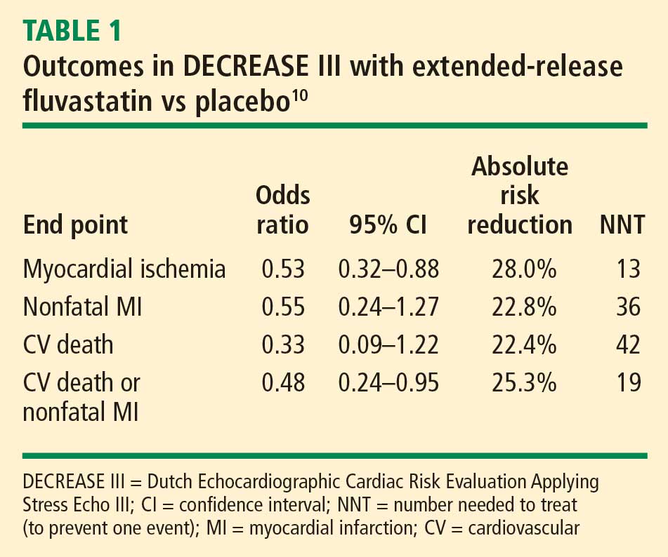
Safety: No effects on liver function or evidence of increased myopathy
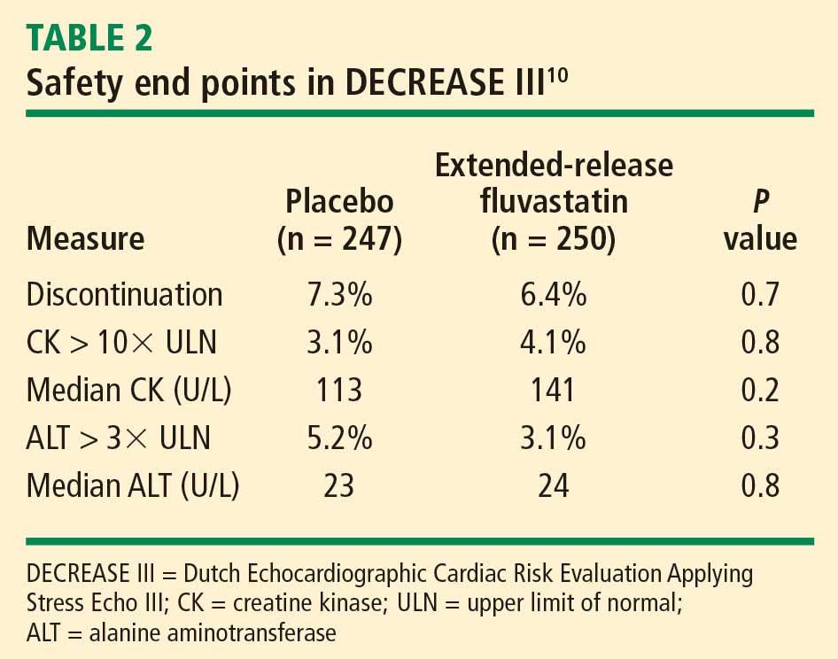
We concluded that initiation of therapy with a long-acting statin should be considered in statin-naïve patients undergoing vascular surgery.
PERIOPERATIVE STATIN USE: PRACTICAL CONSIDERATIONS
Inflammation, not cholesterol, should be the target
The optimal statin choice and the target level of LDL cholesterol immediately prior to surgery remain controversial. It may be that the more potent statins induce more side effects during surgery, but any such claim is speculative since no comparative studies exist. Regardless, the purpose of perioperative statin use should be reduction of the inflammatory stress response to surgery, with the long-term goal being achievement of recommended target lipid levels.
In particular, patients with peripheral arterial disease should have a statin initiated prior to high-risk vascular surgery (if they are not already receiving one), to increase the odds of recovering renal function after surgery (see section below) and to improve long-term outcomes.
Who are the best candidates?
Patients with multiple cardiac risk factors represent an especially high-risk group that benefits the most from statin therapy prior to vascular surgery, as they are likely to have more extensive disease and more extensive inflammation in the coronary artery tree.
Given the low incidence of side effects associated with statins, initiating a statin in patients with multiple cardiac risk factors who are undergoing intermediate-risk surgery may seem appropriate, but no data from large randomized trials are available to support this practice. Caution is in order when extrapolating data from studies conducted in the high-risk vascular surgery context to other surgical settings, since statins may pose hidden side effects such as liver dysfunction and myopathy, which may be missed in patients under anesthesia.
My personal practice is to initiate a statin prior to high-risk surgery or in patients with multiple cardiac risk factors if the risk-factor profile presents a clear indication for long-term statin use. If no risk factors are present, I am more reluctant to initiate a statin because of a lack of supportive data.
Beware the rebound effect with statin withdrawal
Statin withdrawal for several days following surgery is a common practice, since statins are given orally and their pleiotropic effects are underappreciated.
A withdrawal effect leading to abrogation of clinical benefit has been observed with perioperative use of short-acting statins, whose anti-inflammatory properties do not effectively extend to the postoperative period. Acute withdrawal has been associated with an increase in markers of inflammation and oxidative stress, and an increase in cardiac events has been observed with acute withdrawal of statins during periods of instability when compared with continuation of statin therapy.11
For these reasons, a long-acting statin is preferred preoperatively in patients whose oral intake will be compromised for several days after surgery (eg, in gastric surgery). The optimal statin for preventing the withdrawal effect is unknown. We chose extended-release fluvastatin in DECREASE III because its biological effect appears to last at least 4 days12 even though analysis of serum levels of the drug indicates a shorter half-life.
ANOTHER POTENTIAL BENEFIT: ENHANCED RECOVERY OF KIDNEY FUNCTION
Postoperative renal dysfunction is an ominous sign
Renal ischemic reperfusion injury is inevitable after vascular surgery that requires aortic cross-clamping. This is significant, as renal dysfunction after surgery is an ominous long-term sign that indicates abundant atherosclerosis. Complete recovery after acute kidney injury portends an improved long-term outcome, whereas patients with persistent renal dysfunction after vascular surgery have poor long-term outcomes.
A benefit from statins?
Statins may offer an effective means of preventing or shortening the course of acute kidney injury after surgery. Statins have been reported to lengthen survival of chronic kidney disease patients with sepsis or infectious complications and to improve the course of acute kidney injury in aging rats.13–15 These findings prompted my colleagues and I to conduct a retrospective study to evaluate whether statins may ameliorate reperfusion injury in the kidney after aortic cross-clamping.16
Promising findings from an observational review
We reviewed the records of all patients who had undergone vascular surgery at Erasmus Medical Center from January 1995 to June 2006 to examine the relation between preoperative statin use and renal function after suprarenal aortic cross-clamping.16 Of the 1,944 patients who met inclusion criteria, 515 (26.5%) were statin users. Postoperative kidney injury was defined as more than a 10% reduction in creatinine clearance on postoperative day 1 or 2 compared with baseline. Recovery of kidney function was defined as a creatinine clearance of greater than 90% of the baseline value by postoperative day 3.
The clinical characteristics of the populations with and without kidney injury after aortic cross-clamping were similar, including baseline creatinine clearance and serum creatinine.
Acute kidney injury within 2 days of surgery occurred in 664 patients (34%), of which 313 (47%) had complete recovery of kidney function at postoperative day 3. Although the incidence of postoperative kidney injury was similar among statin users and nonusers, statin use was associated with an increased chance of complete recovery of kidney function at day 3 (odds ratio = 2.0; 95% CI, 1.0–3.8).
All-cause mortality was assessed during a mean follow-up of 6.24 years. Statin use was associated with improved long-term survival, regardless of any change in kidney function (hazard ratio for death = 0.60; 95% CI, 0.48–0.75). Among the four broad patient groups, survival was highest among statin users with no postoperative kidney injury, followed by statin users who had kidney injury, then by nonusers of statins with no kidney injury, and finally by nonusers of statins who had kidney injury.
We concluded that perioperative statin use was associated with clinically significant recovery from acute kidney injury after high-risk vascular surgery and, more importantly, with improved long-term survival regardless of the presence of kidney injury. These promising findings require confirmation in prospective trials.
SUMMARY
Vascular surgery carries a high risk of perioperative mortality. Perioperative use of extended-release fluvastatin is associated with a reduced incidence of myocardial ischemia and the composite of MI and cardiovascular death at 30 days following surgery. These beneficial clinical outcomes are achieved without an increase in the incidence of side effects, including liver dysfunction and myopathy. Preoperative initiation of a long-acting statin is a reasonable strategy for reducing the risks associated with vascular surgery, and offers a bridge to postoperative statin continuation to blunt the inflammatory stress of surgery. Ischemic reperfusion injury is a major cause of renal dysfunction following vascular surgery. Statin therapy appears to help restore kidney function after aortic cross-clamping in patients undergoing high-risk vascular surgery.
DISCUSSION
Question from the audience: The majority of patients randomized in DECREASE III had relatively normal cholesterol levels. Do you believe those patients are biologically different from patients with physiologic vascular disease and elevated cholesterol levels?
Dr. Poldermans: We enrolled patients with various baseline cholesterol levels, and we found that these levels were not related to postoperative outcome. It would be a good idea to examine inflammation status just prior to surgery in patients with lower cholesterol levels to see if they have different outcomes from those with high cholesterol.
Question from the audience: If a patient is already on a short-acting statin and we know that he or she won’t be able to take a statin postoperatively, should we change to a long-acting statin just prior to surgery?
Dr. Poldermans: To be honest, this is a financial issue. If you have the opportunity, the best course would be to prescribe a statin with a prolonged half-life or an extended-release formulation. Of course, it’s not always possible to prescribe one particular statin. You have to negotiate what is feasible and hope to initiate the statin as early as possible to reduce risk.
Question from the audience: In studies conducted outside the perioperative setting, such as PROVE IT (Pravastatin or Atorvastatin Evaluation and Infection Therapy) and a substudy of REVERSAL (Reversing Atherosclerosis with Aggressive Lipid Lowering), it took about 30 days after statin initiation for hsCRP levels to minimize, and at least that long for halting of plaque progression to be detected by intravascular ultrasonography. Given that, does it make sense to delay nonurgent surgery in a patient in whom you’re worried about a postoperative MI?
Dr. Poldermans: Rat studies show improved blood flow and reduced thrombosis within hours of statin initiation. In the perioperative setting, therefore, initiating a statin within 30 days may be appropriate, but nobody knows the exact timing for optimal effect. Since there are no data to answer this question, I would not postpone surgery for this reason.
- Cohen MC, Aretz TH. Histological analysis of coronary artery lesions in fatal postoperative myocardial infarction. Cardiovasc Pathol 1999; 8:133–139.
- Dawood MM, Gutpa DK, Southern J, et al. Pathology of fatal perioperative myocardial infarction: implications regarding pathophysiology and prevention. Int J Cardiol 1996; 57:37–44.
- Ridker PM, Cannon CP, Morrow D, et al. C-reactive protein levels and outcomes after statin therapy. N Engl J Med 2005; 352:20–28.
- Poldermans D, Bax J, Boersma E, et al. Guidelines for pre-operative cardiac risk assessment and perioperative cardiac management in non-cardiac surgery. European Society of Cardiology Web site. http://www.escardio.org/guidelines-surveys/esc-guidelines/GuidelinesDocuments/guidelines-perioperative-cardiac-care-FT.pdf. Posted September 1, 2009. Accessed September 3, 2009.
- Durazzo AE, Machado FS, Ikeoka DT, et al. Reduction in cardiovascular events after vascular surgery with atorvastatin: a randomized trial. J Vasc Surg 2004; 39:967–976.
- Lindenauer PK, Pekow P, Wang K, Gutierrez B, Benjamin EM. Lipid-lowering therapy and in-hospital mortality following major noncardiac surgery. JAMA 2004; 291:2092–2099.
- Kertai MD, Boersma E, Westerhout CM, et al. A combination of statins and beta-blockers is independently associated with a reduction in the incidence of perioperative mortality and nonfatal myocardial infarction in patients undergoing abdominal aortic aneurysm surgery. Eur J Vasc Endovasc Surg 2004; 28:343–352.
- O’Neil-Callahan K, Katsimaglis G, Tepper MR, et al. Statins decrease perioperative cardiac complications in patients undergoing noncardiac vascular surgery: the Statins for Risk Reduction in Surgery (StaRRS). J Am Coll Cardiol 2005; 45:336–342.
- Poldermans D, Bax JJ, Kertai MD, et al. Statins are associated with a reduced incidence of perioperative mortality in patients undergoing major noncardiac vascular surgery. Circulation 2003; 107:1848–1851.
- Schouten O, Boersma E, Hoeks SE, et al, for the Dutch Echocardiographic Cardiac Risk Evaluation Applying Stress Echocardiography Study Group. Fluvastatin and perioperative events in patients undergoing vascular surgery. N Engl J Med 2009; 361:980–989.
- Heeschen C, Hamm CW, Laufs U, Snapinn S, Bohm M, White HD. Withdrawal of statins increases event rates in patients with acute coronary syndromes. Circulation 2002; 105:1446–1452.
- Schouten O, Hoeks SE, Welten GM, et al. Effect of statin withdrawal on frequency of cardiac events after vascular surgery. Am J Cardiol 2007; 100:316–320.
- Gupta R, Plantinga LC, Fink NE, et al. Statin use and hospitalization for sepsis in patients with chronic kidney disease. JAMA 2007; 297:1455–1464.
- Yasuda H, Yuen PS, Hu X, et al. Simvastatin improves sepsis-induced mortality and acute kidney injury via renal vascular effects. Kidney Int 2006; 69:1535–1542.
- Sabbatini M, Pisani A, Uccello F, et al. Atorvastatin improves the course of ischemic acute renal failure in aging rats. J Am Soc Nephrol 2004; 15:901–909.
- Welten GMJM, Chonchol M, Schouten O, et al. Statin use is associated with early recovery of kidney injury after vascular surgery and improved long-term outcome. Nephrol Dial Transplant 2008; 23:3867–3873.
Current uncertainty over the best approach for preventing fatal perioperative myocardial infarction (MI) lies in our inability, despite sophisticated testing methods, to detect unstable coronary plaque prior to surgery. Unstable plaque can be present in patients with coronary lumina that appear normal on coronary angiography. Therefore, reliance on medical therapy to blunt inflammation is currently the best practice for minimizing the risk that unstable plaque poses.
Perioperative use of statins is a cornerstone of such therapy. This article briefly reviews the rationale for perioperative statin use in the setting of noncardiac surgery, presents the latest evidence on the clinical effects of perioperative statin use, and considers the potential role for statins in promoting recovery from acute kidney injury after vascular surgery.
FATAL MI: ORIGINS AND APPROACHES TO RISK REDUCTION
Fatal perioperative MI has two potential origins.1,2 One is a culprit coronary plaque that fissures and ruptures, causing a cascade of thrombogenic events (hemorrhage and thrombosis) inside the vessel wall, culminating in an MI. Less often, fatal perioperative MI results from long-lasting myocardial ischemia (a demand/supply mismatch of oxygen), typically as a consequence of a fixed coronary stenosis.
In nearly half of patients with fatal MI, coronary inflammation is a key contributor. In the perioperative setting, surgical stress induces the release of inflammatory cytokines that disrupt smooth muscle cells in the endothelium and contribute to disruption of a nonobstructing coronary plaque, predisposing to acute thrombus formation.
Risk reduction depends on pathophysiology
Strategies for minimizing the risk of perioperative MI depend on the pathophysiology involved. In the case of oxygen demand/supply mismatch as a result of flow-limiting stenosis, a beta-blocker and coronary revascularization, if possible, may be useful.
In the more common case of unstable plaque, a multifactorial strategy appears optimal, involving the following:
- Statin therapy to reduce coronary inflammation
- Aspirin to blunt the prothrombotic milieu postoperatively
- Chronic low-dose beta-blockade to decrease myocardial oxygen demand or inhibit plaque rupture.
A particular role for statins
Ridker et al found that patients with an acute coronary syndrome who experience a decline in high-sensitivity C-reactive protein (hsCRP) level after treatment with a statin have improved clinical outcomes compared with those whose hsCRP level remains high, regardless of their resultant low-density lipoprotein (LDL) cholesterol level.3
Among surgical patients, those most at risk for poor cardiovascular outcomes are those who undergo vascular surgery. In Europe, the cardiovascular death rate in such patients is approximately 2%.4
Retrospective cohort data and data from randomized clinical trials have demonstrated reductions in perioperative cardiac complications with statin use in patients undergoing various types of noncardiac vascular surgery.5–9 In light of these data, my colleagues and I recently undertook a prospective study to examine the effect of perioperative statin use on cardiovascular complications in patients undergoing vascular surgery.10 Key details and findings are surveyed in the following section.
DECREASE III: PROSPECTIVE EVIDENCE FOR ISCHEMIC BENEFIT FROM PERIOPERATIVE STATINS
The Dutch Echocardiographic Cardiac Risk Evaluation Applying Stress Echo III (DECREASE III) was conducted at a single center (Erasmus Medical Center, Rotterdam, the Netherlands) in a randomized, double-blind, placebo-controlled manner.10
Patients and study design
The study population included 497 statin-naïve patients who were scheduled for one of four noncardiac vascular surgical procedures (repair or revascularization for abdominal aortic aneurysm, abdominal aortic stenosis, lower limb arterial stenosis, or carotid artery stenosis). Patients with unstable coronary artery disease or left main disease were excluded.
Patients were randomized to placebo or extended-release fluvastatin (80 mg/day) starting on the day of randomization, which was a median of 37 days before surgery. Treatment was continued until 30 days after surgery.
Extended-release fluvastatin was chosen because its long half-life permits a bridge to the early postoperative period, during which oral medications are not permitted in patients undergoing high-risk vascular surgery.
The primary end point was the occurrence of myocardial ischemia as assessed by three methods:
- Holter monitoring during the first 72 postoperative hours
- Measurement of troponin T on days 1, 3, 7, and 30
- Additional electrocardiographic recordings on days 7 and 30.
The secondary end point was a composite of cardiovascular death and nonfatal MI during the first 30 postoperative days.
Baseline characteristics were similar between the two randomized groups, with a median age approaching 66 years. About three-fourths of the patients were male, one-fourth had a history of MI, one-fourth had angina pectoris, one-fifth had diabetes mellitus, and nearly 30% had a history of cerebrovascular accident or transient ischemic attack.
All patients were being treated with a beta-blocker, about 60% with antiplatelet therapy, more than one-fourth with anticoagulant therapy, nearly half with either an angiotensin-converting enzyme inhibitor or angiotensin receptor blocker, and more than one-fourth with diuretics. There were no significant differences between the groups in the proportion of patients on each of these therapies.
Results: Reductions in inflammatory markers
Baseline levels of hsCRP and interleukin-6 (IL-6) were comparable between the groups. In patients randomized to placebo, the hsCRP level increased by 3%, from a median of 5.80 mg/L at randomization to 6.00 mg/L immediately prior to surgery. In contrast, the hsCRP level in patients randomized to extended-release fluvastatin decreased by 21%, from a median of 5.93 mg/L to 4.66 mg/L. The between-group difference in the change in hsCRP level was statistically significant (P < .001). There was also a significantly greater reduction from baseline in median level of IL-6 among fluvastatin recipients compared with placebo recipients (–33% vs –4%; P < .001).
The specificity of hsCRP for cardiac inflammation is not yet known, but measures of hsCRP and IL-6 can provide a fingerprint of inflammatory activity prior to surgery. Other inflammatory and noninflammatory markers are being investigated to better identify (prior to surgery) those high-risk patients most likely to benefit from perioperative statin use.
Results: Favorable effect on clinical end points
The incidence of the primary end point—myocardial ischemia 30 days after surgery—was significantly lower in the patients randomized to extended-release fluvastatin compared with placebo (10.9% vs 18.9%; P = .016), as was the incidence of the secondary end point of cardiovascular death or nonfatal MI (4.8% vs 10.1%; P = .039).
Safety: No effects on liver function or evidence of increased myopathy
We concluded that initiation of therapy with a long-acting statin should be considered in statin-naïve patients undergoing vascular surgery.
PERIOPERATIVE STATIN USE: PRACTICAL CONSIDERATIONS
Inflammation, not cholesterol, should be the target
The optimal statin choice and the target level of LDL cholesterol immediately prior to surgery remain controversial. It may be that the more potent statins induce more side effects during surgery, but any such claim is speculative since no comparative studies exist. Regardless, the purpose of perioperative statin use should be reduction of the inflammatory stress response to surgery, with the long-term goal being achievement of recommended target lipid levels.
In particular, patients with peripheral arterial disease should have a statin initiated prior to high-risk vascular surgery (if they are not already receiving one), to increase the odds of recovering renal function after surgery (see section below) and to improve long-term outcomes.
Who are the best candidates?
Patients with multiple cardiac risk factors represent an especially high-risk group that benefits the most from statin therapy prior to vascular surgery, as they are likely to have more extensive disease and more extensive inflammation in the coronary artery tree.
Given the low incidence of side effects associated with statins, initiating a statin in patients with multiple cardiac risk factors who are undergoing intermediate-risk surgery may seem appropriate, but no data from large randomized trials are available to support this practice. Caution is in order when extrapolating data from studies conducted in the high-risk vascular surgery context to other surgical settings, since statins may pose hidden side effects such as liver dysfunction and myopathy, which may be missed in patients under anesthesia.
My personal practice is to initiate a statin prior to high-risk surgery or in patients with multiple cardiac risk factors if the risk-factor profile presents a clear indication for long-term statin use. If no risk factors are present, I am more reluctant to initiate a statin because of a lack of supportive data.
Beware the rebound effect with statin withdrawal
Statin withdrawal for several days following surgery is a common practice, since statins are given orally and their pleiotropic effects are underappreciated.
A withdrawal effect leading to abrogation of clinical benefit has been observed with perioperative use of short-acting statins, whose anti-inflammatory properties do not effectively extend to the postoperative period. Acute withdrawal has been associated with an increase in markers of inflammation and oxidative stress, and an increase in cardiac events has been observed with acute withdrawal of statins during periods of instability when compared with continuation of statin therapy.11
For these reasons, a long-acting statin is preferred preoperatively in patients whose oral intake will be compromised for several days after surgery (eg, in gastric surgery). The optimal statin for preventing the withdrawal effect is unknown. We chose extended-release fluvastatin in DECREASE III because its biological effect appears to last at least 4 days12 even though analysis of serum levels of the drug indicates a shorter half-life.
ANOTHER POTENTIAL BENEFIT: ENHANCED RECOVERY OF KIDNEY FUNCTION
Postoperative renal dysfunction is an ominous sign
Renal ischemic reperfusion injury is inevitable after vascular surgery that requires aortic cross-clamping. This is significant, as renal dysfunction after surgery is an ominous long-term sign that indicates abundant atherosclerosis. Complete recovery after acute kidney injury portends an improved long-term outcome, whereas patients with persistent renal dysfunction after vascular surgery have poor long-term outcomes.
A benefit from statins?
Statins may offer an effective means of preventing or shortening the course of acute kidney injury after surgery. Statins have been reported to lengthen survival of chronic kidney disease patients with sepsis or infectious complications and to improve the course of acute kidney injury in aging rats.13–15 These findings prompted my colleagues and I to conduct a retrospective study to evaluate whether statins may ameliorate reperfusion injury in the kidney after aortic cross-clamping.16
Promising findings from an observational review
We reviewed the records of all patients who had undergone vascular surgery at Erasmus Medical Center from January 1995 to June 2006 to examine the relation between preoperative statin use and renal function after suprarenal aortic cross-clamping.16 Of the 1,944 patients who met inclusion criteria, 515 (26.5%) were statin users. Postoperative kidney injury was defined as more than a 10% reduction in creatinine clearance on postoperative day 1 or 2 compared with baseline. Recovery of kidney function was defined as a creatinine clearance of greater than 90% of the baseline value by postoperative day 3.
The clinical characteristics of the populations with and without kidney injury after aortic cross-clamping were similar, including baseline creatinine clearance and serum creatinine.
Acute kidney injury within 2 days of surgery occurred in 664 patients (34%), of which 313 (47%) had complete recovery of kidney function at postoperative day 3. Although the incidence of postoperative kidney injury was similar among statin users and nonusers, statin use was associated with an increased chance of complete recovery of kidney function at day 3 (odds ratio = 2.0; 95% CI, 1.0–3.8).
All-cause mortality was assessed during a mean follow-up of 6.24 years. Statin use was associated with improved long-term survival, regardless of any change in kidney function (hazard ratio for death = 0.60; 95% CI, 0.48–0.75). Among the four broad patient groups, survival was highest among statin users with no postoperative kidney injury, followed by statin users who had kidney injury, then by nonusers of statins with no kidney injury, and finally by nonusers of statins who had kidney injury.
We concluded that perioperative statin use was associated with clinically significant recovery from acute kidney injury after high-risk vascular surgery and, more importantly, with improved long-term survival regardless of the presence of kidney injury. These promising findings require confirmation in prospective trials.
SUMMARY
Vascular surgery carries a high risk of perioperative mortality. Perioperative use of extended-release fluvastatin is associated with a reduced incidence of myocardial ischemia and the composite of MI and cardiovascular death at 30 days following surgery. These beneficial clinical outcomes are achieved without an increase in the incidence of side effects, including liver dysfunction and myopathy. Preoperative initiation of a long-acting statin is a reasonable strategy for reducing the risks associated with vascular surgery, and offers a bridge to postoperative statin continuation to blunt the inflammatory stress of surgery. Ischemic reperfusion injury is a major cause of renal dysfunction following vascular surgery. Statin therapy appears to help restore kidney function after aortic cross-clamping in patients undergoing high-risk vascular surgery.
DISCUSSION
Question from the audience: The majority of patients randomized in DECREASE III had relatively normal cholesterol levels. Do you believe those patients are biologically different from patients with physiologic vascular disease and elevated cholesterol levels?
Dr. Poldermans: We enrolled patients with various baseline cholesterol levels, and we found that these levels were not related to postoperative outcome. It would be a good idea to examine inflammation status just prior to surgery in patients with lower cholesterol levels to see if they have different outcomes from those with high cholesterol.
Question from the audience: If a patient is already on a short-acting statin and we know that he or she won’t be able to take a statin postoperatively, should we change to a long-acting statin just prior to surgery?
Dr. Poldermans: To be honest, this is a financial issue. If you have the opportunity, the best course would be to prescribe a statin with a prolonged half-life or an extended-release formulation. Of course, it’s not always possible to prescribe one particular statin. You have to negotiate what is feasible and hope to initiate the statin as early as possible to reduce risk.
Question from the audience: In studies conducted outside the perioperative setting, such as PROVE IT (Pravastatin or Atorvastatin Evaluation and Infection Therapy) and a substudy of REVERSAL (Reversing Atherosclerosis with Aggressive Lipid Lowering), it took about 30 days after statin initiation for hsCRP levels to minimize, and at least that long for halting of plaque progression to be detected by intravascular ultrasonography. Given that, does it make sense to delay nonurgent surgery in a patient in whom you’re worried about a postoperative MI?
Dr. Poldermans: Rat studies show improved blood flow and reduced thrombosis within hours of statin initiation. In the perioperative setting, therefore, initiating a statin within 30 days may be appropriate, but nobody knows the exact timing for optimal effect. Since there are no data to answer this question, I would not postpone surgery for this reason.
- Cohen MC, Aretz TH. Histological analysis of coronary artery lesions in fatal postoperative myocardial infarction. Cardiovasc Pathol 1999; 8:133–139.
- Dawood MM, Gutpa DK, Southern J, et al. Pathology of fatal perioperative myocardial infarction: implications regarding pathophysiology and prevention. Int J Cardiol 1996; 57:37–44.
- Ridker PM, Cannon CP, Morrow D, et al. C-reactive protein levels and outcomes after statin therapy. N Engl J Med 2005; 352:20–28.
- Poldermans D, Bax J, Boersma E, et al. Guidelines for pre-operative cardiac risk assessment and perioperative cardiac management in non-cardiac surgery. European Society of Cardiology Web site. http://www.escardio.org/guidelines-surveys/esc-guidelines/GuidelinesDocuments/guidelines-perioperative-cardiac-care-FT.pdf. Posted September 1, 2009. Accessed September 3, 2009.
- Durazzo AE, Machado FS, Ikeoka DT, et al. Reduction in cardiovascular events after vascular surgery with atorvastatin: a randomized trial. J Vasc Surg 2004; 39:967–976.
- Lindenauer PK, Pekow P, Wang K, Gutierrez B, Benjamin EM. Lipid-lowering therapy and in-hospital mortality following major noncardiac surgery. JAMA 2004; 291:2092–2099.
- Kertai MD, Boersma E, Westerhout CM, et al. A combination of statins and beta-blockers is independently associated with a reduction in the incidence of perioperative mortality and nonfatal myocardial infarction in patients undergoing abdominal aortic aneurysm surgery. Eur J Vasc Endovasc Surg 2004; 28:343–352.
- O’Neil-Callahan K, Katsimaglis G, Tepper MR, et al. Statins decrease perioperative cardiac complications in patients undergoing noncardiac vascular surgery: the Statins for Risk Reduction in Surgery (StaRRS). J Am Coll Cardiol 2005; 45:336–342.
- Poldermans D, Bax JJ, Kertai MD, et al. Statins are associated with a reduced incidence of perioperative mortality in patients undergoing major noncardiac vascular surgery. Circulation 2003; 107:1848–1851.
- Schouten O, Boersma E, Hoeks SE, et al, for the Dutch Echocardiographic Cardiac Risk Evaluation Applying Stress Echocardiography Study Group. Fluvastatin and perioperative events in patients undergoing vascular surgery. N Engl J Med 2009; 361:980–989.
- Heeschen C, Hamm CW, Laufs U, Snapinn S, Bohm M, White HD. Withdrawal of statins increases event rates in patients with acute coronary syndromes. Circulation 2002; 105:1446–1452.
- Schouten O, Hoeks SE, Welten GM, et al. Effect of statin withdrawal on frequency of cardiac events after vascular surgery. Am J Cardiol 2007; 100:316–320.
- Gupta R, Plantinga LC, Fink NE, et al. Statin use and hospitalization for sepsis in patients with chronic kidney disease. JAMA 2007; 297:1455–1464.
- Yasuda H, Yuen PS, Hu X, et al. Simvastatin improves sepsis-induced mortality and acute kidney injury via renal vascular effects. Kidney Int 2006; 69:1535–1542.
- Sabbatini M, Pisani A, Uccello F, et al. Atorvastatin improves the course of ischemic acute renal failure in aging rats. J Am Soc Nephrol 2004; 15:901–909.
- Welten GMJM, Chonchol M, Schouten O, et al. Statin use is associated with early recovery of kidney injury after vascular surgery and improved long-term outcome. Nephrol Dial Transplant 2008; 23:3867–3873.
KEY POINTS
- The inflammatory and oxidative stress induced by vascular surgery can be blunted by statin therapy.
- Statin therapy started preoperatively can reduce the incidence of myocardial ischemia and the level of inflammatory markers in patients undergoing high-risk vascular surgery.
- The purpose of perioperative statin use should be reduction of the inflammatory stress response to surgery, with the long-term goal being achievement of target lipid levels.
- A long-acting statin is preferred preoperatively to best extend the anti-inflammatory effects into the postoperative period. Statin therapy should be continued postoperatively, if possible, to avoid deleterious acute withdrawal effects.
The experts debate: Perioperative beta-blockade for noncardiac surgery—proven safe or not?
NOTE: The individual co-authors in this debate-based article are responsible only for those views within their respective bylined subsections and those views ascribed to them in the rebuttals and discussion at the end.
Perioperative beta-blockade improves outcomes
By Don Poldermans, MD, PhD
It is my contention that perioperative beta-blockade improves mortality and cardiac outcomes in select high- and intermediate-risk patients undergoing noncardiac surgery. Patients on chronic beta-blocker therapy should have it continued perioperatively. For patients not already on beta-blockade who are at cardiac risk, initiation of low-dose beta-blocker therapy should be considered prior to surgery; such therapy should be started approximately 1 month before surgery, with dose titration to achieve hemodynamic stability. Reports of increased stroke rates with perioperative beta-blockade appear to be due to inappropriate acute administration of high-dose beta-blocker therapy.
THE PHYSIOLOGIC RATIONALE FOR PERIOPERATIVE BETA-BLOCKADE
Perioperative myocardial infarction (MI) can occur by one of two mechanisms, both of which can be attenuated by beta-blockade:
- The stress induced by surgery can cause an asymptomatic coronary plaque to become unstable and rupture, resulting in complete occlusion of a portion of the coronary tree. This type of perioperative MI occurs typically in patients with multiple risk factors for MI absent a critical coronary stenosis. The perioperative risk associated with unstable plaque can be reduced pharmacologically with aspirin, statins, and chronic beta-blocker therapy.
- Alternately, a fixed coronary stenosis can predispose to a mismatch of oxygen demand and supply, leading to myocardial ischemia and infarction. The patient with a fixed coronary lesion typically presents with stable angina pectoris, and the at-risk stenosis is identified through a stress echocardiogram or nuclear scan. The risk conferred by flow-limiting stable plaque can be reduced by coronary revascularization and a short course of beta-blocker therapy prior to surgery.
INITIAL SUPPORTIVE DATA
Mangano and colleagues were the first to evaluate perioperative beta-blockade in a randomized, controlled fashion.1,2 In their study of 200 surgical patients with or at risk for coronary artery disease, oral atenolol administered perioperatively was associated with a 50% reduction (compared with placebo) in the incidence of postoperative myocardial ischemia as measured by three-lead Holter monitoring.2 During 2 years of follow-up, mortality was significantly lower in the atenolol group (10%) than in the placebo group (21%) (P = .019).1
In the Dutch Echocardiographic Cardiac Risk Evaluation Applying Stress Echocardiography (DECREASE I), my research group randomized 112 high-risk patients (as identified by dobutamine echocardiography) to standard perioperative care alone or standard perioperative care plus bisoprolol starting 30 days prior to major vascular surgery.3 The dosage of bisoprolol was titrated to achieve a target heart rate of 60 to 70 beats per minute. Thirty days after surgery, the incidence of the primary end point—a composite of death from cardiac causes or nonfatal MI—was reduced from 34% in the standard-care group to 3.4% in the bisoprolol group (P < .001). Thus, in this unblinded study in a population with proven coronary artery disease, beta-blockade clearly improved outcomes.
Additional studies of perioperative beta-blocker use have produced a wide range of outcomes, with most favoring beta-blockade, albeit usually not to a statistically significant degree.4–13 Notably, only some of these trials were randomized, they used various beta-blocker regimens at various doses, they were conducted in patients with varying degrees of cardiac risk, and many had small sample sizes.
What emerged from these trials was the idea that perioperative beta-blockade in patients with coronary artery disease produces an effect similar to that of long-term beta-blockade in reducing the risk of cardiovascular events in post-MI patients and in those with coronary artery disease and heart failure.
THE POISE STUDY AND ITS IMPLICATIONS
Results of the Perioperative Ischemic Evaluation (POISE) were published in 2008, in which 8,351 noncardiac surgery patients with or at risk of atherosclerotic disease were randomized to placebo or extended-release metoprolol succinate started 2 to 4 hours preoperatively and continued for 30 days.14 Metoprolol was associated with a clear reduction in the primary end point, a composite of cardiovascular death, nonfatal MI, or nonfatal cardiac arrest (5.8% vs 6.9% with placebo; hazard ratio [HR] = 0.84 [95% CI, 0.70–0.99]; P = .0399), but this effect was offset by significant increases in total mortality and stroke incidence in the metoprolol group. Mortality was 3.1% with metoprolol versus 2.3% with placebo (HR = 1.33 [95% CI, 1.03–1.74]; P = .0317), and stroke incidence was 1.0% with metoprolol versus 0.5% with placebo (HR = 2.17 [95% CI, 1.26–3.74]; P = .0053). Cerebral infarction, not bleeding, explained most of the excess mortality with metoprolol.
Of the 60 strokes in POISE, 49 were ischemic in origin, 3 were hemorrhagic, and 8 were of uncertain etiology. Preoperative predictors of stroke were the use of clopidogrel and a history of stroke or transient ischemic attack. Postoperative predictors of stroke included intraoperative bleeding and intraoperative hypotension. These predictors suggest a diseased cerebrovascular tree or unstable hemodynamics during the intraoperative period in the patients who suffered a stroke.
Does dosing explain the rise in mortality and strokes?
Could the fatal outcomes associated with the beta-blocker in POISE be attributed to the dosage of metoprolol? In the study, 100 mg of metoprolol was started immediately prior to surgery, and an additional 100 mg could be given, depending on the hemodynamic response. Maintenance therapy (200 mg/day) was started on the same day, making it possible that a patient could have received as much as 400 mg of metoprolol the day of surgery. The starting dose of metoprolol used in POISE was two to eight times the commonly prescribed dose.
The initial 100-mg dose of metoprolol used in POISE has a similar beta1-receptor blockade potency compared with the 5-mg dose of bisoprolol used in DECREASE I.3 However, in DECREASE I, bisoprolol was initiated 30 days prior to surgery and was titrated, if necessary, according to heart rate. The maintenance dose of bisoprolol was half of the maintenance dose used in POISE. In the later DECREASE trials, the starting dose of bisoprolol was only 2.5 mg. Therefore, there was a huge difference in beta-blocker dosing between POISE and DECREASE.
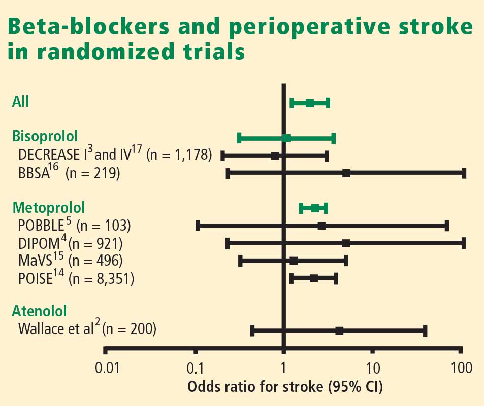
What about timing of beta-blocker initiation?
The POISE findings may also be explained in part by the timing of beta-blocker initiation. Whereas bisoprolol was carefully titrated for 30 days before surgery in DECREASE, metoprolol was initiated just before surgery in POISE, and the maximum recommended dose may have been prescribed during the first 24 hours, although subsequent dosing was 200 mg daily, which is 50% of the maximum daily therapeutic dose. This extremely narrow time window for titration may be important, since the beneficial effects of beta-blockade on coronary plaque stability are likely to take weeks to develop.
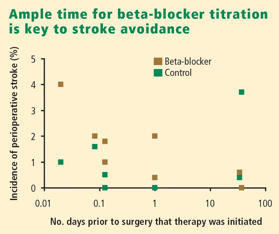
Reassurance from a large case-control study
My colleagues and I conducted a case-control study from among more than 75,000 patients who underwent noncardiac, nonvascular surgery at our institution, Erasmus Medical Center, from 1991 to 2001.18 The cases were the 989 patients who died in the hospital postoperatively; the controls were 1,879 survivors matched with the cases for age, sex, the year the surgery was performed, and the type of surgery. The incidence of perioperative stroke was 0.5%, which is comparable to the rate found in the literature. Risk factors predictive of stroke were the presence of diabetes, cerebrovascular disease, peripheral arterial disease, atrial fibrillation, coronary artery disease, and hypertension. Notably, no relationship was found between chronic beta-blocker use and stroke.
WHAT ABOUT PATIENTS AT INTERMEDIATE RISK?
Because the effect of perioperative beta-blockade has traditionally been ill defined in surgical patients at intermediate risk of cardiovascular events, the DECREASE study group recently completed a study (DECREASE IV) to assess perioperative bisoprolol in terms of cardiac morbidity and mortality in intermediate-risk patients undergoing elective noncardiovascular surgery.17 Enrollees had a score of 1 to 2 on the Revised Cardiac Risk Index of Lee et al,19 which corresponds to an estimated risk of between 1% and 6% for a perioperative cardiovascular event.17
DECREASE IV also aimed to assess the effect of perioperative fluvastatin, so a 2 x 2 factorial design was used in which the study’s 1,066 patients were randomized to receive bisoprolol, fluvastatin, combination treatment, or combination placebo control. Bisoprolol was initiated up to 30 days prior to surgery, and the 2.5-mg daily starting dosage was titrated according to the patient’s heart rate to achieve a target rate of 50 to 70 beats per minute. Fluvastatin was also started up to 30 days prior to surgery. Patients who received bisoprolol (with or without fluvastatin) had a significant reduction in the 30-day incidence of cardiac death and nonfatal MI compared with those who did not receive bisoprolol (2.1% vs 6.0%; HR = 0.34 [95% CI, 0.17–0.67]; P = .002). Fluvastatin was associated with a favorable trend on this end point, but statistical significance was not achieved (P = .17).17
There was no difference among treatment groups in the incidence of stroke (4 strokes in the 533 patients who received bisoprolol vs 3 strokes in the 533 patients who did not),17 which further suggests that the increased stroke rate seen with beta-blockade in POISE may have been due to dosage, timing of initiation, or both.
CONCLUSIONS
Dose-related hypotension may explain POISE findings
Our understanding of postoperative stroke is incomplete, but it appears that dosing of a beta-blocker can be a contributor, especially with respect to the potential side effect of hypotension during surgery. Keep in mind that the average age of patients in POISE was approximately 70 years and that patients were naïve to beta-blockers. Some may have had asymptomatic left ventricular dysfunction, and we know that starting a beta-blocker at a high dose in such patients may lead to hypotension. At my institution we routinely perform echocardiographic screening of all patients scheduled for surgery, and we have found that more than half of the patients with heart failure have it uncovered only through this screening.
It is not the medicine alone that can cause perioperative hypotension; other factors may induce hypotension, requiring beta-blocker titration and careful monitoring of hemodynamics during surgery.
Advice: Start early and titrate dose; continue chronic beta-blockade
My advice is as follows:
- If a patient is on chronic beta-blocker therapy, do not stop it perioperatively. We have seen devastating outcomes in the Netherlands when patients had their beta-blockers stopped immediately before surgery. Consider adjusting the dose, but do not stop it entirely. If a beta-blocker is on board and the patient develops hypotension or bradycardia during surgery, treat the symptoms and check for sepsis.
- In a patient not on a beta-blocker, consider adding one if the patient is at intermediate or high risk of a cardiac event, but start at a low dosage (ie, 2.5 mg/day for bisoprolol and 25 mg/day for metoprolol). Treatment ideally should be started 30 days preoperatively; in the Netherlands, we have the chance to start well in advance of surgery so we can titrate the dose according to hemodynamics.
- If a beta-blocker is not started because of insufficient time for titration, do not add one to treat tachycardia that develops during surgery, since tachycardia may represent a response to normal defense mechanisms.
Safety of perioperative beta-blocker use has not been adequately demonstrated
By P.J. Devereaux, MD, PhD
I contend that perioperative beta-blockade is a practice not grounded in evidence-based medicine, and its overall safety has increasingly come into question as more data from large, high-quality trials have emerged. I will begin with a historical overview of perioperative beta-blocker use, review the results of the POISE trial (for which I was the co-principal investigator), explore the major questions raised by this trial, and conclude with some take-away messages.
THE HISTORY OF PERIOPERATIVE BETA-BLOCKADE
In the 1970s, physicians were encouraged to hold beta-blockers prior to surgery out of concern that these medications may inhibit the required cardiovascular response when patients developed hypotension, and could thereby lead to serious adverse consequences.
In the 1980s, new research associated tachycardia with perioperative cardiovascular events, leading to proposals to implement perioperative beta-blocker use.
In the 1990s, two randomized trials with a total sample size of 312 patients1,3 suggested that perioperative beta-blockers had a large treatment effect in preventing major cardiovascular events and death. These small trials had several methodological limitations:
- One trial3 was unblinded in a setting in which the vast majority of MIs are clinically silent.
- One trial3 was stopped early—after randomizing only 112 patients—for unexpected large treatment effects.
- One of the studies1 failed to follow intention-to-treat principle.
Nevertheless, guidelines developed at the time by the American College of Cardiology and the American Heart Association (ACC/AHA) recommended the use of perioperative beta-blockers on the basis of the physiological rationale and these two small clinical trials. That recommendation was retained in the latest (2007) update of the ACC/AHA perioperative guidelines.20
In 2006, two clinical trials with a total sample size of 1,417 were completed,4,15 surpassing the total size of previous trials by more than fourfold. These two more recent trials did not suffer from the methodological limitations of earlier trials. These trials showed no benefit of perioperative beta-blocker use; in fact, there was a trend toward worse outcomes in the beta-blocker recipients.4,15 Despite these new data, guidelines committees continued to recommend perioperative beta-blockade.20
THE POISE TRIAL
Study design
This was the context into which the POISE results were released in 2008. POISE was a randomized, controlled, blinded trial of patients 45 years or older scheduled for noncardiac surgery who had, or were at high risk of, atherosclerotic disease.14 The intervention consisted of metoprolol succinate (metoprolol controlled release [CR]) or placebo started 2 to 4 hours preoperatively (if heart rate was ≥ 50 beats per minute and systolic blood pressure [SBP] was ≥ 100 mm Hg) and continued for 30 days. The target dosage of metoprolol was 200 mg once daily. No patients received the recommended maximum dosage of 400 mg over 24 hours. The main outcome measure was a 30-day composite of cardiovascular death, nonfatal MI, or nonfatal cardiac arrest.
We randomized 9,298 patients in a 1:1 ratio to metoprolol or placebo. We encountered data fraud at a number of centers that prompted exclusion of data from 474 patients allocated to metoprolol and 473 allocated to placebo. Therefore, the total number of patients available for the intention-to-treat analysis was 8,351, from 190 centers in 23 countries.
Results
The risk of the primary composite outcome was reduced by 16% (relative reduction) in recipients of metoprolol CR compared with placebo recipients (P = .0399). Significantly fewer nonfatal MIs occurred in the metoprolol CR group than in the placebo group (152 [3.6%] vs 215 [5.1%]; P = .0008), leaving little doubt that perioperative beta-blockade prevents MI.
In contrast, total mortality was increased in the beta-blocker group, with 129 deaths among those assigned to metoprolol CR and 97 among those assigned to placebo (P = .0317), and the incidence of stroke was also significantly greater in the metoprolol CR group (1.0% vs. 0.5%; P = .0053).
Consistency with findings from other trials
The POISE data are consistent with those from a 2008 meta-analysis of high-quality randomized controlled trials in noncardiac surgery patients, which showed a significantly greater risk of death among patients assigned to a beta-blocker than among controls who were not (160 deaths [2.8%] vs 127 deaths [2.3%]; odds ratio [OR] = 1.27 [95% CI, 1.01–1.61]).21 This meta-analysis also found a significantly greater risk of nonfatal stroke in beta-blocker recipients compared with controls (38 [0.7%] vs 17 [0.3%]; OR = 2.16 [95% CI, 1.27–3.68]).
I also contend that the DECREASE IV trial supports the POISE findings in that although few strokes were encountered in DECREASE IV, the trend was in the direction of harm in the beta-blocker group, which had 4 strokes among 533 patients versus 3 strokes among 533 patients not receiving the beta-blocker.17
Predictive role of hypotension
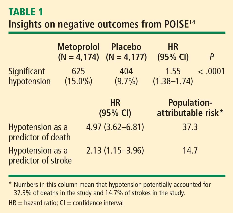
The link between hypotension and death in POISE is consistent with findings from the largest beta-blocker trial undertaken, COMMIT (Clopidogrel and Metoprolol in Myocardial Infarction Trial), in which 45,852 patients with acute MI were randomized to metoprolol or placebo.22 In COMMIT, metoprolol had no effect on 30-day all-cause mortality but significantly reduced the risk of arrhythmic death, a benefit that was countered by a significantly increased risk of death from shock with a beta-blocker in acute MI. Clinically significant hypotension is much more common in the perioperative setting than in acute MI, which may explain the excess number of deaths observed with metoprolol in POISE as opposed to metoprolol’s neutral effect on mortality in COMMIT.
ANSWERING THE CRITICS
Several criticisms have been raised about POISE, as detailed below.
Beta-blocker dose
Some contend that a lower dose of beta-blocker would provide benefit and minimize risk, but this assertion must be supported by evidence from a large clinical trial. The targeted dosage of metoprolol in POISE represents 50% of the maximum daily therapeutic dose. Further, the protocol called for decreasing the dosage to 100 mg/day if SBP dropped to less than 100 mm Hg or if heart rate fell to less than 45 beats per minute.
The two small trials on which guideline recommendations for perioperative beta-blockade are primarily based1,3 had a sample size that was 4% of that in POISE, which calls into question the reliability of their results. The study by Mangano et al used atenolol at a target dosage that was 50% of the maximum daily therapeutic dose,1 the same as with metoprolol in POISE. DECREASE initiated bisoprolol at 25% of the maximum daily therapeutic dose, and allowed for titration to 50% of the maximum daily therapeutic dose.3
As the second largest study of perioperative beta-blockade, the Diabetic Postoperative Mortality and Morbidity (DIPOM) trial enrolled 921 patients who were assigned to placebo or controlled-release metoprolol with a target dosage that was 25% of the maximum daily therapeutic dose.4 The 30-day outcomes from DIPOM showed a trend toward an excess of death and stroke despite using only one-half the dosage in POISE and the same dosage as in DECREASE.
Timing of beta-blocker initiation
Another contention is that earlier beta-blocker initiation would be better. The issue with timing of initiation is not benefit, as POISE showed that starting a beta-blocker hours before surgery results in a reduction in the risk of MI. The issue is whether giving a beta-blocker earlier makes administering the drug safer. Nearly 10% of placebo recipients in POISE developed clinically significant hypotension, which suggests that the titrated dosage of a beta-blocker that appears effective preoperatively is unlikely to inform a safe dose after surgery, when hypotension is common.
The practicality of titrating the dose of beta-blocker prior to surgery also comes into play. Most patients referred to my institution for surgery are seen 1 to 2 weeks in advance, at the earliest. Real-world practice at present simply does not afford us the luxury of seeing patients three to four times before surgery in order to titrate the beta-blocker dose.
POISE did not address chronic beta-blocker therapy
It is important to remember that POISE excluded patients on chronic beta-blocker therapy and thus did not attempt to address the perioperative management of such patients who undergo noncardiac surgery. My suspicion is that perioperative continuation of beta-blockade in these patients is the best course of action, but this too has not been studied robustly, so we need a large controlled trial to confirm that this practice is indeed safe.
CONCLUSIONS
The POISE results suggest that for every 1,000 patients treated, perioperative metoprolol would:
- Prevent 15 MIs, 3 cardiac revascularizations, and 7 new cases of atrial fibrillation
- Result in 8 excess deaths, 5 strokes, 53 cases of clinically significant hypotension, and 42 cases of clinically significant bradycardia.
The central take-away message is that patients are unlikely to want a perioperative beta-blocker if they are unwilling to accept a probable increase in mortality or if they place three times more value on avoiding a perioperative stroke than on avoiding an MI.
It has been 10 years since the recommendation to use perioperatove beta-blockers was incorporated into perioperative practice guidelines. Assuming only 10% of physicians acted on this recommendation, 100 million patients have received a perioperative beta-blocker over this time as a result. If the POISE results are applicable, a full 800,000 of these patients died and another 500,000 suffered perioperative strokes as a result of being given a beta-blocker. This issue is not to be taken lightly, given the evidence to suggest harm.
Though it is possible that an alternative beta-blocker regimen to the one used in POISE may provide benefit without substantial harm, the data suggest this is not probable. The POISE data highlight the risk of making assumptions, as well as the importance of and need for large, high-quality randomized trials in the perioperative setting.
It is time for perioperative medicine to enter the age of evidence-based practice and embrace one of its central tenets: only large trials are reliable when it comes to therapeutic questions.
Rebuttals and discussion
POLDERMANS REBUTTAL: MORE TRANSPARENCY NEEDED IN POISE DOSING DATA
Dr. Poldermans: The initial paper describing the POISE trial design did indeed indicate that it was possible for a patient to receive 400 mg of metoprolol on the first day of treatment. We need to see the actual doses of metoprolol given to all patients in POISE who had a perioperative stroke. If you show me these data, the issue will be much easier to discuss.
Our data from randomized trials are consistent in showing that a titrated dosing regimen using bisoprolol reduces the incidence of postoperative cardiac events with no increase in the number of strokes.
My take-home message is that if you want to use beta-blockers, use them sensibly, use them carefully, and act during surgery. If many of your patients are developing hypotension, then you are doing something wrong.
DEVEREAUX REBUTTAL: A SHIFT IN THINKING IS REQUIRED
Dr. Devereaux: The data from POISE are fully available, and I take issue with Dr. Poldermans’ contention that a patient could have received as much as 400 mg of metoprolol CR on the day of surgery; this was not an option according to protocol. I believe his statement is misleading in the same way that it is misleading to indicate that in the DECREASE trial patients may have received 20 mg of bisoprolol within 24 hours of surgery. It is possible that a patient in DECREASE could have gone to surgery at 2:00 pm and may have taken his or her bisoprolol at 10:00 am that morning. The following morning (in the hospital), it is possible that the patient would have received his or her bisoprolol 10 mg at 7:00 or 8:00 am (ie, 20 mg within 24 hours of surgery). Although this is possible and something similar could have happened within POISE, it does not reflect a patient receiving an effective dose of metoprolol CR 400 mg or bisoprolol 20 mg over a 24-hour period.
I worry about the distortion of reality in perioperative medicine that leads so many of us to believe that randomization is magical despite small sample sizes. Small randomized trials are at profound risk of imbalance between the randomized groups, whether we see it or not, and the results are therefore simply not reliable.
Unless we shift our thinking, we make ourselves susceptible to overconfidence in the benefits of a certain intervention before the data from large clinical trials become available. In the meantime, as we have seen from POISE, an intervention may have negative consequences that are not apparent from small clinical trials.
The reality of excess stroke with perioperative beta-blockers is consistent across all the trials. It does not mean that we cannot find another way to give beta-blockers safely, but if we want to establish safety, we need a large trial that unequivocally demonstrates safety, as opposed to simply using observational data, retrospective cohorts, or comparisons between two nonrandomized trials. Until we have large data sets, it is very difficult to say that we can give beta-blockers safely.
DISCUSSION WITH THE AUDIENCE
Moderator*: Dr. Devereaux, was the hypotension in POISE related to the long-acting beta-blocker itself or to the large dose of if that was used? Similarly, were the strokes a result of the drug itself or of the hypotension?
Dr. Devereaux: I must take issue with your premise that the dose of metoprolol used in POISE was “large.” As I noted, Mangano’s study used its beta-blocker (atenolol) at 50% of its maximum daily therapeutic dose,1 the same proportion used in POISE, and Dr. Poldermans’ own DECREASE trial allowed titration of bisoprolol up to 50% of the maximum daily therapeutic dose.3 The DIPOM trial used half the dose of metoprolol that we used, yet it too yielded a trend toward more death and stroke in the beta-blocker group.4 So it’s not that the dose we used was at some excessive level. At the same time, that does not mean that a smaller dose may not have achieved a similarly significant benefit in cardiac outcomes.
We can’t explain most of the strokes. Because most strokes were ischemic, I suspect that the explanation may lie in the threshold used to define clinically significant hypotension. We used an SBP cutoff of less than 90 mm Hg, but we did not classify large drops in SBP, such as from 180 to 95 mm Hg, as clinically significant hypotension. The high incidence of clinically significant hypotension in the placebo group—about 10%—suggests that hypotension was likely the driving factor for stroke. The beta-blocker exacerbated the hypotension, but its more important effect may have been that it made it harder for the body to overcome the hypotension. That is the exact same signal observed in the COMMIT trial in the setting of acute MI.22
Dr. Poldermans: I’d like to see the intraoperative blood pressure data for the 60 patients who suffered strokes in POISE. We could then find out exactly when the hypotension occurred, what kind of hypotension it was, what the patient’s initial blood pressure reading was, and so on. If we had access to this information, we could determine which occurred first—the hypotension or the stroke.
Dr. Devereaux: Although trials can indicate a signal, they can’t explain with certainty the pathway through which the outcome occurred. For example, we know that beta-blockers prevent MI, but we don’t know how. What’s most impressive about the stroke issue is the consistency across all the perioperative beta-blocker trials: every one shows a direction of excess stroke with beta-blockers.
Question from the audience: The patient groups studied in DECREASE and POISE were different. DECREASE studied a very high-risk vascular surgery group with known coronary artery disease on the basis of echocardiography. POISE included patients undergoing emergency surgery and patients with sepsis. Can you describe the outcomes in POISE solely among the patients who underwent elective vascular surgery, similar to the patients studied in DECREASE?
Dr. Devereaux: In terms of the benefit to bisoprolol in very high-risk patients in DECREASE, remember that it was a study of 112 patients. That’s far too small a trial to establish safety or efficacy. The benefit of perioperative beta-blockade in preventing MI is unequivocal because it’s consistent across all trials. But the real issue is, was it safe?
Interestingly, in POISE, the groups at highest risk looked like they benefited the least, not the most. The notion of targeting high-risk people is not supported by POISE; if anything, the POISE results went in the direction of harm with beta-blockade in high-risk patients. That being said, the P value for interaction is not statistically significant, but it’s heading in the direction of harm. So I wouldn’t take comfort in believing that if we simply target high-risk patients, beta-blockers become safe.
Question from the audience: I believe that the seven or eight studies that showed higher stroke rates with beta-blockers all gave beta-blockade within 24 hours of surgery. Only in DECREASE was it given days and weeks in advance of surgery. Can you comment?
Dr. Poldermans: There’s clearly a relation between the time of beta-blocker initiation and the incidence of stroke. If you look at the randomized trials, you see an increased incidence of stroke in patients in whom beta-blockers are started just prior to surgery but not in patients who are on chronic beta-blockers. In our case-control study,18 we screened more than 185,000 patients for stroke and could not detect an increased incidence of stroke in those on chronic beta-blocker therapy. So stroke indeed has something to do with starting beta-blockers just before surgery.
Dr. Devereaux: In DECREASE IV, bisoprolol was started up to 1 month before surgery, yet there were 4 strokes in the bisoprolol group versus 3 in the control group.17
Dr. Poldermans: Yes, but that difference is not statistically meaningful.
Comment from the audience: I’m uncomfortable with the way Dr. Devereaux stresses the importance of significant findings from large randomized trials but then quibbles about a stroke rate of 4 versus 3, which is not statistically significant. Keep it scientific: either there is or there isn’t a P value that achieves significance.
Though I congratulate you on a great trial, any resident in my program would be fired for pursuing your strategy of perioperative care in POISE, which included using an SBP of 100 mm Hg as the threshold for stopping the beta-blocker regardless of preoperative blood pressure. An SBP of 100 mm Hg is not the definition of hypotension. Most anesthesiologists and perioperative physicians peg the beta-blockade to a reasonable level based on the preoperative blood pressure. They titrate in fluids and titrate in the beta-blocker. Certainly the timing is an issue—we don’t recommend giving it right before the operation.
Dr. Devereaux: I referred to the stroke rates in DECREASE IV because Dr. Poldermans has claimed that the excess in strokes has occurred in all trials but his, yet DECREASE IV was one of his trials and also one in which bisoprolol was started early. I’m not claiming statistical significance between the 3 versus 4 strokes in DECREASE IV, but the stroke trend is in the same direction as in the other trials, so it tells us nothing with regard to safety. My point is, has anyone proven safety? As much as we’d like to imagine that beta-blockers are safe perioperatively—and maybe they are—it has not been proven. The largest trials at present are consistent with a signal toward harm.
It’s easy to criticize the methodology after a trial is done. A number of us came together and thought we had a reasonable protocol for POISE. We found a reduction in the incidence of MI but more strokes and higher mortality with perioperative metoprolol. Does this mean there is not a safe way to give a beta-blocker and derive the benefit? Of course not. But at the moment the evidence does not support a way to give a beta-blocker safely. Do we need to find a way to give it safely? Of course we do.
For example, the design of POISE II is factorial, looking at aspirin versus placebo as well as clonidine versus placebo. There are a number of factors about clonidine that suggest we might be able to achieve the benefits we saw in POISE and avoid the risk, but until we do a large trial, we’re not going to know.
Comment from the audience: It’s important that we clearly understand the conclusion from POISE. It’s not that the administration of beta-blockers is not safe. It’s that the administration of a beta-blocker, as your methodology applied it, was not totally safe. Those are two very different conclusions.
Dr. Devereaux: I would say the conclusion is that the way that beta-blockers are being given has not been proven to be safe. The result is consistent. It’s even consistent with DECREASE IV.
Moderator: I believe in large clinical trials, but they must apply to real-life practice. POISE did not address the practices of many people in this room, particularly regarding the doses of metoprolol used. So it is hard for us to apply the findings in clinical practice. Those of you who design large trials need to think through your trial design very thoroughly, considering the millions of dollars that go into these trials. It’s not fair to clinicians to do a study that may have little clinical relevance.
Dr. Devereaux: Investigators from 190 centers in 23 countries were involved in POISE, and all of them thought we had a reasonable approach and methodology. That doesn’t mean it was perfect, yet the criticisms we are hearing now did not surface while the trial was ongoing.
Comment from the audience: My hospital in Australia contributed to the POISE study. When it started 6 or 7 years ago, the cardiologists were using beta-blockers liberally and haphazardly. It was a huge challenge to convince them that conducting the trial was justifiable—that the case for perioperative beta-blockers had not been absolutely and overwhelmingly proved. They wanted to put beta-blockers in the water supply at the doses we’re talking about.
It would be interesting to do separate analyses of the data from the various countries involved in POISE. In Australia, the percentage of the population on chronic beta-blockers—who therefore would have been ineligible for the trial—is now quite high. Most patients who need to be on beta-blockers long term are on them, whereas that was not the case 15 years ago. The population is changing even while we’re doing the trials. Australian cardiologists are no longer putting every patient on perioperative beta-blockers; they’re thinking about it first.
Dr. Devereaux: A compelling feature of POISE as an international trial is its consistency of outcomes across the planet. No matter where we looked, the outcomes were consistent: in Asia, Europe, North America, and Australia.
Moderator: Would each of you summarize your take-home message for clinicians?
Dr. Devereaux: I urge clinicians to actually read the trials themselves rather than just relying on the advice of guideline writers. It’s important not to allow ourselves to become entrenched in a practice without evidence, just because we’ve done it for so long. If you told your patients the number of MIs prevented and the potential number of excess strokes and deaths, I suspect the average patient would conclude it’s not a great trade-off.
Remember that two-thirds of MIs in the perioperative setting are clinically silent. That doesn’t mean they’re not important, but the strokes, in contrast, are profoundly devastating. One-third of patients with stroke in POISE were dead within 30 days, and of those who survived, 60% were incapacitated, needing help with everyday activities.
I encourage clinicians to read the POISE manuscript with a fresh perspective, regardless of how you’ve practiced until now. Then ask yourself whether you really are comfortable with the safety of perioperative beta-blockers at this time. Of course, that doesn’t mean the evidence won’t change in the future.
Dr. Poldermans: The main imperative is to improve postoperative care. We strongly believe that perioperative beta-blockers work in the general population. If you have a patient who needs to be on a beta-blocker after surgery, why not start it preoperatively? I believe that dosing and timing of initiation are important. If you have the opportunity to start the beta-blocker prior to surgery, do so at a reasonable dose and start early. Patients in whom beta-blockers are started immediately prior to surgery may be worse off, with a higher incidence of stroke.
*Amir K. Jaffer, MD, University of Miami Miller School of Medicine, served as moderator of the debate and the discussion period.
- Mangano DT, Layug EL, Wallace A, Tateo I. Effect of atenolol on mortality and cardiovascular morbidity after noncardiac surgery: Multicenter Study of Perioperative Ischemia Research Group. N Engl J Med 1996; 335:1713–1720.
- Wallace A, Layug B, Tateo I, et al. Prophylactic atenolol reduces postoperative myocardial ischemia: McSPI Research Group. Anesthesiology 1998; 88:7–17.
- Poldermans D, Boersma E, Bax JJ, et al. The effect of bisoprolol on perioperative mortality and myocardial infarction in high-risk patients undergoing vascular surgery: Dutch Echocardiographic Cardiac Risk Evaluation Applying Stress Echocardiography Study Group. N Engl J Med 1999; 341:1789–1794.
- Juul AB, Wetterslev J, Gluud C, et al. Effect of perioperative b-blockade in patients with diabetes undergoing major non-cardiac surgery: randomised placebo controlled, blinded multicentre trial. BMJ 2006; 332:1482.
- Brady AR, Gibbs JS, Greenhalgh RM, et al. Perioperative beta-blockade (POBBLE) for patients undergoing infrarenal vascular surgery: results of a randomized double-blind controlled trial. J Vasc Surg 2005; 41:602–609.
- Cucchiara RF, Benefiel DJ, Matteo RS, DeWood M, Albin MS. Evaluation of esmolol in controlling increases in heart rate and blood pressure during endotracheal intubation in patients undergoing carotid endarterectomy. Anesthesiology 1986; 65:528–531.
- Magnusson J, Thulin T, Werner O, Järhult J, Thomson D. Haemodynamic effects of pretreatment with metoprolol in hypertensive patients undergoing surgery. Br J Anaesth 1986; 58:251–260.
- Stone JG, Foëx P, Sear JW, et al. Myocardial ischemia in untreated hypertensive patients: effect of a single small oral dose of a beta-adrenergic blocking agent. Anesthesiology 1988; 68:495–500.
- Jakobsen CJ, Bille S, Ahlburg P, et al. Perioperative metoprolol reduces the frequency of atrial fibrillation after thoracotomy for lung resection. J Cardiothorac Vasc Anesth 1997; 11:746–751.
- Bayliff CD, Massel DR, Inculet RI, et al. Propranolol for the prevention of postoperative arrhythmias in general thoracic surgery. Ann Thorac Surg 1999; 67:182–186.
- Raby KE, Brull SJ, Timimi F, et al. The effect of heart rate control on myocardial ischemia among high-risk patients after vascular surgery. Anesth Analg 1999; 88:477–482.
- Zaugg M, Tagliente T, Lucchinetti E, et al. Beneficial effects from beta-adrenergic blockade in elderly patients undergoing noncardiac surgery. Anesthesiology 1999; 91:1674–1686.
- Urban MK, Markowitz SM, Gordon MA, et al. Postoperative prophylactic administration of beta-adrenergic blockers in patients at risk for myocardial ischemia. Anesth Analg 2000; 90:1257–1261.
- POISE Study Group. Effects of extended-release metoprolol succinate in patients undergoing non-cardiac surgery (POISE trial): a randomised controlled trial. Lancet 2008; 371:1839–1847.
- Yang H, Raymer K, Butler R, et al. The effects of perioperative beta-blockade: results of the Metoprolol after Vascular Surgery (MaVS) study, a randomized controlled trial. Am Heart J 2006; 152:983–990.
- Zaugg M, Bestman L, Wacker J, et al. Adrenergic receptor genotype but not perioperative bisoprolol therapy may determine cardiovascular outcome in at-risk patients undergoing surgery with spinal block: the Swiss Beta Blocker in Spinal Anesthesia (BBSA) study. Anesthesiology 2007; 107:33–44.
- Dunkelgrun M, Boersma E, Schouten O, et al. Bisoprolol and fluvastatin for reduction of perioperative cardiac mortality and myocardial infarction in intermediate-risk patients undergoing noncardiac surgery. A randomized controlled trial (DECREASE-IV). Ann Surg 2009; 249:921–926.
- Noordzij PG, Poldermans D, Schouten O, et al. Beta-blockers and statins are individually associated with reduced mortality in patients undergoing noncardiac, nonvascular surgery. Coron Artery Dis 2007; 18:67–72.
- Lee TH, Marcantonio ER, Mangione CM, et al. Derivation and prospective validation of a simple index for prediction of cardiac risk of major noncardiac surgery. Circulation 1999; 100:1043–1049.
- Fleisher LA, Beckman JA, Brown KA, et al. ACC/AHA 2007 guidelines on perioperative cardiovascular evaluation and care for noncardiac surgery: a report of the American College of Cardiology/American Heart Association Task Force on Practice Guidelines. J Am Coll Cardiol 2007; 50:1707–1732.
- Bangalore S, Wetterslev J, Pranesh S, et al. Perioperative beta blockers in patients having non-cardiac surgery: a meta-analysis. Lancet 2008; 372:1962–1976.
- COMMIT (Clopidogrel and Metoprolol in Myocardial Infarction Trial) collaborative group. Early intravenous then oral metoprolol in 45,852 patients with acute myocardial infarction: randomised placebo-controlled trial. Lancet 2005; 366:1622–1632.
NOTE: The individual co-authors in this debate-based article are responsible only for those views within their respective bylined subsections and those views ascribed to them in the rebuttals and discussion at the end.
Perioperative beta-blockade improves outcomes
By Don Poldermans, MD, PhD
It is my contention that perioperative beta-blockade improves mortality and cardiac outcomes in select high- and intermediate-risk patients undergoing noncardiac surgery. Patients on chronic beta-blocker therapy should have it continued perioperatively. For patients not already on beta-blockade who are at cardiac risk, initiation of low-dose beta-blocker therapy should be considered prior to surgery; such therapy should be started approximately 1 month before surgery, with dose titration to achieve hemodynamic stability. Reports of increased stroke rates with perioperative beta-blockade appear to be due to inappropriate acute administration of high-dose beta-blocker therapy.
THE PHYSIOLOGIC RATIONALE FOR PERIOPERATIVE BETA-BLOCKADE
Perioperative myocardial infarction (MI) can occur by one of two mechanisms, both of which can be attenuated by beta-blockade:
- The stress induced by surgery can cause an asymptomatic coronary plaque to become unstable and rupture, resulting in complete occlusion of a portion of the coronary tree. This type of perioperative MI occurs typically in patients with multiple risk factors for MI absent a critical coronary stenosis. The perioperative risk associated with unstable plaque can be reduced pharmacologically with aspirin, statins, and chronic beta-blocker therapy.
- Alternately, a fixed coronary stenosis can predispose to a mismatch of oxygen demand and supply, leading to myocardial ischemia and infarction. The patient with a fixed coronary lesion typically presents with stable angina pectoris, and the at-risk stenosis is identified through a stress echocardiogram or nuclear scan. The risk conferred by flow-limiting stable plaque can be reduced by coronary revascularization and a short course of beta-blocker therapy prior to surgery.
INITIAL SUPPORTIVE DATA
Mangano and colleagues were the first to evaluate perioperative beta-blockade in a randomized, controlled fashion.1,2 In their study of 200 surgical patients with or at risk for coronary artery disease, oral atenolol administered perioperatively was associated with a 50% reduction (compared with placebo) in the incidence of postoperative myocardial ischemia as measured by three-lead Holter monitoring.2 During 2 years of follow-up, mortality was significantly lower in the atenolol group (10%) than in the placebo group (21%) (P = .019).1
In the Dutch Echocardiographic Cardiac Risk Evaluation Applying Stress Echocardiography (DECREASE I), my research group randomized 112 high-risk patients (as identified by dobutamine echocardiography) to standard perioperative care alone or standard perioperative care plus bisoprolol starting 30 days prior to major vascular surgery.3 The dosage of bisoprolol was titrated to achieve a target heart rate of 60 to 70 beats per minute. Thirty days after surgery, the incidence of the primary end point—a composite of death from cardiac causes or nonfatal MI—was reduced from 34% in the standard-care group to 3.4% in the bisoprolol group (P < .001). Thus, in this unblinded study in a population with proven coronary artery disease, beta-blockade clearly improved outcomes.
Additional studies of perioperative beta-blocker use have produced a wide range of outcomes, with most favoring beta-blockade, albeit usually not to a statistically significant degree.4–13 Notably, only some of these trials were randomized, they used various beta-blocker regimens at various doses, they were conducted in patients with varying degrees of cardiac risk, and many had small sample sizes.
What emerged from these trials was the idea that perioperative beta-blockade in patients with coronary artery disease produces an effect similar to that of long-term beta-blockade in reducing the risk of cardiovascular events in post-MI patients and in those with coronary artery disease and heart failure.
THE POISE STUDY AND ITS IMPLICATIONS
Results of the Perioperative Ischemic Evaluation (POISE) were published in 2008, in which 8,351 noncardiac surgery patients with or at risk of atherosclerotic disease were randomized to placebo or extended-release metoprolol succinate started 2 to 4 hours preoperatively and continued for 30 days.14 Metoprolol was associated with a clear reduction in the primary end point, a composite of cardiovascular death, nonfatal MI, or nonfatal cardiac arrest (5.8% vs 6.9% with placebo; hazard ratio [HR] = 0.84 [95% CI, 0.70–0.99]; P = .0399), but this effect was offset by significant increases in total mortality and stroke incidence in the metoprolol group. Mortality was 3.1% with metoprolol versus 2.3% with placebo (HR = 1.33 [95% CI, 1.03–1.74]; P = .0317), and stroke incidence was 1.0% with metoprolol versus 0.5% with placebo (HR = 2.17 [95% CI, 1.26–3.74]; P = .0053). Cerebral infarction, not bleeding, explained most of the excess mortality with metoprolol.
Of the 60 strokes in POISE, 49 were ischemic in origin, 3 were hemorrhagic, and 8 were of uncertain etiology. Preoperative predictors of stroke were the use of clopidogrel and a history of stroke or transient ischemic attack. Postoperative predictors of stroke included intraoperative bleeding and intraoperative hypotension. These predictors suggest a diseased cerebrovascular tree or unstable hemodynamics during the intraoperative period in the patients who suffered a stroke.
Does dosing explain the rise in mortality and strokes?
Could the fatal outcomes associated with the beta-blocker in POISE be attributed to the dosage of metoprolol? In the study, 100 mg of metoprolol was started immediately prior to surgery, and an additional 100 mg could be given, depending on the hemodynamic response. Maintenance therapy (200 mg/day) was started on the same day, making it possible that a patient could have received as much as 400 mg of metoprolol the day of surgery. The starting dose of metoprolol used in POISE was two to eight times the commonly prescribed dose.
The initial 100-mg dose of metoprolol used in POISE has a similar beta1-receptor blockade potency compared with the 5-mg dose of bisoprolol used in DECREASE I.3 However, in DECREASE I, bisoprolol was initiated 30 days prior to surgery and was titrated, if necessary, according to heart rate. The maintenance dose of bisoprolol was half of the maintenance dose used in POISE. In the later DECREASE trials, the starting dose of bisoprolol was only 2.5 mg. Therefore, there was a huge difference in beta-blocker dosing between POISE and DECREASE.
What about timing of beta-blocker initiation?
The POISE findings may also be explained in part by the timing of beta-blocker initiation. Whereas bisoprolol was carefully titrated for 30 days before surgery in DECREASE, metoprolol was initiated just before surgery in POISE, and the maximum recommended dose may have been prescribed during the first 24 hours, although subsequent dosing was 200 mg daily, which is 50% of the maximum daily therapeutic dose. This extremely narrow time window for titration may be important, since the beneficial effects of beta-blockade on coronary plaque stability are likely to take weeks to develop.
Reassurance from a large case-control study
My colleagues and I conducted a case-control study from among more than 75,000 patients who underwent noncardiac, nonvascular surgery at our institution, Erasmus Medical Center, from 1991 to 2001.18 The cases were the 989 patients who died in the hospital postoperatively; the controls were 1,879 survivors matched with the cases for age, sex, the year the surgery was performed, and the type of surgery. The incidence of perioperative stroke was 0.5%, which is comparable to the rate found in the literature. Risk factors predictive of stroke were the presence of diabetes, cerebrovascular disease, peripheral arterial disease, atrial fibrillation, coronary artery disease, and hypertension. Notably, no relationship was found between chronic beta-blocker use and stroke.
WHAT ABOUT PATIENTS AT INTERMEDIATE RISK?
Because the effect of perioperative beta-blockade has traditionally been ill defined in surgical patients at intermediate risk of cardiovascular events, the DECREASE study group recently completed a study (DECREASE IV) to assess perioperative bisoprolol in terms of cardiac morbidity and mortality in intermediate-risk patients undergoing elective noncardiovascular surgery.17 Enrollees had a score of 1 to 2 on the Revised Cardiac Risk Index of Lee et al,19 which corresponds to an estimated risk of between 1% and 6% for a perioperative cardiovascular event.17
DECREASE IV also aimed to assess the effect of perioperative fluvastatin, so a 2 x 2 factorial design was used in which the study’s 1,066 patients were randomized to receive bisoprolol, fluvastatin, combination treatment, or combination placebo control. Bisoprolol was initiated up to 30 days prior to surgery, and the 2.5-mg daily starting dosage was titrated according to the patient’s heart rate to achieve a target rate of 50 to 70 beats per minute. Fluvastatin was also started up to 30 days prior to surgery. Patients who received bisoprolol (with or without fluvastatin) had a significant reduction in the 30-day incidence of cardiac death and nonfatal MI compared with those who did not receive bisoprolol (2.1% vs 6.0%; HR = 0.34 [95% CI, 0.17–0.67]; P = .002). Fluvastatin was associated with a favorable trend on this end point, but statistical significance was not achieved (P = .17).17
There was no difference among treatment groups in the incidence of stroke (4 strokes in the 533 patients who received bisoprolol vs 3 strokes in the 533 patients who did not),17 which further suggests that the increased stroke rate seen with beta-blockade in POISE may have been due to dosage, timing of initiation, or both.
CONCLUSIONS
Dose-related hypotension may explain POISE findings
Our understanding of postoperative stroke is incomplete, but it appears that dosing of a beta-blocker can be a contributor, especially with respect to the potential side effect of hypotension during surgery. Keep in mind that the average age of patients in POISE was approximately 70 years and that patients were naïve to beta-blockers. Some may have had asymptomatic left ventricular dysfunction, and we know that starting a beta-blocker at a high dose in such patients may lead to hypotension. At my institution we routinely perform echocardiographic screening of all patients scheduled for surgery, and we have found that more than half of the patients with heart failure have it uncovered only through this screening.
It is not the medicine alone that can cause perioperative hypotension; other factors may induce hypotension, requiring beta-blocker titration and careful monitoring of hemodynamics during surgery.
Advice: Start early and titrate dose; continue chronic beta-blockade
My advice is as follows:
- If a patient is on chronic beta-blocker therapy, do not stop it perioperatively. We have seen devastating outcomes in the Netherlands when patients had their beta-blockers stopped immediately before surgery. Consider adjusting the dose, but do not stop it entirely. If a beta-blocker is on board and the patient develops hypotension or bradycardia during surgery, treat the symptoms and check for sepsis.
- In a patient not on a beta-blocker, consider adding one if the patient is at intermediate or high risk of a cardiac event, but start at a low dosage (ie, 2.5 mg/day for bisoprolol and 25 mg/day for metoprolol). Treatment ideally should be started 30 days preoperatively; in the Netherlands, we have the chance to start well in advance of surgery so we can titrate the dose according to hemodynamics.
- If a beta-blocker is not started because of insufficient time for titration, do not add one to treat tachycardia that develops during surgery, since tachycardia may represent a response to normal defense mechanisms.
Safety of perioperative beta-blocker use has not been adequately demonstrated
By P.J. Devereaux, MD, PhD
I contend that perioperative beta-blockade is a practice not grounded in evidence-based medicine, and its overall safety has increasingly come into question as more data from large, high-quality trials have emerged. I will begin with a historical overview of perioperative beta-blocker use, review the results of the POISE trial (for which I was the co-principal investigator), explore the major questions raised by this trial, and conclude with some take-away messages.
THE HISTORY OF PERIOPERATIVE BETA-BLOCKADE
In the 1970s, physicians were encouraged to hold beta-blockers prior to surgery out of concern that these medications may inhibit the required cardiovascular response when patients developed hypotension, and could thereby lead to serious adverse consequences.
In the 1980s, new research associated tachycardia with perioperative cardiovascular events, leading to proposals to implement perioperative beta-blocker use.
In the 1990s, two randomized trials with a total sample size of 312 patients1,3 suggested that perioperative beta-blockers had a large treatment effect in preventing major cardiovascular events and death. These small trials had several methodological limitations:
- One trial3 was unblinded in a setting in which the vast majority of MIs are clinically silent.
- One trial3 was stopped early—after randomizing only 112 patients—for unexpected large treatment effects.
- One of the studies1 failed to follow intention-to-treat principle.
Nevertheless, guidelines developed at the time by the American College of Cardiology and the American Heart Association (ACC/AHA) recommended the use of perioperative beta-blockers on the basis of the physiological rationale and these two small clinical trials. That recommendation was retained in the latest (2007) update of the ACC/AHA perioperative guidelines.20
In 2006, two clinical trials with a total sample size of 1,417 were completed,4,15 surpassing the total size of previous trials by more than fourfold. These two more recent trials did not suffer from the methodological limitations of earlier trials. These trials showed no benefit of perioperative beta-blocker use; in fact, there was a trend toward worse outcomes in the beta-blocker recipients.4,15 Despite these new data, guidelines committees continued to recommend perioperative beta-blockade.20
THE POISE TRIAL
Study design
This was the context into which the POISE results were released in 2008. POISE was a randomized, controlled, blinded trial of patients 45 years or older scheduled for noncardiac surgery who had, or were at high risk of, atherosclerotic disease.14 The intervention consisted of metoprolol succinate (metoprolol controlled release [CR]) or placebo started 2 to 4 hours preoperatively (if heart rate was ≥ 50 beats per minute and systolic blood pressure [SBP] was ≥ 100 mm Hg) and continued for 30 days. The target dosage of metoprolol was 200 mg once daily. No patients received the recommended maximum dosage of 400 mg over 24 hours. The main outcome measure was a 30-day composite of cardiovascular death, nonfatal MI, or nonfatal cardiac arrest.
We randomized 9,298 patients in a 1:1 ratio to metoprolol or placebo. We encountered data fraud at a number of centers that prompted exclusion of data from 474 patients allocated to metoprolol and 473 allocated to placebo. Therefore, the total number of patients available for the intention-to-treat analysis was 8,351, from 190 centers in 23 countries.
Results
The risk of the primary composite outcome was reduced by 16% (relative reduction) in recipients of metoprolol CR compared with placebo recipients (P = .0399). Significantly fewer nonfatal MIs occurred in the metoprolol CR group than in the placebo group (152 [3.6%] vs 215 [5.1%]; P = .0008), leaving little doubt that perioperative beta-blockade prevents MI.
In contrast, total mortality was increased in the beta-blocker group, with 129 deaths among those assigned to metoprolol CR and 97 among those assigned to placebo (P = .0317), and the incidence of stroke was also significantly greater in the metoprolol CR group (1.0% vs. 0.5%; P = .0053).
Consistency with findings from other trials
The POISE data are consistent with those from a 2008 meta-analysis of high-quality randomized controlled trials in noncardiac surgery patients, which showed a significantly greater risk of death among patients assigned to a beta-blocker than among controls who were not (160 deaths [2.8%] vs 127 deaths [2.3%]; odds ratio [OR] = 1.27 [95% CI, 1.01–1.61]).21 This meta-analysis also found a significantly greater risk of nonfatal stroke in beta-blocker recipients compared with controls (38 [0.7%] vs 17 [0.3%]; OR = 2.16 [95% CI, 1.27–3.68]).
I also contend that the DECREASE IV trial supports the POISE findings in that although few strokes were encountered in DECREASE IV, the trend was in the direction of harm in the beta-blocker group, which had 4 strokes among 533 patients versus 3 strokes among 533 patients not receiving the beta-blocker.17
Predictive role of hypotension
The link between hypotension and death in POISE is consistent with findings from the largest beta-blocker trial undertaken, COMMIT (Clopidogrel and Metoprolol in Myocardial Infarction Trial), in which 45,852 patients with acute MI were randomized to metoprolol or placebo.22 In COMMIT, metoprolol had no effect on 30-day all-cause mortality but significantly reduced the risk of arrhythmic death, a benefit that was countered by a significantly increased risk of death from shock with a beta-blocker in acute MI. Clinically significant hypotension is much more common in the perioperative setting than in acute MI, which may explain the excess number of deaths observed with metoprolol in POISE as opposed to metoprolol’s neutral effect on mortality in COMMIT.
ANSWERING THE CRITICS
Several criticisms have been raised about POISE, as detailed below.
Beta-blocker dose
Some contend that a lower dose of beta-blocker would provide benefit and minimize risk, but this assertion must be supported by evidence from a large clinical trial. The targeted dosage of metoprolol in POISE represents 50% of the maximum daily therapeutic dose. Further, the protocol called for decreasing the dosage to 100 mg/day if SBP dropped to less than 100 mm Hg or if heart rate fell to less than 45 beats per minute.
The two small trials on which guideline recommendations for perioperative beta-blockade are primarily based1,3 had a sample size that was 4% of that in POISE, which calls into question the reliability of their results. The study by Mangano et al used atenolol at a target dosage that was 50% of the maximum daily therapeutic dose,1 the same as with metoprolol in POISE. DECREASE initiated bisoprolol at 25% of the maximum daily therapeutic dose, and allowed for titration to 50% of the maximum daily therapeutic dose.3
As the second largest study of perioperative beta-blockade, the Diabetic Postoperative Mortality and Morbidity (DIPOM) trial enrolled 921 patients who were assigned to placebo or controlled-release metoprolol with a target dosage that was 25% of the maximum daily therapeutic dose.4 The 30-day outcomes from DIPOM showed a trend toward an excess of death and stroke despite using only one-half the dosage in POISE and the same dosage as in DECREASE.
Timing of beta-blocker initiation
Another contention is that earlier beta-blocker initiation would be better. The issue with timing of initiation is not benefit, as POISE showed that starting a beta-blocker hours before surgery results in a reduction in the risk of MI. The issue is whether giving a beta-blocker earlier makes administering the drug safer. Nearly 10% of placebo recipients in POISE developed clinically significant hypotension, which suggests that the titrated dosage of a beta-blocker that appears effective preoperatively is unlikely to inform a safe dose after surgery, when hypotension is common.
The practicality of titrating the dose of beta-blocker prior to surgery also comes into play. Most patients referred to my institution for surgery are seen 1 to 2 weeks in advance, at the earliest. Real-world practice at present simply does not afford us the luxury of seeing patients three to four times before surgery in order to titrate the beta-blocker dose.
POISE did not address chronic beta-blocker therapy
It is important to remember that POISE excluded patients on chronic beta-blocker therapy and thus did not attempt to address the perioperative management of such patients who undergo noncardiac surgery. My suspicion is that perioperative continuation of beta-blockade in these patients is the best course of action, but this too has not been studied robustly, so we need a large controlled trial to confirm that this practice is indeed safe.
CONCLUSIONS
The POISE results suggest that for every 1,000 patients treated, perioperative metoprolol would:
- Prevent 15 MIs, 3 cardiac revascularizations, and 7 new cases of atrial fibrillation
- Result in 8 excess deaths, 5 strokes, 53 cases of clinically significant hypotension, and 42 cases of clinically significant bradycardia.
The central take-away message is that patients are unlikely to want a perioperative beta-blocker if they are unwilling to accept a probable increase in mortality or if they place three times more value on avoiding a perioperative stroke than on avoiding an MI.
It has been 10 years since the recommendation to use perioperatove beta-blockers was incorporated into perioperative practice guidelines. Assuming only 10% of physicians acted on this recommendation, 100 million patients have received a perioperative beta-blocker over this time as a result. If the POISE results are applicable, a full 800,000 of these patients died and another 500,000 suffered perioperative strokes as a result of being given a beta-blocker. This issue is not to be taken lightly, given the evidence to suggest harm.
Though it is possible that an alternative beta-blocker regimen to the one used in POISE may provide benefit without substantial harm, the data suggest this is not probable. The POISE data highlight the risk of making assumptions, as well as the importance of and need for large, high-quality randomized trials in the perioperative setting.
It is time for perioperative medicine to enter the age of evidence-based practice and embrace one of its central tenets: only large trials are reliable when it comes to therapeutic questions.
Rebuttals and discussion
POLDERMANS REBUTTAL: MORE TRANSPARENCY NEEDED IN POISE DOSING DATA
Dr. Poldermans: The initial paper describing the POISE trial design did indeed indicate that it was possible for a patient to receive 400 mg of metoprolol on the first day of treatment. We need to see the actual doses of metoprolol given to all patients in POISE who had a perioperative stroke. If you show me these data, the issue will be much easier to discuss.
Our data from randomized trials are consistent in showing that a titrated dosing regimen using bisoprolol reduces the incidence of postoperative cardiac events with no increase in the number of strokes.
My take-home message is that if you want to use beta-blockers, use them sensibly, use them carefully, and act during surgery. If many of your patients are developing hypotension, then you are doing something wrong.
DEVEREAUX REBUTTAL: A SHIFT IN THINKING IS REQUIRED
Dr. Devereaux: The data from POISE are fully available, and I take issue with Dr. Poldermans’ contention that a patient could have received as much as 400 mg of metoprolol CR on the day of surgery; this was not an option according to protocol. I believe his statement is misleading in the same way that it is misleading to indicate that in the DECREASE trial patients may have received 20 mg of bisoprolol within 24 hours of surgery. It is possible that a patient in DECREASE could have gone to surgery at 2:00 pm and may have taken his or her bisoprolol at 10:00 am that morning. The following morning (in the hospital), it is possible that the patient would have received his or her bisoprolol 10 mg at 7:00 or 8:00 am (ie, 20 mg within 24 hours of surgery). Although this is possible and something similar could have happened within POISE, it does not reflect a patient receiving an effective dose of metoprolol CR 400 mg or bisoprolol 20 mg over a 24-hour period.
I worry about the distortion of reality in perioperative medicine that leads so many of us to believe that randomization is magical despite small sample sizes. Small randomized trials are at profound risk of imbalance between the randomized groups, whether we see it or not, and the results are therefore simply not reliable.
Unless we shift our thinking, we make ourselves susceptible to overconfidence in the benefits of a certain intervention before the data from large clinical trials become available. In the meantime, as we have seen from POISE, an intervention may have negative consequences that are not apparent from small clinical trials.
The reality of excess stroke with perioperative beta-blockers is consistent across all the trials. It does not mean that we cannot find another way to give beta-blockers safely, but if we want to establish safety, we need a large trial that unequivocally demonstrates safety, as opposed to simply using observational data, retrospective cohorts, or comparisons between two nonrandomized trials. Until we have large data sets, it is very difficult to say that we can give beta-blockers safely.
DISCUSSION WITH THE AUDIENCE
Moderator*: Dr. Devereaux, was the hypotension in POISE related to the long-acting beta-blocker itself or to the large dose of if that was used? Similarly, were the strokes a result of the drug itself or of the hypotension?
Dr. Devereaux: I must take issue with your premise that the dose of metoprolol used in POISE was “large.” As I noted, Mangano’s study used its beta-blocker (atenolol) at 50% of its maximum daily therapeutic dose,1 the same proportion used in POISE, and Dr. Poldermans’ own DECREASE trial allowed titration of bisoprolol up to 50% of the maximum daily therapeutic dose.3 The DIPOM trial used half the dose of metoprolol that we used, yet it too yielded a trend toward more death and stroke in the beta-blocker group.4 So it’s not that the dose we used was at some excessive level. At the same time, that does not mean that a smaller dose may not have achieved a similarly significant benefit in cardiac outcomes.
We can’t explain most of the strokes. Because most strokes were ischemic, I suspect that the explanation may lie in the threshold used to define clinically significant hypotension. We used an SBP cutoff of less than 90 mm Hg, but we did not classify large drops in SBP, such as from 180 to 95 mm Hg, as clinically significant hypotension. The high incidence of clinically significant hypotension in the placebo group—about 10%—suggests that hypotension was likely the driving factor for stroke. The beta-blocker exacerbated the hypotension, but its more important effect may have been that it made it harder for the body to overcome the hypotension. That is the exact same signal observed in the COMMIT trial in the setting of acute MI.22
Dr. Poldermans: I’d like to see the intraoperative blood pressure data for the 60 patients who suffered strokes in POISE. We could then find out exactly when the hypotension occurred, what kind of hypotension it was, what the patient’s initial blood pressure reading was, and so on. If we had access to this information, we could determine which occurred first—the hypotension or the stroke.
Dr. Devereaux: Although trials can indicate a signal, they can’t explain with certainty the pathway through which the outcome occurred. For example, we know that beta-blockers prevent MI, but we don’t know how. What’s most impressive about the stroke issue is the consistency across all the perioperative beta-blocker trials: every one shows a direction of excess stroke with beta-blockers.
Question from the audience: The patient groups studied in DECREASE and POISE were different. DECREASE studied a very high-risk vascular surgery group with known coronary artery disease on the basis of echocardiography. POISE included patients undergoing emergency surgery and patients with sepsis. Can you describe the outcomes in POISE solely among the patients who underwent elective vascular surgery, similar to the patients studied in DECREASE?
Dr. Devereaux: In terms of the benefit to bisoprolol in very high-risk patients in DECREASE, remember that it was a study of 112 patients. That’s far too small a trial to establish safety or efficacy. The benefit of perioperative beta-blockade in preventing MI is unequivocal because it’s consistent across all trials. But the real issue is, was it safe?
Interestingly, in POISE, the groups at highest risk looked like they benefited the least, not the most. The notion of targeting high-risk people is not supported by POISE; if anything, the POISE results went in the direction of harm with beta-blockade in high-risk patients. That being said, the P value for interaction is not statistically significant, but it’s heading in the direction of harm. So I wouldn’t take comfort in believing that if we simply target high-risk patients, beta-blockers become safe.
Question from the audience: I believe that the seven or eight studies that showed higher stroke rates with beta-blockers all gave beta-blockade within 24 hours of surgery. Only in DECREASE was it given days and weeks in advance of surgery. Can you comment?
Dr. Poldermans: There’s clearly a relation between the time of beta-blocker initiation and the incidence of stroke. If you look at the randomized trials, you see an increased incidence of stroke in patients in whom beta-blockers are started just prior to surgery but not in patients who are on chronic beta-blockers. In our case-control study,18 we screened more than 185,000 patients for stroke and could not detect an increased incidence of stroke in those on chronic beta-blocker therapy. So stroke indeed has something to do with starting beta-blockers just before surgery.
Dr. Devereaux: In DECREASE IV, bisoprolol was started up to 1 month before surgery, yet there were 4 strokes in the bisoprolol group versus 3 in the control group.17
Dr. Poldermans: Yes, but that difference is not statistically meaningful.
Comment from the audience: I’m uncomfortable with the way Dr. Devereaux stresses the importance of significant findings from large randomized trials but then quibbles about a stroke rate of 4 versus 3, which is not statistically significant. Keep it scientific: either there is or there isn’t a P value that achieves significance.
Though I congratulate you on a great trial, any resident in my program would be fired for pursuing your strategy of perioperative care in POISE, which included using an SBP of 100 mm Hg as the threshold for stopping the beta-blocker regardless of preoperative blood pressure. An SBP of 100 mm Hg is not the definition of hypotension. Most anesthesiologists and perioperative physicians peg the beta-blockade to a reasonable level based on the preoperative blood pressure. They titrate in fluids and titrate in the beta-blocker. Certainly the timing is an issue—we don’t recommend giving it right before the operation.
Dr. Devereaux: I referred to the stroke rates in DECREASE IV because Dr. Poldermans has claimed that the excess in strokes has occurred in all trials but his, yet DECREASE IV was one of his trials and also one in which bisoprolol was started early. I’m not claiming statistical significance between the 3 versus 4 strokes in DECREASE IV, but the stroke trend is in the same direction as in the other trials, so it tells us nothing with regard to safety. My point is, has anyone proven safety? As much as we’d like to imagine that beta-blockers are safe perioperatively—and maybe they are—it has not been proven. The largest trials at present are consistent with a signal toward harm.
It’s easy to criticize the methodology after a trial is done. A number of us came together and thought we had a reasonable protocol for POISE. We found a reduction in the incidence of MI but more strokes and higher mortality with perioperative metoprolol. Does this mean there is not a safe way to give a beta-blocker and derive the benefit? Of course not. But at the moment the evidence does not support a way to give a beta-blocker safely. Do we need to find a way to give it safely? Of course we do.
For example, the design of POISE II is factorial, looking at aspirin versus placebo as well as clonidine versus placebo. There are a number of factors about clonidine that suggest we might be able to achieve the benefits we saw in POISE and avoid the risk, but until we do a large trial, we’re not going to know.
Comment from the audience: It’s important that we clearly understand the conclusion from POISE. It’s not that the administration of beta-blockers is not safe. It’s that the administration of a beta-blocker, as your methodology applied it, was not totally safe. Those are two very different conclusions.
Dr. Devereaux: I would say the conclusion is that the way that beta-blockers are being given has not been proven to be safe. The result is consistent. It’s even consistent with DECREASE IV.
Moderator: I believe in large clinical trials, but they must apply to real-life practice. POISE did not address the practices of many people in this room, particularly regarding the doses of metoprolol used. So it is hard for us to apply the findings in clinical practice. Those of you who design large trials need to think through your trial design very thoroughly, considering the millions of dollars that go into these trials. It’s not fair to clinicians to do a study that may have little clinical relevance.
Dr. Devereaux: Investigators from 190 centers in 23 countries were involved in POISE, and all of them thought we had a reasonable approach and methodology. That doesn’t mean it was perfect, yet the criticisms we are hearing now did not surface while the trial was ongoing.
Comment from the audience: My hospital in Australia contributed to the POISE study. When it started 6 or 7 years ago, the cardiologists were using beta-blockers liberally and haphazardly. It was a huge challenge to convince them that conducting the trial was justifiable—that the case for perioperative beta-blockers had not been absolutely and overwhelmingly proved. They wanted to put beta-blockers in the water supply at the doses we’re talking about.
It would be interesting to do separate analyses of the data from the various countries involved in POISE. In Australia, the percentage of the population on chronic beta-blockers—who therefore would have been ineligible for the trial—is now quite high. Most patients who need to be on beta-blockers long term are on them, whereas that was not the case 15 years ago. The population is changing even while we’re doing the trials. Australian cardiologists are no longer putting every patient on perioperative beta-blockers; they’re thinking about it first.
Dr. Devereaux: A compelling feature of POISE as an international trial is its consistency of outcomes across the planet. No matter where we looked, the outcomes were consistent: in Asia, Europe, North America, and Australia.
Moderator: Would each of you summarize your take-home message for clinicians?
Dr. Devereaux: I urge clinicians to actually read the trials themselves rather than just relying on the advice of guideline writers. It’s important not to allow ourselves to become entrenched in a practice without evidence, just because we’ve done it for so long. If you told your patients the number of MIs prevented and the potential number of excess strokes and deaths, I suspect the average patient would conclude it’s not a great trade-off.
Remember that two-thirds of MIs in the perioperative setting are clinically silent. That doesn’t mean they’re not important, but the strokes, in contrast, are profoundly devastating. One-third of patients with stroke in POISE were dead within 30 days, and of those who survived, 60% were incapacitated, needing help with everyday activities.
I encourage clinicians to read the POISE manuscript with a fresh perspective, regardless of how you’ve practiced until now. Then ask yourself whether you really are comfortable with the safety of perioperative beta-blockers at this time. Of course, that doesn’t mean the evidence won’t change in the future.
Dr. Poldermans: The main imperative is to improve postoperative care. We strongly believe that perioperative beta-blockers work in the general population. If you have a patient who needs to be on a beta-blocker after surgery, why not start it preoperatively? I believe that dosing and timing of initiation are important. If you have the opportunity to start the beta-blocker prior to surgery, do so at a reasonable dose and start early. Patients in whom beta-blockers are started immediately prior to surgery may be worse off, with a higher incidence of stroke.
*Amir K. Jaffer, MD, University of Miami Miller School of Medicine, served as moderator of the debate and the discussion period.
- Mangano DT, Layug EL, Wallace A, Tateo I. Effect of atenolol on mortality and cardiovascular morbidity after noncardiac surgery: Multicenter Study of Perioperative Ischemia Research Group. N Engl J Med 1996; 335:1713–1720.
- Wallace A, Layug B, Tateo I, et al. Prophylactic atenolol reduces postoperative myocardial ischemia: McSPI Research Group. Anesthesiology 1998; 88:7–17.
- Poldermans D, Boersma E, Bax JJ, et al. The effect of bisoprolol on perioperative mortality and myocardial infarction in high-risk patients undergoing vascular surgery: Dutch Echocardiographic Cardiac Risk Evaluation Applying Stress Echocardiography Study Group. N Engl J Med 1999; 341:1789–1794.
- Juul AB, Wetterslev J, Gluud C, et al. Effect of perioperative b-blockade in patients with diabetes undergoing major non-cardiac surgery: randomised placebo controlled, blinded multicentre trial. BMJ 2006; 332:1482.
- Brady AR, Gibbs JS, Greenhalgh RM, et al. Perioperative beta-blockade (POBBLE) for patients undergoing infrarenal vascular surgery: results of a randomized double-blind controlled trial. J Vasc Surg 2005; 41:602–609.
- Cucchiara RF, Benefiel DJ, Matteo RS, DeWood M, Albin MS. Evaluation of esmolol in controlling increases in heart rate and blood pressure during endotracheal intubation in patients undergoing carotid endarterectomy. Anesthesiology 1986; 65:528–531.
- Magnusson J, Thulin T, Werner O, Järhult J, Thomson D. Haemodynamic effects of pretreatment with metoprolol in hypertensive patients undergoing surgery. Br J Anaesth 1986; 58:251–260.
- Stone JG, Foëx P, Sear JW, et al. Myocardial ischemia in untreated hypertensive patients: effect of a single small oral dose of a beta-adrenergic blocking agent. Anesthesiology 1988; 68:495–500.
- Jakobsen CJ, Bille S, Ahlburg P, et al. Perioperative metoprolol reduces the frequency of atrial fibrillation after thoracotomy for lung resection. J Cardiothorac Vasc Anesth 1997; 11:746–751.
- Bayliff CD, Massel DR, Inculet RI, et al. Propranolol for the prevention of postoperative arrhythmias in general thoracic surgery. Ann Thorac Surg 1999; 67:182–186.
- Raby KE, Brull SJ, Timimi F, et al. The effect of heart rate control on myocardial ischemia among high-risk patients after vascular surgery. Anesth Analg 1999; 88:477–482.
- Zaugg M, Tagliente T, Lucchinetti E, et al. Beneficial effects from beta-adrenergic blockade in elderly patients undergoing noncardiac surgery. Anesthesiology 1999; 91:1674–1686.
- Urban MK, Markowitz SM, Gordon MA, et al. Postoperative prophylactic administration of beta-adrenergic blockers in patients at risk for myocardial ischemia. Anesth Analg 2000; 90:1257–1261.
- POISE Study Group. Effects of extended-release metoprolol succinate in patients undergoing non-cardiac surgery (POISE trial): a randomised controlled trial. Lancet 2008; 371:1839–1847.
- Yang H, Raymer K, Butler R, et al. The effects of perioperative beta-blockade: results of the Metoprolol after Vascular Surgery (MaVS) study, a randomized controlled trial. Am Heart J 2006; 152:983–990.
- Zaugg M, Bestman L, Wacker J, et al. Adrenergic receptor genotype but not perioperative bisoprolol therapy may determine cardiovascular outcome in at-risk patients undergoing surgery with spinal block: the Swiss Beta Blocker in Spinal Anesthesia (BBSA) study. Anesthesiology 2007; 107:33–44.
- Dunkelgrun M, Boersma E, Schouten O, et al. Bisoprolol and fluvastatin for reduction of perioperative cardiac mortality and myocardial infarction in intermediate-risk patients undergoing noncardiac surgery. A randomized controlled trial (DECREASE-IV). Ann Surg 2009; 249:921–926.
- Noordzij PG, Poldermans D, Schouten O, et al. Beta-blockers and statins are individually associated with reduced mortality in patients undergoing noncardiac, nonvascular surgery. Coron Artery Dis 2007; 18:67–72.
- Lee TH, Marcantonio ER, Mangione CM, et al. Derivation and prospective validation of a simple index for prediction of cardiac risk of major noncardiac surgery. Circulation 1999; 100:1043–1049.
- Fleisher LA, Beckman JA, Brown KA, et al. ACC/AHA 2007 guidelines on perioperative cardiovascular evaluation and care for noncardiac surgery: a report of the American College of Cardiology/American Heart Association Task Force on Practice Guidelines. J Am Coll Cardiol 2007; 50:1707–1732.
- Bangalore S, Wetterslev J, Pranesh S, et al. Perioperative beta blockers in patients having non-cardiac surgery: a meta-analysis. Lancet 2008; 372:1962–1976.
- COMMIT (Clopidogrel and Metoprolol in Myocardial Infarction Trial) collaborative group. Early intravenous then oral metoprolol in 45,852 patients with acute myocardial infarction: randomised placebo-controlled trial. Lancet 2005; 366:1622–1632.
Perioperative considerations for patients with liver disease
Assessing patients with liver disease for surgery is one of the most common reasons for hepatology consultation in the hospital. This review focuses on practical aspects of evaluating patients with known or suspected liver disease and provides guidance for determining whether it is safe to proceed with surgery in such patients. I begin with a case study to introduce some common clinical challenges and then revisit the case—with relevant teaching points—at the end.
CASE: A MIDDLE-AGED MAN WITH LIVER DISEASE SCHEDULED FOR CARDIAC SURGERY
A 57-year-old man with a history of liver disease is referred for preoperative assessment. It is 6:30 pm, and the patient has just arrived in the hospital; he is scheduled for coronary artery bypass graft surgery (CABG) early tomorrow morning for ischemic heart disease. Ten years ago, he was diagnosed with hepatitis C virus infection; 2 years later, he had a cholecystectomy. He has a remote history of intravenous drug use.
The sub-intern asks for an assessment of operative risk as well as advice on the type of anesthesia to be used.
HEPATIC EFFECTS OF ANESTHESIA
Anesthesiologists are keenly aware of the hepatic effects of anesthesia and that they must carefully choose anesthetics for patients with liver disease. There are a number of at least theoretical concerns about using particular anesthetics:
- Inhaled anesthetics, such as isoflurane, cause systemic vasodilation and depress cardiac output. These effects are of concern since many patients with advanced liver disease already have a hyperdynamic circulation because of peripheral vasodilation.
- Spinal or epidural anesthetics may reduce mean arterial pressure, which is of concern for similar reasons.
- Nitrous oxide has less of a depressive effect unless the patient has concomitant hypercapnia.
Another consideration is the hepatic metabolism of anesthetic agents. Use of halothane, which is 20% metabolized by the liver, is now uncommon, particularly if there is any concern about liver disease. In contrast, enflurane is only 4% metabolized by the liver. Numerous other anesthetics—including isoflurane, desflurane, and sevoflurane—have only minimal hepatic metabolism (< 0.2%), which makes them, along with nitrous oxide, the best anesthetic choices for patients with liver disease.
ASSESSING OPERATIVE RISK
The more important issue in the consultation for our patient is the degree of operative risk associated with his underlying liver disease. A number of factors are pertinent, including the etiology and severity of the liver disease and the type of surgery planned.
Acute liver disease has higher operative risk
Literature dating back 40 years has associated acute viral and alcoholic hepatitis with poor outcomes in surgical patients. Major elective surgery for a patient with suspected acute hepatitis A, for example, should be deferred until the patient has recovered, barring some compelling reason for greater urgency, such as a perforated viscus.
In chronic liver disease, hepatocellular function predicts outcome
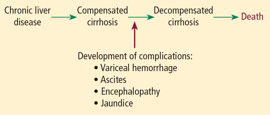
When a patient with liver disease is evaluated for surgery, evidence should be sought to determine whether an index complication has already occurred. Because the patient in our case study had a cholecystectomy several years before, I would also ask, “What did the surgeon say your liver looked like? Did you have any bleeding problems afterwards? Did you develop ascites?”
It is also important to determine whether portal hypertension is present. For a patient with liver disease, otherwise unexplained thrombocytopenia is a useful indicator of portal hypertension.
Systems for scoring liver disease severity
Even a surgical patient with well-compensated liver disease is at risk for developing complications postoperatively, particularly if abdominal surgery is planned. Risk should be assessed in all patients with liver disease using either the Child-Pugh scoring system or the Model for End-Stage Liver Disease (MELD) scoring system.
The Child-Pugh score, which assigns 1 to 3 points according to the presence/absence and levels of each of five simple factors (bilirubin, albumin, prothrombin time/international normalized ratio [INR], ascites, and encephalopathy stage), has been used for decades to assess the severity of liver disease. Patients with Child-Pugh class A disease (score of 5–6) have well-compensated cirrhosis and good synthetic function, and therefore have essentially no restrictions for undergoing surgery. For patients in Child-Pugh class B (score of 7–9), the risk of perioperative complications and mortality is higher and any major hepatic surgery (such as hepatic resection) should be avoided. Patients with class C cirrhosis (score of 10–15) are not candidates for any major elective surgery and should be considered for liver transplantation referral.
The MELD scoring system was developed more recently and is used to prioritize eligibility for liver transplantation. Calculated using a mathematical formula that incorporates three objective patient variables—
creatinine, bilirubin, and INR—the MELD score correlates very well with prognosis. The score can be calculated by an online MELD calculator such as the one at www.unos.org/resources.2 A patient with a high MELD score is unlikely to survive for more than a few months without liver transplantation; a patient with a low MELD score is likely to survive for at least 12 months. Calculating the MELD score is now one of the first assessments in any patient suspected of having cirrhosis.
Risk factors for complications and death
In a retrospective study to identify factors associated with complications and mortality in surgical patients with cirrhosis, Ziser et al reviewed the records of 733 patients with cirrhosis who underwent surgical procedures (except liver transplantation) at the Mayo Clinic over an 11-year period (1980–1991).3 The mortality rate within 30 days of surgery was 11.6%. Long-term follow-up showed that most deaths occurred within the first few months after surgery, when many patients succumbed to pneumonia or renal insufficiency.
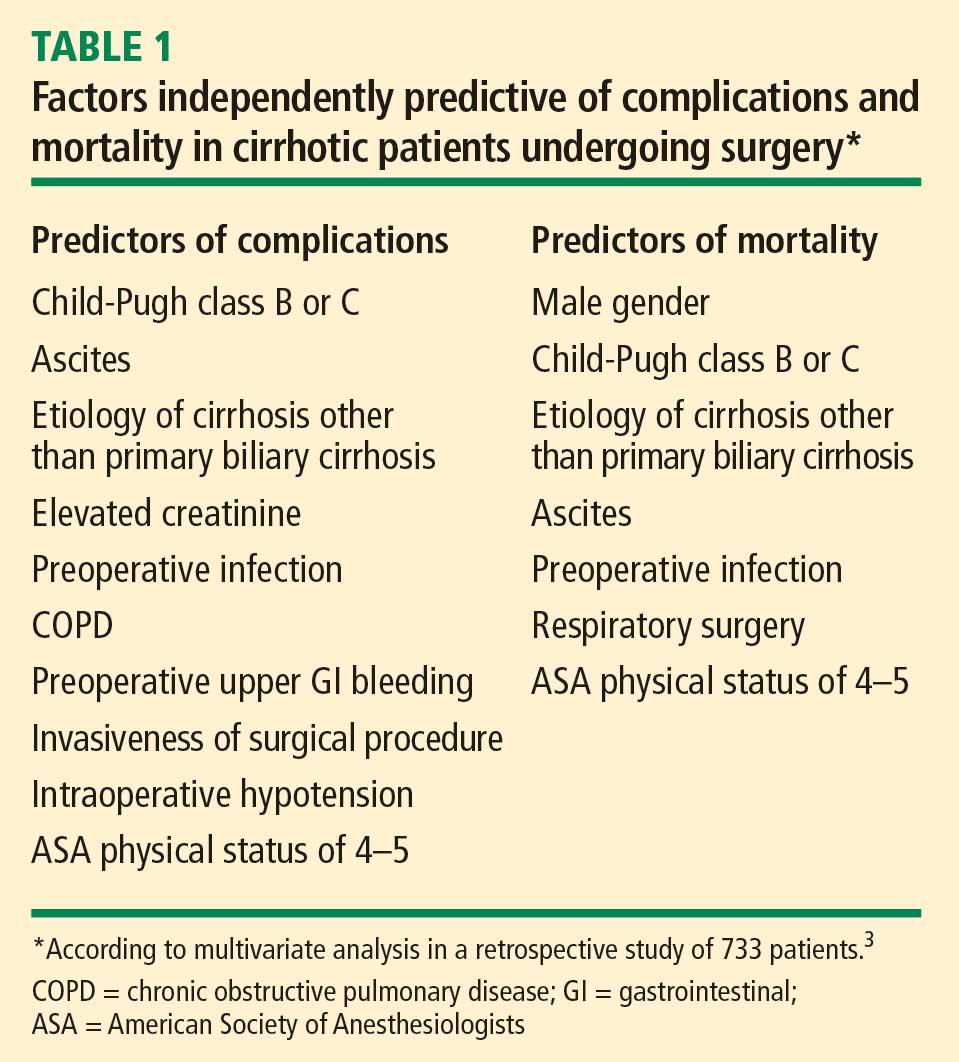
Risk factors have strong cumulative power
The study by Ziser et al also underscored the cumulative effect of risk factors, as the probability of developing a perioperative complication increased dramatically with the number of risk factors (as identified by multiavariate analysis) that a patient had, as follows3:
- 9.3% risk of complications with 1 risk factor
- 14.5% risk with 2 factors
- 33.5% risk with 3 factors
- 63.0% risk with 4 or 5 factors
- 73.3% risk with 6 factors
- 100% risk with 7 or 8 factors.
Postoperative complications: Beware hepatorenal syndrome
The most common postoperative complications in the study by Ziser et al were pneumonia, other infections, ventilation dependency, and ascites.3
Possibly the most ominous perioperative complication in a patient with liver disease is the onset of renal insufficiency, which may be precipitated by a number of factors, including nephrotoxic drugs and intraoperative hypotension. Renal insufficiency is usually a predictor of markedly reduced survival and a sign that hepatorenal syndrome may have developed.
Hepatorenal syndrome, which occurred in 3.3% of patients in the analysis by Ziser et al,3 is the presence of renal failure in a patient with cirrhosis. It is characterized by advanced liver failure and severe sinusoidal portal hypertension. The renal failure is said to be “functional” because significant histological changes are absent on kidney biopsy. Marked arteriolar vasodilation occurs in the extrarenal circulation with renal vasoconstriction leading to reduced glomerular filtration.
IMPORTANCE OF SURGICAL PROCEDURE TYPE
In addition to the patient-specific risk factors discussed above, certain surgical procedures deserve special consideration in patients with liver disease.
Cholecystectomy: Open vs closed
Patients with liver disease have the same indication for cholecystectomy as anyone else does: symptomatic gallstone disease. Patients with cirrhosis who are found to have incidental gallstones on ultrasonography should not undergo cholecystectomy unless the gallstones are symptomatic, as liver function may deteriorate after surgery.
For a patient with liver disease undergoing cholecystectomy, a common concern is whether an open or closed procedure should be done. Conventional wisdom had been that a patient with underlying liver disease (particularly cirrhosis) should have an open procedure so that the surgeon could more easily control bleeding, but that notion has changed in recent years with evidence supporting the safety of a laparoscopic approach in patients with liver disease.
One study supporting this new strategy is a retrospective review of 50 patients who had undergone cholecystectomy for symptomatic gallstone disease at the Mayo Clinic between 1990 and 1997.4 The procedure was open in half of the patients and laparoscopic in the other half. All patients had Child-Pugh class A or B cirrhosis. The indications for surgery were acute cholecystitis, biliary colic, or pancreatitis, and the number of patients with each of these indications was comparable between the open-surgery and laparoscopy groups. Three patients who initially underwent laparoscopy were converted to open cholecystectomy: two for bleeding and one for poor access. The study found that laparoscopic cholecystectomy was associated with statistically significant reductions in operating room time, blood loss, and length of hospital stay. No deaths occurred in either group. The authors concluded that laparoscopic cholecystectomy is safe in patients with cirrhosis and offers several advantages over an open surgical approach.
In light of these findings and other recent evidence, laparoscopic cholecystectomy should be recommended for patients with liver disease unless they have ascites or other evidence of overt hepatic decompensation, in which case cholecystectomy itself is contraindicated.
Cardiac surgery with bypass poses extra risk
Patients with liver disease undergoing open heart surgery with cardiopulmonary bypass are at especially high risk because of the effect on hepatic hemodynamics. This risk was demonstrated in a retrospective review of all patients with cirrhosis who underwent cardiac surgery with cardiopulmonary bypass at the Cleveland Clinic from 1992 to 2002.5 Of the 44 patients identified, 12 (27%) developed hepatic decompensation and 7 (16%) died. Hepatic decompensation was a major factor in all the deaths.
The MELD and Child-Pugh scores correlated well with one another in this study and were highly associated with hepatic decompensation and death. The best cutoff values for predicting mortality and hepatic decompensation were found to be a score greater than 7 in the Child-Pugh system and a score greater than 13 in the MELD system. (For context, receipt of a donor liver via a transplant list in the United States requires a MELD score of at least 15.) The study confirmed that the Child-Pugh score, which is easy to determine at the bedside, remains a reliable predictor of poor outcomes.5
CASE REVISITED: POSTOPERATIVE LIVER FUNCTION DECLINE―HOW SERIOUS IS IT?
Our patient undergoes the CABG procedure, and 3 days later you are asked to see him. According to the sub-intern, although the surgery was successful, the patient is now “in liver failure.” After hearing this news, the family is anxious to discuss liver transplantation.
On examination, the patient is alert and extubated, so he is clearly not encephalopathic. His wound is clean and shows no sign of infection. He appears to be mildly icteric, and he may have some ascites, based on mild flank dullness.
His laboratory test results are as follows:
- Bilirubin, 3.1 mg/dL (normal range, 0.3–1.2)
- INR, 1.2 (0.9–1.2)
- Alanine aminotransferase (ALT), 300 U/L (10–40)
- Creatinine, 0.9 mg/dL (0.6–1.2).
Although the bilirubin and ALT are elevated, it is notable that the creatinine is normal. This pattern is not uncommon after elective surgery in a patient with underlying cirrhosis. Renal dysfunction is the biggest concern in the perioperative management of a patient with liver disease, as it is an indicator that the patient may develop overt hepatic decompensation. Likely reasons for the patient’s ALT elevation are the effects of cardiopulmonary bypass and possible intraoperative hypotension.
The family needs to be told that the patient is not in liver failure and that it is best to wait with the expectation that he will do fine unless other complications supervene.
You advise cautious diuresis, and the ALT falls over the next few days. The bilirubin declines to 2.0 mg/dL. At this point, you advise discharge planning.
One need not wait for the bilirubin to return to normal: after an acute hepatic insult such as ischemic hepatitis or intraoperative hypotension, bilirubin is the last indicator to improve. Bilirubin is in part albumin-bound, and the half-life of albumin is 18 days, so a patient can remain icteric for some time after the rest of the liver function tests have returned to normal.
DISCUSSION
Question from the audience: What are your recommendations regarding platelet transfusion if the platelet count is less than 50,000 in a patient with liver disease?
Dr. Martin: For patients with thrombocytopenia, it is prudent to get the platelet count above 60,000 before any procedure. We will not even do a blind liver biopsy in a patient with a platelet count of less than 60,000.
Question from the audience: A study from the Annals of Surgery concludes that patients with liver disease do poorly with a hemoglobin of less than 10 g/dL. Would you transfuse aggressively before surgery?
Dr. Martin: For a patient with anemia, I don’t like to use aggressive transfusion if cirrhosis is present because the portal pressure may go up and increase the risk of variceal hemorrhage. If there is adequate time for a work-up, one can screen for varices by endoscopy. If there is evidence of overt hepatic decompensation and portal hypertension (esophageal varices, a palpable spleen, and thrombocytopenia), I wouldn’t try to get the hemoglobin much above 10 g/dL.
Question from the audience: How would you modify prophylaxis for deep vein thrombosis following hip or knee replacement surgery in patients with liver disease?
Dr. Martin: I would base it on the INR. Patients who are already mildly coagulopathic tend to be very sensitive to warfarin in the long term. For immediate perioperative prophylaxis, I would not administer anything if the patient had a platelet count below 60,000; otherwise I would probably proceed as usual.
Question from the audience: You said that we shouldn’t operate on patients with acute hepatitis, but we frequently encounter patients with drug-induced hepatitis, such as from anticholesterol drugs. These patients’ ALT and aspartate aminotransferase (AST) levels can remain elevated for 2 or 3 months. How long should we delay surgery? For example, is it dangerous to proceed with a mastectomy a month after discontinuing the drug if the liver enzymes are still around 100 U/L?
Dr. Martin: It’s worth noting that much of the literature on surgery in patients with acute viral hepatitis is 30 or 40 years old. If such a patient had a compelling reason to have surgery, you might wait until the liver enzymes were trending downward and you were confident that the patient was recovering.
Question from the audience: How do you manage patients who have varices or have had variceal bleeding in the past? Many of them are on beta-blockers, such as propranolol, which can cause hypotension intraoperatively.
Dr. Martin: The standard of care is to prescribe beta-blockers for a patient with large varices, or to ablate the varices by endoscopy, which is my practice. In general, I would discontinue propranolol on the morning of surgery. If possible, however, I would have the patient undergo endoscopy before surgery to assess the likelihood of short-term variceal bleeding. If the varices look to be at low risk of bleeding, the beta-blocker can safely be stopped. If they look to be at high risk of bleeding, the surgery should be delayed for a few weeks, if possible, so that the varices can be ablated, which usually takes two or three sessions.
Question from the audience: I deal with many referrals, and I struggle with how aggressive a work-up I should do for patients undergoing elective surgery when a new abnormality is found in one of their liver function tests.
Dr. Martin: I would try to establish whether the abnormality is a chronic problem. Has the patient been told about an abnormal liver test in the past? Ask if the patient has been a blood donor, as measurement of ALT and some hepatitis serologies would have been required. Also ask if he or she has ever taken out a big life insurance policy, which also would have required liver function testing. If the abnormality is chronic, you may proceed with surgery if the bilirubin and INR are normal. In the absence of chronicity, surgery should be delayed for further work-up only in patients with indicators of significant liver disease—either markedly abnormal liver tests, thrombocytopenia, or coagulopathy.
Follow-up question: But patients rarely know whether they’ve had elevated liver enzymes in the past. You said not to worry about enzyme abnormalities unless they are markedly elevated, but how high is that?
Dr. Martin: AST and ALT are indicators of liver injury rather than of synthetic function. The true liver function tests are really albumin, bilirubin, and prothrombin time. Paradoxically, one of the best liver function tests is the platelet count. For me, a red flag for a patient with newly recognized liver disease is any degree of thrombocytopenia or coagulopathy or an elevation of bilirubin above the upper limit of normal. A patient with a platelet count of 90,000 and an INR above 1.2 has significant underlying liver disease, and I would be very concerned. Unless it’s a dire emergency, such a patient would need further evaluation before proceeding with surgery. In contrast, a patient with an ALT of 89, an AST of 65, and normal prothrombin time and platelet count should be safe to proceed to surgery. But such a patient needs an evaluation for liver disease afterward.
Question from the audience: My institution performs many liver resections for metastases or primary liver cancers. Our liver surgeons routinely discontinue statins 2 to 3 weeks before liver surgery, but it has been said at this summit that is not necessary. What’s your opinion?
Dr. Martin: I think that statins get a very bad rap in terms of hepatotoxity. Most patients with metabolic syndrome have hyperlipidemia, which can cause fatty liver disease and hepatic dysfunction. Statins help bring the lipid levels down. Hepatologists do not regard statins as major culprits in causing liver problems. I don’t believe there’s any particular indication to stop them before a patient undergoes hepatic surgery.
Question from the audience: I assess patients 1 or 2 weeks before surgery. For a patient with coagulopathy whom you suspect has underlying liver disease, is there any value in trying to treat the coagulopathy with vitamin K?
Dr. Martin: It can be worthwhile to try 10 mg subcutaneously for 3 days to see whether the situation improves, but if the patient has severe parenchymal liver disease, the vitamin K won’t help much.
- Ginés P, Quintero E, Arroyo V, et al. Compensated cirrhosis: natural history and prognostic factors. Hepatology 1987; 7:122–128.
- MELD/PELD calculator. United Network for Organ Sharing Web site. http://www.unos.org/resources/MeldPeldCalculator.asp?index=98. Accessed May 8, 2009.
- Ziser A, Plevak DJ, Wiesner RH, Rakela J, Offord KP, Brown DL. Morbidity and mortality in cirrhotic patients undergoing anesthesia and surgery. Anesthesiology 1999; 90:42–53.
- Poggio JL, Rowland CM, Gores GJ, et al. A comparison of laparoscopic and open cholecystectomy in patients with compensated cirrhosis and symptomatic gallstone disease. Surgery 2000; 127:405–411.
- Suman A, Barnes DS, Zein NN, et al. Predicting outcome after cardiac surgery in patients with cirrhosis: a comparison of Child-Pugh and MELD scores. Clin Gastroenterol Hepatol 2004; 2:719–723.
Assessing patients with liver disease for surgery is one of the most common reasons for hepatology consultation in the hospital. This review focuses on practical aspects of evaluating patients with known or suspected liver disease and provides guidance for determining whether it is safe to proceed with surgery in such patients. I begin with a case study to introduce some common clinical challenges and then revisit the case—with relevant teaching points—at the end.
CASE: A MIDDLE-AGED MAN WITH LIVER DISEASE SCHEDULED FOR CARDIAC SURGERY
A 57-year-old man with a history of liver disease is referred for preoperative assessment. It is 6:30 pm, and the patient has just arrived in the hospital; he is scheduled for coronary artery bypass graft surgery (CABG) early tomorrow morning for ischemic heart disease. Ten years ago, he was diagnosed with hepatitis C virus infection; 2 years later, he had a cholecystectomy. He has a remote history of intravenous drug use.
The sub-intern asks for an assessment of operative risk as well as advice on the type of anesthesia to be used.
HEPATIC EFFECTS OF ANESTHESIA
Anesthesiologists are keenly aware of the hepatic effects of anesthesia and that they must carefully choose anesthetics for patients with liver disease. There are a number of at least theoretical concerns about using particular anesthetics:
- Inhaled anesthetics, such as isoflurane, cause systemic vasodilation and depress cardiac output. These effects are of concern since many patients with advanced liver disease already have a hyperdynamic circulation because of peripheral vasodilation.
- Spinal or epidural anesthetics may reduce mean arterial pressure, which is of concern for similar reasons.
- Nitrous oxide has less of a depressive effect unless the patient has concomitant hypercapnia.
Another consideration is the hepatic metabolism of anesthetic agents. Use of halothane, which is 20% metabolized by the liver, is now uncommon, particularly if there is any concern about liver disease. In contrast, enflurane is only 4% metabolized by the liver. Numerous other anesthetics—including isoflurane, desflurane, and sevoflurane—have only minimal hepatic metabolism (< 0.2%), which makes them, along with nitrous oxide, the best anesthetic choices for patients with liver disease.
ASSESSING OPERATIVE RISK
The more important issue in the consultation for our patient is the degree of operative risk associated with his underlying liver disease. A number of factors are pertinent, including the etiology and severity of the liver disease and the type of surgery planned.
Acute liver disease has higher operative risk
Literature dating back 40 years has associated acute viral and alcoholic hepatitis with poor outcomes in surgical patients. Major elective surgery for a patient with suspected acute hepatitis A, for example, should be deferred until the patient has recovered, barring some compelling reason for greater urgency, such as a perforated viscus.
In chronic liver disease, hepatocellular function predicts outcome
When a patient with liver disease is evaluated for surgery, evidence should be sought to determine whether an index complication has already occurred. Because the patient in our case study had a cholecystectomy several years before, I would also ask, “What did the surgeon say your liver looked like? Did you have any bleeding problems afterwards? Did you develop ascites?”
It is also important to determine whether portal hypertension is present. For a patient with liver disease, otherwise unexplained thrombocytopenia is a useful indicator of portal hypertension.
Systems for scoring liver disease severity
Even a surgical patient with well-compensated liver disease is at risk for developing complications postoperatively, particularly if abdominal surgery is planned. Risk should be assessed in all patients with liver disease using either the Child-Pugh scoring system or the Model for End-Stage Liver Disease (MELD) scoring system.
The Child-Pugh score, which assigns 1 to 3 points according to the presence/absence and levels of each of five simple factors (bilirubin, albumin, prothrombin time/international normalized ratio [INR], ascites, and encephalopathy stage), has been used for decades to assess the severity of liver disease. Patients with Child-Pugh class A disease (score of 5–6) have well-compensated cirrhosis and good synthetic function, and therefore have essentially no restrictions for undergoing surgery. For patients in Child-Pugh class B (score of 7–9), the risk of perioperative complications and mortality is higher and any major hepatic surgery (such as hepatic resection) should be avoided. Patients with class C cirrhosis (score of 10–15) are not candidates for any major elective surgery and should be considered for liver transplantation referral.
The MELD scoring system was developed more recently and is used to prioritize eligibility for liver transplantation. Calculated using a mathematical formula that incorporates three objective patient variables—
creatinine, bilirubin, and INR—the MELD score correlates very well with prognosis. The score can be calculated by an online MELD calculator such as the one at www.unos.org/resources.2 A patient with a high MELD score is unlikely to survive for more than a few months without liver transplantation; a patient with a low MELD score is likely to survive for at least 12 months. Calculating the MELD score is now one of the first assessments in any patient suspected of having cirrhosis.
Risk factors for complications and death
In a retrospective study to identify factors associated with complications and mortality in surgical patients with cirrhosis, Ziser et al reviewed the records of 733 patients with cirrhosis who underwent surgical procedures (except liver transplantation) at the Mayo Clinic over an 11-year period (1980–1991).3 The mortality rate within 30 days of surgery was 11.6%. Long-term follow-up showed that most deaths occurred within the first few months after surgery, when many patients succumbed to pneumonia or renal insufficiency.
Risk factors have strong cumulative power
The study by Ziser et al also underscored the cumulative effect of risk factors, as the probability of developing a perioperative complication increased dramatically with the number of risk factors (as identified by multiavariate analysis) that a patient had, as follows3:
- 9.3% risk of complications with 1 risk factor
- 14.5% risk with 2 factors
- 33.5% risk with 3 factors
- 63.0% risk with 4 or 5 factors
- 73.3% risk with 6 factors
- 100% risk with 7 or 8 factors.
Postoperative complications: Beware hepatorenal syndrome
The most common postoperative complications in the study by Ziser et al were pneumonia, other infections, ventilation dependency, and ascites.3
Possibly the most ominous perioperative complication in a patient with liver disease is the onset of renal insufficiency, which may be precipitated by a number of factors, including nephrotoxic drugs and intraoperative hypotension. Renal insufficiency is usually a predictor of markedly reduced survival and a sign that hepatorenal syndrome may have developed.
Hepatorenal syndrome, which occurred in 3.3% of patients in the analysis by Ziser et al,3 is the presence of renal failure in a patient with cirrhosis. It is characterized by advanced liver failure and severe sinusoidal portal hypertension. The renal failure is said to be “functional” because significant histological changes are absent on kidney biopsy. Marked arteriolar vasodilation occurs in the extrarenal circulation with renal vasoconstriction leading to reduced glomerular filtration.
IMPORTANCE OF SURGICAL PROCEDURE TYPE
In addition to the patient-specific risk factors discussed above, certain surgical procedures deserve special consideration in patients with liver disease.
Cholecystectomy: Open vs closed
Patients with liver disease have the same indication for cholecystectomy as anyone else does: symptomatic gallstone disease. Patients with cirrhosis who are found to have incidental gallstones on ultrasonography should not undergo cholecystectomy unless the gallstones are symptomatic, as liver function may deteriorate after surgery.
For a patient with liver disease undergoing cholecystectomy, a common concern is whether an open or closed procedure should be done. Conventional wisdom had been that a patient with underlying liver disease (particularly cirrhosis) should have an open procedure so that the surgeon could more easily control bleeding, but that notion has changed in recent years with evidence supporting the safety of a laparoscopic approach in patients with liver disease.
One study supporting this new strategy is a retrospective review of 50 patients who had undergone cholecystectomy for symptomatic gallstone disease at the Mayo Clinic between 1990 and 1997.4 The procedure was open in half of the patients and laparoscopic in the other half. All patients had Child-Pugh class A or B cirrhosis. The indications for surgery were acute cholecystitis, biliary colic, or pancreatitis, and the number of patients with each of these indications was comparable between the open-surgery and laparoscopy groups. Three patients who initially underwent laparoscopy were converted to open cholecystectomy: two for bleeding and one for poor access. The study found that laparoscopic cholecystectomy was associated with statistically significant reductions in operating room time, blood loss, and length of hospital stay. No deaths occurred in either group. The authors concluded that laparoscopic cholecystectomy is safe in patients with cirrhosis and offers several advantages over an open surgical approach.
In light of these findings and other recent evidence, laparoscopic cholecystectomy should be recommended for patients with liver disease unless they have ascites or other evidence of overt hepatic decompensation, in which case cholecystectomy itself is contraindicated.
Cardiac surgery with bypass poses extra risk
Patients with liver disease undergoing open heart surgery with cardiopulmonary bypass are at especially high risk because of the effect on hepatic hemodynamics. This risk was demonstrated in a retrospective review of all patients with cirrhosis who underwent cardiac surgery with cardiopulmonary bypass at the Cleveland Clinic from 1992 to 2002.5 Of the 44 patients identified, 12 (27%) developed hepatic decompensation and 7 (16%) died. Hepatic decompensation was a major factor in all the deaths.
The MELD and Child-Pugh scores correlated well with one another in this study and were highly associated with hepatic decompensation and death. The best cutoff values for predicting mortality and hepatic decompensation were found to be a score greater than 7 in the Child-Pugh system and a score greater than 13 in the MELD system. (For context, receipt of a donor liver via a transplant list in the United States requires a MELD score of at least 15.) The study confirmed that the Child-Pugh score, which is easy to determine at the bedside, remains a reliable predictor of poor outcomes.5
CASE REVISITED: POSTOPERATIVE LIVER FUNCTION DECLINE―HOW SERIOUS IS IT?
Our patient undergoes the CABG procedure, and 3 days later you are asked to see him. According to the sub-intern, although the surgery was successful, the patient is now “in liver failure.” After hearing this news, the family is anxious to discuss liver transplantation.
On examination, the patient is alert and extubated, so he is clearly not encephalopathic. His wound is clean and shows no sign of infection. He appears to be mildly icteric, and he may have some ascites, based on mild flank dullness.
His laboratory test results are as follows:
- Bilirubin, 3.1 mg/dL (normal range, 0.3–1.2)
- INR, 1.2 (0.9–1.2)
- Alanine aminotransferase (ALT), 300 U/L (10–40)
- Creatinine, 0.9 mg/dL (0.6–1.2).
Although the bilirubin and ALT are elevated, it is notable that the creatinine is normal. This pattern is not uncommon after elective surgery in a patient with underlying cirrhosis. Renal dysfunction is the biggest concern in the perioperative management of a patient with liver disease, as it is an indicator that the patient may develop overt hepatic decompensation. Likely reasons for the patient’s ALT elevation are the effects of cardiopulmonary bypass and possible intraoperative hypotension.
The family needs to be told that the patient is not in liver failure and that it is best to wait with the expectation that he will do fine unless other complications supervene.
You advise cautious diuresis, and the ALT falls over the next few days. The bilirubin declines to 2.0 mg/dL. At this point, you advise discharge planning.
One need not wait for the bilirubin to return to normal: after an acute hepatic insult such as ischemic hepatitis or intraoperative hypotension, bilirubin is the last indicator to improve. Bilirubin is in part albumin-bound, and the half-life of albumin is 18 days, so a patient can remain icteric for some time after the rest of the liver function tests have returned to normal.
DISCUSSION
Question from the audience: What are your recommendations regarding platelet transfusion if the platelet count is less than 50,000 in a patient with liver disease?
Dr. Martin: For patients with thrombocytopenia, it is prudent to get the platelet count above 60,000 before any procedure. We will not even do a blind liver biopsy in a patient with a platelet count of less than 60,000.
Question from the audience: A study from the Annals of Surgery concludes that patients with liver disease do poorly with a hemoglobin of less than 10 g/dL. Would you transfuse aggressively before surgery?
Dr. Martin: For a patient with anemia, I don’t like to use aggressive transfusion if cirrhosis is present because the portal pressure may go up and increase the risk of variceal hemorrhage. If there is adequate time for a work-up, one can screen for varices by endoscopy. If there is evidence of overt hepatic decompensation and portal hypertension (esophageal varices, a palpable spleen, and thrombocytopenia), I wouldn’t try to get the hemoglobin much above 10 g/dL.
Question from the audience: How would you modify prophylaxis for deep vein thrombosis following hip or knee replacement surgery in patients with liver disease?
Dr. Martin: I would base it on the INR. Patients who are already mildly coagulopathic tend to be very sensitive to warfarin in the long term. For immediate perioperative prophylaxis, I would not administer anything if the patient had a platelet count below 60,000; otherwise I would probably proceed as usual.
Question from the audience: You said that we shouldn’t operate on patients with acute hepatitis, but we frequently encounter patients with drug-induced hepatitis, such as from anticholesterol drugs. These patients’ ALT and aspartate aminotransferase (AST) levels can remain elevated for 2 or 3 months. How long should we delay surgery? For example, is it dangerous to proceed with a mastectomy a month after discontinuing the drug if the liver enzymes are still around 100 U/L?
Dr. Martin: It’s worth noting that much of the literature on surgery in patients with acute viral hepatitis is 30 or 40 years old. If such a patient had a compelling reason to have surgery, you might wait until the liver enzymes were trending downward and you were confident that the patient was recovering.
Question from the audience: How do you manage patients who have varices or have had variceal bleeding in the past? Many of them are on beta-blockers, such as propranolol, which can cause hypotension intraoperatively.
Dr. Martin: The standard of care is to prescribe beta-blockers for a patient with large varices, or to ablate the varices by endoscopy, which is my practice. In general, I would discontinue propranolol on the morning of surgery. If possible, however, I would have the patient undergo endoscopy before surgery to assess the likelihood of short-term variceal bleeding. If the varices look to be at low risk of bleeding, the beta-blocker can safely be stopped. If they look to be at high risk of bleeding, the surgery should be delayed for a few weeks, if possible, so that the varices can be ablated, which usually takes two or three sessions.
Question from the audience: I deal with many referrals, and I struggle with how aggressive a work-up I should do for patients undergoing elective surgery when a new abnormality is found in one of their liver function tests.
Dr. Martin: I would try to establish whether the abnormality is a chronic problem. Has the patient been told about an abnormal liver test in the past? Ask if the patient has been a blood donor, as measurement of ALT and some hepatitis serologies would have been required. Also ask if he or she has ever taken out a big life insurance policy, which also would have required liver function testing. If the abnormality is chronic, you may proceed with surgery if the bilirubin and INR are normal. In the absence of chronicity, surgery should be delayed for further work-up only in patients with indicators of significant liver disease—either markedly abnormal liver tests, thrombocytopenia, or coagulopathy.
Follow-up question: But patients rarely know whether they’ve had elevated liver enzymes in the past. You said not to worry about enzyme abnormalities unless they are markedly elevated, but how high is that?
Dr. Martin: AST and ALT are indicators of liver injury rather than of synthetic function. The true liver function tests are really albumin, bilirubin, and prothrombin time. Paradoxically, one of the best liver function tests is the platelet count. For me, a red flag for a patient with newly recognized liver disease is any degree of thrombocytopenia or coagulopathy or an elevation of bilirubin above the upper limit of normal. A patient with a platelet count of 90,000 and an INR above 1.2 has significant underlying liver disease, and I would be very concerned. Unless it’s a dire emergency, such a patient would need further evaluation before proceeding with surgery. In contrast, a patient with an ALT of 89, an AST of 65, and normal prothrombin time and platelet count should be safe to proceed to surgery. But such a patient needs an evaluation for liver disease afterward.
Question from the audience: My institution performs many liver resections for metastases or primary liver cancers. Our liver surgeons routinely discontinue statins 2 to 3 weeks before liver surgery, but it has been said at this summit that is not necessary. What’s your opinion?
Dr. Martin: I think that statins get a very bad rap in terms of hepatotoxity. Most patients with metabolic syndrome have hyperlipidemia, which can cause fatty liver disease and hepatic dysfunction. Statins help bring the lipid levels down. Hepatologists do not regard statins as major culprits in causing liver problems. I don’t believe there’s any particular indication to stop them before a patient undergoes hepatic surgery.
Question from the audience: I assess patients 1 or 2 weeks before surgery. For a patient with coagulopathy whom you suspect has underlying liver disease, is there any value in trying to treat the coagulopathy with vitamin K?
Dr. Martin: It can be worthwhile to try 10 mg subcutaneously for 3 days to see whether the situation improves, but if the patient has severe parenchymal liver disease, the vitamin K won’t help much.
- Ginés P, Quintero E, Arroyo V, et al. Compensated cirrhosis: natural history and prognostic factors. Hepatology 1987; 7:122–128.
- MELD/PELD calculator. United Network for Organ Sharing Web site. http://www.unos.org/resources/MeldPeldCalculator.asp?index=98. Accessed May 8, 2009.
- Ziser A, Plevak DJ, Wiesner RH, Rakela J, Offord KP, Brown DL. Morbidity and mortality in cirrhotic patients undergoing anesthesia and surgery. Anesthesiology 1999; 90:42–53.
- Poggio JL, Rowland CM, Gores GJ, et al. A comparison of laparoscopic and open cholecystectomy in patients with compensated cirrhosis and symptomatic gallstone disease. Surgery 2000; 127:405–411.
- Suman A, Barnes DS, Zein NN, et al. Predicting outcome after cardiac surgery in patients with cirrhosis: a comparison of Child-Pugh and MELD scores. Clin Gastroenterol Hepatol 2004; 2:719–723.
KEY POINTS
- Patients with acute hepatitis should delay elective surgery until after their hepatitis resolves.
- Patients with chronic liver disease who have developed any index complication—variceal hemorrhage, ascites, hepatic encephalopathy, or jaundice—are at increased risk for postoperative complications and death.
- The Child-Pugh and MELD scores appear to be comparably effective in predicting surgical outcomes in patients with liver disease.
- Cardiac surgery with cardiopulmonary bypass and abdominal surgery are particularly high-risk procedures in patients with liver disease.
- If cholecystectomy is indicated in a patient with compensated liver disease, laparoscopy should be the initial approach, with conversion to an open procedure only if necessary.
Perioperative management of obstructive sleep apnea: Ready for prime time?
Obstructive sleep apnea (OSA) is characterized by repeated complete or partial collapse of the pharyngeal airway during sleep, causing cessation of airflow (apnea) or shallow breathing (hypopnea). Persons with OSA may have repeated arousals from sleep (to reestablish breathing) with each episode of apnea or hypopnea. The resulting sleep disruption often leads to daytime somnolence and compromised neurocognitive function.
This pattern of sleep arousal, coupled with intermittent hypoxemia, is associated with serious adverse cardiovascular outcomes, including stroke. Among surgical patients, OSA is associated with postoperative complications and the need for increased medical intervention. This review discusses why OSA is important in the perioperative setting, preoperative screening for OSA risk, and perioperative management of patients with likely or confirmed OSA.
OSA AT A GLANCE
Prevalence in the general population
Four percent of middle-aged men and 2% of middle-aged women meet minimal diagnostic criteria for OSA, according to a landmark cohort study from the 1990s.1 This makes OSA more common than asthma among adults. Risk increases with age, as 24% of persons older than 65 years have OSA and up to 50% of nursing home residents have clinically significant OSA.2
Prevalence in the surgical population
The prevalence of OSA in the surgical population is higher than that in the general population, and it can vary widely according to the underlying medical condition. A study of 433 patients undergoing general surgery reported a 3.2% prevalence of OSA,3 but this study excluded patients undergoing cardiac surgery, in whom the risk of OSA is higher. In contrast, the prevalence of OSA among obese bariatric surgery patients has been reported at greater than 70%.4 Notably, the patients in the general surgery study3 who appeared to be at risk for OSA based on screening questions were invited to participate in a sleep study, whereas all patients in the bariatric surgery study4 were evaluated through sleep studies. It is likely that the prevalence of OSA among the general surgery study patients would have been higher if all patients had been evaluated with polysomnography.
Pathophysiology
OSA can occur when any part of the upper airway does not function normally. Upper airway patency is determined by muscle activity, craniofacial and soft tissue structure, and sleep state. During sleep, upper airway muscles are relaxed, which reduces airway patency. Sleep is associated with pharyngeal narrowing and substantially increased inspiratory resistance even among persons without sleep apnea. A person who is awake can compensate for abnormal pharyngeal function through increased muscle activity. During sleep this muscle compensation fails, resulting in partial collapse and subsequent snoring, and sometimes prolonged obstructive hypoventilation. Complete closure results in apnea.
WHY OSA MATTERS
Health consequences of OSA
OSA is associated with serious health consequences, such as increased risk of motor vehicle accidents, stroke, and a number of cardiovascular conditions—hypertension, coronary artery disease, and atrial fibrillation.
Accidents. The daytime hypersomnolence resulting from OSA contributes to reduced vigilance and is likely responsible for an increased incidence of motor vehicle accidents. One study found that among a sample of men and women with unrecognized OSA undergoing polysomnography studies, the likelihood of motor vehicle accidents during the prior 5 years was significantly correlated with the subjects’ apnea-hypopnea index (AHI) score, which reflects the number of apnea or hypopnea episodes per hour of sleep.5 Other studies have demonstrated similar associations.
Stroke. Numerous observational studies have demonstrated an elevated prevalence of OSA among patients with stroke as compared with the general population, but these studies did not adjust for other cerebrovascular risk factors. A recent observational cohort study aimed to address this evidence gap by using proportional hazards analysis to determine the independent effect of OSA on the incidence of stroke or death from any cause among persons with no history of stroke or myocardial infarction.6 Study participants were 1,022 consecutive patients who underwent polysomnography for evaluation of sleep-disordered breathing. OSA was identified in 68% of patients. During the 3.4-year follow-up period, 22 strokes and 50 deaths occurred among the 697 patients with OSA compared with 2 strokes and 14 deaths among the 325 patients without OSA. The probability of survival was significantly lower for patients with OSA compared with their counterparts without OSA (P < .003). After adjustment for other risk factors, OSA was significantly associated with stroke or death (hazard ratio = 1.97; 95% CI, 1.12–3.48).6
Hypertension. Four large studies involving a total of 10,708 patients evaluated for sleep-disordered breathing have established an association between OSA and hypertension risk.7–10 In each study, the risk of hypertension rose linearly with AHI scores. Clinically significant OSA, defined as an AHI score greater than 15, roughly doubled the risk of hypertension compared with the absence of apnea/hypopnea episodes, with odds ratios ranging from 1.37 to 2.89 across the four studies.7–10 Each apnea event per hour of sleep was estimated to increase the odds of developing hypertension by approximately 1%.8 Notably, the effects of OSA on blood pressure are most pronounced in patients younger than age 50.7
Coronary artery disease. The Sleep Heart Health Study evaluated the association between sleep-disordered breathing and cardiovascular disease in 6,424 community-dwelling adults undergoing home polysomnography.11 The population’s median AHI score was 4.4. At least one cardiovascular event was reported by 16% of participants. Sleep-disordered breathing was associated with self-reported heart failure, stroke, and, more modestly, coronary artery disease. A linear relationship was noted between AHI and cardiovascular risk.
Snoring, which is often an indicator for OSA, has also been associated with cardiovascular risk. The Nurses’ Health Study evaluated 71,000 women who completed medical questionnaires that included questions about snoring. Over 8 years of follow-up, the relative risks for cardiovascular disease were 1.46 among occasional snorers (95% CI, 1.23–1.74) and 2.02 among regular snorers (95% CI, 1.62–2.53) in comparison with nonsnorers. Snoring, even without a diagnosis of OSA, emerged as an independent risk factor for cardiovascular disease.12
Atrial fibrillation. OSA has been identified as a predictor of new-onset atrial fibrillation in a retrospective cohort study (hazard ratio = 2.18; 95% CI, 1.34–3.54).13 In a prospective study, patients with atrial fibrillation but normal left ventricular function were found to have significantly higher AHI scores than matched normal controls.14 After adjustment for relevant covariates, the odds ratio for an association between atrial fibrillation and significant sleep-disordered breathing (AHI score > 15) was 3.04 (95% CI, 1.24–7.46).14 In another prospective trial, patients with atrial fibrillation and OSA who underwent cardioversion were at increased risk for a recurrence of atrial fibrillation if OSA was untreated (82% for untreated vs 42% for treated OSA; P = .013).15
An association with postoperative complications
OSA also has been shown to increase postoperative complication rates, increase the need for intensive care intervention, and prolong hospital stays.
Representative evidence. One of the first studies to characterize the postoperative risks of OSA was conducted by Mayo Clinic researchers who retrospectively reviewed 4 years of data for 101 patients with OSA who had had hip or knee replacement surgery within 3 years before (n = 36) or any time after (n = 65) their OSA diagnosis.16 Outcomes were compared with those of 101 matched controls without OSA who underwent the same operations. Only half the patients with diagnosed OSA prior to their operation used continuous positive airway pressure (CPAP) therapy at home prior to hospitalization. Complications occurred among 39% of patients with OSA and among 18% of control patients (P = .001). Serious complications requiring intensive care unit transfer for cardiac ischemia or respiratory failure occurred in 24% of patients with OSA versus only 9% of controls (P = .004), and hospital stays were longer for patients with OSA compared with controls (P < .007). Most complications occurred during the first day after surgery, but a small number occurred as late as postoperative days 4 and 5.
In a separate study designed to evaluate OSA screening tools, postoperative complication rates were assessed in 211 patients who underwent polysomnography to determine the presence or absence of OSA prior to elective surgery.17 Patients undergoing various elective procedures were included, but none were undergoing cardiac or bariatric procedures. The overall rate of postoperative complications was more than twice as high among patients with OSA compared with those without OSA (27.4% vs 12.3%; P = .02). The most common complication was oxygen desaturation (ie, level ≤ 90%), which occurred among 20.6% of patients with OSA versus 9.2% of patients without OSA (P < .04). There were no deaths or serious complications.
Potential causes of complications. In the immediate postoperative period, OSA-associated complications may be attributable to lingering effects of sedatives, which can often lead to respiratory problems. Later in the postoperative course, so-called REM rebound is more likely to be implicated in complications. Patients often experience sleep deprivation in the hospital due to constant interruptions. Once a patient does sleep, the amount of REM sleep increases to compensate for this deprivation. The REM stage is when most apneas and hypopneas occur, so the risk of hypoxemia is greatest in the REM stage. As a result, respiratory and cardiovascular complications such as arrhythmias can increase.
OSA AND THE PREOPERATIVE EVALUATION
Risk factors for OSA
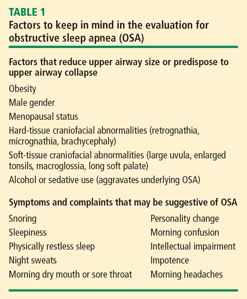
Additionally, certain aspects of perioperative management can increase the risk of OSA in the perioperative setting. For example, general anesthesia can mimic the effects of sleep on the airway, reducing muscle tone and potentially leading to pharyngeal collapse. Normal response to hypercapnia is also diminished under general anesthesia and while patients remain sedated postoperatively, which subdues normal protective arousal mechanisms. This does not pose a problem while the patient remains intubated but highlights the need for respiratory monitoring in the extubated patient who is recovering from the residual effects of sedation.
History and physical examination
What to look for. A number of physical characteristics reveal potential risks for OSA. Obesity and hypertension are well established, as noted above. Large neck circumference (≥ 17 inches in men and ≥ 16 inches in women) is another characteristic associated with OSA. Examination of the upper airway can reveal obstruction due to tonsil enlargement, nasal obstruction, an elongated uvula, or macroglossia. Since retrognathia or micrognathia can produce a narrowed oropharynx, attention to mandible size and position is advised.
Ask about sleep habits. Assessment of OSA risk in the preoperative evaluation need not be lengthy, but patients should be asked about snoring and waking habits, especially frequency of night wakening, to identify possible OSA. Patients generally do not volunteer information about sleep, so it is important to explicitly ask. Responses that suggest OSA include reports of tiredness or sleepiness during the day, or comments by a partner about the patient’s snoring. A patient who reports having a dry mouth in the morning may have nasal congestion or obstruction that leads to mouth breathing. Severe sleep disruption can lead to sleep deprivation, causing personality changes, confusion, intellectual impairment, impotence, or morning headaches (Table 1).
Preoperative screening tools. Screening tools can assist in identifying relevant questions about sleep. Three such tools for OSA have been validated for use in surgical patients: the Berlin questionnaire, the American Society of Anesthesiologists (ASA) checklist, and the STOP questionnaire.17–20 The performance of these tools was evaluated in 177 surgical patients with OSA identified using polysomnography.17 Each tool’s sensitivity, specificity, and positive and negative predictive values were calculated according to polysomnography-based AHI severity. All three tools demonstrated moderately high sensitivity for detecting OSA.17
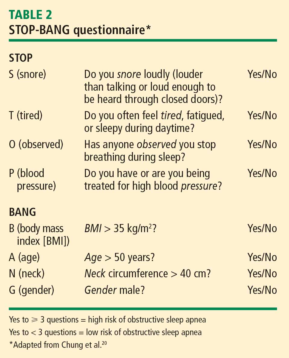
Identifying levels of OSA severity. Physical examination and screening questions may be adequate to identify patients at risk for OSA prior to surgery. Mild OSA (AHI score of 5–15) can generally be managed after surgery, at the patient’s leisure. In contrast, moderate OSA (AHI score of 15–30) and severe OSA (AHI score > 30) can affect perioperative management (see next section). If moderate to severe OSA is suspected, and if there is enough time before surgery to consult a sleep lab, polysomnography can provide a more complete diagnosis.
PERIOPERATIVE MANAGEMENT OF OSA
When in doubt, proceed as if patient has OSA
Evidence of OSA’s association with postoperative complications is emerging, as noted above, but more specific information about risks is needed to develop effective management procedures. For surgical patients who are deemed to be at high risk for OSA, and for whom surgery cannot be delayed for diagnostic tests and OSA treatment, the most prudent course is to proceed with surgery but assume the patient has moderate to severe OSA. Anesthesiologists should be informed when patients are likely to have OSA, as they may choose a different strategy for managing anesthesia during surgery for patients at high risk.
Management recommendations
The ASA published practice guidelines in 2006 for the perioperative management of patients with OSA.19 In view of the paucity of data on the best management strategies, the guidelines were based mostly on expert opinion. Their key recommendations include the following:
- Surgical patients should be screened clinically to determine their OSA risk. Any of the aforementioned screening tools is effective for this purpose.
- For patients with a diagnosis of OSA or who are clinically determined to be at high risk, close attention to airway management is required, extubation should be done when the patient is fully awake (to reduce residual effects of anesthesia and sedatives), and regional anesthesia should be used whenever possible.
- Postoperative pain management in patients with confirmed or suspected OSA should minimize the use of opioids and other sedatives. Such patients also should undergo close pulse oximetry monitoring in a step-down setting after surgery and receive postoperative CPAP therapy as soon as possible.
These ASA recommendations are broadly echoed by a 2003 clinical practice review report of the American Academy of Sleep Medicine, which recommends careful attention during the first 24 hours after surgery in patients with presumed OSA and also cautions that patient-controlled analgesia may not be appropriate.21
Future research questions
Even with the insights reviewed above, many questions about perioperative management of OSA remain, including the following:
- Will the early diagnosis and treatment of OSA—usually with CPAP—improve perioperative and postoperative outcomes?
- What are the costs associated with observed complications of OSA, and will immediate and continued use of CPAP postoperatively prove cost-effective?
- Where should patients with OSA be monitored postoperatively, and for how long?
- Which pain-control strategies are best for patients with OSA?
DISCUSSION
Question from the audience: Have studies of OSA-associated postoperative complications stratified results on the basis of AHI score?
Dr. Shafazand: Yes. In most studies, postoperative complications are more likely to occur among patients with AHI scores that indicate moderate to severe OSA. However, although the AHI is used extensively as a measure of OSA severity, it may not be the best measure. The degree and duration of oxygen desaturation are probably more relevant to the physiologic changes that occur than is the actual apnea or hypopnea event. The more severe the hypoxemia, the greater the risk of complications.
Comment from the audience: I want to reiterate the point from earlier in this summit that consultant physicians should avoid recommending a type of anesthetic in a preoperative consult. Despite the recommendations of the 2006 ASA guidelines,19 many anesthesiologists prefer to use a minimal opioid technique or a general anesthetic for patients with OSA rather than risk losing the airway during the operation and having to perform an emergent intubation.
Dr. Shafazand: I agree. In my own consultations I never presume to make recommendations about the type of anesthesia to be used. The important thing is to have a discussion with the anesthesiologist about the best way to manage patients with OSA, but not in the intraoperative context because the patient is going to be intubated and the airway will be protected. The discussion is really more about how to manage patients once they are extubated.
Question from the audience: Should patients with OSA undergo surgery in outpatient facilities?
Dr. Shafazand: It depends on the type and duration of the procedure. If it is a quick procedure, which is likely for an outpatient facility, with minimal sedation and a period of respiratory observation to ensure that the patient is fully awake, the outpatient setting is probably acceptable, especially if the patient is using CPAP at home. It also depends on the severity of OSA. For patients with more severe OSA, an outpatient facility is not recommended. Unfortunately, data about OSA complications in outpatient facilities are sparse.
Question from the audience: What is the role of overnight pulse oximetry versus a sleep study?
Dr. Shafazand: That is the Achilles’ heel of managing patients with OSA. Sleep labs are overbooked, so it is often not possible to order a sleep study for patients prior to surgery. Some studies have evaluated overnight pulse oximetry, noting the percentage of desaturation or the total time spent at less than 90% saturation during the night or per hour. This approach is probably adequate for screening for suspected severe OSA, but not all patients with OSA will have desaturations. Overnight pulse oximetry is at best a “poor man’s” screening tool—if it is negative, OSA cannot be ruled out.
Question from the audience: What is your opinion of surgical treatments for sleep apnea such as uvulopalatopharyngoplasty (UPPP)?
Dr. Shafazand: For patients with an AHI score below 15 and no comorbidities, some surgical correction may be advisable. For patients with an AHI score above 15, surgery can be recommended in some circumstances—for example, if there is a clear blockage of the nasal passage. But patients with moderate to severe OSA usually continue to require CPAP therapy after surgery. CPAP is still the recommended treatment for moderate to severe OSA, though surgery might help the patient tolerate CPAP better in certain instances by lowering the pressure requirements.
Question from the audience: A minimal number of hospitals actually screen patients for OSA and treat them differently. Do you know why the Joint Commission dropped a proposed safety goal to screen patients for OSA upon admission and treat based on the results?
Dr. Shafazand: I think the biggest problem is that results from the literature are so variable in terms of risks that it’s difficult to draw conclusions. Patients with desaturation are given oxygen to address the immediate problem, but there is no focus on complications. Depending on the study, there are true complications that affect patient safety but also add to the costs of care. Until there are more definitive results in the literature, there is not enough evidence to make and enforce recommendations.
- Young T, Palta M, Dempsey J, Skatrud J, Weber S, Badr S. The occurrence of sleep-disordered breathing among middle-aged adults. N Engl J Med 1993; 328:1230–1235.
- Young T, Peppard PE, Gottlieb DJ. Epidemiology of obstructive sleep apnea: a population health perspective. Am J Respir Crit Care Med 2002; 165:1217–1239.
- Fidan H, Fidan F, Unlu M, et al. Prevalence of sleep apnoea in patients undergoing operation. Sleep Breath 2006; 10:161–165.
- Lopez PP, Stefan B, Schulman CI, Byers PM. Prevalence of sleep apnea in morbidly obese patients who presented for weight loss surgery evaluation: more evidence for routine screening for obstructive sleep apnea before weight loss surgery. Am Surg 2008; 74:834–838.
- Young T, Blustein J, Finn L, Palta M. Sleep-disordered breathing and motor vehicle accidents in a population-based sample of employed adults. Sleep 1997; 20:608–613.
- Yaggi HK, Concato J, Kernana WN, et al. Obstructive sleep apnea as a risk factor for stroke and death. N Engl J Med 2005; 353:2034–2041.
- Grote L, Ploch T, Heitmann J, et al. Sleep-related breathing disorder is an independent risk factor for systemic hypertension. Am J Respir Crit Care Med 1999; 160:1875–1882.
- Lavie P, Herer P, Hoffstein V. Obstructive sleep apnoea syndrome as a risk factor for hypertension: population study. BMJ 2000; 320:479–482.
- Nieto FJ, Young TB, Lind BK, et al. Association of sleep-disordered breathing, sleep apnea, and hypertension in a large community-based study. Sleep Heart Health Study. JAMA 2000; 283:1829–1836.
- Peppard PE, Young T, Palta M, Skatrud J. Prospective study of the association between sleep-disordered breathing and hypertension. N Engl J Med 2000; 342:1378–1384.
- Shahar E, Whitney CW, Redline S, et al. Sleep-disordered breathing and cardiovascular disease: cross-sectional results of the Sleep Heart Health Study. Am J Respir Crit Care Med 2001; 163:19–25.
- Hu FB, Willett WC, Manson JE, et al. Snoring and risk of cardiovascular disease in women. J Am Coll Cardiol 2000; 35:308–313.
- Gami AS, Hodge DO, Herges RM, et al. Obstructive sleep apnea, obesity, and the risk of incident atrial fibrillation. J Am Coll Cardiol 2007; 49:565–571.
- Stevenson IH, Teichtahl H, Cunnington D, et al. Prevalence of sleep disordered breathing in paroxysmal and persistent atrial fibrillation patients with normal left ventricular function. Eur Heart J 2008; 29:1662–1669.
- Kanagala R, Murali NS, Friedman PA, et al. Obstructive sleep apnea and the recurrence of atrial fibrillation. Circulation 2003; 107:2589–2594.
- Gupta RM, Parvizi J, Hanssen AD, Gay PC. Postoperative complications in patients with obstructive sleep apnea syndrome undergoing hip or knee replacement: a case-control study. Mayo Clin Proc 2001; 76:897–905.
- Chung F, Yegneswaran B, Liao P, et al. Validation of the Berlin questionnaire and American Society of Anesthesiologists checklist as screening tools for obstructive sleep apnea in surgical patients. Anesthesiology 2008; 108:822–830.
- Netzer NC, Stoohs RA, Netzer CM, Clark K, Strohl KP. Using the Berlin questionnaire to identify patients at risk for the sleep apnea syndrome. Ann Intern Med 1999; 131:485–491.
- Gross JB, Bachenberg KL, Benumof JL, et al; American Society of Anesthesiologists Task Force on Perioperative Management. Practice guidelines for the perioperative management of patients with obstructive sleep apnea: a report by the American Society of Anesthesiologists Task Force on Perioperative Management of patients with obstructive sleep apnea. Anesthesiology 2006; 104:1081–1093.
- Chung F, Yegneswaran B, Liao P, et al. STOP questionnaire: a tool to screen patients for obstructive sleep apnea. Anesthesiology 2008; 108:812–821.
- Meoli AL, Rosen CL, Kristo D, et al. Upper airway management of the adult patient with obstructive sleep apnea in the perioperative period—avoiding complications. Clinical Practice Review Committee, American Academy of Sleep Medicine. Sleep 2003; 26:1060–1065.
Obstructive sleep apnea (OSA) is characterized by repeated complete or partial collapse of the pharyngeal airway during sleep, causing cessation of airflow (apnea) or shallow breathing (hypopnea). Persons with OSA may have repeated arousals from sleep (to reestablish breathing) with each episode of apnea or hypopnea. The resulting sleep disruption often leads to daytime somnolence and compromised neurocognitive function.
This pattern of sleep arousal, coupled with intermittent hypoxemia, is associated with serious adverse cardiovascular outcomes, including stroke. Among surgical patients, OSA is associated with postoperative complications and the need for increased medical intervention. This review discusses why OSA is important in the perioperative setting, preoperative screening for OSA risk, and perioperative management of patients with likely or confirmed OSA.
OSA AT A GLANCE
Prevalence in the general population
Four percent of middle-aged men and 2% of middle-aged women meet minimal diagnostic criteria for OSA, according to a landmark cohort study from the 1990s.1 This makes OSA more common than asthma among adults. Risk increases with age, as 24% of persons older than 65 years have OSA and up to 50% of nursing home residents have clinically significant OSA.2
Prevalence in the surgical population
The prevalence of OSA in the surgical population is higher than that in the general population, and it can vary widely according to the underlying medical condition. A study of 433 patients undergoing general surgery reported a 3.2% prevalence of OSA,3 but this study excluded patients undergoing cardiac surgery, in whom the risk of OSA is higher. In contrast, the prevalence of OSA among obese bariatric surgery patients has been reported at greater than 70%.4 Notably, the patients in the general surgery study3 who appeared to be at risk for OSA based on screening questions were invited to participate in a sleep study, whereas all patients in the bariatric surgery study4 were evaluated through sleep studies. It is likely that the prevalence of OSA among the general surgery study patients would have been higher if all patients had been evaluated with polysomnography.
Pathophysiology
OSA can occur when any part of the upper airway does not function normally. Upper airway patency is determined by muscle activity, craniofacial and soft tissue structure, and sleep state. During sleep, upper airway muscles are relaxed, which reduces airway patency. Sleep is associated with pharyngeal narrowing and substantially increased inspiratory resistance even among persons without sleep apnea. A person who is awake can compensate for abnormal pharyngeal function through increased muscle activity. During sleep this muscle compensation fails, resulting in partial collapse and subsequent snoring, and sometimes prolonged obstructive hypoventilation. Complete closure results in apnea.
WHY OSA MATTERS
Health consequences of OSA
OSA is associated with serious health consequences, such as increased risk of motor vehicle accidents, stroke, and a number of cardiovascular conditions—hypertension, coronary artery disease, and atrial fibrillation.
Accidents. The daytime hypersomnolence resulting from OSA contributes to reduced vigilance and is likely responsible for an increased incidence of motor vehicle accidents. One study found that among a sample of men and women with unrecognized OSA undergoing polysomnography studies, the likelihood of motor vehicle accidents during the prior 5 years was significantly correlated with the subjects’ apnea-hypopnea index (AHI) score, which reflects the number of apnea or hypopnea episodes per hour of sleep.5 Other studies have demonstrated similar associations.
Stroke. Numerous observational studies have demonstrated an elevated prevalence of OSA among patients with stroke as compared with the general population, but these studies did not adjust for other cerebrovascular risk factors. A recent observational cohort study aimed to address this evidence gap by using proportional hazards analysis to determine the independent effect of OSA on the incidence of stroke or death from any cause among persons with no history of stroke or myocardial infarction.6 Study participants were 1,022 consecutive patients who underwent polysomnography for evaluation of sleep-disordered breathing. OSA was identified in 68% of patients. During the 3.4-year follow-up period, 22 strokes and 50 deaths occurred among the 697 patients with OSA compared with 2 strokes and 14 deaths among the 325 patients without OSA. The probability of survival was significantly lower for patients with OSA compared with their counterparts without OSA (P < .003). After adjustment for other risk factors, OSA was significantly associated with stroke or death (hazard ratio = 1.97; 95% CI, 1.12–3.48).6
Hypertension. Four large studies involving a total of 10,708 patients evaluated for sleep-disordered breathing have established an association between OSA and hypertension risk.7–10 In each study, the risk of hypertension rose linearly with AHI scores. Clinically significant OSA, defined as an AHI score greater than 15, roughly doubled the risk of hypertension compared with the absence of apnea/hypopnea episodes, with odds ratios ranging from 1.37 to 2.89 across the four studies.7–10 Each apnea event per hour of sleep was estimated to increase the odds of developing hypertension by approximately 1%.8 Notably, the effects of OSA on blood pressure are most pronounced in patients younger than age 50.7
Coronary artery disease. The Sleep Heart Health Study evaluated the association between sleep-disordered breathing and cardiovascular disease in 6,424 community-dwelling adults undergoing home polysomnography.11 The population’s median AHI score was 4.4. At least one cardiovascular event was reported by 16% of participants. Sleep-disordered breathing was associated with self-reported heart failure, stroke, and, more modestly, coronary artery disease. A linear relationship was noted between AHI and cardiovascular risk.
Snoring, which is often an indicator for OSA, has also been associated with cardiovascular risk. The Nurses’ Health Study evaluated 71,000 women who completed medical questionnaires that included questions about snoring. Over 8 years of follow-up, the relative risks for cardiovascular disease were 1.46 among occasional snorers (95% CI, 1.23–1.74) and 2.02 among regular snorers (95% CI, 1.62–2.53) in comparison with nonsnorers. Snoring, even without a diagnosis of OSA, emerged as an independent risk factor for cardiovascular disease.12
Atrial fibrillation. OSA has been identified as a predictor of new-onset atrial fibrillation in a retrospective cohort study (hazard ratio = 2.18; 95% CI, 1.34–3.54).13 In a prospective study, patients with atrial fibrillation but normal left ventricular function were found to have significantly higher AHI scores than matched normal controls.14 After adjustment for relevant covariates, the odds ratio for an association between atrial fibrillation and significant sleep-disordered breathing (AHI score > 15) was 3.04 (95% CI, 1.24–7.46).14 In another prospective trial, patients with atrial fibrillation and OSA who underwent cardioversion were at increased risk for a recurrence of atrial fibrillation if OSA was untreated (82% for untreated vs 42% for treated OSA; P = .013).15
An association with postoperative complications
OSA also has been shown to increase postoperative complication rates, increase the need for intensive care intervention, and prolong hospital stays.
Representative evidence. One of the first studies to characterize the postoperative risks of OSA was conducted by Mayo Clinic researchers who retrospectively reviewed 4 years of data for 101 patients with OSA who had had hip or knee replacement surgery within 3 years before (n = 36) or any time after (n = 65) their OSA diagnosis.16 Outcomes were compared with those of 101 matched controls without OSA who underwent the same operations. Only half the patients with diagnosed OSA prior to their operation used continuous positive airway pressure (CPAP) therapy at home prior to hospitalization. Complications occurred among 39% of patients with OSA and among 18% of control patients (P = .001). Serious complications requiring intensive care unit transfer for cardiac ischemia or respiratory failure occurred in 24% of patients with OSA versus only 9% of controls (P = .004), and hospital stays were longer for patients with OSA compared with controls (P < .007). Most complications occurred during the first day after surgery, but a small number occurred as late as postoperative days 4 and 5.
In a separate study designed to evaluate OSA screening tools, postoperative complication rates were assessed in 211 patients who underwent polysomnography to determine the presence or absence of OSA prior to elective surgery.17 Patients undergoing various elective procedures were included, but none were undergoing cardiac or bariatric procedures. The overall rate of postoperative complications was more than twice as high among patients with OSA compared with those without OSA (27.4% vs 12.3%; P = .02). The most common complication was oxygen desaturation (ie, level ≤ 90%), which occurred among 20.6% of patients with OSA versus 9.2% of patients without OSA (P < .04). There were no deaths or serious complications.
Potential causes of complications. In the immediate postoperative period, OSA-associated complications may be attributable to lingering effects of sedatives, which can often lead to respiratory problems. Later in the postoperative course, so-called REM rebound is more likely to be implicated in complications. Patients often experience sleep deprivation in the hospital due to constant interruptions. Once a patient does sleep, the amount of REM sleep increases to compensate for this deprivation. The REM stage is when most apneas and hypopneas occur, so the risk of hypoxemia is greatest in the REM stage. As a result, respiratory and cardiovascular complications such as arrhythmias can increase.
OSA AND THE PREOPERATIVE EVALUATION
Risk factors for OSA
Additionally, certain aspects of perioperative management can increase the risk of OSA in the perioperative setting. For example, general anesthesia can mimic the effects of sleep on the airway, reducing muscle tone and potentially leading to pharyngeal collapse. Normal response to hypercapnia is also diminished under general anesthesia and while patients remain sedated postoperatively, which subdues normal protective arousal mechanisms. This does not pose a problem while the patient remains intubated but highlights the need for respiratory monitoring in the extubated patient who is recovering from the residual effects of sedation.
History and physical examination
What to look for. A number of physical characteristics reveal potential risks for OSA. Obesity and hypertension are well established, as noted above. Large neck circumference (≥ 17 inches in men and ≥ 16 inches in women) is another characteristic associated with OSA. Examination of the upper airway can reveal obstruction due to tonsil enlargement, nasal obstruction, an elongated uvula, or macroglossia. Since retrognathia or micrognathia can produce a narrowed oropharynx, attention to mandible size and position is advised.
Ask about sleep habits. Assessment of OSA risk in the preoperative evaluation need not be lengthy, but patients should be asked about snoring and waking habits, especially frequency of night wakening, to identify possible OSA. Patients generally do not volunteer information about sleep, so it is important to explicitly ask. Responses that suggest OSA include reports of tiredness or sleepiness during the day, or comments by a partner about the patient’s snoring. A patient who reports having a dry mouth in the morning may have nasal congestion or obstruction that leads to mouth breathing. Severe sleep disruption can lead to sleep deprivation, causing personality changes, confusion, intellectual impairment, impotence, or morning headaches (Table 1).
Preoperative screening tools. Screening tools can assist in identifying relevant questions about sleep. Three such tools for OSA have been validated for use in surgical patients: the Berlin questionnaire, the American Society of Anesthesiologists (ASA) checklist, and the STOP questionnaire.17–20 The performance of these tools was evaluated in 177 surgical patients with OSA identified using polysomnography.17 Each tool’s sensitivity, specificity, and positive and negative predictive values were calculated according to polysomnography-based AHI severity. All three tools demonstrated moderately high sensitivity for detecting OSA.17
Identifying levels of OSA severity. Physical examination and screening questions may be adequate to identify patients at risk for OSA prior to surgery. Mild OSA (AHI score of 5–15) can generally be managed after surgery, at the patient’s leisure. In contrast, moderate OSA (AHI score of 15–30) and severe OSA (AHI score > 30) can affect perioperative management (see next section). If moderate to severe OSA is suspected, and if there is enough time before surgery to consult a sleep lab, polysomnography can provide a more complete diagnosis.
PERIOPERATIVE MANAGEMENT OF OSA
When in doubt, proceed as if patient has OSA
Evidence of OSA’s association with postoperative complications is emerging, as noted above, but more specific information about risks is needed to develop effective management procedures. For surgical patients who are deemed to be at high risk for OSA, and for whom surgery cannot be delayed for diagnostic tests and OSA treatment, the most prudent course is to proceed with surgery but assume the patient has moderate to severe OSA. Anesthesiologists should be informed when patients are likely to have OSA, as they may choose a different strategy for managing anesthesia during surgery for patients at high risk.
Management recommendations
The ASA published practice guidelines in 2006 for the perioperative management of patients with OSA.19 In view of the paucity of data on the best management strategies, the guidelines were based mostly on expert opinion. Their key recommendations include the following:
- Surgical patients should be screened clinically to determine their OSA risk. Any of the aforementioned screening tools is effective for this purpose.
- For patients with a diagnosis of OSA or who are clinically determined to be at high risk, close attention to airway management is required, extubation should be done when the patient is fully awake (to reduce residual effects of anesthesia and sedatives), and regional anesthesia should be used whenever possible.
- Postoperative pain management in patients with confirmed or suspected OSA should minimize the use of opioids and other sedatives. Such patients also should undergo close pulse oximetry monitoring in a step-down setting after surgery and receive postoperative CPAP therapy as soon as possible.
These ASA recommendations are broadly echoed by a 2003 clinical practice review report of the American Academy of Sleep Medicine, which recommends careful attention during the first 24 hours after surgery in patients with presumed OSA and also cautions that patient-controlled analgesia may not be appropriate.21
Future research questions
Even with the insights reviewed above, many questions about perioperative management of OSA remain, including the following:
- Will the early diagnosis and treatment of OSA—usually with CPAP—improve perioperative and postoperative outcomes?
- What are the costs associated with observed complications of OSA, and will immediate and continued use of CPAP postoperatively prove cost-effective?
- Where should patients with OSA be monitored postoperatively, and for how long?
- Which pain-control strategies are best for patients with OSA?
DISCUSSION
Question from the audience: Have studies of OSA-associated postoperative complications stratified results on the basis of AHI score?
Dr. Shafazand: Yes. In most studies, postoperative complications are more likely to occur among patients with AHI scores that indicate moderate to severe OSA. However, although the AHI is used extensively as a measure of OSA severity, it may not be the best measure. The degree and duration of oxygen desaturation are probably more relevant to the physiologic changes that occur than is the actual apnea or hypopnea event. The more severe the hypoxemia, the greater the risk of complications.
Comment from the audience: I want to reiterate the point from earlier in this summit that consultant physicians should avoid recommending a type of anesthetic in a preoperative consult. Despite the recommendations of the 2006 ASA guidelines,19 many anesthesiologists prefer to use a minimal opioid technique or a general anesthetic for patients with OSA rather than risk losing the airway during the operation and having to perform an emergent intubation.
Dr. Shafazand: I agree. In my own consultations I never presume to make recommendations about the type of anesthesia to be used. The important thing is to have a discussion with the anesthesiologist about the best way to manage patients with OSA, but not in the intraoperative context because the patient is going to be intubated and the airway will be protected. The discussion is really more about how to manage patients once they are extubated.
Question from the audience: Should patients with OSA undergo surgery in outpatient facilities?
Dr. Shafazand: It depends on the type and duration of the procedure. If it is a quick procedure, which is likely for an outpatient facility, with minimal sedation and a period of respiratory observation to ensure that the patient is fully awake, the outpatient setting is probably acceptable, especially if the patient is using CPAP at home. It also depends on the severity of OSA. For patients with more severe OSA, an outpatient facility is not recommended. Unfortunately, data about OSA complications in outpatient facilities are sparse.
Question from the audience: What is the role of overnight pulse oximetry versus a sleep study?
Dr. Shafazand: That is the Achilles’ heel of managing patients with OSA. Sleep labs are overbooked, so it is often not possible to order a sleep study for patients prior to surgery. Some studies have evaluated overnight pulse oximetry, noting the percentage of desaturation or the total time spent at less than 90% saturation during the night or per hour. This approach is probably adequate for screening for suspected severe OSA, but not all patients with OSA will have desaturations. Overnight pulse oximetry is at best a “poor man’s” screening tool—if it is negative, OSA cannot be ruled out.
Question from the audience: What is your opinion of surgical treatments for sleep apnea such as uvulopalatopharyngoplasty (UPPP)?
Dr. Shafazand: For patients with an AHI score below 15 and no comorbidities, some surgical correction may be advisable. For patients with an AHI score above 15, surgery can be recommended in some circumstances—for example, if there is a clear blockage of the nasal passage. But patients with moderate to severe OSA usually continue to require CPAP therapy after surgery. CPAP is still the recommended treatment for moderate to severe OSA, though surgery might help the patient tolerate CPAP better in certain instances by lowering the pressure requirements.
Question from the audience: A minimal number of hospitals actually screen patients for OSA and treat them differently. Do you know why the Joint Commission dropped a proposed safety goal to screen patients for OSA upon admission and treat based on the results?
Dr. Shafazand: I think the biggest problem is that results from the literature are so variable in terms of risks that it’s difficult to draw conclusions. Patients with desaturation are given oxygen to address the immediate problem, but there is no focus on complications. Depending on the study, there are true complications that affect patient safety but also add to the costs of care. Until there are more definitive results in the literature, there is not enough evidence to make and enforce recommendations.
- Young T, Palta M, Dempsey J, Skatrud J, Weber S, Badr S. The occurrence of sleep-disordered breathing among middle-aged adults. N Engl J Med 1993; 328:1230–1235.
- Young T, Peppard PE, Gottlieb DJ. Epidemiology of obstructive sleep apnea: a population health perspective. Am J Respir Crit Care Med 2002; 165:1217–1239.
- Fidan H, Fidan F, Unlu M, et al. Prevalence of sleep apnoea in patients undergoing operation. Sleep Breath 2006; 10:161–165.
- Lopez PP, Stefan B, Schulman CI, Byers PM. Prevalence of sleep apnea in morbidly obese patients who presented for weight loss surgery evaluation: more evidence for routine screening for obstructive sleep apnea before weight loss surgery. Am Surg 2008; 74:834–838.
- Young T, Blustein J, Finn L, Palta M. Sleep-disordered breathing and motor vehicle accidents in a population-based sample of employed adults. Sleep 1997; 20:608–613.
- Yaggi HK, Concato J, Kernana WN, et al. Obstructive sleep apnea as a risk factor for stroke and death. N Engl J Med 2005; 353:2034–2041.
- Grote L, Ploch T, Heitmann J, et al. Sleep-related breathing disorder is an independent risk factor for systemic hypertension. Am J Respir Crit Care Med 1999; 160:1875–1882.
- Lavie P, Herer P, Hoffstein V. Obstructive sleep apnoea syndrome as a risk factor for hypertension: population study. BMJ 2000; 320:479–482.
- Nieto FJ, Young TB, Lind BK, et al. Association of sleep-disordered breathing, sleep apnea, and hypertension in a large community-based study. Sleep Heart Health Study. JAMA 2000; 283:1829–1836.
- Peppard PE, Young T, Palta M, Skatrud J. Prospective study of the association between sleep-disordered breathing and hypertension. N Engl J Med 2000; 342:1378–1384.
- Shahar E, Whitney CW, Redline S, et al. Sleep-disordered breathing and cardiovascular disease: cross-sectional results of the Sleep Heart Health Study. Am J Respir Crit Care Med 2001; 163:19–25.
- Hu FB, Willett WC, Manson JE, et al. Snoring and risk of cardiovascular disease in women. J Am Coll Cardiol 2000; 35:308–313.
- Gami AS, Hodge DO, Herges RM, et al. Obstructive sleep apnea, obesity, and the risk of incident atrial fibrillation. J Am Coll Cardiol 2007; 49:565–571.
- Stevenson IH, Teichtahl H, Cunnington D, et al. Prevalence of sleep disordered breathing in paroxysmal and persistent atrial fibrillation patients with normal left ventricular function. Eur Heart J 2008; 29:1662–1669.
- Kanagala R, Murali NS, Friedman PA, et al. Obstructive sleep apnea and the recurrence of atrial fibrillation. Circulation 2003; 107:2589–2594.
- Gupta RM, Parvizi J, Hanssen AD, Gay PC. Postoperative complications in patients with obstructive sleep apnea syndrome undergoing hip or knee replacement: a case-control study. Mayo Clin Proc 2001; 76:897–905.
- Chung F, Yegneswaran B, Liao P, et al. Validation of the Berlin questionnaire and American Society of Anesthesiologists checklist as screening tools for obstructive sleep apnea in surgical patients. Anesthesiology 2008; 108:822–830.
- Netzer NC, Stoohs RA, Netzer CM, Clark K, Strohl KP. Using the Berlin questionnaire to identify patients at risk for the sleep apnea syndrome. Ann Intern Med 1999; 131:485–491.
- Gross JB, Bachenberg KL, Benumof JL, et al; American Society of Anesthesiologists Task Force on Perioperative Management. Practice guidelines for the perioperative management of patients with obstructive sleep apnea: a report by the American Society of Anesthesiologists Task Force on Perioperative Management of patients with obstructive sleep apnea. Anesthesiology 2006; 104:1081–1093.
- Chung F, Yegneswaran B, Liao P, et al. STOP questionnaire: a tool to screen patients for obstructive sleep apnea. Anesthesiology 2008; 108:812–821.
- Meoli AL, Rosen CL, Kristo D, et al. Upper airway management of the adult patient with obstructive sleep apnea in the perioperative period—avoiding complications. Clinical Practice Review Committee, American Academy of Sleep Medicine. Sleep 2003; 26:1060–1065.
KEY POINTS
- OSA is more common than asthma in adults, affecting 4% and 2% of middle-aged men and women, respectively.
- OSA is associated with serious health consequences, including increased risks for accidents, stroke, hypertension, coronary artery disease, atrial fibrillation, and postoperative complications.
- Screening tools consisting of only a few questions are available to quickly and effectively identify risk for OSA prior to surgery.
- For surgical patients deemed to be at high risk for OSA, and for whom surgery cannot be delayed for diagnostic tests and OSA treatment, the best course is to proceed with surgery but assume the patient has moderate to severe OSA.
- Use of regional anesthesia, close attention to airway management, vigilant postoperative monitoring of pulse oximetry, and minimal use of opioids are recommended for patients with OSA.
Nuts and bolts of preoperative clinics: The view from three institutions
Weiner Center for Preoperative Evaluation at Brigham and Women’s Hospital
By Angela M. Bader, MD, MPH
When organizing our preoperative clinic at Brigham and Women’s Hospital, we had several goals. Overall, we wanted a standardized process to help us achieve a high level of excellence. We hoped that creating a new system would eliminate ambiguity about who was responsible for following up on a patient’s abnormal laboratory test result—the surgeon, anesthesiologist, or primary care physician. We also wanted to better coordinate the various care teams involved throughout the perioperative period.
STANDARDIZATION HELPS MEET MANY GOALS
Standardization can occur at many levels:
- Performance of assessments and testing
- Organization of the patient chart and medical records
- Systems checks throughout the process to ensure that nothing is missed
- Team-to-team communication.
Documentation requirements apply regardless of institutional structure
When considering any system of preoperative assessment, keep in mind that the hospital must meet and appropriately document compliance with all regulatory, accreditation, and payer requirements and guidelines, such as those of the Joint Commission, the Centers for Medicare and Medicaid Services (CMS), and the National Surgical Quality Improvement Program. For example, the Joint Commission requires that a surgical history and physical examination be done within 30 days of a procedure. An anesthesiology assessment and a nursing assessment are also required. All of these assessments have mandatory elements, including documenting “never events” and ordering appropriate laboratory tests, electrocardiograms (ECGs), and radiographs.
Sometimes administrators of other hospitals say to me, “We can’t afford a preoperative clinic, and we don’t need one.” My response is that regardless of whether a hospital has a preoperative clinic, the regulatory requirements and guidelines must be met: it is not an issue of avoiding certain steps. Having a dedicated preoperative clinic simply shifts the work to a standardized, centralized system and avoids delaying these required steps until the day of surgery, when taking care of a problem involves the most inefficient use of resources.
Tailor system to institutional needs and characteristics
Within the regulatory framework, the organizational scheme of every institution must address issues of volume and acuity, the types of surgery performed, and the time frames required. A system must be able to deal with the preoperative needs of patients undergoing operations that are booked weeks in advance (often the case for orthopedic surgery) as well as those that may not be booked until a day before the procedure (eg, cancer surgery).
Our plan was developed for our very high-volume, tertiary care institution. In 2008, 24,000 patients used our clinic (roughly 100 patients per day).
DESIGN OF THE PREOPERATIVE CLINIC
A nurse practitioner–based model for ‘one-stop shopping’
We decided that the clinic should offer all elements of the preoperative assessment and thereby give patients “one-stop shopping.” Each patient sees a nurse practitioner, who performs the surgical history and physical examination as well as the anesthesiology and nursing assessments. The result is a multidisciplinary approach with a single assessment output. We shifted employees who had been responsible for preoperative assessment in the offices of various surgeons to a central clinic so that all assessments could be standardized, and we provided additional training to enable them to perform various assessments. The nurse practitioners are supervised by an on-site attending physician, as detailed below.
This model offers a number of advantages:
- Patients see a single provider.
- Assessment is facilitated for our surgeons, who may not be completely up-to-date on perioperative risk assessment and management.
- We have a central location for standardized education programs for our physicians, nurses, and residents.
- The clinic’s standardized records and processes facilitate data generation for research and clinical practice improvement.
Independent budgetary and staffing structure
The preoperative clinic is a separate cost center under the leadership of the department of anesthesiology. Resources were shifted to a central location so that as volume increases, we can add resources to meet the additional volume. We contracted with the hospital administration to provide payment for two full-time-equivalent anesthesiologists per day, who serve as on-site attending physicians. The hospital is willing to do this because not only do these attending physicians supervise the anesthesiology assessment, they are the collaborating physicians for the entire perioperative assessment. They review every patient, order tests and write prescriptions as needed, and discuss issues with the primary care physicians and referring specialists.
The preoperative clinic has an anesthesiologist director (me) who reports directly to the hospital’s vice president for surgical services on budget and staffing issues. I also report to the chairman of the department of anesthesiology, though he is not involved in budgetary functions (the hospital contracts with him to provide the anesthesiology staffing). The clerical and nursing staff work directly for the clinic.
The clinic is run in a self-contained area with a central waiting room and space for doing all the assessments and laboratory work internally, including 16 examination rooms and a room for chart organization.
MORE BENEFITS OF STANDARDIZATION
Standardized scheduling ensures reliability
The secretaries in each surgeon’s office schedule appointments through a central computer system after registration and insurance precertification. Our computer system does not allow an operation to be scheduled without an evaluation also being scheduled. The evaluation can involve either a visit or a telephone screen; we provide algorithms so that the surgeons’ secretaries know which is required. This system has substantially reduced the number of walk-ins, allowing for a more even distribution of patients and ensuring that medical records will be available when a patient is seen.
We watch our schedule carefully. Our computer system monitors the time that each patient is in our clinic to determine his or her waiting time and assessment time. It takes about 75 minutes to go through the whole process, including the time for a nurse practitioner to do the surgical history and physical examination and the anesthesiology and nursing assessments, a laboratory technician to do an ECG and laboratory tests if indicated, and completion of all required documentation. Accordingly, patients are scheduled in 75-minute blocks between 7:00 am and 6:30 pm. We do not have evening or weekend hours because of the difficulty of contacting surgeons and primary care physicians when questions arise. It is simply not cost-effective to have to do that type of follow-up on a case after the patient leaves.
Only about 10% of our patients are screened by telephone, since most of our operations are complicated and require in-person assessment (most low-acuity procedures are done at other hospitals). Of the patients who visit the preoperative clinic, about 75% undergo the single assessment model for surgery, anesthesiology, and nursing as described above. The remaining 25% of patients have their history and physical exam completed outside Brigham and Women’s Hospital for insurance reasons; the remainder of their assessment is conducted in our preoperative clinic by a registered nurse and an anesthesiology resident.
Multiple systems checks
Our model also incorporates standardization in the form of multiple systems checks:
- Case presentation. Every case is presented to an attending anesthesiologist, who reviews the ECG (if ordered) before the patient leaves the clinic.
- Post-visit chart check. A registered nurse or nurse practitioner signs off on each chart after the visit, confirming test results and resolution of all paperwork issues.
- Surgical checklist. The end result is a checklist that serves as the front sheet of the operating room chart.
Our ability to use this system of checks to get the chart completed comprehensively and reliably and deliver it to the operating room when needed was key to securing institutional support and funding for the preoperative clinic.
ROLE OF THE ATTENDING ANESTHESIOLOGISTS
Two full-time attending anesthesiologists are present in the preoperative clinic each day. One is responsible largely for supervising the nurse practitioner assessments and reviewing case presentations, while the other also oversees the education and supervision of residents. Residents rotate through the clinic for 2 weeks (one or two at a time) and have a designated curriculum consisting of daily lectures and competencies in preoperative evaluation.
Because our anesthesiologists are expert in preoperative assessment, we require very few outside consults. We can communicate directly with the cardiologists and other physicians and order tests when indicated. We have a clerical assistant who obtains all necessary paperwork and prior testing from outside providers so that the clinicians need not waste time on this.
A GROWING CHALLENGE: ASSESSMENT FOR PROCEDURES IN AMBULATORY SETTINGS
Looking forward, a rapidly growing challenge facing our clinic stems from the tremendous growth in patients who require anesthesia for procedures performed outside the operating room. In these situations, the proceduralists need a system for deciding whether an anesthesiologist must be present for any given case.
We have started to develop appropriate screening processes to ensure that the proceduralists in multiple departments know which patients to refer for preprocedure assessment. We hope to soon develop protocols for high-risk patients and for various procedures such as implanting a pacemaker or defibrillator, catheter procedures, interventional radiology, and endoscopy.
Anesthesia Perioperative Medicine Clinic at University of Chicago
By BobbieJean Sweitzer, MD
Detsky and Naglie have argued that the costs and clinical outcomes associated with any intervention must be compared with those of alternate strategies for treating the same patients,1 and I believe their point applies well to preoperative clinics. Although certain requirements of the Joint Commission and CMS must be met, as noted by Dr. Bader, they can be met in various ways. I will preface my comments by emphasizing that one size does not fit all: every institution must decide the best approach to preoperative assessment based on its patient population, the types of procedures it performs, and the volume it handles.
TRIAGE STREAMLINES THE PROCESS
Our preoperative clinic at the University of Chicago emphasizes triage. Not every patient should have to go to the trouble of coming in to see a provider. In the future, we will likely see more “virtual” preoperative assessments using devices in development, such as handheld ultrasonography machines. Just as patients can have their pacemakers and implantable cardiac defibrillators remotely checked via phone contact, more tools will one day be available for remote assessment.
Although not every surgical patient needs to come in to the preoperative clinic, every patient must have a physical examination. All patients will be seen on the day of surgery, so in some cases the physical exam may be able to wait until then. For example, an airway assessment need not be done ahead of time. Most anesthesiologists are prepared to manage airways on very short notice, so extensive advance planning is not always necessary.
Obtain basic info by questionnaire to save staff time
Information about the patient is key to triage, and it may be either paper- or computer-based. An initial priority should be to develop some mechanism for getting information from patients before the day of their procedure without a visit to the hospital or ambulatory surgery center.
We use a two-page paper questionnaire to obtain basic information from patients, including (among other pertinent questions) age, planned operation, names of the surgeon and primary doctor, past operations and medical history, allergies, a list of medications, social history (drug, alcohol, tobacco use), whether they have ever taken steroids, whether they have high blood pressure, and whether they can comfortably walk up a flight of stairs. We provide the primary care physicians and surgeons with blank questionnaires, which their patients can fill out in their waiting rooms or take home and fax to us (or drop off) later. The questionnaire gives us a good deal of essential information without using staff time.
Various computer-based and Web-based systems are also available for collecting basic patient information. Smaller institutions need not purchase an entire electronic medical record system, which can be very expensive. Some Web-based tools operate on a pay-per-use basis and can be very helpful.
Review the information to guide triage
We then review the patient information to determine the extent of preoperative evaluation required. Some patients, especially those scheduled at an ambulatory surgery center, are healthy enough that they can just come in on the day of surgery for an examination and an update of their information. Others will need an appointment at the clinic before the day of surgery for more extensive preoperative evaluation. For other patients, review of their questionnaire responses may prompt a phone call or e-mail from the clinic for more information to determine whether a day-of-surgery exam will suffice or whether evaluation in advance is needed. When in doubt, concerns raised by the questionnaire should be explored before the day of surgery to avoid surprises and allow sufficient time for a consultation, if needed.
STANDARDIZED GUIDELINES KEEP CARE CONSISTENT
We encourage our staff to minimize preoperative testing and ECGs. A majority of diagnoses are made based on the history and physical exam.2 Generally, a test should confirm what is already suspected and merely provide objective evidence when needed. Testing in this setting should not be done to “find out what is wrong” with a patient.
It is helpful to develop standardized guidelines for preoperative assessment and make them available to everyone in the institution via the Web. The guidelines should address recommended preoperative tests and management practices according to specific patient conditions or surgical procedures. The clear objective is to avoid disagreement about what a patient needs between the provider who evaluated the patient in advance and the surgeon or anesthesiologist who evaluates the patient on the day of surgery.
Our guidelines at the University of Chicago include recommendations for patients on long-term anticoagulant therapy, for patients with coronary stents, for medications that should be discontinued (and those that may be continued) on the day of surgery, and for numerous other conditions and issues. Our testing guidelines list indicated tests for various medical problems, which in turn link to other guidelines. Other links are based on the medications a patient is using or the type of operation that is planned.
We collaborated with our electrophysiology department to create guidelines for managing patients with pacemakers and defibrillators. Almost every patient with one of these devices has a little card associated with the device, and we ask the surgeons to copy the card and send it to the clinic if we will not be directly seeing the patient. Using a national database, the electrophysiology department can determine from the card the type of pacemaker or defibrillator a patient has, and they fax or e-mail us back a page of instructions to let us know whether the device requires special consideration during surgery, whether it should be checked preoperatively, and whether its battery needs replacing. With this system, we have markedly reduced problems on the day of surgery.
CONSULTS HAVE AN IMPORTANT ROLE
Consults should never be requested in order to “clear a patient for surgery.” Consult requests should rather address specific issues, such as, “Is this patient medically optimized?” or “Please address this patient’s hypertension.” In turn, consult notes should provide meaningful information that can be used in a specific way. A clearance letter or simple risk assessment is not helpful.
If a patient has not seen a primary care doctor in a long time, a consult request should (in addition to requesting a global risk assessment) specify any particular concerns, such as, “The patient reports snoring; please address sleep apnea and cardiac risk.”
Case study: Beware consult notes with no specifics
Consider a case we encountered of a 54-year-old man who had a preoperative cardiac risk assessment. The cardiology consultant completed a short form consisting of a multiple-choice check-off list indicating low, moderate, or high cardiac risk. The consultant checked that the patient had low cardiac risk but provided no other instructions or information other than his own contact information.
When we reviewed the patient’s questionnaire, we saw that his medications included metoprolol, clopidogrel, and aspirin even though the patient did not mention that he had coronary artery disease. On this basis, we requested details about his cardiac evaluation from his cardiologist. It turned out that the patient had a history of four catheterizations with several cardiac stents placed. The most recent stent was implanted to overlap a previous stent that had been found “floating” in the blood vessel; this last stent was placed just 6 months before the cardiologist issued the consult note indicating “low cardiac risk.”
The moral is to approach consult notes with caution, especially if they offer no specifics. It actually makes me nervous when a note states “low risk” because if something unexpectedly goes wrong in surgery, it appears that the perioperative team took poor care of the patient even if the complication actually may have stemmed from higher-than-recognized underlying patient risk.
PROVIDE, AND REINFORCE, CLEAR INSTRUCTIONS
We give patients written preoperative instructions that become part of our computerized records. We first verbally give explicit instructions for each medication—ie, whether it can be taken as usual or when it needs to be stopped before surgery (and why). Then we provide the same information in writing, after which we try to have the patient repeat the instructions back to the clinician. We include a phone number that patients can call if they need help understanding their preoperative instructions.
Web-based programs also can provide patients online instructions about their medications. Some services even customize information by providing, for example, lists of local surgeons who are willing to allow a patient to continue on aspirin therapy until the day of surgery.
USE THE RIGHT RESOURCES
Staffing
Our model at the University of Chicago relies mainly on residents in training and physician assistants, but advanced nurse practitioners are well suited to a preoperative clinic as well. These types of providers have background training in history-taking, physical examination, diagnostic testing, and disease management. Registered nurses have more limited abilities, although they may be appropriate for a clinic that deals primarily with healthy patients for whom only history taking and a list of medications is needed. Additionally, our clinic is staffed by one attending anesthesiologist at all times (from among a group of rotating anesthesiologists) as well as medical assistants and clerical staff.
Some clinics perform the surgical history and physical exam at the same time as the anesthesia assessment. I would urge caution with this practice. Just as primary care doctors should not be conducting the anesthesia assessment, nonsurgeons should not be conducting the surgical assessment; doing so puts them out on a limb from a medicolegal standpoint. Advanced nurse practitioners and physician assistants may do surgical assessments under the supervision of a surgeon, but only surgeons should ultimately decide—and document—whether an operation is necessary and what degree of examination is required in advance.
Computer technology for records, messaging, billing
Using electronic medical records and corresponding with colleagues by e-mail make preoperative care much more efficient. We have standardized computer forms for ordering tests and documenting the physical exam. Patients usually understand that electronic medical records are safe and more efficient, and they are often more accepting of their use than practitioners are. Many patients want e-mail access to doctors, to schedule appointments online, and to receive appointment reminders by e-mail.3
Electronic medical records also avoid redundancy. If a patient has been seen in our preoperative clinic and is later scheduled for another surgery (even if a different surgeon is involved), a return visit to our clinic may not be necessary. In some cases, we can send the old work-up stamped “For information only,” which can then be updated by the anesthesiologist on the day of surgery.
A central, standardized process also makes billing more efficient and helps to ensure that payment is received for all services provided. Standardized documentation makes it easier for coders to enter the correct evaluation and management codes and ensures that all required criteria are met.
THE PAYOFF: LIVES AND DOLLARS SAVED
A thorough and efficient preoperative assessment system is cost-effective. Every minute of operating room time is worth $10 to $15,4,5 so delays should be avoided. Everything that is done ahead of time saves money for the whole enterprise by reducing unnecessary case setups and reducing “down time” due to lack of patient, equipment, or staff readiness. We routinely bill for preoperative evaluations when this service goes beyond a routine preoperative assessment based on CMS (and other insurance) requirements. However, a preoperative evaluation is required by CMS and most payers if one wants to be paid for any anesthesia-provided service. As a result, a cost is incurred without offsetting revenue if a case is cancelled on the day of surgery after one performs the anesthesia evaluation.
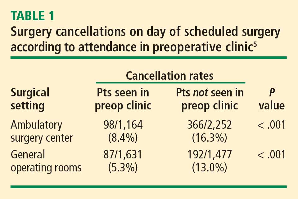
Yesterday I heard someone ask, “Do we really need all this preoperative evaluation? Does it really improve outcomes?” There is some evidence that it does. A study from 2000 based on data from the Australian Incident Monitoring Study found that 11% of the 6,271 critical incidents that occurred following operations were attributable to inadequate preoperative evaluation and that 3% were unequivocally related to problems with preoperative assessment or preparation. More than half of the incidents were deemed preventable.6
Preoperative clinics are good for patients and make good sense economically. We just need to demonstrate to our administrators and to payers that we are offering an excellent service.
Cleveland Clinic IMPACT Center
By Ajay Kumar, MD
Cleveland Clinic is structured differently from most other institutions in that its surgeons, anesthesiologists, and hospitalists are all direct employees of the institution. Despite this unique structure, many aspects of our preoperative clinic—known as the Internal Medicine Preoperative Assessment, Consultation and Treatment (IMPACT) Center—are applicable to other institutions.
Cleveland Clinic is a busy surgical hospital whose preoperative optimization system is designed to provide high-quality care. The IMPACT Center is consulted for most complicated noncardiac surgery patients, and its referral sources include most of the institution’s surgical specialties.
QUEST FOR A BETTER PATIENT EXPERIENCE
When the IMPACT Center was created in 1997, the aim was to focus on the same objectives highlighted by Drs. Bader and Sweitzer: safety, a positive patient experience, enhanced communication, better continuity of care, effective use of resources, and improving throughput by standardizing care.
A prime motivator was the desire to move away from the tendency for presurgical consults to simply “clear the patient for surgery,” and we have indeed evolved considerably from that point. The focus of our perioperative care program today is to comprehensively evaluate risk by taking into account patient-, procedure-, and anesthesia-related factors.
We offer “one-stop shopping,” and our priority is for efficient throughput. We are located in a 12-story building that includes outpatient, preoperative, and surgical clinics and offices. The IMPACT Center is on the first floor along with the preoperative anesthesia consultation and evaluation (PACE) clinic, the laboratory, and ECG and stress-test labs. Patients can undergo radiographic studies on the second floor.
The patient experience counts for a lot. Many of our patients are from another state or country, so efficiency and convenience are especially important. Patients can usually get all assessment and testing done in a single day.
A TIGHTLY MANAGED PROCESS
A ‘smart’ questionnaire starts the process
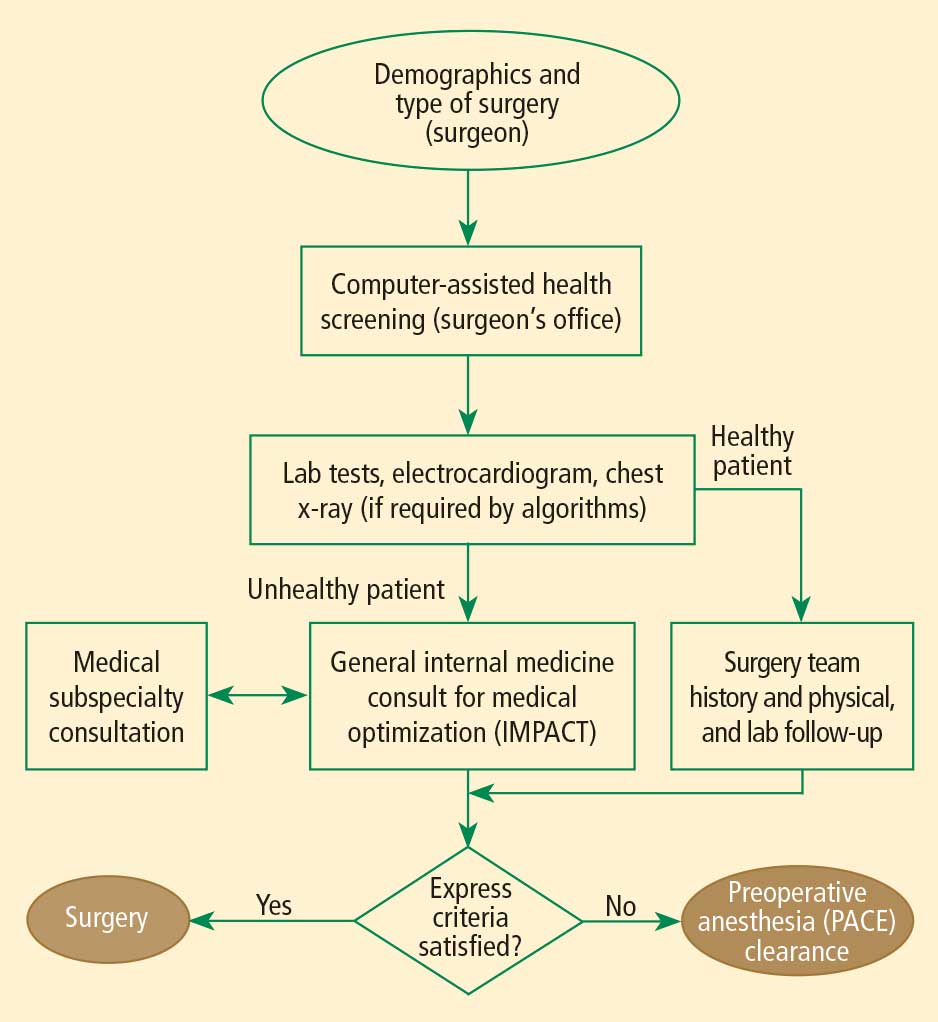
A computerized report based on the questionnaire guides the surgical office in scheduling the patient to specific areas according to algorithms. Based on case complexity and clinical needs, patients are scheduled for the IMPACT Center along with the PACE clinic; if needed (based on the algorithms), patients also are scheduled for laboratory tests or imaging. This standardized approach helps create a safe passage for patients through the preoperative process with less confusion.
Patient is given a personalized binder
Once all appointments and tests are scheduled, the patient is given a binder containing specific information about the procedure and preoperative instructions. The medical appointment at the IMPACT Center is usually scheduled before the PACE clinic appointment. Patients receive an itinerary for all preoperative appointments and surgical office appointments before the planned surgery. The itinerary is planned so that if additional testing is requested, it can be accommodated on the same day.
At the end of the preoperative assessment, the patient receives printed information with specific preoperative instructions, including which medications to continue or stop.
Standardized, computer-based medical records
Our systems and processes have undergone a good deal of evolution. We have integrated our medical records and use a standard outside medical record retrieval process. The template for the history and physical exam is standard for all Cleveland Clinic patients and is used for all presurgical assessments before all noncardiac surgeries. The template is comprehensive, including the history of the present illness, the review of systems, the physical exam, and anesthesia-related issues. All outside documents are scanned into our electronic medical record system and are available for viewing prior to surgery from any computer connected to the system.
Our preoperative assessment guidelines are also kept updated at a central online location so that all providers have access to them.
Staff keeps process running efficiently
The IMPACT Center is managed by the department of hospital medicine and supported by at least 5.5 full-time physicians every day. We also have two registered nurses, two assistants who help with scheduling and testing, and three secretaries who support the doctors, obtain records, and make arrangements with outside doctors for testing if a patient wants to return home before our testing can be completed.
A secretary also keeps a log for each patient seen in the clinic, tracking all pending issues. The day before surgery, the secretary contacts the appropriate office for anything that is still pending. If she gets no response, the matter is transferred to one of our doctors so that the problem can be resolved at once. This strategy allows us to achieve a nearly 0% rate of surgery delay or cancellation attributable to unavailable test results.
Our patient volumes have increased significantly since we started in 1997. Last year more than 15,000 patients visited the IMPACT Center and now we have expanded our services to regional hospitals within the Cleveland Clinic Health System.
INTERDEPARTMENTAL COMMUNICATION IS CRITICAL
Interdepartmental communication is a must for patient safety, so we encourage a culture of communication between the hospitalist and the surgical team. The location of most of our surgical clinics within the same building as the IMPACT Center further facilitates communication, as does the proximity of the PACE clinic. Additionally, one of our IMPACT Center physicians is accessible around the clock to answer to our surgeon or anesthesiologist colleagues as needed.
We regularly assess our process and seek feedback from surgeons and anesthesiologists. We also conduct yearly patient experience surveys to make sure we are providing patients with the highest quality of care.
Discussion
Question from the audience: Our anesthesia assessment department was approached by our surgeons to do both the anesthesia and surgical assessments, but we felt that would put us in a potential legal conflict if a patient who was assessed that way developed problems. Can you comment?
Dr. Bader: Although we do surgical assessments at our preoperative clinic, we don’t make any decisions about whether or not to proceed with an operation. We get an office note from the surgeon that is directed specifically toward the need for surgery, indications for surgery, and surgical consent. We perform the surgical history and physical examination. Our process is essentially the same as when surgeons have a physician assistant do the history and physical examination in their office. Our practitioners are employed by the hospital, so there is no conflict of interest there.
Comment from the audience: I’m a strong believer in hands-on patient contact. Over my 15 years of practice, we have encountered a lot of unexpected problems during the preoperative exam—aortic stenoses, infections, ventricular septal defects—all of which would never have been detected from a screening form.
Dr. Sweitzer: I agree that we pick up many things by seeing the patient in person. I’ve picked up more cases of aortic stenosis as an anesthesiologist in the preoperative clinic than I ever did as an internist, because the population is high-risk. But patients who have such problems tend to have risk factors and be in certain age groups. Studies indicate that the history is more important than the physical exam: the history suggests about 75% of conditions that are present. The physical exam adds only a little more—perhaps another 15%. Our recommendations are very much consistent with the American College of Cardiology and American Heart Association guidelines on preoperative cardiac evaluation.7 It is more important to identify whether a patient has risk factors for coronary artery disease than to find out whether a stress test or ECG is normal. One needs to do a really good history, but it can be done remotely. Based on certain risk factors identified, high-risk patients can be selected who need to come in and have a physical exam.
Question from the audience: Could you elaborate on the electronic medical record system used at the University of Chicago? I’ve heard there’s a steep learning curve when implementing these kinds of systems. They also are very expensive—I’ve heard that some cost $40 to $80 million. Has enhanced revenue flow offset the costs?
Dr. Sweitzer: We have a home-grown system developed with FileMaker Pro by a computer programmer at our institution. It was a lot easier to develop than people tend to think. There are many savvy computer programmers out there; I’ve had medical students assist me with updating it. We’re now considering developing it as a commercial system. Many systems are available for purchase, including Epic, Pyxis, one from General Electric, and many others. They are very expensive, so smaller institutions might want to use a pay-for-service system.
There definitely is a learning curve to switching to electronic medical records, but it is not nearly as steep as many believe. The extra time it takes a clinician to initially make a computer entry rather than write on paper is vastly recouped downstream: the electronic medical record is legible and organized, and it doesn’t get lost or need to be redone. You can bring up a patient record from 6 months before and reuse it as a template.
Dr. Bader: The discussion of cost savings from preoperative clinics usually focuses on savings from avoiding surgery cancellations and delays and from more efficient laboratory testing, but the biggest savings for an institution is better reimbursement through better diagnosis-related groups (DRG) coding. That’s an important reason our institution is funding our clinic. Electronic medical records allow standardization of information so that coders know exactly where to look for the comorbidities and other pertinent information. This increases payments for DRGs, which can be documented for the hospital. This literally runs into millions of dollars a year and more than offsets the costs of the system.
Question from the audience: Dr. Bader, I’m impressed with the number of patients going through your preoperative clinic. How many patients are seen per nurse practitioner in your clinic?
Dr. Bader: The nurse practitioners have 10-hour shifts and see one patient every 75 minutes. The process of seeing a patient takes a lot less time now than with the old system, in which patients saw an anesthesiologist plus a nurse. Our current system eliminates redundancy: questions need to be asked only once.
Question from the audience: My compliance office says that preoperative assessments for early-morning admission patients are good for only 7 days. Is that true?
Dr. Bader: There are sometimes differences between Joint Commission requirements and those of certain insurance companies. That kind of issue needs to be discussed with your hospital compliance office. We program rules into our scheduling system to accommodate different insurance policies and other requirements so that a patient is not scheduled beyond the allowable period.
- Detsky AS, Naglie IG. A clinician’s guide to cost-effectiveness analysis. Ann Intern Med 1990; 113:147–154.
- Sandler G. The importance of the history in the medical clinic and the cost of unnecessary tests. Am Heart J 1980; 100:928–931.
- Blumenthal D, Glaser JP. Information technology comes to medicine. N Engl J Med 2007; 356:2527–2534.
- Strum DP, Vargas LG, May JH. Surgical subspecialty block utilization and capacity planning: a minimal cost analysis model. Anesthesiology 1999; 90:1176–1185.
- Ferschl MB, Tung A, Sweitzer B, Huo D, Glick DB. Preoperative clinic visits reduce operating room cancellations and delays. Anesthesiology 2005; 103:855–859.
- Kluger MT, Tham EJ, Coleman NA, et al. Inadequate preoperative evaluation and preparation: a review of 197 reports from the Australian incident monitoring study. Anaesthesia 2000; 55:1173–1178.
- Fleisher LA, Beckman JA, Brown KA, et al. ACC/AHA 2007 guidelines on perioperative cardiovascular evaluation and care for noncardiac surgery: a report of the American College of Cardiology/American Heart Association Task Force on Practice Guidelines. J Am Coll Cardiol 2007; 50:1707–1732.
Weiner Center for Preoperative Evaluation at Brigham and Women’s Hospital
By Angela M. Bader, MD, MPH
When organizing our preoperative clinic at Brigham and Women’s Hospital, we had several goals. Overall, we wanted a standardized process to help us achieve a high level of excellence. We hoped that creating a new system would eliminate ambiguity about who was responsible for following up on a patient’s abnormal laboratory test result—the surgeon, anesthesiologist, or primary care physician. We also wanted to better coordinate the various care teams involved throughout the perioperative period.
STANDARDIZATION HELPS MEET MANY GOALS
Standardization can occur at many levels:
- Performance of assessments and testing
- Organization of the patient chart and medical records
- Systems checks throughout the process to ensure that nothing is missed
- Team-to-team communication.
Documentation requirements apply regardless of institutional structure
When considering any system of preoperative assessment, keep in mind that the hospital must meet and appropriately document compliance with all regulatory, accreditation, and payer requirements and guidelines, such as those of the Joint Commission, the Centers for Medicare and Medicaid Services (CMS), and the National Surgical Quality Improvement Program. For example, the Joint Commission requires that a surgical history and physical examination be done within 30 days of a procedure. An anesthesiology assessment and a nursing assessment are also required. All of these assessments have mandatory elements, including documenting “never events” and ordering appropriate laboratory tests, electrocardiograms (ECGs), and radiographs.
Sometimes administrators of other hospitals say to me, “We can’t afford a preoperative clinic, and we don’t need one.” My response is that regardless of whether a hospital has a preoperative clinic, the regulatory requirements and guidelines must be met: it is not an issue of avoiding certain steps. Having a dedicated preoperative clinic simply shifts the work to a standardized, centralized system and avoids delaying these required steps until the day of surgery, when taking care of a problem involves the most inefficient use of resources.
Tailor system to institutional needs and characteristics
Within the regulatory framework, the organizational scheme of every institution must address issues of volume and acuity, the types of surgery performed, and the time frames required. A system must be able to deal with the preoperative needs of patients undergoing operations that are booked weeks in advance (often the case for orthopedic surgery) as well as those that may not be booked until a day before the procedure (eg, cancer surgery).
Our plan was developed for our very high-volume, tertiary care institution. In 2008, 24,000 patients used our clinic (roughly 100 patients per day).
DESIGN OF THE PREOPERATIVE CLINIC
A nurse practitioner–based model for ‘one-stop shopping’
We decided that the clinic should offer all elements of the preoperative assessment and thereby give patients “one-stop shopping.” Each patient sees a nurse practitioner, who performs the surgical history and physical examination as well as the anesthesiology and nursing assessments. The result is a multidisciplinary approach with a single assessment output. We shifted employees who had been responsible for preoperative assessment in the offices of various surgeons to a central clinic so that all assessments could be standardized, and we provided additional training to enable them to perform various assessments. The nurse practitioners are supervised by an on-site attending physician, as detailed below.
This model offers a number of advantages:
- Patients see a single provider.
- Assessment is facilitated for our surgeons, who may not be completely up-to-date on perioperative risk assessment and management.
- We have a central location for standardized education programs for our physicians, nurses, and residents.
- The clinic’s standardized records and processes facilitate data generation for research and clinical practice improvement.
Independent budgetary and staffing structure
The preoperative clinic is a separate cost center under the leadership of the department of anesthesiology. Resources were shifted to a central location so that as volume increases, we can add resources to meet the additional volume. We contracted with the hospital administration to provide payment for two full-time-equivalent anesthesiologists per day, who serve as on-site attending physicians. The hospital is willing to do this because not only do these attending physicians supervise the anesthesiology assessment, they are the collaborating physicians for the entire perioperative assessment. They review every patient, order tests and write prescriptions as needed, and discuss issues with the primary care physicians and referring specialists.
The preoperative clinic has an anesthesiologist director (me) who reports directly to the hospital’s vice president for surgical services on budget and staffing issues. I also report to the chairman of the department of anesthesiology, though he is not involved in budgetary functions (the hospital contracts with him to provide the anesthesiology staffing). The clerical and nursing staff work directly for the clinic.
The clinic is run in a self-contained area with a central waiting room and space for doing all the assessments and laboratory work internally, including 16 examination rooms and a room for chart organization.
MORE BENEFITS OF STANDARDIZATION
Standardized scheduling ensures reliability
The secretaries in each surgeon’s office schedule appointments through a central computer system after registration and insurance precertification. Our computer system does not allow an operation to be scheduled without an evaluation also being scheduled. The evaluation can involve either a visit or a telephone screen; we provide algorithms so that the surgeons’ secretaries know which is required. This system has substantially reduced the number of walk-ins, allowing for a more even distribution of patients and ensuring that medical records will be available when a patient is seen.
We watch our schedule carefully. Our computer system monitors the time that each patient is in our clinic to determine his or her waiting time and assessment time. It takes about 75 minutes to go through the whole process, including the time for a nurse practitioner to do the surgical history and physical examination and the anesthesiology and nursing assessments, a laboratory technician to do an ECG and laboratory tests if indicated, and completion of all required documentation. Accordingly, patients are scheduled in 75-minute blocks between 7:00 am and 6:30 pm. We do not have evening or weekend hours because of the difficulty of contacting surgeons and primary care physicians when questions arise. It is simply not cost-effective to have to do that type of follow-up on a case after the patient leaves.
Only about 10% of our patients are screened by telephone, since most of our operations are complicated and require in-person assessment (most low-acuity procedures are done at other hospitals). Of the patients who visit the preoperative clinic, about 75% undergo the single assessment model for surgery, anesthesiology, and nursing as described above. The remaining 25% of patients have their history and physical exam completed outside Brigham and Women’s Hospital for insurance reasons; the remainder of their assessment is conducted in our preoperative clinic by a registered nurse and an anesthesiology resident.
Multiple systems checks
Our model also incorporates standardization in the form of multiple systems checks:
- Case presentation. Every case is presented to an attending anesthesiologist, who reviews the ECG (if ordered) before the patient leaves the clinic.
- Post-visit chart check. A registered nurse or nurse practitioner signs off on each chart after the visit, confirming test results and resolution of all paperwork issues.
- Surgical checklist. The end result is a checklist that serves as the front sheet of the operating room chart.
Our ability to use this system of checks to get the chart completed comprehensively and reliably and deliver it to the operating room when needed was key to securing institutional support and funding for the preoperative clinic.
ROLE OF THE ATTENDING ANESTHESIOLOGISTS
Two full-time attending anesthesiologists are present in the preoperative clinic each day. One is responsible largely for supervising the nurse practitioner assessments and reviewing case presentations, while the other also oversees the education and supervision of residents. Residents rotate through the clinic for 2 weeks (one or two at a time) and have a designated curriculum consisting of daily lectures and competencies in preoperative evaluation.
Because our anesthesiologists are expert in preoperative assessment, we require very few outside consults. We can communicate directly with the cardiologists and other physicians and order tests when indicated. We have a clerical assistant who obtains all necessary paperwork and prior testing from outside providers so that the clinicians need not waste time on this.
A GROWING CHALLENGE: ASSESSMENT FOR PROCEDURES IN AMBULATORY SETTINGS
Looking forward, a rapidly growing challenge facing our clinic stems from the tremendous growth in patients who require anesthesia for procedures performed outside the operating room. In these situations, the proceduralists need a system for deciding whether an anesthesiologist must be present for any given case.
We have started to develop appropriate screening processes to ensure that the proceduralists in multiple departments know which patients to refer for preprocedure assessment. We hope to soon develop protocols for high-risk patients and for various procedures such as implanting a pacemaker or defibrillator, catheter procedures, interventional radiology, and endoscopy.
Anesthesia Perioperative Medicine Clinic at University of Chicago
By BobbieJean Sweitzer, MD
Detsky and Naglie have argued that the costs and clinical outcomes associated with any intervention must be compared with those of alternate strategies for treating the same patients,1 and I believe their point applies well to preoperative clinics. Although certain requirements of the Joint Commission and CMS must be met, as noted by Dr. Bader, they can be met in various ways. I will preface my comments by emphasizing that one size does not fit all: every institution must decide the best approach to preoperative assessment based on its patient population, the types of procedures it performs, and the volume it handles.
TRIAGE STREAMLINES THE PROCESS
Our preoperative clinic at the University of Chicago emphasizes triage. Not every patient should have to go to the trouble of coming in to see a provider. In the future, we will likely see more “virtual” preoperative assessments using devices in development, such as handheld ultrasonography machines. Just as patients can have their pacemakers and implantable cardiac defibrillators remotely checked via phone contact, more tools will one day be available for remote assessment.
Although not every surgical patient needs to come in to the preoperative clinic, every patient must have a physical examination. All patients will be seen on the day of surgery, so in some cases the physical exam may be able to wait until then. For example, an airway assessment need not be done ahead of time. Most anesthesiologists are prepared to manage airways on very short notice, so extensive advance planning is not always necessary.
Obtain basic info by questionnaire to save staff time
Information about the patient is key to triage, and it may be either paper- or computer-based. An initial priority should be to develop some mechanism for getting information from patients before the day of their procedure without a visit to the hospital or ambulatory surgery center.
We use a two-page paper questionnaire to obtain basic information from patients, including (among other pertinent questions) age, planned operation, names of the surgeon and primary doctor, past operations and medical history, allergies, a list of medications, social history (drug, alcohol, tobacco use), whether they have ever taken steroids, whether they have high blood pressure, and whether they can comfortably walk up a flight of stairs. We provide the primary care physicians and surgeons with blank questionnaires, which their patients can fill out in their waiting rooms or take home and fax to us (or drop off) later. The questionnaire gives us a good deal of essential information without using staff time.
Various computer-based and Web-based systems are also available for collecting basic patient information. Smaller institutions need not purchase an entire electronic medical record system, which can be very expensive. Some Web-based tools operate on a pay-per-use basis and can be very helpful.
Review the information to guide triage
We then review the patient information to determine the extent of preoperative evaluation required. Some patients, especially those scheduled at an ambulatory surgery center, are healthy enough that they can just come in on the day of surgery for an examination and an update of their information. Others will need an appointment at the clinic before the day of surgery for more extensive preoperative evaluation. For other patients, review of their questionnaire responses may prompt a phone call or e-mail from the clinic for more information to determine whether a day-of-surgery exam will suffice or whether evaluation in advance is needed. When in doubt, concerns raised by the questionnaire should be explored before the day of surgery to avoid surprises and allow sufficient time for a consultation, if needed.
STANDARDIZED GUIDELINES KEEP CARE CONSISTENT
We encourage our staff to minimize preoperative testing and ECGs. A majority of diagnoses are made based on the history and physical exam.2 Generally, a test should confirm what is already suspected and merely provide objective evidence when needed. Testing in this setting should not be done to “find out what is wrong” with a patient.
It is helpful to develop standardized guidelines for preoperative assessment and make them available to everyone in the institution via the Web. The guidelines should address recommended preoperative tests and management practices according to specific patient conditions or surgical procedures. The clear objective is to avoid disagreement about what a patient needs between the provider who evaluated the patient in advance and the surgeon or anesthesiologist who evaluates the patient on the day of surgery.
Our guidelines at the University of Chicago include recommendations for patients on long-term anticoagulant therapy, for patients with coronary stents, for medications that should be discontinued (and those that may be continued) on the day of surgery, and for numerous other conditions and issues. Our testing guidelines list indicated tests for various medical problems, which in turn link to other guidelines. Other links are based on the medications a patient is using or the type of operation that is planned.
We collaborated with our electrophysiology department to create guidelines for managing patients with pacemakers and defibrillators. Almost every patient with one of these devices has a little card associated with the device, and we ask the surgeons to copy the card and send it to the clinic if we will not be directly seeing the patient. Using a national database, the electrophysiology department can determine from the card the type of pacemaker or defibrillator a patient has, and they fax or e-mail us back a page of instructions to let us know whether the device requires special consideration during surgery, whether it should be checked preoperatively, and whether its battery needs replacing. With this system, we have markedly reduced problems on the day of surgery.
CONSULTS HAVE AN IMPORTANT ROLE
Consults should never be requested in order to “clear a patient for surgery.” Consult requests should rather address specific issues, such as, “Is this patient medically optimized?” or “Please address this patient’s hypertension.” In turn, consult notes should provide meaningful information that can be used in a specific way. A clearance letter or simple risk assessment is not helpful.
If a patient has not seen a primary care doctor in a long time, a consult request should (in addition to requesting a global risk assessment) specify any particular concerns, such as, “The patient reports snoring; please address sleep apnea and cardiac risk.”
Case study: Beware consult notes with no specifics
Consider a case we encountered of a 54-year-old man who had a preoperative cardiac risk assessment. The cardiology consultant completed a short form consisting of a multiple-choice check-off list indicating low, moderate, or high cardiac risk. The consultant checked that the patient had low cardiac risk but provided no other instructions or information other than his own contact information.
When we reviewed the patient’s questionnaire, we saw that his medications included metoprolol, clopidogrel, and aspirin even though the patient did not mention that he had coronary artery disease. On this basis, we requested details about his cardiac evaluation from his cardiologist. It turned out that the patient had a history of four catheterizations with several cardiac stents placed. The most recent stent was implanted to overlap a previous stent that had been found “floating” in the blood vessel; this last stent was placed just 6 months before the cardiologist issued the consult note indicating “low cardiac risk.”
The moral is to approach consult notes with caution, especially if they offer no specifics. It actually makes me nervous when a note states “low risk” because if something unexpectedly goes wrong in surgery, it appears that the perioperative team took poor care of the patient even if the complication actually may have stemmed from higher-than-recognized underlying patient risk.
PROVIDE, AND REINFORCE, CLEAR INSTRUCTIONS
We give patients written preoperative instructions that become part of our computerized records. We first verbally give explicit instructions for each medication—ie, whether it can be taken as usual or when it needs to be stopped before surgery (and why). Then we provide the same information in writing, after which we try to have the patient repeat the instructions back to the clinician. We include a phone number that patients can call if they need help understanding their preoperative instructions.
Web-based programs also can provide patients online instructions about their medications. Some services even customize information by providing, for example, lists of local surgeons who are willing to allow a patient to continue on aspirin therapy until the day of surgery.
USE THE RIGHT RESOURCES
Staffing
Our model at the University of Chicago relies mainly on residents in training and physician assistants, but advanced nurse practitioners are well suited to a preoperative clinic as well. These types of providers have background training in history-taking, physical examination, diagnostic testing, and disease management. Registered nurses have more limited abilities, although they may be appropriate for a clinic that deals primarily with healthy patients for whom only history taking and a list of medications is needed. Additionally, our clinic is staffed by one attending anesthesiologist at all times (from among a group of rotating anesthesiologists) as well as medical assistants and clerical staff.
Some clinics perform the surgical history and physical exam at the same time as the anesthesia assessment. I would urge caution with this practice. Just as primary care doctors should not be conducting the anesthesia assessment, nonsurgeons should not be conducting the surgical assessment; doing so puts them out on a limb from a medicolegal standpoint. Advanced nurse practitioners and physician assistants may do surgical assessments under the supervision of a surgeon, but only surgeons should ultimately decide—and document—whether an operation is necessary and what degree of examination is required in advance.
Computer technology for records, messaging, billing
Using electronic medical records and corresponding with colleagues by e-mail make preoperative care much more efficient. We have standardized computer forms for ordering tests and documenting the physical exam. Patients usually understand that electronic medical records are safe and more efficient, and they are often more accepting of their use than practitioners are. Many patients want e-mail access to doctors, to schedule appointments online, and to receive appointment reminders by e-mail.3
Electronic medical records also avoid redundancy. If a patient has been seen in our preoperative clinic and is later scheduled for another surgery (even if a different surgeon is involved), a return visit to our clinic may not be necessary. In some cases, we can send the old work-up stamped “For information only,” which can then be updated by the anesthesiologist on the day of surgery.
A central, standardized process also makes billing more efficient and helps to ensure that payment is received for all services provided. Standardized documentation makes it easier for coders to enter the correct evaluation and management codes and ensures that all required criteria are met.
THE PAYOFF: LIVES AND DOLLARS SAVED
A thorough and efficient preoperative assessment system is cost-effective. Every minute of operating room time is worth $10 to $15,4,5 so delays should be avoided. Everything that is done ahead of time saves money for the whole enterprise by reducing unnecessary case setups and reducing “down time” due to lack of patient, equipment, or staff readiness. We routinely bill for preoperative evaluations when this service goes beyond a routine preoperative assessment based on CMS (and other insurance) requirements. However, a preoperative evaluation is required by CMS and most payers if one wants to be paid for any anesthesia-provided service. As a result, a cost is incurred without offsetting revenue if a case is cancelled on the day of surgery after one performs the anesthesia evaluation.
Yesterday I heard someone ask, “Do we really need all this preoperative evaluation? Does it really improve outcomes?” There is some evidence that it does. A study from 2000 based on data from the Australian Incident Monitoring Study found that 11% of the 6,271 critical incidents that occurred following operations were attributable to inadequate preoperative evaluation and that 3% were unequivocally related to problems with preoperative assessment or preparation. More than half of the incidents were deemed preventable.6
Preoperative clinics are good for patients and make good sense economically. We just need to demonstrate to our administrators and to payers that we are offering an excellent service.
Cleveland Clinic IMPACT Center
By Ajay Kumar, MD
Cleveland Clinic is structured differently from most other institutions in that its surgeons, anesthesiologists, and hospitalists are all direct employees of the institution. Despite this unique structure, many aspects of our preoperative clinic—known as the Internal Medicine Preoperative Assessment, Consultation and Treatment (IMPACT) Center—are applicable to other institutions.
Cleveland Clinic is a busy surgical hospital whose preoperative optimization system is designed to provide high-quality care. The IMPACT Center is consulted for most complicated noncardiac surgery patients, and its referral sources include most of the institution’s surgical specialties.
QUEST FOR A BETTER PATIENT EXPERIENCE
When the IMPACT Center was created in 1997, the aim was to focus on the same objectives highlighted by Drs. Bader and Sweitzer: safety, a positive patient experience, enhanced communication, better continuity of care, effective use of resources, and improving throughput by standardizing care.
A prime motivator was the desire to move away from the tendency for presurgical consults to simply “clear the patient for surgery,” and we have indeed evolved considerably from that point. The focus of our perioperative care program today is to comprehensively evaluate risk by taking into account patient-, procedure-, and anesthesia-related factors.
We offer “one-stop shopping,” and our priority is for efficient throughput. We are located in a 12-story building that includes outpatient, preoperative, and surgical clinics and offices. The IMPACT Center is on the first floor along with the preoperative anesthesia consultation and evaluation (PACE) clinic, the laboratory, and ECG and stress-test labs. Patients can undergo radiographic studies on the second floor.
The patient experience counts for a lot. Many of our patients are from another state or country, so efficiency and convenience are especially important. Patients can usually get all assessment and testing done in a single day.
A TIGHTLY MANAGED PROCESS
A ‘smart’ questionnaire starts the process
A computerized report based on the questionnaire guides the surgical office in scheduling the patient to specific areas according to algorithms. Based on case complexity and clinical needs, patients are scheduled for the IMPACT Center along with the PACE clinic; if needed (based on the algorithms), patients also are scheduled for laboratory tests or imaging. This standardized approach helps create a safe passage for patients through the preoperative process with less confusion.
Patient is given a personalized binder
Once all appointments and tests are scheduled, the patient is given a binder containing specific information about the procedure and preoperative instructions. The medical appointment at the IMPACT Center is usually scheduled before the PACE clinic appointment. Patients receive an itinerary for all preoperative appointments and surgical office appointments before the planned surgery. The itinerary is planned so that if additional testing is requested, it can be accommodated on the same day.
At the end of the preoperative assessment, the patient receives printed information with specific preoperative instructions, including which medications to continue or stop.
Standardized, computer-based medical records
Our systems and processes have undergone a good deal of evolution. We have integrated our medical records and use a standard outside medical record retrieval process. The template for the history and physical exam is standard for all Cleveland Clinic patients and is used for all presurgical assessments before all noncardiac surgeries. The template is comprehensive, including the history of the present illness, the review of systems, the physical exam, and anesthesia-related issues. All outside documents are scanned into our electronic medical record system and are available for viewing prior to surgery from any computer connected to the system.
Our preoperative assessment guidelines are also kept updated at a central online location so that all providers have access to them.
Staff keeps process running efficiently
The IMPACT Center is managed by the department of hospital medicine and supported by at least 5.5 full-time physicians every day. We also have two registered nurses, two assistants who help with scheduling and testing, and three secretaries who support the doctors, obtain records, and make arrangements with outside doctors for testing if a patient wants to return home before our testing can be completed.
A secretary also keeps a log for each patient seen in the clinic, tracking all pending issues. The day before surgery, the secretary contacts the appropriate office for anything that is still pending. If she gets no response, the matter is transferred to one of our doctors so that the problem can be resolved at once. This strategy allows us to achieve a nearly 0% rate of surgery delay or cancellation attributable to unavailable test results.
Our patient volumes have increased significantly since we started in 1997. Last year more than 15,000 patients visited the IMPACT Center and now we have expanded our services to regional hospitals within the Cleveland Clinic Health System.
INTERDEPARTMENTAL COMMUNICATION IS CRITICAL
Interdepartmental communication is a must for patient safety, so we encourage a culture of communication between the hospitalist and the surgical team. The location of most of our surgical clinics within the same building as the IMPACT Center further facilitates communication, as does the proximity of the PACE clinic. Additionally, one of our IMPACT Center physicians is accessible around the clock to answer to our surgeon or anesthesiologist colleagues as needed.
We regularly assess our process and seek feedback from surgeons and anesthesiologists. We also conduct yearly patient experience surveys to make sure we are providing patients with the highest quality of care.
Discussion
Question from the audience: Our anesthesia assessment department was approached by our surgeons to do both the anesthesia and surgical assessments, but we felt that would put us in a potential legal conflict if a patient who was assessed that way developed problems. Can you comment?
Dr. Bader: Although we do surgical assessments at our preoperative clinic, we don’t make any decisions about whether or not to proceed with an operation. We get an office note from the surgeon that is directed specifically toward the need for surgery, indications for surgery, and surgical consent. We perform the surgical history and physical examination. Our process is essentially the same as when surgeons have a physician assistant do the history and physical examination in their office. Our practitioners are employed by the hospital, so there is no conflict of interest there.
Comment from the audience: I’m a strong believer in hands-on patient contact. Over my 15 years of practice, we have encountered a lot of unexpected problems during the preoperative exam—aortic stenoses, infections, ventricular septal defects—all of which would never have been detected from a screening form.
Dr. Sweitzer: I agree that we pick up many things by seeing the patient in person. I’ve picked up more cases of aortic stenosis as an anesthesiologist in the preoperative clinic than I ever did as an internist, because the population is high-risk. But patients who have such problems tend to have risk factors and be in certain age groups. Studies indicate that the history is more important than the physical exam: the history suggests about 75% of conditions that are present. The physical exam adds only a little more—perhaps another 15%. Our recommendations are very much consistent with the American College of Cardiology and American Heart Association guidelines on preoperative cardiac evaluation.7 It is more important to identify whether a patient has risk factors for coronary artery disease than to find out whether a stress test or ECG is normal. One needs to do a really good history, but it can be done remotely. Based on certain risk factors identified, high-risk patients can be selected who need to come in and have a physical exam.
Question from the audience: Could you elaborate on the electronic medical record system used at the University of Chicago? I’ve heard there’s a steep learning curve when implementing these kinds of systems. They also are very expensive—I’ve heard that some cost $40 to $80 million. Has enhanced revenue flow offset the costs?
Dr. Sweitzer: We have a home-grown system developed with FileMaker Pro by a computer programmer at our institution. It was a lot easier to develop than people tend to think. There are many savvy computer programmers out there; I’ve had medical students assist me with updating it. We’re now considering developing it as a commercial system. Many systems are available for purchase, including Epic, Pyxis, one from General Electric, and many others. They are very expensive, so smaller institutions might want to use a pay-for-service system.
There definitely is a learning curve to switching to electronic medical records, but it is not nearly as steep as many believe. The extra time it takes a clinician to initially make a computer entry rather than write on paper is vastly recouped downstream: the electronic medical record is legible and organized, and it doesn’t get lost or need to be redone. You can bring up a patient record from 6 months before and reuse it as a template.
Dr. Bader: The discussion of cost savings from preoperative clinics usually focuses on savings from avoiding surgery cancellations and delays and from more efficient laboratory testing, but the biggest savings for an institution is better reimbursement through better diagnosis-related groups (DRG) coding. That’s an important reason our institution is funding our clinic. Electronic medical records allow standardization of information so that coders know exactly where to look for the comorbidities and other pertinent information. This increases payments for DRGs, which can be documented for the hospital. This literally runs into millions of dollars a year and more than offsets the costs of the system.
Question from the audience: Dr. Bader, I’m impressed with the number of patients going through your preoperative clinic. How many patients are seen per nurse practitioner in your clinic?
Dr. Bader: The nurse practitioners have 10-hour shifts and see one patient every 75 minutes. The process of seeing a patient takes a lot less time now than with the old system, in which patients saw an anesthesiologist plus a nurse. Our current system eliminates redundancy: questions need to be asked only once.
Question from the audience: My compliance office says that preoperative assessments for early-morning admission patients are good for only 7 days. Is that true?
Dr. Bader: There are sometimes differences between Joint Commission requirements and those of certain insurance companies. That kind of issue needs to be discussed with your hospital compliance office. We program rules into our scheduling system to accommodate different insurance policies and other requirements so that a patient is not scheduled beyond the allowable period.
- Detsky AS, Naglie IG. A clinician’s guide to cost-effectiveness analysis. Ann Intern Med 1990; 113:147–154.
- Sandler G. The importance of the history in the medical clinic and the cost of unnecessary tests. Am Heart J 1980; 100:928–931.
- Blumenthal D, Glaser JP. Information technology comes to medicine. N Engl J Med 2007; 356:2527–2534.
- Strum DP, Vargas LG, May JH. Surgical subspecialty block utilization and capacity planning: a minimal cost analysis model. Anesthesiology 1999; 90:1176–1185.
- Ferschl MB, Tung A, Sweitzer B, Huo D, Glick DB. Preoperative clinic visits reduce operating room cancellations and delays. Anesthesiology 2005; 103:855–859.
- Kluger MT, Tham EJ, Coleman NA, et al. Inadequate preoperative evaluation and preparation: a review of 197 reports from the Australian incident monitoring study. Anaesthesia 2000; 55:1173–1178.
- Fleisher LA, Beckman JA, Brown KA, et al. ACC/AHA 2007 guidelines on perioperative cardiovascular evaluation and care for noncardiac surgery: a report of the American College of Cardiology/American Heart Association Task Force on Practice Guidelines. J Am Coll Cardiol 2007; 50:1707–1732.
KEY POINTS
- Standardizing the preoperative assessment process helps ensure that regulatory, accreditation, and payer requirements and guidelines are met.
- Careful triage based on a patient’s history can help avoid unnecessary assessment of low-risk patients and ensure that necessary assessments for higher-risk patients are completed before the day of surgery.
- Perioperative assessment and management guidelines for various types of surgery and patient risk factors should be developed, continuously updated, and made available online to all providers within the institution.
- Electronic medical records allow standardization of patient information, avoid redundancy, and provide a database for research.
Perioperative management of anemia: Limits of blood transfusion and alternatives to it
Anemia is a potent risk factor for mortality and morbidity in surgical patients, and its management has begun to shift away from allogeneic blood transfusion in recent years. This article reviews the clinical importance of perioperative anemia, the role and shortcomings of blood transfusion, and the pros and cons of alternative approaches to managing perioperative anemia. I conclude with an overview of a program for perioperative blood product use at my institution, Cleveland Clinic.
SIGNIFICANCE OF PERIOPERATIVE ANEMIA
Prevalence depends on many factors
The reported prevalence of anemia in surgical patients varies widely—from 5% to 76%1—and depends on the patient’s disease and comorbidities, the surgical procedure and associated blood loss, and the definition of anemia used. The prevalence of preoperative anemia increases with patient age and is higher in women than in men.2
A multiplier of risk
Anemia is an important multiplier of mortality risk. For example, the presence of anemia raises the relative risk of 2-year mortality from 2.05 to 3.37 in patients with chronic kidney disease, from 2.86 to 3.78 in patients with heart failure, and from 4.86 to 6.07 in patients with concomitant heart failure and chronic kidney disease.3
Adverse effects of anemia have been demonstrated specifically in the perioperative setting as well. A large retrospective cohort study showed that a preoperative hemoglobin concentration of less than 6 g/dL increases the risk of death 30 days after surgery by a factor of 26 relative to a concentration of 12 g/dL or greater in surgical patients who declined blood transfusion for religious reasons.4 The anemia-associated mortality risk was especially pronounced among patients with cardiovascular disease.4 Other studies have demonstrated perioperative anemia to be associated with increases in the risk of death,5 cardiac events,6 pneumonia,7 and postoperative delirium.8
IS BLOOD TRANSFUSION THE ANSWER
The use of allogeneic blood transfusion to manage anemia and blood loss is a concept that originated several centuries ago and has changed little over the years.
Blood supply challenges
Blood collection has historically lagged demand, resulting in a blood supply insufficient to meet transfusion needs. According to the federal government’s 2007 National Blood Collection and Utilization Survey Report, 6.89% of US hospitals reported that they cancelled elective surgery on 1 or more days in the prior year because of a lack of blood availability, and 13.5% experienced at least 1 day in which nonsurgical blood needs could not be met.9 Unless practices are changed to increase blood donation, these unmet tranfusion needs may grow.
Joint Commission set to measure blood management
In response to this challenge, an advisory panel formed by the Joint Commission has identified 17 performance measures related to blood conservation and appropriate transfusion.10 These measures are currently in development, and we expect to see some types of metrics in the near future. Such metrics are likely to further prioritize blood management for US hospitals.
Safety of the blood supply: Viral transmission down, TRALI risk persists
The safety of the blood supply has improved markedly. Sophisticated testing and public demand have led to a dramatic decline in the risk of transfusion-related transmission of HIV, hepatitis C virus, and hepatitis B virus.11
Despite this progress, the risk of transfusion-related acute lung injury (TRALI) has persisted in recent years. TRALI is characterized by acute onset of noncardiogenic pulmonary edema within 6 hours of blood product transfusion. Believed to be immune-mediated, TRALI is thought to occur as antibodies to human leukocyte antigens develop, inducing capillary leak syndrome.12 The patients most commonly affected are those who receive plasma from multiparous female donors. A recent evaluation of transfusion-related fatalities reported to the US Food and Drug Administration (FDA) revealed a continual rise in fatal TRALI cases in the United States from 2001 to 2006.13–15 TRALI was implicated in more than half of all transfusion-related fatalities reported to the FDA in 2006, a higher number than for any other single cause.13
At the same time, there is evidence that hemovigilance can reduce TRALI risk. In the United Kingdom, the Serious Hazards of Transfusion Steering Group introduced in late 2003 a policy of using plasma from male donors as much as possible, in view of the association of TRALI with plasma from multiparous female donors. The effort appeared to pay off: whereas TRALI accounted for 6.8% of all transfusion-related adverse events reported in the United Kingdom during the period 1996–2003,16 this proportion declined to just 1.9% in 2006.17
Finally, despite the progress in screening blood for more established infections like HIV and the hepatitis viruses, some additional infections now must be considered when assessing blood supply safety. These include diseases newly recognized as being transmissible by blood, or for which blood donor screening is not currently available, or that are newly emergent infections for which the potential for spread by transfusion is unknown. For such diseases—which include malaria and West Nile virus—the risk of transmission through transfusion is low, as they are much more likely to be acquired by other means.
Transfusion and outcomes: Not a strong record
Transfusion has never undergone safety and efficacy evaluation by the FDA. Given the challenges of conducting a randomized study of transfusion in the perioperative setting, we may never have high-quality data to assess transfusion in this setting.
A few studies merit mention, however. The Transfusion Requirement in Critical Care (TRICC) trial was conducted in 838 critically ill patients in the intensive care setting.18 Patients were randomized to a strategy of either liberal transfusion (begun when hemoglobin fell below 10 g/dL) or restrictive transfusion (begun when hemoglobin fell below 7 g/dL). Thirty-day mortality was similar between patients in the two strategy groups, but the restrictive strategy was associated with significantly lower mortality in at least two subgroups: patients with myocardial infarction and patients with pulmonary edema. Further subgroup analysis found no benefit of early or aggressive transfusion in patients with coronary artery disease or in those requiring mechanical ventilation.
Rao et al performed a meta-analysis of three large international trials of patients with acute coronary syndromes to determine whether blood transfusion to correct anemia in this setting was associated with improved survival.19 They found significantly higher mortality among patients who underwent transfusion compared with those who did not, prompting them to urge caution in the use of transfusion to maintain arbitrary hematocrit levels in stable patients with ischemic heart disease.
Similarly, a risk-adjusted, propensity-matched analysis of 6,301 patients undergoing noncardiac surgery found that receipt of 4 U of blood or more was a predictor of greater mortality, higher risk of infection, and longer hospital stay.20 Moreover, in an observational cohort study of 11,963 patients who underwent isolated coronary artery bypass graft surgery, each unit of red blood cells transfused was associated with an incrementally increased risk of adverse outcome (eg, mortality, renal injury, need for ventilator support, lengthened hospital stay, infection).21 The latter study found that transfusion was the single factor most reliably associated with increased risk of postoperative morbidity.
Additional studies have echoed these findings—ie, that perioperative blood transfusion has been associated with a host of adverse outcomes, including increased morbidity and length of stay, increased rates of postoperative infection, as well as immunosuppression, viral transmission, and acute transfusion reactions.5,22,23
Outcomes and duration of blood storage
An interesting factor in the relation between transfusion and outcomes is the shelf life of the blood being transfused. The FDA currently allows storage of blood for a maximum of 42 days, but a recent study of patients who received red blood cell transfusions during cardiac surgery found that those who received “older blood” (stored for > 14 days) had significantly higher rates of sepsis, prolonged intubation, renal failure, in-hospital mortality, and 1-year mortality compared with those who received “newer blood” (stored for ≤ 14 days).24
These differing outcomes are generally attributed to the so-called storage defect: as blood gets older, it loses components such as 2,3-DPG and adenosine disphosphate, its red cells lose deformability, and it undergoes buildup of cytokines and free hemoglobin. Increased demand for newer blood in light of the storage defect could further intensify pressures on the blood supply.
MANAGEMENT OF PERIOPERATIVE ANEMIA
In light of these shortcomings of blood transfusion, how should anemia be managed perioperatively to reduce or avoid the need for transfusion?
Preoperative evaluation
Vigilance for anemia and related issues in the preoperative evaluation is fundamental. The evaluation should elicit a history of bleeding tendencies, previous transfusions, and symptoms of anemia. Medications should be reviewed with an eye toward any that may predispose to perioperative bleeding and anemia, such as aspirin, clopidogrel, and anticoagulants. During the physical examination, alertness for pallor and petechiae is key, as is attentiveness to symptoms of anemia such as shortness of breath and fatigue.
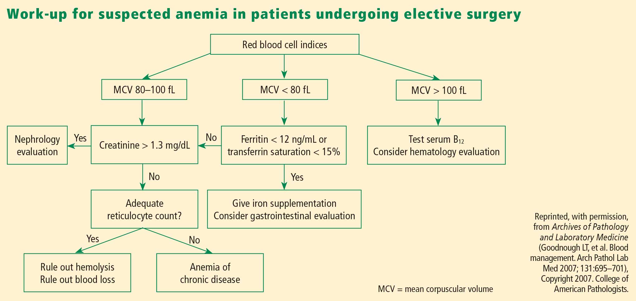
Overview of management options
Once the cause of anemia is identified, the choice for optimal medical management can be made. Choices broadly consist of pharmacologic and technological options. The former include iron supplements and erythropoiesis-stimulating agents. Among other pharmacologic options are thrombin, collagen, fibrin glue, tranexamic acid, and aminocaproic acid, but these agents are less well studied and will not be discussed here. Technological options include preoperative autologous blood donation, cell salvage, and acute normovolemic hemodilution.
In addition to these options, careful management of anticoagulant and antiplatelet medications should be provided, including discontinuation or substitution of drugs that could hamper clotting perioperatively.
PHARMACOLOGIC OPTIONS
Iron supplementation
Oral iron is available in four preparations: ferrous sulfate, ferrous gluconate, ferrous fumarate, and iron polysaccharide. Gastrointestinal side effects may limit these preparations’ tolerability. Iron supplements with a high elemental value will require fewer pills and fewer doses, reducing the risk or frequency of side effects.
Intravenous (IV) iron preparations are much safer now than they were years ago, when anaphylactic reactions were a concern. The ones generally used in the perioperative setting are iron sucrose and iron gluconate. Unlike the older IV preparations, the use of iron sucrose and iron gluconate often requires a second dose. The effect on hemoglobin levels usually occurs starting at 1 week, with the maximum effect achieved at 2 weeks. Hypotension, arthralgia, abdominal discomfort, and back pain are potential side effects of IV iron.
Efficacy and safety of iron supplementation. Evidence of the efficacy of preoperative iron supplementation is mounting. A study of 569 patients undergoing colorectal cancer surgery found that among the 116 patients who were anemic, intraoperative transfusion was needed in a significantly lower proportion of those who received 2 weeks of preoperative oral iron supplementation (200 mg) compared with those who received no iron therapy (9.4% vs 27.4%; P < .05).26 Similarly, in an uncontrolled study, 10 days of IV iron sucrose starting 4 weeks preoperatively significantly increased hemoglobin levels in 20 patients with iron-deficiency anemia prior to elective orthopedic surgery.27
Risks of infection and cancer progression have been concerns with IV iron therapy. However, no significant association between IV iron therapy and bacteremia was identified in a prospective study of 985 patients receiving chronic hemodialysis.28 The effect of IV iron administration on tumor progression has not been prospectively studied.
In general, IV iron, especially the newer forms, is a safer alternative to blood transfusion. Death occurs at a much lower rate with iron than with blood transfusion (0.4 per million vs 4 per million, respectively), as do life-threatening adverse events (4 per million vs 10 per million, respectively), according to a systematic review by the Network for Advancement of Transfusion Alternatives.29
Erythropoiesis-stimulating agents
Erythropoiesis-stimulating agents (ESAs) include epoetin alfa (erythropoietin), first approved by the FDA in 1989, and the more recently introduced darbepoetin alfa. They are approved to treat anemia in several patient populations, but only epoetin alfa is approved by the FDA explicitly for use in patients undergoing major surgery (to reduce the need for blood transfusions). The ESAs have come under intense scrutiny in recent years over their risk-to-benefit ratio, as detailed below.
The preoperative dosing schedule for epoetin alfa is usually three weekly doses (plus a fourth dose on the day of surgery) if the surgery is scheduled 3 or more weeks in advance. However, daily dosing can be used effectively if the preoperative period is less than 3 weeks, provided that it is continued until 4 days after surgery. Oral iron is necessary throughout the course of epoetin alfa therapy.
Efficacy in reducing transfusions. In a systematic review published in 1998, epoetin alfa was shown to minimize perioperative exposure to allogeneic blood transfusion in patients undergoing orthopedic or cardiac surgery.30 Its benefit was greatest in patients at the highest risk of requiring transfusion. It was effective whether given daily or weekly, and did not significantly increase the risk of thrombotic events when used in surgical patients, although some studies did find an excess of thrombotic events with its use.
In three randomized trials conducted in patients undergoing joint arthroplasty (hip or knee), epoetin alfa was associated with substantial and significant reductions in perioperative blood transfusion compared with placebo or preoperative autologous blood donation.31–33 Rates of deep vein thrombosis (DVT) did not differ significantly between the epoetin alfa and placebo groups.
Concerns over perioperative thromboembolic risk. In early 2007, the FDA was made aware of preliminary results of an open-label study in which 681 patients undergoing elective spinal surgery who did not receive prophylactic anticoagulation were randomized to epoetin alfa plus standard-of-care therapy (pneumatic compression) or standard-of-care therapy alone.34,35 The incidence of DVT was 4.7% in patients treated with epoetin alfa compared with 2.1% in those not receiving epoetin alfa. It is important to note that the available ESAs are prothrombotic and increase thrombotic risk significantly, especially in populations like this one in which pharmacologic DVT prophylaxis is not routinely used.
Based in part on this study, the FDA in 2007 required a boxed warning to be added to the ESAs’ package inserts to specify the increased risk of DVT with their use in surgical patients not receiving prophylactic anticoagulation. The warning urges consideration of the use of DVT prophylaxis in surgical patients receiving an ESA.34,35
TECHNOLOGICAL OPTIONS AND OTHER STRATEGIES
Autologous blood donation: A practice in decline
In cases of elective surgery, autologous blood donation can be used to protect against disease transmission and overcome the challenge of blood type compatibility. Preoperative autologous donation of blood has been a prevalent practice, but its use is declining. One reason is that waste is high (approximately 50% at Cleveland Clinic), which makes this practice more costly than is often realized. Also, autologous blood donation increases the likelihood that the patient will be anemic on the day of surgery, so that he or she may still need allogeneic blood after all, defeating the initial purpose. Despite these limitations, preoperative autologous blood donation remains a useful option for a subset of patients with multiple antibodies for whom donor blood may be difficult to obtain.
Cell salvage
Cell salvage is an innovative technology that recovers the patient’s own blood (after being shed from the surgical incision) for transfusion after filtering and washing. It is particularly well suited to procedures that involve massive blood loss. Cell savage requires technical expertise, however, and involves costs associated with both the machine and disposables.
Restricted postoperative phlebotomy
Phlebotomy accounts for a significant amount of blood loss, especially in intensive care patients with arterial lines. The equivalent of 30% of total blood transfused has been reported to be lost to phlebotomy during an intensive care unit stay.36 Triggers for transfusion cannot be assigned universally based on blood loss from phlebotomy but must consider the patient’s hemodynamic status, cardiac reserve, and other clinical characteristics.
PROMOTING RESPONSIBLE BLOOD PRODUCT USE
Blood is expensive, and in recent years hospitals have experienced increases in the cost of blood and blood products. To promote responsible blood use, we have developed a multipronged approach to blood management at Cleveland Clinic. The program’s cornerstone is increased awareness of the risks associated with blood transfusions. The emphasis is on educating staff physicians and other caregivers about the appropriate use of blood products. We also have implemented a new policy requiring staff authorization for all blood requested in nonemergency situations. Additionally, requests for blood components require adherence to an indication-based ordering process. Finally, data about blood use are shared transparently among physicians, encouraging good clinical practice.
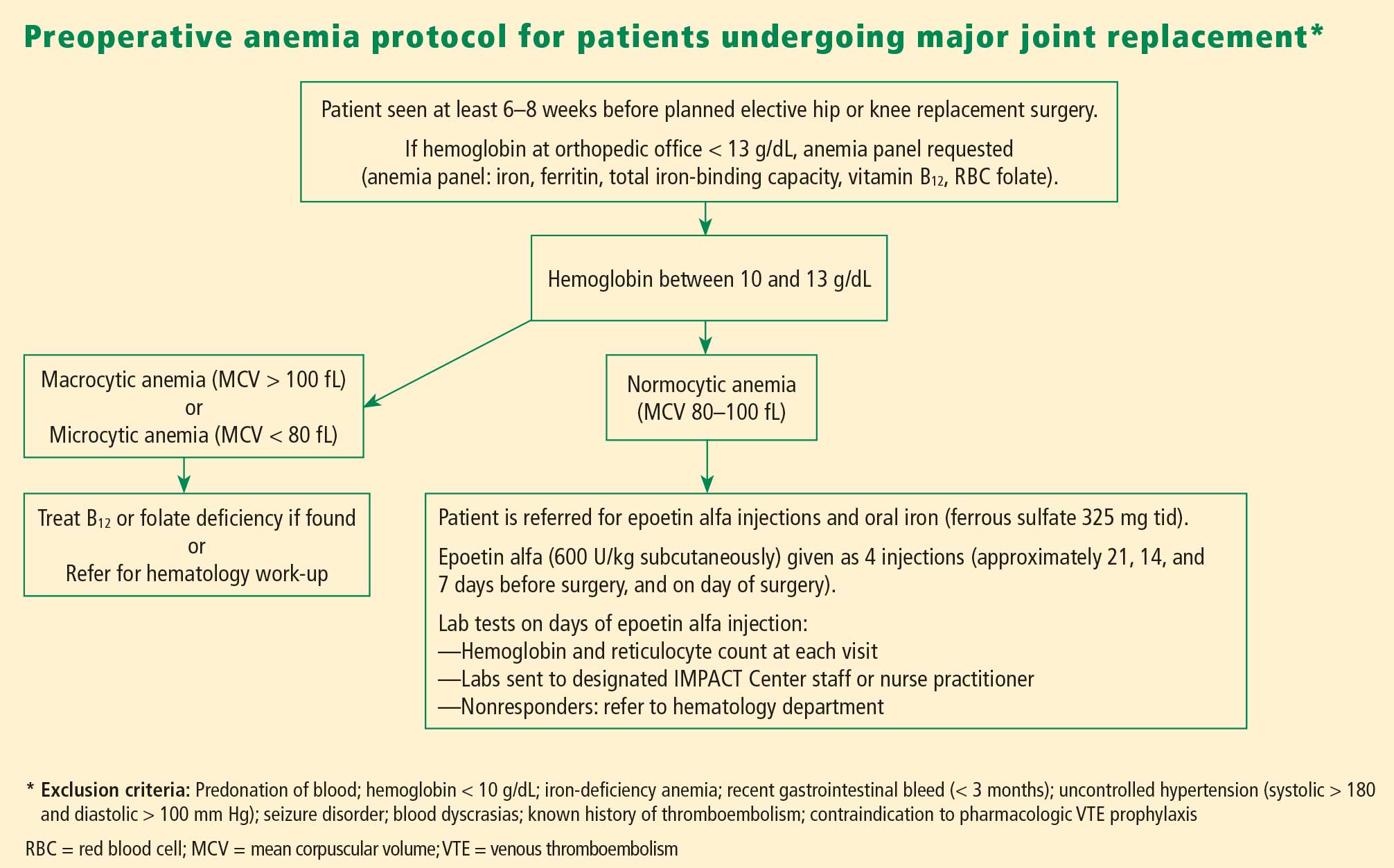
In the first 7 months of the program, we observed decreased utilization of blood products in the inpatient setting. Notably, the reduction in blood use was significantly greater in the surgical population than in the medical population.
CONCULSIONS
Anemia is associated with increased morbidity and mortality in the perioperative setting. Perioperative blood transfusion is one method of raising hemoglobin levels in anemic surgical patients, but it increases perioperative morbidity in the form of acute transfusion reactions, immunosuppression, postoperative infection, and longer hospital stays. Moreover, blood collection continues to lag blood demand. For these reasons, most relevant major medical organizations—including the Association of Blood Banks, the American Red Cross, and the FDA—advise that red blood cell–containing components should not be used to treat anemias that can be corrected with medications. These medical alternatives—all of which can be used in the perioperative setting—include iron supplementation, vitamin B12, and ESAs in select patient groups.
DISCUSSION
Question from the audience: Are there risks involved with autologous blood donation? Are different hemoglobin thresholds used when a patient’s own blood is used?
Dr. Kumar: As I mentioned, preoperative autologous donation is a technique that is less frequently used in our hospital. Autologous transfusion is considered safe only for patients who come to the clinic with normal hemoglobin values. Some patients may not have recovered from their blood loss by the time they come to surgery, so you end up needing to give them more blood because they started out anemic.
Question from the audience: Is there risk to giving patients back their own blood? Do you have to worry about transfusion-induced lung injury, sepsis, or other complications?
Dr. Kumar: As with allogeneic blood, the risk of clerical or clinical error exists with autologous blood: it too needs to be kept on the shelf, taken out, and infused, and the risk of sepsis remains the same.
- Shander A, Knight K, Thurer R, Adamson J, Spence R. Prevalence and outcomes of anemia in surgery: a systematic review of the literature. Am J Med 2004; 116(suppl 7A):58S–69S.
- Kulier A, Gombotz H. Perioperative anemia [in German]. Anaesthesist 2001; 50:73–86.
- Herzog CA, Muster HA, Li S, Collins AJ. Impact of congestive heart failure, chronic kidney disease, and anemia on survival in the Medicare population. J Card Fail 2004; 10:467–472.
- Carson JL, Duff A, Poses RM, et al. Effect of anaemia and cardiovascular disease on surgical mortality and morbidity. Lancet 1996; 348:1055–1060.
- Gruson KI, Aharonoff GB, Egol KA, Zuckerman JD, Koval KJ. The relationship between admission hemoglobin level and outcome after hip fracture. J Orthop Trauma 2002; 16:39–44.
- Nelson AH, Fleisher LA, Rosenbaum SH. Relationship between postoperative anemia and cardiac morbidity in high-risk vascular patients in the intensive care unit. Crit Care Med 1993; 21:860–866.
- Faris PM, Spence RK, Larholt KM, Sampson AR, Frei D. The predictive power of baseline hemoglobin for transfusion risk in surgery patients. Orthopedics 1999; 22(suppl 1):s135–s140.
- Marcantonio ER, Goldman L, Orav EJ, Cook EF, Lee TH. The association of intraoperative factors with the development of postoperative delirium. Am J Med 1998; 105:380–384.
- US Department of Health and Human Services. The 2007 National Blood Collection and Utilization Survey. Available at: http://www.aabb.org/Documents/Programs_and_Services/Data_Center/07nbcusrpt.pdf. Accessed March 9, 2009.
- The Joint Commission. Performance measurement initiatives. Available at http://www.jointcommission.org/PerformanceMeasurement/PerformanceMeasurement/Blood+Management+-+Utilization.htm.
- Busch MP, Kleinman SH, Nemo GJ. Current and emerging infectious risks of blood transfusions. JAMA 2003; 289:959–962.
- Silliman CC, Ambruso DR, Boshkov LK. Transfusion-related acute lung injury. Blood 2005; 105:2266–2273.
- Benjamin RJ. Transfusion and transplantation safety: American Red Cross concerns. PowerPoint slides presented at Department of Health and Human Services Advisory Committee on Blood Safety and Availability meeting, May 10–11, 2007. Available at: http://www.hhs.gov/ophs/bloodsafety/presentations/Benjamin0507.pdf. Accessed March 9, 2009.
- Holness L. Transfusion related acute lung injury (TRALI): the FDA current view. PowerPoint slides presented at FDA Blood Products Advisory Committee meeting, July 22–23, 2004. Available at: http://www.fda.gov/OHRMS/DOCKETS/ac/04/briefing/2004-4057b1_01.pdf. Accessed March 9, 2009.
- Williams AE. Transfusion related acute lung injury (TRALI). PowerPoint slides presented at FDA Blood Products Advisory Committee meeting, April 27, 2007. Available at: http://www.fda.gov/ohrms/dockets/ac/07/slides/2007-4300S2-05.ppt. Accessed March 9, 2009.
- Stainsby D, Cohen H, Jones H, et al. Serious Hazards of Transfusion—Annual Report 2003. Published July 5, 2004. Available at: http://www.shotuk.org/SHOT%20Report%202003.pdf. Accessed April 29, 2009.
- Taylor C, Cohen H, Stainsby D, et al. Serious Hazards of Transfusion—Annual Report 2006. Published November 20, 2007. Available at: http://www.shotuk.org/SHOT_report_2006.pdf. Accessed April 29, 2009.
- Hébert PC, Wells G, Blajchman MA, et al. A multicenter, randomized, controlled clinical trial of transfusion requirements in critical care. Transfusion Requirements in Critical Care Investigators, Canadian Critical Care Trials Group. N Engl J Med 1999; 340:409–417.
- Rao SV, Jollis JG, Harrington RA, et al. Relationship of blood transfusion and clinical outcomes in patients with acute coronary syndromes. JAMA 2004; 292:1555–1562.
- Dunne JR, Malone D, Tracy JK, et al. Perioperative anemia: an independent risk factor for infection, mortality, and resource utilization in surgery. J Surg Res 2002; 102:237–244.
- Koch CG, Li L, Duncan AI, et al. Morbidity and mortality risk associated with red blood cell and blood-component transfusion in isolated coronary artery bypass grafting. Crit Care Med 2006; 34:1608–1616.
- Rady MY, Ryan T, Starr NJ. Perioperative determinants of morbidity and mortality in elderly patients undergoing cardiac surgery. Crit Care Med 1998; 26:225–235.
- Goodnough LT, Brecher ME, Kanter MH, AuBuchon JP. Transfusion medicine. First of two parts—blood transfusion. N Engl J Med 1999; 340:438–447.
- Koch CG, Li L, Sessler DI, et al. Duration of red-cell storage and complications after cardiac surgery. N Engl J Med 2008; 358:1229–1239.
- Goodnough LT, Shander A. Blood management. Arch Pathol Lab Med 2007; 131:695–701.
- Okuyama M, Ikeda K, Shibata T, et al. Preoperative iron supplementation and intraoperative transfusion during colorectal cancer surgery. Surg Today 2005; 35:36–40.
- Theusinger OM, Leyvraz PF, Schanz U, et al. Treatment of iron deficiency anemia in orthopedic surgery with intravenous iron: efficacy and limits: a prospective study. Anesthesiology 2007; 107:923–927.
- Hoen B, Paul-Dauphin A, Kessler M. Intravenous iron administration does not significantly increase the risk of bacteremia in chronic hemodialysis patients. Clin Nephrol 2002; 57:457–461.
- Beris P, Muñoz M, García-Erce JA, et al. Perioperative anaemia management: consensus statement on the role of intravenous iron. Br J Anaesth 2008; 100:599–604.
- Laupacis A, Fergusson D. Erythropoietin to minimize perioperative blood transfusion: a systematic review of randomized trials: The International Study of Perioperative Transfusion (ISPOT) Investigators. Transfus Med 1998; 8:309–317.
- Feagan BG, Wong CJ, Kirkley A, et al. Erythropoietin with iron supplementation to prevent allogeneic blood transfusion in total hip joint arthroplasty: a randomized, controlled trial. Ann Intern Med 2000; 133:845–854.
- Stowell CP, Chandler H, Jové M, et al. An open-label, randomized study to compare the safety and efficacy of perioperative epoetin alfa with preoperative autologous blood donation in total joint arthroplasty. Orthopedics 1999; 22(suppl 1):s105–s112.
- de Andrade JR, Jové M, Landon G, et al. Baseline hemoglobin as a predictor of risk of transfusion and response to Epoetin alfa in orthopedic surgery patients. Am J Orthop 1996; 25:533–542.
- Procrit [package insert]. Raritan, NJ: Ortho Biotech Products LP; 2009.
- Epogen [package insert]. Thousand Oaks, CA: Amgen Inc.; 2009.
- Smoller BR, Kuskall MS. Phlebotomy for diagnostic laboratory tests in adults: pattern of use and effect on transfusion requirements. N Engl J Med 1986; 314:1233–1235.
Anemia is a potent risk factor for mortality and morbidity in surgical patients, and its management has begun to shift away from allogeneic blood transfusion in recent years. This article reviews the clinical importance of perioperative anemia, the role and shortcomings of blood transfusion, and the pros and cons of alternative approaches to managing perioperative anemia. I conclude with an overview of a program for perioperative blood product use at my institution, Cleveland Clinic.
SIGNIFICANCE OF PERIOPERATIVE ANEMIA
Prevalence depends on many factors
The reported prevalence of anemia in surgical patients varies widely—from 5% to 76%1—and depends on the patient’s disease and comorbidities, the surgical procedure and associated blood loss, and the definition of anemia used. The prevalence of preoperative anemia increases with patient age and is higher in women than in men.2
A multiplier of risk
Anemia is an important multiplier of mortality risk. For example, the presence of anemia raises the relative risk of 2-year mortality from 2.05 to 3.37 in patients with chronic kidney disease, from 2.86 to 3.78 in patients with heart failure, and from 4.86 to 6.07 in patients with concomitant heart failure and chronic kidney disease.3
Adverse effects of anemia have been demonstrated specifically in the perioperative setting as well. A large retrospective cohort study showed that a preoperative hemoglobin concentration of less than 6 g/dL increases the risk of death 30 days after surgery by a factor of 26 relative to a concentration of 12 g/dL or greater in surgical patients who declined blood transfusion for religious reasons.4 The anemia-associated mortality risk was especially pronounced among patients with cardiovascular disease.4 Other studies have demonstrated perioperative anemia to be associated with increases in the risk of death,5 cardiac events,6 pneumonia,7 and postoperative delirium.8
IS BLOOD TRANSFUSION THE ANSWER
The use of allogeneic blood transfusion to manage anemia and blood loss is a concept that originated several centuries ago and has changed little over the years.
Blood supply challenges
Blood collection has historically lagged demand, resulting in a blood supply insufficient to meet transfusion needs. According to the federal government’s 2007 National Blood Collection and Utilization Survey Report, 6.89% of US hospitals reported that they cancelled elective surgery on 1 or more days in the prior year because of a lack of blood availability, and 13.5% experienced at least 1 day in which nonsurgical blood needs could not be met.9 Unless practices are changed to increase blood donation, these unmet tranfusion needs may grow.
Joint Commission set to measure blood management
In response to this challenge, an advisory panel formed by the Joint Commission has identified 17 performance measures related to blood conservation and appropriate transfusion.10 These measures are currently in development, and we expect to see some types of metrics in the near future. Such metrics are likely to further prioritize blood management for US hospitals.
Safety of the blood supply: Viral transmission down, TRALI risk persists
The safety of the blood supply has improved markedly. Sophisticated testing and public demand have led to a dramatic decline in the risk of transfusion-related transmission of HIV, hepatitis C virus, and hepatitis B virus.11
Despite this progress, the risk of transfusion-related acute lung injury (TRALI) has persisted in recent years. TRALI is characterized by acute onset of noncardiogenic pulmonary edema within 6 hours of blood product transfusion. Believed to be immune-mediated, TRALI is thought to occur as antibodies to human leukocyte antigens develop, inducing capillary leak syndrome.12 The patients most commonly affected are those who receive plasma from multiparous female donors. A recent evaluation of transfusion-related fatalities reported to the US Food and Drug Administration (FDA) revealed a continual rise in fatal TRALI cases in the United States from 2001 to 2006.13–15 TRALI was implicated in more than half of all transfusion-related fatalities reported to the FDA in 2006, a higher number than for any other single cause.13
At the same time, there is evidence that hemovigilance can reduce TRALI risk. In the United Kingdom, the Serious Hazards of Transfusion Steering Group introduced in late 2003 a policy of using plasma from male donors as much as possible, in view of the association of TRALI with plasma from multiparous female donors. The effort appeared to pay off: whereas TRALI accounted for 6.8% of all transfusion-related adverse events reported in the United Kingdom during the period 1996–2003,16 this proportion declined to just 1.9% in 2006.17
Finally, despite the progress in screening blood for more established infections like HIV and the hepatitis viruses, some additional infections now must be considered when assessing blood supply safety. These include diseases newly recognized as being transmissible by blood, or for which blood donor screening is not currently available, or that are newly emergent infections for which the potential for spread by transfusion is unknown. For such diseases—which include malaria and West Nile virus—the risk of transmission through transfusion is low, as they are much more likely to be acquired by other means.
Transfusion and outcomes: Not a strong record
Transfusion has never undergone safety and efficacy evaluation by the FDA. Given the challenges of conducting a randomized study of transfusion in the perioperative setting, we may never have high-quality data to assess transfusion in this setting.
A few studies merit mention, however. The Transfusion Requirement in Critical Care (TRICC) trial was conducted in 838 critically ill patients in the intensive care setting.18 Patients were randomized to a strategy of either liberal transfusion (begun when hemoglobin fell below 10 g/dL) or restrictive transfusion (begun when hemoglobin fell below 7 g/dL). Thirty-day mortality was similar between patients in the two strategy groups, but the restrictive strategy was associated with significantly lower mortality in at least two subgroups: patients with myocardial infarction and patients with pulmonary edema. Further subgroup analysis found no benefit of early or aggressive transfusion in patients with coronary artery disease or in those requiring mechanical ventilation.
Rao et al performed a meta-analysis of three large international trials of patients with acute coronary syndromes to determine whether blood transfusion to correct anemia in this setting was associated with improved survival.19 They found significantly higher mortality among patients who underwent transfusion compared with those who did not, prompting them to urge caution in the use of transfusion to maintain arbitrary hematocrit levels in stable patients with ischemic heart disease.
Similarly, a risk-adjusted, propensity-matched analysis of 6,301 patients undergoing noncardiac surgery found that receipt of 4 U of blood or more was a predictor of greater mortality, higher risk of infection, and longer hospital stay.20 Moreover, in an observational cohort study of 11,963 patients who underwent isolated coronary artery bypass graft surgery, each unit of red blood cells transfused was associated with an incrementally increased risk of adverse outcome (eg, mortality, renal injury, need for ventilator support, lengthened hospital stay, infection).21 The latter study found that transfusion was the single factor most reliably associated with increased risk of postoperative morbidity.
Additional studies have echoed these findings—ie, that perioperative blood transfusion has been associated with a host of adverse outcomes, including increased morbidity and length of stay, increased rates of postoperative infection, as well as immunosuppression, viral transmission, and acute transfusion reactions.5,22,23
Outcomes and duration of blood storage
An interesting factor in the relation between transfusion and outcomes is the shelf life of the blood being transfused. The FDA currently allows storage of blood for a maximum of 42 days, but a recent study of patients who received red blood cell transfusions during cardiac surgery found that those who received “older blood” (stored for > 14 days) had significantly higher rates of sepsis, prolonged intubation, renal failure, in-hospital mortality, and 1-year mortality compared with those who received “newer blood” (stored for ≤ 14 days).24
These differing outcomes are generally attributed to the so-called storage defect: as blood gets older, it loses components such as 2,3-DPG and adenosine disphosphate, its red cells lose deformability, and it undergoes buildup of cytokines and free hemoglobin. Increased demand for newer blood in light of the storage defect could further intensify pressures on the blood supply.
MANAGEMENT OF PERIOPERATIVE ANEMIA
In light of these shortcomings of blood transfusion, how should anemia be managed perioperatively to reduce or avoid the need for transfusion?
Preoperative evaluation
Vigilance for anemia and related issues in the preoperative evaluation is fundamental. The evaluation should elicit a history of bleeding tendencies, previous transfusions, and symptoms of anemia. Medications should be reviewed with an eye toward any that may predispose to perioperative bleeding and anemia, such as aspirin, clopidogrel, and anticoagulants. During the physical examination, alertness for pallor and petechiae is key, as is attentiveness to symptoms of anemia such as shortness of breath and fatigue.
Overview of management options
Once the cause of anemia is identified, the choice for optimal medical management can be made. Choices broadly consist of pharmacologic and technological options. The former include iron supplements and erythropoiesis-stimulating agents. Among other pharmacologic options are thrombin, collagen, fibrin glue, tranexamic acid, and aminocaproic acid, but these agents are less well studied and will not be discussed here. Technological options include preoperative autologous blood donation, cell salvage, and acute normovolemic hemodilution.
In addition to these options, careful management of anticoagulant and antiplatelet medications should be provided, including discontinuation or substitution of drugs that could hamper clotting perioperatively.
PHARMACOLOGIC OPTIONS
Iron supplementation
Oral iron is available in four preparations: ferrous sulfate, ferrous gluconate, ferrous fumarate, and iron polysaccharide. Gastrointestinal side effects may limit these preparations’ tolerability. Iron supplements with a high elemental value will require fewer pills and fewer doses, reducing the risk or frequency of side effects.
Intravenous (IV) iron preparations are much safer now than they were years ago, when anaphylactic reactions were a concern. The ones generally used in the perioperative setting are iron sucrose and iron gluconate. Unlike the older IV preparations, the use of iron sucrose and iron gluconate often requires a second dose. The effect on hemoglobin levels usually occurs starting at 1 week, with the maximum effect achieved at 2 weeks. Hypotension, arthralgia, abdominal discomfort, and back pain are potential side effects of IV iron.
Efficacy and safety of iron supplementation. Evidence of the efficacy of preoperative iron supplementation is mounting. A study of 569 patients undergoing colorectal cancer surgery found that among the 116 patients who were anemic, intraoperative transfusion was needed in a significantly lower proportion of those who received 2 weeks of preoperative oral iron supplementation (200 mg) compared with those who received no iron therapy (9.4% vs 27.4%; P < .05).26 Similarly, in an uncontrolled study, 10 days of IV iron sucrose starting 4 weeks preoperatively significantly increased hemoglobin levels in 20 patients with iron-deficiency anemia prior to elective orthopedic surgery.27
Risks of infection and cancer progression have been concerns with IV iron therapy. However, no significant association between IV iron therapy and bacteremia was identified in a prospective study of 985 patients receiving chronic hemodialysis.28 The effect of IV iron administration on tumor progression has not been prospectively studied.
In general, IV iron, especially the newer forms, is a safer alternative to blood transfusion. Death occurs at a much lower rate with iron than with blood transfusion (0.4 per million vs 4 per million, respectively), as do life-threatening adverse events (4 per million vs 10 per million, respectively), according to a systematic review by the Network for Advancement of Transfusion Alternatives.29
Erythropoiesis-stimulating agents
Erythropoiesis-stimulating agents (ESAs) include epoetin alfa (erythropoietin), first approved by the FDA in 1989, and the more recently introduced darbepoetin alfa. They are approved to treat anemia in several patient populations, but only epoetin alfa is approved by the FDA explicitly for use in patients undergoing major surgery (to reduce the need for blood transfusions). The ESAs have come under intense scrutiny in recent years over their risk-to-benefit ratio, as detailed below.
The preoperative dosing schedule for epoetin alfa is usually three weekly doses (plus a fourth dose on the day of surgery) if the surgery is scheduled 3 or more weeks in advance. However, daily dosing can be used effectively if the preoperative period is less than 3 weeks, provided that it is continued until 4 days after surgery. Oral iron is necessary throughout the course of epoetin alfa therapy.
Efficacy in reducing transfusions. In a systematic review published in 1998, epoetin alfa was shown to minimize perioperative exposure to allogeneic blood transfusion in patients undergoing orthopedic or cardiac surgery.30 Its benefit was greatest in patients at the highest risk of requiring transfusion. It was effective whether given daily or weekly, and did not significantly increase the risk of thrombotic events when used in surgical patients, although some studies did find an excess of thrombotic events with its use.
In three randomized trials conducted in patients undergoing joint arthroplasty (hip or knee), epoetin alfa was associated with substantial and significant reductions in perioperative blood transfusion compared with placebo or preoperative autologous blood donation.31–33 Rates of deep vein thrombosis (DVT) did not differ significantly between the epoetin alfa and placebo groups.
Concerns over perioperative thromboembolic risk. In early 2007, the FDA was made aware of preliminary results of an open-label study in which 681 patients undergoing elective spinal surgery who did not receive prophylactic anticoagulation were randomized to epoetin alfa plus standard-of-care therapy (pneumatic compression) or standard-of-care therapy alone.34,35 The incidence of DVT was 4.7% in patients treated with epoetin alfa compared with 2.1% in those not receiving epoetin alfa. It is important to note that the available ESAs are prothrombotic and increase thrombotic risk significantly, especially in populations like this one in which pharmacologic DVT prophylaxis is not routinely used.
Based in part on this study, the FDA in 2007 required a boxed warning to be added to the ESAs’ package inserts to specify the increased risk of DVT with their use in surgical patients not receiving prophylactic anticoagulation. The warning urges consideration of the use of DVT prophylaxis in surgical patients receiving an ESA.34,35
TECHNOLOGICAL OPTIONS AND OTHER STRATEGIES
Autologous blood donation: A practice in decline
In cases of elective surgery, autologous blood donation can be used to protect against disease transmission and overcome the challenge of blood type compatibility. Preoperative autologous donation of blood has been a prevalent practice, but its use is declining. One reason is that waste is high (approximately 50% at Cleveland Clinic), which makes this practice more costly than is often realized. Also, autologous blood donation increases the likelihood that the patient will be anemic on the day of surgery, so that he or she may still need allogeneic blood after all, defeating the initial purpose. Despite these limitations, preoperative autologous blood donation remains a useful option for a subset of patients with multiple antibodies for whom donor blood may be difficult to obtain.
Cell salvage
Cell salvage is an innovative technology that recovers the patient’s own blood (after being shed from the surgical incision) for transfusion after filtering and washing. It is particularly well suited to procedures that involve massive blood loss. Cell savage requires technical expertise, however, and involves costs associated with both the machine and disposables.
Restricted postoperative phlebotomy
Phlebotomy accounts for a significant amount of blood loss, especially in intensive care patients with arterial lines. The equivalent of 30% of total blood transfused has been reported to be lost to phlebotomy during an intensive care unit stay.36 Triggers for transfusion cannot be assigned universally based on blood loss from phlebotomy but must consider the patient’s hemodynamic status, cardiac reserve, and other clinical characteristics.
PROMOTING RESPONSIBLE BLOOD PRODUCT USE
Blood is expensive, and in recent years hospitals have experienced increases in the cost of blood and blood products. To promote responsible blood use, we have developed a multipronged approach to blood management at Cleveland Clinic. The program’s cornerstone is increased awareness of the risks associated with blood transfusions. The emphasis is on educating staff physicians and other caregivers about the appropriate use of blood products. We also have implemented a new policy requiring staff authorization for all blood requested in nonemergency situations. Additionally, requests for blood components require adherence to an indication-based ordering process. Finally, data about blood use are shared transparently among physicians, encouraging good clinical practice.
In the first 7 months of the program, we observed decreased utilization of blood products in the inpatient setting. Notably, the reduction in blood use was significantly greater in the surgical population than in the medical population.
CONCULSIONS
Anemia is associated with increased morbidity and mortality in the perioperative setting. Perioperative blood transfusion is one method of raising hemoglobin levels in anemic surgical patients, but it increases perioperative morbidity in the form of acute transfusion reactions, immunosuppression, postoperative infection, and longer hospital stays. Moreover, blood collection continues to lag blood demand. For these reasons, most relevant major medical organizations—including the Association of Blood Banks, the American Red Cross, and the FDA—advise that red blood cell–containing components should not be used to treat anemias that can be corrected with medications. These medical alternatives—all of which can be used in the perioperative setting—include iron supplementation, vitamin B12, and ESAs in select patient groups.
DISCUSSION
Question from the audience: Are there risks involved with autologous blood donation? Are different hemoglobin thresholds used when a patient’s own blood is used?
Dr. Kumar: As I mentioned, preoperative autologous donation is a technique that is less frequently used in our hospital. Autologous transfusion is considered safe only for patients who come to the clinic with normal hemoglobin values. Some patients may not have recovered from their blood loss by the time they come to surgery, so you end up needing to give them more blood because they started out anemic.
Question from the audience: Is there risk to giving patients back their own blood? Do you have to worry about transfusion-induced lung injury, sepsis, or other complications?
Dr. Kumar: As with allogeneic blood, the risk of clerical or clinical error exists with autologous blood: it too needs to be kept on the shelf, taken out, and infused, and the risk of sepsis remains the same.
- Shander A, Knight K, Thurer R, Adamson J, Spence R. Prevalence and outcomes of anemia in surgery: a systematic review of the literature. Am J Med 2004; 116(suppl 7A):58S–69S.
- Kulier A, Gombotz H. Perioperative anemia [in German]. Anaesthesist 2001; 50:73–86.
- Herzog CA, Muster HA, Li S, Collins AJ. Impact of congestive heart failure, chronic kidney disease, and anemia on survival in the Medicare population. J Card Fail 2004; 10:467–472.
- Carson JL, Duff A, Poses RM, et al. Effect of anaemia and cardiovascular disease on surgical mortality and morbidity. Lancet 1996; 348:1055–1060.
- Gruson KI, Aharonoff GB, Egol KA, Zuckerman JD, Koval KJ. The relationship between admission hemoglobin level and outcome after hip fracture. J Orthop Trauma 2002; 16:39–44.
- Nelson AH, Fleisher LA, Rosenbaum SH. Relationship between postoperative anemia and cardiac morbidity in high-risk vascular patients in the intensive care unit. Crit Care Med 1993; 21:860–866.
- Faris PM, Spence RK, Larholt KM, Sampson AR, Frei D. The predictive power of baseline hemoglobin for transfusion risk in surgery patients. Orthopedics 1999; 22(suppl 1):s135–s140.
- Marcantonio ER, Goldman L, Orav EJ, Cook EF, Lee TH. The association of intraoperative factors with the development of postoperative delirium. Am J Med 1998; 105:380–384.
- US Department of Health and Human Services. The 2007 National Blood Collection and Utilization Survey. Available at: http://www.aabb.org/Documents/Programs_and_Services/Data_Center/07nbcusrpt.pdf. Accessed March 9, 2009.
- The Joint Commission. Performance measurement initiatives. Available at http://www.jointcommission.org/PerformanceMeasurement/PerformanceMeasurement/Blood+Management+-+Utilization.htm.
- Busch MP, Kleinman SH, Nemo GJ. Current and emerging infectious risks of blood transfusions. JAMA 2003; 289:959–962.
- Silliman CC, Ambruso DR, Boshkov LK. Transfusion-related acute lung injury. Blood 2005; 105:2266–2273.
- Benjamin RJ. Transfusion and transplantation safety: American Red Cross concerns. PowerPoint slides presented at Department of Health and Human Services Advisory Committee on Blood Safety and Availability meeting, May 10–11, 2007. Available at: http://www.hhs.gov/ophs/bloodsafety/presentations/Benjamin0507.pdf. Accessed March 9, 2009.
- Holness L. Transfusion related acute lung injury (TRALI): the FDA current view. PowerPoint slides presented at FDA Blood Products Advisory Committee meeting, July 22–23, 2004. Available at: http://www.fda.gov/OHRMS/DOCKETS/ac/04/briefing/2004-4057b1_01.pdf. Accessed March 9, 2009.
- Williams AE. Transfusion related acute lung injury (TRALI). PowerPoint slides presented at FDA Blood Products Advisory Committee meeting, April 27, 2007. Available at: http://www.fda.gov/ohrms/dockets/ac/07/slides/2007-4300S2-05.ppt. Accessed March 9, 2009.
- Stainsby D, Cohen H, Jones H, et al. Serious Hazards of Transfusion—Annual Report 2003. Published July 5, 2004. Available at: http://www.shotuk.org/SHOT%20Report%202003.pdf. Accessed April 29, 2009.
- Taylor C, Cohen H, Stainsby D, et al. Serious Hazards of Transfusion—Annual Report 2006. Published November 20, 2007. Available at: http://www.shotuk.org/SHOT_report_2006.pdf. Accessed April 29, 2009.
- Hébert PC, Wells G, Blajchman MA, et al. A multicenter, randomized, controlled clinical trial of transfusion requirements in critical care. Transfusion Requirements in Critical Care Investigators, Canadian Critical Care Trials Group. N Engl J Med 1999; 340:409–417.
- Rao SV, Jollis JG, Harrington RA, et al. Relationship of blood transfusion and clinical outcomes in patients with acute coronary syndromes. JAMA 2004; 292:1555–1562.
- Dunne JR, Malone D, Tracy JK, et al. Perioperative anemia: an independent risk factor for infection, mortality, and resource utilization in surgery. J Surg Res 2002; 102:237–244.
- Koch CG, Li L, Duncan AI, et al. Morbidity and mortality risk associated with red blood cell and blood-component transfusion in isolated coronary artery bypass grafting. Crit Care Med 2006; 34:1608–1616.
- Rady MY, Ryan T, Starr NJ. Perioperative determinants of morbidity and mortality in elderly patients undergoing cardiac surgery. Crit Care Med 1998; 26:225–235.
- Goodnough LT, Brecher ME, Kanter MH, AuBuchon JP. Transfusion medicine. First of two parts—blood transfusion. N Engl J Med 1999; 340:438–447.
- Koch CG, Li L, Sessler DI, et al. Duration of red-cell storage and complications after cardiac surgery. N Engl J Med 2008; 358:1229–1239.
- Goodnough LT, Shander A. Blood management. Arch Pathol Lab Med 2007; 131:695–701.
- Okuyama M, Ikeda K, Shibata T, et al. Preoperative iron supplementation and intraoperative transfusion during colorectal cancer surgery. Surg Today 2005; 35:36–40.
- Theusinger OM, Leyvraz PF, Schanz U, et al. Treatment of iron deficiency anemia in orthopedic surgery with intravenous iron: efficacy and limits: a prospective study. Anesthesiology 2007; 107:923–927.
- Hoen B, Paul-Dauphin A, Kessler M. Intravenous iron administration does not significantly increase the risk of bacteremia in chronic hemodialysis patients. Clin Nephrol 2002; 57:457–461.
- Beris P, Muñoz M, García-Erce JA, et al. Perioperative anaemia management: consensus statement on the role of intravenous iron. Br J Anaesth 2008; 100:599–604.
- Laupacis A, Fergusson D. Erythropoietin to minimize perioperative blood transfusion: a systematic review of randomized trials: The International Study of Perioperative Transfusion (ISPOT) Investigators. Transfus Med 1998; 8:309–317.
- Feagan BG, Wong CJ, Kirkley A, et al. Erythropoietin with iron supplementation to prevent allogeneic blood transfusion in total hip joint arthroplasty: a randomized, controlled trial. Ann Intern Med 2000; 133:845–854.
- Stowell CP, Chandler H, Jové M, et al. An open-label, randomized study to compare the safety and efficacy of perioperative epoetin alfa with preoperative autologous blood donation in total joint arthroplasty. Orthopedics 1999; 22(suppl 1):s105–s112.
- de Andrade JR, Jové M, Landon G, et al. Baseline hemoglobin as a predictor of risk of transfusion and response to Epoetin alfa in orthopedic surgery patients. Am J Orthop 1996; 25:533–542.
- Procrit [package insert]. Raritan, NJ: Ortho Biotech Products LP; 2009.
- Epogen [package insert]. Thousand Oaks, CA: Amgen Inc.; 2009.
- Smoller BR, Kuskall MS. Phlebotomy for diagnostic laboratory tests in adults: pattern of use and effect on transfusion requirements. N Engl J Med 1986; 314:1233–1235.
KEY POINTS
- Anemia is a potent multiplier of morbidity and mortality risk, including in the perioperative setting.
- The Joint Commission plans to implement a performance measure on blood management in the near future.
- While the safety of the blood supply has improved markedly from the standpoint of infection transmission, other risks from transfusion persist, including transfusion-related acute lung injury and emerging infections.
- The preoperative evaluation should elicit a history of bleeding tendencies, previous transfusions, and symptoms of anemia. Medications should be reviewed with an eye toward those that may need to be stopped to avoid a predisposition to bleeding (eg, antiplatelets, anticoagulants).
- Use of ESAs minimizes the need for blood transfusion in patients undergoing orthopedic and other surgeries, but they raise the risk of thromboembolism in the absence of prophylactic anticoagulation.