User login
CCJM delivers practical clinical articles relevant to internists, cardiologists, endocrinologists, and other specialists, all written by known experts.
Copyright © 2019 Cleveland Clinic. All rights reserved. The information provided is for educational purposes only. Use of this website is subject to the disclaimer and privacy policy.
gambling
compulsive behaviors
ammunition
assault rifle
black jack
Boko Haram
bondage
child abuse
cocaine
Daech
drug paraphernalia
explosion
gun
human trafficking
ISIL
ISIS
Islamic caliphate
Islamic state
mixed martial arts
MMA
molestation
national rifle association
NRA
nsfw
pedophile
pedophilia
poker
porn
pornography
psychedelic drug
recreational drug
sex slave rings
slot machine
terrorism
terrorist
Texas hold 'em
UFC
substance abuse
abuseed
abuseer
abusees
abuseing
abusely
abuses
aeolus
aeolused
aeoluser
aeoluses
aeolusing
aeolusly
aeoluss
ahole
aholeed
aholeer
aholees
aholeing
aholely
aholes
alcohol
alcoholed
alcoholer
alcoholes
alcoholing
alcoholly
alcohols
allman
allmaned
allmaner
allmanes
allmaning
allmanly
allmans
alted
altes
alting
altly
alts
analed
analer
anales
analing
anally
analprobe
analprobeed
analprobeer
analprobees
analprobeing
analprobely
analprobes
anals
anilingus
anilingused
anilinguser
anilinguses
anilingusing
anilingusly
anilinguss
anus
anused
anuser
anuses
anusing
anusly
anuss
areola
areolaed
areolaer
areolaes
areolaing
areolaly
areolas
areole
areoleed
areoleer
areolees
areoleing
areolely
areoles
arian
arianed
arianer
arianes
arianing
arianly
arians
aryan
aryaned
aryaner
aryanes
aryaning
aryanly
aryans
asiaed
asiaer
asiaes
asiaing
asialy
asias
ass
ass hole
ass lick
ass licked
ass licker
ass lickes
ass licking
ass lickly
ass licks
assbang
assbanged
assbangeded
assbangeder
assbangedes
assbangeding
assbangedly
assbangeds
assbanger
assbanges
assbanging
assbangly
assbangs
assbangsed
assbangser
assbangses
assbangsing
assbangsly
assbangss
assed
asser
asses
assesed
asseser
asseses
assesing
assesly
assess
assfuck
assfucked
assfucker
assfuckered
assfuckerer
assfuckeres
assfuckering
assfuckerly
assfuckers
assfuckes
assfucking
assfuckly
assfucks
asshat
asshated
asshater
asshates
asshating
asshatly
asshats
assholeed
assholeer
assholees
assholeing
assholely
assholes
assholesed
assholeser
assholeses
assholesing
assholesly
assholess
assing
assly
assmaster
assmastered
assmasterer
assmasteres
assmastering
assmasterly
assmasters
assmunch
assmunched
assmuncher
assmunches
assmunching
assmunchly
assmunchs
asss
asswipe
asswipeed
asswipeer
asswipees
asswipeing
asswipely
asswipes
asswipesed
asswipeser
asswipeses
asswipesing
asswipesly
asswipess
azz
azzed
azzer
azzes
azzing
azzly
azzs
babeed
babeer
babees
babeing
babely
babes
babesed
babeser
babeses
babesing
babesly
babess
ballsac
ballsaced
ballsacer
ballsaces
ballsacing
ballsack
ballsacked
ballsacker
ballsackes
ballsacking
ballsackly
ballsacks
ballsacly
ballsacs
ballsed
ballser
ballses
ballsing
ballsly
ballss
barf
barfed
barfer
barfes
barfing
barfly
barfs
bastard
bastarded
bastarder
bastardes
bastarding
bastardly
bastards
bastardsed
bastardser
bastardses
bastardsing
bastardsly
bastardss
bawdy
bawdyed
bawdyer
bawdyes
bawdying
bawdyly
bawdys
beaner
beanered
beanerer
beaneres
beanering
beanerly
beaners
beardedclam
beardedclamed
beardedclamer
beardedclames
beardedclaming
beardedclamly
beardedclams
beastiality
beastialityed
beastialityer
beastialityes
beastialitying
beastialityly
beastialitys
beatch
beatched
beatcher
beatches
beatching
beatchly
beatchs
beater
beatered
beaterer
beateres
beatering
beaterly
beaters
beered
beerer
beeres
beering
beerly
beeyotch
beeyotched
beeyotcher
beeyotches
beeyotching
beeyotchly
beeyotchs
beotch
beotched
beotcher
beotches
beotching
beotchly
beotchs
biatch
biatched
biatcher
biatches
biatching
biatchly
biatchs
big tits
big titsed
big titser
big titses
big titsing
big titsly
big titss
bigtits
bigtitsed
bigtitser
bigtitses
bigtitsing
bigtitsly
bigtitss
bimbo
bimboed
bimboer
bimboes
bimboing
bimboly
bimbos
bisexualed
bisexualer
bisexuales
bisexualing
bisexually
bisexuals
bitch
bitched
bitcheded
bitcheder
bitchedes
bitcheding
bitchedly
bitcheds
bitcher
bitches
bitchesed
bitcheser
bitcheses
bitchesing
bitchesly
bitchess
bitching
bitchly
bitchs
bitchy
bitchyed
bitchyer
bitchyes
bitchying
bitchyly
bitchys
bleached
bleacher
bleaches
bleaching
bleachly
bleachs
blow job
blow jobed
blow jober
blow jobes
blow jobing
blow jobly
blow jobs
blowed
blower
blowes
blowing
blowjob
blowjobed
blowjober
blowjobes
blowjobing
blowjobly
blowjobs
blowjobsed
blowjobser
blowjobses
blowjobsing
blowjobsly
blowjobss
blowly
blows
boink
boinked
boinker
boinkes
boinking
boinkly
boinks
bollock
bollocked
bollocker
bollockes
bollocking
bollockly
bollocks
bollocksed
bollockser
bollockses
bollocksing
bollocksly
bollockss
bollok
bolloked
bolloker
bollokes
bolloking
bollokly
bolloks
boner
bonered
bonerer
boneres
bonering
bonerly
boners
bonersed
bonerser
bonerses
bonersing
bonersly
bonerss
bong
bonged
bonger
bonges
bonging
bongly
bongs
boob
boobed
boober
boobes
boobies
boobiesed
boobieser
boobieses
boobiesing
boobiesly
boobiess
boobing
boobly
boobs
boobsed
boobser
boobses
boobsing
boobsly
boobss
booby
boobyed
boobyer
boobyes
boobying
boobyly
boobys
booger
boogered
boogerer
boogeres
boogering
boogerly
boogers
bookie
bookieed
bookieer
bookiees
bookieing
bookiely
bookies
bootee
booteeed
booteeer
booteees
booteeing
booteely
bootees
bootie
bootieed
bootieer
bootiees
bootieing
bootiely
booties
booty
bootyed
bootyer
bootyes
bootying
bootyly
bootys
boozeed
boozeer
boozees
boozeing
boozely
boozer
boozered
boozerer
boozeres
boozering
boozerly
boozers
boozes
boozy
boozyed
boozyer
boozyes
boozying
boozyly
boozys
bosomed
bosomer
bosomes
bosoming
bosomly
bosoms
bosomy
bosomyed
bosomyer
bosomyes
bosomying
bosomyly
bosomys
bugger
buggered
buggerer
buggeres
buggering
buggerly
buggers
bukkake
bukkakeed
bukkakeer
bukkakees
bukkakeing
bukkakely
bukkakes
bull shit
bull shited
bull shiter
bull shites
bull shiting
bull shitly
bull shits
bullshit
bullshited
bullshiter
bullshites
bullshiting
bullshitly
bullshits
bullshitsed
bullshitser
bullshitses
bullshitsing
bullshitsly
bullshitss
bullshitted
bullshitteded
bullshitteder
bullshittedes
bullshitteding
bullshittedly
bullshitteds
bullturds
bullturdsed
bullturdser
bullturdses
bullturdsing
bullturdsly
bullturdss
bung
bunged
bunger
bunges
bunging
bungly
bungs
busty
bustyed
bustyer
bustyes
bustying
bustyly
bustys
butt
butt fuck
butt fucked
butt fucker
butt fuckes
butt fucking
butt fuckly
butt fucks
butted
buttes
buttfuck
buttfucked
buttfucker
buttfuckered
buttfuckerer
buttfuckeres
buttfuckering
buttfuckerly
buttfuckers
buttfuckes
buttfucking
buttfuckly
buttfucks
butting
buttly
buttplug
buttpluged
buttpluger
buttpluges
buttpluging
buttplugly
buttplugs
butts
caca
cacaed
cacaer
cacaes
cacaing
cacaly
cacas
cahone
cahoneed
cahoneer
cahonees
cahoneing
cahonely
cahones
cameltoe
cameltoeed
cameltoeer
cameltoees
cameltoeing
cameltoely
cameltoes
carpetmuncher
carpetmunchered
carpetmuncherer
carpetmuncheres
carpetmunchering
carpetmuncherly
carpetmunchers
cawk
cawked
cawker
cawkes
cawking
cawkly
cawks
chinc
chinced
chincer
chinces
chincing
chincly
chincs
chincsed
chincser
chincses
chincsing
chincsly
chincss
chink
chinked
chinker
chinkes
chinking
chinkly
chinks
chode
chodeed
chodeer
chodees
chodeing
chodely
chodes
chodesed
chodeser
chodeses
chodesing
chodesly
chodess
clit
clited
cliter
clites
cliting
clitly
clitoris
clitorised
clitoriser
clitorises
clitorising
clitorisly
clitoriss
clitorus
clitorused
clitoruser
clitoruses
clitorusing
clitorusly
clitoruss
clits
clitsed
clitser
clitses
clitsing
clitsly
clitss
clitty
clittyed
clittyer
clittyes
clittying
clittyly
clittys
cocain
cocaine
cocained
cocaineed
cocaineer
cocainees
cocaineing
cocainely
cocainer
cocaines
cocaining
cocainly
cocains
cock
cock sucker
cock suckered
cock suckerer
cock suckeres
cock suckering
cock suckerly
cock suckers
cockblock
cockblocked
cockblocker
cockblockes
cockblocking
cockblockly
cockblocks
cocked
cocker
cockes
cockholster
cockholstered
cockholsterer
cockholsteres
cockholstering
cockholsterly
cockholsters
cocking
cockknocker
cockknockered
cockknockerer
cockknockeres
cockknockering
cockknockerly
cockknockers
cockly
cocks
cocksed
cockser
cockses
cocksing
cocksly
cocksmoker
cocksmokered
cocksmokerer
cocksmokeres
cocksmokering
cocksmokerly
cocksmokers
cockss
cocksucker
cocksuckered
cocksuckerer
cocksuckeres
cocksuckering
cocksuckerly
cocksuckers
coital
coitaled
coitaler
coitales
coitaling
coitally
coitals
commie
commieed
commieer
commiees
commieing
commiely
commies
condomed
condomer
condomes
condoming
condomly
condoms
coon
cooned
cooner
coones
cooning
coonly
coons
coonsed
coonser
coonses
coonsing
coonsly
coonss
corksucker
corksuckered
corksuckerer
corksuckeres
corksuckering
corksuckerly
corksuckers
cracked
crackwhore
crackwhoreed
crackwhoreer
crackwhorees
crackwhoreing
crackwhorely
crackwhores
crap
craped
craper
crapes
craping
craply
crappy
crappyed
crappyer
crappyes
crappying
crappyly
crappys
cum
cumed
cumer
cumes
cuming
cumly
cummin
cummined
cumminer
cummines
cumming
cumminged
cumminger
cumminges
cumminging
cummingly
cummings
cummining
cumminly
cummins
cums
cumshot
cumshoted
cumshoter
cumshotes
cumshoting
cumshotly
cumshots
cumshotsed
cumshotser
cumshotses
cumshotsing
cumshotsly
cumshotss
cumslut
cumsluted
cumsluter
cumslutes
cumsluting
cumslutly
cumsluts
cumstain
cumstained
cumstainer
cumstaines
cumstaining
cumstainly
cumstains
cunilingus
cunilingused
cunilinguser
cunilinguses
cunilingusing
cunilingusly
cunilinguss
cunnilingus
cunnilingused
cunnilinguser
cunnilinguses
cunnilingusing
cunnilingusly
cunnilinguss
cunny
cunnyed
cunnyer
cunnyes
cunnying
cunnyly
cunnys
cunt
cunted
cunter
cuntes
cuntface
cuntfaceed
cuntfaceer
cuntfacees
cuntfaceing
cuntfacely
cuntfaces
cunthunter
cunthuntered
cunthunterer
cunthunteres
cunthuntering
cunthunterly
cunthunters
cunting
cuntlick
cuntlicked
cuntlicker
cuntlickered
cuntlickerer
cuntlickeres
cuntlickering
cuntlickerly
cuntlickers
cuntlickes
cuntlicking
cuntlickly
cuntlicks
cuntly
cunts
cuntsed
cuntser
cuntses
cuntsing
cuntsly
cuntss
dago
dagoed
dagoer
dagoes
dagoing
dagoly
dagos
dagosed
dagoser
dagoses
dagosing
dagosly
dagoss
dammit
dammited
dammiter
dammites
dammiting
dammitly
dammits
damn
damned
damneded
damneder
damnedes
damneding
damnedly
damneds
damner
damnes
damning
damnit
damnited
damniter
damnites
damniting
damnitly
damnits
damnly
damns
dick
dickbag
dickbaged
dickbager
dickbages
dickbaging
dickbagly
dickbags
dickdipper
dickdippered
dickdipperer
dickdipperes
dickdippering
dickdipperly
dickdippers
dicked
dicker
dickes
dickface
dickfaceed
dickfaceer
dickfacees
dickfaceing
dickfacely
dickfaces
dickflipper
dickflippered
dickflipperer
dickflipperes
dickflippering
dickflipperly
dickflippers
dickhead
dickheaded
dickheader
dickheades
dickheading
dickheadly
dickheads
dickheadsed
dickheadser
dickheadses
dickheadsing
dickheadsly
dickheadss
dicking
dickish
dickished
dickisher
dickishes
dickishing
dickishly
dickishs
dickly
dickripper
dickrippered
dickripperer
dickripperes
dickrippering
dickripperly
dickrippers
dicks
dicksipper
dicksippered
dicksipperer
dicksipperes
dicksippering
dicksipperly
dicksippers
dickweed
dickweeded
dickweeder
dickweedes
dickweeding
dickweedly
dickweeds
dickwhipper
dickwhippered
dickwhipperer
dickwhipperes
dickwhippering
dickwhipperly
dickwhippers
dickzipper
dickzippered
dickzipperer
dickzipperes
dickzippering
dickzipperly
dickzippers
diddle
diddleed
diddleer
diddlees
diddleing
diddlely
diddles
dike
dikeed
dikeer
dikees
dikeing
dikely
dikes
dildo
dildoed
dildoer
dildoes
dildoing
dildoly
dildos
dildosed
dildoser
dildoses
dildosing
dildosly
dildoss
diligaf
diligafed
diligafer
diligafes
diligafing
diligafly
diligafs
dillweed
dillweeded
dillweeder
dillweedes
dillweeding
dillweedly
dillweeds
dimwit
dimwited
dimwiter
dimwites
dimwiting
dimwitly
dimwits
dingle
dingleed
dingleer
dinglees
dingleing
dinglely
dingles
dipship
dipshiped
dipshiper
dipshipes
dipshiping
dipshiply
dipships
dizzyed
dizzyer
dizzyes
dizzying
dizzyly
dizzys
doggiestyleed
doggiestyleer
doggiestylees
doggiestyleing
doggiestylely
doggiestyles
doggystyleed
doggystyleer
doggystylees
doggystyleing
doggystylely
doggystyles
dong
donged
donger
donges
donging
dongly
dongs
doofus
doofused
doofuser
doofuses
doofusing
doofusly
doofuss
doosh
dooshed
doosher
dooshes
dooshing
dooshly
dooshs
dopeyed
dopeyer
dopeyes
dopeying
dopeyly
dopeys
douchebag
douchebaged
douchebager
douchebages
douchebaging
douchebagly
douchebags
douchebagsed
douchebagser
douchebagses
douchebagsing
douchebagsly
douchebagss
doucheed
doucheer
douchees
doucheing
douchely
douches
douchey
doucheyed
doucheyer
doucheyes
doucheying
doucheyly
doucheys
drunk
drunked
drunker
drunkes
drunking
drunkly
drunks
dumass
dumassed
dumasser
dumasses
dumassing
dumassly
dumasss
dumbass
dumbassed
dumbasser
dumbasses
dumbassesed
dumbasseser
dumbasseses
dumbassesing
dumbassesly
dumbassess
dumbassing
dumbassly
dumbasss
dummy
dummyed
dummyer
dummyes
dummying
dummyly
dummys
dyke
dykeed
dykeer
dykees
dykeing
dykely
dykes
dykesed
dykeser
dykeses
dykesing
dykesly
dykess
erotic
eroticed
eroticer
erotices
eroticing
eroticly
erotics
extacy
extacyed
extacyer
extacyes
extacying
extacyly
extacys
extasy
extasyed
extasyer
extasyes
extasying
extasyly
extasys
fack
facked
facker
fackes
facking
fackly
facks
fag
faged
fager
fages
fagg
fagged
faggeded
faggeder
faggedes
faggeding
faggedly
faggeds
fagger
fagges
fagging
faggit
faggited
faggiter
faggites
faggiting
faggitly
faggits
faggly
faggot
faggoted
faggoter
faggotes
faggoting
faggotly
faggots
faggs
faging
fagly
fagot
fagoted
fagoter
fagotes
fagoting
fagotly
fagots
fags
fagsed
fagser
fagses
fagsing
fagsly
fagss
faig
faiged
faiger
faiges
faiging
faigly
faigs
faigt
faigted
faigter
faigtes
faigting
faigtly
faigts
fannybandit
fannybandited
fannybanditer
fannybandites
fannybanditing
fannybanditly
fannybandits
farted
farter
fartes
farting
fartknocker
fartknockered
fartknockerer
fartknockeres
fartknockering
fartknockerly
fartknockers
fartly
farts
felch
felched
felcher
felchered
felcherer
felcheres
felchering
felcherly
felchers
felches
felching
felchinged
felchinger
felchinges
felchinging
felchingly
felchings
felchly
felchs
fellate
fellateed
fellateer
fellatees
fellateing
fellately
fellates
fellatio
fellatioed
fellatioer
fellatioes
fellatioing
fellatioly
fellatios
feltch
feltched
feltcher
feltchered
feltcherer
feltcheres
feltchering
feltcherly
feltchers
feltches
feltching
feltchly
feltchs
feom
feomed
feomer
feomes
feoming
feomly
feoms
fisted
fisteded
fisteder
fistedes
fisteding
fistedly
fisteds
fisting
fistinged
fistinger
fistinges
fistinging
fistingly
fistings
fisty
fistyed
fistyer
fistyes
fistying
fistyly
fistys
floozy
floozyed
floozyer
floozyes
floozying
floozyly
floozys
foad
foaded
foader
foades
foading
foadly
foads
fondleed
fondleer
fondlees
fondleing
fondlely
fondles
foobar
foobared
foobarer
foobares
foobaring
foobarly
foobars
freex
freexed
freexer
freexes
freexing
freexly
freexs
frigg
frigga
friggaed
friggaer
friggaes
friggaing
friggaly
friggas
frigged
frigger
frigges
frigging
friggly
friggs
fubar
fubared
fubarer
fubares
fubaring
fubarly
fubars
fuck
fuckass
fuckassed
fuckasser
fuckasses
fuckassing
fuckassly
fuckasss
fucked
fuckeded
fuckeder
fuckedes
fuckeding
fuckedly
fuckeds
fucker
fuckered
fuckerer
fuckeres
fuckering
fuckerly
fuckers
fuckes
fuckface
fuckfaceed
fuckfaceer
fuckfacees
fuckfaceing
fuckfacely
fuckfaces
fuckin
fuckined
fuckiner
fuckines
fucking
fuckinged
fuckinger
fuckinges
fuckinging
fuckingly
fuckings
fuckining
fuckinly
fuckins
fuckly
fucknugget
fucknuggeted
fucknuggeter
fucknuggetes
fucknuggeting
fucknuggetly
fucknuggets
fucknut
fucknuted
fucknuter
fucknutes
fucknuting
fucknutly
fucknuts
fuckoff
fuckoffed
fuckoffer
fuckoffes
fuckoffing
fuckoffly
fuckoffs
fucks
fucksed
fuckser
fuckses
fucksing
fucksly
fuckss
fucktard
fucktarded
fucktarder
fucktardes
fucktarding
fucktardly
fucktards
fuckup
fuckuped
fuckuper
fuckupes
fuckuping
fuckuply
fuckups
fuckwad
fuckwaded
fuckwader
fuckwades
fuckwading
fuckwadly
fuckwads
fuckwit
fuckwited
fuckwiter
fuckwites
fuckwiting
fuckwitly
fuckwits
fudgepacker
fudgepackered
fudgepackerer
fudgepackeres
fudgepackering
fudgepackerly
fudgepackers
fuk
fuked
fuker
fukes
fuking
fukly
fuks
fvck
fvcked
fvcker
fvckes
fvcking
fvckly
fvcks
fxck
fxcked
fxcker
fxckes
fxcking
fxckly
fxcks
gae
gaeed
gaeer
gaees
gaeing
gaely
gaes
gai
gaied
gaier
gaies
gaiing
gaily
gais
ganja
ganjaed
ganjaer
ganjaes
ganjaing
ganjaly
ganjas
gayed
gayer
gayes
gaying
gayly
gays
gaysed
gayser
gayses
gaysing
gaysly
gayss
gey
geyed
geyer
geyes
geying
geyly
geys
gfc
gfced
gfcer
gfces
gfcing
gfcly
gfcs
gfy
gfyed
gfyer
gfyes
gfying
gfyly
gfys
ghay
ghayed
ghayer
ghayes
ghaying
ghayly
ghays
ghey
gheyed
gheyer
gheyes
gheying
gheyly
gheys
gigolo
gigoloed
gigoloer
gigoloes
gigoloing
gigololy
gigolos
goatse
goatseed
goatseer
goatsees
goatseing
goatsely
goatses
godamn
godamned
godamner
godamnes
godamning
godamnit
godamnited
godamniter
godamnites
godamniting
godamnitly
godamnits
godamnly
godamns
goddam
goddamed
goddamer
goddames
goddaming
goddamly
goddammit
goddammited
goddammiter
goddammites
goddammiting
goddammitly
goddammits
goddamn
goddamned
goddamner
goddamnes
goddamning
goddamnly
goddamns
goddams
goldenshower
goldenshowered
goldenshowerer
goldenshoweres
goldenshowering
goldenshowerly
goldenshowers
gonad
gonaded
gonader
gonades
gonading
gonadly
gonads
gonadsed
gonadser
gonadses
gonadsing
gonadsly
gonadss
gook
gooked
gooker
gookes
gooking
gookly
gooks
gooksed
gookser
gookses
gooksing
gooksly
gookss
gringo
gringoed
gringoer
gringoes
gringoing
gringoly
gringos
gspot
gspoted
gspoter
gspotes
gspoting
gspotly
gspots
gtfo
gtfoed
gtfoer
gtfoes
gtfoing
gtfoly
gtfos
guido
guidoed
guidoer
guidoes
guidoing
guidoly
guidos
handjob
handjobed
handjober
handjobes
handjobing
handjobly
handjobs
hard on
hard oned
hard oner
hard ones
hard oning
hard only
hard ons
hardknight
hardknighted
hardknighter
hardknightes
hardknighting
hardknightly
hardknights
hebe
hebeed
hebeer
hebees
hebeing
hebely
hebes
heeb
heebed
heeber
heebes
heebing
heebly
heebs
hell
helled
heller
helles
helling
hellly
hells
hemp
hemped
hemper
hempes
hemping
hemply
hemps
heroined
heroiner
heroines
heroining
heroinly
heroins
herp
herped
herper
herpes
herpesed
herpeser
herpeses
herpesing
herpesly
herpess
herping
herply
herps
herpy
herpyed
herpyer
herpyes
herpying
herpyly
herpys
hitler
hitlered
hitlerer
hitleres
hitlering
hitlerly
hitlers
hived
hiver
hives
hiving
hivly
hivs
hobag
hobaged
hobager
hobages
hobaging
hobagly
hobags
homey
homeyed
homeyer
homeyes
homeying
homeyly
homeys
homo
homoed
homoer
homoes
homoey
homoeyed
homoeyer
homoeyes
homoeying
homoeyly
homoeys
homoing
homoly
homos
honky
honkyed
honkyer
honkyes
honkying
honkyly
honkys
hooch
hooched
hoocher
hooches
hooching
hoochly
hoochs
hookah
hookahed
hookaher
hookahes
hookahing
hookahly
hookahs
hooker
hookered
hookerer
hookeres
hookering
hookerly
hookers
hoor
hoored
hoorer
hoores
hooring
hoorly
hoors
hootch
hootched
hootcher
hootches
hootching
hootchly
hootchs
hooter
hootered
hooterer
hooteres
hootering
hooterly
hooters
hootersed
hooterser
hooterses
hootersing
hootersly
hooterss
horny
hornyed
hornyer
hornyes
hornying
hornyly
hornys
houstoned
houstoner
houstones
houstoning
houstonly
houstons
hump
humped
humpeded
humpeder
humpedes
humpeding
humpedly
humpeds
humper
humpes
humping
humpinged
humpinger
humpinges
humpinging
humpingly
humpings
humply
humps
husbanded
husbander
husbandes
husbanding
husbandly
husbands
hussy
hussyed
hussyer
hussyes
hussying
hussyly
hussys
hymened
hymener
hymenes
hymening
hymenly
hymens
inbred
inbreded
inbreder
inbredes
inbreding
inbredly
inbreds
incest
incested
incester
incestes
incesting
incestly
incests
injun
injuned
injuner
injunes
injuning
injunly
injuns
jackass
jackassed
jackasser
jackasses
jackassing
jackassly
jackasss
jackhole
jackholeed
jackholeer
jackholees
jackholeing
jackholely
jackholes
jackoff
jackoffed
jackoffer
jackoffes
jackoffing
jackoffly
jackoffs
jap
japed
japer
japes
japing
japly
japs
japsed
japser
japses
japsing
japsly
japss
jerkoff
jerkoffed
jerkoffer
jerkoffes
jerkoffing
jerkoffly
jerkoffs
jerks
jism
jismed
jismer
jismes
jisming
jismly
jisms
jiz
jized
jizer
jizes
jizing
jizly
jizm
jizmed
jizmer
jizmes
jizming
jizmly
jizms
jizs
jizz
jizzed
jizzeded
jizzeder
jizzedes
jizzeding
jizzedly
jizzeds
jizzer
jizzes
jizzing
jizzly
jizzs
junkie
junkieed
junkieer
junkiees
junkieing
junkiely
junkies
junky
junkyed
junkyer
junkyes
junkying
junkyly
junkys
kike
kikeed
kikeer
kikees
kikeing
kikely
kikes
kikesed
kikeser
kikeses
kikesing
kikesly
kikess
killed
killer
killes
killing
killly
kills
kinky
kinkyed
kinkyer
kinkyes
kinkying
kinkyly
kinkys
kkk
kkked
kkker
kkkes
kkking
kkkly
kkks
klan
klaned
klaner
klanes
klaning
klanly
klans
knobend
knobended
knobender
knobendes
knobending
knobendly
knobends
kooch
kooched
koocher
kooches
koochesed
koocheser
koocheses
koochesing
koochesly
koochess
kooching
koochly
koochs
kootch
kootched
kootcher
kootches
kootching
kootchly
kootchs
kraut
krauted
krauter
krautes
krauting
krautly
krauts
kyke
kykeed
kykeer
kykees
kykeing
kykely
kykes
lech
leched
lecher
leches
leching
lechly
lechs
leper
lepered
leperer
leperes
lepering
leperly
lepers
lesbiansed
lesbianser
lesbianses
lesbiansing
lesbiansly
lesbianss
lesbo
lesboed
lesboer
lesboes
lesboing
lesboly
lesbos
lesbosed
lesboser
lesboses
lesbosing
lesbosly
lesboss
lez
lezbianed
lezbianer
lezbianes
lezbianing
lezbianly
lezbians
lezbiansed
lezbianser
lezbianses
lezbiansing
lezbiansly
lezbianss
lezbo
lezboed
lezboer
lezboes
lezboing
lezboly
lezbos
lezbosed
lezboser
lezboses
lezbosing
lezbosly
lezboss
lezed
lezer
lezes
lezing
lezly
lezs
lezzie
lezzieed
lezzieer
lezziees
lezzieing
lezziely
lezzies
lezziesed
lezzieser
lezzieses
lezziesing
lezziesly
lezziess
lezzy
lezzyed
lezzyer
lezzyes
lezzying
lezzyly
lezzys
lmaoed
lmaoer
lmaoes
lmaoing
lmaoly
lmaos
lmfao
lmfaoed
lmfaoer
lmfaoes
lmfaoing
lmfaoly
lmfaos
loined
loiner
loines
loining
loinly
loins
loinsed
loinser
loinses
loinsing
loinsly
loinss
lubeed
lubeer
lubees
lubeing
lubely
lubes
lusty
lustyed
lustyer
lustyes
lustying
lustyly
lustys
massa
massaed
massaer
massaes
massaing
massaly
massas
masterbate
masterbateed
masterbateer
masterbatees
masterbateing
masterbately
masterbates
masterbating
masterbatinged
masterbatinger
masterbatinges
masterbatinging
masterbatingly
masterbatings
masterbation
masterbationed
masterbationer
masterbationes
masterbationing
masterbationly
masterbations
masturbate
masturbateed
masturbateer
masturbatees
masturbateing
masturbately
masturbates
masturbating
masturbatinged
masturbatinger
masturbatinges
masturbatinging
masturbatingly
masturbatings
masturbation
masturbationed
masturbationer
masturbationes
masturbationing
masturbationly
masturbations
methed
mether
methes
mething
methly
meths
militaryed
militaryer
militaryes
militarying
militaryly
militarys
mofo
mofoed
mofoer
mofoes
mofoing
mofoly
mofos
molest
molested
molester
molestes
molesting
molestly
molests
moolie
moolieed
moolieer
mooliees
moolieing
mooliely
moolies
moron
moroned
moroner
morones
moroning
moronly
morons
motherfucka
motherfuckaed
motherfuckaer
motherfuckaes
motherfuckaing
motherfuckaly
motherfuckas
motherfucker
motherfuckered
motherfuckerer
motherfuckeres
motherfuckering
motherfuckerly
motherfuckers
motherfucking
motherfuckinged
motherfuckinger
motherfuckinges
motherfuckinging
motherfuckingly
motherfuckings
mtherfucker
mtherfuckered
mtherfuckerer
mtherfuckeres
mtherfuckering
mtherfuckerly
mtherfuckers
mthrfucker
mthrfuckered
mthrfuckerer
mthrfuckeres
mthrfuckering
mthrfuckerly
mthrfuckers
mthrfucking
mthrfuckinged
mthrfuckinger
mthrfuckinges
mthrfuckinging
mthrfuckingly
mthrfuckings
muff
muffdiver
muffdivered
muffdiverer
muffdiveres
muffdivering
muffdiverly
muffdivers
muffed
muffer
muffes
muffing
muffly
muffs
murdered
murderer
murderes
murdering
murderly
murders
muthafuckaz
muthafuckazed
muthafuckazer
muthafuckazes
muthafuckazing
muthafuckazly
muthafuckazs
muthafucker
muthafuckered
muthafuckerer
muthafuckeres
muthafuckering
muthafuckerly
muthafuckers
mutherfucker
mutherfuckered
mutherfuckerer
mutherfuckeres
mutherfuckering
mutherfuckerly
mutherfuckers
mutherfucking
mutherfuckinged
mutherfuckinger
mutherfuckinges
mutherfuckinging
mutherfuckingly
mutherfuckings
muthrfucking
muthrfuckinged
muthrfuckinger
muthrfuckinges
muthrfuckinging
muthrfuckingly
muthrfuckings
nad
naded
nader
nades
nading
nadly
nads
nadsed
nadser
nadses
nadsing
nadsly
nadss
nakeded
nakeder
nakedes
nakeding
nakedly
nakeds
napalm
napalmed
napalmer
napalmes
napalming
napalmly
napalms
nappy
nappyed
nappyer
nappyes
nappying
nappyly
nappys
nazi
nazied
nazier
nazies
naziing
nazily
nazis
nazism
nazismed
nazismer
nazismes
nazisming
nazismly
nazisms
negro
negroed
negroer
negroes
negroing
negroly
negros
nigga
niggaed
niggaer
niggaes
niggah
niggahed
niggaher
niggahes
niggahing
niggahly
niggahs
niggaing
niggaly
niggas
niggased
niggaser
niggases
niggasing
niggasly
niggass
niggaz
niggazed
niggazer
niggazes
niggazing
niggazly
niggazs
nigger
niggered
niggerer
niggeres
niggering
niggerly
niggers
niggersed
niggerser
niggerses
niggersing
niggersly
niggerss
niggle
niggleed
niggleer
nigglees
niggleing
nigglely
niggles
niglet
nigleted
nigleter
nigletes
nigleting
nigletly
niglets
nimrod
nimroded
nimroder
nimrodes
nimroding
nimrodly
nimrods
ninny
ninnyed
ninnyer
ninnyes
ninnying
ninnyly
ninnys
nooky
nookyed
nookyer
nookyes
nookying
nookyly
nookys
nuccitelli
nuccitellied
nuccitellier
nuccitellies
nuccitelliing
nuccitellily
nuccitellis
nympho
nymphoed
nymphoer
nymphoes
nymphoing
nympholy
nymphos
opium
opiumed
opiumer
opiumes
opiuming
opiumly
opiums
orgies
orgiesed
orgieser
orgieses
orgiesing
orgiesly
orgiess
orgy
orgyed
orgyer
orgyes
orgying
orgyly
orgys
paddy
paddyed
paddyer
paddyes
paddying
paddyly
paddys
paki
pakied
pakier
pakies
pakiing
pakily
pakis
pantie
pantieed
pantieer
pantiees
pantieing
pantiely
panties
pantiesed
pantieser
pantieses
pantiesing
pantiesly
pantiess
panty
pantyed
pantyer
pantyes
pantying
pantyly
pantys
pastie
pastieed
pastieer
pastiees
pastieing
pastiely
pasties
pasty
pastyed
pastyer
pastyes
pastying
pastyly
pastys
pecker
peckered
peckerer
peckeres
peckering
peckerly
peckers
pedo
pedoed
pedoer
pedoes
pedoing
pedoly
pedophile
pedophileed
pedophileer
pedophilees
pedophileing
pedophilely
pedophiles
pedophilia
pedophiliac
pedophiliaced
pedophiliacer
pedophiliaces
pedophiliacing
pedophiliacly
pedophiliacs
pedophiliaed
pedophiliaer
pedophiliaes
pedophiliaing
pedophilialy
pedophilias
pedos
penial
penialed
penialer
peniales
penialing
penially
penials
penile
penileed
penileer
penilees
penileing
penilely
peniles
penis
penised
peniser
penises
penising
penisly
peniss
perversion
perversioned
perversioner
perversiones
perversioning
perversionly
perversions
peyote
peyoteed
peyoteer
peyotees
peyoteing
peyotely
peyotes
phuck
phucked
phucker
phuckes
phucking
phuckly
phucks
pillowbiter
pillowbitered
pillowbiterer
pillowbiteres
pillowbitering
pillowbiterly
pillowbiters
pimp
pimped
pimper
pimpes
pimping
pimply
pimps
pinko
pinkoed
pinkoer
pinkoes
pinkoing
pinkoly
pinkos
pissed
pisseded
pisseder
pissedes
pisseding
pissedly
pisseds
pisser
pisses
pissing
pissly
pissoff
pissoffed
pissoffer
pissoffes
pissoffing
pissoffly
pissoffs
pisss
polack
polacked
polacker
polackes
polacking
polackly
polacks
pollock
pollocked
pollocker
pollockes
pollocking
pollockly
pollocks
poon
pooned
pooner
poones
pooning
poonly
poons
poontang
poontanged
poontanger
poontanges
poontanging
poontangly
poontangs
porn
porned
porner
pornes
porning
pornly
porno
pornoed
pornoer
pornoes
pornography
pornographyed
pornographyer
pornographyes
pornographying
pornographyly
pornographys
pornoing
pornoly
pornos
porns
prick
pricked
pricker
prickes
pricking
prickly
pricks
prig
priged
priger
priges
priging
prigly
prigs
prostitute
prostituteed
prostituteer
prostitutees
prostituteing
prostitutely
prostitutes
prude
prudeed
prudeer
prudees
prudeing
prudely
prudes
punkass
punkassed
punkasser
punkasses
punkassing
punkassly
punkasss
punky
punkyed
punkyer
punkyes
punkying
punkyly
punkys
puss
pussed
pusser
pusses
pussies
pussiesed
pussieser
pussieses
pussiesing
pussiesly
pussiess
pussing
pussly
pusss
pussy
pussyed
pussyer
pussyes
pussying
pussyly
pussypounder
pussypoundered
pussypounderer
pussypounderes
pussypoundering
pussypounderly
pussypounders
pussys
puto
putoed
putoer
putoes
putoing
putoly
putos
queaf
queafed
queafer
queafes
queafing
queafly
queafs
queef
queefed
queefer
queefes
queefing
queefly
queefs
queer
queered
queerer
queeres
queering
queerly
queero
queeroed
queeroer
queeroes
queeroing
queeroly
queeros
queers
queersed
queerser
queerses
queersing
queersly
queerss
quicky
quickyed
quickyer
quickyes
quickying
quickyly
quickys
quim
quimed
quimer
quimes
quiming
quimly
quims
racy
racyed
racyer
racyes
racying
racyly
racys
rape
raped
rapeded
rapeder
rapedes
rapeding
rapedly
rapeds
rapeed
rapeer
rapees
rapeing
rapely
raper
rapered
raperer
raperes
rapering
raperly
rapers
rapes
rapist
rapisted
rapister
rapistes
rapisting
rapistly
rapists
raunch
raunched
rauncher
raunches
raunching
raunchly
raunchs
rectus
rectused
rectuser
rectuses
rectusing
rectusly
rectuss
reefer
reefered
reeferer
reeferes
reefering
reeferly
reefers
reetard
reetarded
reetarder
reetardes
reetarding
reetardly
reetards
reich
reiched
reicher
reiches
reiching
reichly
reichs
retard
retarded
retardeded
retardeder
retardedes
retardeding
retardedly
retardeds
retarder
retardes
retarding
retardly
retards
rimjob
rimjobed
rimjober
rimjobes
rimjobing
rimjobly
rimjobs
ritard
ritarded
ritarder
ritardes
ritarding
ritardly
ritards
rtard
rtarded
rtarder
rtardes
rtarding
rtardly
rtards
rum
rumed
rumer
rumes
ruming
rumly
rump
rumped
rumper
rumpes
rumping
rumply
rumprammer
rumprammered
rumprammerer
rumprammeres
rumprammering
rumprammerly
rumprammers
rumps
rums
ruski
ruskied
ruskier
ruskies
ruskiing
ruskily
ruskis
sadism
sadismed
sadismer
sadismes
sadisming
sadismly
sadisms
sadist
sadisted
sadister
sadistes
sadisting
sadistly
sadists
scag
scaged
scager
scages
scaging
scagly
scags
scantily
scantilyed
scantilyer
scantilyes
scantilying
scantilyly
scantilys
schlong
schlonged
schlonger
schlonges
schlonging
schlongly
schlongs
scrog
scroged
scroger
scroges
scroging
scrogly
scrogs
scrot
scrote
scroted
scroteed
scroteer
scrotees
scroteing
scrotely
scroter
scrotes
scroting
scrotly
scrots
scrotum
scrotumed
scrotumer
scrotumes
scrotuming
scrotumly
scrotums
scrud
scruded
scruder
scrudes
scruding
scrudly
scruds
scum
scumed
scumer
scumes
scuming
scumly
scums
seaman
seamaned
seamaner
seamanes
seamaning
seamanly
seamans
seamen
seamened
seamener
seamenes
seamening
seamenly
seamens
seduceed
seduceer
seducees
seduceing
seducely
seduces
semen
semened
semener
semenes
semening
semenly
semens
shamedame
shamedameed
shamedameer
shamedamees
shamedameing
shamedamely
shamedames
shit
shite
shiteater
shiteatered
shiteaterer
shiteateres
shiteatering
shiteaterly
shiteaters
shited
shiteed
shiteer
shitees
shiteing
shitely
shiter
shites
shitface
shitfaceed
shitfaceer
shitfacees
shitfaceing
shitfacely
shitfaces
shithead
shitheaded
shitheader
shitheades
shitheading
shitheadly
shitheads
shithole
shitholeed
shitholeer
shitholees
shitholeing
shitholely
shitholes
shithouse
shithouseed
shithouseer
shithousees
shithouseing
shithousely
shithouses
shiting
shitly
shits
shitsed
shitser
shitses
shitsing
shitsly
shitss
shitt
shitted
shitteded
shitteder
shittedes
shitteding
shittedly
shitteds
shitter
shittered
shitterer
shitteres
shittering
shitterly
shitters
shittes
shitting
shittly
shitts
shitty
shittyed
shittyer
shittyes
shittying
shittyly
shittys
shiz
shized
shizer
shizes
shizing
shizly
shizs
shooted
shooter
shootes
shooting
shootly
shoots
sissy
sissyed
sissyer
sissyes
sissying
sissyly
sissys
skag
skaged
skager
skages
skaging
skagly
skags
skank
skanked
skanker
skankes
skanking
skankly
skanks
slave
slaveed
slaveer
slavees
slaveing
slavely
slaves
sleaze
sleazeed
sleazeer
sleazees
sleazeing
sleazely
sleazes
sleazy
sleazyed
sleazyer
sleazyes
sleazying
sleazyly
sleazys
slut
slutdumper
slutdumpered
slutdumperer
slutdumperes
slutdumpering
slutdumperly
slutdumpers
sluted
sluter
slutes
sluting
slutkiss
slutkissed
slutkisser
slutkisses
slutkissing
slutkissly
slutkisss
slutly
sluts
slutsed
slutser
slutses
slutsing
slutsly
slutss
smegma
smegmaed
smegmaer
smegmaes
smegmaing
smegmaly
smegmas
smut
smuted
smuter
smutes
smuting
smutly
smuts
smutty
smuttyed
smuttyer
smuttyes
smuttying
smuttyly
smuttys
snatch
snatched
snatcher
snatches
snatching
snatchly
snatchs
sniper
snipered
sniperer
sniperes
snipering
sniperly
snipers
snort
snorted
snorter
snortes
snorting
snortly
snorts
snuff
snuffed
snuffer
snuffes
snuffing
snuffly
snuffs
sodom
sodomed
sodomer
sodomes
sodoming
sodomly
sodoms
spic
spiced
spicer
spices
spicing
spick
spicked
spicker
spickes
spicking
spickly
spicks
spicly
spics
spik
spoof
spoofed
spoofer
spoofes
spoofing
spoofly
spoofs
spooge
spoogeed
spoogeer
spoogees
spoogeing
spoogely
spooges
spunk
spunked
spunker
spunkes
spunking
spunkly
spunks
steamyed
steamyer
steamyes
steamying
steamyly
steamys
stfu
stfued
stfuer
stfues
stfuing
stfuly
stfus
stiffy
stiffyed
stiffyer
stiffyes
stiffying
stiffyly
stiffys
stoneded
stoneder
stonedes
stoneding
stonedly
stoneds
stupided
stupider
stupides
stupiding
stupidly
stupids
suckeded
suckeder
suckedes
suckeding
suckedly
suckeds
sucker
suckes
sucking
suckinged
suckinger
suckinges
suckinging
suckingly
suckings
suckly
sucks
sumofabiatch
sumofabiatched
sumofabiatcher
sumofabiatches
sumofabiatching
sumofabiatchly
sumofabiatchs
tard
tarded
tarder
tardes
tarding
tardly
tards
tawdry
tawdryed
tawdryer
tawdryes
tawdrying
tawdryly
tawdrys
teabagging
teabagginged
teabagginger
teabagginges
teabagginging
teabaggingly
teabaggings
terd
terded
terder
terdes
terding
terdly
terds
teste
testee
testeed
testeeed
testeeer
testeees
testeeing
testeely
testeer
testees
testeing
testely
testes
testesed
testeser
testeses
testesing
testesly
testess
testicle
testicleed
testicleer
testiclees
testicleing
testiclely
testicles
testis
testised
testiser
testises
testising
testisly
testiss
thrusted
thruster
thrustes
thrusting
thrustly
thrusts
thug
thuged
thuger
thuges
thuging
thugly
thugs
tinkle
tinkleed
tinkleer
tinklees
tinkleing
tinklely
tinkles
tit
tited
titer
tites
titfuck
titfucked
titfucker
titfuckes
titfucking
titfuckly
titfucks
titi
titied
titier
tities
titiing
titily
titing
titis
titly
tits
titsed
titser
titses
titsing
titsly
titss
tittiefucker
tittiefuckered
tittiefuckerer
tittiefuckeres
tittiefuckering
tittiefuckerly
tittiefuckers
titties
tittiesed
tittieser
tittieses
tittiesing
tittiesly
tittiess
titty
tittyed
tittyer
tittyes
tittyfuck
tittyfucked
tittyfucker
tittyfuckered
tittyfuckerer
tittyfuckeres
tittyfuckering
tittyfuckerly
tittyfuckers
tittyfuckes
tittyfucking
tittyfuckly
tittyfucks
tittying
tittyly
tittys
toke
tokeed
tokeer
tokees
tokeing
tokely
tokes
toots
tootsed
tootser
tootses
tootsing
tootsly
tootss
tramp
tramped
tramper
trampes
tramping
tramply
tramps
transsexualed
transsexualer
transsexuales
transsexualing
transsexually
transsexuals
trashy
trashyed
trashyer
trashyes
trashying
trashyly
trashys
tubgirl
tubgirled
tubgirler
tubgirles
tubgirling
tubgirlly
tubgirls
turd
turded
turder
turdes
turding
turdly
turds
tush
tushed
tusher
tushes
tushing
tushly
tushs
twat
twated
twater
twates
twating
twatly
twats
twatsed
twatser
twatses
twatsing
twatsly
twatss
undies
undiesed
undieser
undieses
undiesing
undiesly
undiess
unweded
unweder
unwedes
unweding
unwedly
unweds
uzi
uzied
uzier
uzies
uziing
uzily
uzis
vag
vaged
vager
vages
vaging
vagly
vags
valium
valiumed
valiumer
valiumes
valiuming
valiumly
valiums
venous
virgined
virginer
virgines
virgining
virginly
virgins
vixen
vixened
vixener
vixenes
vixening
vixenly
vixens
vodkaed
vodkaer
vodkaes
vodkaing
vodkaly
vodkas
voyeur
voyeured
voyeurer
voyeures
voyeuring
voyeurly
voyeurs
vulgar
vulgared
vulgarer
vulgares
vulgaring
vulgarly
vulgars
wang
wanged
wanger
wanges
wanging
wangly
wangs
wank
wanked
wanker
wankered
wankerer
wankeres
wankering
wankerly
wankers
wankes
wanking
wankly
wanks
wazoo
wazooed
wazooer
wazooes
wazooing
wazooly
wazoos
wedgie
wedgieed
wedgieer
wedgiees
wedgieing
wedgiely
wedgies
weeded
weeder
weedes
weeding
weedly
weeds
weenie
weenieed
weenieer
weeniees
weenieing
weeniely
weenies
weewee
weeweeed
weeweeer
weeweees
weeweeing
weeweely
weewees
weiner
weinered
weinerer
weineres
weinering
weinerly
weiners
weirdo
weirdoed
weirdoer
weirdoes
weirdoing
weirdoly
weirdos
wench
wenched
wencher
wenches
wenching
wenchly
wenchs
wetback
wetbacked
wetbacker
wetbackes
wetbacking
wetbackly
wetbacks
whitey
whiteyed
whiteyer
whiteyes
whiteying
whiteyly
whiteys
whiz
whized
whizer
whizes
whizing
whizly
whizs
whoralicious
whoralicioused
whoraliciouser
whoraliciouses
whoraliciousing
whoraliciously
whoraliciouss
whore
whorealicious
whorealicioused
whorealiciouser
whorealiciouses
whorealiciousing
whorealiciously
whorealiciouss
whored
whoreded
whoreder
whoredes
whoreding
whoredly
whoreds
whoreed
whoreer
whorees
whoreface
whorefaceed
whorefaceer
whorefacees
whorefaceing
whorefacely
whorefaces
whorehopper
whorehoppered
whorehopperer
whorehopperes
whorehoppering
whorehopperly
whorehoppers
whorehouse
whorehouseed
whorehouseer
whorehousees
whorehouseing
whorehousely
whorehouses
whoreing
whorely
whores
whoresed
whoreser
whoreses
whoresing
whoresly
whoress
whoring
whoringed
whoringer
whoringes
whoringing
whoringly
whorings
wigger
wiggered
wiggerer
wiggeres
wiggering
wiggerly
wiggers
woody
woodyed
woodyer
woodyes
woodying
woodyly
woodys
wop
woped
woper
wopes
woping
woply
wops
wtf
wtfed
wtfer
wtfes
wtfing
wtfly
wtfs
xxx
xxxed
xxxer
xxxes
xxxing
xxxly
xxxs
yeasty
yeastyed
yeastyer
yeastyes
yeastying
yeastyly
yeastys
yobbo
yobboed
yobboer
yobboes
yobboing
yobboly
yobbos
zoophile
zoophileed
zoophileer
zoophilees
zoophileing
zoophilely
zoophiles
anal
ass
ass lick
balls
ballsac
bisexual
bleach
causas
cheap
cost of miracles
cunt
display network stats
fart
fda and death
fda AND warn
fda AND warning
fda AND warns
feom
fuck
gfc
humira AND expensive
illegal
madvocate
masturbation
nuccitelli
overdose
porn
shit
snort
texarkana
direct\-acting antivirals
assistance
ombitasvir
support path
harvoni
abbvie
direct-acting antivirals
paritaprevir
advocacy
ledipasvir
vpak
ritonavir with dasabuvir
program
gilead
greedy
financial
needy
fake-ovir
viekira pak
v pak
sofosbuvir
support
oasis
discount
dasabuvir
protest
ritonavir
section[contains(@class, 'nav-hidden')]
footer[@id='footer']
div[contains(@class, 'pane-pub-article-cleveland-clinic')]
div[contains(@class, 'pane-pub-home-cleveland-clinic')]
div[contains(@class, 'pane-pub-topic-cleveland-clinic')]
div[contains(@class, 'panel-panel-inner')]
div[contains(@class, 'pane-node-field-article-topics')]
section[contains(@class, 'footer-nav-section-wrapper')]
No pig in a poke

For the past several years we have published an annual influenza update with the goals of increasing the vaccination rate, updating our readers on the nuances of the current flu strain, and reviewing any advances in diagnosis and therapy. Not in recent history has there been such a feeling of urgency in getting the latest influenza information distributed to patients, governments, schools, workers, and clinicians. In this issue of the Journal, Dr. Steven Gordon, Chairman of Cleveland Clinic’s Department of Infectious Diseases, reviews the current issues related to recognition and management of 2009 pandemic influenza A (H1N1).
The concern in 2009 is not just over the usual seasonal varietal influenza and our suboptimal vaccination delivery, but also over a second influenza virus strain of swine origin that will be coursing through the population in tandem with the usual seasonal virus. This pandemic H1N1 “swine flu” is the odd product of interspecies incubation with resultant in vivo gene reassortment (pig, bird, and human). At the moment, the seasonal flu has not yet hit the United States in force, but at the time of this writing the US Centers for Disease Control and Prevention (CDC) reports 9,079 confirmed hospitalized cases of swine-origin flu, with 593 deaths (www.cdc.gov/h1n1flu/). Cases have been reported in all 50 states, the District of Columbia, and US territories.
Influenza viruses spread via droplet and surface contact between persons, and the swine-origin virus is no exception. Clinically, it behaves like other influenza strains, causing a combination of fever, myalgias, runny nose, sore throat, and cough (but not generally a lot of sneezing). Unlike in many other strains, though, nausea and diarrhea can be a prominent component in a significant minority.
There is a suggestion that patients older than 64 years may be less prone to infection, perhaps because of partial immunity from similar swine-origin flu infections in the distant past. The usual risk factors for severe disease and complications seem to be at play: diabetes, heart disease, asthma, and pregnancy. However, early statistics suggest that pregnant women may be particularly vulnerable for a poor outcome, including acute respiratory distress syndrome. A posting on September 3, 2009, on www.CDC.gov indicates that 6% of deaths from influenza in 2009 have been in pregnant women, significantly higher than the estimated percentage of pregnant women in the population.
As to what we can we do now, mainly we can wash our hands a lot and strongly encourage use of what I hope will be a safe and efficacious vaccination program.
Update on 2009 pandemic influenza A (H1N1) virus
A 69-year-old ohio man with leukemia was treated in another state in late June. During the car trip back to Ohio, he developed a sore throat, fever, cough, and nasal congestion. He was admitted to Cleveland Clinic with a presumed diagnosis of neutropenic fever; his absolute neutrophil count was 0.4 × 10⁹/L (reference range 1.8–7.7). His chest radiograph was normal. He was treated with empiric broad-spectrum antimicrobials. On his second day in the hospital, he was tested for influenza by a polymerase chain reaction (PCR) test, which was positive for influenza A. He was moved to a private room and started on oseltamivir (Tamiflu) and rimantadine (Flumadine). The patient’s previous roommate subsequently tested positive for influenza A, as did two health care workers working on the ward. All patients on the floor received prophylactic oseltamivir.
The patient’s condition worsened, and he subsequently went into respiratory distress with diffuse pulmonary infiltrates. He was transferred to the intensive care unit, where he was intubated. Influenza A was isolated from a bronchoscopic specimen. He subsequently recovered after a prolonged course and was discharged on hospital day 50. Testing by the Ohio Department of Health confirmed that this was the 2009 pandemic influenza A (H1N1) virus.
THE CHALLENGES WE FACE
We are now in the midst of an influenza pandemic of the 2009 influenza A (H1N1) virus, with pandemic defined as “worldwide sustained community transmission.” The circulation of seasonal and 2009 pandemic influenza A (H1N1) strains will make this flu season both interesting and challenging.
The approaches to vaccination, prophylaxis, and treatment will be more complex. As of this writing (mid-September 2009), it is clear that we will be giving two influenza vaccines this season: a trivalent vaccine for seasonal influenza, and a monovalent vaccine for pandemic H1N1. It appears the monovalent vaccine may require only one dose to provide protective immunity.1 Fortunately, the vast majority of cases of pandemic H1N1 are relatively mild and uncomplicated. Still, some people are at higher risk of complications, including young patients, pregnant women, and people with immune deficiency or concomitant health conditions that put them at higher risk of flu-associated complications. Thus, clinicians will need to be educated about whom to test, who needs prophylaxis, and who should not be treated.
As our case demonstrates, unsuspected cases of influenza in hospitalized patients or health care workers working with influenza pose the greatest threat for transmission of influenza within the hospital. Adults hospitalized with influenza tend to present late (more than 48 hours after the onset of symptoms) and tend to have prolonged illness.2 Ambulatory adults shed virus for 3 to 6 days; virus shedding is more prolonged for hospitalized patients. Antiviral agents started within 4 days of illness enhance viral clearance and are associated with a shorter stay.3 Therefore, we should have a low threshold for testing for influenza and for isolating all suspected cases.
This is also creating a paradigm shift for health care workers, who are notorious for working through an illness. If you are sick, stay home! This applies whether you have pandemic H1N1 or something else.
EPIDEMIOLOGY OF PANDEMIC 2009 INFLUENZA A (H1N1) VIRUS
The location of cases can now be found on Google Maps; the US Centers for Disease Control and Prevention (CDC) provides weekly influenza reports at www.cdc.gov/flu/weekly/fluactivity.htm.
Pandemic H1N1 appeared in the spring of 2009, and cases continued to mount all summer in the United States (when influenza is normally absent) and around the world. In Mexico in March and April 2009, 2,155 cases of pneumonia, 821 hospitalizations, and 100 deaths were reported.4
In contrast with seasonal influenza, children and younger adults were hit the hardest in Mexico. The age group 5 through 59 years accounted for 87% of the deaths (usually, they account for about 17%) and 71% of the cases of severe pneumonia (usually, they account for 32%). These observations may be explained in part by the possibility that people who were alive during the 1957 pandemic (which was an H1N1 strain) have some immunity to the new virus. However, the case-fatality rate was highest in people age 65 and older.4
As of July 2009, there were more than 43,000 confirmed cases of pandemic H1N1 in the United States, and actual cases probably exceed 1 million, with more than 400 deaths. An underlying risk factor was identified in more than half of the fatal cases.5 Ten percent of the women who died were pregnant.
Pandemic H1N1 has several distinctive epidemiologic features:
- The distribution of cases is similar across multiple geographic areas.
- The distribution of cases by age group is markedly different than that of seasonal influenza, with more cases in school children and fewer cases in older adults.
- Fewer cases have been reported in older adults, but this group has the highest case-fatality rate.
2009 PANDEMIC H1N1 IS A MONGREL
There are three types of influenza viruses, designated A, B, and C. Type A undergoes antigenic shift (rapid changes) and antigenic drift (gradual changes) from year to year, and so it is the type associated with pandemics. In contrast, type B undergoes antigenic drift only, and type C is relatively stable.
Influenza virus is subtyped on the basis of surface glycoproteins: 16 hemagglutinins and nine neuraminidases. The circulating subtypes change every year; the current circulating human subtypes are a seasonal subtype of H1N1 that is different than the pandemic H1N1 subtype, and H3N2.
The 2009 pandemic H1N1 is a new virus never seen before in North America.6 Genetically, it is a mongrel, coming from three recognized sources (pigs, birds, and humans) which were combined in pigs.7 It is similar to subtypes that circulated in the 1920s through the 1940s.
Most influenza in the Western world comes from Asia every fall, and its arrival is probably facilitated by air travel. The spread is usually unidirectional and is unlikely to contribute to long-term viral evolution.8 It appears that 2009 H1N1 virus is the predominant strain circulating in the current influenza season in the Southern Hemisphere. Virologic studies indicate that the H1N1 virus strain has remained antigenically stable since it appeared in April 2009. Thus, it appears likely that the strain selected by the United States for vaccine manufacturing will match the currently circulating seasonal and pandemic H1N1 strains.
VACCINATION IS THE FIRST LINE OF DEFENSE
In addition to the trivalent vaccine against seasonal influenza, a monovalent vaccine for pandemic H1N1 virus is being produced. The CDC has indicated that 45 million doses of pandemic influenza vaccine are expected in October 2009, with an average of 20 million doses each week thereafter. It is anticipated that half of these will be in multidose vials, that 20% will be in prefilled syringes for children over 5 years old and for pregnant women, and that 20% will be in the form of live-attenuated influenza vaccine (nasal spray). The inhaled vaccine should not be given to children under 2 years old, to children under 5 years old who have recurrent wheezing, or to anyone with severe asthma. Neither vaccine should be given to people allergic to hen eggs, from which the vaccine is produced.
An ample supply of the seasonal trivalent vaccine should be available. Once the CDC has more information about specific product availability of the pandemic H1N1 vaccine, that vaccine will be distributed. It can be given concurrently with seasonal influenza vaccine.
Several definitions should be kept in mind when discussing vaccination strategies. Supply is the number of vaccine doses available for distribution. Availability is the ability of a person recommended to be vaccinated to do so in a local venue. Prioritization is the recommendation to vaccination venues to selectively use vaccine for certain population groups first. Targeting is the recommendation that immunization programs encourage and promote vaccination for certain population groups.
The Advisory Committee on Immunization Practices and the CDC recommend both seasonal and H1N1 vaccinations for anyone 6 months of age or older who is at risk of becoming ill or of transmitting the viruses to others. Based on a review of epidemiologic data, the recommendation is for targeting the following five groups for H1N1 vaccination: children and young adults aged 6 months through 24 years; pregnant women; health care workers and emergency medical service workers; people ages 25 through 64 years who have certain health conditions (eg, diabetes, heart disease, lung disease); and people who live with or care for children younger than 6 months of age. This represents approximately 159 million people in the United States.
If the estimates for the vaccine supply are met, and if pandemic H1N1 vaccine requires only a single injection, there should be no need for prioritization of vaccine. If the supply of pandemic H1N1 vaccine is inadequate, then those groups who are targeted would also receive the first doses of the pandemic H1N1 vaccine. It should be used only with caution after consideration of potential benefits and risks in people who have had Guillain-Barré syndrome during the previous 6 weeks, in people with altered immunocompetence, or in people with medical conditions predisposing to influenza complications.
A mass vaccination campaign involving two separate flu vaccines can pose challenges in execution and messaging for public health officials and politicians. In 1976, an aggressive vaccination program turned into a disaster, as there was no pandemic and the vaccine was associated with adverse effects such as Guillain-Barré syndrome. The government and the medical profession need to prepare for a vaccine controversy and to communicate and continue to explain the plan to the public. As pointed out in a recent op-ed piece,9 we would hope that all expectant women in the fall flu season will get the flu vaccines. We also know that, normally, one in seven pregnancies would be expected to miscarry. The challenge for public health officials and physicians will be to explain to these patients that there may be an association rather than a causal relationship.
In health care workers, the average vaccination rate is only 37%. We should be doing much better. Cleveland Clinic previously increased the rate of vaccination among its employees via a program in which all workers must either be vaccinated or formally declare (on an internal Web site) that they decline to be vaccinated.10 This season, even more resources are being directed at decreasing the barriers to flu vaccinations for our health care workers with the support from hospital leadership.
INFECTION CONTROL IN THE HOSPITAL AND IN THE COMMUNITY
Influenza is very contagious and is spread in droplets via sneezing and coughing (within a 3-foot radius), or via unwashed hands—thus the infection-control campaigns urging you to cover your cough and wash your hands.
As noted, for patients being admitted or transferred to the hospital, we need to have a low threshold for testing for influenza and for isolating patients suspected of having influenza. For patients with suspected or proven seasonal influenza, transmission precautions are those recommended by the CDC for droplet precautions (www.cdc.gov/ncidod/dhqp/gl_isolation_droplet.html). A face mask is deemed adequate to protect transmission when coming within 3 feet of an infected person. CDC guidelines for pandemic H1N1 recommends airborne-transmission-based precautions for health care workers who are in close contact with patients with proven or possible H1N1 (www.cdc.gov/ncidod/dhqp/gl_isolation_airborne.html). This recommendation implies the use of fit-tested N95 respirators and negative air pressure rooms (if available).
The recent Institute of Medicine report, Respiratory Protection for Healthcare Workers in the Workplace Against Novel H1N1 Influenza A (www.iom.edu/CMS/3740/71769/72967/72970.aspx) endorses the current CDC guidelines and recommends following these guidelines until we have evidence that other forms of protection or guidelines are equally or more effective.
Personally, I am against this requirement because it creates a terrible administrative burden with no proven benefit. Requiring a respirator means requiring fit-testing, and this will negatively affect our ability to deliver patient care. Recent studies have shown that surgical masks may not be as effective11 but are probably sufficient. Lim et al12 reported that 79 (37%) of 212 workers who responded to a survey experienced headaches while wearing N95 masks. This remains a controversial issue.
Besides getting the flu shot, what can one do to avoid getting influenza or transmitting to others?
- Cover your cough (cough etiquette) and sneeze.
- Practice good hand hygiene.
- Avoid close contact with people who are sick.
- Do not go to school or work if sick.
A recent study of influenza in households suggested that having the person with flu and household contacts wear face masks and practice hand hygiene within the first 36 hours decreased transmission of flu within the household.13
The United States does have a national influenza pandemic plan that outlines specific roles in the event of a pandemic, and I urge you to peruse it at www.hhs.gov/pandemicflu/plan/.
RECOGNIZING AND DIAGNOSING INFLUENZA
The familiar signs and symptoms of influenza—fever, cough, muscle aches, and headache—are nonspecific. Call et al14 analyzed the diagnostic accuracy of symptoms and signs of influenza and found that fever and cough during an epidemic suggest but do not confirm influenza, and that sneezing in those over age 60 argues against influenza. They concluded that signs and symptoms can tell us whether a patient has an influenza-like illness, but do not confirm or exclude the diagnosis of influenza: “Clinicians need to consider whether influenza is circulating in their communities, and then either treat patients with influenza-like illness empirically or obtain a rapid influenza test.”14
The signs and symptoms of pandemic 2009 H1N1 are the same as for seasonal flu, except that about 25% of patients with pandemic flu develop gastrointestinal symptoms. It has not been more virulent than seasonal influenza to date.
Should you order a test for influenza?
Most people with influenza are neither tested nor treated. Before ordering a test for influenza, ask, “Does this patient actually have influenza?” Patients diagnosed with “influenza” may have a range of infectious and noninfectious causes, such as vasculitis, endocarditis, or any other condition that can cause a fever and cough.
If I truly suspect influenza, I would still only order a test if the results would change how I manage the patient—for example, a patient being admitted to the hospital where isolation would be required.
Pandemic H1N1 will be detected only as influenza A in our current PCR screen for human influenza. The test does not differentiate between seasonal strains of influenza A (which is resistant to oseltamivir) and pandemic H1N1 (which is susceptible to oseltamivir). This means if you intend to treat, you will have to address further complexity.
Testing for influenza
The clinician should be familiar with the types of tests available. Each test has advantages and disadvantages15:
Rapid antigen assay is a point-of-care test that can give results in 15 minutes but unfortunately is only 20% to 30% sensitive, so a negative result does not exclude the diagnosis. The positive predictive value is high, meaning a positive test means the patient does have the flu.
Direct fluorescent antibody testing takes about 2.5 hours to complete and requires special training for technicians. It has a sensitivity of 47%, a positive predictive value of 95%, and a negative predictive value of 92%.
PCR testing takes about 6 hours and has a sensitivity of 98%, a positive predictive value of 100%, and a negative predictive value of 98%. This is probably the best test, in view of its all-around performance, but it is not a point-of-care test.
Culture takes 2 to 3 days, has a sensitivity of 89%, a positive predictive value of 100%, and a negative predictive value of 88%.
These tests can determine that the patient has influenza A, but a confirmatory test is always required to confirm pandemic H1N1. This confirmatory testing can be done by the CDC, by state public health laboratories, and by commercial reference laboratories.
ANTIVIRAL TREATMENT
Since influenza test results do not specify whether the patient has seasonal or pandemic influenza, treatment decisions are a sticky wicket. Most patients with pandemic H1N1 do not need to be tested or treated.
Several drugs are approved for treating influenza and shorten the duration of symptoms by about 1 day. The earlier the treatment is started, the better: the time of antiviral initiation affects influenza viral load and the duration of viral shedding.3
The neuraminidase inhibitors oseltamivir and zanamivir (Relenza) block release of virus from the cell. Resistance to oseltamivir is emerging in seasonal influenza A, while most pandemic H1N1 strains are susceptible.
Oseltamivir resistance in pandemic H1N1
A total of 11 cases of oseltamivir-resistant pandemic H1N1 have been confirmed worldwide, including 3 in the United States (2 in immunosuppressed patients in Seattle, WA). Ten of the 11 cases occurred with oseltamivir exposure. All involved a histidine-to-tyrosine substitution at position 275 (H275Y) of the neuraminidase gene. Most were susceptible to zanamivir.
Supplies of oseltamivir and zanamivir are limited, so they should be used only in those who will benefit the most, ie, those at higher risk of influenza complications. These include children under 5 years old, adults age 65 and older, children and adolescents on long-term aspirin therapy, pregnant women, patients who have chronic conditions or who are immunosuppressed, and residents of long-term care facilities.
- Greenberg MA, Lai MH, Hartel GF. Response after one dose of a monovalent influenza A (H1N1) 2009 vaccine—preliminary report. N Engl J Med 2009;361doi:10.1056/NEJMoa0907413 [published online ahead of print].
- Ison M. Influenza in hospitalized adults: gaining insight into a significant problem. J Infect Dis 2009; 200:485–488.
- Lee N, Chan PKS, Hui DSC, et al. Viral loads and duration of viral shedding in adult patients hospitalized with influenza. J Infect Dis 2009; 200:492–500.
- Chowell G, Bertozzi SM, Colchero MA, et al. Severe respiratory disease concurrent with the circulation of H1N1 influenza. N Engl J Med 2009; 361:674–679.
- Vaillant L, La Ruche G, Tarantola A, Barboza P; for the Epidemic Intelligence Team at InVS. Epidemiology of fatal cases associated with pandemic H1N1 influenza 2009. Euro Surveill 2009; 14(33):1–6. Available online at www.eurosurveillance.org/ViewArticle.aspx?ArticleID=19309.
- Zimmer SM, Burke DS. Historical perspective—emergence of influenza A (H1N1) viruses. N Engl J Med 2009; 361:279–285.
- Garten RJ, Davis CT, Russell CA, et al. Antigenic and genetic characteristics of swine-origin 2009 A(H1N1) influenza viruses circulating in humans. Science 2009; 325:197–201.
- Russell CA, Jones TC, Barr IG, et al. The global circulation of seasonal influenza A (H3N2) viruses. Science 2008; 320:340–346.
- Allen A. Prepare for a vaccine controversy. New York Times. 9/1/2009.
- Bertin M, Scarpelli M, Proctor AW, et al. Novel use of the intranet to document health care personnel participation in a mandatory influenza vaccination reporting program. Am J Infect Control 2007; 35:33–37.
- Johnson DF, Druce JD, Birch C, Grayson ML. A quantitative assessment of the efficacy of surgical and N95 masks to filter influenza virus in patients with acute influenza infection. Clin Infect Dis 2009; 49:275–277.
- Lim EC, Seet RC, Lee KH, Wilder-Smith EP, Chuah BY, Ong BK. Headaches and the N95 face-mask amongst healthcare providers. Acta Neurol Scand 2006; 113:199–202.
- Cowling BJ, Chan KH, Fang VJ, et al. Facemasks and hand hygiene to prevent influenza transmission in households: a randomized trial. Ann Intern Med 2009; 151(6 Oct) [published online ahead of print].
- Call SA, Vollenweider MA, Hornung CA, Simel DL, McKinney WP. Does this patient have influenza? JAMA 2005; 293:987–997.
- Ginocchio CC, Zhang F, Manji R, et al. Evaluation of multiple test methods for the detection of the novel 2009 influenza A (H1N1) during the New York City outbreak. J Clin Virol 2009; 45:191–195.
- US Centers for Disease Control and Prevention. Oseltamivir-resistant novel influenza A (H1N1) virus infection in two immunosuppressed patients—Seattle, Washington, 2009. MMWR 2009; 58:893–896.
A 69-year-old ohio man with leukemia was treated in another state in late June. During the car trip back to Ohio, he developed a sore throat, fever, cough, and nasal congestion. He was admitted to Cleveland Clinic with a presumed diagnosis of neutropenic fever; his absolute neutrophil count was 0.4 × 10⁹/L (reference range 1.8–7.7). His chest radiograph was normal. He was treated with empiric broad-spectrum antimicrobials. On his second day in the hospital, he was tested for influenza by a polymerase chain reaction (PCR) test, which was positive for influenza A. He was moved to a private room and started on oseltamivir (Tamiflu) and rimantadine (Flumadine). The patient’s previous roommate subsequently tested positive for influenza A, as did two health care workers working on the ward. All patients on the floor received prophylactic oseltamivir.
The patient’s condition worsened, and he subsequently went into respiratory distress with diffuse pulmonary infiltrates. He was transferred to the intensive care unit, where he was intubated. Influenza A was isolated from a bronchoscopic specimen. He subsequently recovered after a prolonged course and was discharged on hospital day 50. Testing by the Ohio Department of Health confirmed that this was the 2009 pandemic influenza A (H1N1) virus.
THE CHALLENGES WE FACE
We are now in the midst of an influenza pandemic of the 2009 influenza A (H1N1) virus, with pandemic defined as “worldwide sustained community transmission.” The circulation of seasonal and 2009 pandemic influenza A (H1N1) strains will make this flu season both interesting and challenging.
The approaches to vaccination, prophylaxis, and treatment will be more complex. As of this writing (mid-September 2009), it is clear that we will be giving two influenza vaccines this season: a trivalent vaccine for seasonal influenza, and a monovalent vaccine for pandemic H1N1. It appears the monovalent vaccine may require only one dose to provide protective immunity.1 Fortunately, the vast majority of cases of pandemic H1N1 are relatively mild and uncomplicated. Still, some people are at higher risk of complications, including young patients, pregnant women, and people with immune deficiency or concomitant health conditions that put them at higher risk of flu-associated complications. Thus, clinicians will need to be educated about whom to test, who needs prophylaxis, and who should not be treated.
As our case demonstrates, unsuspected cases of influenza in hospitalized patients or health care workers working with influenza pose the greatest threat for transmission of influenza within the hospital. Adults hospitalized with influenza tend to present late (more than 48 hours after the onset of symptoms) and tend to have prolonged illness.2 Ambulatory adults shed virus for 3 to 6 days; virus shedding is more prolonged for hospitalized patients. Antiviral agents started within 4 days of illness enhance viral clearance and are associated with a shorter stay.3 Therefore, we should have a low threshold for testing for influenza and for isolating all suspected cases.
This is also creating a paradigm shift for health care workers, who are notorious for working through an illness. If you are sick, stay home! This applies whether you have pandemic H1N1 or something else.
EPIDEMIOLOGY OF PANDEMIC 2009 INFLUENZA A (H1N1) VIRUS
The location of cases can now be found on Google Maps; the US Centers for Disease Control and Prevention (CDC) provides weekly influenza reports at www.cdc.gov/flu/weekly/fluactivity.htm.
Pandemic H1N1 appeared in the spring of 2009, and cases continued to mount all summer in the United States (when influenza is normally absent) and around the world. In Mexico in March and April 2009, 2,155 cases of pneumonia, 821 hospitalizations, and 100 deaths were reported.4
In contrast with seasonal influenza, children and younger adults were hit the hardest in Mexico. The age group 5 through 59 years accounted for 87% of the deaths (usually, they account for about 17%) and 71% of the cases of severe pneumonia (usually, they account for 32%). These observations may be explained in part by the possibility that people who were alive during the 1957 pandemic (which was an H1N1 strain) have some immunity to the new virus. However, the case-fatality rate was highest in people age 65 and older.4
As of July 2009, there were more than 43,000 confirmed cases of pandemic H1N1 in the United States, and actual cases probably exceed 1 million, with more than 400 deaths. An underlying risk factor was identified in more than half of the fatal cases.5 Ten percent of the women who died were pregnant.
Pandemic H1N1 has several distinctive epidemiologic features:
- The distribution of cases is similar across multiple geographic areas.
- The distribution of cases by age group is markedly different than that of seasonal influenza, with more cases in school children and fewer cases in older adults.
- Fewer cases have been reported in older adults, but this group has the highest case-fatality rate.
2009 PANDEMIC H1N1 IS A MONGREL
There are three types of influenza viruses, designated A, B, and C. Type A undergoes antigenic shift (rapid changes) and antigenic drift (gradual changes) from year to year, and so it is the type associated with pandemics. In contrast, type B undergoes antigenic drift only, and type C is relatively stable.
Influenza virus is subtyped on the basis of surface glycoproteins: 16 hemagglutinins and nine neuraminidases. The circulating subtypes change every year; the current circulating human subtypes are a seasonal subtype of H1N1 that is different than the pandemic H1N1 subtype, and H3N2.
The 2009 pandemic H1N1 is a new virus never seen before in North America.6 Genetically, it is a mongrel, coming from three recognized sources (pigs, birds, and humans) which were combined in pigs.7 It is similar to subtypes that circulated in the 1920s through the 1940s.
Most influenza in the Western world comes from Asia every fall, and its arrival is probably facilitated by air travel. The spread is usually unidirectional and is unlikely to contribute to long-term viral evolution.8 It appears that 2009 H1N1 virus is the predominant strain circulating in the current influenza season in the Southern Hemisphere. Virologic studies indicate that the H1N1 virus strain has remained antigenically stable since it appeared in April 2009. Thus, it appears likely that the strain selected by the United States for vaccine manufacturing will match the currently circulating seasonal and pandemic H1N1 strains.
VACCINATION IS THE FIRST LINE OF DEFENSE
In addition to the trivalent vaccine against seasonal influenza, a monovalent vaccine for pandemic H1N1 virus is being produced. The CDC has indicated that 45 million doses of pandemic influenza vaccine are expected in October 2009, with an average of 20 million doses each week thereafter. It is anticipated that half of these will be in multidose vials, that 20% will be in prefilled syringes for children over 5 years old and for pregnant women, and that 20% will be in the form of live-attenuated influenza vaccine (nasal spray). The inhaled vaccine should not be given to children under 2 years old, to children under 5 years old who have recurrent wheezing, or to anyone with severe asthma. Neither vaccine should be given to people allergic to hen eggs, from which the vaccine is produced.
An ample supply of the seasonal trivalent vaccine should be available. Once the CDC has more information about specific product availability of the pandemic H1N1 vaccine, that vaccine will be distributed. It can be given concurrently with seasonal influenza vaccine.
Several definitions should be kept in mind when discussing vaccination strategies. Supply is the number of vaccine doses available for distribution. Availability is the ability of a person recommended to be vaccinated to do so in a local venue. Prioritization is the recommendation to vaccination venues to selectively use vaccine for certain population groups first. Targeting is the recommendation that immunization programs encourage and promote vaccination for certain population groups.
The Advisory Committee on Immunization Practices and the CDC recommend both seasonal and H1N1 vaccinations for anyone 6 months of age or older who is at risk of becoming ill or of transmitting the viruses to others. Based on a review of epidemiologic data, the recommendation is for targeting the following five groups for H1N1 vaccination: children and young adults aged 6 months through 24 years; pregnant women; health care workers and emergency medical service workers; people ages 25 through 64 years who have certain health conditions (eg, diabetes, heart disease, lung disease); and people who live with or care for children younger than 6 months of age. This represents approximately 159 million people in the United States.
If the estimates for the vaccine supply are met, and if pandemic H1N1 vaccine requires only a single injection, there should be no need for prioritization of vaccine. If the supply of pandemic H1N1 vaccine is inadequate, then those groups who are targeted would also receive the first doses of the pandemic H1N1 vaccine. It should be used only with caution after consideration of potential benefits and risks in people who have had Guillain-Barré syndrome during the previous 6 weeks, in people with altered immunocompetence, or in people with medical conditions predisposing to influenza complications.
A mass vaccination campaign involving two separate flu vaccines can pose challenges in execution and messaging for public health officials and politicians. In 1976, an aggressive vaccination program turned into a disaster, as there was no pandemic and the vaccine was associated with adverse effects such as Guillain-Barré syndrome. The government and the medical profession need to prepare for a vaccine controversy and to communicate and continue to explain the plan to the public. As pointed out in a recent op-ed piece,9 we would hope that all expectant women in the fall flu season will get the flu vaccines. We also know that, normally, one in seven pregnancies would be expected to miscarry. The challenge for public health officials and physicians will be to explain to these patients that there may be an association rather than a causal relationship.
In health care workers, the average vaccination rate is only 37%. We should be doing much better. Cleveland Clinic previously increased the rate of vaccination among its employees via a program in which all workers must either be vaccinated or formally declare (on an internal Web site) that they decline to be vaccinated.10 This season, even more resources are being directed at decreasing the barriers to flu vaccinations for our health care workers with the support from hospital leadership.
INFECTION CONTROL IN THE HOSPITAL AND IN THE COMMUNITY
Influenza is very contagious and is spread in droplets via sneezing and coughing (within a 3-foot radius), or via unwashed hands—thus the infection-control campaigns urging you to cover your cough and wash your hands.
As noted, for patients being admitted or transferred to the hospital, we need to have a low threshold for testing for influenza and for isolating patients suspected of having influenza. For patients with suspected or proven seasonal influenza, transmission precautions are those recommended by the CDC for droplet precautions (www.cdc.gov/ncidod/dhqp/gl_isolation_droplet.html). A face mask is deemed adequate to protect transmission when coming within 3 feet of an infected person. CDC guidelines for pandemic H1N1 recommends airborne-transmission-based precautions for health care workers who are in close contact with patients with proven or possible H1N1 (www.cdc.gov/ncidod/dhqp/gl_isolation_airborne.html). This recommendation implies the use of fit-tested N95 respirators and negative air pressure rooms (if available).
The recent Institute of Medicine report, Respiratory Protection for Healthcare Workers in the Workplace Against Novel H1N1 Influenza A (www.iom.edu/CMS/3740/71769/72967/72970.aspx) endorses the current CDC guidelines and recommends following these guidelines until we have evidence that other forms of protection or guidelines are equally or more effective.
Personally, I am against this requirement because it creates a terrible administrative burden with no proven benefit. Requiring a respirator means requiring fit-testing, and this will negatively affect our ability to deliver patient care. Recent studies have shown that surgical masks may not be as effective11 but are probably sufficient. Lim et al12 reported that 79 (37%) of 212 workers who responded to a survey experienced headaches while wearing N95 masks. This remains a controversial issue.
Besides getting the flu shot, what can one do to avoid getting influenza or transmitting to others?
- Cover your cough (cough etiquette) and sneeze.
- Practice good hand hygiene.
- Avoid close contact with people who are sick.
- Do not go to school or work if sick.
A recent study of influenza in households suggested that having the person with flu and household contacts wear face masks and practice hand hygiene within the first 36 hours decreased transmission of flu within the household.13
The United States does have a national influenza pandemic plan that outlines specific roles in the event of a pandemic, and I urge you to peruse it at www.hhs.gov/pandemicflu/plan/.
RECOGNIZING AND DIAGNOSING INFLUENZA
The familiar signs and symptoms of influenza—fever, cough, muscle aches, and headache—are nonspecific. Call et al14 analyzed the diagnostic accuracy of symptoms and signs of influenza and found that fever and cough during an epidemic suggest but do not confirm influenza, and that sneezing in those over age 60 argues against influenza. They concluded that signs and symptoms can tell us whether a patient has an influenza-like illness, but do not confirm or exclude the diagnosis of influenza: “Clinicians need to consider whether influenza is circulating in their communities, and then either treat patients with influenza-like illness empirically or obtain a rapid influenza test.”14
The signs and symptoms of pandemic 2009 H1N1 are the same as for seasonal flu, except that about 25% of patients with pandemic flu develop gastrointestinal symptoms. It has not been more virulent than seasonal influenza to date.
Should you order a test for influenza?
Most people with influenza are neither tested nor treated. Before ordering a test for influenza, ask, “Does this patient actually have influenza?” Patients diagnosed with “influenza” may have a range of infectious and noninfectious causes, such as vasculitis, endocarditis, or any other condition that can cause a fever and cough.
If I truly suspect influenza, I would still only order a test if the results would change how I manage the patient—for example, a patient being admitted to the hospital where isolation would be required.
Pandemic H1N1 will be detected only as influenza A in our current PCR screen for human influenza. The test does not differentiate between seasonal strains of influenza A (which is resistant to oseltamivir) and pandemic H1N1 (which is susceptible to oseltamivir). This means if you intend to treat, you will have to address further complexity.
Testing for influenza
The clinician should be familiar with the types of tests available. Each test has advantages and disadvantages15:
Rapid antigen assay is a point-of-care test that can give results in 15 minutes but unfortunately is only 20% to 30% sensitive, so a negative result does not exclude the diagnosis. The positive predictive value is high, meaning a positive test means the patient does have the flu.
Direct fluorescent antibody testing takes about 2.5 hours to complete and requires special training for technicians. It has a sensitivity of 47%, a positive predictive value of 95%, and a negative predictive value of 92%.
PCR testing takes about 6 hours and has a sensitivity of 98%, a positive predictive value of 100%, and a negative predictive value of 98%. This is probably the best test, in view of its all-around performance, but it is not a point-of-care test.
Culture takes 2 to 3 days, has a sensitivity of 89%, a positive predictive value of 100%, and a negative predictive value of 88%.
These tests can determine that the patient has influenza A, but a confirmatory test is always required to confirm pandemic H1N1. This confirmatory testing can be done by the CDC, by state public health laboratories, and by commercial reference laboratories.
ANTIVIRAL TREATMENT
Since influenza test results do not specify whether the patient has seasonal or pandemic influenza, treatment decisions are a sticky wicket. Most patients with pandemic H1N1 do not need to be tested or treated.
Several drugs are approved for treating influenza and shorten the duration of symptoms by about 1 day. The earlier the treatment is started, the better: the time of antiviral initiation affects influenza viral load and the duration of viral shedding.3
The neuraminidase inhibitors oseltamivir and zanamivir (Relenza) block release of virus from the cell. Resistance to oseltamivir is emerging in seasonal influenza A, while most pandemic H1N1 strains are susceptible.
Oseltamivir resistance in pandemic H1N1
A total of 11 cases of oseltamivir-resistant pandemic H1N1 have been confirmed worldwide, including 3 in the United States (2 in immunosuppressed patients in Seattle, WA). Ten of the 11 cases occurred with oseltamivir exposure. All involved a histidine-to-tyrosine substitution at position 275 (H275Y) of the neuraminidase gene. Most were susceptible to zanamivir.
Supplies of oseltamivir and zanamivir are limited, so they should be used only in those who will benefit the most, ie, those at higher risk of influenza complications. These include children under 5 years old, adults age 65 and older, children and adolescents on long-term aspirin therapy, pregnant women, patients who have chronic conditions or who are immunosuppressed, and residents of long-term care facilities.
- Greenberg MA, Lai MH, Hartel GF. Response after one dose of a monovalent influenza A (H1N1) 2009 vaccine—preliminary report. N Engl J Med 2009;361doi:10.1056/NEJMoa0907413 [published online ahead of print].
- Ison M. Influenza in hospitalized adults: gaining insight into a significant problem. J Infect Dis 2009; 200:485–488.
- Lee N, Chan PKS, Hui DSC, et al. Viral loads and duration of viral shedding in adult patients hospitalized with influenza. J Infect Dis 2009; 200:492–500.
- Chowell G, Bertozzi SM, Colchero MA, et al. Severe respiratory disease concurrent with the circulation of H1N1 influenza. N Engl J Med 2009; 361:674–679.
- Vaillant L, La Ruche G, Tarantola A, Barboza P; for the Epidemic Intelligence Team at InVS. Epidemiology of fatal cases associated with pandemic H1N1 influenza 2009. Euro Surveill 2009; 14(33):1–6. Available online at www.eurosurveillance.org/ViewArticle.aspx?ArticleID=19309.
- Zimmer SM, Burke DS. Historical perspective—emergence of influenza A (H1N1) viruses. N Engl J Med 2009; 361:279–285.
- Garten RJ, Davis CT, Russell CA, et al. Antigenic and genetic characteristics of swine-origin 2009 A(H1N1) influenza viruses circulating in humans. Science 2009; 325:197–201.
- Russell CA, Jones TC, Barr IG, et al. The global circulation of seasonal influenza A (H3N2) viruses. Science 2008; 320:340–346.
- Allen A. Prepare for a vaccine controversy. New York Times. 9/1/2009.
- Bertin M, Scarpelli M, Proctor AW, et al. Novel use of the intranet to document health care personnel participation in a mandatory influenza vaccination reporting program. Am J Infect Control 2007; 35:33–37.
- Johnson DF, Druce JD, Birch C, Grayson ML. A quantitative assessment of the efficacy of surgical and N95 masks to filter influenza virus in patients with acute influenza infection. Clin Infect Dis 2009; 49:275–277.
- Lim EC, Seet RC, Lee KH, Wilder-Smith EP, Chuah BY, Ong BK. Headaches and the N95 face-mask amongst healthcare providers. Acta Neurol Scand 2006; 113:199–202.
- Cowling BJ, Chan KH, Fang VJ, et al. Facemasks and hand hygiene to prevent influenza transmission in households: a randomized trial. Ann Intern Med 2009; 151(6 Oct) [published online ahead of print].
- Call SA, Vollenweider MA, Hornung CA, Simel DL, McKinney WP. Does this patient have influenza? JAMA 2005; 293:987–997.
- Ginocchio CC, Zhang F, Manji R, et al. Evaluation of multiple test methods for the detection of the novel 2009 influenza A (H1N1) during the New York City outbreak. J Clin Virol 2009; 45:191–195.
- US Centers for Disease Control and Prevention. Oseltamivir-resistant novel influenza A (H1N1) virus infection in two immunosuppressed patients—Seattle, Washington, 2009. MMWR 2009; 58:893–896.
KEY POINTS
- Vaccination this season will require two vaccines: a trivalent vaccine for seasonal influenza and a monovalent vaccine for 2009 pandemic influenza A (H1N1).
- Recent studies indicate that the monovalent vaccine for 2009 pandemic influenza A (H1N1) may require only one injection.
- To date, 2009 pandemic influenza A (H1N1) virus has not been exceptionally virulent and differs from conventional influenza in that it seems to disproportionately affect children and young adults. Pregnant women are at a higher risk of complications.
- Most people with 2009 pandemic influenza A (H1N1) do not need to be tested, treated, or seen by a clinician.
- Antiviral drugs should be reserved only for those at high risk of influenza complications.
Diabetic ketoacidosis
To the Editor: I read with interest the article by Hu and Isaacson1 on methods to distinguish type 1 from type 2 diabetes.
While a laboratory workup may be helpful in some hyperglycemic patients, I am unsure what value C-peptide testing (it costs approximately $40 at ARUP Laboratories, Salt Lake City, UT) would offer to the patient in question. Even without considering his age (48), two diabetic parents, and weight of 278 lb, the fact that he had controlled his diabetes for 6 years with diet and metformin makes a history of type 1 diabetes impossible. Could he have new-onset autoimmune diabetes complicating type 2 diabetes? His age makes this highly unlikely, and as the authors note, this is the phase of type 1 diabetes when a C-peptide level may still be normal. My guess is that the level was actually sent just “to see,” or as a rough measure of whether his pancreatitis had so impaired his insulin secretion that he would have an insulin-deficiency diabetes in addition to his type 2 diabetes. One hopes, however, that the severity of pancreatitis would be the primary clue to this possibility.
One test won’t break the camel’s back, but I write to promote the “booger rule” coined by a former mentor: ordering a test is like picking your nose—you have to know what you’re going to do with the result before you go digging. This advice encourages clinical problem-solving and reduces phlebotomy-induced anemia, venous access issues, and costs. (In my academic hospitalist practice, I can frequently cancel hundreds of dollars of “morning labs” on a nightly basis.) In some cases it may be crucial: as resident, I was unable to stop a cardiac catheterization we knew could not influence care, and biopsy of a brain mass in an elderly patient (too ill for any cancer care) that caused a lethal hemorrhage. As an attending, I have prevented a diagnostic colonoscopy on a patient with less than a week to live.
- Hu M, Isaacson JH. A 48-year-old man with uncontrolled diabetes. Cleve Clin J Med 2009; 76:413–416.
To the Editor: I read with interest the article by Hu and Isaacson1 on methods to distinguish type 1 from type 2 diabetes.
While a laboratory workup may be helpful in some hyperglycemic patients, I am unsure what value C-peptide testing (it costs approximately $40 at ARUP Laboratories, Salt Lake City, UT) would offer to the patient in question. Even without considering his age (48), two diabetic parents, and weight of 278 lb, the fact that he had controlled his diabetes for 6 years with diet and metformin makes a history of type 1 diabetes impossible. Could he have new-onset autoimmune diabetes complicating type 2 diabetes? His age makes this highly unlikely, and as the authors note, this is the phase of type 1 diabetes when a C-peptide level may still be normal. My guess is that the level was actually sent just “to see,” or as a rough measure of whether his pancreatitis had so impaired his insulin secretion that he would have an insulin-deficiency diabetes in addition to his type 2 diabetes. One hopes, however, that the severity of pancreatitis would be the primary clue to this possibility.
One test won’t break the camel’s back, but I write to promote the “booger rule” coined by a former mentor: ordering a test is like picking your nose—you have to know what you’re going to do with the result before you go digging. This advice encourages clinical problem-solving and reduces phlebotomy-induced anemia, venous access issues, and costs. (In my academic hospitalist practice, I can frequently cancel hundreds of dollars of “morning labs” on a nightly basis.) In some cases it may be crucial: as resident, I was unable to stop a cardiac catheterization we knew could not influence care, and biopsy of a brain mass in an elderly patient (too ill for any cancer care) that caused a lethal hemorrhage. As an attending, I have prevented a diagnostic colonoscopy on a patient with less than a week to live.
- Hu M, Isaacson JH. A 48-year-old man with uncontrolled diabetes. Cleve Clin J Med 2009; 76:413–416.
- Hu M, Isaacson JH. A 48-year-old man with uncontrolled diabetes. Cleve Clin J Med 2009; 76:413–416.
In reply: Diabetic ketoacidosis
In Reply: Dr. Jenkins brings up an important issue in his letter, and in fact we endorse his approach of ordering tests only if they will lead to a change in management. As we outlined in our case, clinical information alone strongly supported the diagnosis of type 2 diabetes mellitus.
In Reply: Dr. Jenkins brings up an important issue in his letter, and in fact we endorse his approach of ordering tests only if they will lead to a change in management. As we outlined in our case, clinical information alone strongly supported the diagnosis of type 2 diabetes mellitus.
In Reply: Dr. Jenkins brings up an important issue in his letter, and in fact we endorse his approach of ordering tests only if they will lead to a change in management. As we outlined in our case, clinical information alone strongly supported the diagnosis of type 2 diabetes mellitus.
Beta-blockers for hypertension: Are they going out of style?
In recent years the role of beta-blockers as a primary tool to treat hypertension has come under question. These drugs have shown disappointing results when used as antihypertensive therapy in patients without heart disease, ie, when used as primary prevention. At the same time, beta-blockers clearly reduce the risk of future cardiovascular events in patients who already have heart disease, eg, who already have had a myocardial infarction or who have congestive heart failure.
Several meta-analyses and a few clinical trials have shown that beta-blockers may have no advantage over other antihypertensive drugs, and in fact may not reduce the risk of stroke as effectively as other classes of blood pressure medications.
Why should this be? Is it that the patients in the antihypertensive trials were mostly older, and that beta-blockers do not work as well in older patients as in younger ones? Or does it have to do with the fact that atenolol (Tenormin) was the drug most often used in the trials? Would newer, different beta-blockers be better?
Hypertension experts currently disagree on how to interpret the available data, and this has led to conflict and confusion among clinicians as to the role of beta-blockers in managing hypertension. Current evidence suggests that older beta-blockers may not be the preferred first-line antihypertensive drugs for hypertensive patients who have no compelling indications for them (eg, heart failure, myocardial infarction, diabetes, high risk of coronary heart disease). However, newer beta-blockers with vasodilatory properties should be considered in cases of uncontrolled or resistant hypertension, especially in younger patients.
Further, while controversy and debate continue over the benefits and adverse effects of one class of antihypertensive drugs vs another, it is indisputable that controlling arterial blood pressure to the recommended goal offers major protection against cardiovascular and renal events in patients with hypertension.1,2
MECHANISM OF ACTION OF BETA-BLOCKERS
Beta-blockers effectively reduce blood pressure in both systolic-diastolic hypertension and isolated systolic hypertension.3–5 Exactly how is not known, but it has been proposed that they may do so by:
Reducing the heart rate and cardiac output. When catecholamines activate beta-1 receptors in the heart, the heart rate and myocardial contractility increase. By blocking beta-1 receptors, beta-blockers reduce the heart rate and myocardial contractility, thus lowering cardiac output and arterial blood pressure.6
Inhibiting renin release. Activation of the renin-angiotensin system is another major pathway that can lead to elevated arterial blood pressure. Renin release is mediated through the sympathetic nervous system via beta-1 receptors on the juxtaglomerular cells of the kidney. Beta-blockers can therefore lower blood pressure by inhibiting renin release.7
Inhibiting central nervous sympathetic outflow, thereby inducing presynaptic blockade, which in turn reduces the release of catecholamines.
Reducing venous return and plasma volume.
Generating nitric oxide, thus reducing peripheral vascular resistance (some agents).8
Reducing vasomotor tone.
Reducing vascular tone.
Improving vascular compliance.
Resetting baroreceptor levels.
Attenuating the pressor response to catecholamines with exercise and stress.
HETEROGENEITY OF BETA-BLOCKERS
Selectivity
Beta-blockers are not all the same. They can be classified into three categories.
Nonselective beta-blockers block both beta-1 and beta-2 adrenergic receptors. It is generally accepted that beta-blockers exert their primary antihypertensive effect by blocking beta-1 adrenergic receptors.6 Of interest, nonselective beta-blockers inhibit beta-2 receptors on arteries and thus cause an unopposed alpha-adrenergic effect, leading to increased peripheral vascular resistance.9 Examples of this category:
- Nadolol (Corgard)
- Pindolol (Visken)
- Propranolol (Inderal)
- Timolol (Blocadren).
Selective beta-blockers specifically block beta-1 receptors alone, although they are known to be nonselective at higher doses. Examples:
- Atenolol (Tenormin)
- Betaxolol (Kerlone)
- Bisoprolol (Zebeta)
- Esmolol (Brevibloc)
- Metoprolol (Lopressor, Toprol).
Beta-blockers with peripheral vasodilatatory effects act either via antagonism of the alpha-1 receptor, as with labetolol (Normodyne) and carvedilol (Coreg),10 or via enhanced release of nitric oxide, as with nebivolol (Bystolic).8
Lipid and water solubility
The lipid solubility and water solubility of each beta-blocker determine its bioavailability and side-effect profile.
Lipid solubility determines the degree to which a beta-blocker penetrates the blood-brain barrier and thereby leads to central nervous system side effects such as lethargy, nightmares, confusion, and depression. Propranolol is highly lipid-soluble; metoprolol and labetalol are moderately so.
Water-soluble beta-blockers such as atenolol have less tissue permeation, have a longer half-life, and cause fewer central nervous system effects and symptoms.11
Routes of elimination
Beta-blockers also differ in their route of elimination.
Atenolol and nadolol are eliminated by the kidney and require dose adjustment in patients with impaired renal function.12,13
On the other hand, propranolol, metoprolol, labetalol, carvedilol, and nebivolol are excreted primarily via hepatic metabolism.13
BETA-BLOCKERS IN THE MANAGEMENT OF HYPERTENSION
Beta-blockers were initially used to treat arrhythmias, but by the early 1970s they were also widely accepted for managing hypertension. 14 Their initial acceptance as one of the first-line classes of drugs for hypertension was based on their better side-effect profile compared with other antihypertensive drugs available at that time.
In the 1980s and 1990s, beta-blockers were listed as preferred first-line antihypertensive drugs along with diuretics in national hypertension guidelines.15 Subsequent updates of the guidelines favored diuretics as initial therapy and relegated all other classes of antihypertensive medications to be alternatives to diuretics.16 Although beta-blockers remain alternative first-line drugs in the latest guidelines (published in 2003; see reference 66), they are the preferred antihypertensive agents for patients with cardiac disease.
The current recommendations reflect the findings from hypertension trials in which patients with myocardial infarction and congestive heart failure had better cardiovascular outcomes if they received these drugs,17–19 including a lower risk of death.20,21 It was widely assumed that beta-blockers would also prevent first episodes of cardiovascular events.
However, to date, there is no evidence that beta-blockers are effective as primary prevention. Several large randomized controlled trials showed no benefit with beta-blockers compared with other antihypertensive drugs—in fact, there were more cardiovascular events with beta-blockers (see below).
Beta-blockers are well tolerated in clinical practice, although they can have side effects that include fatigue, depression, impaired exercise tolerance, sexual dysfunction, and asthma attacks.
Wiysonge et al22 analyzed how many patients withdrew from randomized trials of antihypertensive treatment because of drug-related adverse events. There was no significant difference in the incidence of fatigue, depressive symptoms, or sexual dysfunction with beta-blockers compared with placebo, and trial participants on a beta-blocker were not statistically significantly more likely to discontinue treatment than those receiving a placebo in three trials with 22,729 participants (relative risk [RR] 2.34, 95% confidence interval [CI] 0.84–6.52).
THE CONTROVERSY: WHAT THE TRIALS SHOWED
Messerli et al23 performed a meta-analysis published in 1998 that suggested that beta-blockers may not be as effective as diuretics in preventing cardiovascular events when used as first-line antihypertensive therapy in elderly patients. In 10 randomized controlled trials in 16,164 patients who were treated with either a diuretic or a beta-blocker (atenolol), blood pressure was normalized in two-thirds of diuretic-treated patients but only one-third of patients treated with atenolol as monotherapy. Diuretic therapy was superior with regard to all end points, and beta-blockers were found to be ineffective except in reducing cerebrovascular events.
The LIFE study (Losartan Intervention for Endpoint Reduction in Hypertension)24 compared the angiotensin-receptor blocker losartan (Cozaar) and atenolol in 9,193 patients with hypertension and left ventricular hypertrophy. At 4 years of follow-up, the rate of primary cardiovascular events (death, myocardial infarction, or stroke) was lower in the losartan group than in the atenolol group. The difference was mainly due to a 25% lower incidence of stroke, which was statistically significant. The rates of myocardial infarction and death from cardiovascular causes were not significantly different between the two treatment groups. The systolic blood pressure was 1 mm Hg lower in the losartan group than in the atenolol group, which was statistically significant.
Carlberg et al25 performed another important meta-analysis that questioned whether atenolol reduces rates of cardiovascular morbidity and death in hypertensive patients. The results were surprising: eight randomized controlled trials including more than 6,000 patients and comparing atenolol with placebo or no treatment showed no differences between the treatment groups with regard to the outcomes of all-cause mortality (RR 1.01, 95% CI 0.89–1.15), cardiovascular mortality (RR 0.99, 95% CI 0.83–1.18), or myocardial infarction (RR 0.99, 95% CI 0.83–1.19).
In addition, when atenolol was compared with other antihypertensives in five other randomized controlled trials that included more than 14,000 patients, those treated with atenolol had a higher risk of stroke (RR 1.30, 95% CI 1.12–1.50) and death (RR 1.13, 95% CI 1.02–1.25).
The ASCOT-BPLA trial (Anglo-Scandinavian Cardiac Outcomes Trial—Blood Pressure Lowering Arm)26 had similar results. This trial compared the combination of atenolol plus the diuretic bendroflumethiazide against the combination of the calcium channel blocker amlodipine (Norvasc) plus the angiotensin-converting enzyme (ACE) inhibitor perindopril (Aceon). Although no significant difference was seen in the primary outcome of nonfatal myocardial infarction or fatal coronary heart disease (unadjusted hazard ratio [HR] with amlodipine-perindopril 0.90, 95% CI 0.79–1.02, P = .1052), the amlodipine-plus-perindopril group had significantly fewer strokes (327 vs 422, HR 0.77, 95% CI 0.66–0.89, P = .0003), fewer total cardiovascular events (1,362 vs 1,602, HR 0.84, 95% CI 0.78–0.90, P = .0001), and fewer deaths from any cause (738 vs 820; HR 0.89, 95% CI 0.81–0.99, P = .025).
Lindholm et al27 performed a meta-analysis that included studies of selective beta-blockers (including atenolol) and nonselective beta-blockers, with a follow-up time of more than 2 years. Compared with placebo or no treatment, beta-blockers reduced the risk of stroke by 19% but had no effect on myocardial infarction or all-cause mortality. Compared with other antihypertensive drugs, beta-blockers were less than optimum, and the relative risk of stroke was 16% higher. Atenolol was the beta-blocker used in most of the randomized clinical trials included in this meta-analysis.
The Cochrane group22 found beta-blockers to be inferior to all other antihypertensive drugs with respect to the ability to lower the risk of stroke.
WHY WERE THE RESULTS SO DISAPPOINTING?
Problems with atenolol
Most of the trials in the meta-analyses discussed above used atenolol and other beta-blockers that had no vasodilatory properties.
Further, in most of the trials atenolol was used in a once-daily dosage, whereas ideally it needs to be taken more frequently, based on its pharmacokinetic and pharmacodynamic properties (a half-life of 6–9 hours).3 Neutel et al28 confirmed that atenolol, when taken once daily, leaves the patient unprotected in the last 6 hours of a 24-hour period, as demonstrated by 24-hour ambulatory blood pressure monitoring. It is possible that this short duration of action of atenolol may have contributed to the results observed in clinical trials that used atenolol to treat hypertension.
Differences between older and younger patients
Another possible reason for the disappointing results is that the trials included many elderly patients, in whom beta-blockers may not be as effective. The pathophysiology of hypertension in younger people is different from that in older patients.29 Hemodynamic characteristics of younger hypertensive patients include a high cardiac output and hyperdynamic circulation with a low pulse pressure, while older patients have lower arterial compliance with an elevated vascular resistance.
The notion of choosing antihypertensive medications on the basis of age and age-related pathophysiology is supported by several clinical studies. Randomized controlled trials appear to show that beta-blockers are effective in younger hypertensive patients.30
Conversely, the CAFE (Conduit Artery Function Evaluation) trial,31 a substudy of the main ASCOT trial,26 indicated that betablocker-based therapy was less effective in reducing central aortic pressure than were regimens based on an ACE inhibitor or a calcium channel blocker.
The CAFE researchers recruited 2,073 patients from five ASCOT centers and used radial artery applanation tonometry and pulse-wave analysis to derive central aortic pressures and hemodynamic indices during study visits up to a period of 4 years. Although the two treatment groups achieved similar brachial systolic blood pressures, the central aortic systolic pressure was 4.3 mm Hg lower in the amlodipine group (95% CI 3.3–5.4; P < .0001), and the central aortic pulse pressure was 3.0 mm Hg lower (95% CI 2.1–3.9; P < .0001).
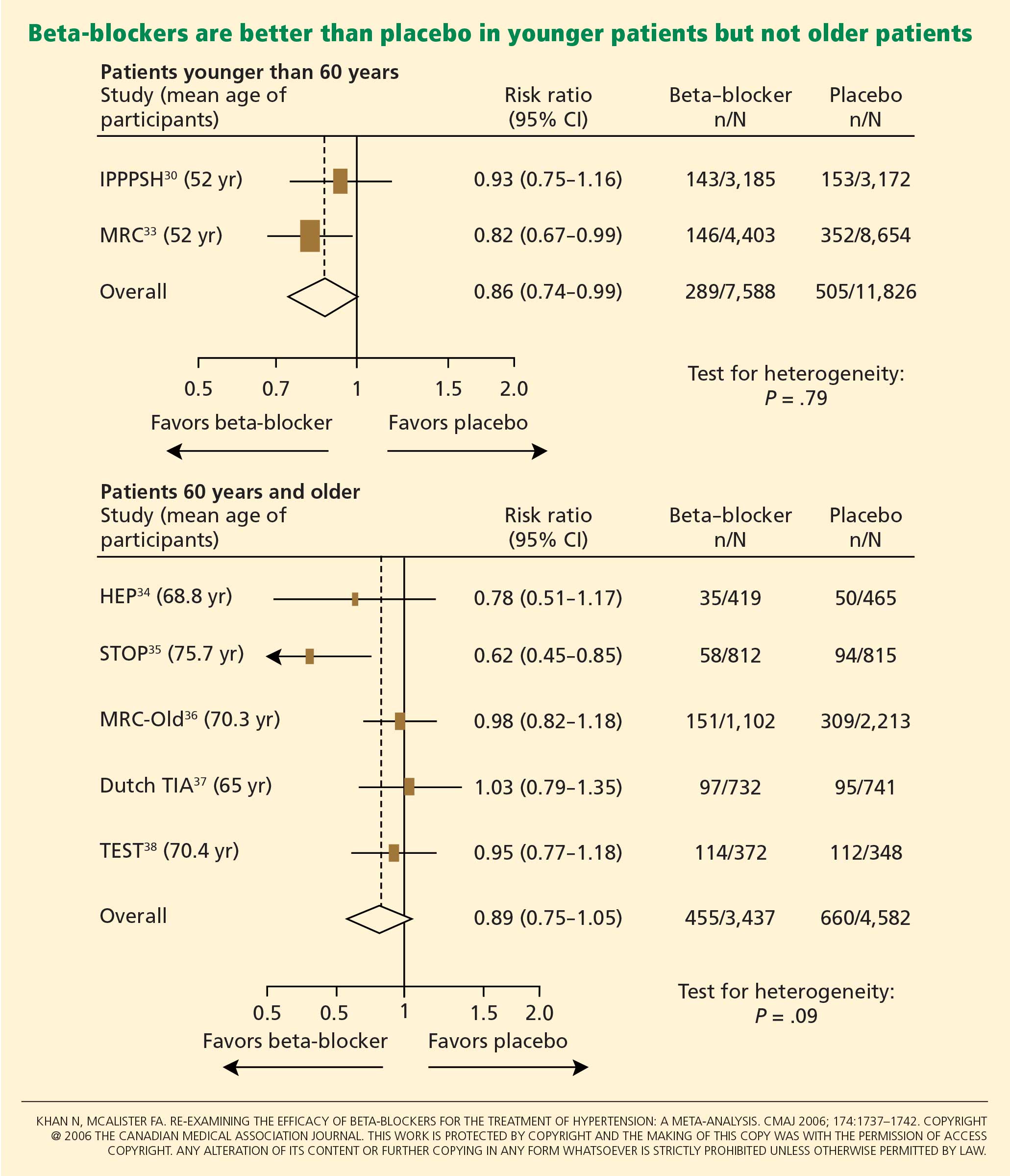
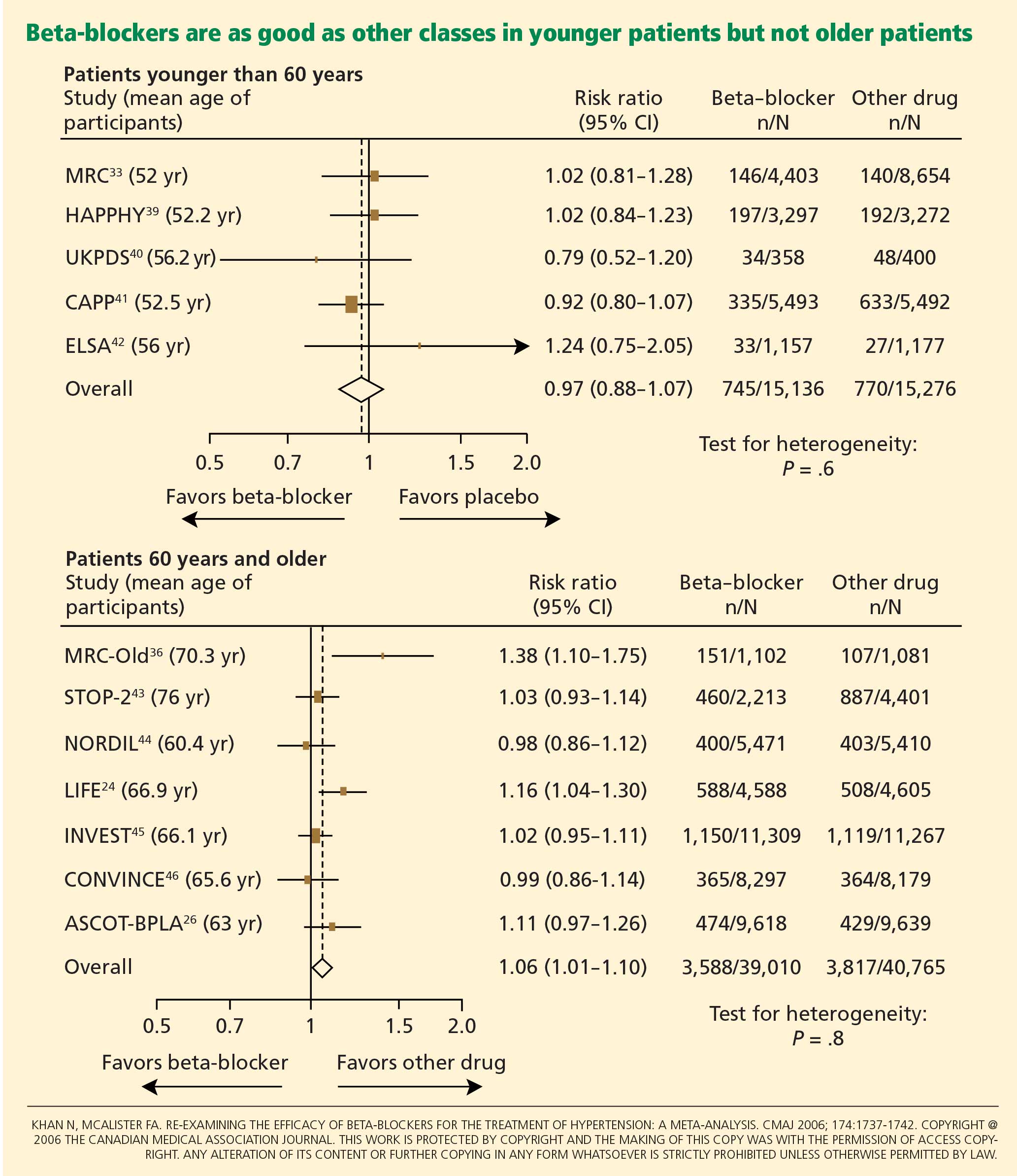
Pulse-wave dyssynchrony
Bangalore et al47 offer an interesting hypothesis to explain the probable adverse effect of beta-blockers. Their theory concerns the effect of these drugs on the arterial pulse wave.
Normally, with each contraction of the left ventricle during systole, an arterial pulse wave is generated and propagated forward to the peripheral arteries. This wave is then reflected back to the heart from the branching points of peripheral arteries. The final form of the pressure wave at the aortic root is a synchronized summation of the forward-traveling wave and the backward-reflected wave.
In healthy people with normal arteries, the reflected wave merges with the forward-traveling wave in diastole and augments coronary blood flow. In patients whose arteries are stiff due to aging or vascular comorbidities, the reflected wave returns faster and merges with the incident wave in systole, resulting in higher left ventricular afterload and less coronary perfusion.48
Bangalore et al47 propose that artificially reducing the heart rate with beta-blockers may further dyssynchronize the pulse wave, adversely affecting coronary perfusion and leading to an increased risk of cardiovascular events and death.
Metabolic side effects
Older beta-blockers, and especially atenolol, have well-known metabolic adverse effects, particularly impairment of glycemic control. This adverse effect appears to occur only with beta-blockers that do not possess vasodilatory properties and thus increase peripheral vascular resistance, which results in lower glucose availability and reduced uptake by skeletal muscles.49
Bangalore et al50 evaluated the effect of beta-blockers in a meta-analysis of 12 studies in 94,492 patients followed up for more than 1 year. Beta-blocker therapy resulted in a 22% higher risk of new-onset diabetes mellitus (RR 1.22, 95% CI 1.12–1.33) than with other nondiuretic antihypertensive agents.
Of note, however, the meta-analysis did not show a significantly higher risk of the onset of diabetes with propranolol or metoprolol than with other nondiuretic antihypertensives when studies of these beta-blockers were separated from atenolol-based studies.
Further, the United Kingdom Prospective Diabetes Study40 found that cardiovascular outcomes in patients with good blood pressure control were similar when atenolol-based therapy was compared with therapy with the ACE inhibitor captopril (Capoten).
A meta-analysis conducted by Balamuthusamy et al51 in 2009 found no higher risk of stroke in patients with hypertension and diabetes mellitus who received beta-blockers than in those who received other antihypertensive medications. However, beta-blockers were associated with a higher risk of death from cardiovascular causes (RR 1.39, 95% CI 1.07–1.804; P < .01) compared with reninangiotensin blockade.
NEWER BETA-BLOCKERS MAY BE BETTER
In the United States, more than 40 million prescriptions for atenolol are written every year, making it by far the most commonly used beta-blocker for the treatment of hypertension. 52 It is clear, however, that atenolol is not an ideal representative of this class of antihypertensive medications.
Preliminary data from studies of newer beta-blockers that possess beneficial vasodilatory properties are encouraging. Animal studies and preliminary human studies find that these new-generation beta-blockers cause fewer adverse metabolic effects and improve endothelial function, measures of arterial stiffness, and cardiovascular outcomes.
Carvedilol
Carvedilol is a nonselective beta-blocker with vasodilatory effects that are thought to be due to its ability to concurrently block alpha-1 receptors in addition to beta receptors. 53 In experiments in vitro and in trials in patients with diabetes and hypertension, carvedilol increased endothelial vasodilation and reduced inflammation and platelet aggregation. These effects may be achieved though antioxidant actions, thereby preserving nitric oxide bioactivity.54,55
In the Glycemic Effects in Diabetes Mellitus: Carvedilol-Metoprolol Comparison in Hypertensives (GEMINI) trial,56 carvedilol was associated with better maintenance of glycemic control in diabetic hypertensive patients than was metoprolol. Insulin sensitivity improved with carvedilol but not with metoprolol, and fewer patients on carvedilol progressed to microalbuminuria.
Nebivolol
Nebivolol is a novel selective beta-blocker with a much higher affinity for beta-1 adrenergic receptors than for beta-2 adrenergic receptors. Among all the beta-blockers in clinical use today, nebivolol has the highest selectivity for beta-1 receptors.8
Nebivolol causes vasodilation through activation of the l-arginine/nitric oxide pathway.57–59 Blockade of synthesis of nitric oxide leads to local arterial stiffness. Endothelial dysfunction is characterized by decreased bioavailability of nitric oxide and has been shown to be a strong predictor of cardiovascular outcomes. By generating nitric oxide, nebivolol reduces peripheral vascular resistance, overcoming a significant side effect of earlier beta-blockers that lowered blood pressure but ultimately increased peripheral vascular tone and resistance.8
In an experiment in a bovine model,60 nebivolol significantly reduced the pulse-wave velocity (a measure of arterial stiffness), while atenolol had no effect. Moreover, evidence for the role of the l-arginine/nitric oxide pathway in the vasodilatory effect of nebivolol was demonstrated by co-infusion of NG-monomethyl-L-arginine, a specific endothelial nitric oxide synthetase inhibitor that attenuated the reduction of pulse-wave velocity by nebivolol.
In studies in hypertensive patients, nebivolol was associated with a better metabolic profile than atenolol, with none of the adverse effects on insulin sensitivity that atenolol had.61 In the Study of Effects of Nebivolol Interventions on Outcomes and Rehospitalization in Seniors With Heart Failure (SENIORS) trial, significantly fewer patients receiving nebivolol died or were admitted to the hospital for cardiovascular reasons compared with those receiving placebo.62
Although these findings are encouraging, we do not yet know if these effects will translate into a significant reduction in cardiovascular outcomes in clinical trials. Large, prospective hypertension outcome trials, particularly to evaluate primary prevention of cardiovascular outcomes, are needed for an evidence-based approach to using the newer beta-blockers as preferred first-line therapy for hypertension.
WHAT RECENT GUIDELINES SAY ABOUT BETA-BLOCKERS
The British National Institute for Health and Clinical Excellence and the British Hypertension Society, in their 2004 guidelines, recommended beta-blockers as one of several first-line antihypertensive medications in young, nonblack patients.63 On the other hand, they advised clinicians to be aware of the reported increase in onset of diabetes mellitus in patients treated with these medications. After the LIFE24 and ASCOT26 study results were published, these guidelines were amended to exclude beta-blockers as preferred routine initial therapy for hypertension.64
More recently, the 2007 European Society of Hypertension and European Society of Cardiology reconsidered the role of beta-blockers, recommending them as an option in both initial and subsequent antihypertensive treatment strategies.65
The current guidelines from the National Heart, Lung, and Blood Institute,66 which were published in 2003, were highly influenced by the results of the Antihypertensive and Lipid-Lowering Treatment to Prevent Heart Attack Trial (ALLHAT),2 and favor diuretics as the first-line therapy. However, they indicate that beta-blockers are a suitable alternative, particularly when a compelling cardiac indication is present.53 We hope that the next update, expected late in 2009, will re-address this issue in the light of more recent data.
- Staessen JA, Wang JG, Thijs L. Cardiovascular protection and blood pressure reduction: a meta-analysis. Lancet 2001; 358:1305–1315.
- ALLHAT Officers and Coordinators for the ALLHAT Collaborative Research Group. The Antihypertensive and Lipid-Lowering Treatment to Prevent Heart Attack Trial. Major outcomes in high-risk hypertensive patients randomized to angiotensin-converting enzyme inhibitor or calcium channel blocker vs diuretic: the Antihypertensive and Lipid-Lowering Treatment to Prevent Heart Attack Trial (ALLHAT). JAMA 2002; 288:2981–2997.
- Neutel JM, Smith DH, Ram CV, et al. Application of ambulatory blood pressure monitoring in differentiating between antihypertensive agents. Am J Med 1993; 94:181–187.
- Materson BJ, Reda DJ, Cushman WC, et al. Single-drug therapy for hypertension in men. A comparison of six antihypertensive agents with placebo. The Department of Veterans Affairs Cooperative Study Group on Antihypertensive Agents. N Engl J Med 1993; 328:914–921.
- Psaty BM, Smith NL, Siscovick DS, et al. Health outcomes associated with antihypertensive therapies used as first-line agents. A systematic review and meta-analysis. JAMA 1997; 277:739–745.
- Frishman W, Silverman R. Clinical pharmacology of the new beta-adrenergic blocking drugs. Part 2. Physiologic and metabolic effects. Am Heart J 1979; 97:797–807.
- Garrett BN, Kaplan NM. Plasma renin activity suppression: duration after withdrawal from beta-adrenergic blockade. Arch Intern Med 1980; 140:1316–1318.
- Pedersen ME, Cockcroft JR. The latest generation of beta-blockers: new pharmacologic properties. Curr Hypertens Rep 2006; 8:279–286.
- Man in’t Veld AJ, Van den Meiracker AH, Schalekamp MA. Do beta-blockers really increase peripheral vascular resistance? Review of the literature and new observations under basal conditions. Am J Hypertens 1988; 1:91–96.
- Pearce CJ, Wallin JD. Labetalol and other agents that block both alpha- and beta-adrenergic receptors. Cleve Clin J Med 1994; 61:59–69.
- Dimsdale JE, Newton RP, Joist T. Neuropsychological side effects of beta-blockers. Arch Intern Med 1989; 149:514–525.
- Agarwal R. Supervised atenolol therapy in the management of hemodialysis hypertension. Kidney Int 1999; 55:1528–1535.
- Sica DA, Black HR. Pharmacologic considerations in the positioning of beta-blockers in antihypertensive therapy. Curr Hypertens Rep 2008; 10:330–335.
- Prichard BN, Gillam GP. Use of propranolol (Inderal) in treatment of hypertension. Br Med J 1964; 19; 2:725–727.
- The fifth report of the Joint National Committee on Detection, Evaluation, and Treatment of High Blood Pressure (JNC V). Arch Intern Med 1993; 153:154–183.
- Moser M. Evolution of the treatment of hypertension from the 1940s to JNC V. Am J Hypertens 1997; 10:2S–8S.
- Houghton T, Freemantle N, Cleland JG. Are beta-blockers effective in patients who develop heart failure soon after myocardial infarction? A meta-regression analysis of randomised trials. Eur J Heart Fail 2000; 2:333–340.
- The Cardiac Insufficiency Bisoprolol Study II (CIBIS-II): a randomised trial. Lancet 1999; 353:9–13.
- Effect of metoprolol CR/XL in chronic heart failure: Metoprolol CR/XL Randomised Intervention Trial in Congestive Heart Failure (MERIT-HF). Lancet 1999; 353:2001–2007.
- Yusuf S, Peto R, Lewis J, Collins R, Sleight P. Beta blockade during and after myocardial infarction: an overview of the randomized trials. Prog Cardiovasc Dis 1985; 27:335–371.
- Brophy JM, Joseph L, Rouleau JL. Beta-blockers in congestive heart failure. A Bayesian meta-analysis. Ann Intern Med 2001; 134:550–560.
- Wiysonge CS, Bradley H, Mayosi BM, et al. Beta-blockers for hypertension. Cochrane Database Syst Rev 2007;CD002003.
- Messerli FH, Grossman E, Goldbourt U. Are beta-blockers efficacious as first-line therapy for hypertension in the elderly? A systematic review. JAMA 1998; 279:1903–1907.
- Dahlöf B, Devereux RB, Kjeldsen SE, et al; for the LIFE study group. Cardiovascular morbidity and mortality in the Losartan Intervention For Endpoint Reduction in Hypertension study (LIFE): a randomised trial against atenolol. Lancet 2002; 359:995–1003.
- Carlberg B, Samuelsson O, Lindholm LH. Atenolol in hypertension: is it a wise choice? Lancet 2004; 364:1684–1689.
- Dahlöf B, Sever PS, Poulter NR, et al; ASCOT Investigators. Prevention of cardiovascular events with an antihypertensive regimen of amlodipine adding perindopril as required versus atenolol adding bendroflumethiazide as required, in the Anglo-Scandinavian Cardiac Outcomes Trial-Blood Pressure Lowering Arm (ASCOT-BPLA): a multicentre randomised controlled trial. Lancet 2005; 366:895–906.
- Lindholm LH, Carlberg B, Samuelsson O. Should beta-blockers remain first choice in the treatment of primary hypertension? A meta-analysis. Lancet 2005; 366:1545–1553.
- Neutel JM, Schnaper H, Cheung DG, Graettinger WF, Weber MA. Antihypertensive effects of beta-blockers administered once daily: 24-hour measurements. Am Heart J 1990; 120:166–171.
- Franklin SS, Gustin W, Wong ND, et al. Hemodynamic patterns of age-related changes in blood pressure. The Framingham Heart Study. Circulation 1997; 96:308–315.
- The IPPPSH Collaborative Group. Cardiovascular risk and risk factors in a randomized trial of treatment based on the beta-blocker oxprenolol: the International Prospective Primary Prevention Study in Hypertension (IPPPSH). J Hypertens 1985; 3:379–392.
- Williams B, Lacy PS, Thom SM, et al; CAFE Investigators. Differential impact of blood pressure-lowering drugs on central aortic pressure and clinical outcomes: principal results of the Conduit Artery Function Evaluation (CAFE) study. Circulation 2006; 113:1213–1225.
- Khan N, McAlister FA. Re-examining the efficacy of beta-blockers for the treatment of hypertension: a meta-analysis. CMAJ 2006; 174:1737–1742.
- Medical Research Council Working Party. MRC trial of treatment of mild hypertension: principal results. BMJ 1985; 291:97–104.
- Coope J, Warrender TS. Randomised trial of treatment of hypertension in elderly patients in primary care. BMJ 1986; 293:1145–1151.
- Dahlöf B, Lindholm LH, Hansson L, et al. Morbidity and mortality in the Swedish Trial in Old Patients with Hypertension (STOP-Hypertension). Lancet 1991; 338:1281–1285.
- MRC Working Party. Medical Research Council trial of treatment of hypertension in older adults: principal results. BMJ 1992; 304:405–412.
- The Dutch TIA Study Group. Trial of secondary prevention with atenolol after transient ischemic attack or nondisabling ischemic stroke. Stroke 1993; 24:543–548.
- Eriksson S, Olofsson B-O, Wester P-O; for the TEST Study Group. Atenolol in secondary prevention after stroke. Cerebrovasc Dis 1995; 5:21–25.
- Wilhelmsen L, Berglund G, Elmfeldt D, et al. Beta-blockers versus diuretics in hypertensive men: main results from the HAPPHY trial. J Hypertens 1987; 5:561–572.
- UK Prospective Diabetes Study Group. Efficacy of atenolol and captopril in reducing risk of macrovascular and microvascular complications in type 2 diabetes: UKPDS 39. BMJ 1998; 317:713–720.
- Hansson L, Lindholm LH, Niskanen L, et al. Effect of angiotensin-converting enzyme inhibition compared with conventional therapy on cardiovascular morbidity and mortality in hypertension: the Captopril Prevention Project (CAPP) randomised trial. Lancet 1999; 353:611–616.
- Lanchetti A, Bond MG, Henning M, et al. Calcium antagonist lacidipine slow down progression of asymptomatic carotid atherosclerosis. Principal results of the European Lacidipine Study on Atherosclerosis (ELSA), a randomized, double-blind, long-term trial. Circulation 2002; 106:2422–2427.
- Hansson L, Lindholm LH, Ekbom T, et al. Randomised trial of old and new antihypertensive drugs in elderly patients: cardiovascular mortality and morbidity in the Swedish Trial in Old Patients with Hypertension-2 study. Lancet 1999; 354:1751–1756.
- Hansson L, Hedner T, Lund-Johansen P, et al. Randomised trial of effects of calcium antagonists compared with diuretics and ß blockers on cardiovascular morbidity and mortality in hypertension: the Nordic Diltiazem (NORDIL) study. Lancet 2000; 356:359–365.
- Pepine CJ, Handsberg EM, Cooper-DeHoff RM, et al. A calcium antagonist vs a non-calcium antagonist hypertension treatment strategy for patients with coronary artery disease. The International Verapamil-Trandolapril Study (INVEST): a randomized controlled trial. JAMA 2003; 290:2805–2816.
- Black HR, Elliott WJ, Grandits G, et al. Principal results of the Controlled Onset Verapamil Investigation of Cardiovascular End Points (CONVINCE) Trial. JAMA 2003; 289:2073–2082.
- Bangalore S, Sawhney S, Messerli FH. Relation of beta-blocker-induced heart rate lowering and cardioprotection in hypertension. J Am Coll Cardiol 2008; 52:1482–1489.
- Boutouyrie P, Vermersch S, Laurent S, Briet M. Cardiovascular risk assessment through target organ damage: role of carotid to femoral pulse wave velocity. Clin Exp Pharmacol Physiol 2008; 35:530–533.
- Kveiborg B, Christiansen B, Major-Petersen A, Torp-Pedersen C. Metabolic effects of beta-adrenoceptor antagonists with special emphasis on carvedilol. Am J Cardiovasc Drugs 2006; 6:209–217.
- Bangalore S, Parkar S, Grossman E, Messerli FH. A meta-analysis of 94,492 patients with hypertension treated with beta-blockers to determine the risk of new-onset diabetes mellitus. Am J Cardiol 2007; 100:1254–1262.
- Balamuthusamy S, Molnar J, Adigopula S, Arora R. Comparative analysis of beta-blockers with other antihypertensive agents on cardiovascular outcomes in hypertensive patients with diabetes mellitus: a systematic review and meta-analysis. Am J Ther 2009; 16:133–142.
- Berenson A. Big drug makers see sales decline with their image. New York Times 2005 Nov 14.
- Bristow MR. Beta-adrenergic receptor blockade in chronic heart failure. Circulation 2000; 101:558–569.
- Giugliano D, Marfella R, Acampora R, Giunta R, Coppola L, D’Onofrio F. Effects of perindopril and carvedilol on endothelium-dependent vascular functions in patients with diabetes and hypertension. Diabetes Care 1998; 21:631–636.
- Lopez BL, Christopher TA, Yue TL, Ruffolo R, Feuerstein GZ, Ma XL. Carvedilol, a new beta-adrenoreceptor blocker antihypertensive drug, protects against free-radical-induced endothelial dysfunction. Pharmacology 1995; 51:165–173.
- Bakris GL, Fonseca V, Katholi RE, et al; GEMINI Investigators. Metabolic effects of carvedilol vs metoprolol in patients with type 2 diabetes mellitus and hypertension: a randomized controlled trial. JAMA 2004; 292:2227–2236.
- Georgescu A, Pluteanu F, Flonta ML, Badila E, Dorobantu M, Popov D. The cellular mechanisms involved in the vasodilator effect of nebivolol on the renal artery. Eur J Pharmacol 2005; 508:159–166.
- Kalinowski L, Dobrucki LW, Szczepanska-Konkel M, et al. Third-generation beta-blockers stimulate nitric oxide release from endothelial cells through ATP efflux: a novel mechanism for antihypertensive action. Circulation 2003; 107:2747–2752.
- Cockcroft JR, Chowienczyk PJ, Brett SE, et al. Nebivolol vasodilates human forearm vasculature: evidence for an L-arginine/NO-dependent mechanism. J Pharmacol Exp Ther 1995; 274:1067–1071.
- McEniery CM, Schmitt M, Qasem A, et al. Nebivolol increases arterial distensibility in vivo. Hypertension 2004; 44:305–310.
- Poirier L, Cleroux J, Nadeau A, Lacourciere Y. Effects of nebivolol and atenolol on insulin sensitivity and haemodynamics in hypertensive patients. J Hypertens 2001; 19:1429–1435.
- Flather MD, Shibata MC, Coats AJ, et al; SENIORS Investigators. Randomized trial to determine the effect of nebivolol on mortality and cardiovascular hospital admission in elderly patients with heart failure (SENIORS). Eur Heart J 2005; 26:215–225.
- Williams B, Poulter NR, Brown MJ, et al; BHS guidelines working party, for the British Hypertension Society. British Hypertension Society guidelines for hypertension management 2004 (BHS-IV): summary. BMJ 2004; 328:634–640.
- Sever P. New hypertension guidelines from the National Institute for Health and Clinical Excellence and the British Hypertension Society. J Renin Angiotensin Aldosterone Syst 2006; 7:61–63.
- Mancia G, De Backer G, Dominiczak A, et al; Management of Arterial Hypertension of the European Society of Hypertension. 2007 Guidelines for the Management of Arterial Hypertension: The Task Force for the Management of Arterial Hypertension of the European Society of Hypertension (ESH) and of the European Society of Cardiology (ESC). J Hypertens 2007; 25:1105–1187.
- Chobanian AV, Bakris GL, Black HR, et al; National Heart, Lung, and Blood Institute Joint National Committee on Prevention, Detection, Evaluation, and Treatment of High Blood Pressure; National High Blood Pressure Education Program Coordinating Committee. The Seventh Report of the Joint National Committee on Prevention, Detection, Evaluation, and Treatment of High Blood Pressure: the JNC 7 report. JAMA 2003; 289:2560–2572.
In recent years the role of beta-blockers as a primary tool to treat hypertension has come under question. These drugs have shown disappointing results when used as antihypertensive therapy in patients without heart disease, ie, when used as primary prevention. At the same time, beta-blockers clearly reduce the risk of future cardiovascular events in patients who already have heart disease, eg, who already have had a myocardial infarction or who have congestive heart failure.
Several meta-analyses and a few clinical trials have shown that beta-blockers may have no advantage over other antihypertensive drugs, and in fact may not reduce the risk of stroke as effectively as other classes of blood pressure medications.
Why should this be? Is it that the patients in the antihypertensive trials were mostly older, and that beta-blockers do not work as well in older patients as in younger ones? Or does it have to do with the fact that atenolol (Tenormin) was the drug most often used in the trials? Would newer, different beta-blockers be better?
Hypertension experts currently disagree on how to interpret the available data, and this has led to conflict and confusion among clinicians as to the role of beta-blockers in managing hypertension. Current evidence suggests that older beta-blockers may not be the preferred first-line antihypertensive drugs for hypertensive patients who have no compelling indications for them (eg, heart failure, myocardial infarction, diabetes, high risk of coronary heart disease). However, newer beta-blockers with vasodilatory properties should be considered in cases of uncontrolled or resistant hypertension, especially in younger patients.
Further, while controversy and debate continue over the benefits and adverse effects of one class of antihypertensive drugs vs another, it is indisputable that controlling arterial blood pressure to the recommended goal offers major protection against cardiovascular and renal events in patients with hypertension.1,2
MECHANISM OF ACTION OF BETA-BLOCKERS
Beta-blockers effectively reduce blood pressure in both systolic-diastolic hypertension and isolated systolic hypertension.3–5 Exactly how is not known, but it has been proposed that they may do so by:
Reducing the heart rate and cardiac output. When catecholamines activate beta-1 receptors in the heart, the heart rate and myocardial contractility increase. By blocking beta-1 receptors, beta-blockers reduce the heart rate and myocardial contractility, thus lowering cardiac output and arterial blood pressure.6
Inhibiting renin release. Activation of the renin-angiotensin system is another major pathway that can lead to elevated arterial blood pressure. Renin release is mediated through the sympathetic nervous system via beta-1 receptors on the juxtaglomerular cells of the kidney. Beta-blockers can therefore lower blood pressure by inhibiting renin release.7
Inhibiting central nervous sympathetic outflow, thereby inducing presynaptic blockade, which in turn reduces the release of catecholamines.
Reducing venous return and plasma volume.
Generating nitric oxide, thus reducing peripheral vascular resistance (some agents).8
Reducing vasomotor tone.
Reducing vascular tone.
Improving vascular compliance.
Resetting baroreceptor levels.
Attenuating the pressor response to catecholamines with exercise and stress.
HETEROGENEITY OF BETA-BLOCKERS
Selectivity
Beta-blockers are not all the same. They can be classified into three categories.
Nonselective beta-blockers block both beta-1 and beta-2 adrenergic receptors. It is generally accepted that beta-blockers exert their primary antihypertensive effect by blocking beta-1 adrenergic receptors.6 Of interest, nonselective beta-blockers inhibit beta-2 receptors on arteries and thus cause an unopposed alpha-adrenergic effect, leading to increased peripheral vascular resistance.9 Examples of this category:
- Nadolol (Corgard)
- Pindolol (Visken)
- Propranolol (Inderal)
- Timolol (Blocadren).
Selective beta-blockers specifically block beta-1 receptors alone, although they are known to be nonselective at higher doses. Examples:
- Atenolol (Tenormin)
- Betaxolol (Kerlone)
- Bisoprolol (Zebeta)
- Esmolol (Brevibloc)
- Metoprolol (Lopressor, Toprol).
Beta-blockers with peripheral vasodilatatory effects act either via antagonism of the alpha-1 receptor, as with labetolol (Normodyne) and carvedilol (Coreg),10 or via enhanced release of nitric oxide, as with nebivolol (Bystolic).8
Lipid and water solubility
The lipid solubility and water solubility of each beta-blocker determine its bioavailability and side-effect profile.
Lipid solubility determines the degree to which a beta-blocker penetrates the blood-brain barrier and thereby leads to central nervous system side effects such as lethargy, nightmares, confusion, and depression. Propranolol is highly lipid-soluble; metoprolol and labetalol are moderately so.
Water-soluble beta-blockers such as atenolol have less tissue permeation, have a longer half-life, and cause fewer central nervous system effects and symptoms.11
Routes of elimination
Beta-blockers also differ in their route of elimination.
Atenolol and nadolol are eliminated by the kidney and require dose adjustment in patients with impaired renal function.12,13
On the other hand, propranolol, metoprolol, labetalol, carvedilol, and nebivolol are excreted primarily via hepatic metabolism.13
BETA-BLOCKERS IN THE MANAGEMENT OF HYPERTENSION
Beta-blockers were initially used to treat arrhythmias, but by the early 1970s they were also widely accepted for managing hypertension. 14 Their initial acceptance as one of the first-line classes of drugs for hypertension was based on their better side-effect profile compared with other antihypertensive drugs available at that time.
In the 1980s and 1990s, beta-blockers were listed as preferred first-line antihypertensive drugs along with diuretics in national hypertension guidelines.15 Subsequent updates of the guidelines favored diuretics as initial therapy and relegated all other classes of antihypertensive medications to be alternatives to diuretics.16 Although beta-blockers remain alternative first-line drugs in the latest guidelines (published in 2003; see reference 66), they are the preferred antihypertensive agents for patients with cardiac disease.
The current recommendations reflect the findings from hypertension trials in which patients with myocardial infarction and congestive heart failure had better cardiovascular outcomes if they received these drugs,17–19 including a lower risk of death.20,21 It was widely assumed that beta-blockers would also prevent first episodes of cardiovascular events.
However, to date, there is no evidence that beta-blockers are effective as primary prevention. Several large randomized controlled trials showed no benefit with beta-blockers compared with other antihypertensive drugs—in fact, there were more cardiovascular events with beta-blockers (see below).
Beta-blockers are well tolerated in clinical practice, although they can have side effects that include fatigue, depression, impaired exercise tolerance, sexual dysfunction, and asthma attacks.
Wiysonge et al22 analyzed how many patients withdrew from randomized trials of antihypertensive treatment because of drug-related adverse events. There was no significant difference in the incidence of fatigue, depressive symptoms, or sexual dysfunction with beta-blockers compared with placebo, and trial participants on a beta-blocker were not statistically significantly more likely to discontinue treatment than those receiving a placebo in three trials with 22,729 participants (relative risk [RR] 2.34, 95% confidence interval [CI] 0.84–6.52).
THE CONTROVERSY: WHAT THE TRIALS SHOWED
Messerli et al23 performed a meta-analysis published in 1998 that suggested that beta-blockers may not be as effective as diuretics in preventing cardiovascular events when used as first-line antihypertensive therapy in elderly patients. In 10 randomized controlled trials in 16,164 patients who were treated with either a diuretic or a beta-blocker (atenolol), blood pressure was normalized in two-thirds of diuretic-treated patients but only one-third of patients treated with atenolol as monotherapy. Diuretic therapy was superior with regard to all end points, and beta-blockers were found to be ineffective except in reducing cerebrovascular events.
The LIFE study (Losartan Intervention for Endpoint Reduction in Hypertension)24 compared the angiotensin-receptor blocker losartan (Cozaar) and atenolol in 9,193 patients with hypertension and left ventricular hypertrophy. At 4 years of follow-up, the rate of primary cardiovascular events (death, myocardial infarction, or stroke) was lower in the losartan group than in the atenolol group. The difference was mainly due to a 25% lower incidence of stroke, which was statistically significant. The rates of myocardial infarction and death from cardiovascular causes were not significantly different between the two treatment groups. The systolic blood pressure was 1 mm Hg lower in the losartan group than in the atenolol group, which was statistically significant.
Carlberg et al25 performed another important meta-analysis that questioned whether atenolol reduces rates of cardiovascular morbidity and death in hypertensive patients. The results were surprising: eight randomized controlled trials including more than 6,000 patients and comparing atenolol with placebo or no treatment showed no differences between the treatment groups with regard to the outcomes of all-cause mortality (RR 1.01, 95% CI 0.89–1.15), cardiovascular mortality (RR 0.99, 95% CI 0.83–1.18), or myocardial infarction (RR 0.99, 95% CI 0.83–1.19).
In addition, when atenolol was compared with other antihypertensives in five other randomized controlled trials that included more than 14,000 patients, those treated with atenolol had a higher risk of stroke (RR 1.30, 95% CI 1.12–1.50) and death (RR 1.13, 95% CI 1.02–1.25).
The ASCOT-BPLA trial (Anglo-Scandinavian Cardiac Outcomes Trial—Blood Pressure Lowering Arm)26 had similar results. This trial compared the combination of atenolol plus the diuretic bendroflumethiazide against the combination of the calcium channel blocker amlodipine (Norvasc) plus the angiotensin-converting enzyme (ACE) inhibitor perindopril (Aceon). Although no significant difference was seen in the primary outcome of nonfatal myocardial infarction or fatal coronary heart disease (unadjusted hazard ratio [HR] with amlodipine-perindopril 0.90, 95% CI 0.79–1.02, P = .1052), the amlodipine-plus-perindopril group had significantly fewer strokes (327 vs 422, HR 0.77, 95% CI 0.66–0.89, P = .0003), fewer total cardiovascular events (1,362 vs 1,602, HR 0.84, 95% CI 0.78–0.90, P = .0001), and fewer deaths from any cause (738 vs 820; HR 0.89, 95% CI 0.81–0.99, P = .025).
Lindholm et al27 performed a meta-analysis that included studies of selective beta-blockers (including atenolol) and nonselective beta-blockers, with a follow-up time of more than 2 years. Compared with placebo or no treatment, beta-blockers reduced the risk of stroke by 19% but had no effect on myocardial infarction or all-cause mortality. Compared with other antihypertensive drugs, beta-blockers were less than optimum, and the relative risk of stroke was 16% higher. Atenolol was the beta-blocker used in most of the randomized clinical trials included in this meta-analysis.
The Cochrane group22 found beta-blockers to be inferior to all other antihypertensive drugs with respect to the ability to lower the risk of stroke.
WHY WERE THE RESULTS SO DISAPPOINTING?
Problems with atenolol
Most of the trials in the meta-analyses discussed above used atenolol and other beta-blockers that had no vasodilatory properties.
Further, in most of the trials atenolol was used in a once-daily dosage, whereas ideally it needs to be taken more frequently, based on its pharmacokinetic and pharmacodynamic properties (a half-life of 6–9 hours).3 Neutel et al28 confirmed that atenolol, when taken once daily, leaves the patient unprotected in the last 6 hours of a 24-hour period, as demonstrated by 24-hour ambulatory blood pressure monitoring. It is possible that this short duration of action of atenolol may have contributed to the results observed in clinical trials that used atenolol to treat hypertension.
Differences between older and younger patients
Another possible reason for the disappointing results is that the trials included many elderly patients, in whom beta-blockers may not be as effective. The pathophysiology of hypertension in younger people is different from that in older patients.29 Hemodynamic characteristics of younger hypertensive patients include a high cardiac output and hyperdynamic circulation with a low pulse pressure, while older patients have lower arterial compliance with an elevated vascular resistance.
The notion of choosing antihypertensive medications on the basis of age and age-related pathophysiology is supported by several clinical studies. Randomized controlled trials appear to show that beta-blockers are effective in younger hypertensive patients.30
Conversely, the CAFE (Conduit Artery Function Evaluation) trial,31 a substudy of the main ASCOT trial,26 indicated that betablocker-based therapy was less effective in reducing central aortic pressure than were regimens based on an ACE inhibitor or a calcium channel blocker.
The CAFE researchers recruited 2,073 patients from five ASCOT centers and used radial artery applanation tonometry and pulse-wave analysis to derive central aortic pressures and hemodynamic indices during study visits up to a period of 4 years. Although the two treatment groups achieved similar brachial systolic blood pressures, the central aortic systolic pressure was 4.3 mm Hg lower in the amlodipine group (95% CI 3.3–5.4; P < .0001), and the central aortic pulse pressure was 3.0 mm Hg lower (95% CI 2.1–3.9; P < .0001).
Pulse-wave dyssynchrony
Bangalore et al47 offer an interesting hypothesis to explain the probable adverse effect of beta-blockers. Their theory concerns the effect of these drugs on the arterial pulse wave.
Normally, with each contraction of the left ventricle during systole, an arterial pulse wave is generated and propagated forward to the peripheral arteries. This wave is then reflected back to the heart from the branching points of peripheral arteries. The final form of the pressure wave at the aortic root is a synchronized summation of the forward-traveling wave and the backward-reflected wave.
In healthy people with normal arteries, the reflected wave merges with the forward-traveling wave in diastole and augments coronary blood flow. In patients whose arteries are stiff due to aging or vascular comorbidities, the reflected wave returns faster and merges with the incident wave in systole, resulting in higher left ventricular afterload and less coronary perfusion.48
Bangalore et al47 propose that artificially reducing the heart rate with beta-blockers may further dyssynchronize the pulse wave, adversely affecting coronary perfusion and leading to an increased risk of cardiovascular events and death.
Metabolic side effects
Older beta-blockers, and especially atenolol, have well-known metabolic adverse effects, particularly impairment of glycemic control. This adverse effect appears to occur only with beta-blockers that do not possess vasodilatory properties and thus increase peripheral vascular resistance, which results in lower glucose availability and reduced uptake by skeletal muscles.49
Bangalore et al50 evaluated the effect of beta-blockers in a meta-analysis of 12 studies in 94,492 patients followed up for more than 1 year. Beta-blocker therapy resulted in a 22% higher risk of new-onset diabetes mellitus (RR 1.22, 95% CI 1.12–1.33) than with other nondiuretic antihypertensive agents.
Of note, however, the meta-analysis did not show a significantly higher risk of the onset of diabetes with propranolol or metoprolol than with other nondiuretic antihypertensives when studies of these beta-blockers were separated from atenolol-based studies.
Further, the United Kingdom Prospective Diabetes Study40 found that cardiovascular outcomes in patients with good blood pressure control were similar when atenolol-based therapy was compared with therapy with the ACE inhibitor captopril (Capoten).
A meta-analysis conducted by Balamuthusamy et al51 in 2009 found no higher risk of stroke in patients with hypertension and diabetes mellitus who received beta-blockers than in those who received other antihypertensive medications. However, beta-blockers were associated with a higher risk of death from cardiovascular causes (RR 1.39, 95% CI 1.07–1.804; P < .01) compared with reninangiotensin blockade.
NEWER BETA-BLOCKERS MAY BE BETTER
In the United States, more than 40 million prescriptions for atenolol are written every year, making it by far the most commonly used beta-blocker for the treatment of hypertension. 52 It is clear, however, that atenolol is not an ideal representative of this class of antihypertensive medications.
Preliminary data from studies of newer beta-blockers that possess beneficial vasodilatory properties are encouraging. Animal studies and preliminary human studies find that these new-generation beta-blockers cause fewer adverse metabolic effects and improve endothelial function, measures of arterial stiffness, and cardiovascular outcomes.
Carvedilol
Carvedilol is a nonselective beta-blocker with vasodilatory effects that are thought to be due to its ability to concurrently block alpha-1 receptors in addition to beta receptors. 53 In experiments in vitro and in trials in patients with diabetes and hypertension, carvedilol increased endothelial vasodilation and reduced inflammation and platelet aggregation. These effects may be achieved though antioxidant actions, thereby preserving nitric oxide bioactivity.54,55
In the Glycemic Effects in Diabetes Mellitus: Carvedilol-Metoprolol Comparison in Hypertensives (GEMINI) trial,56 carvedilol was associated with better maintenance of glycemic control in diabetic hypertensive patients than was metoprolol. Insulin sensitivity improved with carvedilol but not with metoprolol, and fewer patients on carvedilol progressed to microalbuminuria.
Nebivolol
Nebivolol is a novel selective beta-blocker with a much higher affinity for beta-1 adrenergic receptors than for beta-2 adrenergic receptors. Among all the beta-blockers in clinical use today, nebivolol has the highest selectivity for beta-1 receptors.8
Nebivolol causes vasodilation through activation of the l-arginine/nitric oxide pathway.57–59 Blockade of synthesis of nitric oxide leads to local arterial stiffness. Endothelial dysfunction is characterized by decreased bioavailability of nitric oxide and has been shown to be a strong predictor of cardiovascular outcomes. By generating nitric oxide, nebivolol reduces peripheral vascular resistance, overcoming a significant side effect of earlier beta-blockers that lowered blood pressure but ultimately increased peripheral vascular tone and resistance.8
In an experiment in a bovine model,60 nebivolol significantly reduced the pulse-wave velocity (a measure of arterial stiffness), while atenolol had no effect. Moreover, evidence for the role of the l-arginine/nitric oxide pathway in the vasodilatory effect of nebivolol was demonstrated by co-infusion of NG-monomethyl-L-arginine, a specific endothelial nitric oxide synthetase inhibitor that attenuated the reduction of pulse-wave velocity by nebivolol.
In studies in hypertensive patients, nebivolol was associated with a better metabolic profile than atenolol, with none of the adverse effects on insulin sensitivity that atenolol had.61 In the Study of Effects of Nebivolol Interventions on Outcomes and Rehospitalization in Seniors With Heart Failure (SENIORS) trial, significantly fewer patients receiving nebivolol died or were admitted to the hospital for cardiovascular reasons compared with those receiving placebo.62
Although these findings are encouraging, we do not yet know if these effects will translate into a significant reduction in cardiovascular outcomes in clinical trials. Large, prospective hypertension outcome trials, particularly to evaluate primary prevention of cardiovascular outcomes, are needed for an evidence-based approach to using the newer beta-blockers as preferred first-line therapy for hypertension.
WHAT RECENT GUIDELINES SAY ABOUT BETA-BLOCKERS
The British National Institute for Health and Clinical Excellence and the British Hypertension Society, in their 2004 guidelines, recommended beta-blockers as one of several first-line antihypertensive medications in young, nonblack patients.63 On the other hand, they advised clinicians to be aware of the reported increase in onset of diabetes mellitus in patients treated with these medications. After the LIFE24 and ASCOT26 study results were published, these guidelines were amended to exclude beta-blockers as preferred routine initial therapy for hypertension.64
More recently, the 2007 European Society of Hypertension and European Society of Cardiology reconsidered the role of beta-blockers, recommending them as an option in both initial and subsequent antihypertensive treatment strategies.65
The current guidelines from the National Heart, Lung, and Blood Institute,66 which were published in 2003, were highly influenced by the results of the Antihypertensive and Lipid-Lowering Treatment to Prevent Heart Attack Trial (ALLHAT),2 and favor diuretics as the first-line therapy. However, they indicate that beta-blockers are a suitable alternative, particularly when a compelling cardiac indication is present.53 We hope that the next update, expected late in 2009, will re-address this issue in the light of more recent data.
- Staessen JA, Wang JG, Thijs L. Cardiovascular protection and blood pressure reduction: a meta-analysis. Lancet 2001; 358:1305–1315.
- ALLHAT Officers and Coordinators for the ALLHAT Collaborative Research Group. The Antihypertensive and Lipid-Lowering Treatment to Prevent Heart Attack Trial. Major outcomes in high-risk hypertensive patients randomized to angiotensin-converting enzyme inhibitor or calcium channel blocker vs diuretic: the Antihypertensive and Lipid-Lowering Treatment to Prevent Heart Attack Trial (ALLHAT). JAMA 2002; 288:2981–2997.
- Neutel JM, Smith DH, Ram CV, et al. Application of ambulatory blood pressure monitoring in differentiating between antihypertensive agents. Am J Med 1993; 94:181–187.
- Materson BJ, Reda DJ, Cushman WC, et al. Single-drug therapy for hypertension in men. A comparison of six antihypertensive agents with placebo. The Department of Veterans Affairs Cooperative Study Group on Antihypertensive Agents. N Engl J Med 1993; 328:914–921.
- Psaty BM, Smith NL, Siscovick DS, et al. Health outcomes associated with antihypertensive therapies used as first-line agents. A systematic review and meta-analysis. JAMA 1997; 277:739–745.
- Frishman W, Silverman R. Clinical pharmacology of the new beta-adrenergic blocking drugs. Part 2. Physiologic and metabolic effects. Am Heart J 1979; 97:797–807.
- Garrett BN, Kaplan NM. Plasma renin activity suppression: duration after withdrawal from beta-adrenergic blockade. Arch Intern Med 1980; 140:1316–1318.
- Pedersen ME, Cockcroft JR. The latest generation of beta-blockers: new pharmacologic properties. Curr Hypertens Rep 2006; 8:279–286.
- Man in’t Veld AJ, Van den Meiracker AH, Schalekamp MA. Do beta-blockers really increase peripheral vascular resistance? Review of the literature and new observations under basal conditions. Am J Hypertens 1988; 1:91–96.
- Pearce CJ, Wallin JD. Labetalol and other agents that block both alpha- and beta-adrenergic receptors. Cleve Clin J Med 1994; 61:59–69.
- Dimsdale JE, Newton RP, Joist T. Neuropsychological side effects of beta-blockers. Arch Intern Med 1989; 149:514–525.
- Agarwal R. Supervised atenolol therapy in the management of hemodialysis hypertension. Kidney Int 1999; 55:1528–1535.
- Sica DA, Black HR. Pharmacologic considerations in the positioning of beta-blockers in antihypertensive therapy. Curr Hypertens Rep 2008; 10:330–335.
- Prichard BN, Gillam GP. Use of propranolol (Inderal) in treatment of hypertension. Br Med J 1964; 19; 2:725–727.
- The fifth report of the Joint National Committee on Detection, Evaluation, and Treatment of High Blood Pressure (JNC V). Arch Intern Med 1993; 153:154–183.
- Moser M. Evolution of the treatment of hypertension from the 1940s to JNC V. Am J Hypertens 1997; 10:2S–8S.
- Houghton T, Freemantle N, Cleland JG. Are beta-blockers effective in patients who develop heart failure soon after myocardial infarction? A meta-regression analysis of randomised trials. Eur J Heart Fail 2000; 2:333–340.
- The Cardiac Insufficiency Bisoprolol Study II (CIBIS-II): a randomised trial. Lancet 1999; 353:9–13.
- Effect of metoprolol CR/XL in chronic heart failure: Metoprolol CR/XL Randomised Intervention Trial in Congestive Heart Failure (MERIT-HF). Lancet 1999; 353:2001–2007.
- Yusuf S, Peto R, Lewis J, Collins R, Sleight P. Beta blockade during and after myocardial infarction: an overview of the randomized trials. Prog Cardiovasc Dis 1985; 27:335–371.
- Brophy JM, Joseph L, Rouleau JL. Beta-blockers in congestive heart failure. A Bayesian meta-analysis. Ann Intern Med 2001; 134:550–560.
- Wiysonge CS, Bradley H, Mayosi BM, et al. Beta-blockers for hypertension. Cochrane Database Syst Rev 2007;CD002003.
- Messerli FH, Grossman E, Goldbourt U. Are beta-blockers efficacious as first-line therapy for hypertension in the elderly? A systematic review. JAMA 1998; 279:1903–1907.
- Dahlöf B, Devereux RB, Kjeldsen SE, et al; for the LIFE study group. Cardiovascular morbidity and mortality in the Losartan Intervention For Endpoint Reduction in Hypertension study (LIFE): a randomised trial against atenolol. Lancet 2002; 359:995–1003.
- Carlberg B, Samuelsson O, Lindholm LH. Atenolol in hypertension: is it a wise choice? Lancet 2004; 364:1684–1689.
- Dahlöf B, Sever PS, Poulter NR, et al; ASCOT Investigators. Prevention of cardiovascular events with an antihypertensive regimen of amlodipine adding perindopril as required versus atenolol adding bendroflumethiazide as required, in the Anglo-Scandinavian Cardiac Outcomes Trial-Blood Pressure Lowering Arm (ASCOT-BPLA): a multicentre randomised controlled trial. Lancet 2005; 366:895–906.
- Lindholm LH, Carlberg B, Samuelsson O. Should beta-blockers remain first choice in the treatment of primary hypertension? A meta-analysis. Lancet 2005; 366:1545–1553.
- Neutel JM, Schnaper H, Cheung DG, Graettinger WF, Weber MA. Antihypertensive effects of beta-blockers administered once daily: 24-hour measurements. Am Heart J 1990; 120:166–171.
- Franklin SS, Gustin W, Wong ND, et al. Hemodynamic patterns of age-related changes in blood pressure. The Framingham Heart Study. Circulation 1997; 96:308–315.
- The IPPPSH Collaborative Group. Cardiovascular risk and risk factors in a randomized trial of treatment based on the beta-blocker oxprenolol: the International Prospective Primary Prevention Study in Hypertension (IPPPSH). J Hypertens 1985; 3:379–392.
- Williams B, Lacy PS, Thom SM, et al; CAFE Investigators. Differential impact of blood pressure-lowering drugs on central aortic pressure and clinical outcomes: principal results of the Conduit Artery Function Evaluation (CAFE) study. Circulation 2006; 113:1213–1225.
- Khan N, McAlister FA. Re-examining the efficacy of beta-blockers for the treatment of hypertension: a meta-analysis. CMAJ 2006; 174:1737–1742.
- Medical Research Council Working Party. MRC trial of treatment of mild hypertension: principal results. BMJ 1985; 291:97–104.
- Coope J, Warrender TS. Randomised trial of treatment of hypertension in elderly patients in primary care. BMJ 1986; 293:1145–1151.
- Dahlöf B, Lindholm LH, Hansson L, et al. Morbidity and mortality in the Swedish Trial in Old Patients with Hypertension (STOP-Hypertension). Lancet 1991; 338:1281–1285.
- MRC Working Party. Medical Research Council trial of treatment of hypertension in older adults: principal results. BMJ 1992; 304:405–412.
- The Dutch TIA Study Group. Trial of secondary prevention with atenolol after transient ischemic attack or nondisabling ischemic stroke. Stroke 1993; 24:543–548.
- Eriksson S, Olofsson B-O, Wester P-O; for the TEST Study Group. Atenolol in secondary prevention after stroke. Cerebrovasc Dis 1995; 5:21–25.
- Wilhelmsen L, Berglund G, Elmfeldt D, et al. Beta-blockers versus diuretics in hypertensive men: main results from the HAPPHY trial. J Hypertens 1987; 5:561–572.
- UK Prospective Diabetes Study Group. Efficacy of atenolol and captopril in reducing risk of macrovascular and microvascular complications in type 2 diabetes: UKPDS 39. BMJ 1998; 317:713–720.
- Hansson L, Lindholm LH, Niskanen L, et al. Effect of angiotensin-converting enzyme inhibition compared with conventional therapy on cardiovascular morbidity and mortality in hypertension: the Captopril Prevention Project (CAPP) randomised trial. Lancet 1999; 353:611–616.
- Lanchetti A, Bond MG, Henning M, et al. Calcium antagonist lacidipine slow down progression of asymptomatic carotid atherosclerosis. Principal results of the European Lacidipine Study on Atherosclerosis (ELSA), a randomized, double-blind, long-term trial. Circulation 2002; 106:2422–2427.
- Hansson L, Lindholm LH, Ekbom T, et al. Randomised trial of old and new antihypertensive drugs in elderly patients: cardiovascular mortality and morbidity in the Swedish Trial in Old Patients with Hypertension-2 study. Lancet 1999; 354:1751–1756.
- Hansson L, Hedner T, Lund-Johansen P, et al. Randomised trial of effects of calcium antagonists compared with diuretics and ß blockers on cardiovascular morbidity and mortality in hypertension: the Nordic Diltiazem (NORDIL) study. Lancet 2000; 356:359–365.
- Pepine CJ, Handsberg EM, Cooper-DeHoff RM, et al. A calcium antagonist vs a non-calcium antagonist hypertension treatment strategy for patients with coronary artery disease. The International Verapamil-Trandolapril Study (INVEST): a randomized controlled trial. JAMA 2003; 290:2805–2816.
- Black HR, Elliott WJ, Grandits G, et al. Principal results of the Controlled Onset Verapamil Investigation of Cardiovascular End Points (CONVINCE) Trial. JAMA 2003; 289:2073–2082.
- Bangalore S, Sawhney S, Messerli FH. Relation of beta-blocker-induced heart rate lowering and cardioprotection in hypertension. J Am Coll Cardiol 2008; 52:1482–1489.
- Boutouyrie P, Vermersch S, Laurent S, Briet M. Cardiovascular risk assessment through target organ damage: role of carotid to femoral pulse wave velocity. Clin Exp Pharmacol Physiol 2008; 35:530–533.
- Kveiborg B, Christiansen B, Major-Petersen A, Torp-Pedersen C. Metabolic effects of beta-adrenoceptor antagonists with special emphasis on carvedilol. Am J Cardiovasc Drugs 2006; 6:209–217.
- Bangalore S, Parkar S, Grossman E, Messerli FH. A meta-analysis of 94,492 patients with hypertension treated with beta-blockers to determine the risk of new-onset diabetes mellitus. Am J Cardiol 2007; 100:1254–1262.
- Balamuthusamy S, Molnar J, Adigopula S, Arora R. Comparative analysis of beta-blockers with other antihypertensive agents on cardiovascular outcomes in hypertensive patients with diabetes mellitus: a systematic review and meta-analysis. Am J Ther 2009; 16:133–142.
- Berenson A. Big drug makers see sales decline with their image. New York Times 2005 Nov 14.
- Bristow MR. Beta-adrenergic receptor blockade in chronic heart failure. Circulation 2000; 101:558–569.
- Giugliano D, Marfella R, Acampora R, Giunta R, Coppola L, D’Onofrio F. Effects of perindopril and carvedilol on endothelium-dependent vascular functions in patients with diabetes and hypertension. Diabetes Care 1998; 21:631–636.
- Lopez BL, Christopher TA, Yue TL, Ruffolo R, Feuerstein GZ, Ma XL. Carvedilol, a new beta-adrenoreceptor blocker antihypertensive drug, protects against free-radical-induced endothelial dysfunction. Pharmacology 1995; 51:165–173.
- Bakris GL, Fonseca V, Katholi RE, et al; GEMINI Investigators. Metabolic effects of carvedilol vs metoprolol in patients with type 2 diabetes mellitus and hypertension: a randomized controlled trial. JAMA 2004; 292:2227–2236.
- Georgescu A, Pluteanu F, Flonta ML, Badila E, Dorobantu M, Popov D. The cellular mechanisms involved in the vasodilator effect of nebivolol on the renal artery. Eur J Pharmacol 2005; 508:159–166.
- Kalinowski L, Dobrucki LW, Szczepanska-Konkel M, et al. Third-generation beta-blockers stimulate nitric oxide release from endothelial cells through ATP efflux: a novel mechanism for antihypertensive action. Circulation 2003; 107:2747–2752.
- Cockcroft JR, Chowienczyk PJ, Brett SE, et al. Nebivolol vasodilates human forearm vasculature: evidence for an L-arginine/NO-dependent mechanism. J Pharmacol Exp Ther 1995; 274:1067–1071.
- McEniery CM, Schmitt M, Qasem A, et al. Nebivolol increases arterial distensibility in vivo. Hypertension 2004; 44:305–310.
- Poirier L, Cleroux J, Nadeau A, Lacourciere Y. Effects of nebivolol and atenolol on insulin sensitivity and haemodynamics in hypertensive patients. J Hypertens 2001; 19:1429–1435.
- Flather MD, Shibata MC, Coats AJ, et al; SENIORS Investigators. Randomized trial to determine the effect of nebivolol on mortality and cardiovascular hospital admission in elderly patients with heart failure (SENIORS). Eur Heart J 2005; 26:215–225.
- Williams B, Poulter NR, Brown MJ, et al; BHS guidelines working party, for the British Hypertension Society. British Hypertension Society guidelines for hypertension management 2004 (BHS-IV): summary. BMJ 2004; 328:634–640.
- Sever P. New hypertension guidelines from the National Institute for Health and Clinical Excellence and the British Hypertension Society. J Renin Angiotensin Aldosterone Syst 2006; 7:61–63.
- Mancia G, De Backer G, Dominiczak A, et al; Management of Arterial Hypertension of the European Society of Hypertension. 2007 Guidelines for the Management of Arterial Hypertension: The Task Force for the Management of Arterial Hypertension of the European Society of Hypertension (ESH) and of the European Society of Cardiology (ESC). J Hypertens 2007; 25:1105–1187.
- Chobanian AV, Bakris GL, Black HR, et al; National Heart, Lung, and Blood Institute Joint National Committee on Prevention, Detection, Evaluation, and Treatment of High Blood Pressure; National High Blood Pressure Education Program Coordinating Committee. The Seventh Report of the Joint National Committee on Prevention, Detection, Evaluation, and Treatment of High Blood Pressure: the JNC 7 report. JAMA 2003; 289:2560–2572.
KEY POINTS
- No evidence exists that beta-blockers prevent first episodes of cardiovascular events in patients with hypertension, and in some trials, outcomes were worse with beta-blockers than with antihypertensive drugs of other classes.
- Younger hypertensive patients have hemodynamic characteristics that would seem to be amenable to beta-blocker therapy. However, most clinical trials of beta blockers did not stratify patients by age.
- Most trials of the antihypertensive effects of beta-blockers used atenolol (Tenormin), which is not an ideal representative of this class of drugs.
- Newer beta-blockers with vasodilatory properties may overcome the adverse effect of increased peripheral vascular resistance that occurs with older agents such as atenolol.
Role of MRI in breast cancer management
A 52-year-old woman presents to your outpatient clinic with a newly palpable marble-sized lump in the upper outer quadrant of her right breast. The mass is firm and non-tender. She performs breast self-examinations regularly and first noticed the mass 3 weeks ago. Your examination confirms a palpable lesion. Annual screening mammograms at another facility have been negative, including the last one 6 months ago. The woman is otherwise in good health.
When she was younger, she had two pregnancies and gave birth to two children, whom she did not breastfeed. She reached menopause at age 49 and has never been on hormone replacement therapy. Neither she nor anyone in her family has had breast cancer, and she has never undergone breast biopsy.
She says that a woman she spoke with in the waiting room, a 2-year breast cancer survivor, told her that her primary cancer had been “finally diagnosed” with magnetic resonance imaging (MRI). This patient is now urging you to order an MRI. What should you do?
DIFFERENT IMAGING TESTS FOR DIFFERENT INDICATIONS
Different imaging tests are indicated in different situations. Screening mammography is the standard of care for women who have no signs or symptoms of breast cancer. When a screening examination shows abnormal or equivocal findings or when a patient presents with symptoms (eg, a palpable lesion, breast pain, nipple discharge), further characterization with a tailored imaging examination is warranted. Such an examination might include diagnostic mammography, ultrasonography, or MRI.
This article reviews MRI’s role in breast cancer management with respect to the other imaging tests currently in use.
MAMMOGRAPHY IS THE SCREENING TEST OF CHOICE
Screening mammography, as distinguished from diagnostic mammography, is an x-ray examination of breast tissue that obtains high-quality images while using a very low dose of ionizing radiation. It is performed with the breast tissue compressed to optimize image quality. The examination time is short, usually 15 to 20 minutes. No contrast agents are used. The malignant lesions it detects differ from normal fibroglandular tissue in their x-ray attenuation, appearing as asymmetric soft-tissue densities, architectural distortion, masses, or abnormal calcifications.
The American Cancer Society1 recommends mammography as the method of choice to screen for nonpalpable, clinically occult breast cancers in women over age 40 and in younger women with certain risk factors.
The rationale for screening mammography is supported by evidence of significant reductions in death rates from breast cancer in patients who undergo routine mammographic screening. Tabar et al,2 in the Swedish Two-County Trial, found a 30% lower rate of death from breast cancer in women ages 40 to 74 who were invited to undergo screening: the reduction was 34% for women age 50 to 74 and 12% for women age 40 to 49. The authors attributed the smaller benefit in the younger group to a tendency toward more rapid tumor progression in that age group.
Mammography is somewhat less sensitive and specific in women with dense breasts,3 in younger women, and in women on hormone replacement therapy.4 For example, a recent population-based study of seven mammography registries in the United States5 reported that the sensitivity of mammography for detecting breast cancer, adjusted for breast density and age, ranged from 63% for extremely dense breast tissue to 87% for entirely fatty breast tissue, and from 69% for women age 40 to 44 to 83% for women age 80 to 89. The adjusted specificities ranged from 89% for extremely dense breast tissue to 97% for entirely fatty breast tissue. The adjusted specificities in women not on hormone replacement therapy ranged from 91% for those age 40 to 44 to 94% for those age 80 to 89. In women on hormone replacement therapy, the adjusted specificity was about 92% for all ages taken together.
Digital is often better than film
Screening mammography has improved with the development of digital technology,6 in which images are acquired directly from an x-ray-sensitive solid-state receptor, without film. This process differs from digital computer-aided detection techniques, in which conventional analog films are optically scanned, generating a secondary digital image, which is then used for subsequent computerized analysis.
In 2005, the first large national trial comparing digital mammography with conventional mammography—the Digital Mammographic Imaging Screening Trial6—showed that digital mammography held a statistically significant advantage over conventional film-screen mammography in three subgroups7:
- Women under age 50
- Women with radiographically dense breast tissue
- Premenopausal or perimenopausal women. It is hoped that these encouraging results will be confirmed and extended by other trials now in progress.
DIAGNOSTIC MAMOGRAPHY FOR FURTHER WORKUP
Screening mammography yields findings of uncertain significance or frank concern in roughly 1 out of 10 examinations. In these cases, the examination is considered to be incomplete. If the finding is of unclear significance, previous mammograms, if available, may reveal whether the finding has remained the same or changed over time. If a worrisome change has occurred or if no prior films can be obtained, a diagnostic study with additional imaging must be carried out so that the radiologist can decide if a lesion is actually present.
Diagnostic mammography is a tailored examination that may include special projections to better visualize a specific region of concern, spot-compression views to disperse dense breast tissue, or magnification views to characterize microcalcifications. In cases of known breast cancer, diagnostic mammography helps detect additional foci of cancer in the same or in the contralateral breast.8
DIRECTED ULTRASONOGRAPHY TO EVALUATE REGIONS OF CONCERN
While ultrasonography is not part of the standard breast cancer screening protocol,9 directed or “targeted” breast ultrasonography is routinely used in the diagnostic workup to evaluate particular regions of concern. Ultrasonography is used in combination with diagnostic mammography to evaluate mammographic masses, palpable lumps, asymmetric tissue, and architectural distortions.
Breast ultrasonography can usually distinguish cystic lesions from solid lesions, and it is used to guide core biopsy or fine needle aspiration of suspicious breast lesions. It is relatively inexpensive, widely available, and reliable when performed by a skilled and knowledgeable operator.
HOW MRI WORKS
Magnetic resonance imaging takes advantage of the magnetic properties of hydrogen nuclei (protons) in breast tissue. A small fraction of the protons in the patient are brought into alignment with a strong magnetic field within the MRI scanner. Then, the protons are exposed to a brief pulse of radiofrequency energy, which displaces their magnetic vectors. As the protons “relax” and realign along the applied magnetic field, energy is released. This energy, the electromagnetic magnetic resonance signal, is detected and electronically processed to construct an image, exploiting the different “relaxation times” of the different tissues in the breast to generate image contrast.
A standard breast MRI examination requires an intravenous paramagnetic contrast agent, usually a gadolinium chelate, to increase the sensitivity of the study. Gadolinium-based contrast material causes shortening of the T1 relaxation time of tissues in which the contrast agent accumulates, thereby increasing signal intensity (or “enhancement”) in those tissues.
Contrast enhancement may occur in malignant tissues with defective or “leaky” capillaries, but it also can occur in benign tissues, such as normal lymph nodes or benign proliferative processes. Thus, the finding of contrast enhancement does not by itself establish the diagnosis of breast cancer.
The patient must remain still
The patient is positioned prone for about 30 to 40 minutes inside the MRI scanner with the breasts encompassed by specially designed imaging coils, which maximize the signal strength and achieve high spatial resolution. The prone position also minimizes motion of breast tissue and transmitted physiologic motions, further ensuring good image quality.
Contrast enhancement over time
To display how contrast enhancement resolves over time, a series of scans must be obtained. First, a baseline scan is recorded. Then, the contrast material is given, and multiple postcontrast scans are obtained at equally spaced time intervals, typically 1 to 1.5 minutes apart. Usually five to seven postcontrast scans are recorded. During this time, the patient must continue to lie still without moving.
For each individual volume element (voxel) of breast tissue, which may measure 1 mm3 or less, a curve representing contrast enhancement vs time can be constructed. Such curves tend to show one of three typical trajectories or patterns, known as “washout,” “plateau,” and “progressive.” With additional postprocessing, these three contrast enhancement patterns are reduced to color coding and are mapped onto the gray-scale MRI image in the form of a color overlay, so that overall enhancement patterns in both breasts can be discerned at a glance by the radiologist.
These enhancement patterns initially were believed to be reliable indicators of malignant and benign conditions, but further experience has shown considerable overlap of the enhancement patterns between benign and malignant tissues. Thus, the diagnostic value of enhancement patterns is limited. As a rule of thumb, the washout pattern of enhancement (rapid uptake of contrast material followed by rapid washout) is thought to indicate malignancy in 60% to 70% of lesions that are suspicious in other respects.
Abnormal contrast enhancement of the suspicious region must be considered along with morphologic features, the degree of enhancement in adjacent normal-appearing tissue, and the correlation with mammographic or ultrasonographic findings.
Better for invasive ductal carcinoma than invasive lobular carcinoma or ductal carcinoma in situ
At present, we have no foolproof method of diagnosing cancer by MRI alone, though in many cases invasive ductal carcinoma can be predicted with a high degree of confidence. The accuracy of breast MRI is lower for “non-mass-like” enhancement, as is often seen in invasive lobular carcinoma and ductal carcinoma in situ.
Contraindications, problems
MRI for breast cancer evaluation is contraindicated in women with cardiac pacemakers, implanted neurostimulators, and certain older models of aneurysm clips and cardiac prosthetic valves. However, this is becoming less of a problem as MRI-compatible devices of recent design become more prevalent. To ensure safety, patients should complete a screening questionnaire for ferromagnetic devices before they are allowed to undergo breast MRI.
Claustrophobia may preclude an MRI study, but this is less of an issue now, as the newer “short-bore” magnet designs reduce the sensation of confinement.
A problem of increasing importance today is patient obesity: obese patients may not fit into the MRI scanner. So-called “open” MRI scanners are not a good alternative, as they cannot provide the high-resolution images of uniform quality required for breast MRI.
Another factor affecting the use of MRI for breast cancer diagnosis is the limited number of facilities that offer it. Head, spine, and orthopedic MRI services are more widely available.
Cost may be an issue. A breast MRI evaluation costs about 10 times more than screening mammography and may not be covered by health insurance, although coverage for this indication appears to be improving gradually.
MRI IS SENSITIVE, BUT NOT SO SPECIFIC
The role of MRI in evaluating breast disease has been studied and debated since contrast-enhanced breast MRI was introduced in 1985.10 Interest has grown steadily as evidence of its usefulness has accumulated. Improvements in MRI scanners have included better image resolution, dedicated breast coils, and rapid dynamic contrast-enhanced imaging.
The overall sensitivity of MRI for breast cancer is relatively high, with estimates ranging from 85% to 100%.11 In invasive ductal carcinoma, its sensitivity approaches 100%.12 Sensitivities for invasive lobular carcinoma and ductal carcinoma in situ are lower and not yet well defined.
In contrast, MRI’s specificity for breast cancer is much more variable, ranging from 37% to 100%. The discrepancies among estimates of specificity are attributed to multiple confounding methodologic factors in the studies to date, such as differences in imaging protocols, patient selection criteria, patient ages, interpretation criteria, and the level of experience of the interpreting radiologist.12
False-positive results may be caused by benign conditions such as fibroadenomas, intramammary lymph nodes, proliferative and nonproliferative fibrocystic changes, and mastitis, as well as by radial scars, atypical ductal hyperplasia, and lobular carcinoma in situ.13–15 In premenopausal women, the menstrual cycle may bring about regional physiologic variation in enhancement of the normal breast parenchyma, which may either simulate the appearance of a lesion or obscure a true lesion.16
Thus, breast MRI may detect cancer that is occult to mammography, but it also carries the risk of worrisome incidental findings that may only be resolved by biopsy. Such uncertain findings are troubling for both the radiologist and the patient when mammography, ultrasonography, and the physical examination are all normal. Clearly, breast MRI cannot be counted on to reassure the “worried well” patient.
MRI is not for screening in the general population
While its high sensitivity for invasive ductal carcinoma17 would seem to make breast MRI attractive for breast cancer screening, it has the disadvantages of lower sensitivity for invasive lobular carcinoma and ductal carcinoma in situ,17,18 as well as the potential to raise suspicions of breast cancer that may be difficult to resolve. For these reasons, MRI is not suitable for routine breast cancer screening in asymptomatic women, although it is recommended for patents in some high-risk groups, as we discuss later.
Data from the Memorial Sloan-Kettering Cancer Center suggest that MRI can detect mammographically occult breast cancer in high-risk populations.19 This study evaluated 367 women at high risk (ie, with a personal history of breast cancer, lobular carcinoma in situ, or atypia, or with a family history of breast cancer). Biopsy was recommended in 64 (17%) of the women on the basis of MRI findings. Biopsy revealed cancer in 14 (24%) of 59 women who underwent biopsy. Subgroup analysis further suggested a 50% positive predictive value of biopsy based on MRI findings in women with both a positive family history and a personal history of breast cancer.
Further studies of breast MRI for screening high-risk populations are under way in North America and Europe.
CLINICAL APPLICATIONS OF MRI OF THE BREAST
MRI has been shown to be useful in:
- Staging biopsy-proven primary breast carcinoma
- Detecting an occult primary breast cancer in a patient with proven axillary node involvement but negative results on mammography and ultrasonography
- Ascertaining the extent of disease after lumpectomy with positive margins or close margins
- Investigating suspected pectoralis muscle invasion
- Assessing response to chemotherapy, including preoperative chemotherapy
- Looking for suspected recurrent disease, such as in a postsurgical scar
- A compelling clinical presentation with negative or equivocal imaging results
- Problem solving, ie, workup of uncertain imaging findings that could not be resolved even after special mammographic and ultrasonographic techniques were used
- Needle localization and guided biopsy
- Known or suspected rupture of breast implants
- Screening patients with certain well-defined risk factors for breast cancer.
The current standard of practice does not support the use of MRI to replace problem-solving mammography and ultrasonography. A negative MRI study does not preclude biopsy of a suspicious lesion found with mammography or directed ultrasonography.
Lesion characterization and staging
Surgical options for treating breast cancer are breast-conserving surgery and mastectomy, taking into account the tumor size, multifocality or multicentricity, local extent vs distant spread, nodal status, and patient preference. Studies have shown that MRI is more accurate than mammography and ultrasonography in defining the extent of tumor burden as characterized by tumor size and multifocality or multicentricity.20,21
Preoperative MRI also has been shown to change therapeutic decisions when additional disease was detected and then proven by image-guided biopsy.19 In a study by Fischer et al22 in 336 women with breast cancer, MRI led to a change in therapy in 19.6% of patients by demonstrating unsuspected multifocal or multicentric ipsilateral lesions or contralateral carcinomas. In all cases, a confirming tissue diagnosis, either before or after MRI, was obtained before surgery. Given the potential for false-positive findings on breast MRI, biopsy of newly detected suspicious lesions is generally necessary before mastectomy is contemplated.
MRI in the follow-up assessment
After excisional biopsy, MRI may help determine the presence or absence of residual tumor if there are positive or close margins shown by surgical pathology, or if residual microcalcifications persist on the postbiopsy mammogram.23 The time between surgical biopsy and follow-up MRI affects the sensitivity of MRI for residual tumor. Frei et al24 reported a sensitivity of 89% to 94% when imaging was done at least 28 days after excision.21
MRI also is useful in identifying and differentiating tumor recurrence from postsurgical or postradiation scar when conventional imaging is indeterminate.25 In a study of 45 women with suspected tumor recurrence after lumpectomy, with or without radiotherapy and chemotherapy, Lewis-Jones et al26 reported a sensitivity of 100% and a specificity of 94% for MRI in detecting new tumor vs posttreatment fibrosis.26
Inflammatory changes in the breast tissue after surgery and radiation therapy limit the accuracy of MRI. Tissue enhancement can be seen in the operative bed for up to 6 months after surgery and for up to 24 months after radiation therapy. In general, local tumor recurrence appears after this interval. Therefore, the postsurgical timing of the MRI examination is important.
Problem solving
At times, mammographic findings are unclear as to whether a suspected lesion is truly present, often because of the radiographic density of fibroglandular breast tissue. In some cases, a lesion’s morphology is indeterminate for malignancy. These equivocal findings can usually be resolved with the combined use of tailored mammographic views, as noted above (eg, magnification or compression views), and directed ultrasonography.
If the findings are still inconclusive after this additional workup, MRI may be useful. Newer, improved MRI scanners can show structures as small as 0.5 mm, which helps the radiologist discern lesion morphology. Moreover, contrast-enhanced and temporally resolved imaging provides estimates of spatially localized enhancement patterns and kinetics, which in turn may offer clues as to whether a lesion is benign or malignant.
Screening patients at high risk
The National Comprehensive Cancer Network27 currently recommends screening with both mammography and MRI starting at age 20 to 25 for women at high risk for hereditary breast cancer and ovarian cancer. Risk factors include the following:
- A known BRCA1 or BRCA2 mutation in the patient or a family member
- A personal history of breast cancer, with two or more close blood relatives with breast or epithelial ovarian cancer at any age
- A close male blood relative with breast cancer
- A personal history of epithelial ovarian cancer
- Being in an ethnic group with a higher frequency of deleterious mutations (eg, Ashkenazi Jews)
- Mutations in p53 (Li-Fraumeni syndrome) or PTEN (Cowden syndrome).
MRI IN THE PREOPERATIVE EVALUATION: THE DEBATE
Numerous reports have shown that MRI can detect additional foci of breast cancer in a substantial number of women with a new diagnosis of breast cancer. While some argue that detecting these additional lesions should improve outcomes after the first operation and, hopefully, lead to lower rates of recurrence, the long-term consequences of MRI-directed changes in treatment have not been fully studied. Below is a summary of the arguments both against and for the use of breast MRI in staging.
The argument against preoperative MRI
Mastectomy was the routine treatment for breast cancer into the 1980s. The arrival of breast conservation surgery combined with radiation therapy offered major advantages with similarly low recurrence rates. Based on the results of controlled clinical trials with mortality as the end point, breast conservation therapy and mastectomy confer equivalent risk to the patient. Any increase in the rate of mastectomy prompted by MRI findings would represent a setback in the standard of care. And since radiation therapy is presumed to eradicate or delay progression of residual disease in most women who undergo conservation therapy, preoperative MRI would have little or no impact on rates of recurrence or death. Thus, MRI should not be used routinely in the workup of new breast cancers.28
The argument for preoperative MRI
The upper threshold amount of residual disease that can be eradicated by radiation therapy is not yet well established. There are as yet no MRI criteria for assessing the likelihood of standard treatment failure in individual patients with multifocal or multicentric disease, or with occult cancer in the contralateral breast. Although the rate of recurrence after breast conservation is low, it is not zero, and each patient should be offered the best possible chance for successful treatment. Detecting widespread disease can obviate inappropriate attempts at conservation, in which both lumpectomy with positive margins and re-excision with positive margins are carried out before the full extent of the disease burden is understood. Knowledge of the extent of disease at presentation will help the patient to make a more informed decision when presented with treatment options. A staging MRI examination showing only a single cancer lesion may permit the patient to choose conservation therapy with a high degree of confidence that no macroscopic disease will be missed at surgery.29
Challenges for future clinical trials
These issues will not be easy to resolve. Definitive answers can only come from controlled clinical trials with mortality as the end point, but for the data from these trials to be useful, the trials must use standardized MRI technique and interpretation criteria. Such standardization has yet to be accomplished.
In the absence of such guidance, it seems reasonable to use MRI for staging within the known limitations of the technique and with secure histologic confirmation whenever widespread disease is suspected from the MRI findings. In this way, the patient and her surgeon can select a treatment plan based on the most realistic assessment of disease burden.
CASE RESOLVED
MRI to assess extent of disease (Figure 1) showed two enhancing lesions with irregular borders in the region of the proven cancer. The MRI enhancement kinetics of the lesions were consistent with malignancy. MRI also showed several additional, unsuspected, small, irregular lesions in the 12-o’clock region.
On the basis of these findings, a second ultrasonographic examination of the right breast was carried out, targeting the 12-o’clock region. One of the MRI-detected lesions was located, and biopsy showed invasive breast cancer of the same cell type as the palpable mass. With this evidence of multiple malignant lesions in the same breast, it was concluded that breast-conserving surgery would not be feasible. The patient underwent mastectomy with pathologic confirmation of the MRI findings.
Comment. This case demonstrates how breast MRI, when used appropriately, can lead to objective pathologic results that support the clinical decision to perform a mastectomy rather than breast conservation therapy.
- American Cancer Society. Breast cancer facts and figures 2008–2009. Atlanta: American Cancer Society. http:/www.cancer.org/docroot/STT/content/STT_1x_Breast_Cancer_Facts_Figures_2007-2008_08.asp. Accessed 10/9/2009.
- Tabar L, Fagerberg G, Chen HH, et al. Efficacy of breast cancer screening by age: new results from the Swedish Two-County Trial. Cancer 1995; 75:2507–2517.
- Vachon CM, van Gils CH, Sellers TA, et al. Mammographic density, breast cancer risk and risk prediction. Breast Cancer Res 2007; 9:217.
- Warren R. Hormones and mammographic breast density. Maturitas 2004; 49:67–78.
- Carney PA, Miglioretti DL, Yankaskas BC, et al. Individual and combined effects of age, breast density, and hormone replacement therapy use on the accuracy of screening mammography. Ann Intern Med 2003; 138:168–175.
- Pisano ED, Gatsonis C, Hendrick RE, et al. Diagnostic performance of digital versus film mammography for breast-cancer screening. N Engl J Med 2005; 53:1773–1783.
- Pisano ED, Hendrick RE, Yaffe MJ, et al. Diagnostic accuracy of digital versus film mammography: exploratory analysis of selected population subgroups in DMIST. Radiology 2008; 246:376–383.
- Barlow WE, Lehman CD, Zheng Y, et al. Performance of diagnostic mammography for women with signs or symptoms of breast cancer. J Natl Cancer Inst 2002; 94:1151–1159.
- Berg WA, Blume JD, Cormack JB, et al. Combined screening with ultrasound and mammography vs mammography alone in women at elevated risk of breast cancer. JAMA 2008; 299:2151–2163.
- Heywang SH, Hahn D, Schmid H, et al. MR imaging of the breast using gadolinium-DTPA. J Comput Assist Tomogr 1986; 10:199–204.
- Heywang-Kobrunner SH, Viehweg P, Heinig A, Kuchler CH. Contrast-enhanced MRI of the breast: accuracy, value, controversies, solutions. Eur J Radiol 1997; 24:94–108.
- Lee CH. Problem solving MR imaging of the breast. Radiol Clin North Am 2004; 42:919–934.
- Heywang SH, Wolf A, Pruss E, Hilbertz T, Eiermann W, Permanetter W. MR imaging of the breast with Gd-DTPA: use and limitations. Radiology 1989; 171:95–103.
- Gilles R, Guinebretière JM, Lucidarme O, et al. Nonpalpable breast tumors: diagnosis with contrast-enhanced subtraction dynamic MR imaging. Radiology 1994; 191:625–631.
- Fobben ES, Rubin CZ, Kalisher L, Dembner AG, Seltzer MH, Santoro EJ. Breast MR imaging with commercially available techniques: radiologic-pathologic correlation. Radiology 1995; 196:143–152.
- Müller-Schimpfle M, Ohmenhaüser K, Stoll P, Dietz K, Claussen CD. Menstrual cycle and age: influence on parenchymal contrast medium enhancement in MR imaging of the breast. Radiology 1997; 203:145–149.
- Orel SG, Schnall MD, Powell CM, et al. Staging of suspected breast cancer: effect of MR imaging and MR-guided biopsy. Radiology 1995; 196:115–122.
- Weinstein SP, Orel SG, Heller R, et al. MR imaging of the breast in patients with invasive lobular carcinoma. AJR Am J Roentgenol 2001; 176:399–406.
- Morris EA, Liberman L, Ballon DJ, et al. MRI of occult breast carcinoma in a high-risk population. AJR Am J Roentgenol 2003; 181:619–626.
- Esserman L, Hylton N, Yassa L, Barclay J, Frankel S, Sickles E. Utility of magnetic resonance imaging in the management of breast cancer: evidence for improved preoperative staging. J Clin Oncol 1999; 17:110–119.
- Boetes C, Mus RD, Holland R, et al. Breast tumors: comparative accuracy of MR imaging relative to mammography and US for demonstrating extent. Radiology 1995; 197:743–747.
- Fischer U, Kopa L, Grabbe E. Breast carcinoma: effect of preoperative contrast-enhanced MR imaging on the therapeutic approach. Radiology 1999; 213:881–888.
- Orel SG, Reynolds C, Schnall MD, Solin LJ, Fraker DL, Sullivan DC. Breast carcinoma: MR imaging before re-excisional biopsy. Radiology 1997; 205:429–436.
- Frei KA, Kinkel K, Bonel HM, Lu Y, Esserman LJ, Hylton NM. MR imaging of the breast in patients with positive margins after lumpectomy: influence of the time interval between lumpectomy and MR imaging. AJR Am J Roentgenol 2000; 175:1577–1584.
- Gilles R, Guinebretière JM, Shapeero LG, et al. Assessment of breast cancer recurrence with contrast-enhanced subtraction MR imaging: preliminary results in 26 patients. Radiology 1993; 188:473–478.
- Lewis-Jones HG, Whitehouse GH, Leinster SJ. The role of magnetic resonance imaging in the assessment of local recurrent breast carcinoma. Clinical Radiol 1991; 43:197–204.
- National Comprehensive Cancer Network. Clinical practice guidelines in oncology. Genetic/familial high-risk assessment: breast and ovarian cancer. www.nccn.org/professionals/physician_gls/f_guidelines.asp. Accessed May 4, 2009.
- Morrow M, Freedman G. A clinical oncology perspective on the use of breast MR. Magn Reson Imaging Clin N Am 2006; 14:363–378.
- Schnall M. MR imaging of cancer extent: is there clinical relevance? Magn Reson Imaging Clin N Am 2006; 14:379–381.
A 52-year-old woman presents to your outpatient clinic with a newly palpable marble-sized lump in the upper outer quadrant of her right breast. The mass is firm and non-tender. She performs breast self-examinations regularly and first noticed the mass 3 weeks ago. Your examination confirms a palpable lesion. Annual screening mammograms at another facility have been negative, including the last one 6 months ago. The woman is otherwise in good health.
When she was younger, she had two pregnancies and gave birth to two children, whom she did not breastfeed. She reached menopause at age 49 and has never been on hormone replacement therapy. Neither she nor anyone in her family has had breast cancer, and she has never undergone breast biopsy.
She says that a woman she spoke with in the waiting room, a 2-year breast cancer survivor, told her that her primary cancer had been “finally diagnosed” with magnetic resonance imaging (MRI). This patient is now urging you to order an MRI. What should you do?
DIFFERENT IMAGING TESTS FOR DIFFERENT INDICATIONS
Different imaging tests are indicated in different situations. Screening mammography is the standard of care for women who have no signs or symptoms of breast cancer. When a screening examination shows abnormal or equivocal findings or when a patient presents with symptoms (eg, a palpable lesion, breast pain, nipple discharge), further characterization with a tailored imaging examination is warranted. Such an examination might include diagnostic mammography, ultrasonography, or MRI.
This article reviews MRI’s role in breast cancer management with respect to the other imaging tests currently in use.
MAMMOGRAPHY IS THE SCREENING TEST OF CHOICE
Screening mammography, as distinguished from diagnostic mammography, is an x-ray examination of breast tissue that obtains high-quality images while using a very low dose of ionizing radiation. It is performed with the breast tissue compressed to optimize image quality. The examination time is short, usually 15 to 20 minutes. No contrast agents are used. The malignant lesions it detects differ from normal fibroglandular tissue in their x-ray attenuation, appearing as asymmetric soft-tissue densities, architectural distortion, masses, or abnormal calcifications.
The American Cancer Society1 recommends mammography as the method of choice to screen for nonpalpable, clinically occult breast cancers in women over age 40 and in younger women with certain risk factors.
The rationale for screening mammography is supported by evidence of significant reductions in death rates from breast cancer in patients who undergo routine mammographic screening. Tabar et al,2 in the Swedish Two-County Trial, found a 30% lower rate of death from breast cancer in women ages 40 to 74 who were invited to undergo screening: the reduction was 34% for women age 50 to 74 and 12% for women age 40 to 49. The authors attributed the smaller benefit in the younger group to a tendency toward more rapid tumor progression in that age group.
Mammography is somewhat less sensitive and specific in women with dense breasts,3 in younger women, and in women on hormone replacement therapy.4 For example, a recent population-based study of seven mammography registries in the United States5 reported that the sensitivity of mammography for detecting breast cancer, adjusted for breast density and age, ranged from 63% for extremely dense breast tissue to 87% for entirely fatty breast tissue, and from 69% for women age 40 to 44 to 83% for women age 80 to 89. The adjusted specificities ranged from 89% for extremely dense breast tissue to 97% for entirely fatty breast tissue. The adjusted specificities in women not on hormone replacement therapy ranged from 91% for those age 40 to 44 to 94% for those age 80 to 89. In women on hormone replacement therapy, the adjusted specificity was about 92% for all ages taken together.
Digital is often better than film
Screening mammography has improved with the development of digital technology,6 in which images are acquired directly from an x-ray-sensitive solid-state receptor, without film. This process differs from digital computer-aided detection techniques, in which conventional analog films are optically scanned, generating a secondary digital image, which is then used for subsequent computerized analysis.
In 2005, the first large national trial comparing digital mammography with conventional mammography—the Digital Mammographic Imaging Screening Trial6—showed that digital mammography held a statistically significant advantage over conventional film-screen mammography in three subgroups7:
- Women under age 50
- Women with radiographically dense breast tissue
- Premenopausal or perimenopausal women. It is hoped that these encouraging results will be confirmed and extended by other trials now in progress.
DIAGNOSTIC MAMOGRAPHY FOR FURTHER WORKUP
Screening mammography yields findings of uncertain significance or frank concern in roughly 1 out of 10 examinations. In these cases, the examination is considered to be incomplete. If the finding is of unclear significance, previous mammograms, if available, may reveal whether the finding has remained the same or changed over time. If a worrisome change has occurred or if no prior films can be obtained, a diagnostic study with additional imaging must be carried out so that the radiologist can decide if a lesion is actually present.
Diagnostic mammography is a tailored examination that may include special projections to better visualize a specific region of concern, spot-compression views to disperse dense breast tissue, or magnification views to characterize microcalcifications. In cases of known breast cancer, diagnostic mammography helps detect additional foci of cancer in the same or in the contralateral breast.8
DIRECTED ULTRASONOGRAPHY TO EVALUATE REGIONS OF CONCERN
While ultrasonography is not part of the standard breast cancer screening protocol,9 directed or “targeted” breast ultrasonography is routinely used in the diagnostic workup to evaluate particular regions of concern. Ultrasonography is used in combination with diagnostic mammography to evaluate mammographic masses, palpable lumps, asymmetric tissue, and architectural distortions.
Breast ultrasonography can usually distinguish cystic lesions from solid lesions, and it is used to guide core biopsy or fine needle aspiration of suspicious breast lesions. It is relatively inexpensive, widely available, and reliable when performed by a skilled and knowledgeable operator.
HOW MRI WORKS
Magnetic resonance imaging takes advantage of the magnetic properties of hydrogen nuclei (protons) in breast tissue. A small fraction of the protons in the patient are brought into alignment with a strong magnetic field within the MRI scanner. Then, the protons are exposed to a brief pulse of radiofrequency energy, which displaces their magnetic vectors. As the protons “relax” and realign along the applied magnetic field, energy is released. This energy, the electromagnetic magnetic resonance signal, is detected and electronically processed to construct an image, exploiting the different “relaxation times” of the different tissues in the breast to generate image contrast.
A standard breast MRI examination requires an intravenous paramagnetic contrast agent, usually a gadolinium chelate, to increase the sensitivity of the study. Gadolinium-based contrast material causes shortening of the T1 relaxation time of tissues in which the contrast agent accumulates, thereby increasing signal intensity (or “enhancement”) in those tissues.
Contrast enhancement may occur in malignant tissues with defective or “leaky” capillaries, but it also can occur in benign tissues, such as normal lymph nodes or benign proliferative processes. Thus, the finding of contrast enhancement does not by itself establish the diagnosis of breast cancer.
The patient must remain still
The patient is positioned prone for about 30 to 40 minutes inside the MRI scanner with the breasts encompassed by specially designed imaging coils, which maximize the signal strength and achieve high spatial resolution. The prone position also minimizes motion of breast tissue and transmitted physiologic motions, further ensuring good image quality.
Contrast enhancement over time
To display how contrast enhancement resolves over time, a series of scans must be obtained. First, a baseline scan is recorded. Then, the contrast material is given, and multiple postcontrast scans are obtained at equally spaced time intervals, typically 1 to 1.5 minutes apart. Usually five to seven postcontrast scans are recorded. During this time, the patient must continue to lie still without moving.
For each individual volume element (voxel) of breast tissue, which may measure 1 mm3 or less, a curve representing contrast enhancement vs time can be constructed. Such curves tend to show one of three typical trajectories or patterns, known as “washout,” “plateau,” and “progressive.” With additional postprocessing, these three contrast enhancement patterns are reduced to color coding and are mapped onto the gray-scale MRI image in the form of a color overlay, so that overall enhancement patterns in both breasts can be discerned at a glance by the radiologist.
These enhancement patterns initially were believed to be reliable indicators of malignant and benign conditions, but further experience has shown considerable overlap of the enhancement patterns between benign and malignant tissues. Thus, the diagnostic value of enhancement patterns is limited. As a rule of thumb, the washout pattern of enhancement (rapid uptake of contrast material followed by rapid washout) is thought to indicate malignancy in 60% to 70% of lesions that are suspicious in other respects.
Abnormal contrast enhancement of the suspicious region must be considered along with morphologic features, the degree of enhancement in adjacent normal-appearing tissue, and the correlation with mammographic or ultrasonographic findings.
Better for invasive ductal carcinoma than invasive lobular carcinoma or ductal carcinoma in situ
At present, we have no foolproof method of diagnosing cancer by MRI alone, though in many cases invasive ductal carcinoma can be predicted with a high degree of confidence. The accuracy of breast MRI is lower for “non-mass-like” enhancement, as is often seen in invasive lobular carcinoma and ductal carcinoma in situ.
Contraindications, problems
MRI for breast cancer evaluation is contraindicated in women with cardiac pacemakers, implanted neurostimulators, and certain older models of aneurysm clips and cardiac prosthetic valves. However, this is becoming less of a problem as MRI-compatible devices of recent design become more prevalent. To ensure safety, patients should complete a screening questionnaire for ferromagnetic devices before they are allowed to undergo breast MRI.
Claustrophobia may preclude an MRI study, but this is less of an issue now, as the newer “short-bore” magnet designs reduce the sensation of confinement.
A problem of increasing importance today is patient obesity: obese patients may not fit into the MRI scanner. So-called “open” MRI scanners are not a good alternative, as they cannot provide the high-resolution images of uniform quality required for breast MRI.
Another factor affecting the use of MRI for breast cancer diagnosis is the limited number of facilities that offer it. Head, spine, and orthopedic MRI services are more widely available.
Cost may be an issue. A breast MRI evaluation costs about 10 times more than screening mammography and may not be covered by health insurance, although coverage for this indication appears to be improving gradually.
MRI IS SENSITIVE, BUT NOT SO SPECIFIC
The role of MRI in evaluating breast disease has been studied and debated since contrast-enhanced breast MRI was introduced in 1985.10 Interest has grown steadily as evidence of its usefulness has accumulated. Improvements in MRI scanners have included better image resolution, dedicated breast coils, and rapid dynamic contrast-enhanced imaging.
The overall sensitivity of MRI for breast cancer is relatively high, with estimates ranging from 85% to 100%.11 In invasive ductal carcinoma, its sensitivity approaches 100%.12 Sensitivities for invasive lobular carcinoma and ductal carcinoma in situ are lower and not yet well defined.
In contrast, MRI’s specificity for breast cancer is much more variable, ranging from 37% to 100%. The discrepancies among estimates of specificity are attributed to multiple confounding methodologic factors in the studies to date, such as differences in imaging protocols, patient selection criteria, patient ages, interpretation criteria, and the level of experience of the interpreting radiologist.12
False-positive results may be caused by benign conditions such as fibroadenomas, intramammary lymph nodes, proliferative and nonproliferative fibrocystic changes, and mastitis, as well as by radial scars, atypical ductal hyperplasia, and lobular carcinoma in situ.13–15 In premenopausal women, the menstrual cycle may bring about regional physiologic variation in enhancement of the normal breast parenchyma, which may either simulate the appearance of a lesion or obscure a true lesion.16
Thus, breast MRI may detect cancer that is occult to mammography, but it also carries the risk of worrisome incidental findings that may only be resolved by biopsy. Such uncertain findings are troubling for both the radiologist and the patient when mammography, ultrasonography, and the physical examination are all normal. Clearly, breast MRI cannot be counted on to reassure the “worried well” patient.
MRI is not for screening in the general population
While its high sensitivity for invasive ductal carcinoma17 would seem to make breast MRI attractive for breast cancer screening, it has the disadvantages of lower sensitivity for invasive lobular carcinoma and ductal carcinoma in situ,17,18 as well as the potential to raise suspicions of breast cancer that may be difficult to resolve. For these reasons, MRI is not suitable for routine breast cancer screening in asymptomatic women, although it is recommended for patents in some high-risk groups, as we discuss later.
Data from the Memorial Sloan-Kettering Cancer Center suggest that MRI can detect mammographically occult breast cancer in high-risk populations.19 This study evaluated 367 women at high risk (ie, with a personal history of breast cancer, lobular carcinoma in situ, or atypia, or with a family history of breast cancer). Biopsy was recommended in 64 (17%) of the women on the basis of MRI findings. Biopsy revealed cancer in 14 (24%) of 59 women who underwent biopsy. Subgroup analysis further suggested a 50% positive predictive value of biopsy based on MRI findings in women with both a positive family history and a personal history of breast cancer.
Further studies of breast MRI for screening high-risk populations are under way in North America and Europe.
CLINICAL APPLICATIONS OF MRI OF THE BREAST
MRI has been shown to be useful in:
- Staging biopsy-proven primary breast carcinoma
- Detecting an occult primary breast cancer in a patient with proven axillary node involvement but negative results on mammography and ultrasonography
- Ascertaining the extent of disease after lumpectomy with positive margins or close margins
- Investigating suspected pectoralis muscle invasion
- Assessing response to chemotherapy, including preoperative chemotherapy
- Looking for suspected recurrent disease, such as in a postsurgical scar
- A compelling clinical presentation with negative or equivocal imaging results
- Problem solving, ie, workup of uncertain imaging findings that could not be resolved even after special mammographic and ultrasonographic techniques were used
- Needle localization and guided biopsy
- Known or suspected rupture of breast implants
- Screening patients with certain well-defined risk factors for breast cancer.
The current standard of practice does not support the use of MRI to replace problem-solving mammography and ultrasonography. A negative MRI study does not preclude biopsy of a suspicious lesion found with mammography or directed ultrasonography.
Lesion characterization and staging
Surgical options for treating breast cancer are breast-conserving surgery and mastectomy, taking into account the tumor size, multifocality or multicentricity, local extent vs distant spread, nodal status, and patient preference. Studies have shown that MRI is more accurate than mammography and ultrasonography in defining the extent of tumor burden as characterized by tumor size and multifocality or multicentricity.20,21
Preoperative MRI also has been shown to change therapeutic decisions when additional disease was detected and then proven by image-guided biopsy.19 In a study by Fischer et al22 in 336 women with breast cancer, MRI led to a change in therapy in 19.6% of patients by demonstrating unsuspected multifocal or multicentric ipsilateral lesions or contralateral carcinomas. In all cases, a confirming tissue diagnosis, either before or after MRI, was obtained before surgery. Given the potential for false-positive findings on breast MRI, biopsy of newly detected suspicious lesions is generally necessary before mastectomy is contemplated.
MRI in the follow-up assessment
After excisional biopsy, MRI may help determine the presence or absence of residual tumor if there are positive or close margins shown by surgical pathology, or if residual microcalcifications persist on the postbiopsy mammogram.23 The time between surgical biopsy and follow-up MRI affects the sensitivity of MRI for residual tumor. Frei et al24 reported a sensitivity of 89% to 94% when imaging was done at least 28 days after excision.21
MRI also is useful in identifying and differentiating tumor recurrence from postsurgical or postradiation scar when conventional imaging is indeterminate.25 In a study of 45 women with suspected tumor recurrence after lumpectomy, with or without radiotherapy and chemotherapy, Lewis-Jones et al26 reported a sensitivity of 100% and a specificity of 94% for MRI in detecting new tumor vs posttreatment fibrosis.26
Inflammatory changes in the breast tissue after surgery and radiation therapy limit the accuracy of MRI. Tissue enhancement can be seen in the operative bed for up to 6 months after surgery and for up to 24 months after radiation therapy. In general, local tumor recurrence appears after this interval. Therefore, the postsurgical timing of the MRI examination is important.
Problem solving
At times, mammographic findings are unclear as to whether a suspected lesion is truly present, often because of the radiographic density of fibroglandular breast tissue. In some cases, a lesion’s morphology is indeterminate for malignancy. These equivocal findings can usually be resolved with the combined use of tailored mammographic views, as noted above (eg, magnification or compression views), and directed ultrasonography.
If the findings are still inconclusive after this additional workup, MRI may be useful. Newer, improved MRI scanners can show structures as small as 0.5 mm, which helps the radiologist discern lesion morphology. Moreover, contrast-enhanced and temporally resolved imaging provides estimates of spatially localized enhancement patterns and kinetics, which in turn may offer clues as to whether a lesion is benign or malignant.
Screening patients at high risk
The National Comprehensive Cancer Network27 currently recommends screening with both mammography and MRI starting at age 20 to 25 for women at high risk for hereditary breast cancer and ovarian cancer. Risk factors include the following:
- A known BRCA1 or BRCA2 mutation in the patient or a family member
- A personal history of breast cancer, with two or more close blood relatives with breast or epithelial ovarian cancer at any age
- A close male blood relative with breast cancer
- A personal history of epithelial ovarian cancer
- Being in an ethnic group with a higher frequency of deleterious mutations (eg, Ashkenazi Jews)
- Mutations in p53 (Li-Fraumeni syndrome) or PTEN (Cowden syndrome).
MRI IN THE PREOPERATIVE EVALUATION: THE DEBATE
Numerous reports have shown that MRI can detect additional foci of breast cancer in a substantial number of women with a new diagnosis of breast cancer. While some argue that detecting these additional lesions should improve outcomes after the first operation and, hopefully, lead to lower rates of recurrence, the long-term consequences of MRI-directed changes in treatment have not been fully studied. Below is a summary of the arguments both against and for the use of breast MRI in staging.
The argument against preoperative MRI
Mastectomy was the routine treatment for breast cancer into the 1980s. The arrival of breast conservation surgery combined with radiation therapy offered major advantages with similarly low recurrence rates. Based on the results of controlled clinical trials with mortality as the end point, breast conservation therapy and mastectomy confer equivalent risk to the patient. Any increase in the rate of mastectomy prompted by MRI findings would represent a setback in the standard of care. And since radiation therapy is presumed to eradicate or delay progression of residual disease in most women who undergo conservation therapy, preoperative MRI would have little or no impact on rates of recurrence or death. Thus, MRI should not be used routinely in the workup of new breast cancers.28
The argument for preoperative MRI
The upper threshold amount of residual disease that can be eradicated by radiation therapy is not yet well established. There are as yet no MRI criteria for assessing the likelihood of standard treatment failure in individual patients with multifocal or multicentric disease, or with occult cancer in the contralateral breast. Although the rate of recurrence after breast conservation is low, it is not zero, and each patient should be offered the best possible chance for successful treatment. Detecting widespread disease can obviate inappropriate attempts at conservation, in which both lumpectomy with positive margins and re-excision with positive margins are carried out before the full extent of the disease burden is understood. Knowledge of the extent of disease at presentation will help the patient to make a more informed decision when presented with treatment options. A staging MRI examination showing only a single cancer lesion may permit the patient to choose conservation therapy with a high degree of confidence that no macroscopic disease will be missed at surgery.29
Challenges for future clinical trials
These issues will not be easy to resolve. Definitive answers can only come from controlled clinical trials with mortality as the end point, but for the data from these trials to be useful, the trials must use standardized MRI technique and interpretation criteria. Such standardization has yet to be accomplished.
In the absence of such guidance, it seems reasonable to use MRI for staging within the known limitations of the technique and with secure histologic confirmation whenever widespread disease is suspected from the MRI findings. In this way, the patient and her surgeon can select a treatment plan based on the most realistic assessment of disease burden.
CASE RESOLVED
MRI to assess extent of disease (Figure 1) showed two enhancing lesions with irregular borders in the region of the proven cancer. The MRI enhancement kinetics of the lesions were consistent with malignancy. MRI also showed several additional, unsuspected, small, irregular lesions in the 12-o’clock region.
On the basis of these findings, a second ultrasonographic examination of the right breast was carried out, targeting the 12-o’clock region. One of the MRI-detected lesions was located, and biopsy showed invasive breast cancer of the same cell type as the palpable mass. With this evidence of multiple malignant lesions in the same breast, it was concluded that breast-conserving surgery would not be feasible. The patient underwent mastectomy with pathologic confirmation of the MRI findings.
Comment. This case demonstrates how breast MRI, when used appropriately, can lead to objective pathologic results that support the clinical decision to perform a mastectomy rather than breast conservation therapy.
- American Cancer Society. Breast cancer facts and figures 2008–2009. Atlanta: American Cancer Society. http:/www.cancer.org/docroot/STT/content/STT_1x_Breast_Cancer_Facts_Figures_2007-2008_08.asp. Accessed 10/9/2009.
- Tabar L, Fagerberg G, Chen HH, et al. Efficacy of breast cancer screening by age: new results from the Swedish Two-County Trial. Cancer 1995; 75:2507–2517.
- Vachon CM, van Gils CH, Sellers TA, et al. Mammographic density, breast cancer risk and risk prediction. Breast Cancer Res 2007; 9:217.
- Warren R. Hormones and mammographic breast density. Maturitas 2004; 49:67–78.
- Carney PA, Miglioretti DL, Yankaskas BC, et al. Individual and combined effects of age, breast density, and hormone replacement therapy use on the accuracy of screening mammography. Ann Intern Med 2003; 138:168–175.
- Pisano ED, Gatsonis C, Hendrick RE, et al. Diagnostic performance of digital versus film mammography for breast-cancer screening. N Engl J Med 2005; 53:1773–1783.
- Pisano ED, Hendrick RE, Yaffe MJ, et al. Diagnostic accuracy of digital versus film mammography: exploratory analysis of selected population subgroups in DMIST. Radiology 2008; 246:376–383.
- Barlow WE, Lehman CD, Zheng Y, et al. Performance of diagnostic mammography for women with signs or symptoms of breast cancer. J Natl Cancer Inst 2002; 94:1151–1159.
- Berg WA, Blume JD, Cormack JB, et al. Combined screening with ultrasound and mammography vs mammography alone in women at elevated risk of breast cancer. JAMA 2008; 299:2151–2163.
- Heywang SH, Hahn D, Schmid H, et al. MR imaging of the breast using gadolinium-DTPA. J Comput Assist Tomogr 1986; 10:199–204.
- Heywang-Kobrunner SH, Viehweg P, Heinig A, Kuchler CH. Contrast-enhanced MRI of the breast: accuracy, value, controversies, solutions. Eur J Radiol 1997; 24:94–108.
- Lee CH. Problem solving MR imaging of the breast. Radiol Clin North Am 2004; 42:919–934.
- Heywang SH, Wolf A, Pruss E, Hilbertz T, Eiermann W, Permanetter W. MR imaging of the breast with Gd-DTPA: use and limitations. Radiology 1989; 171:95–103.
- Gilles R, Guinebretière JM, Lucidarme O, et al. Nonpalpable breast tumors: diagnosis with contrast-enhanced subtraction dynamic MR imaging. Radiology 1994; 191:625–631.
- Fobben ES, Rubin CZ, Kalisher L, Dembner AG, Seltzer MH, Santoro EJ. Breast MR imaging with commercially available techniques: radiologic-pathologic correlation. Radiology 1995; 196:143–152.
- Müller-Schimpfle M, Ohmenhaüser K, Stoll P, Dietz K, Claussen CD. Menstrual cycle and age: influence on parenchymal contrast medium enhancement in MR imaging of the breast. Radiology 1997; 203:145–149.
- Orel SG, Schnall MD, Powell CM, et al. Staging of suspected breast cancer: effect of MR imaging and MR-guided biopsy. Radiology 1995; 196:115–122.
- Weinstein SP, Orel SG, Heller R, et al. MR imaging of the breast in patients with invasive lobular carcinoma. AJR Am J Roentgenol 2001; 176:399–406.
- Morris EA, Liberman L, Ballon DJ, et al. MRI of occult breast carcinoma in a high-risk population. AJR Am J Roentgenol 2003; 181:619–626.
- Esserman L, Hylton N, Yassa L, Barclay J, Frankel S, Sickles E. Utility of magnetic resonance imaging in the management of breast cancer: evidence for improved preoperative staging. J Clin Oncol 1999; 17:110–119.
- Boetes C, Mus RD, Holland R, et al. Breast tumors: comparative accuracy of MR imaging relative to mammography and US for demonstrating extent. Radiology 1995; 197:743–747.
- Fischer U, Kopa L, Grabbe E. Breast carcinoma: effect of preoperative contrast-enhanced MR imaging on the therapeutic approach. Radiology 1999; 213:881–888.
- Orel SG, Reynolds C, Schnall MD, Solin LJ, Fraker DL, Sullivan DC. Breast carcinoma: MR imaging before re-excisional biopsy. Radiology 1997; 205:429–436.
- Frei KA, Kinkel K, Bonel HM, Lu Y, Esserman LJ, Hylton NM. MR imaging of the breast in patients with positive margins after lumpectomy: influence of the time interval between lumpectomy and MR imaging. AJR Am J Roentgenol 2000; 175:1577–1584.
- Gilles R, Guinebretière JM, Shapeero LG, et al. Assessment of breast cancer recurrence with contrast-enhanced subtraction MR imaging: preliminary results in 26 patients. Radiology 1993; 188:473–478.
- Lewis-Jones HG, Whitehouse GH, Leinster SJ. The role of magnetic resonance imaging in the assessment of local recurrent breast carcinoma. Clinical Radiol 1991; 43:197–204.
- National Comprehensive Cancer Network. Clinical practice guidelines in oncology. Genetic/familial high-risk assessment: breast and ovarian cancer. www.nccn.org/professionals/physician_gls/f_guidelines.asp. Accessed May 4, 2009.
- Morrow M, Freedman G. A clinical oncology perspective on the use of breast MR. Magn Reson Imaging Clin N Am 2006; 14:363–378.
- Schnall M. MR imaging of cancer extent: is there clinical relevance? Magn Reson Imaging Clin N Am 2006; 14:379–381.
KEY POINTS
- Whether rates of death and local recurrence are reduced when additional breast tumors found by MRI are treated remains to be seen.
- MRI contrast enhancement occurs in many cancers, but it may occur for benign reasons; thus, the finding of contrast enhancement does not establish the diagnosis of breast cancer.
- The National Comprehensive Cancer Network currently recommends screening with both mammography and MRI starting at age 20 to 25 for women at high risk of hereditary breast cancer and ovarian cancer.
- A breast MRI evaluation costs about 10 times more than screening mammography and may not be covered by health insurance, but coverage for this indication appears to be improving gradually.

Influenza in long-term care facilities: Preventable, detectable, treatable
Influenza vaccination of residents of long-term care facilities (and of health care workers at these facilities) is critical for the prevention of influenza in this frail population. Detection, chemoprophylaxis, and treatment have limitations. Infection control measures should be in place during and between outbreaks. Acute care facilities such as emergency departments and hospitals can assist by testing residents of long-term care facilities who present with influenza-like illness during seasonal epidemics of influenza, and by notifying the receiving facility if a patient with influenza would be arriving.
THE EXTENT OF THE PROBLEM
From 5% to 20% of the US population, including residents and health care workers in long-term care facilities, are infected with influenza every year.1,2 The proportion of those infected who develop clinical illness ranges from 40% to 80%. Each influenza illness is associated with an average of 10 days of respiratory sickness, resulting in approximately 3 days of bed confinement or restricted activity. About 30% to 50% of patients with microbiologically confirmed influenza seek medical care, of whom 16% undergo laboratory tests, 17% undergo radiologic tests, and 75% are recommended an over-the-counter drug or are prescribed a medication. Annual influenza-related hospitalizations range from 200,000 to 400,000, depending on seasonal variations in virulence.2,3 Thus, influenza causes 1.3 hospitalizations per 1,000 people, and 25% of these are in people age 65 and older. In the United States, about 40,000 to 60,000 people die of influenza every year, and 90% of these are age 65 and older.4
POLICIES TO FIGHT ANTIVIRAL RESISTANCE
In January 2006, the US Centers for Disease Control and Prevention (CDC) recommended against the use of the adamantanes—ie, amantadine (Symmetrel) and rimantadine (Flumadine)—for the treatment or prevention of influenza because of a high level of resistance in circulating influenza A/H3N2 in the community. Unfortunately, this resistance trend has not reversed since then, with 96% to 100% of influenza A/H3N2 isolates in the United States showing resistance.5 During the 2007–2008 influenza season, influenza A/H1N1 isolates resistant to oseltamivir (Tamiflu) emerged in Europe, particularly in Norway and France. In the United States, influenza A/H1N1 resistance to oseltamivir increased from 0.7% in the 2006–2007 season, to 10.9% in the 2007–2008 season, and to 98% during the 2008–2009 season.6,7
Fortunately, all oseltamivir-resistant isolates remain susceptible to zanamivir (Relenza). In April 2009, a new influenza A/H1N1 variant (previously referred to as swine-origin influenza virus, or SOIV) emerged in North America and spread to many countries worldwide, and the World Health Organization eventually declared a pandemic. This new variant is susceptible to oseltamivir and zanamivir but resistant to the adamantanes. Resistance patterns for future influenza seasons cannot be predicted, but the current extent of influenza resistance and its development over the past decade8 are alarming.
WHY IS INFLUENZA MORE SERIOUS IN LONG-TERM CARE RESIDENTS?
Influenza is usually introduced to long-term care facilities by workers and visitors. Inside, the closed environment and limited mobility of residents facilitate transmission of infection.
The clinical presentation of influenza in residents of long-term care facilities can be subtle, with a blunted febrile response and a decline in mental and functional status.9
Residents commonly have underlying diseases that can be exacerbated by influenza infection, such as congestive heart failure, chronic obstructive lung disease, chronic kidney disease, and dementia. In addition, residents are at higher risk of serious influenza-related complications than are community-dwelling elderly people.
Impaired oral intake, limited dexterity, and altered consciousness may limit treatment options, thus further adversely affecting outcomes. Bacterial pneumonia secondary to influenza has dire consequences in long-term care residents. Rates of hospitalization for pneumonia and influenza and for exacerbation of chronic lung disease are higher in these patients than in their community-dwelling counterparts.10 Death rates are also higher, exceeding 5% during influenza epidemics.11
PREVENTING INFLUENZA IN LONG-TERM CARE FACILITIES
Immunizing residents is essential
Vaccination is the most important measure in preventing influenza in long-term care facilities, and vaccination programs should include residents and health care workers.
Influenza outbreaks are more common in facilities where the rate of immunization is below 80%, as well as in larger facilities (with > 100 beds), suggesting that herd immunity may play a role.12 Unfortunately, influenza vaccine coverage rates vary widely in this patient population—from 57% to 98% in one report.13
The live-attenuated intranasal vaccine is approved only for healthy people under age 50, so most residents of long-term care facilities should receive only the inactivated trivalent intramuscular vaccine.
The effectiveness of a vaccine in preventing influenza depends in part on the adequacy of the match between vaccine serotypes and circulating strains. Studies of all types—randomized, observational, case-control, and cohort studies, as well as meta-analyses and systematic reviews—have shown preventive efficacy rates of influenza vaccination in elderly residents of long-term care facilities to be 23% to 43% for influenza-like illness, 0% to 58% for influenza, 46% for pneumonia, 45% for hospitalization, 42% for death from influenza or pneumonia, and 60% for death from all causes.14,15 Vaccine performance improved after adjustment for confounders.
Obviously, this protection is variable and incomplete, since influenza outbreaks continue to occur even in long-term care facilities in which most residents are vaccinated.13
Vaccination works, despite the controversy
Whether influenza vaccination prevents deaths in elderly people—or how many deaths it prevents—is a subject of ongoing controversy. 16 Even though influenza vaccination coverage in the elderly increased from 15% to 65% since 1980, the specific influenza-related death rate did not decrease.17
It has been suggested that cohort studies may have overestimated the mortality benefit of influenza vaccination in the elderly because of “frailty selection bias” (ie, extremely frail elderly patients are less likely to be vaccinated and are more likely to die for reasons other than influenza than are less frail, vaccinated elderly people) and because of the use of nonspecific end points such as all-cause mortality. 16 Similarly, observational studies may have overestimated the in-hospital mortality benefit of influenza vaccination in older patients with pneumonia occurring outside of influenza season because of the “healthy user effect” (ie, residual confounding by functional and socioeconomic status).18
One nested case-control study in immunocompetent elderly patients showed that influenza vaccination was not associated with a reduced risk of community-acquired pneumonia after adjusting for the presence and severity of comorbidities.19
Since death is a rare end point, it is hard to show a reduction in the death rate with vaccination in randomized controlled studies. The absolute risk reduction in hospitalization and death with vaccination is two to five times higher in elderly patients at high risk than in the healthy elderly.20
The mortality benefit in elderly patients is increased with annual revaccination, with one death prevented for every 300 vaccinations, and one for every 200 revaccinations.21
The response to influenza vaccination is reduced in elderly people because of immune senescence, and higher doses of vaccine have been shown to be more immunogenic and remain safe.22 This enhanced antibody response may be maintained for subsequent antigenically different influenza variants, even against viruses appearing more than 10 years after vaccination. 23 The 2008–2009 influenza vaccine does not protect against the new, pandemic influenza A/H1N1 variant; efforts to produce such a vaccine are under way.
Influenza vaccination is safe. Recent data showed no association between immunization and Guillain-Barré syndrome.24 In fact, influenza itself may be a triggering agent for Guillain-Barré syndrome during major influenza outbreaks.25
DURATION OF SEROPROTECTION IS MORE THAN 6 MONTHS
Every effort should be made to vaccinate residents of long-term care facilities and their caregivers as early as possible in the influenza season to allow an adequate antibody response to develop before the onset of an influenza outbreak.
In the past, there has been concern that the influenza-vaccine-induced antibody response declines more rapidly in the elderly and may fall below seroprotective levels within 4 months of vaccination. But a recent review of 14 published studies argued against that notion, showing that if seroprotection is achieved in the first month after vaccination, it is then maintained for more than 6 months.26 That review also showed that seroconversion varies inversely with preimmunization titers, but not with age.
Moreover, a prospective study27 in 303 residents of a long-term care facility reported that seroprotection did not correlate with nutritional status. In the study, vaccination was very effective despite a high prevalence of nutritional deficiencies. This study also indicated that although an influenza antibody titer greater than 1:40 is considered protective in the general population, long-term care facility residents may require higher levels for effective immunization.
A recent survey showed that a national shortage of influenza vaccine results in decreased immunization rates in residents and in health care workers in long-term care facilities. 28 In that survey, only 2.3% of facilities expressed concern about emergency preparedness, and this has significant implications for a possible influenza pandemic.
VACCINATION PROGRAMS
Standing-order programs have been shown to significantly increase vaccination rates in ambulatory and hospital settings. However, a recent survey showed that only 9% of long-term care facilities use such programs.29 The greatest use of such programs was in government-owned, nonprofit, dual-certified (ie, by both Medicare and Medicaid), and independent long-term care facilities, and in facilities with a lower index of disease acuity. Use varied substantially by state.
The Healthy People 2010 goal of 90% vaccination may be attained by implementing written protocols for documenting immunization—and refusal of immunization—in a consistent place in the patient’s medical record.30
VACCINATION OF WORKERS
When health care workers in long-term care facilities are vaccinated against influenza, significantly fewer residents die31,32 or develop influenza-like illness, particularly when residents themselves are vaccinated.33 Additional benefits include decreased need for consultations with general practitioners or admissions to the hospital for influenza-like illness during periods of moderate influenza activity.32
The policy of mandatory influenza vaccination for health care workers has its proponents34 and opponents.35 When a large tertiary care center adopted mandatory vaccination, vaccination rates increased significantly over time.36 The Centers for Medicare & Medicaid Services recently mandated public reporting of vaccination rates in health care workers and residents of long-term care facilities, and compliance is expected to increase as a result.
BETWEEN INFLUENZA OUTBREAKS
Studies show that hygienic measures prevent transmission of respiratory viruses.37 Therefore, the cornerstones of any program to prevent transmission of influenza and other microorganisms in long-term care facilities are hand hygiene, cough and sneeze etiquette, maintaining a distance of 3 feet between beds, and education of residents and health care workers.
Wash your hands!
Hands should be washed before and after direct contact with patients or with inanimate objects in their immediate vicinity.38 Contrary to popular belief, hands should also be washed before and after wearing gloves, particularly when handling an invasive device for patient care, and after contact with bodily fluids, excretions, mucous membranes, non-intact skin, or wound dressings. Hands should be washed not only between patients but also during care for the same patient if moving from a contaminated body site to a clean site.
Hands should be washed for 15 to 20 seconds using soap and water or an alcohol-based foam or gel; when hands are visibly soiled, only soap and water should be used. It is not known whether adding virucidals or antiseptics to normal hand-washing further decreases the spread of these viruses.
Continuous education, feedback interventions, and patient-awareness programs can improve hand-washing compliance, but they are not sufficient. Easily accessible dispensers for alcohol-based waterless antiseptic foam or gel can significantly improve hand hygiene rates among health care workers.39
DETECTING INFLUENZA OUTBREAKS
Direct fluorescent antigen detection and nucleic acid detection by polymerase chain reaction (PCR) are the tests recommended for early detection of influenza outbreaks in long-term care facilities. Rapid point-of-care tests to detect influenza antigen are only 60% to 70% sensitive,13,40 viral culture takes several days, and serologic diagnosis requires documentation of seroconversion at least 2 weeks apart, and so none of these is adequate for the early detection of an influenza outbreak.
There is no widely available test to differentiate influenza A/H1N1 from A/H3N2, or to test for drug resistance. In addition, the PCR tests widely used today do not differentiate between seasonal influenza A/H1N1 and the new, pandemic influenza A/H1N1 variant. Efforts are under way to produce and distribute such a test.
Controlling influenza outbreaks
An outbreak should be declared when two or more residents develop an influenza-like illness within 72 hours of each other during the influenza season.41 After influenza infection is confirmed by laboratory testing, testing of all residents who subsequently develop an influenza-like illness may not be feasible, and other respiratory viruses may be responsible for mixed outbreaks during influenza epidemics, particularly respiratory syncytial virus.
Roommates of residents who test positive for influenza have a risk of acquiring influenza three times higher than that of residents living in single rooms.42 Obviously, private rooms for all residents would be optimal, but this is not practical in most facilities. “Cohorting” (ie, housing infected residents together) is reasonable, but in this situation they might infect each other with other viruses or with influenza viruses of different serotypes.
When an outbreak occurs, potential solutions include closing subunits of the facility to new admissions, limiting movement of residents and health care workers from affected to unaffected units, and using curtains or other barriers between roommates. But most important during seasonal outbreaks are hand hygiene, proper cough and sneeze etiquette, and 3-foot separation between roommates. A simulation model showed that during an influenza pandemic, preventing ill residents of long-term care facilities from making contact with other residents may reduce rates of illness and death by about 60%.43 If a long-term care facility resident is visibly coughing and cannot cover his or her mouth, health care workers should wear a mask when within 3 feet of the patient or when entering the room of a resident with confirmed influenza.
CHEMOPROPHYLAXIS DURING INFLUENZA OUTBREAKS
When influenza outbreaks are recognized early in long-term care facilities, appropriate infection control measures and chemoprophylaxis can be started. Chemoprophylaxis should also be considered in residents in whom influenza vaccination is contraindicated, such as those with severe egg allergy.
Rimantadine is preferred over amantadine in residents of long-term care facilities, since amantadine is associated with a much higher rate of adverse events (18.6% vs 1.9%), especially confusion (10.6% vs 0.6%), resulting in more frequent discontinuation (17.3% vs 1.9%).44 In addition, viral resistance to amantadine develops in about 30% of those who receive it. In long-term care facilities, viral resistance occurs not only when it is used for the management of influenza, but also when it is used to treat Parkinson disease.45
Oseltamivir prophylaxis for 6 weeks in influenza-vaccinated, frail elderly residents of long-term care facilities during influenza epidemics was 91% effective in preventing laboratory-confirmed clinical influenza, and 85% effective in preventing a secondary bacterial complication such as pneumonia or sinusitis.46 The rate of adverse events associated with oseltamivir in that population was similar to that with placebo. Importantly, oseltamivir was not associated with suppression of antibody response to influenza infection or vaccination.
Oseltamivir is very effective in terminating influenza outbreaks in long-term care facilities, even when amantadine fails.4,13 When used in that manner, it is also associated with decreased antibiotic prescriptions, hospitalizations, deaths,47 and substantial cost-savings, even when compared with amantadine, which has a much lower acquisition cost but a higher rate of adverse events, lower efficacy, and individualized dosing requirements.48
Zanamivir prophylaxis for 2 weeks given to unvaccinated residents was 29% effective in preventing all symptomatic influenza confirmed by laboratory testing (by culture, PCR, or seroconversion). It was 65% effective in preventing symptomatic culture-confirmed influenza, 70% effective in preventing febrile, laboratory-confirmed influenza, and 21% effective in preventing complications.49 It was well tolerated in this population and was not associated with the emergence of zanamivir resistance. Zanamivir also provides 61% additional protective efficacy over rimantadine in vaccinated residents of long-term care facilities,50 primarily because of the emergence of viruses resistant to rimantadine. However, up to 50% of elderly people may have difficulty loading and priming the Diskhaler device used to deliver zanamivir.51
While several studies have shown chemoprophylaxis to be effective, it is not possible to ascribe the decrease in cases during an outbreak entirely to antiviral drugs since most studies were not placebo-controlled, and since the natural tendency of outbreaks is to subside.
TREATMENT OF INFLUENZA IN LONG-TERM CARE FACILITIES
A mild illness in someone without a high-risk underlying disease can be managed with symptomatic measures alone, and testing for influenza is at the discretion of the caregiver. Patients who develop a moderate or severe influenza-like illness during an influenza epidemic and who have a high-risk underlying disease or who require hospitalization should be tested for influenza by a sensitive test such as direct fluorescent antigen testing or PCR and should be started on empiric anti-influenza therapy.41
Most elderly residents of long-term care facilities do have comorbid conditions and are thus at high risk of complications of influenza. During influenza outbreaks, clinicians caring for these patients—and for similar patients in outpatient settings, emergency departments, and acute-care hospitals—should consider testing for influenza, even if the patient has only a mild influenza-like illness.
Of note, no randomized treatment study has been done specifically in residents of long-term care facilities.52 Pooled analysis of data from 321 patients at high risk (76 were age 65 or older) in zanamivir treatment studies showed that those who received the drug were sick for 2.5 fewer days than those who received placebo.53 In addition, zanamivir recipients returned to normal activities 3 days sooner, had an 11% reduction in the median total symptom score over 1 to 5 days, and had a rate of complications requiring antibiotics 43% less than placebo recipients. Rates of adverse events were similar in both groups.
A multicenter, randomized, open-label, controlled trial of oseltamivir in the treatment of influenza in high-risk Chinese patients with chronic respiratory diseases (chronic bronchitis, obstructive emphysema, bronchial asthma, or bronchiectasis) or chronic cardiac disease showed that oseltamivir significantly reduced the duration of influenza symptoms by 36.8%, the severity of symptoms by 43.1%, the duration of fever by 45.2%, the time to return to baseline health status by 5 days, and the need for antibiotics, without increasing the total cost of medical care.54
Oseltamivir-resistant isolates of seasonal influenza A/H1N1 do not cause different or more severe symptoms than do oseltamivir-susceptible isolates, and they remain susceptible to zanamivir and the adamantanes. There is no widely available test to differentiate influenza A/H1N1 from A/H3N2 or to test for drug resistance. Influenza B and A/H3N2 remain susceptible to both of the neuraminidase inhibitors, ie, oseltamivir and zanamivir.
All currently circulating influenza A/H3N2 isolates remain resistant to the adamantanes. Influenza B is intrinsically resistant to the adamantanes, and some data show that oseltamivir is less effective against influenza B than against influenza A.
Zanamivir is not available in many pharmacies, is not recommended in patients with underlying reactive airway disease, and requires dexterity for administration.
Amantadine is more likely to cause confusion in the elderly than rimantadine, and it is more susceptible to treatment-emergent antiviral resistance.
No parenteral anti-influenza drug is available, but zanamivir and the experimental drug peramivir are undergoing study for parenteral use. An inhaled, long-acting neuraminidase inhibitor, CS-8958, is currently under study.55 Other agents currently under development include T-705, a polymerase inhibitor, and DAS181, an attachment inhibitor.
Complementary and alternative therapies for influenza are not established. A recent review of 14 randomized, controlled trials56 revealed that the evidence is weak and limited by small sample sizes, poor methodologic quality, or clinically irrelevant effect sizes.
- Cifu A, Levinson W. Influenza. JAMA 2000; 284:2847–2849.
- Sullivan KM. Health impact of influenza in the United States. Pharmacoeconomics 1996; 9(suppl 3):26–33.
- Fiore AE, Shay DK, Broder K, et al; US Centers for Disease Control and Prevention. Prevention and control of influenza: recommendations of the Advisory Committee on Immunization Practices (ACIP), 2008. MMWR Recomm Rep 2008; 57:1–60.
- Sprenger MJ, Mulder PG, Beyer WE, Van Strik R, Masurel N. Impact of influenza on mortality in relation to age and underlying disease, 1967–1989. Int J Epidemiol 1993; 22:334–340.
- Deyde VM, Xu X, Bright RA, et al. Surveillance of resistance to adamantanes among influenza A(H3N2) and A(H1N1) viruses isolated worldwide. J Infect Dis 2007; 196:249–257.
- Centers for Disease Control and Prevention (CDC). Update: Influenza activity—United States, September 28, 2008–January 31, 2009. MMWR Morb Mortal Wkly Rep 2009; 58:115–119.
- CDC issues interim recommendations for the use of influenza antiviral medications in the setting of oseltamivir resistance among circulating influenza A (H1N1) viruses, 2008–09 influenza season. Distributed via Health Alert Network. 2008. www2a.cdc.gov/HAN/ArchiveSys/ViewMsgV.asp?AlertNum=00279. Accessed 7/14/2009.
- Weinstock DM, Zuccotti G. The evolution of influenza resistance and treatment. JAMA 2009; 301:1066–1069.
- Barker WH, Borisute H, Cox C. A study of the impact of influenza on the functional status of frail older people. Arch Intern Med 1998; 158:645–650.
- Menec VH, MacWilliams L, Aoki FY. Hospitalization and deaths due to respiratory illnesses during influenza seasons: a comparison of community residents, senior housing residents, and nursing home residents. J Gerontol A Biol Sci Med Sci 2002; 57:M629–M635.
- Kingston BJ, Wright CV. Influenza in the nursing home. Am Fam Physician 2002; 65:75–78.
- Whitley RJ, Monto AS. Prevention and treatment of influenza in high-risk groups: children, pregnant women, immunocompromised hosts, and nursing home residents. J Infect Dis 2006; 194(suppl 2):S133–S138.
- Monto AS, Rotthoff J, Teich E, et al. Detection and control of influenza outbreaks in well-vaccinated nursing home populations. Clin Infect Dis 2004; 39:459–464.
- Jefferson T, Rivetti D, Rivetti A, Rudin M, Di Pietrantonj C, Demicheli V. Efficacy and effectiveness of influenza vaccines in elderly people: a systematic review. Lancet 2005; 366:1165–1174.
- Rivetti D, Jefferson T, Thomas R, et al. Vaccines for preventing influenza in the elderly. Cochrane Database Syst Rev 2006; 3:CD004876.
- Simonsen L, Taylor RJ, Viboud C, Miller MA, Jackson LA. Mortality benefits of influenza vaccination in elderly people: an ongoing controversy. Lancet Infect Dis 2007; 7:658–666.
- Simonsen L, Reichert TA, Viboud C, Blackwelder WC, Taylor RJ, Miller MA. Impact of influenza vaccination on seasonal mortality in the US elderly population. Arch Intern Med 2005; 165:265–272.
- Eurich DT, Marrie TJ, Johnstone J, Majumdar SR. Mortality reduction with influenza vaccine in patients with pneumonia outside “flu” season: pleiotropic benefits or residual confounding? Am J Respir Crit Care Med 2008; 178:527–533.
- Jackson ML, Nelson JC, Weiss NS, Neuzil KM, Barlow W, Jackson LA. Influenza vaccination and risk of community-acquired pneumonia in immunocompetent elderly people: a population-based, nested case-control study. Lancet 2008; 372:398–405.
- Hak E, Nordin J, Wei F, et al. Influence of high-risk medical conditions on the effectiveness of influenza vaccination among elderly members of 3 large managed-care organizations. Clin Infect Dis 2002; 35:370–377.
- Voordouw AC, Sturkenboom MC, Dieleman JP, et al. Annual revaccination against influenza and mortality risk in community-dwelling elderly persons. JAMA 2004; 292:2089–2095.
- Keitel WA, Atmar RL, Cate TR, et al. Safety of high doses of influenza vaccine and effect on antibody responses in elderly persons. Arch Intern Med 2006; 166:1121–1127.
- Keitel WA, Atmar RL, Nino D, Cate TR, Couch RB. Increasing doses of an inactivated influenza A/H1N1 vaccine induce increasing levels of cross-reacting antibody to subsequent, antigenically different, variants. J Infect Dis 2008; 198:1016–1018.
- Hughes RA, Charlton J, Latinovic R, Gulliford MC. No association between immunization and Guillain-Barré syndrome in the United Kingdom, 1992 to 2000. Arch Intern Med 2006; 166:1301–1304.
- Sivadon-Tardy V, Orlikowski D, Porcher R, et al. Guillain-Barré syndrome and influenza virus infection. Clin Infect Dis 2009; 48:48–56.
- Skowronski DM, Tweed SA, De Serres G. Rapid decline of influenza vaccine-induced antibody in the elderly: is it real, or is it relevant? J Infect Dis 2008; 197:490–502.
- Paccalin M, Plouzeau C, Bouche G, et al. Lack of correlation between nutritional status and seroprotection against influenza in a long term care facility. Scand J Infect Dis 2006; 38:894–897.
- Mody L, Langa KM, Malani PN. Impact of the 2004–2005 influenza vaccine shortage on immunization practices in long-term care facilities. Infect Control Hosp Epidemiol 2006; 27:383–387.
- Shefer A, McKibben L, Bardenheier B, Bratzler D, Roberts H. Characteristics of long-term care facilities associated with standing order programs to deliver influenza and pneumococcal vaccinations to residents in 13 states. J Am Med Dir Assoc 2005; 6:97–104.
- Bardenheier BH, Shefer A, McKibben L, Roberts H, Rhew D, Bratzler D. Factors predictive of increased influenza and pneumococcal vaccination coverage in long-term care facilities: the CMS-CDC standing orders program project. J Am Med Dir Assoc 2005; 6:291–299.
- Carman WF, Elder AG, Wallace LA, et al. Effects of influenza vaccination of health-care workers on mortality of elderly people in long-term care: a randomised controlled trial. Lancet 2000; 355:93–97.
- Hayward AC, Harling R, Wetten S, et al. Effectiveness of an influenza vaccine programme for care home staff to prevent death, morbidity, and health service use among residents: cluster randomised controlled trial. BMJ 2006; 333:1241.
- Thomas RE, Jefferson T, Demicheli V, Rivetti D. Influenza vaccination for healthcare workers who work with the elderly. Cochrane Database Syst Rev 2006; 3:CD005187.
- Backer H. Counterpoint: in favor of mandatory influenza vaccine for all health care workers. Clin Infect Dis 2006; 42:1144–1147.
- Finch M. Point: mandatory influenza vaccination for all health care workers? Seven reasons to say “no”. Clin Infect Dis 2006; 42:1141–1143.
- Bertin M, Scarpelli M, Proctor AW, et al. Novel use of the intranet to document health care personnel participation in a mandatory influenza vaccination reporting program. Am J Infect Control 2007; 35:33–37.
- Jefferson T, Foxlee R, Del Mar C, et al. Physical interventions to interrupt or reduce the spread of respiratory viruses: systematic review. BMJ 2008; 336:77–80.
- Pittet D, Allegranzi B, Sax H, et al; WHO Global Patient Safety Challenge, World Alliance for Patient Safety. Evidence-based model for hand transmission during patient care and the role of improved practices. Lancet Infect Dis 2006; 6:641–652.
- Bischoff WE, Reynolds TM, Sessler CN, Edmond MB, Wenzel RP. Handwashing compliance by health care workers: the impact of introducing an accessible, alcohol-based hand antiseptic. Arch Intern Med 2000; 160:1017–1021.
- US Centers for Disease Control and Prevention. Influenza-testing and antiviral-agent prescribing practices—Connecticut, Minnesota, New Mexico, and New York, 2006–07 influenza season. MMWR Morb Mortal Wkly Rep 2008; 57:61–65.
- Harper SA, Bradley JS, Englund JA, et al; Expert Panel of the Infectious Diseases Society of America. Seasonal influenza in adults and children—diagnosis, treatment, chemoprophylaxis, and institutional outbreak management: clinical practice guidelines of the Infectious Diseases Society of America. Clin Infect Dis 2009; 48:1003–1032.
- Drinka PJ, Krause PF, Nest LJ, Goodman BM, Gravenstein S. Risk of acquiring influenza B in a nursing home from a culture-positive roommate. J Am Geriatr Soc 2005; 53:1437.
- Haber MJ, Shay DK, Davis XM, et al. Effectiveness of interventions to reduce contact rates during a simulated influenza pandemic. Emerg Infect Dis 2007; 13:581–589.
- Keyser LA, Karl M, Nafziger AN, Bertino JS. Comparison of central nervous system adverse effects of amantadine and rimantadine used as sequential prophylaxis of influenza A in elderly nursing home patients. Arch Intern Med 2000; 160:1485–1488.
- Saito R, Oshitani H, Masuda H, Suzuki H. Detection of amantadineresistant influenza A virus strains in nursing homes by PCR-restriction fragment length polymorphism analysis with nasopharyngeal swabs. J Clin Microbiol 2002; 40:84–88.
- Peters PH, Gravenstein S, Norwood P, et al. Long-term use of oseltamivir for the prophylaxis of influenza in a vaccinated frail older population. J Am Geriatr Soc 2001; 49:1025–1031.
- Bowles SK, Kennie N, Ruston L, Simor A, Louie M, Collins V. Influenza outbreak in a long-term-care facility: considerations for pharmacy. Am J Health Syst Pharm 1999; 56:2303–2307.
- Risebrough NA, Bowles SK, Simor AE, McGeer A, Oh PI. Economic evaluation of oseltamivir phosphate for postexposure prophylaxis of influenza in long-term care facilities. J Am Geriatr Soc 2005; 53:444–451.
- Ambrozaitis A, Gravenstein S, van Essen GA, et al. Inhaled zanamivir versus placebo for the prevention of influenza outbreaks in an unvaccinated long-term care population. J Am Med Dir Assoc 2005; 6:367–374.
- Gravenstein S, Drinka P, Osterweil D, et al. Inhaled zanamivir versus rimantadine for the control of influenza in a highly vaccinated long-term care population. J Am Med Dir Assoc 2005; 6:359–366.
- Diggory P, Fernandez C, Humphrey A, Jones V, Murphy M. Comparison of elderly people’s technique in using two dry powder inhalers to deliver zanamivir: randomised controlled trial. BMJ 2001; 322:577–579.
- Glezen WP. Clinical practice: prevention and treatment of seasonal influenza. N Engl J Med 2008; 359:2579–2585.
- Lalezari J, Campion K, Keene O, Silagy C. Zanamivir for the treatment of influenza A and B infection in high-risk patients: a pooled analysis of randomized controlled trials. Arch Intern Med 2001; 161:212–217.
- Lin JT, Yu XZ, Cui DJ, et al. A multicentre, randomized, controlled trial of oseltamivir in the treatment of influenza in a high-risk Chinese population. Curr Med Res Opin 2006; 22:75–82.
- Hayden F. Developing new antiviral agents for influenza treatment: what does the future hold? Clin Infect Dis 2009; 48( suppl 1):S3–S13.
- Guo R, Pittler MH, Ernst E. Complementary medicine for treating or preventing influenza or influenza-like illness. Am J Med 2007; 120:923–929.
Influenza vaccination of residents of long-term care facilities (and of health care workers at these facilities) is critical for the prevention of influenza in this frail population. Detection, chemoprophylaxis, and treatment have limitations. Infection control measures should be in place during and between outbreaks. Acute care facilities such as emergency departments and hospitals can assist by testing residents of long-term care facilities who present with influenza-like illness during seasonal epidemics of influenza, and by notifying the receiving facility if a patient with influenza would be arriving.
THE EXTENT OF THE PROBLEM
From 5% to 20% of the US population, including residents and health care workers in long-term care facilities, are infected with influenza every year.1,2 The proportion of those infected who develop clinical illness ranges from 40% to 80%. Each influenza illness is associated with an average of 10 days of respiratory sickness, resulting in approximately 3 days of bed confinement or restricted activity. About 30% to 50% of patients with microbiologically confirmed influenza seek medical care, of whom 16% undergo laboratory tests, 17% undergo radiologic tests, and 75% are recommended an over-the-counter drug or are prescribed a medication. Annual influenza-related hospitalizations range from 200,000 to 400,000, depending on seasonal variations in virulence.2,3 Thus, influenza causes 1.3 hospitalizations per 1,000 people, and 25% of these are in people age 65 and older. In the United States, about 40,000 to 60,000 people die of influenza every year, and 90% of these are age 65 and older.4
POLICIES TO FIGHT ANTIVIRAL RESISTANCE
In January 2006, the US Centers for Disease Control and Prevention (CDC) recommended against the use of the adamantanes—ie, amantadine (Symmetrel) and rimantadine (Flumadine)—for the treatment or prevention of influenza because of a high level of resistance in circulating influenza A/H3N2 in the community. Unfortunately, this resistance trend has not reversed since then, with 96% to 100% of influenza A/H3N2 isolates in the United States showing resistance.5 During the 2007–2008 influenza season, influenza A/H1N1 isolates resistant to oseltamivir (Tamiflu) emerged in Europe, particularly in Norway and France. In the United States, influenza A/H1N1 resistance to oseltamivir increased from 0.7% in the 2006–2007 season, to 10.9% in the 2007–2008 season, and to 98% during the 2008–2009 season.6,7
Fortunately, all oseltamivir-resistant isolates remain susceptible to zanamivir (Relenza). In April 2009, a new influenza A/H1N1 variant (previously referred to as swine-origin influenza virus, or SOIV) emerged in North America and spread to many countries worldwide, and the World Health Organization eventually declared a pandemic. This new variant is susceptible to oseltamivir and zanamivir but resistant to the adamantanes. Resistance patterns for future influenza seasons cannot be predicted, but the current extent of influenza resistance and its development over the past decade8 are alarming.
WHY IS INFLUENZA MORE SERIOUS IN LONG-TERM CARE RESIDENTS?
Influenza is usually introduced to long-term care facilities by workers and visitors. Inside, the closed environment and limited mobility of residents facilitate transmission of infection.
The clinical presentation of influenza in residents of long-term care facilities can be subtle, with a blunted febrile response and a decline in mental and functional status.9
Residents commonly have underlying diseases that can be exacerbated by influenza infection, such as congestive heart failure, chronic obstructive lung disease, chronic kidney disease, and dementia. In addition, residents are at higher risk of serious influenza-related complications than are community-dwelling elderly people.
Impaired oral intake, limited dexterity, and altered consciousness may limit treatment options, thus further adversely affecting outcomes. Bacterial pneumonia secondary to influenza has dire consequences in long-term care residents. Rates of hospitalization for pneumonia and influenza and for exacerbation of chronic lung disease are higher in these patients than in their community-dwelling counterparts.10 Death rates are also higher, exceeding 5% during influenza epidemics.11
PREVENTING INFLUENZA IN LONG-TERM CARE FACILITIES
Immunizing residents is essential
Vaccination is the most important measure in preventing influenza in long-term care facilities, and vaccination programs should include residents and health care workers.
Influenza outbreaks are more common in facilities where the rate of immunization is below 80%, as well as in larger facilities (with > 100 beds), suggesting that herd immunity may play a role.12 Unfortunately, influenza vaccine coverage rates vary widely in this patient population—from 57% to 98% in one report.13
The live-attenuated intranasal vaccine is approved only for healthy people under age 50, so most residents of long-term care facilities should receive only the inactivated trivalent intramuscular vaccine.
The effectiveness of a vaccine in preventing influenza depends in part on the adequacy of the match between vaccine serotypes and circulating strains. Studies of all types—randomized, observational, case-control, and cohort studies, as well as meta-analyses and systematic reviews—have shown preventive efficacy rates of influenza vaccination in elderly residents of long-term care facilities to be 23% to 43% for influenza-like illness, 0% to 58% for influenza, 46% for pneumonia, 45% for hospitalization, 42% for death from influenza or pneumonia, and 60% for death from all causes.14,15 Vaccine performance improved after adjustment for confounders.
Obviously, this protection is variable and incomplete, since influenza outbreaks continue to occur even in long-term care facilities in which most residents are vaccinated.13
Vaccination works, despite the controversy
Whether influenza vaccination prevents deaths in elderly people—or how many deaths it prevents—is a subject of ongoing controversy. 16 Even though influenza vaccination coverage in the elderly increased from 15% to 65% since 1980, the specific influenza-related death rate did not decrease.17
It has been suggested that cohort studies may have overestimated the mortality benefit of influenza vaccination in the elderly because of “frailty selection bias” (ie, extremely frail elderly patients are less likely to be vaccinated and are more likely to die for reasons other than influenza than are less frail, vaccinated elderly people) and because of the use of nonspecific end points such as all-cause mortality. 16 Similarly, observational studies may have overestimated the in-hospital mortality benefit of influenza vaccination in older patients with pneumonia occurring outside of influenza season because of the “healthy user effect” (ie, residual confounding by functional and socioeconomic status).18
One nested case-control study in immunocompetent elderly patients showed that influenza vaccination was not associated with a reduced risk of community-acquired pneumonia after adjusting for the presence and severity of comorbidities.19
Since death is a rare end point, it is hard to show a reduction in the death rate with vaccination in randomized controlled studies. The absolute risk reduction in hospitalization and death with vaccination is two to five times higher in elderly patients at high risk than in the healthy elderly.20
The mortality benefit in elderly patients is increased with annual revaccination, with one death prevented for every 300 vaccinations, and one for every 200 revaccinations.21
The response to influenza vaccination is reduced in elderly people because of immune senescence, and higher doses of vaccine have been shown to be more immunogenic and remain safe.22 This enhanced antibody response may be maintained for subsequent antigenically different influenza variants, even against viruses appearing more than 10 years after vaccination. 23 The 2008–2009 influenza vaccine does not protect against the new, pandemic influenza A/H1N1 variant; efforts to produce such a vaccine are under way.
Influenza vaccination is safe. Recent data showed no association between immunization and Guillain-Barré syndrome.24 In fact, influenza itself may be a triggering agent for Guillain-Barré syndrome during major influenza outbreaks.25
DURATION OF SEROPROTECTION IS MORE THAN 6 MONTHS
Every effort should be made to vaccinate residents of long-term care facilities and their caregivers as early as possible in the influenza season to allow an adequate antibody response to develop before the onset of an influenza outbreak.
In the past, there has been concern that the influenza-vaccine-induced antibody response declines more rapidly in the elderly and may fall below seroprotective levels within 4 months of vaccination. But a recent review of 14 published studies argued against that notion, showing that if seroprotection is achieved in the first month after vaccination, it is then maintained for more than 6 months.26 That review also showed that seroconversion varies inversely with preimmunization titers, but not with age.
Moreover, a prospective study27 in 303 residents of a long-term care facility reported that seroprotection did not correlate with nutritional status. In the study, vaccination was very effective despite a high prevalence of nutritional deficiencies. This study also indicated that although an influenza antibody titer greater than 1:40 is considered protective in the general population, long-term care facility residents may require higher levels for effective immunization.
A recent survey showed that a national shortage of influenza vaccine results in decreased immunization rates in residents and in health care workers in long-term care facilities. 28 In that survey, only 2.3% of facilities expressed concern about emergency preparedness, and this has significant implications for a possible influenza pandemic.
VACCINATION PROGRAMS
Standing-order programs have been shown to significantly increase vaccination rates in ambulatory and hospital settings. However, a recent survey showed that only 9% of long-term care facilities use such programs.29 The greatest use of such programs was in government-owned, nonprofit, dual-certified (ie, by both Medicare and Medicaid), and independent long-term care facilities, and in facilities with a lower index of disease acuity. Use varied substantially by state.
The Healthy People 2010 goal of 90% vaccination may be attained by implementing written protocols for documenting immunization—and refusal of immunization—in a consistent place in the patient’s medical record.30
VACCINATION OF WORKERS
When health care workers in long-term care facilities are vaccinated against influenza, significantly fewer residents die31,32 or develop influenza-like illness, particularly when residents themselves are vaccinated.33 Additional benefits include decreased need for consultations with general practitioners or admissions to the hospital for influenza-like illness during periods of moderate influenza activity.32
The policy of mandatory influenza vaccination for health care workers has its proponents34 and opponents.35 When a large tertiary care center adopted mandatory vaccination, vaccination rates increased significantly over time.36 The Centers for Medicare & Medicaid Services recently mandated public reporting of vaccination rates in health care workers and residents of long-term care facilities, and compliance is expected to increase as a result.
BETWEEN INFLUENZA OUTBREAKS
Studies show that hygienic measures prevent transmission of respiratory viruses.37 Therefore, the cornerstones of any program to prevent transmission of influenza and other microorganisms in long-term care facilities are hand hygiene, cough and sneeze etiquette, maintaining a distance of 3 feet between beds, and education of residents and health care workers.
Wash your hands!
Hands should be washed before and after direct contact with patients or with inanimate objects in their immediate vicinity.38 Contrary to popular belief, hands should also be washed before and after wearing gloves, particularly when handling an invasive device for patient care, and after contact with bodily fluids, excretions, mucous membranes, non-intact skin, or wound dressings. Hands should be washed not only between patients but also during care for the same patient if moving from a contaminated body site to a clean site.
Hands should be washed for 15 to 20 seconds using soap and water or an alcohol-based foam or gel; when hands are visibly soiled, only soap and water should be used. It is not known whether adding virucidals or antiseptics to normal hand-washing further decreases the spread of these viruses.
Continuous education, feedback interventions, and patient-awareness programs can improve hand-washing compliance, but they are not sufficient. Easily accessible dispensers for alcohol-based waterless antiseptic foam or gel can significantly improve hand hygiene rates among health care workers.39
DETECTING INFLUENZA OUTBREAKS
Direct fluorescent antigen detection and nucleic acid detection by polymerase chain reaction (PCR) are the tests recommended for early detection of influenza outbreaks in long-term care facilities. Rapid point-of-care tests to detect influenza antigen are only 60% to 70% sensitive,13,40 viral culture takes several days, and serologic diagnosis requires documentation of seroconversion at least 2 weeks apart, and so none of these is adequate for the early detection of an influenza outbreak.
There is no widely available test to differentiate influenza A/H1N1 from A/H3N2, or to test for drug resistance. In addition, the PCR tests widely used today do not differentiate between seasonal influenza A/H1N1 and the new, pandemic influenza A/H1N1 variant. Efforts are under way to produce and distribute such a test.
Controlling influenza outbreaks
An outbreak should be declared when two or more residents develop an influenza-like illness within 72 hours of each other during the influenza season.41 After influenza infection is confirmed by laboratory testing, testing of all residents who subsequently develop an influenza-like illness may not be feasible, and other respiratory viruses may be responsible for mixed outbreaks during influenza epidemics, particularly respiratory syncytial virus.
Roommates of residents who test positive for influenza have a risk of acquiring influenza three times higher than that of residents living in single rooms.42 Obviously, private rooms for all residents would be optimal, but this is not practical in most facilities. “Cohorting” (ie, housing infected residents together) is reasonable, but in this situation they might infect each other with other viruses or with influenza viruses of different serotypes.
When an outbreak occurs, potential solutions include closing subunits of the facility to new admissions, limiting movement of residents and health care workers from affected to unaffected units, and using curtains or other barriers between roommates. But most important during seasonal outbreaks are hand hygiene, proper cough and sneeze etiquette, and 3-foot separation between roommates. A simulation model showed that during an influenza pandemic, preventing ill residents of long-term care facilities from making contact with other residents may reduce rates of illness and death by about 60%.43 If a long-term care facility resident is visibly coughing and cannot cover his or her mouth, health care workers should wear a mask when within 3 feet of the patient or when entering the room of a resident with confirmed influenza.
CHEMOPROPHYLAXIS DURING INFLUENZA OUTBREAKS
When influenza outbreaks are recognized early in long-term care facilities, appropriate infection control measures and chemoprophylaxis can be started. Chemoprophylaxis should also be considered in residents in whom influenza vaccination is contraindicated, such as those with severe egg allergy.
Rimantadine is preferred over amantadine in residents of long-term care facilities, since amantadine is associated with a much higher rate of adverse events (18.6% vs 1.9%), especially confusion (10.6% vs 0.6%), resulting in more frequent discontinuation (17.3% vs 1.9%).44 In addition, viral resistance to amantadine develops in about 30% of those who receive it. In long-term care facilities, viral resistance occurs not only when it is used for the management of influenza, but also when it is used to treat Parkinson disease.45
Oseltamivir prophylaxis for 6 weeks in influenza-vaccinated, frail elderly residents of long-term care facilities during influenza epidemics was 91% effective in preventing laboratory-confirmed clinical influenza, and 85% effective in preventing a secondary bacterial complication such as pneumonia or sinusitis.46 The rate of adverse events associated with oseltamivir in that population was similar to that with placebo. Importantly, oseltamivir was not associated with suppression of antibody response to influenza infection or vaccination.
Oseltamivir is very effective in terminating influenza outbreaks in long-term care facilities, even when amantadine fails.4,13 When used in that manner, it is also associated with decreased antibiotic prescriptions, hospitalizations, deaths,47 and substantial cost-savings, even when compared with amantadine, which has a much lower acquisition cost but a higher rate of adverse events, lower efficacy, and individualized dosing requirements.48
Zanamivir prophylaxis for 2 weeks given to unvaccinated residents was 29% effective in preventing all symptomatic influenza confirmed by laboratory testing (by culture, PCR, or seroconversion). It was 65% effective in preventing symptomatic culture-confirmed influenza, 70% effective in preventing febrile, laboratory-confirmed influenza, and 21% effective in preventing complications.49 It was well tolerated in this population and was not associated with the emergence of zanamivir resistance. Zanamivir also provides 61% additional protective efficacy over rimantadine in vaccinated residents of long-term care facilities,50 primarily because of the emergence of viruses resistant to rimantadine. However, up to 50% of elderly people may have difficulty loading and priming the Diskhaler device used to deliver zanamivir.51
While several studies have shown chemoprophylaxis to be effective, it is not possible to ascribe the decrease in cases during an outbreak entirely to antiviral drugs since most studies were not placebo-controlled, and since the natural tendency of outbreaks is to subside.
TREATMENT OF INFLUENZA IN LONG-TERM CARE FACILITIES
A mild illness in someone without a high-risk underlying disease can be managed with symptomatic measures alone, and testing for influenza is at the discretion of the caregiver. Patients who develop a moderate or severe influenza-like illness during an influenza epidemic and who have a high-risk underlying disease or who require hospitalization should be tested for influenza by a sensitive test such as direct fluorescent antigen testing or PCR and should be started on empiric anti-influenza therapy.41
Most elderly residents of long-term care facilities do have comorbid conditions and are thus at high risk of complications of influenza. During influenza outbreaks, clinicians caring for these patients—and for similar patients in outpatient settings, emergency departments, and acute-care hospitals—should consider testing for influenza, even if the patient has only a mild influenza-like illness.
Of note, no randomized treatment study has been done specifically in residents of long-term care facilities.52 Pooled analysis of data from 321 patients at high risk (76 were age 65 or older) in zanamivir treatment studies showed that those who received the drug were sick for 2.5 fewer days than those who received placebo.53 In addition, zanamivir recipients returned to normal activities 3 days sooner, had an 11% reduction in the median total symptom score over 1 to 5 days, and had a rate of complications requiring antibiotics 43% less than placebo recipients. Rates of adverse events were similar in both groups.
A multicenter, randomized, open-label, controlled trial of oseltamivir in the treatment of influenza in high-risk Chinese patients with chronic respiratory diseases (chronic bronchitis, obstructive emphysema, bronchial asthma, or bronchiectasis) or chronic cardiac disease showed that oseltamivir significantly reduced the duration of influenza symptoms by 36.8%, the severity of symptoms by 43.1%, the duration of fever by 45.2%, the time to return to baseline health status by 5 days, and the need for antibiotics, without increasing the total cost of medical care.54
Oseltamivir-resistant isolates of seasonal influenza A/H1N1 do not cause different or more severe symptoms than do oseltamivir-susceptible isolates, and they remain susceptible to zanamivir and the adamantanes. There is no widely available test to differentiate influenza A/H1N1 from A/H3N2 or to test for drug resistance. Influenza B and A/H3N2 remain susceptible to both of the neuraminidase inhibitors, ie, oseltamivir and zanamivir.
All currently circulating influenza A/H3N2 isolates remain resistant to the adamantanes. Influenza B is intrinsically resistant to the adamantanes, and some data show that oseltamivir is less effective against influenza B than against influenza A.
Zanamivir is not available in many pharmacies, is not recommended in patients with underlying reactive airway disease, and requires dexterity for administration.
Amantadine is more likely to cause confusion in the elderly than rimantadine, and it is more susceptible to treatment-emergent antiviral resistance.
No parenteral anti-influenza drug is available, but zanamivir and the experimental drug peramivir are undergoing study for parenteral use. An inhaled, long-acting neuraminidase inhibitor, CS-8958, is currently under study.55 Other agents currently under development include T-705, a polymerase inhibitor, and DAS181, an attachment inhibitor.
Complementary and alternative therapies for influenza are not established. A recent review of 14 randomized, controlled trials56 revealed that the evidence is weak and limited by small sample sizes, poor methodologic quality, or clinically irrelevant effect sizes.
- Cifu A, Levinson W. Influenza. JAMA 2000; 284:2847–2849.
- Sullivan KM. Health impact of influenza in the United States. Pharmacoeconomics 1996; 9(suppl 3):26–33.
- Fiore AE, Shay DK, Broder K, et al; US Centers for Disease Control and Prevention. Prevention and control of influenza: recommendations of the Advisory Committee on Immunization Practices (ACIP), 2008. MMWR Recomm Rep 2008; 57:1–60.
- Sprenger MJ, Mulder PG, Beyer WE, Van Strik R, Masurel N. Impact of influenza on mortality in relation to age and underlying disease, 1967–1989. Int J Epidemiol 1993; 22:334–340.
- Deyde VM, Xu X, Bright RA, et al. Surveillance of resistance to adamantanes among influenza A(H3N2) and A(H1N1) viruses isolated worldwide. J Infect Dis 2007; 196:249–257.
- Centers for Disease Control and Prevention (CDC). Update: Influenza activity—United States, September 28, 2008–January 31, 2009. MMWR Morb Mortal Wkly Rep 2009; 58:115–119.
- CDC issues interim recommendations for the use of influenza antiviral medications in the setting of oseltamivir resistance among circulating influenza A (H1N1) viruses, 2008–09 influenza season. Distributed via Health Alert Network. 2008. www2a.cdc.gov/HAN/ArchiveSys/ViewMsgV.asp?AlertNum=00279. Accessed 7/14/2009.
- Weinstock DM, Zuccotti G. The evolution of influenza resistance and treatment. JAMA 2009; 301:1066–1069.
- Barker WH, Borisute H, Cox C. A study of the impact of influenza on the functional status of frail older people. Arch Intern Med 1998; 158:645–650.
- Menec VH, MacWilliams L, Aoki FY. Hospitalization and deaths due to respiratory illnesses during influenza seasons: a comparison of community residents, senior housing residents, and nursing home residents. J Gerontol A Biol Sci Med Sci 2002; 57:M629–M635.
- Kingston BJ, Wright CV. Influenza in the nursing home. Am Fam Physician 2002; 65:75–78.
- Whitley RJ, Monto AS. Prevention and treatment of influenza in high-risk groups: children, pregnant women, immunocompromised hosts, and nursing home residents. J Infect Dis 2006; 194(suppl 2):S133–S138.
- Monto AS, Rotthoff J, Teich E, et al. Detection and control of influenza outbreaks in well-vaccinated nursing home populations. Clin Infect Dis 2004; 39:459–464.
- Jefferson T, Rivetti D, Rivetti A, Rudin M, Di Pietrantonj C, Demicheli V. Efficacy and effectiveness of influenza vaccines in elderly people: a systematic review. Lancet 2005; 366:1165–1174.
- Rivetti D, Jefferson T, Thomas R, et al. Vaccines for preventing influenza in the elderly. Cochrane Database Syst Rev 2006; 3:CD004876.
- Simonsen L, Taylor RJ, Viboud C, Miller MA, Jackson LA. Mortality benefits of influenza vaccination in elderly people: an ongoing controversy. Lancet Infect Dis 2007; 7:658–666.
- Simonsen L, Reichert TA, Viboud C, Blackwelder WC, Taylor RJ, Miller MA. Impact of influenza vaccination on seasonal mortality in the US elderly population. Arch Intern Med 2005; 165:265–272.
- Eurich DT, Marrie TJ, Johnstone J, Majumdar SR. Mortality reduction with influenza vaccine in patients with pneumonia outside “flu” season: pleiotropic benefits or residual confounding? Am J Respir Crit Care Med 2008; 178:527–533.
- Jackson ML, Nelson JC, Weiss NS, Neuzil KM, Barlow W, Jackson LA. Influenza vaccination and risk of community-acquired pneumonia in immunocompetent elderly people: a population-based, nested case-control study. Lancet 2008; 372:398–405.
- Hak E, Nordin J, Wei F, et al. Influence of high-risk medical conditions on the effectiveness of influenza vaccination among elderly members of 3 large managed-care organizations. Clin Infect Dis 2002; 35:370–377.
- Voordouw AC, Sturkenboom MC, Dieleman JP, et al. Annual revaccination against influenza and mortality risk in community-dwelling elderly persons. JAMA 2004; 292:2089–2095.
- Keitel WA, Atmar RL, Cate TR, et al. Safety of high doses of influenza vaccine and effect on antibody responses in elderly persons. Arch Intern Med 2006; 166:1121–1127.
- Keitel WA, Atmar RL, Nino D, Cate TR, Couch RB. Increasing doses of an inactivated influenza A/H1N1 vaccine induce increasing levels of cross-reacting antibody to subsequent, antigenically different, variants. J Infect Dis 2008; 198:1016–1018.
- Hughes RA, Charlton J, Latinovic R, Gulliford MC. No association between immunization and Guillain-Barré syndrome in the United Kingdom, 1992 to 2000. Arch Intern Med 2006; 166:1301–1304.
- Sivadon-Tardy V, Orlikowski D, Porcher R, et al. Guillain-Barré syndrome and influenza virus infection. Clin Infect Dis 2009; 48:48–56.
- Skowronski DM, Tweed SA, De Serres G. Rapid decline of influenza vaccine-induced antibody in the elderly: is it real, or is it relevant? J Infect Dis 2008; 197:490–502.
- Paccalin M, Plouzeau C, Bouche G, et al. Lack of correlation between nutritional status and seroprotection against influenza in a long term care facility. Scand J Infect Dis 2006; 38:894–897.
- Mody L, Langa KM, Malani PN. Impact of the 2004–2005 influenza vaccine shortage on immunization practices in long-term care facilities. Infect Control Hosp Epidemiol 2006; 27:383–387.
- Shefer A, McKibben L, Bardenheier B, Bratzler D, Roberts H. Characteristics of long-term care facilities associated with standing order programs to deliver influenza and pneumococcal vaccinations to residents in 13 states. J Am Med Dir Assoc 2005; 6:97–104.
- Bardenheier BH, Shefer A, McKibben L, Roberts H, Rhew D, Bratzler D. Factors predictive of increased influenza and pneumococcal vaccination coverage in long-term care facilities: the CMS-CDC standing orders program project. J Am Med Dir Assoc 2005; 6:291–299.
- Carman WF, Elder AG, Wallace LA, et al. Effects of influenza vaccination of health-care workers on mortality of elderly people in long-term care: a randomised controlled trial. Lancet 2000; 355:93–97.
- Hayward AC, Harling R, Wetten S, et al. Effectiveness of an influenza vaccine programme for care home staff to prevent death, morbidity, and health service use among residents: cluster randomised controlled trial. BMJ 2006; 333:1241.
- Thomas RE, Jefferson T, Demicheli V, Rivetti D. Influenza vaccination for healthcare workers who work with the elderly. Cochrane Database Syst Rev 2006; 3:CD005187.
- Backer H. Counterpoint: in favor of mandatory influenza vaccine for all health care workers. Clin Infect Dis 2006; 42:1144–1147.
- Finch M. Point: mandatory influenza vaccination for all health care workers? Seven reasons to say “no”. Clin Infect Dis 2006; 42:1141–1143.
- Bertin M, Scarpelli M, Proctor AW, et al. Novel use of the intranet to document health care personnel participation in a mandatory influenza vaccination reporting program. Am J Infect Control 2007; 35:33–37.
- Jefferson T, Foxlee R, Del Mar C, et al. Physical interventions to interrupt or reduce the spread of respiratory viruses: systematic review. BMJ 2008; 336:77–80.
- Pittet D, Allegranzi B, Sax H, et al; WHO Global Patient Safety Challenge, World Alliance for Patient Safety. Evidence-based model for hand transmission during patient care and the role of improved practices. Lancet Infect Dis 2006; 6:641–652.
- Bischoff WE, Reynolds TM, Sessler CN, Edmond MB, Wenzel RP. Handwashing compliance by health care workers: the impact of introducing an accessible, alcohol-based hand antiseptic. Arch Intern Med 2000; 160:1017–1021.
- US Centers for Disease Control and Prevention. Influenza-testing and antiviral-agent prescribing practices—Connecticut, Minnesota, New Mexico, and New York, 2006–07 influenza season. MMWR Morb Mortal Wkly Rep 2008; 57:61–65.
- Harper SA, Bradley JS, Englund JA, et al; Expert Panel of the Infectious Diseases Society of America. Seasonal influenza in adults and children—diagnosis, treatment, chemoprophylaxis, and institutional outbreak management: clinical practice guidelines of the Infectious Diseases Society of America. Clin Infect Dis 2009; 48:1003–1032.
- Drinka PJ, Krause PF, Nest LJ, Goodman BM, Gravenstein S. Risk of acquiring influenza B in a nursing home from a culture-positive roommate. J Am Geriatr Soc 2005; 53:1437.
- Haber MJ, Shay DK, Davis XM, et al. Effectiveness of interventions to reduce contact rates during a simulated influenza pandemic. Emerg Infect Dis 2007; 13:581–589.
- Keyser LA, Karl M, Nafziger AN, Bertino JS. Comparison of central nervous system adverse effects of amantadine and rimantadine used as sequential prophylaxis of influenza A in elderly nursing home patients. Arch Intern Med 2000; 160:1485–1488.
- Saito R, Oshitani H, Masuda H, Suzuki H. Detection of amantadineresistant influenza A virus strains in nursing homes by PCR-restriction fragment length polymorphism analysis with nasopharyngeal swabs. J Clin Microbiol 2002; 40:84–88.
- Peters PH, Gravenstein S, Norwood P, et al. Long-term use of oseltamivir for the prophylaxis of influenza in a vaccinated frail older population. J Am Geriatr Soc 2001; 49:1025–1031.
- Bowles SK, Kennie N, Ruston L, Simor A, Louie M, Collins V. Influenza outbreak in a long-term-care facility: considerations for pharmacy. Am J Health Syst Pharm 1999; 56:2303–2307.
- Risebrough NA, Bowles SK, Simor AE, McGeer A, Oh PI. Economic evaluation of oseltamivir phosphate for postexposure prophylaxis of influenza in long-term care facilities. J Am Geriatr Soc 2005; 53:444–451.
- Ambrozaitis A, Gravenstein S, van Essen GA, et al. Inhaled zanamivir versus placebo for the prevention of influenza outbreaks in an unvaccinated long-term care population. J Am Med Dir Assoc 2005; 6:367–374.
- Gravenstein S, Drinka P, Osterweil D, et al. Inhaled zanamivir versus rimantadine for the control of influenza in a highly vaccinated long-term care population. J Am Med Dir Assoc 2005; 6:359–366.
- Diggory P, Fernandez C, Humphrey A, Jones V, Murphy M. Comparison of elderly people’s technique in using two dry powder inhalers to deliver zanamivir: randomised controlled trial. BMJ 2001; 322:577–579.
- Glezen WP. Clinical practice: prevention and treatment of seasonal influenza. N Engl J Med 2008; 359:2579–2585.
- Lalezari J, Campion K, Keene O, Silagy C. Zanamivir for the treatment of influenza A and B infection in high-risk patients: a pooled analysis of randomized controlled trials. Arch Intern Med 2001; 161:212–217.
- Lin JT, Yu XZ, Cui DJ, et al. A multicentre, randomized, controlled trial of oseltamivir in the treatment of influenza in a high-risk Chinese population. Curr Med Res Opin 2006; 22:75–82.
- Hayden F. Developing new antiviral agents for influenza treatment: what does the future hold? Clin Infect Dis 2009; 48( suppl 1):S3–S13.
- Guo R, Pittler MH, Ernst E. Complementary medicine for treating or preventing influenza or influenza-like illness. Am J Med 2007; 120:923–929.
KEY POINTS
- When health care workers in long-term care facilities are vaccinated against influenza, significantly fewer residents die or develop influenza-like illness, particularly when residents are also vaccinated.
- Easily accessible dispensers for alcohol-based antiseptic foam or gel can significantly improve hand hygiene rates in health care workers.
- If a patient in a long-term care facility is visibly coughing and cannot cover his or her mouth, health care workers should wear a mask when within 3 feet of the patient.
- All isolates of pandemic influenza A/H1N1 (previously called swine-origin influenza virus) are susceptible to zanamivir (Relenza) and oseltamivir (Tamiflu), but are resistant to amantadine (Symmetrel) and rimantadine (Flumadine).
Should catheter ablation be the first line of treatment for atrial fibrillation?
Catheter ablation for atrial fibrillation has evolved since it was introduced a decade ago. It will continue to improve as we gain experience with the procedure, better understand the pathophysiology of atrial fibrillation, and develop new technologies for imaging, catheter navigation, and more effective ablation of atrial tissue. The topic is reviewed by Chowdhury et al1 in this issue of the Cleveland Clinic Journal of Medicine.
An important question is whether catheter ablation should replace antiarrhythmic drugs as the first line of therapy. The answer will be determined by the procedure’s success rate, complication rate, cost, and long-term outcomes compared with drug therapy.
RELATIVELY FEW RANDOMIZED TRIALS, BUT ENCOURAGING RESULTS
Relatively few randomized trials have compared catheter ablation and medical therapy.
In patients with paroxysmal atrial fibrillation, three important randomized trials2–4 have shown catheter ablation to be superior to antiarrhythmic drug therapy. In these trials, freedom from atrial fibrillation or atrial flutter was achieved in 63% to 93% of patients who underwent ablation compared with 17% to 35% of those assigned to drug therapy. However, more than one ablation procedure may be required to achieve success rates in the higher range. Further, these studies were done at “high-volume” centers, and they excluded patients with major comorbidities.
Persistent or long-standing atrial fibrillation is more complex than paroxysmal atrial fibrillation. It is more often accompanied by significant comorbidities, and comparative trials have generally excluded patients with these attributes. Fewer of such patients obtain complete success (ie, cure), and more of them need a second ablation procedure.
Oral et al5 randomly assigned patients with long-standing atrial fibrillation to be treated with amiodarone (Cordarone) or catheter ablation. The analysis of this study was complicated by a high rate of crossover from the drug therapy group to the ablation group. Twenty-five (32%) of the 77 patients assigned to undergo ablation needed a second procedure, but at 12 months 74% were in sinus rhythm without amiodarone, compared with only 4% treated with amiodarone without ablation.
These results indicate that ablation is more effective than medical therapy for paroxysmal atrial fibrillation, and it appears to be more effective than drugs alone for long-standing persistent atrial fibrillation. In addition, quality of life was better after ablation, and complications were relatively few.2–4
The limitations are that the trials were done at hospitals in which the ablation teams had a lot of experience, did many ablation procedures per year, and tracked their outcomes carefully: other hospitals may not be able to achieve the same results. Moreover, many patients referred for ablation have heart failure, significant valvular disease, or left atrial enlargement, which would have excluded them from the published trials.
MORE STUDIES UNDER WAY
Two other initiatives may help define the role of ablation for atrial fibrillation.
The Cardiac Ablation vs Antiarrhythmic Drug Therapy for Atrial Fibrillation (the CABANA) trial is a multicenter randomized longitudinal study designed to determine whether ablation is more effective than drug therapy. Target enrollment is 3,000 patients.
The National Cardiovascular Data Registry is exploring the possibility of establishing a registry for ablation of atrial fibrillation. This database could be used by physicians, hospitals, the Centers for Medicare & Medicaid Services, and the US Food and Drug Administration to track overall outcomes of these complex procedures.
FOR NOW, DRUGS ARE STILL THE FIRST-LINE TREATMENT
For now, I believe that antiarrhythmic drugs should remain the first line of treatment for atrial fibrillation until cumulative evidence from additional randomized multicenter trials proves otherwise. However, the threshold for deciding to do an ablation procedure is getting lower, and it is reasonable for patients to make an informed decision to move directly to ablation as an alternative to drug therapy if that is their preference.
To make these decisions, patients need accurate information about success rates and the risk of complications at the center where the procedure is to be performed. At Cleveland Clinic, where more than 4,300 ablation procedures have been performed for atrial fibrillation, substantial resources are devoted to tracking outcomes. As the government and insurance companies focus on pay for performance and as ablation procedures for atrial fibrillation become more widespread and new technologies are introduced, it will be especially important for hospitals to track their own costs and outcomes.
The cumulative experience from well-designed clinical trials will provide guidance, but unless hospitals verify that they achieve results equivalent to those in the trials, physicians should be cautious about recommending ablation as the first-line therapy.
- Chowdhury P, Lewis R, Schweikert R, Cummings JE. Catheter ablation for the treatment of atrial fibrillation. Cleve Clin J Med 2009; 76:543–550.
- Pappone C, Augello G, Sala S, et al. A randomized trial of circumferential pulmonary vein ablation versus antiarrhythmic drug therapy in paroxysmal atrial fibrillation. the APAF Study. J Am Coll Cardiol 2006; 48:2340–2347.
- Jais P, Cauchemez , Macle L, et al. Catheter ablation versus antiarrhythmic drugs for atrial fibrillation: the A4 Study. Circulation 2008; 118:2498–2505.
- Wilber DJ, Pappone C, Neuzil P, et al; The Thermocool AF Investigators. Recurrent atrial arrhythmias and quality-of-life in patients with paroxysmal atrial fibrillation treated by radiofrequency catheter ablation compared to antiarrhythmic drug therapy: final results of the Thermocool AF trial. Presented at the Heart Rhythm Society Annual Scientific Sessions, May 13–19 2009, Boston, MA.
- Oral H, Pappone C, Chugh A, et al. Circumferential pulmonary-vein ablation for chronic atrial fibrillation. N Engl J Med 2006; 354:934–941.
Catheter ablation for atrial fibrillation has evolved since it was introduced a decade ago. It will continue to improve as we gain experience with the procedure, better understand the pathophysiology of atrial fibrillation, and develop new technologies for imaging, catheter navigation, and more effective ablation of atrial tissue. The topic is reviewed by Chowdhury et al1 in this issue of the Cleveland Clinic Journal of Medicine.
An important question is whether catheter ablation should replace antiarrhythmic drugs as the first line of therapy. The answer will be determined by the procedure’s success rate, complication rate, cost, and long-term outcomes compared with drug therapy.
RELATIVELY FEW RANDOMIZED TRIALS, BUT ENCOURAGING RESULTS
Relatively few randomized trials have compared catheter ablation and medical therapy.
In patients with paroxysmal atrial fibrillation, three important randomized trials2–4 have shown catheter ablation to be superior to antiarrhythmic drug therapy. In these trials, freedom from atrial fibrillation or atrial flutter was achieved in 63% to 93% of patients who underwent ablation compared with 17% to 35% of those assigned to drug therapy. However, more than one ablation procedure may be required to achieve success rates in the higher range. Further, these studies were done at “high-volume” centers, and they excluded patients with major comorbidities.
Persistent or long-standing atrial fibrillation is more complex than paroxysmal atrial fibrillation. It is more often accompanied by significant comorbidities, and comparative trials have generally excluded patients with these attributes. Fewer of such patients obtain complete success (ie, cure), and more of them need a second ablation procedure.
Oral et al5 randomly assigned patients with long-standing atrial fibrillation to be treated with amiodarone (Cordarone) or catheter ablation. The analysis of this study was complicated by a high rate of crossover from the drug therapy group to the ablation group. Twenty-five (32%) of the 77 patients assigned to undergo ablation needed a second procedure, but at 12 months 74% were in sinus rhythm without amiodarone, compared with only 4% treated with amiodarone without ablation.
These results indicate that ablation is more effective than medical therapy for paroxysmal atrial fibrillation, and it appears to be more effective than drugs alone for long-standing persistent atrial fibrillation. In addition, quality of life was better after ablation, and complications were relatively few.2–4
The limitations are that the trials were done at hospitals in which the ablation teams had a lot of experience, did many ablation procedures per year, and tracked their outcomes carefully: other hospitals may not be able to achieve the same results. Moreover, many patients referred for ablation have heart failure, significant valvular disease, or left atrial enlargement, which would have excluded them from the published trials.
MORE STUDIES UNDER WAY
Two other initiatives may help define the role of ablation for atrial fibrillation.
The Cardiac Ablation vs Antiarrhythmic Drug Therapy for Atrial Fibrillation (the CABANA) trial is a multicenter randomized longitudinal study designed to determine whether ablation is more effective than drug therapy. Target enrollment is 3,000 patients.
The National Cardiovascular Data Registry is exploring the possibility of establishing a registry for ablation of atrial fibrillation. This database could be used by physicians, hospitals, the Centers for Medicare & Medicaid Services, and the US Food and Drug Administration to track overall outcomes of these complex procedures.
FOR NOW, DRUGS ARE STILL THE FIRST-LINE TREATMENT
For now, I believe that antiarrhythmic drugs should remain the first line of treatment for atrial fibrillation until cumulative evidence from additional randomized multicenter trials proves otherwise. However, the threshold for deciding to do an ablation procedure is getting lower, and it is reasonable for patients to make an informed decision to move directly to ablation as an alternative to drug therapy if that is their preference.
To make these decisions, patients need accurate information about success rates and the risk of complications at the center where the procedure is to be performed. At Cleveland Clinic, where more than 4,300 ablation procedures have been performed for atrial fibrillation, substantial resources are devoted to tracking outcomes. As the government and insurance companies focus on pay for performance and as ablation procedures for atrial fibrillation become more widespread and new technologies are introduced, it will be especially important for hospitals to track their own costs and outcomes.
The cumulative experience from well-designed clinical trials will provide guidance, but unless hospitals verify that they achieve results equivalent to those in the trials, physicians should be cautious about recommending ablation as the first-line therapy.
- Chowdhury P, Lewis R, Schweikert R, Cummings JE. Catheter ablation for the treatment of atrial fibrillation. Cleve Clin J Med 2009; 76:543–550.
- Pappone C, Augello G, Sala S, et al. A randomized trial of circumferential pulmonary vein ablation versus antiarrhythmic drug therapy in paroxysmal atrial fibrillation. the APAF Study. J Am Coll Cardiol 2006; 48:2340–2347.
- Jais P, Cauchemez , Macle L, et al. Catheter ablation versus antiarrhythmic drugs for atrial fibrillation: the A4 Study. Circulation 2008; 118:2498–2505.
- Wilber DJ, Pappone C, Neuzil P, et al; The Thermocool AF Investigators. Recurrent atrial arrhythmias and quality-of-life in patients with paroxysmal atrial fibrillation treated by radiofrequency catheter ablation compared to antiarrhythmic drug therapy: final results of the Thermocool AF trial. Presented at the Heart Rhythm Society Annual Scientific Sessions, May 13–19 2009, Boston, MA.
- Oral H, Pappone C, Chugh A, et al. Circumferential pulmonary-vein ablation for chronic atrial fibrillation. N Engl J Med 2006; 354:934–941.
Ablation of atrial fibrillation: What can we tell our patients?
More patients with atrial fibrillation are asking their physicians about catheter-based radiofrequency ablation as a treatment option.
Indeed, in the mere 10 years or so since this procedure was introduced, it has shown promising clinical results. Still, it is not yet available at many medical centers, and it is not yet considered the first-line treatment for atrial fibrillation.1 Moreover, some patients may have unrealistic expectations about it, such as being able to stop taking anticoagulant drugs afterward. It is therefore important for health care professionals not only to recognize which patients may benefit from catheter-based treatment, but also to educate them about it so they have reasonable expectations.
See related editorial and patient information at http://my.clevelandclinic.org/heart/services/tests/procedures/ablation.aspx
In this article, we briefly review the mechanisms of catheter ablation of atrial fibrillation and discuss its current indications, with an emphasis on how to determine which patients with atrial fibrillation are candidates for this new procedure.
NUMBERS ARE RISING, AND DRUG THERAPY HAS LIMITATIONS
Atrial fibrillation is a disease of the elderly: about 70% of patients are between the ages of 65 and 85.2 As the elderly segment of the US population increases, the number of people with atrial fibrillation is expected to more than double by the year 2050.3
This growing prevalence and the increasing socioeconomic burden are two reasons people are looking to new treatments such as catheter and surgical ablation.
Several randomized clinical trials,4–7 most importantly the Atrial Fibrillation Follow-up Investigation of Rhythm Management (AFFIRM) trial,7 found that attempting to restore and maintain sinus rhythm with antiarrhythmic drugs imparts no significant benefit in terms of survival compared with a strategy of controlling the heart rate only. However, recent studies, including an analysis by the AFFIRM investigators,8 suggest that if sinus rhythm could be achieved without the adverse effects of antiarrythmic drugs, then rhythm control may have a survival benefit over rate control. These studies, combined with improving techniques and tools for catheter ablation of atrial fibrillation, have made ablative treatment an attractive option and an emerging trend.
HOW ATRIAL FIBRILLATION STARTS AND HOW IT IS MAINTAINED
Several studies9–11 showed that left atrial myocardial cells extend into the pulmonary veins. These “myocardial sleeves,” which vary in extent between individuals, have short refractory periods and can cause conduction delays, which may create the conditions needed for arrhythmias.12,13
In landmark studies in the 1990s, Haïssaguerre et al14 and Jais et al15 showed that most focal triggers are in the myocardial sleeves at the junction of the pulmonary vein and the left atrium. These investigators went on to show that catheter-based ablation of these ectopic foci could eliminate atrial fibrillation in some patients.
Ectopic foci in the pulmonary veins fire rapidly and chaotically, generating impulses that enter the left atrium and begin to generate wavelets of reentry. These wavelets may be perpetuated if the conduction velocity is slow, the refractory period is short, and atrial mass is high.1,16,17
Some experts thought that by surgically interrupting the path of these wavelets and reducing the atrial mass, one could terminate atrial fibrillation. This model is the basis of the surgical maze procedure developed by Cox et al in the 1990s,18 which planted the seed for catheter ablation of atrial fibrillation.
DEFINITIONS OF ATRIAL FIBRILLATION
A 2007 consensus document1 prepared jointly by several heart societies emphasizes the need to classify the types of atrial fibrillation consistently, as recommendations for different treatments are based primarily on the type of atrial fibrillation. Although some patients may have atrial fibrillation that falls into more than one of these categories, it should be categorized by its most frequent pattern. These definitions apply only to episodes that last at least 30 seconds and have no identifiable reversible cause, such as acute pulmonary disease or hyperthyroidism.
Paroxysmal atrial fibrillation is defined as at least two episodes that terminate spontaneously within 7 days.
Persistent atrial fibrillation is defined as lasting more than 7 days, or lasting less than 7 days but necessitating pharmacologic or electrical cardioversion.
Permanent atrial fibrillation is defined as lasting more than 1 year.
THE TECHNIQUE
Catheter ablation is usually done as an outpatient procedure. As the procedure can take 3 to 5 hours, most patients receive conscious sedation or general anesthesia. A catheter is inserted into a femoral vein and advanced into the right atrium. Then, the atrial septum is punctured to gain access to the left atrium, and a radiofrequency ablation catheter and a mapping catheter are inserted (Figure 1).
What to ablate?
Various approaches are being used in catheter-based ablation of atrial fibrillation.1,12,19–29
Since most of the triggers of atrial fibrillation are located within the pulmonary veins, one can use an empiric anatomic approach, creating a ring of ablation lesions around the outside of the ostium of each of the four pulmonary veins (but not within the vein itself), or a single ring around the ostia of the two left pulmonary veins and another around the two right pulmonary veins. The aim is to electrically isolate these veins.
Refinements to this procedure involve making additional lines of lesions in the atrium, similar to those in the Cox maze procedure; a line across the roof of the left atrium connecting the ring of lesions around the left and right superior pulmonary veins; a line across the mitral isthmus (between the mitral valve and the left inferior pulmonary vein); and a line connecting either the roof line or the left or right circumferential lesion to the mitral annulus anteriorly. The aim of these additional lesions is to interrupt the re-entrant circuits that keep atrial fibrillation going, and they may make the procedure more effective in cases of persistent or permanent atrial fibrillation than it would be without these lesions.
An electrophysiologic approach involves using intracardiac electrocardiography to locate specific drivers of fibrillation and areas of complex fractionated atrial electrograms, which can be ablated. This is a more tailored approach, and it may be more effective. In addition, one can ablate, then attempt to induce fibrillation electrically or with drugs, and then, if fibrillation ensues, do more ablation.
Fluoroscopy vs intracardiac echocardiography
Fluoroscopy, intracardiac echocardiography, three-dimensional mapping, and pulmonary venography have all been used to guide left atrial pulmonary vein ablation.
Until recently, electrophysiologists used fluoroscopy, but now most use intracardiac echocardiography and other imaging techniques. Intracardiac echocardiography provides information that fluoroscopy cannot. It can show, from moment to moment, the anatomic structures, the position of the catheter, and if there are thrombi in the left atrium. It can also help optimize the use of radiofrequency energy by monitoring for microbubbles, which represent tissue overheating.1
Three-dimensional mapping and navigation techniques help define the anatomy and help guide the catheter, especially with previously acquired computed tomography (CT) or magnetic resonance imaging (MRI).30–32 Likewise, pulmonary venography can also show the shape and size of the pulmonary ostia. This modality can guide catheter manipulation and assessment for pulmonary venous stenosis resulting from prior ablation.1
INDICATIONS FOR CATHETER ABLATION
An absolute contraindication to catheter ablation is left atrial thrombus. Because of the risk of dislodging an existing thrombus during the procedure and causing a stroke, the committee recommends that patients with persistent atrial fibrillation who are in atrial fibrillation at the time of the procedure undergo transesophageal echocardiography to screen for thrombus.
An individualized decision
The decision to proceed with catheter ablation must be individualized on the basis of the risk of complications, the likely benefits, and the likelihood of success.1
Factors that increase the risk of iatrogenic complications such as myocardial perforation and thromboembolism include older age and comorbid conditions.
Factors that increase the chance of significant benefit include more severe symptoms and heart failure. Hsu et al33 found that the ejection fraction increased by 21 plus or minus 13 percentage points in heart failure patients who underwent the procedure.
Success rates for catheter-based ablation are lower in patients with persistent atrial fibrillation than in those with paroxysmal atrial fibrillation. Oral and colleagues34 reported that the recurrence rate in patients with persistent atrial fibrillation was 75%, compared with 29% in patients with paroxysmal atrial fibrillation. In addition, the chances of a successful outcome are lower in those with marked dilation of the left atrium.
Lifelong anticoagulation is still needed
When weighing the pros and cons of catheter-based procedures, health care providers need to also emphasize to patients that the procedure does not allow them to forgo anticoagulation. Even after ablation, patients with atrial fibrillation still face a formidable risk of thromboembolic events, and most electrophysiologists suggest lifelong anticoagulation, especially in patients with other risk factors for stroke. We discuss the guidelines for anticoagulation later in this review.
EFFECTIVENESS
Crandall et al,35 in an excellent review of the literature, estimated that ablation treatment is successful in approximately 60% to 70% of patients, that 10% to 40% of patients require a second ablation procedure, and that 10% to 15% still need antiarrhythmic drugs.
The specifics of each procedure are beyond the scope of this review, but outcomes have been shown to be better with individualized ablation therapy than with an anatomic approach. Oral et al28 showed that this tailored approach could provide success rates up to 77%; the repeat-procedure rate was 18%, and the risk of complications was low. The tailored approach allows operators to target triggers in locations other than in the pulmonary veins, including those in the thoracic veins and superior vena cava, during mapping. In addition, this approach is often needed in persistent atrial fibrillation, where left atrial substrate is likely to play a larger role than in paroxysmal atrial fibrillation and can be specifically targeted using this method.
When using pulmonary vein isolation by itself, studies have shown better outcomes in treating paroxysmal atrial fibrillation than persistent atrial fibrillation.34 These outcomes have been shown to improve with an individualized approach to ablation therapy.
Haïssaguerre et al36 used various tailored ablation techniques to terminate persistent atrial fibrillation and were able to terminate the rhythm in 87% of their patients. Eleven months after the procedure, 95% of the patients in whom it succeeded remained free of arrhythmia.
Randomized controlled trials now confirm that left atrial ablation is superior to antiarrhythmic drug therapy in maintaining sinus rhythm over time.19,37,38
COMPLICATIONS
It is important that patients understand the risks associated with the procedure. Advancing technology in imaging and catheters and our growing understanding of atrial fibrillation are not only able to optimize ablation outcomes, but also to minimize complications.
Complications of catheter-based treatment of atrial fibrillation described by the expert consensus committee1 include:
- Cardiac tamponade
- Pulmonary vein stenosis
- Phrenic nerve injury
- Esophageal injury, atrioesophageal fistula
- Periesophageal vagal injury
- Thromboembolic events
- Vascular complications
- Acute coronary artery occlusion (rare)
- Air emboli from catheters and sheath
- Catheter entrapment in the mitral valve
- Tachyarrhythmias
- Radiation exposure
- Mitral valve trauma.
Cardiac tamponade
Cardiac tamponade due to accidental puncture or excessive heat accumulation, steam expansion, and perforation of the atrial wall occurs in about 6% of patients,20 but this number varies. Limiting the power delivered to tissue to less than 25 or 35 W may reduce the incidence of this complication.1 The expertise of the physician and the type of imaging used (eg, transesophageal or intracardiac echocardiography) are also factors.
Pulmonary vein stenosis
Pulmonary vein stenosis was seen after the first pulmonary vein isolation techniques were tried, when ablation within the pulmonary vein caused high rates of this complication.1 Improved knowledge of anatomy and better visualization using intracardiac echocardiography have led to a significantly lower rate of pulmonary vein stenosis.20 The current rate is 0.5% to 2%.12,20
Nevertheless, it is important for referring physicians to recognize the symptoms of pulmonary vein stenosis, as they will likely be the first providers to see a patient with these symptoms, which can be mistaken for pneumonia or congestive heart failure. The symptoms include cough, dyspnea, pneumonia, and hemoptysis that may occur early or late (weeks) after ablation.
CT, MRI, and ventilation-perfusion scanning can be used to diagnose pulmonary vein stenosis. Its treatment includes stenting the narrowed vein.
Esophageal injury
Esophageal injury, specifically formation of an atrioesophageal fistula, is a life-threatening complication of this treatment.1 The esophagus passes very close to the left atrial posterior wall and is therefore at risk of thermal injury during ablation.
Health care professionals should be alert to the symptoms of this complication, which include dysphagia, odynophagia, hematemesis, signs of intermittent cardiac or neurologic ischemia, persistent fever, bacteremia, fungemia, leukocytosis, and melena.12 These symptoms may arise weeks after the procedure.
Any patient who has recently undergone catheter ablation and who presents with some of these symptoms needs a prompt workup with MRI or CT. Endoscopy is contraindicated because it can introduce air into the esophagus, which may result in air embolism to the brain. Atrioesophageal fistula is generally fatal, but emergency surgery may be an option.
Thromboembolism
Thromboembolic events are another worrisome complication. The reported incidence rate ranges between 0% and 7%.1
Appropriate anticoagulation protocols can minimize the risk. Patients should take warfarin (Coumadin) for at least 3 weeks before undergoing ablation if they have paroxysmal atrial fibrillation and a CHADS2 score of 1 or higher (1 point each for having congestive heart failure, hypertension, age > 75 years, or diabetes; 2 points for having a prior stroke or transient ischemic attack), or if they have persistent atrial fibrillation regardless of the CHADS2 score. The target international normalized ratio (INR) is in the therapeutic range, ie, 2 to 3. Patients who have paroxysmal atrial fibrillation and a CHADS2 score of 0 may be treated with aspirin or warfarin before the procedure. Patients who have been taking warfarin should be “bridged” with subcutaneous low-molecular-weight heparin or intravenous unfractionated heparin before ablation, eg, by stopping the warfarin several days before the procedure and substituting enoxaparin (Lovenox) 0.5 to 1 mg/kg twice daily until the evening before the procedure.
To screen for thrombi in the left atrium, transesophageal echocardiography should be performed before the procedure in patients who have not been receiving warfarin, or whose INRs have not consistently been in the therapeutic range of 2 to 3, or who have persistent atrial fibrillation and are in atrial fibrillation at the time of the procedure.
During the procedure, anticoagulation is maintained with a heparin infusion. After the procedure, warfarin is restarted along with a low-molecular-weight heparin or unfractionated heparin. The heparin is stopped when the INR is in the therapeutic range, but warfarin should be continued for at least 3 months. Selected patients with a CHADS2 score of 1 may be switched to aspirin therapy after several months, and those with a score of 0 may be switched to aspirin or no therapy.12
We still lack data from large-scale trials about long-term thromboembolic complications of ablation therapy. Most electrophysiologists prefer to continue anticoagulation indefinitely and would consider terminating it only with great caution.
Arrhythmias
After ablation, new atrial arrhythmias such as atrial flutter are common, with a wide range of reported incidence rates.39 Most cases respond poorly to antiarrythmic drugs, but temporizing measures are recommended, since about half will resolve spontaneously. For this reason, experts generally recommend waiting 2 to 3 months after an ablation procedure before performing a repeat ablative procedure.1,12 Close monitoring is recommended during the months following catheter ablation.
COST AND QUALITY OF LIFE
The cost of catheter ablation needs to be taken into account when considering the procedure for an individual patient.
Catheter ablation is expensive, but so is ongoing medical treatment. In the United States, catheter ablation costs between $17,000 and $21,000 initially, with an ongoing cost of $1,500 to $2,000 per year.3 In comparison, medical therapy costs $4,000 to $5,000 per year. Therefore, catheter ablation would take 4 to 8 years to pay for itself.2
Quality of life also remains a key factor in determining whether to pursue this treatment option. Initial studies showed a trend toward better quality of life with catheter ablation than with medical therapy. In a nonrandomized study published in 2003, Pappone et al40 assessed quality of life in 109 patients who underwent ablation and in 102 medically treated patients, using the 36-item Short-Form General Health Survey. At baseline, both groups similarly rated their quality of life significantly lower than people of the same age and sex in the general population (P < .001). By 6 months, quality-of-life scores in the ablation group had risen to the same level as in the general population, while they stayed the same in the medically treated group. However, data are still limited, and, like the cost of the procedure, estimated quality of life needs to be weighed for the individual patient.
FUTURE DEVELOPMENTS
There are exciting developments in imaging and catheter systems for ablation of atrial fibrillation. It is hoped that these new technologies will improve success rates and reduce complication rates.
In imaging, digital fusion of CT and MRI with electroanatomic mapping shows the anatomy of the junction of the left atrium and pulmonary vein in real time. (Currently, CT and MRI have to be done prior to ablative techniques.)
New ablation systems are being developed that use extreme cold, lasers, and ultrasound. An advantage of these new ablation systems is that they have balloon-tipped catheters, which are placed near the pulmonary vein ostium to deliver full circumferential ablation.41,42
- Calkins H, Brugada J, Packer DL, et al. HRS/EHRA/ECAS expert consensus statement on catheter and surgical ablation of atrial fibrillation: recommendations for personnel, policy, procedures and follow-up. A report of the Heart Rhythm Society (HRS) Task Force on Catheter and Surgical Ablation of Atrial Fibrillation developed in partnership with the European Heart Rhythm Association (EHRA) and the European Cardiac Arrhythmia Society (ECAS); in collaboration with the American College of Cardiology (ACC), American Heart Association (AHA), and the Society of Thoracic Surgeons (STS). Endorsed and approved by the governing bodies of the American College of Cardiology, the American Heart Association, the European Cardiac Arrhythmia Society, the European Heart Rhythm Association, the Society of Thoracic Surgeons, and the Heart Rhythm Society. Europace 2007; 9:335–379.
- Fuster V, Ryden LE, Cannom DS, et al. ACC/AHA/ESC 2006 guidelines for the management of patients with atrial fibrillation: full text: a report of the American College of Cardiology/American Heart Association Task Force on practice guidelines and the European Society of Cardiology Committee for Practice Guidelines (Writing Committee to Revise the 2001 guidelines for the management of patients with atrial fibrillation) developed in collaboration with the European Heart Rhythm Association and the Heart Rhythm Society. Europace 2006; 8:651–745.
- Estes NA. Catheter ablation of atrial fibrillation: is the burn worth the buck? J Cardiovasc Electrophysiol 2007; 18:914–916.
- Hohnloser SH, Kuck KH, Lilienthal J. Rhythm or rate control in atrial fibrillation—Pharmacological Intervention in Atrial Fibrillation (PIAF): a randomized trial. Lancet 2000; 356:1789–1794.
- Van Gelder IC, Hagens VE, Bosker HA, et al. A comparison of rate control and rhythm control in patients with recurrent persistent atrial fibrillation. N Engl J Med 2002; 347:1834–1840.
- Carlsson J, Miketic S, Windeler J, et al. Randomized trial of rate-control versus rhythm-control in persistent atrial fibrillation: the Strategies of Treatment of Atrial Fibrillation (STAF) study. J Am Coll Cardiol 2003; 41:1690–1696.
- Wyse DG, Waldo AL, DiMarco JP, et al. A comparison of rate control and rhythm control in patients with atrial fibrillation. N Engl J Med 2002; 347:1825–1833.
- Corley SD, Epstein AE, DiMarco JP, et al. Relationships between sinus rhythm, treatment, and survival in the Atrial Fibrillation Follow-Up Investigation of Rhythm Management (AFFIRM) Study. Circulation 2004; 109:1509–1513.
- Chugh A, Morady F. Atrial fibrillation: catheter ablation. J Interv Card Electrophysiol 2006; 16:15–26.
- Ho SY, Cabrera JA, Tran VH, Farré J, Anderson RH, Sànchez-Quintana D. Architecture of the pulmonary veins: relevance to radiofrequency ablation. Heart 2001; 86:265–270.
- Saito T, Waki K, Becker A. Left atrial myocardial extension onto pulmonary veins in humans: anatomic observations relevant for atrial arrhythmias. J Cardiovasc Electrophysiol 2000; 11:888–894.
- Natale A, Raviele A, Arentz T, et al. Venice Chart international consensus document on atrial fibrillation ablation. J Cardiovasc Electrophysiol 2007; 18:560–580.
- Hocini M, Ho SY, Kawara T, et al. Electrical conduction in canine pulmonary veins: electrophysiological and anatomic correlation. Circulation 2002; 105:2442–2448.
- Haïssaguerre M, Marcus FI, Fischer B, Clementy J. Radiofrequency catheter ablation in unusual mechanisms of atrial fibrillation: report of three cases. J Cardiovasc Electrophysiol 1994; 5:743–751.
- Jais P, Haïssaguerre M, Shah DC, et al. A focal source of atrial fibrillation treated by discrete radiofrequency ablation. Circulation 1997; 95:572–576.
- Moe GK, Rheinboldt WD, Abildskov JA. A computer model of atrial fibrillation. Am Heart J 1964; 67:200–220.
- Allessie MA, Lammers WJEP, Bonke FIM, Hollen SJ. Experimental evaluation of Moe’s multiple wavelet hypothesis of atrial fibrillation. In:Zipes DP, Jalife J, editors. Cardiac Electrophysiology and Arrhythmias. New York: Grune & Stratton; 1985:265–275.
- Cox JL, Canavan TE, Schuessler RB, et al. The surgical treatment of atrial fibrillation. II. Intraoperative electrophysiologic mapping and description of the electrophysiologic basis of atrial flutter and atrial fibrillation. J Thorac Cardiovasc Surg 1991; 101:406–426.
- Pappone C, Augello G, Sala S, et al. A randomized trial of circumferential pulmonary vein ablation versus antiarrhythmic drug therapy in paroxysmal atrial fibrillation: the APAF Study. J Am Coll Cardiol 2006; 48:2340–2347.
- Robbins IM, Colvin EV, Doyle TP, et al. Pulmonary vein stenosis after catheter ablation of atrial fibrillation. Circulation 1998; 98;1769–1775.
- Pappone C, Santinelli V, Manguso F, et al. Pulmonary vein denervation enhances long-term benefit after circumferential ablation for paroxysmal atrial fibrillation. Circulation 2004; 109:327–334.
- Pappone C, Manguso F, Vicedomini G, et al. Prevention of iatrogenic atrial tachycardia after ablation of atrial fibrillation: a prospective randomized study comparing circumferential pulmonary vein ablation with a modified approach. Circulation 2004; 110:3036–3042.
- Patterson E, Po SS, Scherlag BJ, Lazzara R. Triggered firing in pulmonary veins initiated by in vitro autonomic nerve stimulation. Heart Rhythm 2005; 2:624–631.
- Liu L, Nattel S. Differing sympathetic and vagal effects on atrial fibrillation in dogs: role of refractoriness heterogeneity. Am J Physiol 1997; 273:H805–H816.
- Sanders P, Berenfeld O, Hocini M, et al. Spectral analysis identifies sites of high-frequency activity maintaining atrial fibrillation in humans. Circulation 2005; 112:789–797.
- Lin YJ, Tai CT, Kao T, et al. Frequency analysis in different types of paroxysmal atrial fibrillation. J Am Coll Cardiol 2006; 47:1401–1407.
- Jais P, Hocini M, Hsu LF, et al. Technique and results of linear ablation at the mitral isthmus. Circulation 2004; 110:2996–3002.
- Oral H, Chugh A, Good E, et al. A tailored approach to catheter ablation of paroxysmal atrial fibrillation. Circulation 2006; 113:1824–1831.
- Macle L, Jais P, Weerasooriya R, et al. Irrigated-tip catheter ablation of pulmonary veins for treatment of atrial fibrillation. J Cardiovasc Electrophysiol 2002; 13:1067–1073.
- de Groot NM, Bootsma M, van der Velde ET, Schalij MJ. Three-dimensional catheter positioning during radiofrequency ablation in patients: first application of a real time position management system. J Cardiovasc Electrophysiol 2000; 11:1183–1192.
- Macle L, Jaïs P, Scavée C, et al. Pulmonary vein disconnection using the LocaLisa three-dimensional nonfluoroscopic catheter imaging system. J Cardiovasc Electrophysiol 2003; 14:693–697.
- Schreieck J, Ndrepepa G, Zrenner B, et al. Radiofrequency ablation of cardiac arrhythmias using a three-dimensional real time position management and mapping system. Pacing Clin Electrophysiol 2002; 25:1699–1707.
- Hsu LF, Jais P, Sanders P, et al. Catheter ablation for atrial fibrillation in congestive heart failure. N Engl J Med 2004; 351:2373–2383.
- Oral H, Knight BP, Tada H, et al. Pulmonary vein isolation for paroxysmal and persistent atrial fibrillation. Circulation 2002; 105:1077–1081.
- Crandall MA, Bradley DJ, Packer DL, Asirvatham SJ. Contemporary management of atrial fibrillation: update on anticoagulation and invasive management strategies. Mayo Clin Proc 2009; 84:643–662.
- Haïssaguerre M, Hocini M, Sanders P, et al. Catheter ablation of long-lasting persistent atrial fibrillation: clinical outcome and mechanisms of subsequent arrhythmias. J Cardiovasc Electrophysiol 2005; 16:1138–1147.
- Wazni OM, Marrouche NF, Martin DO, et al. Radiofrequency ablation vs antiarrhythmic drugs as first-line treatment of symptomatic atrial fibrillation: a randomized trial. JAMA 2005; 293:2634–2640.
- Jaïs P, Cauchemez B, Macle L, et al. Catheter ablation versus antiarrhythmic drugs for atrial fibrillation: the A4 study. Circulation 2008; 118:2498–2505.
- Chugh A, Oral H, Lemola K, et al. Prevalence, mechanisms, and clinical significance of macroreentrant atrial tachycardia during and following left atrial ablation for atrial fibrillation. Heart Rhythm 2005; 2:464–471.
- Pappone C, Rosanio S, Augello G, et al. Mortality, morbidity, and qualityof lilfe after circumferential pulmonary vein ablation for atrial fibrillation. Outcomes from a controlled nonrandomized long-term study. J Am Coll Cardiol 2003; 42:185–197.
- Garan A, Al-Ahmad A, Mihalik T, et al. Cryoablation of the pulmonary veins using a novel balloon catheter. J Interv Card Electrophysiol 2006; 15:79–81.
- Meininger GR, Calkins H, Lickfett L, et al. Initial experience with a novel focused ultrasound ablation system for ring ablation outside the pulmonary vein. J Interv Card Electrophysiol 2003; 8:141–148.
More patients with atrial fibrillation are asking their physicians about catheter-based radiofrequency ablation as a treatment option.
Indeed, in the mere 10 years or so since this procedure was introduced, it has shown promising clinical results. Still, it is not yet available at many medical centers, and it is not yet considered the first-line treatment for atrial fibrillation.1 Moreover, some patients may have unrealistic expectations about it, such as being able to stop taking anticoagulant drugs afterward. It is therefore important for health care professionals not only to recognize which patients may benefit from catheter-based treatment, but also to educate them about it so they have reasonable expectations.
See related editorial and patient information at http://my.clevelandclinic.org/heart/services/tests/procedures/ablation.aspx
In this article, we briefly review the mechanisms of catheter ablation of atrial fibrillation and discuss its current indications, with an emphasis on how to determine which patients with atrial fibrillation are candidates for this new procedure.
NUMBERS ARE RISING, AND DRUG THERAPY HAS LIMITATIONS
Atrial fibrillation is a disease of the elderly: about 70% of patients are between the ages of 65 and 85.2 As the elderly segment of the US population increases, the number of people with atrial fibrillation is expected to more than double by the year 2050.3
This growing prevalence and the increasing socioeconomic burden are two reasons people are looking to new treatments such as catheter and surgical ablation.
Several randomized clinical trials,4–7 most importantly the Atrial Fibrillation Follow-up Investigation of Rhythm Management (AFFIRM) trial,7 found that attempting to restore and maintain sinus rhythm with antiarrhythmic drugs imparts no significant benefit in terms of survival compared with a strategy of controlling the heart rate only. However, recent studies, including an analysis by the AFFIRM investigators,8 suggest that if sinus rhythm could be achieved without the adverse effects of antiarrythmic drugs, then rhythm control may have a survival benefit over rate control. These studies, combined with improving techniques and tools for catheter ablation of atrial fibrillation, have made ablative treatment an attractive option and an emerging trend.
HOW ATRIAL FIBRILLATION STARTS AND HOW IT IS MAINTAINED
Several studies9–11 showed that left atrial myocardial cells extend into the pulmonary veins. These “myocardial sleeves,” which vary in extent between individuals, have short refractory periods and can cause conduction delays, which may create the conditions needed for arrhythmias.12,13
In landmark studies in the 1990s, Haïssaguerre et al14 and Jais et al15 showed that most focal triggers are in the myocardial sleeves at the junction of the pulmonary vein and the left atrium. These investigators went on to show that catheter-based ablation of these ectopic foci could eliminate atrial fibrillation in some patients.
Ectopic foci in the pulmonary veins fire rapidly and chaotically, generating impulses that enter the left atrium and begin to generate wavelets of reentry. These wavelets may be perpetuated if the conduction velocity is slow, the refractory period is short, and atrial mass is high.1,16,17
Some experts thought that by surgically interrupting the path of these wavelets and reducing the atrial mass, one could terminate atrial fibrillation. This model is the basis of the surgical maze procedure developed by Cox et al in the 1990s,18 which planted the seed for catheter ablation of atrial fibrillation.
DEFINITIONS OF ATRIAL FIBRILLATION
A 2007 consensus document1 prepared jointly by several heart societies emphasizes the need to classify the types of atrial fibrillation consistently, as recommendations for different treatments are based primarily on the type of atrial fibrillation. Although some patients may have atrial fibrillation that falls into more than one of these categories, it should be categorized by its most frequent pattern. These definitions apply only to episodes that last at least 30 seconds and have no identifiable reversible cause, such as acute pulmonary disease or hyperthyroidism.
Paroxysmal atrial fibrillation is defined as at least two episodes that terminate spontaneously within 7 days.
Persistent atrial fibrillation is defined as lasting more than 7 days, or lasting less than 7 days but necessitating pharmacologic or electrical cardioversion.
Permanent atrial fibrillation is defined as lasting more than 1 year.
THE TECHNIQUE
Catheter ablation is usually done as an outpatient procedure. As the procedure can take 3 to 5 hours, most patients receive conscious sedation or general anesthesia. A catheter is inserted into a femoral vein and advanced into the right atrium. Then, the atrial septum is punctured to gain access to the left atrium, and a radiofrequency ablation catheter and a mapping catheter are inserted (Figure 1).
What to ablate?
Various approaches are being used in catheter-based ablation of atrial fibrillation.1,12,19–29
Since most of the triggers of atrial fibrillation are located within the pulmonary veins, one can use an empiric anatomic approach, creating a ring of ablation lesions around the outside of the ostium of each of the four pulmonary veins (but not within the vein itself), or a single ring around the ostia of the two left pulmonary veins and another around the two right pulmonary veins. The aim is to electrically isolate these veins.
Refinements to this procedure involve making additional lines of lesions in the atrium, similar to those in the Cox maze procedure; a line across the roof of the left atrium connecting the ring of lesions around the left and right superior pulmonary veins; a line across the mitral isthmus (between the mitral valve and the left inferior pulmonary vein); and a line connecting either the roof line or the left or right circumferential lesion to the mitral annulus anteriorly. The aim of these additional lesions is to interrupt the re-entrant circuits that keep atrial fibrillation going, and they may make the procedure more effective in cases of persistent or permanent atrial fibrillation than it would be without these lesions.
An electrophysiologic approach involves using intracardiac electrocardiography to locate specific drivers of fibrillation and areas of complex fractionated atrial electrograms, which can be ablated. This is a more tailored approach, and it may be more effective. In addition, one can ablate, then attempt to induce fibrillation electrically or with drugs, and then, if fibrillation ensues, do more ablation.
Fluoroscopy vs intracardiac echocardiography
Fluoroscopy, intracardiac echocardiography, three-dimensional mapping, and pulmonary venography have all been used to guide left atrial pulmonary vein ablation.
Until recently, electrophysiologists used fluoroscopy, but now most use intracardiac echocardiography and other imaging techniques. Intracardiac echocardiography provides information that fluoroscopy cannot. It can show, from moment to moment, the anatomic structures, the position of the catheter, and if there are thrombi in the left atrium. It can also help optimize the use of radiofrequency energy by monitoring for microbubbles, which represent tissue overheating.1
Three-dimensional mapping and navigation techniques help define the anatomy and help guide the catheter, especially with previously acquired computed tomography (CT) or magnetic resonance imaging (MRI).30–32 Likewise, pulmonary venography can also show the shape and size of the pulmonary ostia. This modality can guide catheter manipulation and assessment for pulmonary venous stenosis resulting from prior ablation.1
INDICATIONS FOR CATHETER ABLATION
An absolute contraindication to catheter ablation is left atrial thrombus. Because of the risk of dislodging an existing thrombus during the procedure and causing a stroke, the committee recommends that patients with persistent atrial fibrillation who are in atrial fibrillation at the time of the procedure undergo transesophageal echocardiography to screen for thrombus.
An individualized decision
The decision to proceed with catheter ablation must be individualized on the basis of the risk of complications, the likely benefits, and the likelihood of success.1
Factors that increase the risk of iatrogenic complications such as myocardial perforation and thromboembolism include older age and comorbid conditions.
Factors that increase the chance of significant benefit include more severe symptoms and heart failure. Hsu et al33 found that the ejection fraction increased by 21 plus or minus 13 percentage points in heart failure patients who underwent the procedure.
Success rates for catheter-based ablation are lower in patients with persistent atrial fibrillation than in those with paroxysmal atrial fibrillation. Oral and colleagues34 reported that the recurrence rate in patients with persistent atrial fibrillation was 75%, compared with 29% in patients with paroxysmal atrial fibrillation. In addition, the chances of a successful outcome are lower in those with marked dilation of the left atrium.
Lifelong anticoagulation is still needed
When weighing the pros and cons of catheter-based procedures, health care providers need to also emphasize to patients that the procedure does not allow them to forgo anticoagulation. Even after ablation, patients with atrial fibrillation still face a formidable risk of thromboembolic events, and most electrophysiologists suggest lifelong anticoagulation, especially in patients with other risk factors for stroke. We discuss the guidelines for anticoagulation later in this review.
EFFECTIVENESS
Crandall et al,35 in an excellent review of the literature, estimated that ablation treatment is successful in approximately 60% to 70% of patients, that 10% to 40% of patients require a second ablation procedure, and that 10% to 15% still need antiarrhythmic drugs.
The specifics of each procedure are beyond the scope of this review, but outcomes have been shown to be better with individualized ablation therapy than with an anatomic approach. Oral et al28 showed that this tailored approach could provide success rates up to 77%; the repeat-procedure rate was 18%, and the risk of complications was low. The tailored approach allows operators to target triggers in locations other than in the pulmonary veins, including those in the thoracic veins and superior vena cava, during mapping. In addition, this approach is often needed in persistent atrial fibrillation, where left atrial substrate is likely to play a larger role than in paroxysmal atrial fibrillation and can be specifically targeted using this method.
When using pulmonary vein isolation by itself, studies have shown better outcomes in treating paroxysmal atrial fibrillation than persistent atrial fibrillation.34 These outcomes have been shown to improve with an individualized approach to ablation therapy.
Haïssaguerre et al36 used various tailored ablation techniques to terminate persistent atrial fibrillation and were able to terminate the rhythm in 87% of their patients. Eleven months after the procedure, 95% of the patients in whom it succeeded remained free of arrhythmia.
Randomized controlled trials now confirm that left atrial ablation is superior to antiarrhythmic drug therapy in maintaining sinus rhythm over time.19,37,38
COMPLICATIONS
It is important that patients understand the risks associated with the procedure. Advancing technology in imaging and catheters and our growing understanding of atrial fibrillation are not only able to optimize ablation outcomes, but also to minimize complications.
Complications of catheter-based treatment of atrial fibrillation described by the expert consensus committee1 include:
- Cardiac tamponade
- Pulmonary vein stenosis
- Phrenic nerve injury
- Esophageal injury, atrioesophageal fistula
- Periesophageal vagal injury
- Thromboembolic events
- Vascular complications
- Acute coronary artery occlusion (rare)
- Air emboli from catheters and sheath
- Catheter entrapment in the mitral valve
- Tachyarrhythmias
- Radiation exposure
- Mitral valve trauma.
Cardiac tamponade
Cardiac tamponade due to accidental puncture or excessive heat accumulation, steam expansion, and perforation of the atrial wall occurs in about 6% of patients,20 but this number varies. Limiting the power delivered to tissue to less than 25 or 35 W may reduce the incidence of this complication.1 The expertise of the physician and the type of imaging used (eg, transesophageal or intracardiac echocardiography) are also factors.
Pulmonary vein stenosis
Pulmonary vein stenosis was seen after the first pulmonary vein isolation techniques were tried, when ablation within the pulmonary vein caused high rates of this complication.1 Improved knowledge of anatomy and better visualization using intracardiac echocardiography have led to a significantly lower rate of pulmonary vein stenosis.20 The current rate is 0.5% to 2%.12,20
Nevertheless, it is important for referring physicians to recognize the symptoms of pulmonary vein stenosis, as they will likely be the first providers to see a patient with these symptoms, which can be mistaken for pneumonia or congestive heart failure. The symptoms include cough, dyspnea, pneumonia, and hemoptysis that may occur early or late (weeks) after ablation.
CT, MRI, and ventilation-perfusion scanning can be used to diagnose pulmonary vein stenosis. Its treatment includes stenting the narrowed vein.
Esophageal injury
Esophageal injury, specifically formation of an atrioesophageal fistula, is a life-threatening complication of this treatment.1 The esophagus passes very close to the left atrial posterior wall and is therefore at risk of thermal injury during ablation.
Health care professionals should be alert to the symptoms of this complication, which include dysphagia, odynophagia, hematemesis, signs of intermittent cardiac or neurologic ischemia, persistent fever, bacteremia, fungemia, leukocytosis, and melena.12 These symptoms may arise weeks after the procedure.
Any patient who has recently undergone catheter ablation and who presents with some of these symptoms needs a prompt workup with MRI or CT. Endoscopy is contraindicated because it can introduce air into the esophagus, which may result in air embolism to the brain. Atrioesophageal fistula is generally fatal, but emergency surgery may be an option.
Thromboembolism
Thromboembolic events are another worrisome complication. The reported incidence rate ranges between 0% and 7%.1
Appropriate anticoagulation protocols can minimize the risk. Patients should take warfarin (Coumadin) for at least 3 weeks before undergoing ablation if they have paroxysmal atrial fibrillation and a CHADS2 score of 1 or higher (1 point each for having congestive heart failure, hypertension, age > 75 years, or diabetes; 2 points for having a prior stroke or transient ischemic attack), or if they have persistent atrial fibrillation regardless of the CHADS2 score. The target international normalized ratio (INR) is in the therapeutic range, ie, 2 to 3. Patients who have paroxysmal atrial fibrillation and a CHADS2 score of 0 may be treated with aspirin or warfarin before the procedure. Patients who have been taking warfarin should be “bridged” with subcutaneous low-molecular-weight heparin or intravenous unfractionated heparin before ablation, eg, by stopping the warfarin several days before the procedure and substituting enoxaparin (Lovenox) 0.5 to 1 mg/kg twice daily until the evening before the procedure.
To screen for thrombi in the left atrium, transesophageal echocardiography should be performed before the procedure in patients who have not been receiving warfarin, or whose INRs have not consistently been in the therapeutic range of 2 to 3, or who have persistent atrial fibrillation and are in atrial fibrillation at the time of the procedure.
During the procedure, anticoagulation is maintained with a heparin infusion. After the procedure, warfarin is restarted along with a low-molecular-weight heparin or unfractionated heparin. The heparin is stopped when the INR is in the therapeutic range, but warfarin should be continued for at least 3 months. Selected patients with a CHADS2 score of 1 may be switched to aspirin therapy after several months, and those with a score of 0 may be switched to aspirin or no therapy.12
We still lack data from large-scale trials about long-term thromboembolic complications of ablation therapy. Most electrophysiologists prefer to continue anticoagulation indefinitely and would consider terminating it only with great caution.
Arrhythmias
After ablation, new atrial arrhythmias such as atrial flutter are common, with a wide range of reported incidence rates.39 Most cases respond poorly to antiarrythmic drugs, but temporizing measures are recommended, since about half will resolve spontaneously. For this reason, experts generally recommend waiting 2 to 3 months after an ablation procedure before performing a repeat ablative procedure.1,12 Close monitoring is recommended during the months following catheter ablation.
COST AND QUALITY OF LIFE
The cost of catheter ablation needs to be taken into account when considering the procedure for an individual patient.
Catheter ablation is expensive, but so is ongoing medical treatment. In the United States, catheter ablation costs between $17,000 and $21,000 initially, with an ongoing cost of $1,500 to $2,000 per year.3 In comparison, medical therapy costs $4,000 to $5,000 per year. Therefore, catheter ablation would take 4 to 8 years to pay for itself.2
Quality of life also remains a key factor in determining whether to pursue this treatment option. Initial studies showed a trend toward better quality of life with catheter ablation than with medical therapy. In a nonrandomized study published in 2003, Pappone et al40 assessed quality of life in 109 patients who underwent ablation and in 102 medically treated patients, using the 36-item Short-Form General Health Survey. At baseline, both groups similarly rated their quality of life significantly lower than people of the same age and sex in the general population (P < .001). By 6 months, quality-of-life scores in the ablation group had risen to the same level as in the general population, while they stayed the same in the medically treated group. However, data are still limited, and, like the cost of the procedure, estimated quality of life needs to be weighed for the individual patient.
FUTURE DEVELOPMENTS
There are exciting developments in imaging and catheter systems for ablation of atrial fibrillation. It is hoped that these new technologies will improve success rates and reduce complication rates.
In imaging, digital fusion of CT and MRI with electroanatomic mapping shows the anatomy of the junction of the left atrium and pulmonary vein in real time. (Currently, CT and MRI have to be done prior to ablative techniques.)
New ablation systems are being developed that use extreme cold, lasers, and ultrasound. An advantage of these new ablation systems is that they have balloon-tipped catheters, which are placed near the pulmonary vein ostium to deliver full circumferential ablation.41,42
- Calkins H, Brugada J, Packer DL, et al. HRS/EHRA/ECAS expert consensus statement on catheter and surgical ablation of atrial fibrillation: recommendations for personnel, policy, procedures and follow-up. A report of the Heart Rhythm Society (HRS) Task Force on Catheter and Surgical Ablation of Atrial Fibrillation developed in partnership with the European Heart Rhythm Association (EHRA) and the European Cardiac Arrhythmia Society (ECAS); in collaboration with the American College of Cardiology (ACC), American Heart Association (AHA), and the Society of Thoracic Surgeons (STS). Endorsed and approved by the governing bodies of the American College of Cardiology, the American Heart Association, the European Cardiac Arrhythmia Society, the European Heart Rhythm Association, the Society of Thoracic Surgeons, and the Heart Rhythm Society. Europace 2007; 9:335–379.
- Fuster V, Ryden LE, Cannom DS, et al. ACC/AHA/ESC 2006 guidelines for the management of patients with atrial fibrillation: full text: a report of the American College of Cardiology/American Heart Association Task Force on practice guidelines and the European Society of Cardiology Committee for Practice Guidelines (Writing Committee to Revise the 2001 guidelines for the management of patients with atrial fibrillation) developed in collaboration with the European Heart Rhythm Association and the Heart Rhythm Society. Europace 2006; 8:651–745.
- Estes NA. Catheter ablation of atrial fibrillation: is the burn worth the buck? J Cardiovasc Electrophysiol 2007; 18:914–916.
- Hohnloser SH, Kuck KH, Lilienthal J. Rhythm or rate control in atrial fibrillation—Pharmacological Intervention in Atrial Fibrillation (PIAF): a randomized trial. Lancet 2000; 356:1789–1794.
- Van Gelder IC, Hagens VE, Bosker HA, et al. A comparison of rate control and rhythm control in patients with recurrent persistent atrial fibrillation. N Engl J Med 2002; 347:1834–1840.
- Carlsson J, Miketic S, Windeler J, et al. Randomized trial of rate-control versus rhythm-control in persistent atrial fibrillation: the Strategies of Treatment of Atrial Fibrillation (STAF) study. J Am Coll Cardiol 2003; 41:1690–1696.
- Wyse DG, Waldo AL, DiMarco JP, et al. A comparison of rate control and rhythm control in patients with atrial fibrillation. N Engl J Med 2002; 347:1825–1833.
- Corley SD, Epstein AE, DiMarco JP, et al. Relationships between sinus rhythm, treatment, and survival in the Atrial Fibrillation Follow-Up Investigation of Rhythm Management (AFFIRM) Study. Circulation 2004; 109:1509–1513.
- Chugh A, Morady F. Atrial fibrillation: catheter ablation. J Interv Card Electrophysiol 2006; 16:15–26.
- Ho SY, Cabrera JA, Tran VH, Farré J, Anderson RH, Sànchez-Quintana D. Architecture of the pulmonary veins: relevance to radiofrequency ablation. Heart 2001; 86:265–270.
- Saito T, Waki K, Becker A. Left atrial myocardial extension onto pulmonary veins in humans: anatomic observations relevant for atrial arrhythmias. J Cardiovasc Electrophysiol 2000; 11:888–894.
- Natale A, Raviele A, Arentz T, et al. Venice Chart international consensus document on atrial fibrillation ablation. J Cardiovasc Electrophysiol 2007; 18:560–580.
- Hocini M, Ho SY, Kawara T, et al. Electrical conduction in canine pulmonary veins: electrophysiological and anatomic correlation. Circulation 2002; 105:2442–2448.
- Haïssaguerre M, Marcus FI, Fischer B, Clementy J. Radiofrequency catheter ablation in unusual mechanisms of atrial fibrillation: report of three cases. J Cardiovasc Electrophysiol 1994; 5:743–751.
- Jais P, Haïssaguerre M, Shah DC, et al. A focal source of atrial fibrillation treated by discrete radiofrequency ablation. Circulation 1997; 95:572–576.
- Moe GK, Rheinboldt WD, Abildskov JA. A computer model of atrial fibrillation. Am Heart J 1964; 67:200–220.
- Allessie MA, Lammers WJEP, Bonke FIM, Hollen SJ. Experimental evaluation of Moe’s multiple wavelet hypothesis of atrial fibrillation. In:Zipes DP, Jalife J, editors. Cardiac Electrophysiology and Arrhythmias. New York: Grune & Stratton; 1985:265–275.
- Cox JL, Canavan TE, Schuessler RB, et al. The surgical treatment of atrial fibrillation. II. Intraoperative electrophysiologic mapping and description of the electrophysiologic basis of atrial flutter and atrial fibrillation. J Thorac Cardiovasc Surg 1991; 101:406–426.
- Pappone C, Augello G, Sala S, et al. A randomized trial of circumferential pulmonary vein ablation versus antiarrhythmic drug therapy in paroxysmal atrial fibrillation: the APAF Study. J Am Coll Cardiol 2006; 48:2340–2347.
- Robbins IM, Colvin EV, Doyle TP, et al. Pulmonary vein stenosis after catheter ablation of atrial fibrillation. Circulation 1998; 98;1769–1775.
- Pappone C, Santinelli V, Manguso F, et al. Pulmonary vein denervation enhances long-term benefit after circumferential ablation for paroxysmal atrial fibrillation. Circulation 2004; 109:327–334.
- Pappone C, Manguso F, Vicedomini G, et al. Prevention of iatrogenic atrial tachycardia after ablation of atrial fibrillation: a prospective randomized study comparing circumferential pulmonary vein ablation with a modified approach. Circulation 2004; 110:3036–3042.
- Patterson E, Po SS, Scherlag BJ, Lazzara R. Triggered firing in pulmonary veins initiated by in vitro autonomic nerve stimulation. Heart Rhythm 2005; 2:624–631.
- Liu L, Nattel S. Differing sympathetic and vagal effects on atrial fibrillation in dogs: role of refractoriness heterogeneity. Am J Physiol 1997; 273:H805–H816.
- Sanders P, Berenfeld O, Hocini M, et al. Spectral analysis identifies sites of high-frequency activity maintaining atrial fibrillation in humans. Circulation 2005; 112:789–797.
- Lin YJ, Tai CT, Kao T, et al. Frequency analysis in different types of paroxysmal atrial fibrillation. J Am Coll Cardiol 2006; 47:1401–1407.
- Jais P, Hocini M, Hsu LF, et al. Technique and results of linear ablation at the mitral isthmus. Circulation 2004; 110:2996–3002.
- Oral H, Chugh A, Good E, et al. A tailored approach to catheter ablation of paroxysmal atrial fibrillation. Circulation 2006; 113:1824–1831.
- Macle L, Jais P, Weerasooriya R, et al. Irrigated-tip catheter ablation of pulmonary veins for treatment of atrial fibrillation. J Cardiovasc Electrophysiol 2002; 13:1067–1073.
- de Groot NM, Bootsma M, van der Velde ET, Schalij MJ. Three-dimensional catheter positioning during radiofrequency ablation in patients: first application of a real time position management system. J Cardiovasc Electrophysiol 2000; 11:1183–1192.
- Macle L, Jaïs P, Scavée C, et al. Pulmonary vein disconnection using the LocaLisa three-dimensional nonfluoroscopic catheter imaging system. J Cardiovasc Electrophysiol 2003; 14:693–697.
- Schreieck J, Ndrepepa G, Zrenner B, et al. Radiofrequency ablation of cardiac arrhythmias using a three-dimensional real time position management and mapping system. Pacing Clin Electrophysiol 2002; 25:1699–1707.
- Hsu LF, Jais P, Sanders P, et al. Catheter ablation for atrial fibrillation in congestive heart failure. N Engl J Med 2004; 351:2373–2383.
- Oral H, Knight BP, Tada H, et al. Pulmonary vein isolation for paroxysmal and persistent atrial fibrillation. Circulation 2002; 105:1077–1081.
- Crandall MA, Bradley DJ, Packer DL, Asirvatham SJ. Contemporary management of atrial fibrillation: update on anticoagulation and invasive management strategies. Mayo Clin Proc 2009; 84:643–662.
- Haïssaguerre M, Hocini M, Sanders P, et al. Catheter ablation of long-lasting persistent atrial fibrillation: clinical outcome and mechanisms of subsequent arrhythmias. J Cardiovasc Electrophysiol 2005; 16:1138–1147.
- Wazni OM, Marrouche NF, Martin DO, et al. Radiofrequency ablation vs antiarrhythmic drugs as first-line treatment of symptomatic atrial fibrillation: a randomized trial. JAMA 2005; 293:2634–2640.
- Jaïs P, Cauchemez B, Macle L, et al. Catheter ablation versus antiarrhythmic drugs for atrial fibrillation: the A4 study. Circulation 2008; 118:2498–2505.
- Chugh A, Oral H, Lemola K, et al. Prevalence, mechanisms, and clinical significance of macroreentrant atrial tachycardia during and following left atrial ablation for atrial fibrillation. Heart Rhythm 2005; 2:464–471.
- Pappone C, Rosanio S, Augello G, et al. Mortality, morbidity, and qualityof lilfe after circumferential pulmonary vein ablation for atrial fibrillation. Outcomes from a controlled nonrandomized long-term study. J Am Coll Cardiol 2003; 42:185–197.
- Garan A, Al-Ahmad A, Mihalik T, et al. Cryoablation of the pulmonary veins using a novel balloon catheter. J Interv Card Electrophysiol 2006; 15:79–81.
- Meininger GR, Calkins H, Lickfett L, et al. Initial experience with a novel focused ultrasound ablation system for ring ablation outside the pulmonary vein. J Interv Card Electrophysiol 2003; 8:141–148.
KEY POINTS
- During the procedure, scar tissue is created in rings around the ostia of the pulmonary veins and in other locations in the left atrium to electrically isolate triggers of fibrillation and areas that maintain it.
- Results of the procedure are superior to those of drug therapy. Success rates are higher for those with paroxysmal atrial fibrillation than for those with persistent atrial fibrillation.
- The main indication for this procedure is failure of drug therapy or inability to tolerate drug therapy.
- Patients must understand that ablation therapy will not eliminate the need to take anticoagulant drugs.
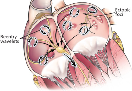
Vertebroplasty, evidence, and health care reform: What is quality care?

Should only evidence-based medicine be reimbursed?
In this time of intense discussion about ways to reduce health costs, and of President Obama’s desire to include efficacy and safety outcome data in the dialogue of how to deliver health services to everyone (although perhaps not every possible health service to everyone), the practical and philosophical implications of studies like these are worth pondering. Like it or not, the concept that all health care services will be paid for on demand by third-party payers is not a sustainable model of health care.
Randomized placebo-controlled trials are the cornerstone of evidence-based medicine. But at their best they provide only an approximation of the truth. Sample size is always a limitation. Patients and physicians in the office or operating suite do not always behave exactly like those in clinical trials. Yet, well-designed clinical trials are often considered to be the best we can do. Practice guidelines and US Food and Drug Administration approvals are based more on the results of randomized clinical trials and less on information from clinical registries and real-world observational outcome studies (which have technical foibles of their own). Approval for devices and procedures does not historically get the same type of regulatory scrutiny, but health care payers in the future may be less likely to cover the cost of procedures that lack proof of efficacy from rigorously conducted outcome studies. The development of quality care measures also depends on appropriate conduct and application of these trials.
The deadly sins and decision-making
We physicians generally take umbrage with external oversight of our decision-making. It is our job and our responsibility to balance the science (evidence-based medicine) and the art (experience and gestalt) of clinical care for the benefit of our patients. But as I thought about the impact these new studies may exert on vertebroplasty utilization, I also wondered about the factors that influence our decision-making process. For example, we have long had solid data on the value of treating systolic hypertension (we undertreat), and of treating uncomplicated urinary tract infections with only 3 days of antibiotics (we overtreat). Performance indicators suggest that this solid evidence has only a modest influence on practice patterns. Why?
I recently heard Dr. Louis B. Rice, Professor of Medicine at Case Western Reserve University and Chief of the Medical Service at the Louis Stokes Cleveland VA Medical Center, discuss the possible impact of some of the seven deadly sins on clinical decision-making. A similar analysis applies when thinking about why some treatments continue to be offered despite good evidence of only limited efficacy.
Pride plays a role. We believe that our own clinical skills will permit us to select the ideal patient to undergo a procedure or therapy, whereas such cherry-picking of patients does not generally occur in large clinical trials. This argument (and others of “external validity,” in the lingo of evidence-based medicine) has been put forth to defend the continued use of some procedures that may not have fared well against sham controls in clinical trials, and these procedures continue to flourish.
Pride may also apply to the feeling we physicians have for doing something right for our patients. This feeling may push us to believe we can succeed where an impersonal clinical trial failed. I suspect this is most keenly felt when the therapy is a procedure that depends on our own individual skills. I suspect that internists and subspecialists with special interest in osteoporosis will interpret these trials differently than surgeons and interventional radiologists who are routinely performing these procedures.
Avarice must be considered, and regulatory controls in the future may limit financial gain from these therapies. But I am not convinced that monetary greed drives all clinical decisions that go against the grain of evidence-based medicine.
And then there is gluttony: we and our patients want it all. We do not want to hear that our patient cannot be provided the most recent therapeutic advance—it might work.
Placebo effect, other issues in ‘negative’ studies
A number of factors in these trials of vertebroplasty need to be dissected and discussed. Not the least is the apparent salubrious effect of the sham procedure. This was documented previously with intra-articular injections of saline (placebo) in studies of hyaluronate joint injections for the pain of knee osteoarthritis,3 in which either type of injection provided significant pain relief. Are these truly markedly positive effects of the sham but invasive maneuvers in the vertebroplasty studies, or are we witnessing the natural history of pain resolution in these disorders (in the absence of a true nonintervention control group that could help make this distinction)?
Crossover issues in one of the vertebroplasty papers will certainly generate letters to the editor. Were patients really blinded to their procedure throughout? Which subsets of patients might have responded better or worse? What about balloon kyphoplasty?
We plan to publish commentaries from proceduralists and medical experts in osteoporosis to critique these key clinical trials for us and to put these issues into clinical perspective.
What role for evidence-based medicine?
In the meantime, I urge you to peruse these papers along with the op-ed pieces in your local newspapers as catalysts to reconsider the role evidence-based medicine should play in our daily one-on-one routine with patients, as well as in the redesign of our health care delivery and reimbursement systems. I don’t think that clinical conundrums can be resolved with a simple look at P values and confidence intervals; clinical trials are not the total story. As physicians, we always need to put the trial results into a clinical perspective. Nonetheless, our personal belief of efficacy (or lack of efficacy) also should not be the total story as we make decisions with individual patients and allocate resources within the health care system.
In the end, it should be all about giving high-quality care to the patient sitting in front of us. A question to be addressed is how well we can assess the quality of a given treatment prior to its administration.
- Kallmes DF, Comstock BA, Heagerty PJ, et al. A randomized trial of vertebroplasty for osteoporotic spinal fractures. N Engl J Med 2009; 361:569–579.
- Buchbinder R, Osborne RH, Ebeling PR, et al. A randomized trial of vertebroplasty for painful osteoporotic vertebral fractures. N Engl J Med 2009; 361:557–568.
- Lundsgaard C, Dufour N, Fallentin E, Winkel P, Gluud C. Intra-articular sodium hyaluronate 2 mL versus physiological saline 20 mL versus physiological saline 2 mL for painful knee osteoarthritis: a randomized clinical trial. Scand J Rheumatol 2008; 37:142–150.
Should only evidence-based medicine be reimbursed?
In this time of intense discussion about ways to reduce health costs, and of President Obama’s desire to include efficacy and safety outcome data in the dialogue of how to deliver health services to everyone (although perhaps not every possible health service to everyone), the practical and philosophical implications of studies like these are worth pondering. Like it or not, the concept that all health care services will be paid for on demand by third-party payers is not a sustainable model of health care.
Randomized placebo-controlled trials are the cornerstone of evidence-based medicine. But at their best they provide only an approximation of the truth. Sample size is always a limitation. Patients and physicians in the office or operating suite do not always behave exactly like those in clinical trials. Yet, well-designed clinical trials are often considered to be the best we can do. Practice guidelines and US Food and Drug Administration approvals are based more on the results of randomized clinical trials and less on information from clinical registries and real-world observational outcome studies (which have technical foibles of their own). Approval for devices and procedures does not historically get the same type of regulatory scrutiny, but health care payers in the future may be less likely to cover the cost of procedures that lack proof of efficacy from rigorously conducted outcome studies. The development of quality care measures also depends on appropriate conduct and application of these trials.
The deadly sins and decision-making
We physicians generally take umbrage with external oversight of our decision-making. It is our job and our responsibility to balance the science (evidence-based medicine) and the art (experience and gestalt) of clinical care for the benefit of our patients. But as I thought about the impact these new studies may exert on vertebroplasty utilization, I also wondered about the factors that influence our decision-making process. For example, we have long had solid data on the value of treating systolic hypertension (we undertreat), and of treating uncomplicated urinary tract infections with only 3 days of antibiotics (we overtreat). Performance indicators suggest that this solid evidence has only a modest influence on practice patterns. Why?
I recently heard Dr. Louis B. Rice, Professor of Medicine at Case Western Reserve University and Chief of the Medical Service at the Louis Stokes Cleveland VA Medical Center, discuss the possible impact of some of the seven deadly sins on clinical decision-making. A similar analysis applies when thinking about why some treatments continue to be offered despite good evidence of only limited efficacy.
Pride plays a role. We believe that our own clinical skills will permit us to select the ideal patient to undergo a procedure or therapy, whereas such cherry-picking of patients does not generally occur in large clinical trials. This argument (and others of “external validity,” in the lingo of evidence-based medicine) has been put forth to defend the continued use of some procedures that may not have fared well against sham controls in clinical trials, and these procedures continue to flourish.
Pride may also apply to the feeling we physicians have for doing something right for our patients. This feeling may push us to believe we can succeed where an impersonal clinical trial failed. I suspect this is most keenly felt when the therapy is a procedure that depends on our own individual skills. I suspect that internists and subspecialists with special interest in osteoporosis will interpret these trials differently than surgeons and interventional radiologists who are routinely performing these procedures.
Avarice must be considered, and regulatory controls in the future may limit financial gain from these therapies. But I am not convinced that monetary greed drives all clinical decisions that go against the grain of evidence-based medicine.
And then there is gluttony: we and our patients want it all. We do not want to hear that our patient cannot be provided the most recent therapeutic advance—it might work.
Placebo effect, other issues in ‘negative’ studies
A number of factors in these trials of vertebroplasty need to be dissected and discussed. Not the least is the apparent salubrious effect of the sham procedure. This was documented previously with intra-articular injections of saline (placebo) in studies of hyaluronate joint injections for the pain of knee osteoarthritis,3 in which either type of injection provided significant pain relief. Are these truly markedly positive effects of the sham but invasive maneuvers in the vertebroplasty studies, or are we witnessing the natural history of pain resolution in these disorders (in the absence of a true nonintervention control group that could help make this distinction)?
Crossover issues in one of the vertebroplasty papers will certainly generate letters to the editor. Were patients really blinded to their procedure throughout? Which subsets of patients might have responded better or worse? What about balloon kyphoplasty?
We plan to publish commentaries from proceduralists and medical experts in osteoporosis to critique these key clinical trials for us and to put these issues into clinical perspective.
What role for evidence-based medicine?
In the meantime, I urge you to peruse these papers along with the op-ed pieces in your local newspapers as catalysts to reconsider the role evidence-based medicine should play in our daily one-on-one routine with patients, as well as in the redesign of our health care delivery and reimbursement systems. I don’t think that clinical conundrums can be resolved with a simple look at P values and confidence intervals; clinical trials are not the total story. As physicians, we always need to put the trial results into a clinical perspective. Nonetheless, our personal belief of efficacy (or lack of efficacy) also should not be the total story as we make decisions with individual patients and allocate resources within the health care system.
In the end, it should be all about giving high-quality care to the patient sitting in front of us. A question to be addressed is how well we can assess the quality of a given treatment prior to its administration.
- Kallmes DF, Comstock BA, Heagerty PJ, et al. A randomized trial of vertebroplasty for osteoporotic spinal fractures. N Engl J Med 2009; 361:569–579.
- Buchbinder R, Osborne RH, Ebeling PR, et al. A randomized trial of vertebroplasty for painful osteoporotic vertebral fractures. N Engl J Med 2009; 361:557–568.
- Lundsgaard C, Dufour N, Fallentin E, Winkel P, Gluud C. Intra-articular sodium hyaluronate 2 mL versus physiological saline 20 mL versus physiological saline 2 mL for painful knee osteoarthritis: a randomized clinical trial. Scand J Rheumatol 2008; 37:142–150.