User login
CCJM delivers practical clinical articles relevant to internists, cardiologists, endocrinologists, and other specialists, all written by known experts.
Copyright © 2019 Cleveland Clinic. All rights reserved. The information provided is for educational purposes only. Use of this website is subject to the disclaimer and privacy policy.
gambling
compulsive behaviors
ammunition
assault rifle
black jack
Boko Haram
bondage
child abuse
cocaine
Daech
drug paraphernalia
explosion
gun
human trafficking
ISIL
ISIS
Islamic caliphate
Islamic state
mixed martial arts
MMA
molestation
national rifle association
NRA
nsfw
pedophile
pedophilia
poker
porn
pornography
psychedelic drug
recreational drug
sex slave rings
slot machine
terrorism
terrorist
Texas hold 'em
UFC
substance abuse
abuseed
abuseer
abusees
abuseing
abusely
abuses
aeolus
aeolused
aeoluser
aeoluses
aeolusing
aeolusly
aeoluss
ahole
aholeed
aholeer
aholees
aholeing
aholely
aholes
alcohol
alcoholed
alcoholer
alcoholes
alcoholing
alcoholly
alcohols
allman
allmaned
allmaner
allmanes
allmaning
allmanly
allmans
alted
altes
alting
altly
alts
analed
analer
anales
analing
anally
analprobe
analprobeed
analprobeer
analprobees
analprobeing
analprobely
analprobes
anals
anilingus
anilingused
anilinguser
anilinguses
anilingusing
anilingusly
anilinguss
anus
anused
anuser
anuses
anusing
anusly
anuss
areola
areolaed
areolaer
areolaes
areolaing
areolaly
areolas
areole
areoleed
areoleer
areolees
areoleing
areolely
areoles
arian
arianed
arianer
arianes
arianing
arianly
arians
aryan
aryaned
aryaner
aryanes
aryaning
aryanly
aryans
asiaed
asiaer
asiaes
asiaing
asialy
asias
ass
ass hole
ass lick
ass licked
ass licker
ass lickes
ass licking
ass lickly
ass licks
assbang
assbanged
assbangeded
assbangeder
assbangedes
assbangeding
assbangedly
assbangeds
assbanger
assbanges
assbanging
assbangly
assbangs
assbangsed
assbangser
assbangses
assbangsing
assbangsly
assbangss
assed
asser
asses
assesed
asseser
asseses
assesing
assesly
assess
assfuck
assfucked
assfucker
assfuckered
assfuckerer
assfuckeres
assfuckering
assfuckerly
assfuckers
assfuckes
assfucking
assfuckly
assfucks
asshat
asshated
asshater
asshates
asshating
asshatly
asshats
assholeed
assholeer
assholees
assholeing
assholely
assholes
assholesed
assholeser
assholeses
assholesing
assholesly
assholess
assing
assly
assmaster
assmastered
assmasterer
assmasteres
assmastering
assmasterly
assmasters
assmunch
assmunched
assmuncher
assmunches
assmunching
assmunchly
assmunchs
asss
asswipe
asswipeed
asswipeer
asswipees
asswipeing
asswipely
asswipes
asswipesed
asswipeser
asswipeses
asswipesing
asswipesly
asswipess
azz
azzed
azzer
azzes
azzing
azzly
azzs
babeed
babeer
babees
babeing
babely
babes
babesed
babeser
babeses
babesing
babesly
babess
ballsac
ballsaced
ballsacer
ballsaces
ballsacing
ballsack
ballsacked
ballsacker
ballsackes
ballsacking
ballsackly
ballsacks
ballsacly
ballsacs
ballsed
ballser
ballses
ballsing
ballsly
ballss
barf
barfed
barfer
barfes
barfing
barfly
barfs
bastard
bastarded
bastarder
bastardes
bastarding
bastardly
bastards
bastardsed
bastardser
bastardses
bastardsing
bastardsly
bastardss
bawdy
bawdyed
bawdyer
bawdyes
bawdying
bawdyly
bawdys
beaner
beanered
beanerer
beaneres
beanering
beanerly
beaners
beardedclam
beardedclamed
beardedclamer
beardedclames
beardedclaming
beardedclamly
beardedclams
beastiality
beastialityed
beastialityer
beastialityes
beastialitying
beastialityly
beastialitys
beatch
beatched
beatcher
beatches
beatching
beatchly
beatchs
beater
beatered
beaterer
beateres
beatering
beaterly
beaters
beered
beerer
beeres
beering
beerly
beeyotch
beeyotched
beeyotcher
beeyotches
beeyotching
beeyotchly
beeyotchs
beotch
beotched
beotcher
beotches
beotching
beotchly
beotchs
biatch
biatched
biatcher
biatches
biatching
biatchly
biatchs
big tits
big titsed
big titser
big titses
big titsing
big titsly
big titss
bigtits
bigtitsed
bigtitser
bigtitses
bigtitsing
bigtitsly
bigtitss
bimbo
bimboed
bimboer
bimboes
bimboing
bimboly
bimbos
bisexualed
bisexualer
bisexuales
bisexualing
bisexually
bisexuals
bitch
bitched
bitcheded
bitcheder
bitchedes
bitcheding
bitchedly
bitcheds
bitcher
bitches
bitchesed
bitcheser
bitcheses
bitchesing
bitchesly
bitchess
bitching
bitchly
bitchs
bitchy
bitchyed
bitchyer
bitchyes
bitchying
bitchyly
bitchys
bleached
bleacher
bleaches
bleaching
bleachly
bleachs
blow job
blow jobed
blow jober
blow jobes
blow jobing
blow jobly
blow jobs
blowed
blower
blowes
blowing
blowjob
blowjobed
blowjober
blowjobes
blowjobing
blowjobly
blowjobs
blowjobsed
blowjobser
blowjobses
blowjobsing
blowjobsly
blowjobss
blowly
blows
boink
boinked
boinker
boinkes
boinking
boinkly
boinks
bollock
bollocked
bollocker
bollockes
bollocking
bollockly
bollocks
bollocksed
bollockser
bollockses
bollocksing
bollocksly
bollockss
bollok
bolloked
bolloker
bollokes
bolloking
bollokly
bolloks
boner
bonered
bonerer
boneres
bonering
bonerly
boners
bonersed
bonerser
bonerses
bonersing
bonersly
bonerss
bong
bonged
bonger
bonges
bonging
bongly
bongs
boob
boobed
boober
boobes
boobies
boobiesed
boobieser
boobieses
boobiesing
boobiesly
boobiess
boobing
boobly
boobs
boobsed
boobser
boobses
boobsing
boobsly
boobss
booby
boobyed
boobyer
boobyes
boobying
boobyly
boobys
booger
boogered
boogerer
boogeres
boogering
boogerly
boogers
bookie
bookieed
bookieer
bookiees
bookieing
bookiely
bookies
bootee
booteeed
booteeer
booteees
booteeing
booteely
bootees
bootie
bootieed
bootieer
bootiees
bootieing
bootiely
booties
booty
bootyed
bootyer
bootyes
bootying
bootyly
bootys
boozeed
boozeer
boozees
boozeing
boozely
boozer
boozered
boozerer
boozeres
boozering
boozerly
boozers
boozes
boozy
boozyed
boozyer
boozyes
boozying
boozyly
boozys
bosomed
bosomer
bosomes
bosoming
bosomly
bosoms
bosomy
bosomyed
bosomyer
bosomyes
bosomying
bosomyly
bosomys
bugger
buggered
buggerer
buggeres
buggering
buggerly
buggers
bukkake
bukkakeed
bukkakeer
bukkakees
bukkakeing
bukkakely
bukkakes
bull shit
bull shited
bull shiter
bull shites
bull shiting
bull shitly
bull shits
bullshit
bullshited
bullshiter
bullshites
bullshiting
bullshitly
bullshits
bullshitsed
bullshitser
bullshitses
bullshitsing
bullshitsly
bullshitss
bullshitted
bullshitteded
bullshitteder
bullshittedes
bullshitteding
bullshittedly
bullshitteds
bullturds
bullturdsed
bullturdser
bullturdses
bullturdsing
bullturdsly
bullturdss
bung
bunged
bunger
bunges
bunging
bungly
bungs
busty
bustyed
bustyer
bustyes
bustying
bustyly
bustys
butt
butt fuck
butt fucked
butt fucker
butt fuckes
butt fucking
butt fuckly
butt fucks
butted
buttes
buttfuck
buttfucked
buttfucker
buttfuckered
buttfuckerer
buttfuckeres
buttfuckering
buttfuckerly
buttfuckers
buttfuckes
buttfucking
buttfuckly
buttfucks
butting
buttly
buttplug
buttpluged
buttpluger
buttpluges
buttpluging
buttplugly
buttplugs
butts
caca
cacaed
cacaer
cacaes
cacaing
cacaly
cacas
cahone
cahoneed
cahoneer
cahonees
cahoneing
cahonely
cahones
cameltoe
cameltoeed
cameltoeer
cameltoees
cameltoeing
cameltoely
cameltoes
carpetmuncher
carpetmunchered
carpetmuncherer
carpetmuncheres
carpetmunchering
carpetmuncherly
carpetmunchers
cawk
cawked
cawker
cawkes
cawking
cawkly
cawks
chinc
chinced
chincer
chinces
chincing
chincly
chincs
chincsed
chincser
chincses
chincsing
chincsly
chincss
chink
chinked
chinker
chinkes
chinking
chinkly
chinks
chode
chodeed
chodeer
chodees
chodeing
chodely
chodes
chodesed
chodeser
chodeses
chodesing
chodesly
chodess
clit
clited
cliter
clites
cliting
clitly
clitoris
clitorised
clitoriser
clitorises
clitorising
clitorisly
clitoriss
clitorus
clitorused
clitoruser
clitoruses
clitorusing
clitorusly
clitoruss
clits
clitsed
clitser
clitses
clitsing
clitsly
clitss
clitty
clittyed
clittyer
clittyes
clittying
clittyly
clittys
cocain
cocaine
cocained
cocaineed
cocaineer
cocainees
cocaineing
cocainely
cocainer
cocaines
cocaining
cocainly
cocains
cock
cock sucker
cock suckered
cock suckerer
cock suckeres
cock suckering
cock suckerly
cock suckers
cockblock
cockblocked
cockblocker
cockblockes
cockblocking
cockblockly
cockblocks
cocked
cocker
cockes
cockholster
cockholstered
cockholsterer
cockholsteres
cockholstering
cockholsterly
cockholsters
cocking
cockknocker
cockknockered
cockknockerer
cockknockeres
cockknockering
cockknockerly
cockknockers
cockly
cocks
cocksed
cockser
cockses
cocksing
cocksly
cocksmoker
cocksmokered
cocksmokerer
cocksmokeres
cocksmokering
cocksmokerly
cocksmokers
cockss
cocksucker
cocksuckered
cocksuckerer
cocksuckeres
cocksuckering
cocksuckerly
cocksuckers
coital
coitaled
coitaler
coitales
coitaling
coitally
coitals
commie
commieed
commieer
commiees
commieing
commiely
commies
condomed
condomer
condomes
condoming
condomly
condoms
coon
cooned
cooner
coones
cooning
coonly
coons
coonsed
coonser
coonses
coonsing
coonsly
coonss
corksucker
corksuckered
corksuckerer
corksuckeres
corksuckering
corksuckerly
corksuckers
cracked
crackwhore
crackwhoreed
crackwhoreer
crackwhorees
crackwhoreing
crackwhorely
crackwhores
crap
craped
craper
crapes
craping
craply
crappy
crappyed
crappyer
crappyes
crappying
crappyly
crappys
cum
cumed
cumer
cumes
cuming
cumly
cummin
cummined
cumminer
cummines
cumming
cumminged
cumminger
cumminges
cumminging
cummingly
cummings
cummining
cumminly
cummins
cums
cumshot
cumshoted
cumshoter
cumshotes
cumshoting
cumshotly
cumshots
cumshotsed
cumshotser
cumshotses
cumshotsing
cumshotsly
cumshotss
cumslut
cumsluted
cumsluter
cumslutes
cumsluting
cumslutly
cumsluts
cumstain
cumstained
cumstainer
cumstaines
cumstaining
cumstainly
cumstains
cunilingus
cunilingused
cunilinguser
cunilinguses
cunilingusing
cunilingusly
cunilinguss
cunnilingus
cunnilingused
cunnilinguser
cunnilinguses
cunnilingusing
cunnilingusly
cunnilinguss
cunny
cunnyed
cunnyer
cunnyes
cunnying
cunnyly
cunnys
cunt
cunted
cunter
cuntes
cuntface
cuntfaceed
cuntfaceer
cuntfacees
cuntfaceing
cuntfacely
cuntfaces
cunthunter
cunthuntered
cunthunterer
cunthunteres
cunthuntering
cunthunterly
cunthunters
cunting
cuntlick
cuntlicked
cuntlicker
cuntlickered
cuntlickerer
cuntlickeres
cuntlickering
cuntlickerly
cuntlickers
cuntlickes
cuntlicking
cuntlickly
cuntlicks
cuntly
cunts
cuntsed
cuntser
cuntses
cuntsing
cuntsly
cuntss
dago
dagoed
dagoer
dagoes
dagoing
dagoly
dagos
dagosed
dagoser
dagoses
dagosing
dagosly
dagoss
dammit
dammited
dammiter
dammites
dammiting
dammitly
dammits
damn
damned
damneded
damneder
damnedes
damneding
damnedly
damneds
damner
damnes
damning
damnit
damnited
damniter
damnites
damniting
damnitly
damnits
damnly
damns
dick
dickbag
dickbaged
dickbager
dickbages
dickbaging
dickbagly
dickbags
dickdipper
dickdippered
dickdipperer
dickdipperes
dickdippering
dickdipperly
dickdippers
dicked
dicker
dickes
dickface
dickfaceed
dickfaceer
dickfacees
dickfaceing
dickfacely
dickfaces
dickflipper
dickflippered
dickflipperer
dickflipperes
dickflippering
dickflipperly
dickflippers
dickhead
dickheaded
dickheader
dickheades
dickheading
dickheadly
dickheads
dickheadsed
dickheadser
dickheadses
dickheadsing
dickheadsly
dickheadss
dicking
dickish
dickished
dickisher
dickishes
dickishing
dickishly
dickishs
dickly
dickripper
dickrippered
dickripperer
dickripperes
dickrippering
dickripperly
dickrippers
dicks
dicksipper
dicksippered
dicksipperer
dicksipperes
dicksippering
dicksipperly
dicksippers
dickweed
dickweeded
dickweeder
dickweedes
dickweeding
dickweedly
dickweeds
dickwhipper
dickwhippered
dickwhipperer
dickwhipperes
dickwhippering
dickwhipperly
dickwhippers
dickzipper
dickzippered
dickzipperer
dickzipperes
dickzippering
dickzipperly
dickzippers
diddle
diddleed
diddleer
diddlees
diddleing
diddlely
diddles
dike
dikeed
dikeer
dikees
dikeing
dikely
dikes
dildo
dildoed
dildoer
dildoes
dildoing
dildoly
dildos
dildosed
dildoser
dildoses
dildosing
dildosly
dildoss
diligaf
diligafed
diligafer
diligafes
diligafing
diligafly
diligafs
dillweed
dillweeded
dillweeder
dillweedes
dillweeding
dillweedly
dillweeds
dimwit
dimwited
dimwiter
dimwites
dimwiting
dimwitly
dimwits
dingle
dingleed
dingleer
dinglees
dingleing
dinglely
dingles
dipship
dipshiped
dipshiper
dipshipes
dipshiping
dipshiply
dipships
dizzyed
dizzyer
dizzyes
dizzying
dizzyly
dizzys
doggiestyleed
doggiestyleer
doggiestylees
doggiestyleing
doggiestylely
doggiestyles
doggystyleed
doggystyleer
doggystylees
doggystyleing
doggystylely
doggystyles
dong
donged
donger
donges
donging
dongly
dongs
doofus
doofused
doofuser
doofuses
doofusing
doofusly
doofuss
doosh
dooshed
doosher
dooshes
dooshing
dooshly
dooshs
dopeyed
dopeyer
dopeyes
dopeying
dopeyly
dopeys
douchebag
douchebaged
douchebager
douchebages
douchebaging
douchebagly
douchebags
douchebagsed
douchebagser
douchebagses
douchebagsing
douchebagsly
douchebagss
doucheed
doucheer
douchees
doucheing
douchely
douches
douchey
doucheyed
doucheyer
doucheyes
doucheying
doucheyly
doucheys
drunk
drunked
drunker
drunkes
drunking
drunkly
drunks
dumass
dumassed
dumasser
dumasses
dumassing
dumassly
dumasss
dumbass
dumbassed
dumbasser
dumbasses
dumbassesed
dumbasseser
dumbasseses
dumbassesing
dumbassesly
dumbassess
dumbassing
dumbassly
dumbasss
dummy
dummyed
dummyer
dummyes
dummying
dummyly
dummys
dyke
dykeed
dykeer
dykees
dykeing
dykely
dykes
dykesed
dykeser
dykeses
dykesing
dykesly
dykess
erotic
eroticed
eroticer
erotices
eroticing
eroticly
erotics
extacy
extacyed
extacyer
extacyes
extacying
extacyly
extacys
extasy
extasyed
extasyer
extasyes
extasying
extasyly
extasys
fack
facked
facker
fackes
facking
fackly
facks
fag
faged
fager
fages
fagg
fagged
faggeded
faggeder
faggedes
faggeding
faggedly
faggeds
fagger
fagges
fagging
faggit
faggited
faggiter
faggites
faggiting
faggitly
faggits
faggly
faggot
faggoted
faggoter
faggotes
faggoting
faggotly
faggots
faggs
faging
fagly
fagot
fagoted
fagoter
fagotes
fagoting
fagotly
fagots
fags
fagsed
fagser
fagses
fagsing
fagsly
fagss
faig
faiged
faiger
faiges
faiging
faigly
faigs
faigt
faigted
faigter
faigtes
faigting
faigtly
faigts
fannybandit
fannybandited
fannybanditer
fannybandites
fannybanditing
fannybanditly
fannybandits
farted
farter
fartes
farting
fartknocker
fartknockered
fartknockerer
fartknockeres
fartknockering
fartknockerly
fartknockers
fartly
farts
felch
felched
felcher
felchered
felcherer
felcheres
felchering
felcherly
felchers
felches
felching
felchinged
felchinger
felchinges
felchinging
felchingly
felchings
felchly
felchs
fellate
fellateed
fellateer
fellatees
fellateing
fellately
fellates
fellatio
fellatioed
fellatioer
fellatioes
fellatioing
fellatioly
fellatios
feltch
feltched
feltcher
feltchered
feltcherer
feltcheres
feltchering
feltcherly
feltchers
feltches
feltching
feltchly
feltchs
feom
feomed
feomer
feomes
feoming
feomly
feoms
fisted
fisteded
fisteder
fistedes
fisteding
fistedly
fisteds
fisting
fistinged
fistinger
fistinges
fistinging
fistingly
fistings
fisty
fistyed
fistyer
fistyes
fistying
fistyly
fistys
floozy
floozyed
floozyer
floozyes
floozying
floozyly
floozys
foad
foaded
foader
foades
foading
foadly
foads
fondleed
fondleer
fondlees
fondleing
fondlely
fondles
foobar
foobared
foobarer
foobares
foobaring
foobarly
foobars
freex
freexed
freexer
freexes
freexing
freexly
freexs
frigg
frigga
friggaed
friggaer
friggaes
friggaing
friggaly
friggas
frigged
frigger
frigges
frigging
friggly
friggs
fubar
fubared
fubarer
fubares
fubaring
fubarly
fubars
fuck
fuckass
fuckassed
fuckasser
fuckasses
fuckassing
fuckassly
fuckasss
fucked
fuckeded
fuckeder
fuckedes
fuckeding
fuckedly
fuckeds
fucker
fuckered
fuckerer
fuckeres
fuckering
fuckerly
fuckers
fuckes
fuckface
fuckfaceed
fuckfaceer
fuckfacees
fuckfaceing
fuckfacely
fuckfaces
fuckin
fuckined
fuckiner
fuckines
fucking
fuckinged
fuckinger
fuckinges
fuckinging
fuckingly
fuckings
fuckining
fuckinly
fuckins
fuckly
fucknugget
fucknuggeted
fucknuggeter
fucknuggetes
fucknuggeting
fucknuggetly
fucknuggets
fucknut
fucknuted
fucknuter
fucknutes
fucknuting
fucknutly
fucknuts
fuckoff
fuckoffed
fuckoffer
fuckoffes
fuckoffing
fuckoffly
fuckoffs
fucks
fucksed
fuckser
fuckses
fucksing
fucksly
fuckss
fucktard
fucktarded
fucktarder
fucktardes
fucktarding
fucktardly
fucktards
fuckup
fuckuped
fuckuper
fuckupes
fuckuping
fuckuply
fuckups
fuckwad
fuckwaded
fuckwader
fuckwades
fuckwading
fuckwadly
fuckwads
fuckwit
fuckwited
fuckwiter
fuckwites
fuckwiting
fuckwitly
fuckwits
fudgepacker
fudgepackered
fudgepackerer
fudgepackeres
fudgepackering
fudgepackerly
fudgepackers
fuk
fuked
fuker
fukes
fuking
fukly
fuks
fvck
fvcked
fvcker
fvckes
fvcking
fvckly
fvcks
fxck
fxcked
fxcker
fxckes
fxcking
fxckly
fxcks
gae
gaeed
gaeer
gaees
gaeing
gaely
gaes
gai
gaied
gaier
gaies
gaiing
gaily
gais
ganja
ganjaed
ganjaer
ganjaes
ganjaing
ganjaly
ganjas
gayed
gayer
gayes
gaying
gayly
gays
gaysed
gayser
gayses
gaysing
gaysly
gayss
gey
geyed
geyer
geyes
geying
geyly
geys
gfc
gfced
gfcer
gfces
gfcing
gfcly
gfcs
gfy
gfyed
gfyer
gfyes
gfying
gfyly
gfys
ghay
ghayed
ghayer
ghayes
ghaying
ghayly
ghays
ghey
gheyed
gheyer
gheyes
gheying
gheyly
gheys
gigolo
gigoloed
gigoloer
gigoloes
gigoloing
gigololy
gigolos
goatse
goatseed
goatseer
goatsees
goatseing
goatsely
goatses
godamn
godamned
godamner
godamnes
godamning
godamnit
godamnited
godamniter
godamnites
godamniting
godamnitly
godamnits
godamnly
godamns
goddam
goddamed
goddamer
goddames
goddaming
goddamly
goddammit
goddammited
goddammiter
goddammites
goddammiting
goddammitly
goddammits
goddamn
goddamned
goddamner
goddamnes
goddamning
goddamnly
goddamns
goddams
goldenshower
goldenshowered
goldenshowerer
goldenshoweres
goldenshowering
goldenshowerly
goldenshowers
gonad
gonaded
gonader
gonades
gonading
gonadly
gonads
gonadsed
gonadser
gonadses
gonadsing
gonadsly
gonadss
gook
gooked
gooker
gookes
gooking
gookly
gooks
gooksed
gookser
gookses
gooksing
gooksly
gookss
gringo
gringoed
gringoer
gringoes
gringoing
gringoly
gringos
gspot
gspoted
gspoter
gspotes
gspoting
gspotly
gspots
gtfo
gtfoed
gtfoer
gtfoes
gtfoing
gtfoly
gtfos
guido
guidoed
guidoer
guidoes
guidoing
guidoly
guidos
handjob
handjobed
handjober
handjobes
handjobing
handjobly
handjobs
hard on
hard oned
hard oner
hard ones
hard oning
hard only
hard ons
hardknight
hardknighted
hardknighter
hardknightes
hardknighting
hardknightly
hardknights
hebe
hebeed
hebeer
hebees
hebeing
hebely
hebes
heeb
heebed
heeber
heebes
heebing
heebly
heebs
hell
helled
heller
helles
helling
hellly
hells
hemp
hemped
hemper
hempes
hemping
hemply
hemps
heroined
heroiner
heroines
heroining
heroinly
heroins
herp
herped
herper
herpes
herpesed
herpeser
herpeses
herpesing
herpesly
herpess
herping
herply
herps
herpy
herpyed
herpyer
herpyes
herpying
herpyly
herpys
hitler
hitlered
hitlerer
hitleres
hitlering
hitlerly
hitlers
hived
hiver
hives
hiving
hivly
hivs
hobag
hobaged
hobager
hobages
hobaging
hobagly
hobags
homey
homeyed
homeyer
homeyes
homeying
homeyly
homeys
homo
homoed
homoer
homoes
homoey
homoeyed
homoeyer
homoeyes
homoeying
homoeyly
homoeys
homoing
homoly
homos
honky
honkyed
honkyer
honkyes
honkying
honkyly
honkys
hooch
hooched
hoocher
hooches
hooching
hoochly
hoochs
hookah
hookahed
hookaher
hookahes
hookahing
hookahly
hookahs
hooker
hookered
hookerer
hookeres
hookering
hookerly
hookers
hoor
hoored
hoorer
hoores
hooring
hoorly
hoors
hootch
hootched
hootcher
hootches
hootching
hootchly
hootchs
hooter
hootered
hooterer
hooteres
hootering
hooterly
hooters
hootersed
hooterser
hooterses
hootersing
hootersly
hooterss
horny
hornyed
hornyer
hornyes
hornying
hornyly
hornys
houstoned
houstoner
houstones
houstoning
houstonly
houstons
hump
humped
humpeded
humpeder
humpedes
humpeding
humpedly
humpeds
humper
humpes
humping
humpinged
humpinger
humpinges
humpinging
humpingly
humpings
humply
humps
husbanded
husbander
husbandes
husbanding
husbandly
husbands
hussy
hussyed
hussyer
hussyes
hussying
hussyly
hussys
hymened
hymener
hymenes
hymening
hymenly
hymens
inbred
inbreded
inbreder
inbredes
inbreding
inbredly
inbreds
incest
incested
incester
incestes
incesting
incestly
incests
injun
injuned
injuner
injunes
injuning
injunly
injuns
jackass
jackassed
jackasser
jackasses
jackassing
jackassly
jackasss
jackhole
jackholeed
jackholeer
jackholees
jackholeing
jackholely
jackholes
jackoff
jackoffed
jackoffer
jackoffes
jackoffing
jackoffly
jackoffs
jap
japed
japer
japes
japing
japly
japs
japsed
japser
japses
japsing
japsly
japss
jerkoff
jerkoffed
jerkoffer
jerkoffes
jerkoffing
jerkoffly
jerkoffs
jerks
jism
jismed
jismer
jismes
jisming
jismly
jisms
jiz
jized
jizer
jizes
jizing
jizly
jizm
jizmed
jizmer
jizmes
jizming
jizmly
jizms
jizs
jizz
jizzed
jizzeded
jizzeder
jizzedes
jizzeding
jizzedly
jizzeds
jizzer
jizzes
jizzing
jizzly
jizzs
junkie
junkieed
junkieer
junkiees
junkieing
junkiely
junkies
junky
junkyed
junkyer
junkyes
junkying
junkyly
junkys
kike
kikeed
kikeer
kikees
kikeing
kikely
kikes
kikesed
kikeser
kikeses
kikesing
kikesly
kikess
killed
killer
killes
killing
killly
kills
kinky
kinkyed
kinkyer
kinkyes
kinkying
kinkyly
kinkys
kkk
kkked
kkker
kkkes
kkking
kkkly
kkks
klan
klaned
klaner
klanes
klaning
klanly
klans
knobend
knobended
knobender
knobendes
knobending
knobendly
knobends
kooch
kooched
koocher
kooches
koochesed
koocheser
koocheses
koochesing
koochesly
koochess
kooching
koochly
koochs
kootch
kootched
kootcher
kootches
kootching
kootchly
kootchs
kraut
krauted
krauter
krautes
krauting
krautly
krauts
kyke
kykeed
kykeer
kykees
kykeing
kykely
kykes
lech
leched
lecher
leches
leching
lechly
lechs
leper
lepered
leperer
leperes
lepering
leperly
lepers
lesbiansed
lesbianser
lesbianses
lesbiansing
lesbiansly
lesbianss
lesbo
lesboed
lesboer
lesboes
lesboing
lesboly
lesbos
lesbosed
lesboser
lesboses
lesbosing
lesbosly
lesboss
lez
lezbianed
lezbianer
lezbianes
lezbianing
lezbianly
lezbians
lezbiansed
lezbianser
lezbianses
lezbiansing
lezbiansly
lezbianss
lezbo
lezboed
lezboer
lezboes
lezboing
lezboly
lezbos
lezbosed
lezboser
lezboses
lezbosing
lezbosly
lezboss
lezed
lezer
lezes
lezing
lezly
lezs
lezzie
lezzieed
lezzieer
lezziees
lezzieing
lezziely
lezzies
lezziesed
lezzieser
lezzieses
lezziesing
lezziesly
lezziess
lezzy
lezzyed
lezzyer
lezzyes
lezzying
lezzyly
lezzys
lmaoed
lmaoer
lmaoes
lmaoing
lmaoly
lmaos
lmfao
lmfaoed
lmfaoer
lmfaoes
lmfaoing
lmfaoly
lmfaos
loined
loiner
loines
loining
loinly
loins
loinsed
loinser
loinses
loinsing
loinsly
loinss
lubeed
lubeer
lubees
lubeing
lubely
lubes
lusty
lustyed
lustyer
lustyes
lustying
lustyly
lustys
massa
massaed
massaer
massaes
massaing
massaly
massas
masterbate
masterbateed
masterbateer
masterbatees
masterbateing
masterbately
masterbates
masterbating
masterbatinged
masterbatinger
masterbatinges
masterbatinging
masterbatingly
masterbatings
masterbation
masterbationed
masterbationer
masterbationes
masterbationing
masterbationly
masterbations
masturbate
masturbateed
masturbateer
masturbatees
masturbateing
masturbately
masturbates
masturbating
masturbatinged
masturbatinger
masturbatinges
masturbatinging
masturbatingly
masturbatings
masturbation
masturbationed
masturbationer
masturbationes
masturbationing
masturbationly
masturbations
methed
mether
methes
mething
methly
meths
militaryed
militaryer
militaryes
militarying
militaryly
militarys
mofo
mofoed
mofoer
mofoes
mofoing
mofoly
mofos
molest
molested
molester
molestes
molesting
molestly
molests
moolie
moolieed
moolieer
mooliees
moolieing
mooliely
moolies
moron
moroned
moroner
morones
moroning
moronly
morons
motherfucka
motherfuckaed
motherfuckaer
motherfuckaes
motherfuckaing
motherfuckaly
motherfuckas
motherfucker
motherfuckered
motherfuckerer
motherfuckeres
motherfuckering
motherfuckerly
motherfuckers
motherfucking
motherfuckinged
motherfuckinger
motherfuckinges
motherfuckinging
motherfuckingly
motherfuckings
mtherfucker
mtherfuckered
mtherfuckerer
mtherfuckeres
mtherfuckering
mtherfuckerly
mtherfuckers
mthrfucker
mthrfuckered
mthrfuckerer
mthrfuckeres
mthrfuckering
mthrfuckerly
mthrfuckers
mthrfucking
mthrfuckinged
mthrfuckinger
mthrfuckinges
mthrfuckinging
mthrfuckingly
mthrfuckings
muff
muffdiver
muffdivered
muffdiverer
muffdiveres
muffdivering
muffdiverly
muffdivers
muffed
muffer
muffes
muffing
muffly
muffs
murdered
murderer
murderes
murdering
murderly
murders
muthafuckaz
muthafuckazed
muthafuckazer
muthafuckazes
muthafuckazing
muthafuckazly
muthafuckazs
muthafucker
muthafuckered
muthafuckerer
muthafuckeres
muthafuckering
muthafuckerly
muthafuckers
mutherfucker
mutherfuckered
mutherfuckerer
mutherfuckeres
mutherfuckering
mutherfuckerly
mutherfuckers
mutherfucking
mutherfuckinged
mutherfuckinger
mutherfuckinges
mutherfuckinging
mutherfuckingly
mutherfuckings
muthrfucking
muthrfuckinged
muthrfuckinger
muthrfuckinges
muthrfuckinging
muthrfuckingly
muthrfuckings
nad
naded
nader
nades
nading
nadly
nads
nadsed
nadser
nadses
nadsing
nadsly
nadss
nakeded
nakeder
nakedes
nakeding
nakedly
nakeds
napalm
napalmed
napalmer
napalmes
napalming
napalmly
napalms
nappy
nappyed
nappyer
nappyes
nappying
nappyly
nappys
nazi
nazied
nazier
nazies
naziing
nazily
nazis
nazism
nazismed
nazismer
nazismes
nazisming
nazismly
nazisms
negro
negroed
negroer
negroes
negroing
negroly
negros
nigga
niggaed
niggaer
niggaes
niggah
niggahed
niggaher
niggahes
niggahing
niggahly
niggahs
niggaing
niggaly
niggas
niggased
niggaser
niggases
niggasing
niggasly
niggass
niggaz
niggazed
niggazer
niggazes
niggazing
niggazly
niggazs
nigger
niggered
niggerer
niggeres
niggering
niggerly
niggers
niggersed
niggerser
niggerses
niggersing
niggersly
niggerss
niggle
niggleed
niggleer
nigglees
niggleing
nigglely
niggles
niglet
nigleted
nigleter
nigletes
nigleting
nigletly
niglets
nimrod
nimroded
nimroder
nimrodes
nimroding
nimrodly
nimrods
ninny
ninnyed
ninnyer
ninnyes
ninnying
ninnyly
ninnys
nooky
nookyed
nookyer
nookyes
nookying
nookyly
nookys
nuccitelli
nuccitellied
nuccitellier
nuccitellies
nuccitelliing
nuccitellily
nuccitellis
nympho
nymphoed
nymphoer
nymphoes
nymphoing
nympholy
nymphos
opium
opiumed
opiumer
opiumes
opiuming
opiumly
opiums
orgies
orgiesed
orgieser
orgieses
orgiesing
orgiesly
orgiess
orgy
orgyed
orgyer
orgyes
orgying
orgyly
orgys
paddy
paddyed
paddyer
paddyes
paddying
paddyly
paddys
paki
pakied
pakier
pakies
pakiing
pakily
pakis
pantie
pantieed
pantieer
pantiees
pantieing
pantiely
panties
pantiesed
pantieser
pantieses
pantiesing
pantiesly
pantiess
panty
pantyed
pantyer
pantyes
pantying
pantyly
pantys
pastie
pastieed
pastieer
pastiees
pastieing
pastiely
pasties
pasty
pastyed
pastyer
pastyes
pastying
pastyly
pastys
pecker
peckered
peckerer
peckeres
peckering
peckerly
peckers
pedo
pedoed
pedoer
pedoes
pedoing
pedoly
pedophile
pedophileed
pedophileer
pedophilees
pedophileing
pedophilely
pedophiles
pedophilia
pedophiliac
pedophiliaced
pedophiliacer
pedophiliaces
pedophiliacing
pedophiliacly
pedophiliacs
pedophiliaed
pedophiliaer
pedophiliaes
pedophiliaing
pedophilialy
pedophilias
pedos
penial
penialed
penialer
peniales
penialing
penially
penials
penile
penileed
penileer
penilees
penileing
penilely
peniles
penis
penised
peniser
penises
penising
penisly
peniss
perversion
perversioned
perversioner
perversiones
perversioning
perversionly
perversions
peyote
peyoteed
peyoteer
peyotees
peyoteing
peyotely
peyotes
phuck
phucked
phucker
phuckes
phucking
phuckly
phucks
pillowbiter
pillowbitered
pillowbiterer
pillowbiteres
pillowbitering
pillowbiterly
pillowbiters
pimp
pimped
pimper
pimpes
pimping
pimply
pimps
pinko
pinkoed
pinkoer
pinkoes
pinkoing
pinkoly
pinkos
pissed
pisseded
pisseder
pissedes
pisseding
pissedly
pisseds
pisser
pisses
pissing
pissly
pissoff
pissoffed
pissoffer
pissoffes
pissoffing
pissoffly
pissoffs
pisss
polack
polacked
polacker
polackes
polacking
polackly
polacks
pollock
pollocked
pollocker
pollockes
pollocking
pollockly
pollocks
poon
pooned
pooner
poones
pooning
poonly
poons
poontang
poontanged
poontanger
poontanges
poontanging
poontangly
poontangs
porn
porned
porner
pornes
porning
pornly
porno
pornoed
pornoer
pornoes
pornography
pornographyed
pornographyer
pornographyes
pornographying
pornographyly
pornographys
pornoing
pornoly
pornos
porns
prick
pricked
pricker
prickes
pricking
prickly
pricks
prig
priged
priger
priges
priging
prigly
prigs
prostitute
prostituteed
prostituteer
prostitutees
prostituteing
prostitutely
prostitutes
prude
prudeed
prudeer
prudees
prudeing
prudely
prudes
punkass
punkassed
punkasser
punkasses
punkassing
punkassly
punkasss
punky
punkyed
punkyer
punkyes
punkying
punkyly
punkys
puss
pussed
pusser
pusses
pussies
pussiesed
pussieser
pussieses
pussiesing
pussiesly
pussiess
pussing
pussly
pusss
pussy
pussyed
pussyer
pussyes
pussying
pussyly
pussypounder
pussypoundered
pussypounderer
pussypounderes
pussypoundering
pussypounderly
pussypounders
pussys
puto
putoed
putoer
putoes
putoing
putoly
putos
queaf
queafed
queafer
queafes
queafing
queafly
queafs
queef
queefed
queefer
queefes
queefing
queefly
queefs
queer
queered
queerer
queeres
queering
queerly
queero
queeroed
queeroer
queeroes
queeroing
queeroly
queeros
queers
queersed
queerser
queerses
queersing
queersly
queerss
quicky
quickyed
quickyer
quickyes
quickying
quickyly
quickys
quim
quimed
quimer
quimes
quiming
quimly
quims
racy
racyed
racyer
racyes
racying
racyly
racys
rape
raped
rapeded
rapeder
rapedes
rapeding
rapedly
rapeds
rapeed
rapeer
rapees
rapeing
rapely
raper
rapered
raperer
raperes
rapering
raperly
rapers
rapes
rapist
rapisted
rapister
rapistes
rapisting
rapistly
rapists
raunch
raunched
rauncher
raunches
raunching
raunchly
raunchs
rectus
rectused
rectuser
rectuses
rectusing
rectusly
rectuss
reefer
reefered
reeferer
reeferes
reefering
reeferly
reefers
reetard
reetarded
reetarder
reetardes
reetarding
reetardly
reetards
reich
reiched
reicher
reiches
reiching
reichly
reichs
retard
retarded
retardeded
retardeder
retardedes
retardeding
retardedly
retardeds
retarder
retardes
retarding
retardly
retards
rimjob
rimjobed
rimjober
rimjobes
rimjobing
rimjobly
rimjobs
ritard
ritarded
ritarder
ritardes
ritarding
ritardly
ritards
rtard
rtarded
rtarder
rtardes
rtarding
rtardly
rtards
rum
rumed
rumer
rumes
ruming
rumly
rump
rumped
rumper
rumpes
rumping
rumply
rumprammer
rumprammered
rumprammerer
rumprammeres
rumprammering
rumprammerly
rumprammers
rumps
rums
ruski
ruskied
ruskier
ruskies
ruskiing
ruskily
ruskis
sadism
sadismed
sadismer
sadismes
sadisming
sadismly
sadisms
sadist
sadisted
sadister
sadistes
sadisting
sadistly
sadists
scag
scaged
scager
scages
scaging
scagly
scags
scantily
scantilyed
scantilyer
scantilyes
scantilying
scantilyly
scantilys
schlong
schlonged
schlonger
schlonges
schlonging
schlongly
schlongs
scrog
scroged
scroger
scroges
scroging
scrogly
scrogs
scrot
scrote
scroted
scroteed
scroteer
scrotees
scroteing
scrotely
scroter
scrotes
scroting
scrotly
scrots
scrotum
scrotumed
scrotumer
scrotumes
scrotuming
scrotumly
scrotums
scrud
scruded
scruder
scrudes
scruding
scrudly
scruds
scum
scumed
scumer
scumes
scuming
scumly
scums
seaman
seamaned
seamaner
seamanes
seamaning
seamanly
seamans
seamen
seamened
seamener
seamenes
seamening
seamenly
seamens
seduceed
seduceer
seducees
seduceing
seducely
seduces
semen
semened
semener
semenes
semening
semenly
semens
shamedame
shamedameed
shamedameer
shamedamees
shamedameing
shamedamely
shamedames
shit
shite
shiteater
shiteatered
shiteaterer
shiteateres
shiteatering
shiteaterly
shiteaters
shited
shiteed
shiteer
shitees
shiteing
shitely
shiter
shites
shitface
shitfaceed
shitfaceer
shitfacees
shitfaceing
shitfacely
shitfaces
shithead
shitheaded
shitheader
shitheades
shitheading
shitheadly
shitheads
shithole
shitholeed
shitholeer
shitholees
shitholeing
shitholely
shitholes
shithouse
shithouseed
shithouseer
shithousees
shithouseing
shithousely
shithouses
shiting
shitly
shits
shitsed
shitser
shitses
shitsing
shitsly
shitss
shitt
shitted
shitteded
shitteder
shittedes
shitteding
shittedly
shitteds
shitter
shittered
shitterer
shitteres
shittering
shitterly
shitters
shittes
shitting
shittly
shitts
shitty
shittyed
shittyer
shittyes
shittying
shittyly
shittys
shiz
shized
shizer
shizes
shizing
shizly
shizs
shooted
shooter
shootes
shooting
shootly
shoots
sissy
sissyed
sissyer
sissyes
sissying
sissyly
sissys
skag
skaged
skager
skages
skaging
skagly
skags
skank
skanked
skanker
skankes
skanking
skankly
skanks
slave
slaveed
slaveer
slavees
slaveing
slavely
slaves
sleaze
sleazeed
sleazeer
sleazees
sleazeing
sleazely
sleazes
sleazy
sleazyed
sleazyer
sleazyes
sleazying
sleazyly
sleazys
slut
slutdumper
slutdumpered
slutdumperer
slutdumperes
slutdumpering
slutdumperly
slutdumpers
sluted
sluter
slutes
sluting
slutkiss
slutkissed
slutkisser
slutkisses
slutkissing
slutkissly
slutkisss
slutly
sluts
slutsed
slutser
slutses
slutsing
slutsly
slutss
smegma
smegmaed
smegmaer
smegmaes
smegmaing
smegmaly
smegmas
smut
smuted
smuter
smutes
smuting
smutly
smuts
smutty
smuttyed
smuttyer
smuttyes
smuttying
smuttyly
smuttys
snatch
snatched
snatcher
snatches
snatching
snatchly
snatchs
sniper
snipered
sniperer
sniperes
snipering
sniperly
snipers
snort
snorted
snorter
snortes
snorting
snortly
snorts
snuff
snuffed
snuffer
snuffes
snuffing
snuffly
snuffs
sodom
sodomed
sodomer
sodomes
sodoming
sodomly
sodoms
spic
spiced
spicer
spices
spicing
spick
spicked
spicker
spickes
spicking
spickly
spicks
spicly
spics
spik
spoof
spoofed
spoofer
spoofes
spoofing
spoofly
spoofs
spooge
spoogeed
spoogeer
spoogees
spoogeing
spoogely
spooges
spunk
spunked
spunker
spunkes
spunking
spunkly
spunks
steamyed
steamyer
steamyes
steamying
steamyly
steamys
stfu
stfued
stfuer
stfues
stfuing
stfuly
stfus
stiffy
stiffyed
stiffyer
stiffyes
stiffying
stiffyly
stiffys
stoneded
stoneder
stonedes
stoneding
stonedly
stoneds
stupided
stupider
stupides
stupiding
stupidly
stupids
suckeded
suckeder
suckedes
suckeding
suckedly
suckeds
sucker
suckes
sucking
suckinged
suckinger
suckinges
suckinging
suckingly
suckings
suckly
sucks
sumofabiatch
sumofabiatched
sumofabiatcher
sumofabiatches
sumofabiatching
sumofabiatchly
sumofabiatchs
tard
tarded
tarder
tardes
tarding
tardly
tards
tawdry
tawdryed
tawdryer
tawdryes
tawdrying
tawdryly
tawdrys
teabagging
teabagginged
teabagginger
teabagginges
teabagginging
teabaggingly
teabaggings
terd
terded
terder
terdes
terding
terdly
terds
teste
testee
testeed
testeeed
testeeer
testeees
testeeing
testeely
testeer
testees
testeing
testely
testes
testesed
testeser
testeses
testesing
testesly
testess
testicle
testicleed
testicleer
testiclees
testicleing
testiclely
testicles
testis
testised
testiser
testises
testising
testisly
testiss
thrusted
thruster
thrustes
thrusting
thrustly
thrusts
thug
thuged
thuger
thuges
thuging
thugly
thugs
tinkle
tinkleed
tinkleer
tinklees
tinkleing
tinklely
tinkles
tit
tited
titer
tites
titfuck
titfucked
titfucker
titfuckes
titfucking
titfuckly
titfucks
titi
titied
titier
tities
titiing
titily
titing
titis
titly
tits
titsed
titser
titses
titsing
titsly
titss
tittiefucker
tittiefuckered
tittiefuckerer
tittiefuckeres
tittiefuckering
tittiefuckerly
tittiefuckers
titties
tittiesed
tittieser
tittieses
tittiesing
tittiesly
tittiess
titty
tittyed
tittyer
tittyes
tittyfuck
tittyfucked
tittyfucker
tittyfuckered
tittyfuckerer
tittyfuckeres
tittyfuckering
tittyfuckerly
tittyfuckers
tittyfuckes
tittyfucking
tittyfuckly
tittyfucks
tittying
tittyly
tittys
toke
tokeed
tokeer
tokees
tokeing
tokely
tokes
toots
tootsed
tootser
tootses
tootsing
tootsly
tootss
tramp
tramped
tramper
trampes
tramping
tramply
tramps
transsexualed
transsexualer
transsexuales
transsexualing
transsexually
transsexuals
trashy
trashyed
trashyer
trashyes
trashying
trashyly
trashys
tubgirl
tubgirled
tubgirler
tubgirles
tubgirling
tubgirlly
tubgirls
turd
turded
turder
turdes
turding
turdly
turds
tush
tushed
tusher
tushes
tushing
tushly
tushs
twat
twated
twater
twates
twating
twatly
twats
twatsed
twatser
twatses
twatsing
twatsly
twatss
undies
undiesed
undieser
undieses
undiesing
undiesly
undiess
unweded
unweder
unwedes
unweding
unwedly
unweds
uzi
uzied
uzier
uzies
uziing
uzily
uzis
vag
vaged
vager
vages
vaging
vagly
vags
valium
valiumed
valiumer
valiumes
valiuming
valiumly
valiums
venous
virgined
virginer
virgines
virgining
virginly
virgins
vixen
vixened
vixener
vixenes
vixening
vixenly
vixens
vodkaed
vodkaer
vodkaes
vodkaing
vodkaly
vodkas
voyeur
voyeured
voyeurer
voyeures
voyeuring
voyeurly
voyeurs
vulgar
vulgared
vulgarer
vulgares
vulgaring
vulgarly
vulgars
wang
wanged
wanger
wanges
wanging
wangly
wangs
wank
wanked
wanker
wankered
wankerer
wankeres
wankering
wankerly
wankers
wankes
wanking
wankly
wanks
wazoo
wazooed
wazooer
wazooes
wazooing
wazooly
wazoos
wedgie
wedgieed
wedgieer
wedgiees
wedgieing
wedgiely
wedgies
weeded
weeder
weedes
weeding
weedly
weeds
weenie
weenieed
weenieer
weeniees
weenieing
weeniely
weenies
weewee
weeweeed
weeweeer
weeweees
weeweeing
weeweely
weewees
weiner
weinered
weinerer
weineres
weinering
weinerly
weiners
weirdo
weirdoed
weirdoer
weirdoes
weirdoing
weirdoly
weirdos
wench
wenched
wencher
wenches
wenching
wenchly
wenchs
wetback
wetbacked
wetbacker
wetbackes
wetbacking
wetbackly
wetbacks
whitey
whiteyed
whiteyer
whiteyes
whiteying
whiteyly
whiteys
whiz
whized
whizer
whizes
whizing
whizly
whizs
whoralicious
whoralicioused
whoraliciouser
whoraliciouses
whoraliciousing
whoraliciously
whoraliciouss
whore
whorealicious
whorealicioused
whorealiciouser
whorealiciouses
whorealiciousing
whorealiciously
whorealiciouss
whored
whoreded
whoreder
whoredes
whoreding
whoredly
whoreds
whoreed
whoreer
whorees
whoreface
whorefaceed
whorefaceer
whorefacees
whorefaceing
whorefacely
whorefaces
whorehopper
whorehoppered
whorehopperer
whorehopperes
whorehoppering
whorehopperly
whorehoppers
whorehouse
whorehouseed
whorehouseer
whorehousees
whorehouseing
whorehousely
whorehouses
whoreing
whorely
whores
whoresed
whoreser
whoreses
whoresing
whoresly
whoress
whoring
whoringed
whoringer
whoringes
whoringing
whoringly
whorings
wigger
wiggered
wiggerer
wiggeres
wiggering
wiggerly
wiggers
woody
woodyed
woodyer
woodyes
woodying
woodyly
woodys
wop
woped
woper
wopes
woping
woply
wops
wtf
wtfed
wtfer
wtfes
wtfing
wtfly
wtfs
xxx
xxxed
xxxer
xxxes
xxxing
xxxly
xxxs
yeasty
yeastyed
yeastyer
yeastyes
yeastying
yeastyly
yeastys
yobbo
yobboed
yobboer
yobboes
yobboing
yobboly
yobbos
zoophile
zoophileed
zoophileer
zoophilees
zoophileing
zoophilely
zoophiles
anal
ass
ass lick
balls
ballsac
bisexual
bleach
causas
cheap
cost of miracles
cunt
display network stats
fart
fda and death
fda AND warn
fda AND warning
fda AND warns
feom
fuck
gfc
humira AND expensive
illegal
madvocate
masturbation
nuccitelli
overdose
porn
shit
snort
texarkana
direct\-acting antivirals
assistance
ombitasvir
support path
harvoni
abbvie
direct-acting antivirals
paritaprevir
advocacy
ledipasvir
vpak
ritonavir with dasabuvir
program
gilead
greedy
financial
needy
fake-ovir
viekira pak
v pak
sofosbuvir
support
oasis
discount
dasabuvir
protest
ritonavir
section[contains(@class, 'nav-hidden')]
footer[@id='footer']
div[contains(@class, 'pane-pub-article-cleveland-clinic')]
div[contains(@class, 'pane-pub-home-cleveland-clinic')]
div[contains(@class, 'pane-pub-topic-cleveland-clinic')]
div[contains(@class, 'panel-panel-inner')]
div[contains(@class, 'pane-node-field-article-topics')]
section[contains(@class, 'footer-nav-section-wrapper')]
Epidemiology of gout
Both the incidence and the prevalence of gout appear to be increasing worldwide.1,2 Risk factors for the development of gout are related to our increasing longevity, dietary and lifestyle changes, and an increased prevalence of comorbid conditions. Patients with conditions such as hypertension, diabetes, cardiovascular disease, and the metabolic syndrome have an increased risk of developing hyperuricemia and gout,1,2 and such conditions are frequently managed by primary care physicians. This paper discusses how these conditions, along with diet, alcohol intake, and lifestyle, contribute to the increasing prevalence of hyperuricemia and gout.1
PREVALENCE AND INCIDENCE: BOTH ARE RISING
The Third National Health and Nutrition Examination Survey (NHANES III) estimated the prevalence of gout in the US population to be 5.1 million between 1988 and 1994.3 Data from a US managed care claims database revealed an increase in gout prevalence from 2.9 cases per 1,000 persons in 1990 to 5.2 cases per 1,000 persons in 1999.4 Other studies indicate that gout is becoming more prevalent in societies such as New Zealand and Taiwan as well as in the United States.5,6
The NHANES III data show that gout affected more than 3 million men aged 40 years or older, and 1.7 million women aged 40 years or older, in the period from 1988 to 1994.3 The estimated prevalence of gout among men aged 40 years or older makes this disease more common than rheumatoid arthritis and the most common form of inflammatory arthritis in adult men.7–9
RISK FACTORS FOR GOUT DEVELOPMENT
Sex and age
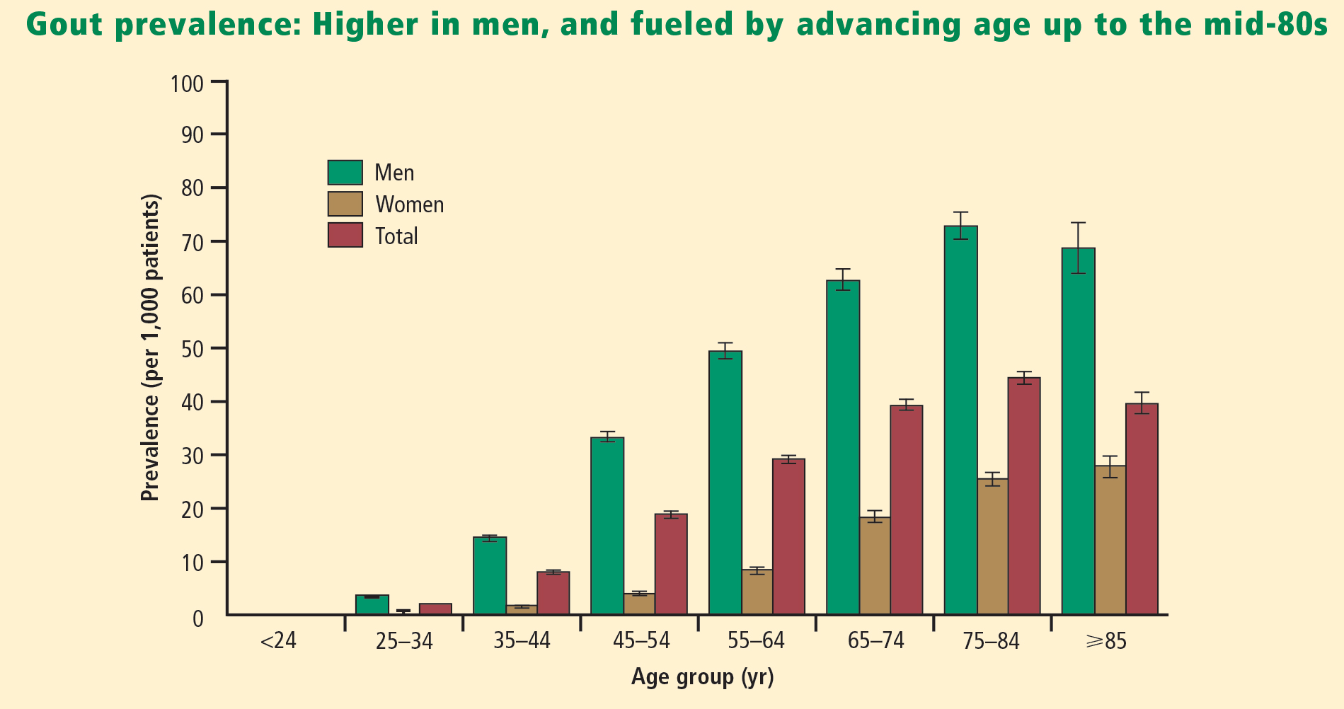
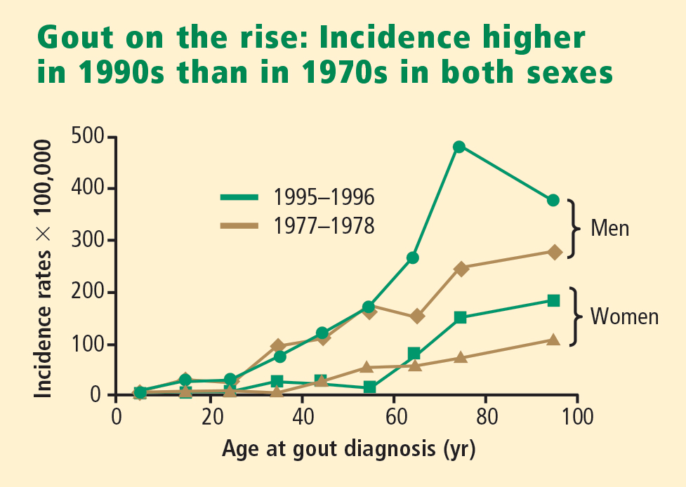
Men have higher serum urate levels than women do, which increases their risk of developing gout. Development of gout before age 30 years is overwhelmingly more likely in men than in women.7,9,10 The prevalence of gout in men increases with advancing age and peaks between ages 75 and 84 years.6 Women have an increased risk of developing gout after menopause; the risk begins to rise at about age 45 years with the decrease in estrogen levels.10,11 The incidence of gout becomes approximately equal between the sexes after age 60 years.10,12
It is important for clinicians to bear these factors in mind when taking the patient history and considering gout in the differential diagnosis. Although gout is more common in men, the diagnosis of gout should still be considered in women, particularly postmenopausal women.10,11
Medications
The use of diuretics is a significant risk factor for the development of gout. Diuretic use results in increased reabsorption of uric acid in the kidney, leading to hyperuricemia.1,7,10,13 If reasonable, an alternative antihypertensive agent should be prescribed. Low-dose aspirin, commonly prescribed for cardioprotection, also increases urate levels slightly in elderly patients,14 but should not be discontinued if indicated. Cyclosporine, which increases tubular reabsorption of urate,1 can result in a type of rapidly ascending gout that frequently is polyarticular.1,15 This is often encountered in transplant patients taking cyclosporine as an immunosuppressant. Hyperuricemia is also seen in patients taking pyrazinamide, ethambutol, and niacin. Physicians should be aware of the risks and benefits of prescribing any of these drugs to a patient with gout and consider that they may precipitate a gout flare in a previously unaffected patient.
Comorbidities
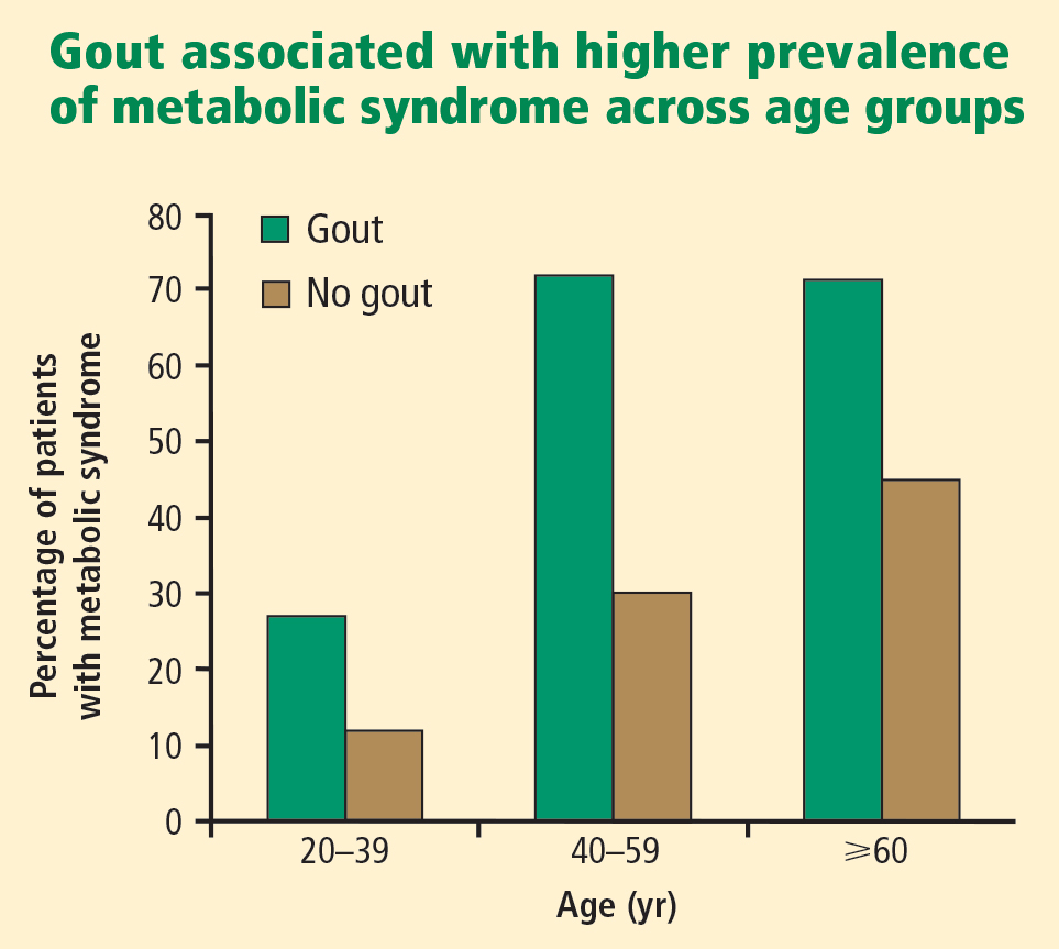
Primary care physicians regularly see patients with hypertension, diabetes, hyperlipidemia, chronic kidney disease, cardiovascular disease, and the metabolic syndrome. As patients with these comorbidities are at an increased risk for developing gout,1,10,11 it may be beneficial in these patients to inquire whether they have had any bouts of arthritic pain and periodically evaluate the serum urate level (which is no longer included on chemistry profiles).
Obesity/high body mass index
Obesity and high body mass index significantly contribute to the risk of developing gout.13,16 Choi and colleagues observed that the risk of gout is very low for men with a body mass index between 21 and 22 but is increased threefold for men whose body mass index is 35 or greater.13 Obesity is associated with increased serum urate levels attributable to increased urate production and decreased renal urate excretion. A weight loss program may reduce the risk of gout by decreasing urate levels over time.13,16
Diet and alcohol consumption
A study by Choi and colleagues found that high intake of alcohol (beer more so than hard liquor or wine) and a diet rich in meat (particularly red meat, wild game, or organ meat) and seafood (particularly shellfish and some larger saltwater fish) increase the risk for developing gout.17,18 Purine-rich vegetables, which were previously eliminated in low-purine diets, were found not to have any association with hyperuricemia and did not increase the risk of gout.17,18 Frequent consumption of dairy products was found to slightly reduce the risk of gout and hyperuricemia.17,18
Although manipulation of diet and alcohol consumption alone rarely achieves the desired degree of serum urate reduction, adherence to changes in diet and alcohol intake will reduce flares of gout and assist in lowering serum urate levels.
CONCLUSIONS
Diet, alcohol consumption, and lifestyle choices can increase the risk of developing gout, but making recommended lifestyle changes does not replace pharmacologic treatments for existing gout or associated comorbidities. Furthermore, very few patients are likely to follow through with lifestyle changes such as weight reduction and a low-cholesterol diet.19–21 Therefore, physician awareness of the factors that contribute to the development of gout is important, as is identification of at-risk patients before they develop manifestations of the disease. Increased vigilance in monitoring serum urate levels and monitoring for gout development in at-risk patients may help to improve the standard of care for gout.
Acknowledgment
Dr. Weaver thanks Kenneth G. Saag, MD, MSc, for his contributions to the lecture on which this paper is based.
- Choi HK, Mount DB, Reginato AM. Pathogenesis of gout. Ann Intern Med 2005; 143:499–516.
- Arromdee E, Michet CJ, Crowson CS, O’Fallon WM, Gabriel SE. Epidemiology of gout: is the incidence rising? J Rheumatol 2002; 29:2403–2406.
- Kramer HM, Curhan G. The association between gout and nephrolithiasis: the National Health and Nutrition Examination Survey III, 1988–1994. Am J Kidney Dis 2002; 40:37–42.
- Wallace KL, Riedel AA, Joseph-Ridge N, Wortmann R. Increasing prevalence of gout and hyperuricemia over 10 years among older adults in a managed care population. J Rheumatol 2004; 31: 1582–1587.
- Roddy E, Zhang W, Doherty M. The changing epidemiology of gout. Nat Clin Pract Rheumatol 2007; 3:443–449.
- Mikuls TR, Farrar JT, Bilker WB, Fernandes S, Schumacher HR Jr, Saag KG. Gout epidemiology: results from the UK General Practice Research Database, 1990–1999. Ann Rheum Dis 2005; 64:267–272.
- Roubenoff R, Klag MJ, Mead LA, Liang KY, Seidler AJ, Hochberg MC. Incidence and risk factors for gout in white men. JAMA 1991; 266:3004–3007.
- Choi HK, Atkinson K, Karlson EW, Willett W, Curhan G. Alcohol intake and risk of incident gout in men: a prospective study. Lancet 2004; 363:1277–1281.
- Lawrence RC, Helmick CG, Arnett FC, et al. Estimates of the prevalence of arthritis and selected musculoskeletal disorders in the United States. Arthritis Rheum 1998; 41:778–799.
- Rott KT, Agudelo CA. Gout. JAMA 2003; 289:2857–2860.
- Choi HK, Ford ES, Li C, Curhan G. Prevalence of the metabolic syndrome in patients with gout: the Third National Health and Nutrition Examination Survey. Arthritis Rheum 2007; 57:109–115.
- Abbott RD, Brand FN, Kannel WB, Castelli WP. Gout and coronary heart disease: the Framingham Study. J Clin Epidemiol 1988; 41:237–242.
- Choi HK, Atkinson K, Karlson EW, Curhan G. Obesity, weight change, hypertension, diuretic use, and risk of gout in men: the health professionals follow-up study. Arch Intern Med 2005; 165:742–748.
- Caspi D, Lubart E, Graff E, Habot B, Yaron M, Segal R. The effect of mini-dose aspirin on renal function and uric acid handling in elderly patients. Arthritis Rheum 2000; 43:103–108.
- Edwards NL. Gout. B. Clinical and laboratory features. In: Klippel JH, Crofford LJ, Stone JH, Weyand CM, eds. Primer on the Rheumatic Diseases. 12th ed. Atlanta, GA: Arthritis Foundation; 2001: 313–319.
- Dessein PH, Shipton EA, Stanwix AE, Joffe BI, Ramokgadi J. Beneficial effects of weight loss associated with moderate calorie/carbohydrate restriction, and increased proportional intake of protein and unsaturated fat on serum urate and lipoprotein levels in gout: a pilot study. Ann Rheum Dis 2000; 59:539–543.
- Choi HK, Atkinson K, Karlson EW, Willett W, Curhan G. Purine-rich foods, dairy and protein intake, and the risk of gout in men. N Engl J Med 2004; 350:1093–1103.
- Choi HK. Diet, alcohol, and gout: how do we advise patients given recent developments? Curr Rheumatol Rep 2005; 7:220–226.
- Fam AG. Gout: excess calories, purines, and alcohol intake and beyond. Response to a urate-lowering diet. J Rheumatol 2005; 32:773–777.
- Emmerson BT. The management of gout. N Engl J Med 1996; 334:445–451.
- Levinson W, Cohen MS, Brady D, Duffy FD. To change or not to change: “sounds like you have a dilemma.” Ann Intern Med 2001; 135:386–391.
Both the incidence and the prevalence of gout appear to be increasing worldwide.1,2 Risk factors for the development of gout are related to our increasing longevity, dietary and lifestyle changes, and an increased prevalence of comorbid conditions. Patients with conditions such as hypertension, diabetes, cardiovascular disease, and the metabolic syndrome have an increased risk of developing hyperuricemia and gout,1,2 and such conditions are frequently managed by primary care physicians. This paper discusses how these conditions, along with diet, alcohol intake, and lifestyle, contribute to the increasing prevalence of hyperuricemia and gout.1
PREVALENCE AND INCIDENCE: BOTH ARE RISING
The Third National Health and Nutrition Examination Survey (NHANES III) estimated the prevalence of gout in the US population to be 5.1 million between 1988 and 1994.3 Data from a US managed care claims database revealed an increase in gout prevalence from 2.9 cases per 1,000 persons in 1990 to 5.2 cases per 1,000 persons in 1999.4 Other studies indicate that gout is becoming more prevalent in societies such as New Zealand and Taiwan as well as in the United States.5,6
The NHANES III data show that gout affected more than 3 million men aged 40 years or older, and 1.7 million women aged 40 years or older, in the period from 1988 to 1994.3 The estimated prevalence of gout among men aged 40 years or older makes this disease more common than rheumatoid arthritis and the most common form of inflammatory arthritis in adult men.7–9
RISK FACTORS FOR GOUT DEVELOPMENT
Sex and age
Men have higher serum urate levels than women do, which increases their risk of developing gout. Development of gout before age 30 years is overwhelmingly more likely in men than in women.7,9,10 The prevalence of gout in men increases with advancing age and peaks between ages 75 and 84 years.6 Women have an increased risk of developing gout after menopause; the risk begins to rise at about age 45 years with the decrease in estrogen levels.10,11 The incidence of gout becomes approximately equal between the sexes after age 60 years.10,12
It is important for clinicians to bear these factors in mind when taking the patient history and considering gout in the differential diagnosis. Although gout is more common in men, the diagnosis of gout should still be considered in women, particularly postmenopausal women.10,11
Medications
The use of diuretics is a significant risk factor for the development of gout. Diuretic use results in increased reabsorption of uric acid in the kidney, leading to hyperuricemia.1,7,10,13 If reasonable, an alternative antihypertensive agent should be prescribed. Low-dose aspirin, commonly prescribed for cardioprotection, also increases urate levels slightly in elderly patients,14 but should not be discontinued if indicated. Cyclosporine, which increases tubular reabsorption of urate,1 can result in a type of rapidly ascending gout that frequently is polyarticular.1,15 This is often encountered in transplant patients taking cyclosporine as an immunosuppressant. Hyperuricemia is also seen in patients taking pyrazinamide, ethambutol, and niacin. Physicians should be aware of the risks and benefits of prescribing any of these drugs to a patient with gout and consider that they may precipitate a gout flare in a previously unaffected patient.
Comorbidities
Primary care physicians regularly see patients with hypertension, diabetes, hyperlipidemia, chronic kidney disease, cardiovascular disease, and the metabolic syndrome. As patients with these comorbidities are at an increased risk for developing gout,1,10,11 it may be beneficial in these patients to inquire whether they have had any bouts of arthritic pain and periodically evaluate the serum urate level (which is no longer included on chemistry profiles).
Obesity/high body mass index
Obesity and high body mass index significantly contribute to the risk of developing gout.13,16 Choi and colleagues observed that the risk of gout is very low for men with a body mass index between 21 and 22 but is increased threefold for men whose body mass index is 35 or greater.13 Obesity is associated with increased serum urate levels attributable to increased urate production and decreased renal urate excretion. A weight loss program may reduce the risk of gout by decreasing urate levels over time.13,16
Diet and alcohol consumption
A study by Choi and colleagues found that high intake of alcohol (beer more so than hard liquor or wine) and a diet rich in meat (particularly red meat, wild game, or organ meat) and seafood (particularly shellfish and some larger saltwater fish) increase the risk for developing gout.17,18 Purine-rich vegetables, which were previously eliminated in low-purine diets, were found not to have any association with hyperuricemia and did not increase the risk of gout.17,18 Frequent consumption of dairy products was found to slightly reduce the risk of gout and hyperuricemia.17,18
Although manipulation of diet and alcohol consumption alone rarely achieves the desired degree of serum urate reduction, adherence to changes in diet and alcohol intake will reduce flares of gout and assist in lowering serum urate levels.
CONCLUSIONS
Diet, alcohol consumption, and lifestyle choices can increase the risk of developing gout, but making recommended lifestyle changes does not replace pharmacologic treatments for existing gout or associated comorbidities. Furthermore, very few patients are likely to follow through with lifestyle changes such as weight reduction and a low-cholesterol diet.19–21 Therefore, physician awareness of the factors that contribute to the development of gout is important, as is identification of at-risk patients before they develop manifestations of the disease. Increased vigilance in monitoring serum urate levels and monitoring for gout development in at-risk patients may help to improve the standard of care for gout.
Acknowledgment
Dr. Weaver thanks Kenneth G. Saag, MD, MSc, for his contributions to the lecture on which this paper is based.
- Choi HK, Mount DB, Reginato AM. Pathogenesis of gout. Ann Intern Med 2005; 143:499–516.
- Arromdee E, Michet CJ, Crowson CS, O’Fallon WM, Gabriel SE. Epidemiology of gout: is the incidence rising? J Rheumatol 2002; 29:2403–2406.
- Kramer HM, Curhan G. The association between gout and nephrolithiasis: the National Health and Nutrition Examination Survey III, 1988–1994. Am J Kidney Dis 2002; 40:37–42.
- Wallace KL, Riedel AA, Joseph-Ridge N, Wortmann R. Increasing prevalence of gout and hyperuricemia over 10 years among older adults in a managed care population. J Rheumatol 2004; 31: 1582–1587.
- Roddy E, Zhang W, Doherty M. The changing epidemiology of gout. Nat Clin Pract Rheumatol 2007; 3:443–449.
- Mikuls TR, Farrar JT, Bilker WB, Fernandes S, Schumacher HR Jr, Saag KG. Gout epidemiology: results from the UK General Practice Research Database, 1990–1999. Ann Rheum Dis 2005; 64:267–272.
- Roubenoff R, Klag MJ, Mead LA, Liang KY, Seidler AJ, Hochberg MC. Incidence and risk factors for gout in white men. JAMA 1991; 266:3004–3007.
- Choi HK, Atkinson K, Karlson EW, Willett W, Curhan G. Alcohol intake and risk of incident gout in men: a prospective study. Lancet 2004; 363:1277–1281.
- Lawrence RC, Helmick CG, Arnett FC, et al. Estimates of the prevalence of arthritis and selected musculoskeletal disorders in the United States. Arthritis Rheum 1998; 41:778–799.
- Rott KT, Agudelo CA. Gout. JAMA 2003; 289:2857–2860.
- Choi HK, Ford ES, Li C, Curhan G. Prevalence of the metabolic syndrome in patients with gout: the Third National Health and Nutrition Examination Survey. Arthritis Rheum 2007; 57:109–115.
- Abbott RD, Brand FN, Kannel WB, Castelli WP. Gout and coronary heart disease: the Framingham Study. J Clin Epidemiol 1988; 41:237–242.
- Choi HK, Atkinson K, Karlson EW, Curhan G. Obesity, weight change, hypertension, diuretic use, and risk of gout in men: the health professionals follow-up study. Arch Intern Med 2005; 165:742–748.
- Caspi D, Lubart E, Graff E, Habot B, Yaron M, Segal R. The effect of mini-dose aspirin on renal function and uric acid handling in elderly patients. Arthritis Rheum 2000; 43:103–108.
- Edwards NL. Gout. B. Clinical and laboratory features. In: Klippel JH, Crofford LJ, Stone JH, Weyand CM, eds. Primer on the Rheumatic Diseases. 12th ed. Atlanta, GA: Arthritis Foundation; 2001: 313–319.
- Dessein PH, Shipton EA, Stanwix AE, Joffe BI, Ramokgadi J. Beneficial effects of weight loss associated with moderate calorie/carbohydrate restriction, and increased proportional intake of protein and unsaturated fat on serum urate and lipoprotein levels in gout: a pilot study. Ann Rheum Dis 2000; 59:539–543.
- Choi HK, Atkinson K, Karlson EW, Willett W, Curhan G. Purine-rich foods, dairy and protein intake, and the risk of gout in men. N Engl J Med 2004; 350:1093–1103.
- Choi HK. Diet, alcohol, and gout: how do we advise patients given recent developments? Curr Rheumatol Rep 2005; 7:220–226.
- Fam AG. Gout: excess calories, purines, and alcohol intake and beyond. Response to a urate-lowering diet. J Rheumatol 2005; 32:773–777.
- Emmerson BT. The management of gout. N Engl J Med 1996; 334:445–451.
- Levinson W, Cohen MS, Brady D, Duffy FD. To change or not to change: “sounds like you have a dilemma.” Ann Intern Med 2001; 135:386–391.
KEY POINTS
- Although gout is more common in men, the diagnosis of gout should still be considered in women, particularly postmenopausal women.
- Use of diuretics is a significant risk factor for gout.
- Patients with hypertension, diabetes, hyperlipidemia, chronic kidney disease, or the metabolic syndrome are at increased risk for developing gout. Vigilance for gout is especially indicated in patients with metabolic syndrome.
The role of hyperuricemia and gout in kidney and cardiovascular disease
Hyperuricemia is a metabolic problem that has become quite common over the past several decades. The main clinical issues associated with hyperuricemia are gouty arthritis, gouty tophi, and uric acid kidney stones. For decades, these have been the main indications for lowering serum urate levels. Well-established nonarticular associations of hyperuricemia in gout include chronic kidney disease, coronary artery disease, and hypertension.1 Recent animal studies and epidemiologic studies have shed new light on the relationship between urate and comorbid disease processes. This article describes our evolving understanding of the association of hyperuricemia and gout with kidney disease, hypertension, and cardiovascular disease, and also reviews the kidney’s role in regulation of urate levels in the body.
SOURCES, DISTRIBUTION, AND ELIMINATION OF URATE
Uric acid is the end product of purine metabolism in humans. Sources of purine are either endogenous, from de novo synthesis and nucleic acid breakdown (approximately 600 mg/day), or exogenous, from dietary purine intake (approximately 100 mg/day).2 In the steady state, this daily production and ingestion of approximately 700 mg of uric acid is balanced by daily elimination of an equal amount of uric acid from the body. Approximately 30% of this daily loss is through the gut, with subsequent bacterial intestinal uricolysis. The other 70% (roughly 500 mg daily) needs to be excreted by the kidneys.
In humans, plasma urate is freely filtered at the glomerulus, but the fractional excretion of the filtered uric acid is less than 10%. This demonstrates a dominance of reabsorptive processes in humans, and these processes are handled primarily by the selective urate transport protein known as URAT1 in the proximal convoluted tubules. In normouricemic individuals, there is a balance between the daily production and ingestion of uric acid and the daily excretion of it. In most patients with primary hyperuricemia and gout, the fractional excretion of uric acid by the kidneys is relatively diminished, resulting in an imbalance of uric acid homeostasis, and serum urate exceeds saturability (6.8 mg/dL at physiologic temperature and pH). As this imbalance persists over years and decades, the miscible serum uric acid pool expands and urate may be deposited as part of an insoluble urate pool in the joints and soft tissues, referred to as tophi.
In the past, most investigators have focused on the destructive and proinflammatory role of insoluble deposited urate crystals, but new evidence is accumulating that rising levels of soluble urate in body fluids may also be harmful and lead to kidney disease, hypertension, and cardiovascular disease.
RENAL MANIFESTATIONS OF HYPERURICEMIA
Urate is strongly associated with renal disease but traditionally has not been considered to have a causal role in kidney dysfunction. The exceptions have been uric acid kidney stones and the acute uric acid nephropathy associated with chemotherapy and tumor lysis syndrome. New epidemiologic studies in humans, as well as an animal model of mild hyperuricemia leading to microvascular changes in the glomerular afferent arterioles, shed new light on a possible direct role of urate in the genesis of idiopathic chronic kidney disease.
Longstanding reluctance to implicate urate in kidney disease
Significant impairment of renal function was reported in up to 40% of patients with gout in studies conducted before the advent of effective urate-lowering therapies.3,4 In these older studies, renal failure was the eventual cause of death in 18% to 25% of patients with gout.3,4 However, any primary causal relationship for urate in this very high incidence of kidney disease was questioned for decades in light of the many other conditions and factors associated with hyperuricemia and gout that may contribute to kidney disease, such as hypertension, diabetes mellitus, alcohol abuse, non-steroidal anti-inflammatory drug use, and lead toxicity.
Recent epidemiologic studies establish the connection
More recently, two large prospective population studies from Japan examined the relationship between serum urate level and development of kidney disease using multiple covariate analysis to adjust for age, blood pressure, body mass index, proteinuria, hematocrit, hyperlipidemia, fasting glucose, and serum creatinine.5,6
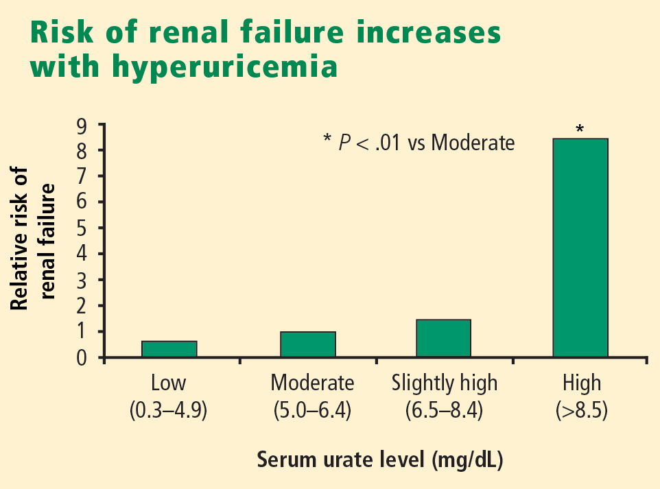
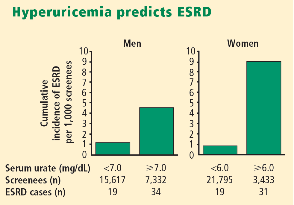
ASSOCIATION OF HYPERTENSION AND HYPERURICEMIA
The strong association between hypertension and hyperuricemia has been recognized for more than a century. In his original description of essential hypertension in 1879, Frederick A. Mohamed noted that many of his subjects came from gouty families.7 Studies from the 1950s and 1960s showed the prevalence of hyperuricemia in hypertensive subjects to be between 20% and 40%.8,9 The prevalence of hypertension among gouty patients is Hyperuricemia predicts ESRD between 25% and 50%.10 In 1972, Kahn et al found that a rising level of serum urate is an independent risk factor for hypertension.11 A year later, Klein et al demonstrated a linear relationship between serum urate level and systolic blood pressure in both black and white subjects.12
Six large epidemiologic studies published over the past 7 years have found that serum urate level predicts the later development of hypertension.13–18 The most recent of these is the Normative Aging Study,18 which showed that the serum urate level independently predicts the development of hypertension when using age-adjusted and multivariate models that include body mass, abdominal girth, alcohol use, serum lipid levels, plasma glucose level, and smoking status.
A potential mechanism emerges
No clear causal or mechanistic link between elevated serum urate and the development of hypertension was evident until a rat model of mild hyperuricemia was found to be associated with the development of an initial salt-insensitive hypertension that was reversible with restoration of normal urate levels.19 However, if the urate-induced hypertension was allowed to persist, the rat would develop a salt-sensitive hypertension that was irreversible even if normouricemia was reestablished.19
This mechanism was tested in a small pilot study of 5 children with previously untreated essential hypertension who received monotherapy with allopurinol for 1 month.20 All 5 children had substantial drops in their continuously monitored ambulatory blood pressure, and 4 of the 5 developed normal blood pressure (as assessed by continuous monitoring). After the allopurinol was stopped, the blood pressure of all 5 children rebounded to baseline levels.20 A larger blinded, randomized, placebo-controlled crossover trial using the same intervention with similar results has been presented recently at national meetings.21
ASSOCIATION BETWEEN CARDIOVASCULAR DISEASE AND HYPERURICEMIA
There is considerable documentation that urate levels correlate with many recognized cardiovascular risk factors, including age, male gender, hypertension, diabetes mellitus, hypertriglyceridemia, obesity, and insulin resistance. Because of these relationships, the observed association between serum urate elevations and cardiovascular disease was considered to be “epiphenomenal” and not causal. Many older epidemiologic studies that demonstrated the increased cardiovascular risk associated with higher urate levels used traditional statistical techniques that could not prove the “independence” of urate as a risk factor.22
An independent effect is finally documented
Three studies published in the past several years demonstrate an independent risk relationship between hyperuricemia and cardiovascular disease, including acute myocardial infarction.23–25
Multivariate regression analysis of the Multiple Risk Factor Intervention Trial (MRFIT) database demonstrated hyperuricemia to be an independent risk factor for acute myocardial infarction.23
Similarly, the Italian Progetto Ipertensione Umbria Monitoraggio Ambulatoriale (PIUMA) study showed that after adjustment for age, sex, diabetes, serum lipid levels, serum creatinine, left ventricular hypertrophy, blood pressure, and diuretic use, serum urate levels in the highest quartile were associated with increased risk of all cardiovascular events (relative risk [RR] = 1.73) and fatal cardiovascular events (RR = 1.96) compared with urate levels in the second quartile.24
Finally, follow-up of 4,385 participants in the Rotterdam Study over an average of 8.4 years revealed that high urate levels were a strong predictor of both myocardial infarction and stroke, even after adjustment for other vascular risk factors.25
CONCLUSIONS
Based on the preponderance of recent epidemiologic studies, it appears that an elevated serum urate level is an independent risk factor for kidney disease, hypertension, and cardiovascular disease. The precise mechanisms for urate-induced tissue injury remain unclear, and no large study to date has convincingly demonstrated that lowering serum urate levels can prevent or lessen the risk of these potentially severe complications of hyperuricemia.
It should also be emphasized that the treatment of hyperuricemia with medications such as allopurinol or probenecid is currently not indicated in patients with hypertension, kidney disease, or heart disease. The use of these agents is supported only in the treatment of gout, the treatment of uric acid kidney stones, and the prevention of tumor lysis syndrome.
- Mikuls TR, Farrar JT, Bilker WB, Fernandes S, Schumacher HR Jr, Saag KG. Gout epidemiology: results from the UK General Practice Research Database, 1990–1999. Ann Rheum Dis 2005; 64:267–272.
- Richards J, Weinman EJ. Uric acid and renal disease. J Nephrol 1966; 9:160–166.
- Berger L, Yü TF. Renal function in gout. IV. An analysis of 524 gouty subjects including long-term follow-up studies. Am J Med 1975; 59:605–613.
- Talbot JH, Terplan KL. The kidney in gout. Medicine 1960; 39:405–468.
- Tomita M, Mizuno S, Yamanaka H, et al. Does hyperuricemia affect mortality? A prospective cohort study of Japanese male workers. J Epidemiol 2000; 10:403–409.
- Iseki K, Ikemiya Y, Inoue T, Iseki C, Kinjo K, Takishita S. Significance of hyperuricemia as a risk factor for developing ESRD in a screened cohort. Am J Kidney Dis 2004; 44:642–650.
- Mohamed FA. On chronic Bright’s disease, and its essential symptoms. Lancet 1879; 1:399–401.
- Weiss TE, Segaloff A, Moore C. Gout and diabetes. Metabolism 1957; 6:103.
- Kinsey D, Walther R, Sise HS, Whitelaw G, Smithwick R. Incidence of hyperuricemia in 400 hypertensive patients. Circulation 1961; 24:972.
- Becker MA, Jolly M. Hyperuricemia and associated diseases. Rheum Dis Clin North Am 2006; 32:275–293.
- Kahn HA, Medalie JH, Neufeld HN, Riss E, Goldbourt U. The incidence of hypertension and associated factors: the Israel ischemic heart disease study. Am Heart J 1972; 84:171–182.
- Klein R, Klein BE, Cornoni JC, Maready J, Cassel JC, Tyroler HA. Serum uric acid. Its relationships to coronary heart disease risk factors and cardiovascular disease, Evans County, Georgia. Arch Intern Med 1973; 132:401–410.
- Nakanishi N, Okamoto M, Yoshida H, Matsuo Y, Suzuki K, Tatara K. Serum uric acid and risk for development of hypertension and impaired fasting glucose or type II diabetes in Japanese male office workers. Eur J Epidemiol 2003; 18:523–530.
- Alper AB Jr, Chen W, Yau L, Srinivasan SR, Berenson GS, Hamm LL. Childhood uric acid predicts adult blood pressure: the Bogalusa Heart Study. Hypertension 2005; 45:34–38.
- Masuo K, Kawaguchi H, Mikami H, Ogihara T, Tuck ML. Serum uric acid and plasma norepinephrine concentrations predict subsequent weight gain and blood pressure elevation. Hypertension 2003; 42:474–480.
- Nagahama K, Inoue T, Iseki K, et al. Hyperuricemia as a predictor of hypertension in a screened cohort in Okinawa, Japan. Hypertens Res 2004; 27:835–841.
- Sundström J, Sullivan L, D’Agostino RB, Levy D, Kannel WB, Vasan RS. Relations of serum uric acid to longitudinal blood pressure tracking and hypertension incidence. Hypertension 2005; 45:28–33.
- Perlstein TS, Gumieniak O, Williams GH, et al. Uric acid and the development of hypertension: the Normative Aging Study. Hypertension 2006; 48:1031–1036.
- Mazzali M, Hughes J, Kim YG, et al. Elevated uric acid increases blood pressure in the rat by a novel crystal-independent mechanism. Hypertension 2001; 38:1101–1106.
- Feig DI, Nakagawa T, Karumanchi SA, et al. Hypothesis: uric acid, nephron number, and the pathogenesis of essential hypertension. Kidney Int 2004; 66:281–287.
- Feig DI, Mazzali M, Kang DH, et al. Serum uric acid: a risk factor and a target for treatment? J Am Soc Nephrol 2006; 17(4 Suppl):S69–S73.
- Rich MW. Uric acid: is it a risk factor for cardiovascular disease? Am J Cardiol 2000; 85:1018–1021.
- Krishnan E, Baker JF, Furst DE, Schumacher HR Jr. Gout and the risk of acute myocardial infarction. Arthritis Rheum 2006; 54:2688–2696.
- Verdecchia P, Schillaci G, Reboldi G, Santeusanio F, Porcellati C, Brunetti P. Relation between serum uric acid and risk of cardiovascular disease in essential hypertension. The PIUMA study. Hypertension 2000; 36:1072–1078.
- Bos MJ , Koudstaal PJ, Hofman A, Witteman JC, Breteler MM. Uric acid is a risk factor for myocardial infarction and stroke: the Rotterdam Study. Stroke 2006; 37:1503–1507.
Hyperuricemia is a metabolic problem that has become quite common over the past several decades. The main clinical issues associated with hyperuricemia are gouty arthritis, gouty tophi, and uric acid kidney stones. For decades, these have been the main indications for lowering serum urate levels. Well-established nonarticular associations of hyperuricemia in gout include chronic kidney disease, coronary artery disease, and hypertension.1 Recent animal studies and epidemiologic studies have shed new light on the relationship between urate and comorbid disease processes. This article describes our evolving understanding of the association of hyperuricemia and gout with kidney disease, hypertension, and cardiovascular disease, and also reviews the kidney’s role in regulation of urate levels in the body.
SOURCES, DISTRIBUTION, AND ELIMINATION OF URATE
Uric acid is the end product of purine metabolism in humans. Sources of purine are either endogenous, from de novo synthesis and nucleic acid breakdown (approximately 600 mg/day), or exogenous, from dietary purine intake (approximately 100 mg/day).2 In the steady state, this daily production and ingestion of approximately 700 mg of uric acid is balanced by daily elimination of an equal amount of uric acid from the body. Approximately 30% of this daily loss is through the gut, with subsequent bacterial intestinal uricolysis. The other 70% (roughly 500 mg daily) needs to be excreted by the kidneys.
In humans, plasma urate is freely filtered at the glomerulus, but the fractional excretion of the filtered uric acid is less than 10%. This demonstrates a dominance of reabsorptive processes in humans, and these processes are handled primarily by the selective urate transport protein known as URAT1 in the proximal convoluted tubules. In normouricemic individuals, there is a balance between the daily production and ingestion of uric acid and the daily excretion of it. In most patients with primary hyperuricemia and gout, the fractional excretion of uric acid by the kidneys is relatively diminished, resulting in an imbalance of uric acid homeostasis, and serum urate exceeds saturability (6.8 mg/dL at physiologic temperature and pH). As this imbalance persists over years and decades, the miscible serum uric acid pool expands and urate may be deposited as part of an insoluble urate pool in the joints and soft tissues, referred to as tophi.
In the past, most investigators have focused on the destructive and proinflammatory role of insoluble deposited urate crystals, but new evidence is accumulating that rising levels of soluble urate in body fluids may also be harmful and lead to kidney disease, hypertension, and cardiovascular disease.
RENAL MANIFESTATIONS OF HYPERURICEMIA
Urate is strongly associated with renal disease but traditionally has not been considered to have a causal role in kidney dysfunction. The exceptions have been uric acid kidney stones and the acute uric acid nephropathy associated with chemotherapy and tumor lysis syndrome. New epidemiologic studies in humans, as well as an animal model of mild hyperuricemia leading to microvascular changes in the glomerular afferent arterioles, shed new light on a possible direct role of urate in the genesis of idiopathic chronic kidney disease.
Longstanding reluctance to implicate urate in kidney disease
Significant impairment of renal function was reported in up to 40% of patients with gout in studies conducted before the advent of effective urate-lowering therapies.3,4 In these older studies, renal failure was the eventual cause of death in 18% to 25% of patients with gout.3,4 However, any primary causal relationship for urate in this very high incidence of kidney disease was questioned for decades in light of the many other conditions and factors associated with hyperuricemia and gout that may contribute to kidney disease, such as hypertension, diabetes mellitus, alcohol abuse, non-steroidal anti-inflammatory drug use, and lead toxicity.
Recent epidemiologic studies establish the connection
More recently, two large prospective population studies from Japan examined the relationship between serum urate level and development of kidney disease using multiple covariate analysis to adjust for age, blood pressure, body mass index, proteinuria, hematocrit, hyperlipidemia, fasting glucose, and serum creatinine.5,6
ASSOCIATION OF HYPERTENSION AND HYPERURICEMIA
The strong association between hypertension and hyperuricemia has been recognized for more than a century. In his original description of essential hypertension in 1879, Frederick A. Mohamed noted that many of his subjects came from gouty families.7 Studies from the 1950s and 1960s showed the prevalence of hyperuricemia in hypertensive subjects to be between 20% and 40%.8,9 The prevalence of hypertension among gouty patients is Hyperuricemia predicts ESRD between 25% and 50%.10 In 1972, Kahn et al found that a rising level of serum urate is an independent risk factor for hypertension.11 A year later, Klein et al demonstrated a linear relationship between serum urate level and systolic blood pressure in both black and white subjects.12
Six large epidemiologic studies published over the past 7 years have found that serum urate level predicts the later development of hypertension.13–18 The most recent of these is the Normative Aging Study,18 which showed that the serum urate level independently predicts the development of hypertension when using age-adjusted and multivariate models that include body mass, abdominal girth, alcohol use, serum lipid levels, plasma glucose level, and smoking status.
A potential mechanism emerges
No clear causal or mechanistic link between elevated serum urate and the development of hypertension was evident until a rat model of mild hyperuricemia was found to be associated with the development of an initial salt-insensitive hypertension that was reversible with restoration of normal urate levels.19 However, if the urate-induced hypertension was allowed to persist, the rat would develop a salt-sensitive hypertension that was irreversible even if normouricemia was reestablished.19
This mechanism was tested in a small pilot study of 5 children with previously untreated essential hypertension who received monotherapy with allopurinol for 1 month.20 All 5 children had substantial drops in their continuously monitored ambulatory blood pressure, and 4 of the 5 developed normal blood pressure (as assessed by continuous monitoring). After the allopurinol was stopped, the blood pressure of all 5 children rebounded to baseline levels.20 A larger blinded, randomized, placebo-controlled crossover trial using the same intervention with similar results has been presented recently at national meetings.21
ASSOCIATION BETWEEN CARDIOVASCULAR DISEASE AND HYPERURICEMIA
There is considerable documentation that urate levels correlate with many recognized cardiovascular risk factors, including age, male gender, hypertension, diabetes mellitus, hypertriglyceridemia, obesity, and insulin resistance. Because of these relationships, the observed association between serum urate elevations and cardiovascular disease was considered to be “epiphenomenal” and not causal. Many older epidemiologic studies that demonstrated the increased cardiovascular risk associated with higher urate levels used traditional statistical techniques that could not prove the “independence” of urate as a risk factor.22
An independent effect is finally documented
Three studies published in the past several years demonstrate an independent risk relationship between hyperuricemia and cardiovascular disease, including acute myocardial infarction.23–25
Multivariate regression analysis of the Multiple Risk Factor Intervention Trial (MRFIT) database demonstrated hyperuricemia to be an independent risk factor for acute myocardial infarction.23
Similarly, the Italian Progetto Ipertensione Umbria Monitoraggio Ambulatoriale (PIUMA) study showed that after adjustment for age, sex, diabetes, serum lipid levels, serum creatinine, left ventricular hypertrophy, blood pressure, and diuretic use, serum urate levels in the highest quartile were associated with increased risk of all cardiovascular events (relative risk [RR] = 1.73) and fatal cardiovascular events (RR = 1.96) compared with urate levels in the second quartile.24
Finally, follow-up of 4,385 participants in the Rotterdam Study over an average of 8.4 years revealed that high urate levels were a strong predictor of both myocardial infarction and stroke, even after adjustment for other vascular risk factors.25
CONCLUSIONS
Based on the preponderance of recent epidemiologic studies, it appears that an elevated serum urate level is an independent risk factor for kidney disease, hypertension, and cardiovascular disease. The precise mechanisms for urate-induced tissue injury remain unclear, and no large study to date has convincingly demonstrated that lowering serum urate levels can prevent or lessen the risk of these potentially severe complications of hyperuricemia.
It should also be emphasized that the treatment of hyperuricemia with medications such as allopurinol or probenecid is currently not indicated in patients with hypertension, kidney disease, or heart disease. The use of these agents is supported only in the treatment of gout, the treatment of uric acid kidney stones, and the prevention of tumor lysis syndrome.
- Mikuls TR, Farrar JT, Bilker WB, Fernandes S, Schumacher HR Jr, Saag KG. Gout epidemiology: results from the UK General Practice Research Database, 1990–1999. Ann Rheum Dis 2005; 64:267–272.
- Richards J, Weinman EJ. Uric acid and renal disease. J Nephrol 1966; 9:160–166.
- Berger L, Yü TF. Renal function in gout. IV. An analysis of 524 gouty subjects including long-term follow-up studies. Am J Med 1975; 59:605–613.
- Talbot JH, Terplan KL. The kidney in gout. Medicine 1960; 39:405–468.
- Tomita M, Mizuno S, Yamanaka H, et al. Does hyperuricemia affect mortality? A prospective cohort study of Japanese male workers. J Epidemiol 2000; 10:403–409.
- Iseki K, Ikemiya Y, Inoue T, Iseki C, Kinjo K, Takishita S. Significance of hyperuricemia as a risk factor for developing ESRD in a screened cohort. Am J Kidney Dis 2004; 44:642–650.
- Mohamed FA. On chronic Bright’s disease, and its essential symptoms. Lancet 1879; 1:399–401.
- Weiss TE, Segaloff A, Moore C. Gout and diabetes. Metabolism 1957; 6:103.
- Kinsey D, Walther R, Sise HS, Whitelaw G, Smithwick R. Incidence of hyperuricemia in 400 hypertensive patients. Circulation 1961; 24:972.
- Becker MA, Jolly M. Hyperuricemia and associated diseases. Rheum Dis Clin North Am 2006; 32:275–293.
- Kahn HA, Medalie JH, Neufeld HN, Riss E, Goldbourt U. The incidence of hypertension and associated factors: the Israel ischemic heart disease study. Am Heart J 1972; 84:171–182.
- Klein R, Klein BE, Cornoni JC, Maready J, Cassel JC, Tyroler HA. Serum uric acid. Its relationships to coronary heart disease risk factors and cardiovascular disease, Evans County, Georgia. Arch Intern Med 1973; 132:401–410.
- Nakanishi N, Okamoto M, Yoshida H, Matsuo Y, Suzuki K, Tatara K. Serum uric acid and risk for development of hypertension and impaired fasting glucose or type II diabetes in Japanese male office workers. Eur J Epidemiol 2003; 18:523–530.
- Alper AB Jr, Chen W, Yau L, Srinivasan SR, Berenson GS, Hamm LL. Childhood uric acid predicts adult blood pressure: the Bogalusa Heart Study. Hypertension 2005; 45:34–38.
- Masuo K, Kawaguchi H, Mikami H, Ogihara T, Tuck ML. Serum uric acid and plasma norepinephrine concentrations predict subsequent weight gain and blood pressure elevation. Hypertension 2003; 42:474–480.
- Nagahama K, Inoue T, Iseki K, et al. Hyperuricemia as a predictor of hypertension in a screened cohort in Okinawa, Japan. Hypertens Res 2004; 27:835–841.
- Sundström J, Sullivan L, D’Agostino RB, Levy D, Kannel WB, Vasan RS. Relations of serum uric acid to longitudinal blood pressure tracking and hypertension incidence. Hypertension 2005; 45:28–33.
- Perlstein TS, Gumieniak O, Williams GH, et al. Uric acid and the development of hypertension: the Normative Aging Study. Hypertension 2006; 48:1031–1036.
- Mazzali M, Hughes J, Kim YG, et al. Elevated uric acid increases blood pressure in the rat by a novel crystal-independent mechanism. Hypertension 2001; 38:1101–1106.
- Feig DI, Nakagawa T, Karumanchi SA, et al. Hypothesis: uric acid, nephron number, and the pathogenesis of essential hypertension. Kidney Int 2004; 66:281–287.
- Feig DI, Mazzali M, Kang DH, et al. Serum uric acid: a risk factor and a target for treatment? J Am Soc Nephrol 2006; 17(4 Suppl):S69–S73.
- Rich MW. Uric acid: is it a risk factor for cardiovascular disease? Am J Cardiol 2000; 85:1018–1021.
- Krishnan E, Baker JF, Furst DE, Schumacher HR Jr. Gout and the risk of acute myocardial infarction. Arthritis Rheum 2006; 54:2688–2696.
- Verdecchia P, Schillaci G, Reboldi G, Santeusanio F, Porcellati C, Brunetti P. Relation between serum uric acid and risk of cardiovascular disease in essential hypertension. The PIUMA study. Hypertension 2000; 36:1072–1078.
- Bos MJ , Koudstaal PJ, Hofman A, Witteman JC, Breteler MM. Uric acid is a risk factor for myocardial infarction and stroke: the Rotterdam Study. Stroke 2006; 37:1503–1507.
KEY POINTS
- Hyperuricemia significantly increases the risk of renal failure and end-stage renal disease.
- Larger, more rigorous trials are under way to assess preliminary findings suggesting that urate-lowering therapy might normalize blood pressure in hypertensive adolescent patients.
- Use of urate-lowering therapies to treat hyperuricemia is not currently supported in patients with kidney disease, heart disease, or hypertension in the absence of gout or uric acid kidney stones.
The gout diagnosis
The presence of urate crystals in synovial fluid is the gold standard for diagnosing gout,1 yet clinicians—both primary care physicians and rheumatologists—may not routinely perform synovial fluid analysis even when evaluating a patient who presents with an acute inflammatory arthritis.2 This paper discusses the various reasons why this is so and reviews several important resulting clinical issues: how a presumptive diagnosis of gout is made, when to measure the serum urate level, and special considerations in the differential diagnosis.
SYNOVIAL FLUID ANALYSIS: WHY IS THE GOLD STANDARD NOT MORE ROUTINE?
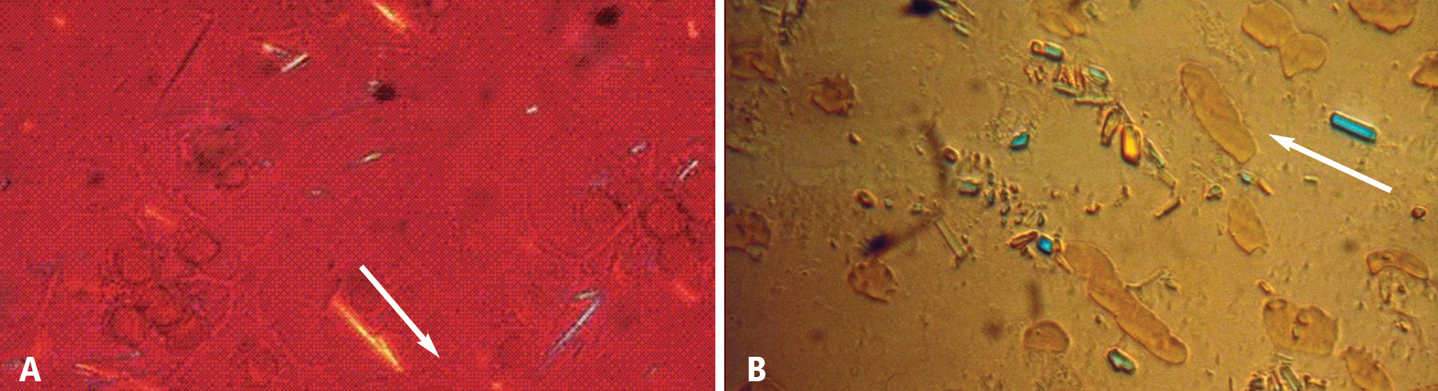
Occasionally, the aspirated joint does not appear to contain any joint fluid and the clinician may be concerned about the possibility of a “dry tap.” Other possible reasons include lack of experience with synovial fluid aspiration and evaluation, or limited access to the polarizing microscopes used to examine synovial fluid. Time is another factor; in a busy primary care practice, where patients are usually seen approximately every 7 to 11 minutes, there may not be time to aspirate a joint. The urgency of fluid examination is another issue, as synovial fluid must be examined immediately, since the crystals can become smaller, less numerous, and less birefringent with time.4
THE CLINICAL, OR PRESUMPTIVE, DIAGNOSIS
In the appropriate clinical scenario, a presumptive diagnosis of gout can be made on the basis of typical clinical features and the presence of hyperuricemia.1,2
Expert societies offer guidance, but no validation studies to date
Evidence-based recommendations for the diagnosis of gout from the European League Against Rheumatism (EULAR) state that in acute attacks, the rapid development of severe pain, swelling, and tenderness that peaks within 6 to 12 hours, especially with overlying erythema, is highly suggestive of crystal inflammation although not specific for gout.5 These recommendations further state that for typical presentations of gout (such as recurrent podagra [gouty pain in the great toe] with hyperuricemia), a clinical diagnosis alone is reasonably accurate.5
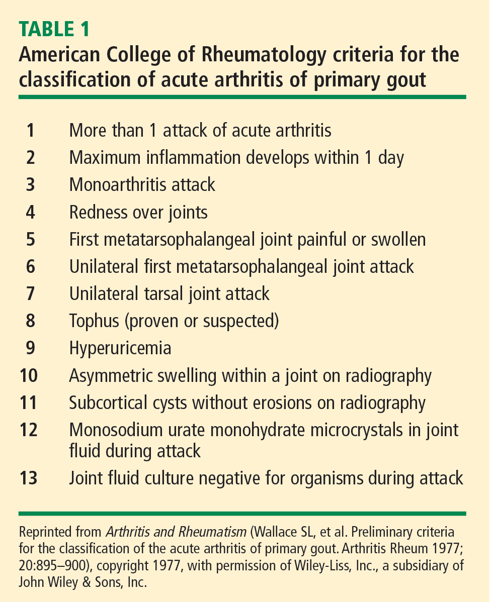
- The presence of urate crystals in joint fluid
- A tophus containing urate crystals
- Fulfillment of 6 or more of the criteria in Table 1.
No subsequent studies have been published on the validity or usefulness of any of these diagnostic criteria.
What must inform the presumptive diagnosis
Both the EULAR recommendations and the ACR criteria state that although the gold standard for diagnosing gout is the presence of urate crystals on synovial fluid analysis, a clinical diagnosis of gout can be made on the basis of certain patient criteria. This clinical, or presumptive, diagnosis of gout should be made based on the following:
- A careful patient and family history, including questions regarding comorbid conditions frequently associated with gout (such as hypertriglyceridemia, diabetes, coronary heart disease, hypertension, and the metabolic syndrome) and whether the patient has had previous similar episodes of acute joint pain and swelling in the absence of trauma
- Thorough identification of all current medications, some of which may be associated with hyperuricemia
- A thorough physical examination.
THE PHYSICAL EXAMINATION FOR GOUT
Examination of patients with a history suggestive of gout should include not only the joints but also the extensor surface of the forearms and feet. When patients are seen for a visit and gout is suspected, they should be instructed to remove their shoes and socks and roll up their sleeves to allow examination for evidence of tophi, which would suggest a past history of gouty arthritis. The ear, knee, and olecranon bursa are other common sites for tophi,3 so patients should also be asked to roll up their pants and sleeves and remove any head coverings.7 In the late stages of gouty arthritis, multiple joints may be involved, which can cause the condition to be confused with other diagnoses such as psoriatic arthritis or erosive osteoarthritis.7
ACUTE PRESENTATIONS OF GOUT
The typical gout presentation is remarkable for very intense pain that often occurs at night when the extremities are colder. Precipitation of urate in the distal extremities can occur when the extremities are horizontal and tend to become cold.8
Approximately 90% of initial gout attacks are monoarticular, leaving only 10% of cases that are oligoarticular or polyarticular.7 If more than one joint is involved, especially if the patient has a family history suggestive of gout or takes a medication that causes hyperuricemia, gout should be considered in the differential diagnosis even if the patient denies having a prior gout attack.
Frequently, patients will call their primary care physician during a gout attack but are not be able to schedule an appointment until after the attack has resolved. When possible, patients should be seen during the attack to confirm whether the attack is due to gout. A diagnosis of gout should not be made over the phone when a patient describes pain in the great toe, as only 50% of initial gout attacks occur in the great toe7 and it is not known what proportion of acute pain episodes in the great toe are attributable to gout. The most common cause of pain in the great toe is osteoarthritis.

SPECIAL CONSIDERATIONS FOR THE PRESUMPTIVE DIAGNOSIS OF GOUT
How long have acute attacks been occurring?
In a clinical scenario in which synovial fluid aspiration cannot be performed, the appropriateness of a presumptive diagnosis can be assessed by a discussion with the patient about how long he or she has been experiencing acute attacks of joint pain. If the attacks have occurred for more than 10 years, tophi will likely be present.3 After even longer periods, gout may become polyarticular.7 In postmenopausal women, the distal interphalangeal joints may be involved,3 which may lead to a misdiagnosis of osteoarthritis, as these joints are typically affected by osteoarthritis.
Is the patient taking a urate-raising medication?
Certain medications have been associated with hyper-uricemia, including cyclosporine and thiazide diuretics.9 If a patient has been taking one of these medications, gout should be considered in the differential diagnosis if the patient presents with acute joint pain.
It has been argued that a reduction in joint pain and swelling after the use of colchicine confirms a diagnosis of gout. However, other conditions—such as tendonitis, calcium pyrophosphate dihydrate (CPPD) crystal deposition disease (pseudogout),3 and rheumatoid arthritis (RA)—can also improve after treatment with colchicine.1
Be vigilant for fever
Another consideration in making a clinical diagnosis of gout is the association with a low-grade fever; these patients may feel as if they have the flu.8 Acute gout may also cause a high fever and an elevated white blood cell (WBC) count;3 in this situation, synovial fluid aspiration must be performed to exclude septic arthritis, either alone or in the presence of gouty arthritis. In situations where septic arthritis is suspected, an emergency visit to a rheumatologist is indicated for synovial fluid aspiration to be performed, as gout and sepsis can coexist.5 In such instances, Gram staining and culture of the synovial fluid should still be performed even if monosodium urate crystals are identified.5
MEASUREMENT OF SERUM URATE LEVELS
Measuring serum urate levels during an acute attack, treating the acute attack with anti-inflammatory medications, and reevaluating the patient in the office 2 weeks after the acute attack are all recommended in the management of a patient with gout. If the serum urate level was not elevated during the acute attack, it is likely to be elevated 2 weeks later if the patient has gout.10 Elevated levels of serum urate during the intercritical periods are predictive of future gout attacks.11 Measuring serum urate during the initial attack and then 2 weeks later yields two serum urate levels that can be compared to assist in considering a presumed diagnosis of gout. A study by Rigby and Wood concluded that in patients with low serum urate levels (< 4 mg/dL) 2 weeks following an inflammatory arthritis attack, a diagnosis of gout is unlikely.12
DIFFERENTIAL DIAGNOSIS OF GOUT
Rheumatoid arthritis
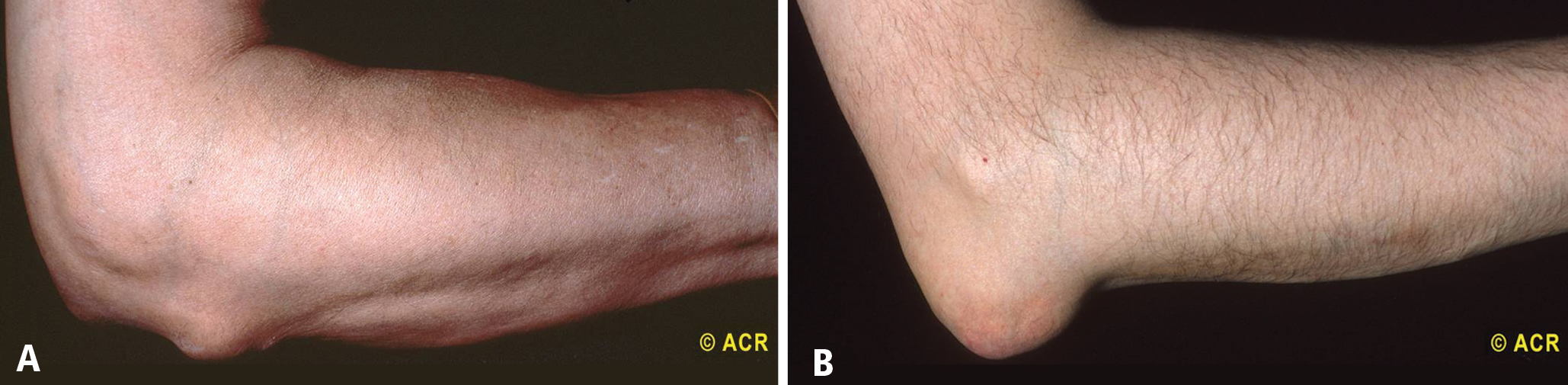
CPPD crystal deposition disease (pseudogout)
CPPD crystal deposition disease, or pseudogout, must also be included in the differential diagnosis of gout. This disease usually occurs in joints previously affected by osteoarthritis or joints that have been injured in the past.15 Attacks of CPPD crystal deposition disease commonly occur in the knee, in the wrist at the base of the thumb, or in the shoulder.15 Radiographic examination may reveal a line of calcification along the cartilage outlining the joint.15 Like gout, pseudogout attacks can occur spontaneously or after trauma, surgery, or a severe illness such as myocardial infarction or stroke.16
The presentation of pseudogout can be very similar to an acute attack of gout. The difference is seen when evaluating the crystals through a polarizing microscope. CPPD crystals are weakly positively birefringent (Figure 1B), in contrast to the negatively birefringent crystals seen with gout (Figure 1A).7 If a polarizing microscope is not available, the crystals usually can be distinguished by their differing shapes: urate crystals are fine and needlelike, whereas CPPD crystals are rhomboid (Figure 1).
Septic arthritis
When the differential diagnosis includes septic arthritis, the joint must be aspirated; a presumed diagnosis cannot be made. Among patients with an acute gouty attack, low-grade fever is reported during the attack in 29% of gout patients and 38% of patients with CPPD crystal deposition disease.14 Temperatures of 101°F or higher are not usually seen in patients with gout or CPPD crystal deposition disease and suggest an infection, although patients with septic arthritis may be afebrile, especially if they are taking immunosuppressive therapy or glucocorticoids, which can inhibit a febrile response. Synovial fluid analysis in patients with gout and septic arthritis can reveal WBC counts above 100,000 per mm3, whereas synovial fluid WBC counts above 50,000 per mm3 are more common in infection.
As noted earlier, gout and septic arthritis can coexist. In a patient presenting with a fever and a warm erythematous swollen joint, synovial fluid aspiration must be performed and evaluated for the presence of crystals and bacteria. The patient may require treatment for both causes of acute monoarticular arthritis.
In a patient undergoing renal dialysis, where gout or pseudogout can occur and where there is frequent intravascular manipulation, a septic joint can occur simultaneously.3,14 In this situation, not only must joint aspiration be performed, but the synovial fluid also needs to be evaluated for both crystals and bacteria. Again, the patient may require treatment for both causes of acute monoarticular arthritis.
CONCLUSIONS
The gold standard for diagnosing gout remains synovial fluid aspiration and analysis. In clinical situations when joint aspiration cannot be performed, the EULAR recommendations5 and the ACR criteria6 provide guidance for making a clinical or presumptive diagnosis of gout. A thorough patient history—both personal and family—and physical examination are critical in making a presumed diagnosis of gout. If the patient presents during an acute attack, serum urate measurement may be useful in making a clinical diagnosis if it reveals an elevated level. When the patient returns for follow-up 2 weeks later, a second serum urate measurement should be taken to allow comparison of the two levels. If the serum urate level is elevated at the follow-up visit, the EULAR recommendations state that a clinical diagnosis of gout can be made if the patient had an acute attack of arthritis in the great toe.
As noted in the EULAR recommendations, the future research agenda should include validating the clinical manifestations of gout against a diagnosis established by identification of urate crystals on synovial fluid analysis.5 Until this task can be completed, clinicians should become familiarized with the technique of joint aspiration so that in situations where a clinical or presumptive diagnosis of gout cannot be made—including cases where the differential diagnosis includes a septic joint—clinicians will be able to perform aspiration with confidence.
- Jelley MJ, Wortmann R. Practical steps in the diagnosis and management of gout. BioDrugs 2000; 14:99–107.
- Eggebeen AT. Gout: an update. Am Fam Physician 2007; 76:801–808, 811–812.
- Rott KT, Agudelo CA. Gout. JAMA 2003; 289:2857–2860.
- Poór G, Mituszova M. History, classification and epidemiology of crystal-related arthropathies. In: Hochberg MC, Silman AJ, Smolen JS, Weinblatt ME, Weisman MH, eds. Rheumatology. 3rd ed, vol 2. Edinburgh: Mosby; 2003:1893–1901.
- Zhang W, Doherty M, Pascual E, et al. EULAR evidence based recommendations for gout. Part I: Diagnosis. Report of a task force of the Standing Committee for International Clinical Studies Including Therapeutics (ESCISIT). Ann Rheum Dis 2006; 65:1301–1311.
- Wallace SL, Robinson H, Masi AT, Decker JL, McCarty DJ, Yü TF. Preliminary criteria for the classification of the acute arthritis of primary gout. Arthritis Rheum 1977; 20:895–900.
- Harris MD, Siegel LB, Alloway JA. Gout and hyperuricemia. Am Fam Physician 1999; 59:925–934.
- Saag KG, Mikuls TR. Recent advances in the epidemiology of gout. Curr Rheumatol Rep 2005; 7:235–241.
- Edwards NL. Gout. B. Clinical and laboratory features. In: Klippel JH, Crofford LJ, Stone JH, Weyand CM, eds. Primer on the Rheumatic Diseases. 12th ed. Atlanta, GA: Arthritis Foundation; 2001:313–319.
- Urano W, Yamanaka H, Tsutani H, et al. The inflammatory process in the mechanism of decreased serum uric acid concentrations during acute gouty arthritis. J Rheumatol 2002; 29:1950–1953.
- Shoji A, Yamanaka H, Kamatani N. A retrospective study of the relationship between serum urate level and recurrent attacks of gouty arthritis: evidence for reduction of recurrent gouty arthritis with anti-hyperuricemic therapy. Arthritis Rheum 2004; 51:321–325.
- Rigby AS, Wood PH. Serum uric acid levels and gout: what does this herald for the population? Clin Exp Rheumatol 1994; 12:395–400.
- George DL. Skin and rheumatic disease. In: Hochberg MC, Silman AJ, Smolen JS, Weinblatt ME, Weisman MH, eds. Rheumatology. 3rd ed, vol 1. Edinburgh: Mosby; 2003:279–292.
- Ho G Jr, DeNuccio M. Gout and pseudogout in hospitalized patients. Arch Intern Med 1993; 153:2787–2790.
- Agarwal AK. Gout and pseudogout. Prim Care 1993; 20:839–855.
- Halverson PB, Ryan LM. Arthritis associated with calcium-containing crystals. In: Klippel JH, Crofford LJ, Stone JH, Weyand CM, eds. Primer on the Rheumatic Diseases. 12th ed. Atlanta, GA: Arthritis Foundation; 2001:299–306.
The presence of urate crystals in synovial fluid is the gold standard for diagnosing gout,1 yet clinicians—both primary care physicians and rheumatologists—may not routinely perform synovial fluid analysis even when evaluating a patient who presents with an acute inflammatory arthritis.2 This paper discusses the various reasons why this is so and reviews several important resulting clinical issues: how a presumptive diagnosis of gout is made, when to measure the serum urate level, and special considerations in the differential diagnosis.
SYNOVIAL FLUID ANALYSIS: WHY IS THE GOLD STANDARD NOT MORE ROUTINE?
Occasionally, the aspirated joint does not appear to contain any joint fluid and the clinician may be concerned about the possibility of a “dry tap.” Other possible reasons include lack of experience with synovial fluid aspiration and evaluation, or limited access to the polarizing microscopes used to examine synovial fluid. Time is another factor; in a busy primary care practice, where patients are usually seen approximately every 7 to 11 minutes, there may not be time to aspirate a joint. The urgency of fluid examination is another issue, as synovial fluid must be examined immediately, since the crystals can become smaller, less numerous, and less birefringent with time.4
THE CLINICAL, OR PRESUMPTIVE, DIAGNOSIS
In the appropriate clinical scenario, a presumptive diagnosis of gout can be made on the basis of typical clinical features and the presence of hyperuricemia.1,2
Expert societies offer guidance, but no validation studies to date
Evidence-based recommendations for the diagnosis of gout from the European League Against Rheumatism (EULAR) state that in acute attacks, the rapid development of severe pain, swelling, and tenderness that peaks within 6 to 12 hours, especially with overlying erythema, is highly suggestive of crystal inflammation although not specific for gout.5 These recommendations further state that for typical presentations of gout (such as recurrent podagra [gouty pain in the great toe] with hyperuricemia), a clinical diagnosis alone is reasonably accurate.5
- The presence of urate crystals in joint fluid
- A tophus containing urate crystals
- Fulfillment of 6 or more of the criteria in Table 1.
No subsequent studies have been published on the validity or usefulness of any of these diagnostic criteria.
What must inform the presumptive diagnosis
Both the EULAR recommendations and the ACR criteria state that although the gold standard for diagnosing gout is the presence of urate crystals on synovial fluid analysis, a clinical diagnosis of gout can be made on the basis of certain patient criteria. This clinical, or presumptive, diagnosis of gout should be made based on the following:
- A careful patient and family history, including questions regarding comorbid conditions frequently associated with gout (such as hypertriglyceridemia, diabetes, coronary heart disease, hypertension, and the metabolic syndrome) and whether the patient has had previous similar episodes of acute joint pain and swelling in the absence of trauma
- Thorough identification of all current medications, some of which may be associated with hyperuricemia
- A thorough physical examination.
THE PHYSICAL EXAMINATION FOR GOUT
Examination of patients with a history suggestive of gout should include not only the joints but also the extensor surface of the forearms and feet. When patients are seen for a visit and gout is suspected, they should be instructed to remove their shoes and socks and roll up their sleeves to allow examination for evidence of tophi, which would suggest a past history of gouty arthritis. The ear, knee, and olecranon bursa are other common sites for tophi,3 so patients should also be asked to roll up their pants and sleeves and remove any head coverings.7 In the late stages of gouty arthritis, multiple joints may be involved, which can cause the condition to be confused with other diagnoses such as psoriatic arthritis or erosive osteoarthritis.7
ACUTE PRESENTATIONS OF GOUT
The typical gout presentation is remarkable for very intense pain that often occurs at night when the extremities are colder. Precipitation of urate in the distal extremities can occur when the extremities are horizontal and tend to become cold.8
Approximately 90% of initial gout attacks are monoarticular, leaving only 10% of cases that are oligoarticular or polyarticular.7 If more than one joint is involved, especially if the patient has a family history suggestive of gout or takes a medication that causes hyperuricemia, gout should be considered in the differential diagnosis even if the patient denies having a prior gout attack.
Frequently, patients will call their primary care physician during a gout attack but are not be able to schedule an appointment until after the attack has resolved. When possible, patients should be seen during the attack to confirm whether the attack is due to gout. A diagnosis of gout should not be made over the phone when a patient describes pain in the great toe, as only 50% of initial gout attacks occur in the great toe7 and it is not known what proportion of acute pain episodes in the great toe are attributable to gout. The most common cause of pain in the great toe is osteoarthritis.
SPECIAL CONSIDERATIONS FOR THE PRESUMPTIVE DIAGNOSIS OF GOUT
How long have acute attacks been occurring?
In a clinical scenario in which synovial fluid aspiration cannot be performed, the appropriateness of a presumptive diagnosis can be assessed by a discussion with the patient about how long he or she has been experiencing acute attacks of joint pain. If the attacks have occurred for more than 10 years, tophi will likely be present.3 After even longer periods, gout may become polyarticular.7 In postmenopausal women, the distal interphalangeal joints may be involved,3 which may lead to a misdiagnosis of osteoarthritis, as these joints are typically affected by osteoarthritis.
Is the patient taking a urate-raising medication?
Certain medications have been associated with hyper-uricemia, including cyclosporine and thiazide diuretics.9 If a patient has been taking one of these medications, gout should be considered in the differential diagnosis if the patient presents with acute joint pain.
It has been argued that a reduction in joint pain and swelling after the use of colchicine confirms a diagnosis of gout. However, other conditions—such as tendonitis, calcium pyrophosphate dihydrate (CPPD) crystal deposition disease (pseudogout),3 and rheumatoid arthritis (RA)—can also improve after treatment with colchicine.1
Be vigilant for fever
Another consideration in making a clinical diagnosis of gout is the association with a low-grade fever; these patients may feel as if they have the flu.8 Acute gout may also cause a high fever and an elevated white blood cell (WBC) count;3 in this situation, synovial fluid aspiration must be performed to exclude septic arthritis, either alone or in the presence of gouty arthritis. In situations where septic arthritis is suspected, an emergency visit to a rheumatologist is indicated for synovial fluid aspiration to be performed, as gout and sepsis can coexist.5 In such instances, Gram staining and culture of the synovial fluid should still be performed even if monosodium urate crystals are identified.5
MEASUREMENT OF SERUM URATE LEVELS
Measuring serum urate levels during an acute attack, treating the acute attack with anti-inflammatory medications, and reevaluating the patient in the office 2 weeks after the acute attack are all recommended in the management of a patient with gout. If the serum urate level was not elevated during the acute attack, it is likely to be elevated 2 weeks later if the patient has gout.10 Elevated levels of serum urate during the intercritical periods are predictive of future gout attacks.11 Measuring serum urate during the initial attack and then 2 weeks later yields two serum urate levels that can be compared to assist in considering a presumed diagnosis of gout. A study by Rigby and Wood concluded that in patients with low serum urate levels (< 4 mg/dL) 2 weeks following an inflammatory arthritis attack, a diagnosis of gout is unlikely.12
DIFFERENTIAL DIAGNOSIS OF GOUT
Rheumatoid arthritis
CPPD crystal deposition disease (pseudogout)
CPPD crystal deposition disease, or pseudogout, must also be included in the differential diagnosis of gout. This disease usually occurs in joints previously affected by osteoarthritis or joints that have been injured in the past.15 Attacks of CPPD crystal deposition disease commonly occur in the knee, in the wrist at the base of the thumb, or in the shoulder.15 Radiographic examination may reveal a line of calcification along the cartilage outlining the joint.15 Like gout, pseudogout attacks can occur spontaneously or after trauma, surgery, or a severe illness such as myocardial infarction or stroke.16
The presentation of pseudogout can be very similar to an acute attack of gout. The difference is seen when evaluating the crystals through a polarizing microscope. CPPD crystals are weakly positively birefringent (Figure 1B), in contrast to the negatively birefringent crystals seen with gout (Figure 1A).7 If a polarizing microscope is not available, the crystals usually can be distinguished by their differing shapes: urate crystals are fine and needlelike, whereas CPPD crystals are rhomboid (Figure 1).
Septic arthritis
When the differential diagnosis includes septic arthritis, the joint must be aspirated; a presumed diagnosis cannot be made. Among patients with an acute gouty attack, low-grade fever is reported during the attack in 29% of gout patients and 38% of patients with CPPD crystal deposition disease.14 Temperatures of 101°F or higher are not usually seen in patients with gout or CPPD crystal deposition disease and suggest an infection, although patients with septic arthritis may be afebrile, especially if they are taking immunosuppressive therapy or glucocorticoids, which can inhibit a febrile response. Synovial fluid analysis in patients with gout and septic arthritis can reveal WBC counts above 100,000 per mm3, whereas synovial fluid WBC counts above 50,000 per mm3 are more common in infection.
As noted earlier, gout and septic arthritis can coexist. In a patient presenting with a fever and a warm erythematous swollen joint, synovial fluid aspiration must be performed and evaluated for the presence of crystals and bacteria. The patient may require treatment for both causes of acute monoarticular arthritis.
In a patient undergoing renal dialysis, where gout or pseudogout can occur and where there is frequent intravascular manipulation, a septic joint can occur simultaneously.3,14 In this situation, not only must joint aspiration be performed, but the synovial fluid also needs to be evaluated for both crystals and bacteria. Again, the patient may require treatment for both causes of acute monoarticular arthritis.
CONCLUSIONS
The gold standard for diagnosing gout remains synovial fluid aspiration and analysis. In clinical situations when joint aspiration cannot be performed, the EULAR recommendations5 and the ACR criteria6 provide guidance for making a clinical or presumptive diagnosis of gout. A thorough patient history—both personal and family—and physical examination are critical in making a presumed diagnosis of gout. If the patient presents during an acute attack, serum urate measurement may be useful in making a clinical diagnosis if it reveals an elevated level. When the patient returns for follow-up 2 weeks later, a second serum urate measurement should be taken to allow comparison of the two levels. If the serum urate level is elevated at the follow-up visit, the EULAR recommendations state that a clinical diagnosis of gout can be made if the patient had an acute attack of arthritis in the great toe.
As noted in the EULAR recommendations, the future research agenda should include validating the clinical manifestations of gout against a diagnosis established by identification of urate crystals on synovial fluid analysis.5 Until this task can be completed, clinicians should become familiarized with the technique of joint aspiration so that in situations where a clinical or presumptive diagnosis of gout cannot be made—including cases where the differential diagnosis includes a septic joint—clinicians will be able to perform aspiration with confidence.
- Jelley MJ, Wortmann R. Practical steps in the diagnosis and management of gout. BioDrugs 2000; 14:99–107.
- Eggebeen AT. Gout: an update. Am Fam Physician 2007; 76:801–808, 811–812.
- Rott KT, Agudelo CA. Gout. JAMA 2003; 289:2857–2860.
- Poór G, Mituszova M. History, classification and epidemiology of crystal-related arthropathies. In: Hochberg MC, Silman AJ, Smolen JS, Weinblatt ME, Weisman MH, eds. Rheumatology. 3rd ed, vol 2. Edinburgh: Mosby; 2003:1893–1901.
- Zhang W, Doherty M, Pascual E, et al. EULAR evidence based recommendations for gout. Part I: Diagnosis. Report of a task force of the Standing Committee for International Clinical Studies Including Therapeutics (ESCISIT). Ann Rheum Dis 2006; 65:1301–1311.
- Wallace SL, Robinson H, Masi AT, Decker JL, McCarty DJ, Yü TF. Preliminary criteria for the classification of the acute arthritis of primary gout. Arthritis Rheum 1977; 20:895–900.
- Harris MD, Siegel LB, Alloway JA. Gout and hyperuricemia. Am Fam Physician 1999; 59:925–934.
- Saag KG, Mikuls TR. Recent advances in the epidemiology of gout. Curr Rheumatol Rep 2005; 7:235–241.
- Edwards NL. Gout. B. Clinical and laboratory features. In: Klippel JH, Crofford LJ, Stone JH, Weyand CM, eds. Primer on the Rheumatic Diseases. 12th ed. Atlanta, GA: Arthritis Foundation; 2001:313–319.
- Urano W, Yamanaka H, Tsutani H, et al. The inflammatory process in the mechanism of decreased serum uric acid concentrations during acute gouty arthritis. J Rheumatol 2002; 29:1950–1953.
- Shoji A, Yamanaka H, Kamatani N. A retrospective study of the relationship between serum urate level and recurrent attacks of gouty arthritis: evidence for reduction of recurrent gouty arthritis with anti-hyperuricemic therapy. Arthritis Rheum 2004; 51:321–325.
- Rigby AS, Wood PH. Serum uric acid levels and gout: what does this herald for the population? Clin Exp Rheumatol 1994; 12:395–400.
- George DL. Skin and rheumatic disease. In: Hochberg MC, Silman AJ, Smolen JS, Weinblatt ME, Weisman MH, eds. Rheumatology. 3rd ed, vol 1. Edinburgh: Mosby; 2003:279–292.
- Ho G Jr, DeNuccio M. Gout and pseudogout in hospitalized patients. Arch Intern Med 1993; 153:2787–2790.
- Agarwal AK. Gout and pseudogout. Prim Care 1993; 20:839–855.
- Halverson PB, Ryan LM. Arthritis associated with calcium-containing crystals. In: Klippel JH, Crofford LJ, Stone JH, Weyand CM, eds. Primer on the Rheumatic Diseases. 12th ed. Atlanta, GA: Arthritis Foundation; 2001:299–306.
KEY POINTS
- If the serum urate level was not elevated when measured during an acute attack of arthritis, it will likely be elevated at 2-week follow-up if the patient does indeed have gout.
- Gouty tophi are typically found in the olecranon bursa, whereas rheumatoid nodules are usually located on the extensor surface of the forearm.
- Urate crystals of gout are negatively bifringent and fine and needlelike in shape, whereas the crystals of pseudogout are weakly positively birefringent and rhomboid.
- Gout and septic arthritis can coexist; when the differential diagnosis includes septic arthritis, joint aspiration is required.
- Until criteria for the presumptive diagnosis of gout are validated, clinicians should become familiar with the technique of joint aspiration.
The practical management of gout
To apropriately manage gout, it is important to distinguish between treatment of acute gout attacks and management of the underlying metabolic defect. While acute attacks are treated with anti-inflammatory agents, the underlying hyperuricemia must be addressed by lowering the serum urate concentration to levels that lead to prevention of acute flares, together with consideration of the contributing role of the patient’s lifestyle factors and comorbidities. This article surveys treatment options for both acute gout attacks and the underlying hyperuricemic state, focusing on considerations to guide therapy selection and optimize prospects for treatment success.
ACUTE GOUTY ARTHRITIS: ANTI-INFLAMMATORY AGENTS AND KEY ISSUES FOR THEIR USE
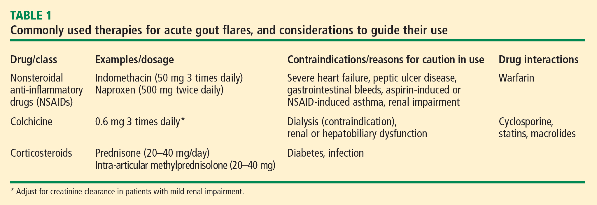
One of the most important considerations in selecting an anti-inflammatory medication is how the patient’s comorbidities, such as renal disease, or concurrent medications may influence the choice of agent, as outlined in Table 1.
Nonsteroidal anti-inflammatory drugs (NSAIDs)
A number of NSAIDs are available to treat acute flares of gout.1–3 When used at a full anti-inflammatory dose, all NSAIDs appear to be equally effective.
NSAIDs must be used cautiously in patients who have any of a number of comorbid conditions, as detailed in Table 1. If a patient is otherwise healthy—without significant renal, cardiovascular, or gastrointestinal disease—and has no history of aspirin allergy, NSAIDs are the treatment of choice for acute gout attacks. Use of a proton pump inhibitor can improve gastrointestinal tolerance of NSAIDs and reduce the likelihood of gastric bleeding but may not avoid other concerns. Indomethacin can cause headache or even confusion, particularly in the elderly.
Colchicine
Colchicine can be an effective alternative for acute therapy.4,5 If a patient with previously documented gout can be coached to begin colchicine at the first hint of a gout attack, a full-blown attack often can be prevented. A colchicine regimen of 0.5 or 0.6 mg 3 times daily, although not well studied, may be effective while limiting the diarrhea, nausea, and vomiting that is predictable with hourly colchicine dosing.6,7 Colchicine must be used cautiously in patients with renal or liver disease and is contraindicated in patients undergoing dialysis.8
Corticosteroids
Systemic corticosteroids are often used for polyarticular gout or in patients with contraindications to NSAIDs or colchicine.9 When they are used in diabetic patients, glycemic control must be monitored, and an increased insulin dose can be prescribed temporarily until glucose levels normalize.
Corticosteroids may also be injected directly into the joint, as this approach offers reduced risks compared with oral administration. Direct injection is especially useful in patients with attacks that involve only one or two joints.
TREATING THE UNDERLYING HYPERURICEMIA THROUGH URATE-LOWERING STRATEGIES
Terminating the acute flare manages gout symptoms but does not treat the underlying disease. Crystals often remain in the joint after flares have resolved. Addressing the underlying metabolic condition requires lowering serum urate levels, which can deplete crystals and reduce or prevent gout flares.
The goals of urate-lowering therapy are to reduce serum urate levels to less than 6 mg/dL in order to mobilize and deplete crystals with minimal toxicity.10
Role of lifestyle interventions
As discussed by Weaver earlier in this supplement, obesity and certain patterns of food and alcohol consumption can increase the risk of developing hyperuricemia and gout. In addition to weight loss, dietary changes.such as reducing intake of animal purines, high-fructose sweeteners, and alcohol, and increasing intake of vitamin C or bing cherries.may lower serum urate levels modestly (ie, by 1 or 2 mg/dL).11–15 While lifestyle interventions may be all that is needed in some patients with early mild gout, such interventions generally do not replace the need for urate-lowering drug therapy in cases of existing gout. Accordingly, this discussion focuses on medications used to treat hyperuricemia in the United States: the xanthine oxidase inhibitor allopurinol and the uricosuric agent probenecid.
Initiating urate-lowering drug therapy
Chronic therapy should be discussed with the patient early in the course of the disease. Treatment recommendations need to be individualized based on the patient’s overall health, comorbidities, and willingness to adhere to chronic treatment.
Initiation of urate-lowering therapy is appropriate to consider after the acute attack has fully resolved and the patient has been stable for 1 to 2 weeks. If a patient’s serum urate level is very high (eg, > 10 mg/dL), urate-lowering therapy may be initiated even after a single attack, as progression is more likely to occur with higher levels. Treatment should be initiated long before tophi or persistent joint damage develop. If the patient already has objective radiographic evidence of gouty changes in the joints, or if tophi or nephrolithiasis are present when the patient is first seen, urate-lowering therapy should be started.
Concurrent low-dose anti-inflammatory prophylaxis
Abrupt decreases (or increases) in serum urate levels may precipitate gout flares. For this reason, anti-inflammatory prophylaxis should be used when urate-lowering therapy is initiated, as it can quickly reduce serum urate levels. Colchicine (0.6 mg once or twice daily)8 or NSAIDs (eg, naproxen 250 mg/day) prescribed at lower than full anti-inflammatory doses may be used to prevent flares in this setting. When using long-term colchicine in a patient with renal disease, lower doses must be used and the patient should be monitored closely for reversible axonal neuromyopathy and vacuolar myopathy or rhabdomyolysis; the latter complication may be more frequent in patients taking concurrent statin or macrolide therapy. There are no controlled studies on the benefits and safety of prophylactic NSAID use in gouty patients with comorbidities.
Borstad et al documented in a placebo-controlled study that colchicine prophylaxis at the time of allopurinol initiation reduces flares but does not completely abolish them.16 From 0 to 3 months after therapy initiation, the mean number of flares was 0.57 in patients who received colchicine versus 1.91 in patients who received placebo (P = .022); from 3 to 6 months after initiation, the mean number of flares was 0 versus 1.05 in the respective patient groups (P = .033).16
Depending on the body’s urate load, it may take many months to deplete crystals. There is evidence that prophylaxis should be used for at least 3 to 6 months to reduce the risk of mobilization flares.16 Patients should be warned during this time that gout flares may still occur and should be treated promptly. Prophylaxis should continue longer in patients with tophi, often until the tophi have resolved.
Allopurinol
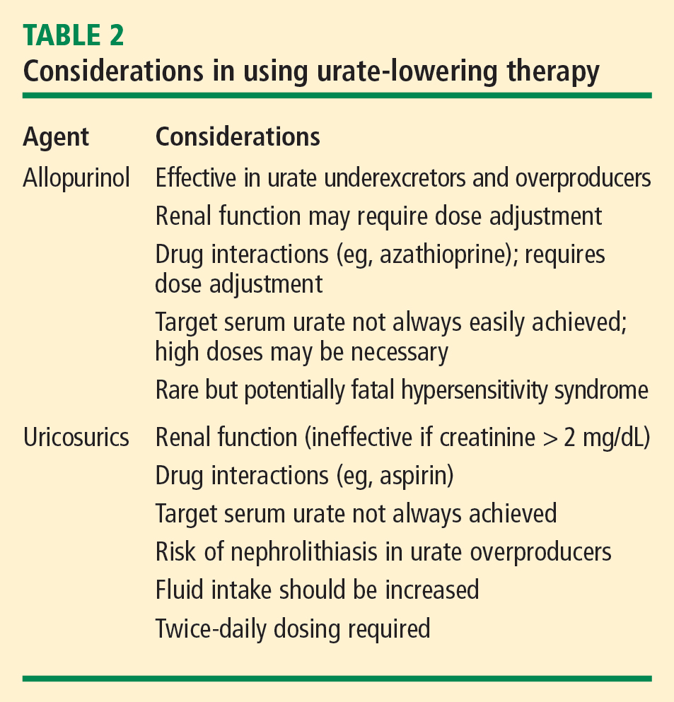
The rare but potentially fatal hypersensitivity syndrome is a concern with allopurinol. If a rash develops in a patient taking allopurinol, the drug should be discontinued, as rash can be a precursor of severe systemic hypersensitivity.
Renal function must be considered in allopurinol dosing, and treatment should always be initiated at lower doses in patients with renal disease.18 However, recent reports indicate that allopurinol doses can be gradually and safely increased to an effective level that achieves a serum urate concentration of less than 6 mg/dL even in patients with reduced kidney function.19,20
Uricosurics
The uricosuric agent probenecid works by increasing the urinary excretion of urate.10 Probenecid is commonly dosed at 500 mg twice daily, with a maximum daily dose of 2 g, in an attempt to reach the target serum urate level. Probenecid is unlikely to be effective if the patient’s serum creatinine is greater than 2 mg/dL.21
When a uricosuric agent is used, a 24-hour urine urate measurement must be taken to identify and exclude urate overproducers (patients with more than 800 to 1,000 mg of uric acid in a good 24-hour collection), as such patients are at risk for uric acid kidney stones. Additionally, aspirin interferes with probenecid’s effect on the renal tubules. The ideal candidate for uricosuric therapy has good kidney function, is not a urate overproducer, and is willing to drink 8 glasses of water a day to minimize the risk of kidney stones (Table 2).
Evidence supporting urate targets and continuous maintenance of urate reductions
If a serum urate level of less than 6 mg/dL is achieved and maintained, gout flares will be reduced and crystals can be depleted from inside the joint.22–25 Additionally, the size of tophi can be reduced and their recurrence prevented.26–28 Serum urate levels below 4 mg/dL can result in more rapid dissolution of tophi.26

Patient commitment and education are essential
No treatment plan will succeed without the commitment of the patient, so discussion to determine the patient’s willingness to commit to lifetime therapy is warranted. A number of surveys have shown that the rate of continued use of allopurinol after it is initially prescribed is less than 50%.17 If the physician or nurse monitors adherence, however, treatment is more likely to be successful.30
Patients need more education about gout, as education may improve adherence and treatment success. Patient education material is available from the Arthritis Foundation (www.arthritis.org/disease-center.php) and the Gout & Uric Acid Education Society (www.gouteducation.org).
CONCLUSION
A comprehensive treatment strategy is critical to ensure ideal gout therapy. Acute flares should be addressed as rapidly as possible with an anti-inflammatory agent selected on the basis of the patient’s comorbidities and other medications. Most patients require chronic urate-lowering therapy to deplete crystals from joints and prevent flares. Initiation of urate-lowering therapy should be considered early in the disease course, following resolution of the acute attack. Low-dose anti-inflammatory prophylaxis should be initiated when any urate-lowering therapy is started. Regular monitoring of serum urate will ensure effective dosing to achieve a target serum urate level of less than 6 mg/dL. Once urate deposits are depleted, acute flares should cease.
- Altman RD, Honig S, Levin JM, Lightfoot RW. Ketoprofen versus indomethacin in patients with acute gouty arthritis: a multicenter, double blind comparative study. J Rheumatol 1988; 15:1422–1426.
- Schumacher HR Jr, Boice JA, Daikh DI, et al. Randomised double blind trial of etoricoxib and indometacin in treatment of acute gouty arthritis. BMJ 2002; 324:1488–1492.
- Willburger RE, Mysler E, Derbot J, et al. Lumiracoxib 400 mg once daily is comparable to indomethacin 50 mg three times daily for the treatment of acute flares of gout. Rheumatology (Oxford) 2007; 46:1126–1132.
- Ahern MJ, Reid C, Gordon T, et al. Does colchicine work? The results of the first controlled study in acute gout. Aust N Z J Med 1987; 17:301–304.
- Schlesinger N, Moore DF, Sun JD, Schumacher HR Jr. A survey of current evaluation and treatment of gout. J Rheumatol 2006; 33:2050–2052.
- Rozenberg S, Lang T, Laatar A, Koeger AC, Orcel P, Bourgerois P. Diversity of opinions on the management of gout in France. A survey of 750 rheumatologists. Rev Rhum Engl Ed 1996; 63:255–261.
- Schlesinger N, Schumacher R, Catton M, Maxwell L. Colchicine for acute gout. Cochrane Database Syst Rev 2006; 18:CD006190.
- Rott KT, Agudelo CA. Gout. JAMA 2003; 289:2857–2860.
- Alloway JA, Moriarity MJ, Hoogland YT, Nashel DJ. Comparison of triamcinolone acetonide with indomethacin in the treatment of acute gouty arthritis. J Rheumatol 1993; 20:111–113.
- Schumacher HR Jr, Chen LX. Newer therapeutic approaches: gout. Rheum Dis Clin North Am 2006; 32:235–244, xii.
- Choi HK. Diet, alcohol, and gout: how do we advise patients given recent developments? Curr Rheumatol Rep 2005; 7:220–226.
- Johnson MW, Mitch WE. The risks of asymptomatic hyperuricaemia and the use of uricosuric diuretics. Drugs 1981; 21:220–225.
- Berger L, Yü TF. Renal function in gout. IV. An analysis of 524 gouty subjects including long-term follow-up studies. Am J Med 1975; 59:605–613.
- Stein HB, Hasan A, Fox IH. Ascorbic acid-induced uricosuria. A consequence of megavitamin therapy. Ann Intern Med 1976; 84:385–388.
- Choi JW, Ford ES, Gao X, Choi HK. Sugar-sweetened soft drinks, diet soft drinks, and serum uric acid level: the Third National Health and Nutrition Examination Survey. Arthritis Rheum 2008; 59:109–116.
- Borstad GC, Bryant LR, Abel MP, Scroggie DA, Harris MD, Alloway JA. Colchicine for prophylaxis of acute flares when initiating allopurinol for chronic gouty arthritis. J Rheumatol 2004; 31:2429–2432.
- Sarawate CA, Patel PA, Schumacher HR, Yang W, Brewer KK, Bakst AW. Serum urate levels and gout flares: analysis from managed care data. J Clin Rheumatol 2006; 12:61–65.
- Hande KR, Noone RM, Stone WJ. Severe allopurinol toxicity. Description and guidelines for prevention in patients with renal insufficiency. Am J Med 1984; 76:47–56.
- Vázquez-Mellado J, Morales EM, Pacheco-Tena C, Burgos-Vargas R. Relation between adverse events associated with allopurinol and renal function in patients with gout. Ann Rheum Dis 2001; 60:981–983.
- Dalbeth N, Kumar S, Stamp L, Gow P. Dose adjustment of allopurinol according to creatinine clearance does not provide adequate control of hyperuricemia in patients with gout. J Rheumatol 2006; 33:1646–1650.
- Emmerson BT. The management of gout. N Engl J Med 1996; 334:445–451.
- Becker MA, Schumacher HR Jr, Wortmann, RL, et al. Febuxostat compared with allopurinol in patients with hyperuricemia and gout. N Engl J Med 2005; 353:2450–2461.
- Li-Yu J, Clayburne G, Sieck M, et al. Treatment of chronic gout. Can we determine when urate stores are depleted enough to prevent attacks of gout? J Rheumatol 2001; 28:577–580.
- Shoji A, Yamanaka H, Kamatani N. A retrospective study of the relationship between serum urate level and recurrent attacks of gouty arthritis: evidence for reduction of recurrent gouty arthritis with anti-hyperuricemic therapy. Arthritis Rheum 2004; 51:321–325.
- Pascual E, Sivera F. Time required for disappearance of urate crystals from synovial fluid after successful hypouricaemic treatment relates to the duration of gout. Ann Rheum Dis 2007; 66:1056–1058.
- Perez-Ruiz F, Calabozo M, Pijoan JI, Herrero-Beites AM, Ruibal A. Effect of urate-lowering therapy on the velocity of size reduction of tophi in chronic gout. Arthritis Rheum 2002; 47:356–360
- McCarthy GM, Barthelemy CR, Veum JA, Wortmann RL. Influence of antihyperuricemic therapy on the clinical and radiographic progression of gout. Arthritis Rheum 1991; 34:1489–1494.
- van Lieshout-Zuidema MF, Breedveld FC. Withdrawal of long-term antihyperuricemic therapy in tophaceous gout. J Rheumatol 1993; 20:1383–1385.
- Bull PW, Scott JT. Intermittent control of hyperuricemia in the treatment of gout. J Rheumatol 1989; 16:1246–1248.
- Murphy-Bielicki, Schumacher HR. How does patient education affect gout? Clin Rheumatol Pract 1984; 2:77–80.
To apropriately manage gout, it is important to distinguish between treatment of acute gout attacks and management of the underlying metabolic defect. While acute attacks are treated with anti-inflammatory agents, the underlying hyperuricemia must be addressed by lowering the serum urate concentration to levels that lead to prevention of acute flares, together with consideration of the contributing role of the patient’s lifestyle factors and comorbidities. This article surveys treatment options for both acute gout attacks and the underlying hyperuricemic state, focusing on considerations to guide therapy selection and optimize prospects for treatment success.
ACUTE GOUTY ARTHRITIS: ANTI-INFLAMMATORY AGENTS AND KEY ISSUES FOR THEIR USE
One of the most important considerations in selecting an anti-inflammatory medication is how the patient’s comorbidities, such as renal disease, or concurrent medications may influence the choice of agent, as outlined in Table 1.
Nonsteroidal anti-inflammatory drugs (NSAIDs)
A number of NSAIDs are available to treat acute flares of gout.1–3 When used at a full anti-inflammatory dose, all NSAIDs appear to be equally effective.
NSAIDs must be used cautiously in patients who have any of a number of comorbid conditions, as detailed in Table 1. If a patient is otherwise healthy—without significant renal, cardiovascular, or gastrointestinal disease—and has no history of aspirin allergy, NSAIDs are the treatment of choice for acute gout attacks. Use of a proton pump inhibitor can improve gastrointestinal tolerance of NSAIDs and reduce the likelihood of gastric bleeding but may not avoid other concerns. Indomethacin can cause headache or even confusion, particularly in the elderly.
Colchicine
Colchicine can be an effective alternative for acute therapy.4,5 If a patient with previously documented gout can be coached to begin colchicine at the first hint of a gout attack, a full-blown attack often can be prevented. A colchicine regimen of 0.5 or 0.6 mg 3 times daily, although not well studied, may be effective while limiting the diarrhea, nausea, and vomiting that is predictable with hourly colchicine dosing.6,7 Colchicine must be used cautiously in patients with renal or liver disease and is contraindicated in patients undergoing dialysis.8
Corticosteroids
Systemic corticosteroids are often used for polyarticular gout or in patients with contraindications to NSAIDs or colchicine.9 When they are used in diabetic patients, glycemic control must be monitored, and an increased insulin dose can be prescribed temporarily until glucose levels normalize.
Corticosteroids may also be injected directly into the joint, as this approach offers reduced risks compared with oral administration. Direct injection is especially useful in patients with attacks that involve only one or two joints.
TREATING THE UNDERLYING HYPERURICEMIA THROUGH URATE-LOWERING STRATEGIES
Terminating the acute flare manages gout symptoms but does not treat the underlying disease. Crystals often remain in the joint after flares have resolved. Addressing the underlying metabolic condition requires lowering serum urate levels, which can deplete crystals and reduce or prevent gout flares.
The goals of urate-lowering therapy are to reduce serum urate levels to less than 6 mg/dL in order to mobilize and deplete crystals with minimal toxicity.10
Role of lifestyle interventions
As discussed by Weaver earlier in this supplement, obesity and certain patterns of food and alcohol consumption can increase the risk of developing hyperuricemia and gout. In addition to weight loss, dietary changes.such as reducing intake of animal purines, high-fructose sweeteners, and alcohol, and increasing intake of vitamin C or bing cherries.may lower serum urate levels modestly (ie, by 1 or 2 mg/dL).11–15 While lifestyle interventions may be all that is needed in some patients with early mild gout, such interventions generally do not replace the need for urate-lowering drug therapy in cases of existing gout. Accordingly, this discussion focuses on medications used to treat hyperuricemia in the United States: the xanthine oxidase inhibitor allopurinol and the uricosuric agent probenecid.
Initiating urate-lowering drug therapy
Chronic therapy should be discussed with the patient early in the course of the disease. Treatment recommendations need to be individualized based on the patient’s overall health, comorbidities, and willingness to adhere to chronic treatment.
Initiation of urate-lowering therapy is appropriate to consider after the acute attack has fully resolved and the patient has been stable for 1 to 2 weeks. If a patient’s serum urate level is very high (eg, > 10 mg/dL), urate-lowering therapy may be initiated even after a single attack, as progression is more likely to occur with higher levels. Treatment should be initiated long before tophi or persistent joint damage develop. If the patient already has objective radiographic evidence of gouty changes in the joints, or if tophi or nephrolithiasis are present when the patient is first seen, urate-lowering therapy should be started.
Concurrent low-dose anti-inflammatory prophylaxis
Abrupt decreases (or increases) in serum urate levels may precipitate gout flares. For this reason, anti-inflammatory prophylaxis should be used when urate-lowering therapy is initiated, as it can quickly reduce serum urate levels. Colchicine (0.6 mg once or twice daily)8 or NSAIDs (eg, naproxen 250 mg/day) prescribed at lower than full anti-inflammatory doses may be used to prevent flares in this setting. When using long-term colchicine in a patient with renal disease, lower doses must be used and the patient should be monitored closely for reversible axonal neuromyopathy and vacuolar myopathy or rhabdomyolysis; the latter complication may be more frequent in patients taking concurrent statin or macrolide therapy. There are no controlled studies on the benefits and safety of prophylactic NSAID use in gouty patients with comorbidities.
Borstad et al documented in a placebo-controlled study that colchicine prophylaxis at the time of allopurinol initiation reduces flares but does not completely abolish them.16 From 0 to 3 months after therapy initiation, the mean number of flares was 0.57 in patients who received colchicine versus 1.91 in patients who received placebo (P = .022); from 3 to 6 months after initiation, the mean number of flares was 0 versus 1.05 in the respective patient groups (P = .033).16
Depending on the body’s urate load, it may take many months to deplete crystals. There is evidence that prophylaxis should be used for at least 3 to 6 months to reduce the risk of mobilization flares.16 Patients should be warned during this time that gout flares may still occur and should be treated promptly. Prophylaxis should continue longer in patients with tophi, often until the tophi have resolved.
Allopurinol
The rare but potentially fatal hypersensitivity syndrome is a concern with allopurinol. If a rash develops in a patient taking allopurinol, the drug should be discontinued, as rash can be a precursor of severe systemic hypersensitivity.
Renal function must be considered in allopurinol dosing, and treatment should always be initiated at lower doses in patients with renal disease.18 However, recent reports indicate that allopurinol doses can be gradually and safely increased to an effective level that achieves a serum urate concentration of less than 6 mg/dL even in patients with reduced kidney function.19,20
Uricosurics
The uricosuric agent probenecid works by increasing the urinary excretion of urate.10 Probenecid is commonly dosed at 500 mg twice daily, with a maximum daily dose of 2 g, in an attempt to reach the target serum urate level. Probenecid is unlikely to be effective if the patient’s serum creatinine is greater than 2 mg/dL.21
When a uricosuric agent is used, a 24-hour urine urate measurement must be taken to identify and exclude urate overproducers (patients with more than 800 to 1,000 mg of uric acid in a good 24-hour collection), as such patients are at risk for uric acid kidney stones. Additionally, aspirin interferes with probenecid’s effect on the renal tubules. The ideal candidate for uricosuric therapy has good kidney function, is not a urate overproducer, and is willing to drink 8 glasses of water a day to minimize the risk of kidney stones (Table 2).
Evidence supporting urate targets and continuous maintenance of urate reductions
If a serum urate level of less than 6 mg/dL is achieved and maintained, gout flares will be reduced and crystals can be depleted from inside the joint.22–25 Additionally, the size of tophi can be reduced and their recurrence prevented.26–28 Serum urate levels below 4 mg/dL can result in more rapid dissolution of tophi.26
Patient commitment and education are essential
No treatment plan will succeed without the commitment of the patient, so discussion to determine the patient’s willingness to commit to lifetime therapy is warranted. A number of surveys have shown that the rate of continued use of allopurinol after it is initially prescribed is less than 50%.17 If the physician or nurse monitors adherence, however, treatment is more likely to be successful.30
Patients need more education about gout, as education may improve adherence and treatment success. Patient education material is available from the Arthritis Foundation (www.arthritis.org/disease-center.php) and the Gout & Uric Acid Education Society (www.gouteducation.org).
CONCLUSION
A comprehensive treatment strategy is critical to ensure ideal gout therapy. Acute flares should be addressed as rapidly as possible with an anti-inflammatory agent selected on the basis of the patient’s comorbidities and other medications. Most patients require chronic urate-lowering therapy to deplete crystals from joints and prevent flares. Initiation of urate-lowering therapy should be considered early in the disease course, following resolution of the acute attack. Low-dose anti-inflammatory prophylaxis should be initiated when any urate-lowering therapy is started. Regular monitoring of serum urate will ensure effective dosing to achieve a target serum urate level of less than 6 mg/dL. Once urate deposits are depleted, acute flares should cease.
- Altman RD, Honig S, Levin JM, Lightfoot RW. Ketoprofen versus indomethacin in patients with acute gouty arthritis: a multicenter, double blind comparative study. J Rheumatol 1988; 15:1422–1426.
- Schumacher HR Jr, Boice JA, Daikh DI, et al. Randomised double blind trial of etoricoxib and indometacin in treatment of acute gouty arthritis. BMJ 2002; 324:1488–1492.
- Willburger RE, Mysler E, Derbot J, et al. Lumiracoxib 400 mg once daily is comparable to indomethacin 50 mg three times daily for the treatment of acute flares of gout. Rheumatology (Oxford) 2007; 46:1126–1132.
- Ahern MJ, Reid C, Gordon T, et al. Does colchicine work? The results of the first controlled study in acute gout. Aust N Z J Med 1987; 17:301–304.
- Schlesinger N, Moore DF, Sun JD, Schumacher HR Jr. A survey of current evaluation and treatment of gout. J Rheumatol 2006; 33:2050–2052.
- Rozenberg S, Lang T, Laatar A, Koeger AC, Orcel P, Bourgerois P. Diversity of opinions on the management of gout in France. A survey of 750 rheumatologists. Rev Rhum Engl Ed 1996; 63:255–261.
- Schlesinger N, Schumacher R, Catton M, Maxwell L. Colchicine for acute gout. Cochrane Database Syst Rev 2006; 18:CD006190.
- Rott KT, Agudelo CA. Gout. JAMA 2003; 289:2857–2860.
- Alloway JA, Moriarity MJ, Hoogland YT, Nashel DJ. Comparison of triamcinolone acetonide with indomethacin in the treatment of acute gouty arthritis. J Rheumatol 1993; 20:111–113.
- Schumacher HR Jr, Chen LX. Newer therapeutic approaches: gout. Rheum Dis Clin North Am 2006; 32:235–244, xii.
- Choi HK. Diet, alcohol, and gout: how do we advise patients given recent developments? Curr Rheumatol Rep 2005; 7:220–226.
- Johnson MW, Mitch WE. The risks of asymptomatic hyperuricaemia and the use of uricosuric diuretics. Drugs 1981; 21:220–225.
- Berger L, Yü TF. Renal function in gout. IV. An analysis of 524 gouty subjects including long-term follow-up studies. Am J Med 1975; 59:605–613.
- Stein HB, Hasan A, Fox IH. Ascorbic acid-induced uricosuria. A consequence of megavitamin therapy. Ann Intern Med 1976; 84:385–388.
- Choi JW, Ford ES, Gao X, Choi HK. Sugar-sweetened soft drinks, diet soft drinks, and serum uric acid level: the Third National Health and Nutrition Examination Survey. Arthritis Rheum 2008; 59:109–116.
- Borstad GC, Bryant LR, Abel MP, Scroggie DA, Harris MD, Alloway JA. Colchicine for prophylaxis of acute flares when initiating allopurinol for chronic gouty arthritis. J Rheumatol 2004; 31:2429–2432.
- Sarawate CA, Patel PA, Schumacher HR, Yang W, Brewer KK, Bakst AW. Serum urate levels and gout flares: analysis from managed care data. J Clin Rheumatol 2006; 12:61–65.
- Hande KR, Noone RM, Stone WJ. Severe allopurinol toxicity. Description and guidelines for prevention in patients with renal insufficiency. Am J Med 1984; 76:47–56.
- Vázquez-Mellado J, Morales EM, Pacheco-Tena C, Burgos-Vargas R. Relation between adverse events associated with allopurinol and renal function in patients with gout. Ann Rheum Dis 2001; 60:981–983.
- Dalbeth N, Kumar S, Stamp L, Gow P. Dose adjustment of allopurinol according to creatinine clearance does not provide adequate control of hyperuricemia in patients with gout. J Rheumatol 2006; 33:1646–1650.
- Emmerson BT. The management of gout. N Engl J Med 1996; 334:445–451.
- Becker MA, Schumacher HR Jr, Wortmann, RL, et al. Febuxostat compared with allopurinol in patients with hyperuricemia and gout. N Engl J Med 2005; 353:2450–2461.
- Li-Yu J, Clayburne G, Sieck M, et al. Treatment of chronic gout. Can we determine when urate stores are depleted enough to prevent attacks of gout? J Rheumatol 2001; 28:577–580.
- Shoji A, Yamanaka H, Kamatani N. A retrospective study of the relationship between serum urate level and recurrent attacks of gouty arthritis: evidence for reduction of recurrent gouty arthritis with anti-hyperuricemic therapy. Arthritis Rheum 2004; 51:321–325.
- Pascual E, Sivera F. Time required for disappearance of urate crystals from synovial fluid after successful hypouricaemic treatment relates to the duration of gout. Ann Rheum Dis 2007; 66:1056–1058.
- Perez-Ruiz F, Calabozo M, Pijoan JI, Herrero-Beites AM, Ruibal A. Effect of urate-lowering therapy on the velocity of size reduction of tophi in chronic gout. Arthritis Rheum 2002; 47:356–360
- McCarthy GM, Barthelemy CR, Veum JA, Wortmann RL. Influence of antihyperuricemic therapy on the clinical and radiographic progression of gout. Arthritis Rheum 1991; 34:1489–1494.
- van Lieshout-Zuidema MF, Breedveld FC. Withdrawal of long-term antihyperuricemic therapy in tophaceous gout. J Rheumatol 1993; 20:1383–1385.
- Bull PW, Scott JT. Intermittent control of hyperuricemia in the treatment of gout. J Rheumatol 1989; 16:1246–1248.
- Murphy-Bielicki, Schumacher HR. How does patient education affect gout? Clin Rheumatol Pract 1984; 2:77–80.
KEY POINTS
- A patient’s comorbidities and other medications should guide the choice of anti-inflammatory agent for acute attacks.
- NSAIDs are the treatment of choice for acute gout attacks; colchicine and corticosteroids are alternatives when NSAIDs are contraindicated.
- Urate-lowering therapy to address underyling hyperuricemia is generally a lifelong commitment, as intermittent therapy can lead to recurrent gout flares.
Dyspnea, arthralgias, and muscle weakness
Q: Which condition is most likely?
- Rheumatoid arthritis with pulmonary involvement
- Hypertrophic pulmonary osteoarthropathy
- Polymyositis-dermatomyositis with pulmonary involvement
- Systemic lupus erythematosus with pulmonary involvement
A: The patient’s symptoms and physical findings suggest polymyositis-dermatomyositis with associated interstitial lung disease.
Rheumatoid arthritis can also cause lung disease and proximal myopathy, but early physical findings in the hands would include symmetrical joint effusions and soft tissue swelling of the metacarpophalangeal joints.
Patients with hypertrophic pulmonary osteoarthropathy present with arthralgias without weakness. Radiographic findings such as osteophytosis and tufting of terminal processes in the hands would support its diagnosis.
A small number of patients with systemic lupus erythematosus develop deforming arthritis with hand involvement that is either erosive (rhupus hand) or nonerosive (Jaccoud arthropathy, or lupus hand), but interstitial lung disease is rare in lupus, making this combination unlikely.
MULTIPLE PATHS TO DIAGNOSIS
Physical examination, review of systems, laboratory screening, radiographic findings, lung biopsy, electromyography, and muscle biopsy may be used in conjunction.
The criteria of Bohan and Peter are often used to diagnose polymyositis-dermatomyositis: symmetric proximal muscle weakness, elevated muscle enzymes, electromyographic changes consistent with myopathy, and compatible histologic findings on muscle biopsy, with or without the characteristic dermatologic manifestations.1,2 However, the diagnosis can be made in the typical clinical setting on the basis of characteristic levels of anti-Jo-1 antinuclear antibody and elevated serum muscle enzyme.
Depending on the criteria used, the incidence of interstitial lung disease in various studies of polymyositis-dermatomyositis ranged from 5% to 46%.3 Pulmonary involvement can present in one of three forms:
- Sudden onset of dyspnea and fever with alveolar infiltrates on chest radiography and ground-glass opacities on high-resolution chest CT
- Progressive dyspnea with radiographic findings of chronic interstitial lung disease
- No clinical symptoms, but with abnormal findings on chest radiography.4
In a minority of patients, lung disease precedes the onset of muscle or skin disease. Much more commonly, patients present with skin and muscle involvement first. In these patients, pulmonary involvement is typically seen 2 to 5 years after the diagnosis.2 Patients with interstitial lung disease are more likely to have arthralgias and arthritis than are those without lung involvement. Interestingly, the finding of microangiopathy on nail fold capillaroscopy strongly suggests pulmonary disease.3
Laboratory findings
Creatine kinase elevation is a marker of disease activity in the muscles. Aldolase, aspartate aminotransferase, and alanine aminotransferase levels may also be elevated but are not muscle-specific. Anti-Jo-1 antinuclear antibody is characteristic, although it can be negative in some patients.
Pulmonary function testing
Restrictive lung physiology with impaired diffusing capacity is the predominant pattern noted.
Lung biopsy findings
Polymyositis-dermatomyositis-associated interstitial lung disease is not limited to one histologic pattern. Nonspecific interstitial pneumonitis is the most common, but usual interstitial pneumonia, organizing pneumonia, and diffuse alveolar damage are also described.2 Patients with nonspecific interstitial pneumonitis and organizing pneumonia are suspected to have a better response to immunosuppression and better survival, although controlled studies are absent.
CT appearance
Polymyositis-dermatomyositis complicated by interstitial lung disease does not have a distinct appearance on high-resolution CT. However, the radiographic changes in most cases will suggest the underlying pathology, and this can be used to guide therapy. Most common are bibasilar subpleural ground-glass and reticular opacities that curiously spare the immediate 1 to 2 mm of subpleural parenchyma.1,3 This pattern is very suggestive of fibrotic nonspecific interstitial pneumonitis. Patchy consolidations with air bronchograms suggest organizing pneumonia. Bibasilar, subpleural honeycomb cystic changes and traction bronchiectasis are noted in usual interstitial pneumonia and suggest fibrosis, which will not improve with therapy. In patients whose disease is progressive, the areas of consolidation often evolve into honeycomb cystic changes.2,4
TREATMENT IS WITH STEROIDS AND OTHER IMMUNOSUPPRESSIVES
Oral corticosteroids in dosages of 0.5 to 1 mg/kg are first-line therapy. Clinically, muscle disease often improves before lung disease, and treatment may be extended to several months. The histologic pattern suggested by CT or by pathologic study of surgical lung biopsy specimens is a better predictor of treatment response than clinical presentation. Nonspecific interstitial pneumonitis and organizing pneumonia have the highest steroid response rates.3 However, many patients do not respond to steroids alone—only 44% in one study.5 Furthermore, treatment response does not indicate recovery, as the disease may relapse.
The addition of immunosuppressive therapy with cyclophosphamide (Cytoxan) may halt deterioration in patients with polymyositis-dermatomyositis-associated interstitial lung disease who are steroid-resistant or may be useful as a steroid-sparing agent in recurrent disease after initial steroid withdrawal. In some cases, therapy with cyclophosphamide improved oxygenation and led to resolution of abnormalities on chest radiography.6,7
Azathioprine (Imuran), methotrexate, and hydroxychloroquine (Plaquenil) have all been used as part of a steroid-sparing regimen.1 Tacrolimus (FK 506; Prograf) and rituximab (Rituxan) are emerging therapies, especially for patients who cannot tolerate cytotoxic immunosuppressive agents or who progress despite them.8,9
PROGNOSIS IS WORSE IF LUNG DISEASE IS PRESENT
The presence of interstitial lung disease increases the risk of death in polymyositis-dermatomyositis. Additionally, clinicians must assess interstitial lung disease separately from muscle or skin disease, as there does not have to be correlation between the activity in the separate organs. Fortunately, the treatment for lung, muscle, and skin involvement is often the same.
Several elements suggest poor prognosis. An acute and aggressive presentation often heralds a poor outcome.1 Neutrophil alveolitis on bronchoalveolar lavage and a very low diffusing capacity (< 45%) have both been associated with a poorer prognosis.3 The histologic pattern not only predicts treatment response but also prognosis. Patients whose lung biopsies reveal nonspecific interstitial pneumonitis or organizing pneumonia have a better outcome than do patients with usual interstitial pneumonia or diffuse alveolar damage.
In one study,1 36 patients with polymyositis-dermatomyositis and interstitial lung disease were followed for 5 years. Resolution was noted in 19.4%, improvement in 55.6%, and deterioration in 25%. Overall, the survival rate was 86.5% at 5 years, and the death rate attributable to pulmonary complications was 13.9% in patients with interstitial lung disease.1
- Douglas WW, Tazelaar HD, Hartman TE, et al. Polymyositis-dermatomyositis-associated interstitial lung disease. Am J Respir Crit Care Med 2001; 164:1182–1185.
- Marie I, Hatron PY, Hachulla E, Wallaert B, Michon-Pasturel U, Devulder B. Pulmonary involvement in polymyositis and in dermatomyositis. J Rheumatol 1998; 25:1336–1343.
- Marie I, Hachulla E, Cherin P, et al. Interstitial lung disease in polymyositis and dermatomyositis. Arthritis Rheum 2002; 47:614–622.
- Akira M, Hara H, Sakatani M. Interstitial lung disease in association with polymyositis-dermatomyositis: long-term follow-up CT evaluation in seven patients. Radiology 1999; 210:333–338.
- Nawata Y, Kurasawa K, Takabayashi K, et al. Corticosteroid resistant interstitial pneumonitis in dermatomyositis/polymyositis: prediction and treatment with cyclosporine. J Rheumatol 1999; 26:1527–1533.
- Schnabel A, Reuter M, Gross WL. Intravenous pulse cyclophosphamide in the treatment of interstitial lung disease due to collagen vascular disease. Arthritis Rheum 1998; 41:1215–1220.
- Shinohara T, Hidaka T, Matsuki Y, et al. Rapidly progressive interstitial lung disease associated with dermatomyositis responding to intravenous cyclophosphamide pulse therapy. Intern Med 1997; 36:519–523.
- Wilkes MR, Sereika SM, Fertig N, Lucas MR, Oddis CV. Treatment of antisynthetase-associated interstitial lung disease with tacrolimus. Arthritis Rheum 2005; 52:2439–2446.
- Ytterberg SR. Treatment of refractory polymyositis and dermatomyositis. Curr Rheumatol Rep 2006; 8:167–173.
Q: Which condition is most likely?
- Rheumatoid arthritis with pulmonary involvement
- Hypertrophic pulmonary osteoarthropathy
- Polymyositis-dermatomyositis with pulmonary involvement
- Systemic lupus erythematosus with pulmonary involvement
A: The patient’s symptoms and physical findings suggest polymyositis-dermatomyositis with associated interstitial lung disease.
Rheumatoid arthritis can also cause lung disease and proximal myopathy, but early physical findings in the hands would include symmetrical joint effusions and soft tissue swelling of the metacarpophalangeal joints.
Patients with hypertrophic pulmonary osteoarthropathy present with arthralgias without weakness. Radiographic findings such as osteophytosis and tufting of terminal processes in the hands would support its diagnosis.
A small number of patients with systemic lupus erythematosus develop deforming arthritis with hand involvement that is either erosive (rhupus hand) or nonerosive (Jaccoud arthropathy, or lupus hand), but interstitial lung disease is rare in lupus, making this combination unlikely.
MULTIPLE PATHS TO DIAGNOSIS
Physical examination, review of systems, laboratory screening, radiographic findings, lung biopsy, electromyography, and muscle biopsy may be used in conjunction.
The criteria of Bohan and Peter are often used to diagnose polymyositis-dermatomyositis: symmetric proximal muscle weakness, elevated muscle enzymes, electromyographic changes consistent with myopathy, and compatible histologic findings on muscle biopsy, with or without the characteristic dermatologic manifestations.1,2 However, the diagnosis can be made in the typical clinical setting on the basis of characteristic levels of anti-Jo-1 antinuclear antibody and elevated serum muscle enzyme.
Depending on the criteria used, the incidence of interstitial lung disease in various studies of polymyositis-dermatomyositis ranged from 5% to 46%.3 Pulmonary involvement can present in one of three forms:
- Sudden onset of dyspnea and fever with alveolar infiltrates on chest radiography and ground-glass opacities on high-resolution chest CT
- Progressive dyspnea with radiographic findings of chronic interstitial lung disease
- No clinical symptoms, but with abnormal findings on chest radiography.4
In a minority of patients, lung disease precedes the onset of muscle or skin disease. Much more commonly, patients present with skin and muscle involvement first. In these patients, pulmonary involvement is typically seen 2 to 5 years after the diagnosis.2 Patients with interstitial lung disease are more likely to have arthralgias and arthritis than are those without lung involvement. Interestingly, the finding of microangiopathy on nail fold capillaroscopy strongly suggests pulmonary disease.3
Laboratory findings
Creatine kinase elevation is a marker of disease activity in the muscles. Aldolase, aspartate aminotransferase, and alanine aminotransferase levels may also be elevated but are not muscle-specific. Anti-Jo-1 antinuclear antibody is characteristic, although it can be negative in some patients.
Pulmonary function testing
Restrictive lung physiology with impaired diffusing capacity is the predominant pattern noted.
Lung biopsy findings
Polymyositis-dermatomyositis-associated interstitial lung disease is not limited to one histologic pattern. Nonspecific interstitial pneumonitis is the most common, but usual interstitial pneumonia, organizing pneumonia, and diffuse alveolar damage are also described.2 Patients with nonspecific interstitial pneumonitis and organizing pneumonia are suspected to have a better response to immunosuppression and better survival, although controlled studies are absent.
CT appearance
Polymyositis-dermatomyositis complicated by interstitial lung disease does not have a distinct appearance on high-resolution CT. However, the radiographic changes in most cases will suggest the underlying pathology, and this can be used to guide therapy. Most common are bibasilar subpleural ground-glass and reticular opacities that curiously spare the immediate 1 to 2 mm of subpleural parenchyma.1,3 This pattern is very suggestive of fibrotic nonspecific interstitial pneumonitis. Patchy consolidations with air bronchograms suggest organizing pneumonia. Bibasilar, subpleural honeycomb cystic changes and traction bronchiectasis are noted in usual interstitial pneumonia and suggest fibrosis, which will not improve with therapy. In patients whose disease is progressive, the areas of consolidation often evolve into honeycomb cystic changes.2,4
TREATMENT IS WITH STEROIDS AND OTHER IMMUNOSUPPRESSIVES
Oral corticosteroids in dosages of 0.5 to 1 mg/kg are first-line therapy. Clinically, muscle disease often improves before lung disease, and treatment may be extended to several months. The histologic pattern suggested by CT or by pathologic study of surgical lung biopsy specimens is a better predictor of treatment response than clinical presentation. Nonspecific interstitial pneumonitis and organizing pneumonia have the highest steroid response rates.3 However, many patients do not respond to steroids alone—only 44% in one study.5 Furthermore, treatment response does not indicate recovery, as the disease may relapse.
The addition of immunosuppressive therapy with cyclophosphamide (Cytoxan) may halt deterioration in patients with polymyositis-dermatomyositis-associated interstitial lung disease who are steroid-resistant or may be useful as a steroid-sparing agent in recurrent disease after initial steroid withdrawal. In some cases, therapy with cyclophosphamide improved oxygenation and led to resolution of abnormalities on chest radiography.6,7
Azathioprine (Imuran), methotrexate, and hydroxychloroquine (Plaquenil) have all been used as part of a steroid-sparing regimen.1 Tacrolimus (FK 506; Prograf) and rituximab (Rituxan) are emerging therapies, especially for patients who cannot tolerate cytotoxic immunosuppressive agents or who progress despite them.8,9
PROGNOSIS IS WORSE IF LUNG DISEASE IS PRESENT
The presence of interstitial lung disease increases the risk of death in polymyositis-dermatomyositis. Additionally, clinicians must assess interstitial lung disease separately from muscle or skin disease, as there does not have to be correlation between the activity in the separate organs. Fortunately, the treatment for lung, muscle, and skin involvement is often the same.
Several elements suggest poor prognosis. An acute and aggressive presentation often heralds a poor outcome.1 Neutrophil alveolitis on bronchoalveolar lavage and a very low diffusing capacity (< 45%) have both been associated with a poorer prognosis.3 The histologic pattern not only predicts treatment response but also prognosis. Patients whose lung biopsies reveal nonspecific interstitial pneumonitis or organizing pneumonia have a better outcome than do patients with usual interstitial pneumonia or diffuse alveolar damage.
In one study,1 36 patients with polymyositis-dermatomyositis and interstitial lung disease were followed for 5 years. Resolution was noted in 19.4%, improvement in 55.6%, and deterioration in 25%. Overall, the survival rate was 86.5% at 5 years, and the death rate attributable to pulmonary complications was 13.9% in patients with interstitial lung disease.1
- Douglas WW, Tazelaar HD, Hartman TE, et al. Polymyositis-dermatomyositis-associated interstitial lung disease. Am J Respir Crit Care Med 2001; 164:1182–1185.
- Marie I, Hatron PY, Hachulla E, Wallaert B, Michon-Pasturel U, Devulder B. Pulmonary involvement in polymyositis and in dermatomyositis. J Rheumatol 1998; 25:1336–1343.
- Marie I, Hachulla E, Cherin P, et al. Interstitial lung disease in polymyositis and dermatomyositis. Arthritis Rheum 2002; 47:614–622.
- Akira M, Hara H, Sakatani M. Interstitial lung disease in association with polymyositis-dermatomyositis: long-term follow-up CT evaluation in seven patients. Radiology 1999; 210:333–338.
- Nawata Y, Kurasawa K, Takabayashi K, et al. Corticosteroid resistant interstitial pneumonitis in dermatomyositis/polymyositis: prediction and treatment with cyclosporine. J Rheumatol 1999; 26:1527–1533.
- Schnabel A, Reuter M, Gross WL. Intravenous pulse cyclophosphamide in the treatment of interstitial lung disease due to collagen vascular disease. Arthritis Rheum 1998; 41:1215–1220.
- Shinohara T, Hidaka T, Matsuki Y, et al. Rapidly progressive interstitial lung disease associated with dermatomyositis responding to intravenous cyclophosphamide pulse therapy. Intern Med 1997; 36:519–523.
- Wilkes MR, Sereika SM, Fertig N, Lucas MR, Oddis CV. Treatment of antisynthetase-associated interstitial lung disease with tacrolimus. Arthritis Rheum 2005; 52:2439–2446.
- Ytterberg SR. Treatment of refractory polymyositis and dermatomyositis. Curr Rheumatol Rep 2006; 8:167–173.
Monitoring pulmonary complications in long-term childhood cancer survivors: Guidelines for the primary care physician
Children who undergo radiotherapy, chemotherapy, or surgery for cancer face a risk of complications later in life, including pulmonary fibrosis and pneumonitis.
These long-term cancer survivors need systematic, lifelong surveillance, in a program that takes into account their individual risk (based on therapeutic exposures, genetic predisposition, lifestyle behaviors, and comorbid health conditions).1 Optimally, they would receive their care at multidisciplinary follow-up clinics organized by pediatric oncologists at tertiary care centers. However, access to such centers is limited, making this an option for relatively few. Consequently, as childhood cancer survivors age, internists and family practitioners may need to assume an increasing amount of responsibility for their follow-up care.
Because individual primary care providers are unlikely to follow more than a handful of survivors, specialists have developed guidelines for survivors of pediatric cancer. Working with established multidisciplinary clinics may help ensure appropriate follow-up for this population of patients.
This review summarizes the late effects of cancer therapy on the lungs and an approach to surveillance for the generalist or pulmonologist. We also review the quality of the evidence upon which these recommendations are based.
NUMBERS ON THE RISE
An estimated 1 of every 330 children develops cancer before age 19. With cure rates exceeding 75% for many pediatric malignancies, the number of survivors of childhood cancer, currently in excess of 270,000, will continue to increase.2
THE CHILDREN’S ONCOLOGY GROUP GUIDELINES
The Children’s Oncology Group (COG)3 released its first set of guidelines in 2003 for the follow-up care of patients treated for pediatric malignancies; the current version is available at www.survivorshipguidelines.org. The guidelines contain comprehensive screening recommendations, including those related to pulmonary toxicity, which can be used to standardize care.

Patient education materials accompany the guidelines, offering detailed information on guideline-specific topics in order to promote health maintenance.
HOW WE SEARCHED THE LITERATURE
We performed an extensive review of the literature via MEDLINE for the years 1975–2005. Key search terms were “childhood cancer,” “late effects,” and “pulmonary toxicity,” combined with keywords for each therapeutic exposure. References from selected articles were used to broaden the search. From several hundred citations, fewer than 30 were selected as best illustrating the relevant associations.
RISK IS THREE TIMES HIGHER IN CANCER SURVIVORS
The Childhood Cancer Survivor Study4 is the largest database of late effects, with more than 12,000 survivors of childhood cancer diagnosed between 1970 and 1986. Its data suggest that the risk of pulmonary conditions is more than three times higher in cancer survivors than in their siblings, as manifested by pulmonary signs (abnormal chest wall growth), symptoms (chronic cough, use of supplemental oxygen, exercise-induced shortness of breath), or specific diagnoses (lung fibrosis, recurrent pneumonia, pleurisy, bronchitis, recurrent sinus infection, or tonsillitis). Limitations: these data are retrospective, and the outcomes were detected by self-report and were not validated by review of medical records. Thus, the figures highlight the fact that pulmonary late effects are an important problem but do not give us a way to calculate risk exactly.
Other limitations of the literature: Treatments are constantly evolving, often in attempts to minimize late effects, and newer agents will need to be monitored for pulmonary toxicities. As noted, much of the available information is from studies of survivors of adult cancer; the potential for late effects of similar therapies in children is inferred. Most conclusions—and especially those based upon prospective serial evaluations—derive from small cohorts. For all treatments, the complications in the very long term remain undefined. What we know is summarized below.
CANCER THERAPY CAUSES FIBROSIS, PNEUMONITIS
The courses of these diseases are poorly characterized, since few longitudinal studies have been done. However, like most of the late effects of cancer therapy, pulmonary toxicity may first become apparent during the treatment and persist, or it may not appear until years later. Signs and symptoms may be static, progressive, or reversible.
ANGIOGENESIS MAY CONTRIBUTE TO FIBROSIS
On a microscopic level, pulmonary fibrosis is characterized by epithelial injury, fibroproliferation, and excessive extracellular matrix deposition.6–8
Evidence is mounting that these findings result in part from angiogenesis. Although this has not been studied in long-term cancer survivors, evidence of neovascularization was seen both in an animal model of lung fibrosis and in patients with idiopathic pulmonary fibrosis.6–8 High plasma concentrations of angiogenic cytokines (eg, tumor necrosis factor alpha, interleukin 8, and endothelin 1) have been found in these situations. Antiangiogenic agents and other immune modulators such as thalidomide may be beneficial in patients with lung fibrosis.7
On a macroscopic level, pulmonary fibrosis results in loss of lung volume in older children and in adults. In contrast, in younger children, interference with growth of both the lung and the chest wall may contribute to pulmonary dysfunction.
CANCER TYPES AND TREATMENTS VARY BY AGE
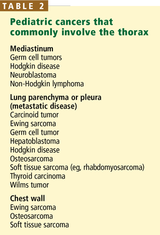
Cancers that commonly involve the thorax are listed in Table 2. Neuroblastoma, hepatoblastoma, extragonadal germ cell tumors, and Wilms tumor typically are diseases of young children; osteosarcoma, Ewing sarcoma, thyroid carcinoma, and Hodgkin disease are most common in older children and adolescents; soft tissue sarcoma and non-Hodgkin lymphoma span all age groups.
Surgery can, in some cases, control the cancer, as with mediastinal neuroblastoma and Ewing sarcoma of the chest wall.
Radiation to the chest remains a major component of treatment for Hodgkin disease, unresected thoracic Ewing sarcoma, soft tissue sarcoma with lung involvement or thyroid carcinoma, and Wilms tumor. Central nervous system tumors and leukemias, the most common pediatric malignancies, may require radiation to the spinal cord—with resulting radiation exposure of the lungs. Total-body irradiation is a component of many preparative regimens for stem cell transplantation.
Chemotherapy remains a mainstay for all types of tumors, and patients with germ cell tumors, Hodgkin disease, and brain tumors are at particular risk of pulmonary toxicity due to heavy reliance on bleomycin (Blenoxane) (for germ cell tumors, Hodgkin disease) and the nitrosoureas (for brain tumors).
RADIATION-INDUCED LUNG DAMAGE
The lungs are particularly sensitive to radiation, and pulmonary problems occur most often in patients with malignant diseases of the chest that are treated with radiation, ie, those involving the mediastinum, the lung parenchyma, or the chest wall.
Abnormal radiographic findings or restrictive changes on pulmonary function testing have been reported in more than 30% of patients who received radiation directly or indirectly to the lung.9–12 These changes have been detected months to years after radiation therapy, most often in patients who suffered radiation pneumonitis as an acute toxicity.
The amount of damage depends on the cumulative dose, how many treatments (“fractions”) this cumulative dose was divided into (dividing the radiation dose into smaller dose fractions can reduce toxicity), the volume of lung tissue involved, and the patient’s age (the younger, the worse) at the time of treatment.
Cumulative dose. Whole-lung irradiation is limited to 12 Gy, although localized areas of cancer can be treated with much higher doses.
Clinically apparent pneumonitis with cough, fever, or dyspnea generally occurs only in survivors who received more than 30 Gy in standard fractions to more than 50% of the lung. However, in 12 survivors of Wilms tumor who received median total doses of approximately 20 Gy to both lungs 7 to 14 years previously, 8 patients had dyspnea on exertion and radiographic evidence of interstitial and pleural thickening.13 Mean total lung volumes and the diffusing capacity of the lung for carbon monoxide (DLCO) were reduced in all patients to approximately 60% of predicted values.
In a prospective study of adults with Hodgkin disease treated after 1980, 145 patients were examined 3 years after receiving more than 44 Gy limited to the mantle area.9 None were experiencing symptoms; however, 30% to 40% had a forced vital capacity (FVC) less than 80% of predicted, and 7% had a reduced DLCO. Some of these patients also had received bleomycin (see below), which may have exacerbated pulmonary toxicity. Asymptomatic restrictive and obstructive lung changes have been detected after lower doses of whole-lung radiation (11–14 Gy) were given for other malignant diseases.11,14
In a recent study of adults and older adolescents with stage I and IIA Hodgkin disease treated with radiation alone (40–45 Gy to involved fields, including the mediastinum), late pulmonary effects observed were minimal.10 While FVC, residual volume, forced expiratory volume in 1 second (FEV1), DLCO, and total lung capacity (TLC) were significantly lower at the end of radiation therapy than before treatment, all except DLCO returned nearly to normal within 1 year. The decrease in DLCO remained stable, but the forced expiratory flow rate between 25% and 75% of FVC (FEF 25%–75%) was significantly lower 3 years after treatment than at baseline.
The high doses of total lung irradiation cited in several of these reports are rarely used in today’s cancer protocols. For example, patients receiving radiation as adjunctive treatment for Hodgkin disease now receive lower doses (< 21 Gy), which are restricted to involved fields, and the pulmonary toxicity is less than in the past.
In one series, 159 children and adolescents with unfavorable Hodgkin disease treated from 1993 to 2000 received six cycles of chemotherapy followed by response-based, involved-field radiation therapy. Patients who achieved a complete response after the first two cycles of chemotherapy got 15 Gy, and those who achieved a partial response got 25.5 Gy to all sites of bulky lymphadenopathy.15 All patients underwent pulmonary function testing. Only 24 (30.8%) of them had pulmonary toxicity, which was limited to asymptomatic deficits of restriction and diffusion.
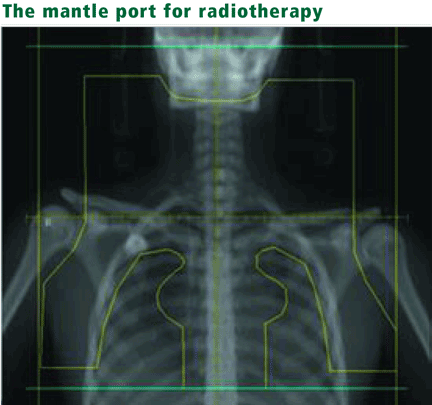
Nevertheless, the lungs receive some radiation even when they are not the target, such as in patients with malignant brain tumors, and this exposure can contribute to the development of lung disease, although these patients are likely to have no symptoms during day-to-day activities. Innovations in targeted radiation delivery (eg, conformal radiation) should further limit damage to normal lung tissue.
Age at the time of treatment also may influence the type and incidence of pulmonary sequelae. In older children and adults, radiation for thoracic malignancy results in pulmonary fibrosis with loss of lung volume. Similar injury can occur in younger children, but pulmonary function may also be compromised by inhibited growth of the supportive structures and the chest wall. One report suggests that children younger than 3 years at the time of therapy experience more chronic toxicity.11
In contrast, after bone marrow transplantation, children seem to be at less risk of significant late pulmonary dysfunction than adults are, despite similar preparatory regimens.16 This may in part reflect a lower incidence of severe graft-vs-host disease involving the lung. Nonetheless, in a recent report of children treated with fractionated total-body irradiation between 1985 and 1993, restrictive pulmonary diseases were found in 30 of 42 patients at a median of 3.1 years after treatment (range 0.5–17 years).17 Most of these patients had asymptomatic mild restrictive disease, and the one patient with severe changes had previously received thoracic radiation for treatment of neuroblastoma. In about half of the cases, pulmonary function abnormalities were permanent although not progressive.
CHEMOTHERAPY-RELATED LUNG DAMAGE
A growing list of chemotherapeutic agents appears to cause pulmonary disease in long-term survivors.5
Bleomycin
Bleomycin toxicity is the prototype for chemotherapy-related lung injury: bleomycin was the first chemotherapy drug shown to cause lung injury, this effect is suggested by a large database, and the mechanism is typical.5,18 Preclinical studies have attributed bleomycin’s toxicity to its tendency to promote free radicals.
Although interstitial pneumonitis and pulmonary fibrosis have been reported in children, clinically apparent bleomycin pneumonopathy is most frequent in older adults. Usually, the abnormalities began within 3 months of therapy and persisted or progressed. Like the acute toxicity, it is dose-dependent and more common above a threshold cumulative dose of 400 units/m2. Above this dose, 10% of adult patients without other risk factors develop fibrosis; data are not available for these doses in children. At lower doses, fibrosis occurs sporadically in fewer than 5% of patients, with a 1% to 2% mortality rate. In some series, bleomycin toxicity was anticipated on the basis of DLCO abnormalities.
Bleomycin pulmonary toxicity is variably exacerbated by concurrent or previous radiation therapy.
Increased oxygen concentrations associated with general anesthesia have also been found to exacerbate prior bleomycin-induced pulmonary injury.19,20 In one instance (reviewed by Zaniboni et al20), the patient recovered with corticosteroid treatment.
Alkylating agents
Carmustine (also called BCNU; brand names BiCNU, Gliadel) and lomustine (CCNU; CeeNU) are thought to cause dose-related lung injury. When cumulative carmustine doses are greater than 1,500 mg/m2, more than 50% of patients develop symptoms.21
In a careful clinicopathologic review of 31 children with brain tumors, restrictive changes with lung fibrosis were reported up to 17 years after treatment, most often with carmustine 100 mg/m2 every 6 to 8 weeks for up to 2 years.22 Four of the 8 patients still alive at the time of study experienced shortness of breath and coughing; 6 showed a characteristic pattern of upper zone fibrosis on chest radiography and computed tomography; all 8 survivors had restrictive findings on pulmonary function testing, with vital capacities of about 50% of normal. Toxicity increases with more intensive dose-scheduling.
Cyclophosphamide (Cytoxan) may cause delayed-onset pulmonary fibrosis with severe restrictive lung disease in association with marked reductions in the anteroposterior diameter of the chest, although the evidence is less convincing than with carmustine and lomustine, coming from case reports and small series.23
Melphalan (Alkeran), generally in doses used in stem cell transplant conditioning regimens, is also thought to cause pulmonary fibrosis.24
Busulfan most predictably causes toxicity when it is used in transplantation doses (ie, more than 500 mg), and may be associated with a progressive, potentially fatal restrictive lung disease.25 A current trend is to adjust the dose on the basis of pharmacokinetic analysis, which we hope will reduce toxicity.
Other agents
Methotrexate (MTX; Trexall) also has been associated with chronic pneumonitis and fibrosis.26 This probably occurs with an incidence well below 1% and may be idiosyncratic and not dose-related. Asymptomatic changes in pulmonary function tests that do not predict clinically significant problems have most frequently been associated with low-dose oral administration during more than 3 years.27 This is a treatment approach used in patients with psoriasis or rheumatoid arthritis, but is now obsolete for pediatric cancer. Intravenous and, rarely, intrathecal administration also have been associated with pulmonary toxicity.28 A single report of two patients who developed diffuse interstitial pulmonary infiltrates and chronic pulmonary changes links vinblastine to these sequelae.29
LUNG INJURY AFTER BONE MARROW TRANSPLANTATION
Hematopoietic stem cell transplantation is associated with various late pulmonary complications. The factors that influence these complications are similar to those discussed earlier. However, the intensity of therapy in patients undergoing transplantation and the additive effects of previous therapies magnify the risks.
Patients undergoing total-body irradiation as part of their preparation for transplantation have a high incidence of late pulmonary complications.25,30–32 Busulfan, carmustine, bleomycin, and cyclophosphamide, also commonly used conditioning chemotherapies, are known to cause pneumonitis and fibrosis after transplantation.33
Some acute pulmonary toxicities can have long-standing effects, including serious pulmonary infections, idiopathic pneumonia syndrome, bronchiolitis obliterans, acute respiratory distress syndrome, or other damage related to graft-vs-host disease.
OTHER RISK FACTORS FOR LUNG DAMAGE
Additional factors contributing to chronic pulmonary toxicity include superimposed infection, underlying pneumonopathy (eg, asthma), cigarette use, respirator toxicity, chronic graft-vs-host disease, and the effects of chronic pulmonary involvement by tumor or reaction to tumor. For example, a subset of patients with Langerhans cell histiocytosis can develop histiocytic pulmonary infiltrates or honeycombing with severe chronic restrictive lung disease unrelated to therapy or the presence of active tumor.
Although not well documented, scuba diving also has been said to exacerbate pulmonary fibrosis through increased underwater pressures and high oxygen levels.34
Lung lobectomy during childhood appears to have no significant impact on long-term pulmonary function,35–37 but the effect of lung surgery for children with cancer is not well defined.
GET THE PATIENT’S TREATMENT SUMMARY
Regardless of the setting for follow-up, the first step in any evaluation is to obtain the patient’s medical history and especially a treatment summary. The treatment summary should outline the cancer diagnosis, involved sites of disease, age at diagnosis, specific treatments (surgery, chemotherapy, radiation), and other key interventions and events during and after cancer therapy. Sample forms for physicians and patients are available at www.survivorshipguidelines.org.
Before the long-term survivor of childhood cancer graduates from the care of a pediatric oncologist, this treatment record and possible long-term problems should be reviewed with the family and, in the case of an adolescent, with the patient. Correspondence between the pediatric oncologist and subsequent caregivers should also include a treatment summary. The treatment summary allows the survivor or his health care provider to interface with the COG guidelines to determine recommended follow-up care. The primary care physician and the patient both should have copies of this document.
We are developing an interactive Web-based version of a standardized summary form, designed to interface with an automated version of the COG guidelines in order to generate individualized follow-up recommendations.
ASK ABOUT LUNG SYMPTOMS
We recommend that health care providers investigate symptoms of pulmonary dysfunction, and specifically ask about chronic cough with or without fever, shortness of breath, and dyspnea on exertion during yearly health care visits.
Baseline pulmonary function testing (including DLCO and spirometry) and chest radiography are recommended 2 or more years after completion of therapy to document persistent deficits and determine the need for continued monitoring. Reevaluation of pulmonary function should be considered in patients with established deficits who require general anesthesia and for those treated with bleomycin.
Scuba diving remains controversial for long-term survivors. Consequently, patients with risk factors for lung disease should be encouraged to consult with a pulmonary specialist to determine if diving poses a health threat to their pulmonary status. If clinical pulmonary dysfunction is identified, referral to colleagues in pulmonology for additional evaluation and treatment is essential. Increasing familiarity of primary care providers with surveillance concepts is a key element in survivorship care.
Smoking cessation can enhance the health of all patients and is particularly important among long-term survivors, especially those who received treatments predisposing to pulmonary injury. Strategies for cessation and patient information can be found at www.cdc.gov/tobacco/how2quit.htm.
Clinicians can take advantage of every patient interaction to assess readiness for smoking cessation and assist patients in this goal. Following the principles of patient-centered counseling, physicians can guide patients into considering a change of behavior with advice and encouragement. Whenever possible, physicians should personalize the risks of smoking as well as the short-term and long-term benefits. As smokers prepare to quit, their physicians can assist in developing a plan that includes a quit date.
Many pharmacologic agents are available to assist patients, ie, nicotine inhalers, sprays, gum, and transdermal patches; the antidepressants bupropion (Wellbutrin) and nortriptiline (Pamelor); the alpha-2 adrenergic agonist clonidine (Catapres); and, most recently, the nicotine receptor partial agonist varenicline (Chantix).38
Follow-up to prevent relapse is an important part of this process.
Acknowledgment. This work was supported in part by the Swim Across America Foundation and the Campini Foundation.
- Hewitt M, Weiner S, Simone J. The epidemiology of childhood cancer. Washington DC: The National Academies Press, 2003:20–36.
- Ries L, Eisner M, Kosary C, et al. SEER cancer statistics review, 1975–2001. Bethesda, MD: National Cancer Institute; 2004.
- Landier W, Bhatia S, Eshelman DA, et al. Development of risk-based guidelines for pediatric cancer survivors: The Children’s Oncology Group long-term follow-up guidelines from the Children’s Oncology Group Late Effects Committee and nursing discipline. J Clin Oncol 2004; 22:4979–4990.
- Mertens AC, Yasui Y, Liu Y, et al. Pulmonary complications in survivors of childhood and adolescent cancer. A report from the Childhood Cancer Survivor Study. Cancer 2002; 95:2431–2441.
- Bhatia S, Blatt J, Meadows A. Late effects of childhood cancer and its treatment. In:Pizzo P, Poplack D. Principles and Practice of Pediatric Oncology. Philadelphia: Lippincott Williams & Wilkins, 2006.
- Anscher MS, Chen L, Rabbani Z, et al. Recent progress in defining mechanisms and potential targets for prevention of normal tissue injury after radiation therapy. Int J Radiat Oncol Biol Phys 2005; 62:255–259.
- Bhatt N, Baran CP, Allen J, et al. Promising pharmacologic innovations in treating pulmonary fibrosis. Curr Opin Pharmacol 2006; 6:284–292.
- Abid SH, Malhotra V, Perry MC. Radiation-induced and chemotherapy-induced pulmonary injury. Curr Opin Oncol 2001: 13:242–248.
- Horning SJ, Adhikari A, Rizk N, et al. Effect of treatment for Hodgkin’s disease on pulmonary function: results of a prospective study. J Clin Oncol 1994; 12:297–305.
- Villani F, Viviani S, Bonfante V, et al. Late pulmonary effects in favorable stage I and IIA Hodgkin’s disease treated with radiotherapy alone. Am J Clin Oncol 2000; 23:18–21.
- Miller RW, Fusner JE, Fink RJ, et al. Pulmonary function abnormalities in long-term survivors of childhood cancer. Med Pediatr Oncol 1986; 14:202–207.
- Weiner DJ, Maity A, Carlson CA, et al. Pulmonary function abnormalities in children treated with whole lung irradiation. Pediatr Blood Cancer 2006; 46:222–227.
- Wohl ME, Griscom NT, Traggis DG, et al. Effects of therapeutic irradiation delivered in early childhood upon subsequent lung function. Pediatrics 1975; 55:507–516.
- Hudson MM, Greenwald C, Thompson E, et al. Efficacy and toxicity of multiagent chemotherapy and low-dose involved-field radiotherapy in children and adolescents with Hodgkin’s disease. J Clin Oncol 1993; 11:100–108.
- Hudson MM, Krasin M, Link MP, et al. Risk-adapted, combined-modality therapy with VAMP/COP and response-based, involved-field radiation for unfavorable pediatric Hodgkin’s disease. J Clin Oncol 2004; 22:4541–4550.
- Quigley PM, Yeager AM, Loughlin GM. The effects of bone marrow transplantation on pulmonary function in children. Pediatr Pulmonol 1994; 18:361–367.
- Faraci M, Barra S, Cohen A, et al. Very late nonfatal consequences of fractionated TBI in children undergoing bone marrow transplant. Int J Radiat Oncol Biol Phys 2005; 63:1568–1575.
- Eigen H, Wyszomierski D. Bleomycin lung injury in children. Pathophysiology and guidelines for management. Am J Pediatr Hematol Oncol 1985; 7:71–78.
- Goldiner PL, Schweizer O. The hazards of anesthesia and surgery in bleomycin-treated patients. Semin Oncol 1979; 6:121–124.
- Zaniboni A, Prabhu S, Audisio RA. Chemotherapy and anaesthetic drugs: too little is known. Lancet Oncol 2005; 6:176–181.
- Aronin PA, Mahaley MS, Rudnick SA, et al. Prediction of BCNU pulmonary toxicity in patients with malignant gliomas: an assessment of risk factors. N Engl J Med 1980; 303:183–188.
- O’Driscoll BR, Hasleton PS, Taylor PM, et al. Active lung fibrosis up to 17 years after chemotherapy with carmustine (BCNU) in childhood. N Engl J Med 1990; 323:378–382.
- Alvarado CS, Boat TF, Newman AJ. Late-onset pulmonary fibrosis and chest deformity in two children treated with cyclophosphamide. J Pediatr 1978; 92:443–446.
- Codling BW, Chakera TM. Pulmonary fibrosis following therapy with melphalan for multiple myeloma. J Clin Pathol 1972; 25:668–673.
- Bruno B, Souillet G, Bertrand Y, et al. Effects of allogeneic bone marrow transplantation on pulmonary function in 80 children in a single paediatric centre. Bone Marrow Transplant 2004; 34:143–147.
- Lateef O, Shakoor N, Balk RA. Methotrexate pulmonary toxicity. Expert Opin Drug Saf 2005; 4:723–730.
- Cottin V, Tebob J, Massonnet B, et al. Pulmonary function in patients receiving long-term methotrexate. Chest 1996; 109:933–938.
- Gutin PH, Green MR, Bleyer WA, et al. Methotrexate pneumonitis induced by intrathecal methotrexate therapy: a case report with pharmacokinetic data. Cancer 1976; 38:1529–1534.
- Konits PH, Aisner J, Sutherland JC, et al. Possible pulmonary toxicity secondary to vinblastine. Cancer 1982; 50:2771–2774.
- Cerveri I, Zoia MC, Fulgoni P, et al. Late pulmonary sequelae after childhood bone marrow transplantation. Thorax 1999; 54:131–135.
- Cerveri I, Fulgoni P, Giorgiani G, et al. Lung function abnormalities after bone marrow transplantation in children: has the trend recently changed? Chest 2001; 120:1900–1906.
- Neve V, Foot AB, Michon J, et al. Longitudinal clinical and functional pulmonary follow-up after megatherapy, fractionated total body irradiation, and autologous bone marrow transplantation for metastatic neuroblastoma. Med Pediatr Oncol 1999; 32:170–176.
- Nenadov Beck M, Meresse V, Hartmann O, et al. Long-term pulmonary sequelae after autologous bone marrow transplantation in children without total body irradiation. Bone Marrow Transplant 1995; 16:771–775.
- Huls G, ten Bokkel Huinink D. Bleomycin and scuba diving: to dive or not to dive? Neth J Med 2003; 61:50–53.
- Kreisel D, Krupnick AS, Huddleston CB. Outcomes and late complications after pulmonary resections in the pediatric population. Semin Thorac Cardiovasc Surg 2004; 16:215–219.
- Lezama-del Valle Valle P, Blakely ML, Lobe TE. Physiologic consequences of pneumonectomy. Long-term consequences of pneumonectomy done in children. Chest Surg Clin North Am 1999; 9:485–495.
- Tobias JD, Bozeman PM, Mackert PW, et al. Postoperative outcome following thoracotomy in the pediatric oncology patient with diminished pulmonary function. J Surg Oncol 1993; 52:105–109.
- Drugs for Tobacco Dependence. Treatment Guidelines from the Medical Letter. June, 2003: 1(10):65–68.
Children who undergo radiotherapy, chemotherapy, or surgery for cancer face a risk of complications later in life, including pulmonary fibrosis and pneumonitis.
These long-term cancer survivors need systematic, lifelong surveillance, in a program that takes into account their individual risk (based on therapeutic exposures, genetic predisposition, lifestyle behaviors, and comorbid health conditions).1 Optimally, they would receive their care at multidisciplinary follow-up clinics organized by pediatric oncologists at tertiary care centers. However, access to such centers is limited, making this an option for relatively few. Consequently, as childhood cancer survivors age, internists and family practitioners may need to assume an increasing amount of responsibility for their follow-up care.
Because individual primary care providers are unlikely to follow more than a handful of survivors, specialists have developed guidelines for survivors of pediatric cancer. Working with established multidisciplinary clinics may help ensure appropriate follow-up for this population of patients.
This review summarizes the late effects of cancer therapy on the lungs and an approach to surveillance for the generalist or pulmonologist. We also review the quality of the evidence upon which these recommendations are based.
NUMBERS ON THE RISE
An estimated 1 of every 330 children develops cancer before age 19. With cure rates exceeding 75% for many pediatric malignancies, the number of survivors of childhood cancer, currently in excess of 270,000, will continue to increase.2
THE CHILDREN’S ONCOLOGY GROUP GUIDELINES
The Children’s Oncology Group (COG)3 released its first set of guidelines in 2003 for the follow-up care of patients treated for pediatric malignancies; the current version is available at www.survivorshipguidelines.org. The guidelines contain comprehensive screening recommendations, including those related to pulmonary toxicity, which can be used to standardize care.
Patient education materials accompany the guidelines, offering detailed information on guideline-specific topics in order to promote health maintenance.
HOW WE SEARCHED THE LITERATURE
We performed an extensive review of the literature via MEDLINE for the years 1975–2005. Key search terms were “childhood cancer,” “late effects,” and “pulmonary toxicity,” combined with keywords for each therapeutic exposure. References from selected articles were used to broaden the search. From several hundred citations, fewer than 30 were selected as best illustrating the relevant associations.
RISK IS THREE TIMES HIGHER IN CANCER SURVIVORS
The Childhood Cancer Survivor Study4 is the largest database of late effects, with more than 12,000 survivors of childhood cancer diagnosed between 1970 and 1986. Its data suggest that the risk of pulmonary conditions is more than three times higher in cancer survivors than in their siblings, as manifested by pulmonary signs (abnormal chest wall growth), symptoms (chronic cough, use of supplemental oxygen, exercise-induced shortness of breath), or specific diagnoses (lung fibrosis, recurrent pneumonia, pleurisy, bronchitis, recurrent sinus infection, or tonsillitis). Limitations: these data are retrospective, and the outcomes were detected by self-report and were not validated by review of medical records. Thus, the figures highlight the fact that pulmonary late effects are an important problem but do not give us a way to calculate risk exactly.
Other limitations of the literature: Treatments are constantly evolving, often in attempts to minimize late effects, and newer agents will need to be monitored for pulmonary toxicities. As noted, much of the available information is from studies of survivors of adult cancer; the potential for late effects of similar therapies in children is inferred. Most conclusions—and especially those based upon prospective serial evaluations—derive from small cohorts. For all treatments, the complications in the very long term remain undefined. What we know is summarized below.
CANCER THERAPY CAUSES FIBROSIS, PNEUMONITIS
The courses of these diseases are poorly characterized, since few longitudinal studies have been done. However, like most of the late effects of cancer therapy, pulmonary toxicity may first become apparent during the treatment and persist, or it may not appear until years later. Signs and symptoms may be static, progressive, or reversible.
ANGIOGENESIS MAY CONTRIBUTE TO FIBROSIS
On a microscopic level, pulmonary fibrosis is characterized by epithelial injury, fibroproliferation, and excessive extracellular matrix deposition.6–8
Evidence is mounting that these findings result in part from angiogenesis. Although this has not been studied in long-term cancer survivors, evidence of neovascularization was seen both in an animal model of lung fibrosis and in patients with idiopathic pulmonary fibrosis.6–8 High plasma concentrations of angiogenic cytokines (eg, tumor necrosis factor alpha, interleukin 8, and endothelin 1) have been found in these situations. Antiangiogenic agents and other immune modulators such as thalidomide may be beneficial in patients with lung fibrosis.7
On a macroscopic level, pulmonary fibrosis results in loss of lung volume in older children and in adults. In contrast, in younger children, interference with growth of both the lung and the chest wall may contribute to pulmonary dysfunction.
CANCER TYPES AND TREATMENTS VARY BY AGE
Cancers that commonly involve the thorax are listed in Table 2. Neuroblastoma, hepatoblastoma, extragonadal germ cell tumors, and Wilms tumor typically are diseases of young children; osteosarcoma, Ewing sarcoma, thyroid carcinoma, and Hodgkin disease are most common in older children and adolescents; soft tissue sarcoma and non-Hodgkin lymphoma span all age groups.
Surgery can, in some cases, control the cancer, as with mediastinal neuroblastoma and Ewing sarcoma of the chest wall.
Radiation to the chest remains a major component of treatment for Hodgkin disease, unresected thoracic Ewing sarcoma, soft tissue sarcoma with lung involvement or thyroid carcinoma, and Wilms tumor. Central nervous system tumors and leukemias, the most common pediatric malignancies, may require radiation to the spinal cord—with resulting radiation exposure of the lungs. Total-body irradiation is a component of many preparative regimens for stem cell transplantation.
Chemotherapy remains a mainstay for all types of tumors, and patients with germ cell tumors, Hodgkin disease, and brain tumors are at particular risk of pulmonary toxicity due to heavy reliance on bleomycin (Blenoxane) (for germ cell tumors, Hodgkin disease) and the nitrosoureas (for brain tumors).
RADIATION-INDUCED LUNG DAMAGE
The lungs are particularly sensitive to radiation, and pulmonary problems occur most often in patients with malignant diseases of the chest that are treated with radiation, ie, those involving the mediastinum, the lung parenchyma, or the chest wall.
Abnormal radiographic findings or restrictive changes on pulmonary function testing have been reported in more than 30% of patients who received radiation directly or indirectly to the lung.9–12 These changes have been detected months to years after radiation therapy, most often in patients who suffered radiation pneumonitis as an acute toxicity.
The amount of damage depends on the cumulative dose, how many treatments (“fractions”) this cumulative dose was divided into (dividing the radiation dose into smaller dose fractions can reduce toxicity), the volume of lung tissue involved, and the patient’s age (the younger, the worse) at the time of treatment.
Cumulative dose. Whole-lung irradiation is limited to 12 Gy, although localized areas of cancer can be treated with much higher doses.
Clinically apparent pneumonitis with cough, fever, or dyspnea generally occurs only in survivors who received more than 30 Gy in standard fractions to more than 50% of the lung. However, in 12 survivors of Wilms tumor who received median total doses of approximately 20 Gy to both lungs 7 to 14 years previously, 8 patients had dyspnea on exertion and radiographic evidence of interstitial and pleural thickening.13 Mean total lung volumes and the diffusing capacity of the lung for carbon monoxide (DLCO) were reduced in all patients to approximately 60% of predicted values.
In a prospective study of adults with Hodgkin disease treated after 1980, 145 patients were examined 3 years after receiving more than 44 Gy limited to the mantle area.9 None were experiencing symptoms; however, 30% to 40% had a forced vital capacity (FVC) less than 80% of predicted, and 7% had a reduced DLCO. Some of these patients also had received bleomycin (see below), which may have exacerbated pulmonary toxicity. Asymptomatic restrictive and obstructive lung changes have been detected after lower doses of whole-lung radiation (11–14 Gy) were given for other malignant diseases.11,14
In a recent study of adults and older adolescents with stage I and IIA Hodgkin disease treated with radiation alone (40–45 Gy to involved fields, including the mediastinum), late pulmonary effects observed were minimal.10 While FVC, residual volume, forced expiratory volume in 1 second (FEV1), DLCO, and total lung capacity (TLC) were significantly lower at the end of radiation therapy than before treatment, all except DLCO returned nearly to normal within 1 year. The decrease in DLCO remained stable, but the forced expiratory flow rate between 25% and 75% of FVC (FEF 25%–75%) was significantly lower 3 years after treatment than at baseline.
The high doses of total lung irradiation cited in several of these reports are rarely used in today’s cancer protocols. For example, patients receiving radiation as adjunctive treatment for Hodgkin disease now receive lower doses (< 21 Gy), which are restricted to involved fields, and the pulmonary toxicity is less than in the past.
In one series, 159 children and adolescents with unfavorable Hodgkin disease treated from 1993 to 2000 received six cycles of chemotherapy followed by response-based, involved-field radiation therapy. Patients who achieved a complete response after the first two cycles of chemotherapy got 15 Gy, and those who achieved a partial response got 25.5 Gy to all sites of bulky lymphadenopathy.15 All patients underwent pulmonary function testing. Only 24 (30.8%) of them had pulmonary toxicity, which was limited to asymptomatic deficits of restriction and diffusion.
Nevertheless, the lungs receive some radiation even when they are not the target, such as in patients with malignant brain tumors, and this exposure can contribute to the development of lung disease, although these patients are likely to have no symptoms during day-to-day activities. Innovations in targeted radiation delivery (eg, conformal radiation) should further limit damage to normal lung tissue.
Age at the time of treatment also may influence the type and incidence of pulmonary sequelae. In older children and adults, radiation for thoracic malignancy results in pulmonary fibrosis with loss of lung volume. Similar injury can occur in younger children, but pulmonary function may also be compromised by inhibited growth of the supportive structures and the chest wall. One report suggests that children younger than 3 years at the time of therapy experience more chronic toxicity.11
In contrast, after bone marrow transplantation, children seem to be at less risk of significant late pulmonary dysfunction than adults are, despite similar preparatory regimens.16 This may in part reflect a lower incidence of severe graft-vs-host disease involving the lung. Nonetheless, in a recent report of children treated with fractionated total-body irradiation between 1985 and 1993, restrictive pulmonary diseases were found in 30 of 42 patients at a median of 3.1 years after treatment (range 0.5–17 years).17 Most of these patients had asymptomatic mild restrictive disease, and the one patient with severe changes had previously received thoracic radiation for treatment of neuroblastoma. In about half of the cases, pulmonary function abnormalities were permanent although not progressive.
CHEMOTHERAPY-RELATED LUNG DAMAGE
A growing list of chemotherapeutic agents appears to cause pulmonary disease in long-term survivors.5
Bleomycin
Bleomycin toxicity is the prototype for chemotherapy-related lung injury: bleomycin was the first chemotherapy drug shown to cause lung injury, this effect is suggested by a large database, and the mechanism is typical.5,18 Preclinical studies have attributed bleomycin’s toxicity to its tendency to promote free radicals.
Although interstitial pneumonitis and pulmonary fibrosis have been reported in children, clinically apparent bleomycin pneumonopathy is most frequent in older adults. Usually, the abnormalities began within 3 months of therapy and persisted or progressed. Like the acute toxicity, it is dose-dependent and more common above a threshold cumulative dose of 400 units/m2. Above this dose, 10% of adult patients without other risk factors develop fibrosis; data are not available for these doses in children. At lower doses, fibrosis occurs sporadically in fewer than 5% of patients, with a 1% to 2% mortality rate. In some series, bleomycin toxicity was anticipated on the basis of DLCO abnormalities.
Bleomycin pulmonary toxicity is variably exacerbated by concurrent or previous radiation therapy.
Increased oxygen concentrations associated with general anesthesia have also been found to exacerbate prior bleomycin-induced pulmonary injury.19,20 In one instance (reviewed by Zaniboni et al20), the patient recovered with corticosteroid treatment.
Alkylating agents
Carmustine (also called BCNU; brand names BiCNU, Gliadel) and lomustine (CCNU; CeeNU) are thought to cause dose-related lung injury. When cumulative carmustine doses are greater than 1,500 mg/m2, more than 50% of patients develop symptoms.21
In a careful clinicopathologic review of 31 children with brain tumors, restrictive changes with lung fibrosis were reported up to 17 years after treatment, most often with carmustine 100 mg/m2 every 6 to 8 weeks for up to 2 years.22 Four of the 8 patients still alive at the time of study experienced shortness of breath and coughing; 6 showed a characteristic pattern of upper zone fibrosis on chest radiography and computed tomography; all 8 survivors had restrictive findings on pulmonary function testing, with vital capacities of about 50% of normal. Toxicity increases with more intensive dose-scheduling.
Cyclophosphamide (Cytoxan) may cause delayed-onset pulmonary fibrosis with severe restrictive lung disease in association with marked reductions in the anteroposterior diameter of the chest, although the evidence is less convincing than with carmustine and lomustine, coming from case reports and small series.23
Melphalan (Alkeran), generally in doses used in stem cell transplant conditioning regimens, is also thought to cause pulmonary fibrosis.24
Busulfan most predictably causes toxicity when it is used in transplantation doses (ie, more than 500 mg), and may be associated with a progressive, potentially fatal restrictive lung disease.25 A current trend is to adjust the dose on the basis of pharmacokinetic analysis, which we hope will reduce toxicity.
Other agents
Methotrexate (MTX; Trexall) also has been associated with chronic pneumonitis and fibrosis.26 This probably occurs with an incidence well below 1% and may be idiosyncratic and not dose-related. Asymptomatic changes in pulmonary function tests that do not predict clinically significant problems have most frequently been associated with low-dose oral administration during more than 3 years.27 This is a treatment approach used in patients with psoriasis or rheumatoid arthritis, but is now obsolete for pediatric cancer. Intravenous and, rarely, intrathecal administration also have been associated with pulmonary toxicity.28 A single report of two patients who developed diffuse interstitial pulmonary infiltrates and chronic pulmonary changes links vinblastine to these sequelae.29
LUNG INJURY AFTER BONE MARROW TRANSPLANTATION
Hematopoietic stem cell transplantation is associated with various late pulmonary complications. The factors that influence these complications are similar to those discussed earlier. However, the intensity of therapy in patients undergoing transplantation and the additive effects of previous therapies magnify the risks.
Patients undergoing total-body irradiation as part of their preparation for transplantation have a high incidence of late pulmonary complications.25,30–32 Busulfan, carmustine, bleomycin, and cyclophosphamide, also commonly used conditioning chemotherapies, are known to cause pneumonitis and fibrosis after transplantation.33
Some acute pulmonary toxicities can have long-standing effects, including serious pulmonary infections, idiopathic pneumonia syndrome, bronchiolitis obliterans, acute respiratory distress syndrome, or other damage related to graft-vs-host disease.
OTHER RISK FACTORS FOR LUNG DAMAGE
Additional factors contributing to chronic pulmonary toxicity include superimposed infection, underlying pneumonopathy (eg, asthma), cigarette use, respirator toxicity, chronic graft-vs-host disease, and the effects of chronic pulmonary involvement by tumor or reaction to tumor. For example, a subset of patients with Langerhans cell histiocytosis can develop histiocytic pulmonary infiltrates or honeycombing with severe chronic restrictive lung disease unrelated to therapy or the presence of active tumor.
Although not well documented, scuba diving also has been said to exacerbate pulmonary fibrosis through increased underwater pressures and high oxygen levels.34
Lung lobectomy during childhood appears to have no significant impact on long-term pulmonary function,35–37 but the effect of lung surgery for children with cancer is not well defined.
GET THE PATIENT’S TREATMENT SUMMARY
Regardless of the setting for follow-up, the first step in any evaluation is to obtain the patient’s medical history and especially a treatment summary. The treatment summary should outline the cancer diagnosis, involved sites of disease, age at diagnosis, specific treatments (surgery, chemotherapy, radiation), and other key interventions and events during and after cancer therapy. Sample forms for physicians and patients are available at www.survivorshipguidelines.org.
Before the long-term survivor of childhood cancer graduates from the care of a pediatric oncologist, this treatment record and possible long-term problems should be reviewed with the family and, in the case of an adolescent, with the patient. Correspondence between the pediatric oncologist and subsequent caregivers should also include a treatment summary. The treatment summary allows the survivor or his health care provider to interface with the COG guidelines to determine recommended follow-up care. The primary care physician and the patient both should have copies of this document.
We are developing an interactive Web-based version of a standardized summary form, designed to interface with an automated version of the COG guidelines in order to generate individualized follow-up recommendations.
ASK ABOUT LUNG SYMPTOMS
We recommend that health care providers investigate symptoms of pulmonary dysfunction, and specifically ask about chronic cough with or without fever, shortness of breath, and dyspnea on exertion during yearly health care visits.
Baseline pulmonary function testing (including DLCO and spirometry) and chest radiography are recommended 2 or more years after completion of therapy to document persistent deficits and determine the need for continued monitoring. Reevaluation of pulmonary function should be considered in patients with established deficits who require general anesthesia and for those treated with bleomycin.
Scuba diving remains controversial for long-term survivors. Consequently, patients with risk factors for lung disease should be encouraged to consult with a pulmonary specialist to determine if diving poses a health threat to their pulmonary status. If clinical pulmonary dysfunction is identified, referral to colleagues in pulmonology for additional evaluation and treatment is essential. Increasing familiarity of primary care providers with surveillance concepts is a key element in survivorship care.
Smoking cessation can enhance the health of all patients and is particularly important among long-term survivors, especially those who received treatments predisposing to pulmonary injury. Strategies for cessation and patient information can be found at www.cdc.gov/tobacco/how2quit.htm.
Clinicians can take advantage of every patient interaction to assess readiness for smoking cessation and assist patients in this goal. Following the principles of patient-centered counseling, physicians can guide patients into considering a change of behavior with advice and encouragement. Whenever possible, physicians should personalize the risks of smoking as well as the short-term and long-term benefits. As smokers prepare to quit, their physicians can assist in developing a plan that includes a quit date.
Many pharmacologic agents are available to assist patients, ie, nicotine inhalers, sprays, gum, and transdermal patches; the antidepressants bupropion (Wellbutrin) and nortriptiline (Pamelor); the alpha-2 adrenergic agonist clonidine (Catapres); and, most recently, the nicotine receptor partial agonist varenicline (Chantix).38
Follow-up to prevent relapse is an important part of this process.
Acknowledgment. This work was supported in part by the Swim Across America Foundation and the Campini Foundation.
- Hewitt M, Weiner S, Simone J. The epidemiology of childhood cancer. Washington DC: The National Academies Press, 2003:20–36.
- Ries L, Eisner M, Kosary C, et al. SEER cancer statistics review, 1975–2001. Bethesda, MD: National Cancer Institute; 2004.
- Landier W, Bhatia S, Eshelman DA, et al. Development of risk-based guidelines for pediatric cancer survivors: The Children’s Oncology Group long-term follow-up guidelines from the Children’s Oncology Group Late Effects Committee and nursing discipline. J Clin Oncol 2004; 22:4979–4990.
- Mertens AC, Yasui Y, Liu Y, et al. Pulmonary complications in survivors of childhood and adolescent cancer. A report from the Childhood Cancer Survivor Study. Cancer 2002; 95:2431–2441.
- Bhatia S, Blatt J, Meadows A. Late effects of childhood cancer and its treatment. In:Pizzo P, Poplack D. Principles and Practice of Pediatric Oncology. Philadelphia: Lippincott Williams & Wilkins, 2006.
- Anscher MS, Chen L, Rabbani Z, et al. Recent progress in defining mechanisms and potential targets for prevention of normal tissue injury after radiation therapy. Int J Radiat Oncol Biol Phys 2005; 62:255–259.
- Bhatt N, Baran CP, Allen J, et al. Promising pharmacologic innovations in treating pulmonary fibrosis. Curr Opin Pharmacol 2006; 6:284–292.
- Abid SH, Malhotra V, Perry MC. Radiation-induced and chemotherapy-induced pulmonary injury. Curr Opin Oncol 2001: 13:242–248.
- Horning SJ, Adhikari A, Rizk N, et al. Effect of treatment for Hodgkin’s disease on pulmonary function: results of a prospective study. J Clin Oncol 1994; 12:297–305.
- Villani F, Viviani S, Bonfante V, et al. Late pulmonary effects in favorable stage I and IIA Hodgkin’s disease treated with radiotherapy alone. Am J Clin Oncol 2000; 23:18–21.
- Miller RW, Fusner JE, Fink RJ, et al. Pulmonary function abnormalities in long-term survivors of childhood cancer. Med Pediatr Oncol 1986; 14:202–207.
- Weiner DJ, Maity A, Carlson CA, et al. Pulmonary function abnormalities in children treated with whole lung irradiation. Pediatr Blood Cancer 2006; 46:222–227.
- Wohl ME, Griscom NT, Traggis DG, et al. Effects of therapeutic irradiation delivered in early childhood upon subsequent lung function. Pediatrics 1975; 55:507–516.
- Hudson MM, Greenwald C, Thompson E, et al. Efficacy and toxicity of multiagent chemotherapy and low-dose involved-field radiotherapy in children and adolescents with Hodgkin’s disease. J Clin Oncol 1993; 11:100–108.
- Hudson MM, Krasin M, Link MP, et al. Risk-adapted, combined-modality therapy with VAMP/COP and response-based, involved-field radiation for unfavorable pediatric Hodgkin’s disease. J Clin Oncol 2004; 22:4541–4550.
- Quigley PM, Yeager AM, Loughlin GM. The effects of bone marrow transplantation on pulmonary function in children. Pediatr Pulmonol 1994; 18:361–367.
- Faraci M, Barra S, Cohen A, et al. Very late nonfatal consequences of fractionated TBI in children undergoing bone marrow transplant. Int J Radiat Oncol Biol Phys 2005; 63:1568–1575.
- Eigen H, Wyszomierski D. Bleomycin lung injury in children. Pathophysiology and guidelines for management. Am J Pediatr Hematol Oncol 1985; 7:71–78.
- Goldiner PL, Schweizer O. The hazards of anesthesia and surgery in bleomycin-treated patients. Semin Oncol 1979; 6:121–124.
- Zaniboni A, Prabhu S, Audisio RA. Chemotherapy and anaesthetic drugs: too little is known. Lancet Oncol 2005; 6:176–181.
- Aronin PA, Mahaley MS, Rudnick SA, et al. Prediction of BCNU pulmonary toxicity in patients with malignant gliomas: an assessment of risk factors. N Engl J Med 1980; 303:183–188.
- O’Driscoll BR, Hasleton PS, Taylor PM, et al. Active lung fibrosis up to 17 years after chemotherapy with carmustine (BCNU) in childhood. N Engl J Med 1990; 323:378–382.
- Alvarado CS, Boat TF, Newman AJ. Late-onset pulmonary fibrosis and chest deformity in two children treated with cyclophosphamide. J Pediatr 1978; 92:443–446.
- Codling BW, Chakera TM. Pulmonary fibrosis following therapy with melphalan for multiple myeloma. J Clin Pathol 1972; 25:668–673.
- Bruno B, Souillet G, Bertrand Y, et al. Effects of allogeneic bone marrow transplantation on pulmonary function in 80 children in a single paediatric centre. Bone Marrow Transplant 2004; 34:143–147.
- Lateef O, Shakoor N, Balk RA. Methotrexate pulmonary toxicity. Expert Opin Drug Saf 2005; 4:723–730.
- Cottin V, Tebob J, Massonnet B, et al. Pulmonary function in patients receiving long-term methotrexate. Chest 1996; 109:933–938.
- Gutin PH, Green MR, Bleyer WA, et al. Methotrexate pneumonitis induced by intrathecal methotrexate therapy: a case report with pharmacokinetic data. Cancer 1976; 38:1529–1534.
- Konits PH, Aisner J, Sutherland JC, et al. Possible pulmonary toxicity secondary to vinblastine. Cancer 1982; 50:2771–2774.
- Cerveri I, Zoia MC, Fulgoni P, et al. Late pulmonary sequelae after childhood bone marrow transplantation. Thorax 1999; 54:131–135.
- Cerveri I, Fulgoni P, Giorgiani G, et al. Lung function abnormalities after bone marrow transplantation in children: has the trend recently changed? Chest 2001; 120:1900–1906.
- Neve V, Foot AB, Michon J, et al. Longitudinal clinical and functional pulmonary follow-up after megatherapy, fractionated total body irradiation, and autologous bone marrow transplantation for metastatic neuroblastoma. Med Pediatr Oncol 1999; 32:170–176.
- Nenadov Beck M, Meresse V, Hartmann O, et al. Long-term pulmonary sequelae after autologous bone marrow transplantation in children without total body irradiation. Bone Marrow Transplant 1995; 16:771–775.
- Huls G, ten Bokkel Huinink D. Bleomycin and scuba diving: to dive or not to dive? Neth J Med 2003; 61:50–53.
- Kreisel D, Krupnick AS, Huddleston CB. Outcomes and late complications after pulmonary resections in the pediatric population. Semin Thorac Cardiovasc Surg 2004; 16:215–219.
- Lezama-del Valle Valle P, Blakely ML, Lobe TE. Physiologic consequences of pneumonectomy. Long-term consequences of pneumonectomy done in children. Chest Surg Clin North Am 1999; 9:485–495.
- Tobias JD, Bozeman PM, Mackert PW, et al. Postoperative outcome following thoracotomy in the pediatric oncology patient with diminished pulmonary function. J Surg Oncol 1993; 52:105–109.
- Drugs for Tobacco Dependence. Treatment Guidelines from the Medical Letter. June, 2003: 1(10):65–68.
KEY POINTS
- Radiation therapy causes pulmonary fibrosis, interstitial pneumonitis, and restrictive or obstructive lung disease. The risk is dose-dependent and increases with concomitant chemotherapy, younger age at treatment, atopic history, and smoking.
- Alkylating agents cause pulmonary fibrosis. Bleomycin can cause interstitial pneumonitis, pulmonary fibrosis, or, very rarely, acute respiratory distress syndrome.
- Cancer survivors should have a yearly history and physical examination, plus pulmonary function testing and radiography at baseline and repeated as clinically indicated.
- All patients who smoke should be encouraged to quit.
Blood smear analysis in babesiosis, ehrlichiosis, relapsing fever, malaria, and Chagas disease
Blood smear analysis, while commonly used to evaluate hematologic conditions, is infrequently used to diagnose infectious diseases. This is because of the rarity of diseases for which blood smear analysis is indicated. Consequently, such testing is often overlooked when it is diagnostically important.
Nonspecific changes may include morphologic changes in leukocytes and erythrocytes (eg, toxic granulations, macrocytosis).1 And with certain pathogens, identifying organisms in a peripheral blood smear allows for a rapid diagnosis.
This paper discusses the epidemiology, clinical manifestations, laboratory findings, and management of five infectious diseases in which direct visualization of the organism in the blood plays a major diagnostic role. Our intent is to summarize the clinical findings that should prompt blood smear analysis so that these uncommon conditions are not overlooked.
BABESIOSIS
Babesiosis, a tick-borne protozoal disease, occurs principally in the United States and Europe. Of the more than 100 species of Babesia, two account for almost all human disease: B microti and B divergens.
Both species are transmitted by Ixodes ticks, although patients often do not recall being bitten. The disease may rarely complicate blood transfusion. Most cases occur from May to September, when tick exposure is highest. The incubation period varies from 1 to 4 weeks.
Common in the Northeast, usually asymptomatic
B microti infection occurs predominantly in the United States. Rodents, especially the white-footed mouse, are the principal reservoir.2,3 Endemic areas, where seropositivity rates range from 4% to 21%, include the coastal areas and islands off of Massachusetts, particularly Cape Cod, Nantucket, and Martha’s Vineyard; the islands near New York City, especially Long Island, Shelter Island, and Fire Island; Block Island, off the coast of Rhode Island; and certain areas in Connecticut.4 WA-1, a species that is morphologically identical to B microti, is emerging in California and Washington.5,6
Infection with B microti is usually asymptomatic. Elderly and immunosuppressed people, especially those without a spleen or with impaired cellular immunity, are more likely to become ill. Symptoms, including fever, malaise, headache, nausea, and generalized aching, may last weeks to months.
About one-fourth of patients with babesiosis are coinfected with the Lyme disease bacterium (Borrelia burgdorferi) and often have more severe illness.3
Hepatomegaly, splenomegaly, jaundice, and dark urine are common findings in patients with symptoms. Severe hemolysis, often accompanied by thrombocytopenia, leukopenia, and atypical lymphocytosis, is more common in high-risk patients. Hepatic transaminases may be elevated. Urinalysis may show proteinuria and hemoglobinuria. Acute respiratory distress syndrome has been reported in severe cases.7–10
B divergens infection: A serious but rare disease seen in Europe
B divergens is found mainly in Europe. Altogether, fewer than 50 cases of infection have been reported in France, Spain, Germany, Great Britain, Ireland, Yugoslavia, and the former Soviet Union.11,12 Cattle are the principal reservoir of infection.
Infection with B divergens causes a rare but devastating disease mainly in asplenic people, usually resulting in coma and death. No cases of subclinical infection have been reported. The clinical course is fulminant, and hemolytic anemia is common.2,3
Suspect babesiosis in endemic areas in cases of prolonged ‘flu’
Babesiosis should be considered when a patient residing in or traveling from an endemic area presents with a prolonged flu-like illness and hemolysis, with or without organomegaly and jaundice.
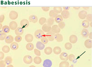
The protozoa may resemble the rings of malaria parasites. Distinguishing traits include exoerythrocytic organisms; the absence of pigmented granules in infected red blood cells; and a “maltese cross,” a rare pattern produced by tetrads of Babesia merozoites.13 An infected erythrocyte may contain up to eight parasites.
Serologic and polymerase chain reaction tests are useful when the organism is not visible.14,15
Treat patients with severe disease
Most patients with B microti infection have a mild illness that resolves without treatment. Treatment is recommended for those with severe infection and in those with high-level parasitemia. Agents with consistent activity against B microti include clindamycin (Cleocin), azithromycin (Zithromax), ato-vaquone (Mepron), doxycycline (Vibramycin), and quinine (Quinamm). Combination therapy with either clindamycin and quinine or azithromycin and atovaquone is recommended. B divergens infection has been successfully treated with a combination of clindamycin, quinine, and exchange transfusion.16
EHRLICHIOSIS
Ehrlichiosis, nicknamed Rocky Mountain “spotless” fever, is a seasonal, tick-borne disease caused by obligate intracellular bacteria. Bacteria of the genus Ehrlichia grow within the cytoplasmic vacuoles of leukocytes and cause mainly zoonotic infections. Several species, especially Ehrlichia chaffeensis and Anaplasma phagocytophilum, are recognized as human pathogens.17,18E chaffeensis infects mononuclear cells, causing a condition known as human monocytic ehrlichiosis (HME). A phagocytophilum infects neutrophils, producing a condition called human granulocytic anaplasmosis (HGA).
Deer are the principal reservoir for E chaffeensis19; white-footed mice, other rodents, and deer are the principal reservoirs for A phagocytophilum. HME is transmitted by Dermacentor and Ixodes ticks, and HGA by Ixodes ticks. Human infection usually occurs in the spring and summer, when tick exposure is greatest. Co-infection of ticks with the organisms causing Lyme disease or babesiosis may result in simultaneous transmission of these diseases.
More than 1,000 cases of HME have been reported in the southeastern, south-central, and mid-Atlantic regions of the United States.20 The prevalence of HME in the United States appears to follow that of Rocky Mountain spotted fever. Some cases have been described in New England and in the Pacific Northwest. The more than 600 reported cases of HGA have come from Wisconsin, Minnesota, Connecticut, New York, Massachusetts, California, Florida, and Western Europe.21,22 The distribution of HGA follows that of Lyme disease, because the two diseases share the same tick vector.
Acute onset of fever and myalgias
HME and HGA have an incubation period of 1 to 2 weeks. The symptoms are similar and are usually acute, ranging from mild to severe. Most patients have fever, chills, malaise, headache, and myalgias. Many also have nausea, vomiting, cough, and arthralgias. Symptoms are similar to those in Rocky Mountain spotted fever (caused by Rickettsia rickettsii), except that rash is uncommon in HME (seen in approximately a third of patients) and rare in HGA.23–25 Neurologic findings, such as altered sensorium and neck stiffness, may be accompanied by lymphocytic pleocytosis and elevated protein levels in the cerebrospinal fluid.26 Subclinical and subacute presentations (eg, a fever lasting up to 2 months) are uncommon. No chronic cases have been reported.
The estimated death rate is 1% to 10%, and hospitalization rates are as high as 60%. Most deaths occur in the elderly, often following such complications as congestive heart failure,27 cardiac tamponade, respiratory or renal failure, seizures, and coma. Patients with human immunodeficiency virus infection also have a poor prognosis. Convalescence may be prolonged.
Laboratory abnormalities include leukopenia, thrombocytopenia, and elevated hepatic transaminase levels. Leukopenia may be associated with lymphopenia or neutropenia. Lymphopenia occurs early in the course of illness and is usually followed by an atypical lymphocytosis. Prolonged symptoms are associated with a decreased total neutrophil count and an increased band neutrophil count.28
Suspect ehrlichiosis in endemic areas in patients with fever, leukopenia, or thrombocytopenia
Ehrlichiosis should be suspected when a febrile patient with leukopenia or thrombocytopenia has been exposed to ticks in an endemic area. Even patients whose cell counts and liver enzyme levels are normal should be evaluated if the clinical and epidemiologic situations suggest this disease.
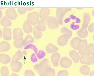
Other diagnostic tests include polymerase chain reaction and serologic assays, which are highly sensitive and specific.30,31 Because the organisms are difficult to culture in vitro, blood cultures are not useful diagnostically.
Treatment
Doxycycline 100 mg twice daily for 7 to 10 days is the treatment of choice for both HME and HGA. No role has been defined for fluoroquinolones for treating these diseases. Avoiding ticks and removing ticks promptly are the best preventive strategies.
RELAPSING FEVER
Relapsing fever is an acute febrile illness caused by spirochetes of the genus Borrelia. The disease has two forms: tick-borne, in which human infection is zoonotic, and louse-borne, in which humans are the only known reservoir of infection.32
Few tick-borne cases in the United States
Tick-borne disease is caused by many species of Borrelia. Those found in the United States occur in the western mountains and high deserts and plains of the Southwest.33 Fewer than 30 cases of tick-borne relapsing fever are diagnosed in the United States annually.
Tick-borne relapsing fever is transmitted by soft-bodied argasid ticks (Ornithodoros genus), which feed for less than an hour (usually at night) and can survive for years without a blood meal. They stay close to human and animal habitations. Exposure often occurs in cabins, under buildings, in caves, near woodpiles, and in rooms shared with animals. Rodents are the primary animal reservoir. In contrast, most other tick-borne diseases—babesiosis, ehrlichiosis, Lyme disease, Rocky Mountain spotted fever, Colorado tick fever—are transmitted by hard-bodied ixodid ticks, which live in brush and forested areas and attach to passersby, on whom they feed for days if not removed.34–36
Louse-borne disease is endemic in Africa
Louse-borne relapsing fever is caused by a single species, B recurrentis, endemic in Ethiopia and Sudan. It may occur sporadically or in epidemics. War, famine, and mass migrations predispose to epidemics with death rates ranging from 30% to 70% if untreated.37,38 Disease is spread between humans by the human body louse (Pediculus humanus).
Relapsing high fevers
The clinical manifestations of tick-borne and louse-borne relapsing fever are similar, although louse-borne relapsing fever often has a longer incubation period and a longer duration of illness. Bacteremia is heralded by the acute onset of high fever (usually above 39°C [102.2°F]), accompanied by headache, nausea, myalgias, and arthralgias. On average, clinical illness remits in 3 days in tick-borne relapsing fever, but may take 5 to 6 days in louse-borne relapsing fever. Physical findings may include altered sensorium, petechiae, hepatosplenomegaly, and conjunctival suffusion. The fever culminates in a “crisis,” characterized by rigors and a precipitous rise in temperature, pulse, and blood pressure. This is followed by defervescence, diaphoresis, and hypotension. The risk of death is highest during this period and immediately afterwards.
With resolution of the bacteremia, an afebrile period ensues, lasting 4 to 14 days. Fever then recurs, although usually milder, again associated with bacteremia. On average, people with tick-borne relapsing fever have three febrile relapses; those with louse-borne relapsing fever have one.39 Relapse occurs because of antigenic variation, in which a major surface antigen of the spirochete is changed to evade the host’s immune system.40–42
Borrelia may invade organs and the nervous system
With each episode of bacteremia, spirochetes may penetrate the brain, heart, liver, eye, or inner ear. Involvement of the central nervous system is more common with tick-borne than with louse-borne relapsing fever. Nervous system involvement may include facial palsy, myelitis, radiculopathy, aphasia, hemiplegia, stupor, or coma.43,44 Myocarditis, common in both forms of relapsing fever, portends a poor prognosis.45 Invasion of the eye or ear may result in visual impairment or dizziness. Bleeding disorders, manifested by epistaxis, petechiae, and ecchymoses, are typical of louse-borne disease and may be associated with low-grade disseminated intravascular coagulation.46 Splenomegaly is more common in louse-borne than in tick-borne disease.
Auxiliary test findings
Laboratory findings include normocytic anemia, leukocytosis, and thrombocytopenia. Liver enzyme levels may be elevated and coagulation tests may be prolonged. Patients with cardiac involvement may have a prolonged QTc interval. Cardiomegaly and pulmonary edema may be seen on chest radiography. The cerebrospinal fluid in patients with neurologic involvement has a mononuclear pleocytosis and a mildly elevated protein concentration.
Suspect if recurrent fever in endemic areas

Treatment
Relapsing fever can be successfully treated with tetracycline, penicillin, or erythromycin.47 The preferred regimen for a nonpregnant adult with louse-borne relapsing fever is a single 0.5-g dose of tetracycline. In tick-borne relapsing fever, 0.5 g of tetracycline four times daily for 5 to 10 days is recommended. Meningitis or encephalitis is usually treated with parenteral penicillin or ceftriaxone (Rocephin). The death rate in treated disease is usually less than 5%.39 Treatment can induce a Jarisch-Herxheimer reaction (rigors and hypotension, resembling a febrile crisis), and patients must be watched closely for the first 4 hours after the antibiotic is given. Avoiding ticks and practicing good personal hygiene to prevent acquiring lice are the major preventive strategies.
MALARIA
Malaria is caused by protozoa of the genus Plasmodium. Injected during a mosquito bite, sporozoites enter the bloodstream and travel to the liver. During the asymptomatic hepatic stage, the sporozoites multiply within hepatocytes to form mature schizonts. Within 1 to 2 weeks, the schizonts rupture, releasing thousands of merozoites into the bloodstream. The merozoites enter red blood cells, where they mature through the trophozoite stage to become schizonts, which release more merozoites into the blood that may infect other red blood cells. Symptoms occur during this erythrocytic stage, usually 1 to 4 weeks after a mosquito bite.
Four Plasmodia species regularly cause human disease.48 All can digest hemoglobin and cause hemolysis. P falciparum is uniquely dangerous, in part because it can alter the erythrocyte surface and obstruct the microcirculation.49,50 Further, P falciparum can cause high levels of parasitemia because it can infect red blood cells in all stages of their maturation. In contrast, P vivax and P ovale invade only reticulocytes, so levels of parasitemia are low (< 1%). P vivax and P ovale produce dormant liver forms (hypnozoites), which may cause relapses, usually within 3 years of exposure. Falciparum malaria does not have a dormant form, so relapses do not occur. P malariae infects only mature red blood cells, producing low levels of parasitemia. A fifth malaria species once thought to be confined to monkeys, P knowlesi, is a rare cause of severe human disease.51
Up to a half-billion cases of malaria occur worldwide each year.52 Although the disease predominates among residents of endemic areas, about 30,000 travelers from industrialized countries are infected each year. Fewer than 1,000 cases are reported in the United States annually, which were usually acquired during travel to endemic areas.53,54 The risk of transmission varies from region to region, with highest rates (listed in descending order) occurring in Oceania, sub-Saharan Africa, the Indian subcontinent, Southeast Asia, South America, and Central America.55 More than 3 million people die of malaria annually, most of them in sub-Saharan Africa.
Malaria is transmitted principally by the bite of the female Anopheles mosquito but can also be transmitted by blood transfusion, by the use of contaminated needles, congenitally, and through organ transplantation. Because immigrants to the United States from endemic areas may harbor the parasites for months to years, malaria may rarely be acquired by autochthonous transmission, in which a parasitized individual infects competent vectors, which then bite uninfected persons.55 The disease may also be transmitted from parasitized mosquitoes that arrive on an aircraft (“airport malaria”). There is no animal reservoir for human malaria parasites.
Recurring fever
Fever is universally present, irrespective of the species causing infection. Common symptoms include malaise, chills, headache, myalgias, abdominal pain, night sweats, nausea, and diarrhea. Febrile episodes, initially frequent and irregular, may become regular, producing temperature spikes every second or third day depending on the species. The severity of the paroxysms usually diminishes over time. Eventually, anemia, thrombocytopenia, jaundice, splenomegaly, and hepatomegaly occur.
Falciparum malaria may result in pulmonary edema, renal failure, gastroenteritis, bleeding, or hypoglycemia. Cerebral disease, presenting as altered sensorium or seizures, can be fatal.56 Death, common in untreated patients, correlates with the degree of parasitemia.57 Severe disease often occurs in children, pregnant women, asplenic patients, and nonimmune adults.
Infection with P vivax or P ovale does not produce the microvascular complications of falciparum malaria, and thrombocytopenia is less common. P malariae infection may lead to immune complex deposition and nephrotic syndrome. Symptoms, although mild, are often prolonged.
Consider malaria in travelers with recurring fever
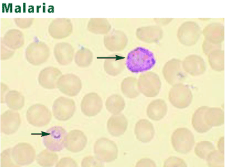
Antigen-capture test kits are useful for rapid diagnosis. These portable devices detect parasitic antigens from a drop of blood in just 15 minutes.61 Polymerase chain reaction techniques offer high sensitivity and specificity but are more expensive and less available.62
The management of patients with suspected malaria should be done by experienced health care providers. Recommendations about malaria treatment are beyond the scope of this review. The choice of antimalarial drugs requires knowledge of the regional distribution of drug resistance and the adverse effects of the agents. Patients with non-falciparum malaria rarely require hospitalization.
Malaria is prevented by mosquito avoidance and chemoprophylaxis.
AMERICAN TRYPANOSOMIASIS (CHAGAS DISEASE)
American trypanosomiasis is a zoonotic, protozoal disease caused by Trypanosoma cruzi.
The parasite is transmitted by bloodsucking triatomine insects, or “kissing bugs.”63 These insects favor mud-brick and clay houses, where they live in wall cracks, under furniture, and behind pictures. The insects acquire the organism by feeding on infected animals or on humans that have circulating trypomastigotes. The organisms then multiply in the gut of the insects and are transmitted to a second vertebrate host as the insect defecates following a blood meal. The parasite then enters the body through the skin, conjunctivae, or mucous membranes. After entering the body, the parasite disseminates through the bloodstream, invading many cell types, especially muscle and nerve.
American trypanosomiasis occurs in Central and South America, Mexico, and the southern United States. Between 16 and 18 million people are infected with the parasite, and nearly 50,000 die annually, usually from cardiac complications. Once confined to rural areas, the disease is now common in cities. The majority of reported cases come from Brazil. Only a few cases have been reported in the United States, but an estimated 50,000 to 100,000 immigrants are thought to be infected.64–68
Transmission of the parasite may also occur with blood transfusion, with organ transplantation, and congenitally.63,69,70 An increase in transfusion-related cases is expected in the United States because of an influx of migrant workers from Mexico and Central America.
Acute phase ranges from asymptomatic to multiorgan involvement
Most infected people are asymptomatic carriers. Only 1% become acutely ill, but up to a third of those infected may develop chronic symptoms decades later. Acute Chagas disease, usually seen in children, is characterized by fever, malaise, and anorexia, often accompanied by vomiting, diarrhea, and rash.64,71,72 The heart, liver, spleen, and lymph nodes become enlarged. A red and indurated nodule or furuncle (chagoma) appears at the inoculation site. If inoculation occurs across the conjunctivae, painless edema of the palpebrae and periocular tissues may be observed (Romaña sign). Generalized lymphadenopathy and hepatosplenomegaly may also be seen.
Myocarditis and meningoencephalitis occur in some cases of acute Chagas disease. Myocardial inflammation may extend to the pericardium, causing pericardial effusion, and to the endocardium, causing thrombus formation. All cardiac chambers become enlarged and the conduction system is disrupted.73 Brain damage from meningoencephalitis usually occurs in infants and young children, and may result in death.
This acute phase lasts 4 to 8 weeks and is characterized by profound parasitemia, tissue invasion, and inflammation. Following the acute phase, an asymptomatic latent or indeterminate phase lasting 10 to 40 years occurs.74 Less than half of those in clinical latency enter a chronic phase of disease.
Severe chronic phase may occur decades later
The chronic phase, which occurs years to decades after the initial infection, is characterized by cardiac, esophageal, and colonic enlargement. Cardiac involvement is associated with congestive heart failure, arrhythmias, and cardiac arrest. Intracardiac thrombi may embolize, causing systemic and pulmonary infarctions.71–73,75 Enlargement of the esophagus is associated with dysphagia, chest pain, weight loss, and sometimes perforation or aspiration-related pneumonitis. Colonic enlargement may result in constipation, abdominal distention, and intestinal obstruction. Sometimes, the small bowel, ureters, and bronchi become dilated as well. These findings are the result of low-grade parasitemia, tissue inflammation, and immune-mediated disruption of the microvasculature.71,76
Diagnosis: Protozoa evident in acute phase
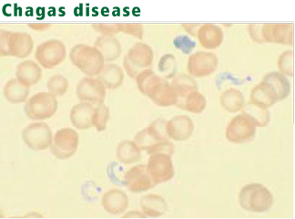
Serologic tests are most useful for diagnosing chronic Chagas disease.77 The organism can be identified by polymerase chain reaction, but the sensitivity of this test is highly variable.
Treatment reduces symptoms
Two drugs are used to treat American trypanosomiasis. Nifurtimox (Lampit) reduces symptom duration and severity, as well as mortality rates, in acute and congenital Chagas disease; however, fewer than 75% of patients have a parasitologic cure, and adverse effects limit tolerability.78 Benznidazole (Rochagan, Radanil) has an efficacy similar to that of nifurtimox and is considered the drug of choice in Latin America. These drugs are not commercially available in the United States. Therapy may help during the indeterminate phase but is rarely effective for chronic Chagas disease. Treatment of Chagas disease with triazoles is under evaluation.
- Kroft SH. Infectious diseases manifested in the peripheral blood. Clin Lab Med 2002; 22:253–277.
- Meldrum SC, Birkhead GS, White DJ, Benach JL, Morse DL. Human babesiosis in New York State: an epidemiological description of 136 cases. Clin Infect Dis 1992; 15:1019–1023.
- Pruthi RK, Marshall WF, Wiltsie JC, Persing DH. Human babesiosis. Mayo Clin Proc 1995; 70:853–862.
- Eskow ES, Krause PJ, Spielman A, Freeman K, Aslanzadeh J. Southern extension of the range of human babesiosis in the eastern United States. J Clin Microbiol 1999; 37:2051–2052.
- Quick RE, Herwaldt BL, Thomford JW, et al. Babesiosis in Washington State: a new species of Babesia? Ann Intern Med 1993; 119:284–290.
- Persing DH, Herwaldt BL, Glaser C, et al. Infection with a babesia-like organism in northern California. N Engl J Med 1995; 332:298–303.
- Gorenflot A, Moubri K, Precigout E, Carcy B, Schetters TP. Human babesiosis. Ann Trop Med Parasitol 1998; 92:489–501.
- Villar BF, White DJ, Benach JL. Human babesiosis. Prog Clin Parasitol 1991; 2:129–143.
- Sun T, Tenenbaum MJ, Greenspan J, et al. Morphologic and clinical observations in human infection with Babesia microti. J Infect Dis 1983; 148:239–248.
- Benach JL, Habicht GS. Clinical characteristics of human babesiosis. J Infect Dis 1981; 144:481.
- Boustani MR, Gelfand JA. Babesiosis. Clin Infect Dis 1996; 22:611–615.
- Meer-Scherrer L, Adelson M, Mordechai E, Lottaz B, Tilton R. Babesia microti infection in Europe. Curr Microbiol 2004; 48:435–437.
- Krause PJ. Babesiosis diagnosis and treatment. Vector Borne Zoonotic Dis 2003: 3:45–51.
- Persing DH, Mathiesen D, Marshall WF, et al. Detection of Babesia microti by polymerase chain reaction. J Clin Microbiol 1992; 30:2097–2103.
- Chisholm ES, Sulzer AJ, Ruebush TK. Indirect immunofluorescence test for human Babesia microti infection: antigen specificity. Am J Trop Med Hyg 1986; 35:921–925.
- Berry A, Morassin B, Kamar N, Magnaval JF. Clinical picture: human babesiosis. Lancet 2001; 357:341.
- Maeda K, Markowitz N, Hawley R, Ristic M, Cox D, McDade JE. Human infection with Ehrlichia canis, a leukocytic rickettsia. N Engl J Med 1987; 316:853–856.
- Chen SM, Dumler JS, Bakken JS, Walker DH. Identification of a granulocytotrophic Ehrlichia species as the etiologic agent of human disease. J Clin Microbiol 1994; 32:589–595.
- Lockhart JM, Davidson WR, Stallknecht DE, Dawson JE, Howerth EW. Isolation of Ehrlichia chaffeensis from wild white-tailed deer (Odocoileus virginianus) confirms their role as natural reservoir hosts. J Clin Microbiol 1997; 35:1681–1686.
- Stone JH, Dierberg K, Aram G, Dumler JS. Human monocytic erhlichiosis. JAMA 2004; 292:2263–2270.
- Bakken JS, Dumler JS. Human granulocytic ehrlichiosis. Clin Infect Dis 2000; 31:554–560.
- Dumler JS, Bakken JS. Ehrlichial diseases of humans: emerging tick-borne infections. Clin Infect Dis 1995; 20:1102–1110.
- Everett ED, Evans KA, Henry RB, McDonald G. Human ehrlichiosis in adults after tick exposure. Diagnosis using polymerase chain reaction. Ann Intern Med 1994; 120:730–735.
- Fishbein DB, Dawson JE, Robinson LE. Human ehrlichiosis in the United States, 1985 to 1990. Ann Intern Med 1994; 120:736–743.
- Bakken JS, Krueth J, Wilson-Nordskog C, Tilden RL, Asanovich K, Dumler JS. Clinical and laboratory characteristics of human granulocytic ehrlichiosis. JAMA 1996; 275:199–205.
- Ratnasamy N, Everett ED, Roland WE, McDonald G, Caldwell CW. Central nervous system manifestations of human ehrlichiosis. Clin Infect Dis 1996; 23:314–319.
- Vanek NN, Kazi S, Cepero NM, Tang S, Rex JH. Human ehrlichiosis causing left ventricular dilation and dysfunction. Clin Infect Dis 1996; 22:386–387.
- Bakken JS, Aguero-Rosenfeld ME, Tilden RL, et al. Serial measurements of hematologic counts during the active phase of human granulocytic ehrlichiosis. Clin Infect Dis 2001; 32:862–870.
- Hamilton KS, Standaert SM, Kinney MC. Characteristic peripheral blood findings in human ehrlichiosis. Mod Path 2004; 17:512–517.
- Dawson JE, Fishbein DB, Eng TR, Redus MA, Green NR. Diagnosis of human ehrlichiosis with the indirect fluorescent antibody test: kinetics and specificity. J Infect Dis 1990; 162:91–95.
- Walls JJ, Caturegli P, Bakken JS, Asanovich KM, Dumler JS. Improved sensitivity of PCR for diagnosis of human granulocytic ehrlichiosis using epank1 genes of Ehrlichia phagocytophila-group ehrlichiae. J Clin Microbiol 2000; 38:354–356.
- Barbour AG. Relapsing fever. In:Goodman JL, Dennis DT, Sonenshine DE, editors. Tick-Borne Diseases of Humans. Washington, DC: ASM Press; 2005:268.
- Barbour AG, Hayes SF. Biology of Borrelia species. Microbiol Rev 1986; 50:381–400.
- Hoogstraal H. Ticks and spirochetes. Acta Trop 1979; 36:133–136.
- Sonenshine D. Biology of Ticks. New York, NY: Oxford University Press; 1991.
- Felsenfeld O. Borrelia; Strains, Vectors, Human, and Animal Borreliosis. St Louis, MO: Warren H Greene, Inc; 1971.
- Bryceson AD, Parry EH, Perine PL, Warrell DA, Vukotich D, Leithead CS. Louse-borne relapsing fever. Q J Med 1970; 39:129–170.
- Judge DM, Samuel I, Perine PL, Vukotic D, Ababa A. Louse-borne relapsing fever in man. Arch Pathol 1974; 97:136–170.
- Southern P, Sanford J. Relapsing fever. A clinical and microbiological review. Medicine 1969; 48:129–149.
- Barbour AG. Antigenic variation of a relapsing fever Borrelia species. Annu Rev Microbiol 1990; 44:155–171.
- Stoenner HG, Dodd T, Larsen C. Antigenic variation of Borrelia hermsii. J Exp Med 1982; 156:1297–1311.
- Barbour AG. Immunobiology of relapsing fever. Contrib Microbiol Immunol 1987; 8:125–137.
- Scott R. Neurological complications of relapsing fever. Lancet 1944; 247:436–438.
- Cadavid D, Barbour AG. Neuroborreliosis during relapsing fever: review of the clinical manifestations, pathology, and treatment of infections in humans and experimental animals. Clin Infect Dis 1998; 26:151–164.
- Wengrower D, Knobler H, Gillis S, Chajek-Shaul T. Myocarditis in tick-borne relapsing fever. J Infect Dis 1984; 149:1033.
- Perine PL, Parry EH, Vukotich D, Warrell DA, Bryceson AD. Bleeding in louse-borne relapsing fever. I. Clinical studies in 37 patients. Trans R Soc Trop Med Hyg 1971; 65:776–781.
- Rhee KY, Johnson WD. Borrelia species (relapsing fever). In:Mandell GL, Bennett JE, Dolin R, editors. Mandell, Douglas, and Bennett’s Principles and Practice of Infectious Diseases. 6th ed. Philadelphia, PA: Elsevier Churchill Livingstone; 2005:2795–2798.
- Greenwood BM, Bojang K, Whitty CJ, Targett GA. Malaria. Lancet 2005; 365:1487–1498.
- Newbold C, Craig A, Kyes S, Rowe A, Fernandez-Reyes D, Fagan T. Cytoadherence, pathogenesis and the infected red cell surface in Plasmodium falciparum. Int J Parasitol 1999; 29:927–937.
- Oh SS, Chishti AH, Palek J, Liu SC. Erythrocyte membrane alterations in Plasmodium falciparum malaria sequestration. Curr Opin Hematol 1997; 4:148–154.
- Singh B, Kim Sung L, Matusop A, et al. A large focus of naturally acquired Plasmodium knowlesi infections in human beings. Lancet 2004; 363:1017–1024.
- Fairhurst RM, Wellems TE. Plasmodium species (malaria). In:Mandell GL, Bennett JE, Dolin R, editors. Mandell, Douglas, and Bennett’s Principles and Practice of Infectious Diseases. 6th ed. Philadelphia, PA: Elsevier Churchill Livingstone; 2005:3121,3122.
- Filler S, Causer LM, Newman RD, et al Centers for Disease Control and Prevention (CDC). . Malaria surveillance—United States, 2001. MMWR Surveill Summ 2003; 52:1–14.
- Olliaro P, Cattani J, Wirth D. Malaria, the submerged disease. JAMA 1996; 275:230–233.
- Kain KC, Keystone JS. Malaria in travelers. Epidemiology, disease, and prevention. Infect Dis Clin North Am 1998; 12:267–284.
- World Health Organization, Division of Control of Tropical Diseases. Severe and complicated malaria. Trans R Soc Trop Med Hyg 1990; 84(suppl 2):1–65.
- Field JW. Blood examination and prognosis in acute falciparum malaria. Trans R Soc Trop Med Hyg 1949; 43:33–48.
- Microscopic procedures for diagnosing malaria. Appendix MMWR Surveill Summ 2003; 52:15–16.
- Lema OE, Carter JY, Nagelkerke N, et al. Comparison of five methods of malaria detection in the outpatient setting. Am J Trop Med Hyg 1999; 60:177–182.
- White NJ. The treatment of malaria. N Engl J Med 1996; 335:800–806.
- Moody A. Rapid diagnostic tests for malaria parasites. Clin Microbiol Rev 2002; 15:66–78.
- Makler MT, Palmer CJ, Ager AL. A review of practical techniques for the diagnosis of malaria. Ann Trop Med Parasitol 1998; 92:419–433.
- Kirchhoff LV. Trypanosoma species (American trypanosomiasis, Chagas’ disease): Biology of trypanosomes. In:Mandell GL, Bennett JE, Dolin R, editors. Mandell, Douglas, and Bennett’s Principles and Practice of Infectious Diseases. 6th ed. Philadelphia, PA: Elsevier Churchill Livingstone; 2005:3157–3158.
- Barrett MP, Burchmore RJ, Stich A, et al. The trypanosomiases. Lancet 2003; 362:1469–1480.
- Chagas disease. Interruption of transmission. Wkly Epidemiol Rec 1997; 71:1
- Control of Chagas disease. Report of a WHO Expert Committee. World Health Organ Tech Rep Ser 1991; 811:1–95.
- Kirchhoff LV. American trypanosomiasis (Chagas’ disease). In:Guerrant R, Walker DH, Weller PF, editors. Tropical Infectious Diseases: Principles, Pathogens and Practice; vol. 1. Philadelphia, PA: Churchill Livingstone; 1999:785.
- Kirchhoff LV. American trypanosomiasis (Chagas’ disease). Gastroenterol Clin North Am 1996; 25:517–533.
- Wanderley DM, Corrêa FM. Epidemiology of Chagas’ heart disease. Sao Paul Med J 1995; 113:742–749.
- Schmuñis GA. Trypanosoma cruzi, the etiologic agent of Chagas’ disease: status in the blood supply in endemic and nonendemic countries. Transfusion 1991; 31:547–557.
- Köberle F. Chagas’ disease and Chagas’ syndromes: the pathology of American trypanosomiasis. Adv Parasitol 1968; 6:63–116.
- Dias E, Laranja FS, Miranda A, Nobrega G. Chagas’ disease; a clinical, epidemiologic, and pathologic study. Circulation 1956; 14:1035–1060.
- Prata A, Andrade Z, Guimaraes AC. Chagas’ heart disease. In:Shaper AG, Hutt MS, Fejfar Z, editors. Cardiovascular disease in the tropics. London, England: British Medical Association; 1974:264.
- Dias JC. The indeterminate form of human chronic Chagas’ disease. A clinical epidemiological study. Rev Soc Bras Med Trop 1989; 22:147–156.
- Lopes ER, Tafuri WL. Involvement of the autonomic nervous system in Chagas’ heart disease. Rev Soc Bras Med Trop 1983; 16:206.
- Meneghelli UG. Chagas’ disease: a model of denervation in the study of digestive tract motility. Braz J Med Biol Res 1985; 18:255–264.
- Pirard M, Iihoshi N, Boelaert M, Basanta P, Lopez F, Van der Stuyft P. The validity of serologic tests for Trypanosoma cruzi and the effectiveness of transfusional screening strategies in a hyperendemic region. Transfusion 2005; 45:554–561.
- Kirchhoff LV. Trypanosoma species (American trypanosomiasis, Chagas’ disease): Biology of trypanosomes. In:Mandell GL, Bennett JE, Dolin R, editors. Mandell, Douglas, and Bennett’s Principles and Practice of Infectious Diseases. 6th ed. Philadelphia, PA: Elsevier Churchill Livingstone; 2005:3162–3163.
Blood smear analysis, while commonly used to evaluate hematologic conditions, is infrequently used to diagnose infectious diseases. This is because of the rarity of diseases for which blood smear analysis is indicated. Consequently, such testing is often overlooked when it is diagnostically important.
Nonspecific changes may include morphologic changes in leukocytes and erythrocytes (eg, toxic granulations, macrocytosis).1 And with certain pathogens, identifying organisms in a peripheral blood smear allows for a rapid diagnosis.
This paper discusses the epidemiology, clinical manifestations, laboratory findings, and management of five infectious diseases in which direct visualization of the organism in the blood plays a major diagnostic role. Our intent is to summarize the clinical findings that should prompt blood smear analysis so that these uncommon conditions are not overlooked.
BABESIOSIS
Babesiosis, a tick-borne protozoal disease, occurs principally in the United States and Europe. Of the more than 100 species of Babesia, two account for almost all human disease: B microti and B divergens.
Both species are transmitted by Ixodes ticks, although patients often do not recall being bitten. The disease may rarely complicate blood transfusion. Most cases occur from May to September, when tick exposure is highest. The incubation period varies from 1 to 4 weeks.
Common in the Northeast, usually asymptomatic
B microti infection occurs predominantly in the United States. Rodents, especially the white-footed mouse, are the principal reservoir.2,3 Endemic areas, where seropositivity rates range from 4% to 21%, include the coastal areas and islands off of Massachusetts, particularly Cape Cod, Nantucket, and Martha’s Vineyard; the islands near New York City, especially Long Island, Shelter Island, and Fire Island; Block Island, off the coast of Rhode Island; and certain areas in Connecticut.4 WA-1, a species that is morphologically identical to B microti, is emerging in California and Washington.5,6
Infection with B microti is usually asymptomatic. Elderly and immunosuppressed people, especially those without a spleen or with impaired cellular immunity, are more likely to become ill. Symptoms, including fever, malaise, headache, nausea, and generalized aching, may last weeks to months.
About one-fourth of patients with babesiosis are coinfected with the Lyme disease bacterium (Borrelia burgdorferi) and often have more severe illness.3
Hepatomegaly, splenomegaly, jaundice, and dark urine are common findings in patients with symptoms. Severe hemolysis, often accompanied by thrombocytopenia, leukopenia, and atypical lymphocytosis, is more common in high-risk patients. Hepatic transaminases may be elevated. Urinalysis may show proteinuria and hemoglobinuria. Acute respiratory distress syndrome has been reported in severe cases.7–10
B divergens infection: A serious but rare disease seen in Europe
B divergens is found mainly in Europe. Altogether, fewer than 50 cases of infection have been reported in France, Spain, Germany, Great Britain, Ireland, Yugoslavia, and the former Soviet Union.11,12 Cattle are the principal reservoir of infection.
Infection with B divergens causes a rare but devastating disease mainly in asplenic people, usually resulting in coma and death. No cases of subclinical infection have been reported. The clinical course is fulminant, and hemolytic anemia is common.2,3
Suspect babesiosis in endemic areas in cases of prolonged ‘flu’
Babesiosis should be considered when a patient residing in or traveling from an endemic area presents with a prolonged flu-like illness and hemolysis, with or without organomegaly and jaundice.
The protozoa may resemble the rings of malaria parasites. Distinguishing traits include exoerythrocytic organisms; the absence of pigmented granules in infected red blood cells; and a “maltese cross,” a rare pattern produced by tetrads of Babesia merozoites.13 An infected erythrocyte may contain up to eight parasites.
Serologic and polymerase chain reaction tests are useful when the organism is not visible.14,15
Treat patients with severe disease
Most patients with B microti infection have a mild illness that resolves without treatment. Treatment is recommended for those with severe infection and in those with high-level parasitemia. Agents with consistent activity against B microti include clindamycin (Cleocin), azithromycin (Zithromax), ato-vaquone (Mepron), doxycycline (Vibramycin), and quinine (Quinamm). Combination therapy with either clindamycin and quinine or azithromycin and atovaquone is recommended. B divergens infection has been successfully treated with a combination of clindamycin, quinine, and exchange transfusion.16
EHRLICHIOSIS
Ehrlichiosis, nicknamed Rocky Mountain “spotless” fever, is a seasonal, tick-borne disease caused by obligate intracellular bacteria. Bacteria of the genus Ehrlichia grow within the cytoplasmic vacuoles of leukocytes and cause mainly zoonotic infections. Several species, especially Ehrlichia chaffeensis and Anaplasma phagocytophilum, are recognized as human pathogens.17,18E chaffeensis infects mononuclear cells, causing a condition known as human monocytic ehrlichiosis (HME). A phagocytophilum infects neutrophils, producing a condition called human granulocytic anaplasmosis (HGA).
Deer are the principal reservoir for E chaffeensis19; white-footed mice, other rodents, and deer are the principal reservoirs for A phagocytophilum. HME is transmitted by Dermacentor and Ixodes ticks, and HGA by Ixodes ticks. Human infection usually occurs in the spring and summer, when tick exposure is greatest. Co-infection of ticks with the organisms causing Lyme disease or babesiosis may result in simultaneous transmission of these diseases.
More than 1,000 cases of HME have been reported in the southeastern, south-central, and mid-Atlantic regions of the United States.20 The prevalence of HME in the United States appears to follow that of Rocky Mountain spotted fever. Some cases have been described in New England and in the Pacific Northwest. The more than 600 reported cases of HGA have come from Wisconsin, Minnesota, Connecticut, New York, Massachusetts, California, Florida, and Western Europe.21,22 The distribution of HGA follows that of Lyme disease, because the two diseases share the same tick vector.
Acute onset of fever and myalgias
HME and HGA have an incubation period of 1 to 2 weeks. The symptoms are similar and are usually acute, ranging from mild to severe. Most patients have fever, chills, malaise, headache, and myalgias. Many also have nausea, vomiting, cough, and arthralgias. Symptoms are similar to those in Rocky Mountain spotted fever (caused by Rickettsia rickettsii), except that rash is uncommon in HME (seen in approximately a third of patients) and rare in HGA.23–25 Neurologic findings, such as altered sensorium and neck stiffness, may be accompanied by lymphocytic pleocytosis and elevated protein levels in the cerebrospinal fluid.26 Subclinical and subacute presentations (eg, a fever lasting up to 2 months) are uncommon. No chronic cases have been reported.
The estimated death rate is 1% to 10%, and hospitalization rates are as high as 60%. Most deaths occur in the elderly, often following such complications as congestive heart failure,27 cardiac tamponade, respiratory or renal failure, seizures, and coma. Patients with human immunodeficiency virus infection also have a poor prognosis. Convalescence may be prolonged.
Laboratory abnormalities include leukopenia, thrombocytopenia, and elevated hepatic transaminase levels. Leukopenia may be associated with lymphopenia or neutropenia. Lymphopenia occurs early in the course of illness and is usually followed by an atypical lymphocytosis. Prolonged symptoms are associated with a decreased total neutrophil count and an increased band neutrophil count.28
Suspect ehrlichiosis in endemic areas in patients with fever, leukopenia, or thrombocytopenia
Ehrlichiosis should be suspected when a febrile patient with leukopenia or thrombocytopenia has been exposed to ticks in an endemic area. Even patients whose cell counts and liver enzyme levels are normal should be evaluated if the clinical and epidemiologic situations suggest this disease.
Other diagnostic tests include polymerase chain reaction and serologic assays, which are highly sensitive and specific.30,31 Because the organisms are difficult to culture in vitro, blood cultures are not useful diagnostically.
Treatment
Doxycycline 100 mg twice daily for 7 to 10 days is the treatment of choice for both HME and HGA. No role has been defined for fluoroquinolones for treating these diseases. Avoiding ticks and removing ticks promptly are the best preventive strategies.
RELAPSING FEVER
Relapsing fever is an acute febrile illness caused by spirochetes of the genus Borrelia. The disease has two forms: tick-borne, in which human infection is zoonotic, and louse-borne, in which humans are the only known reservoir of infection.32
Few tick-borne cases in the United States
Tick-borne disease is caused by many species of Borrelia. Those found in the United States occur in the western mountains and high deserts and plains of the Southwest.33 Fewer than 30 cases of tick-borne relapsing fever are diagnosed in the United States annually.
Tick-borne relapsing fever is transmitted by soft-bodied argasid ticks (Ornithodoros genus), which feed for less than an hour (usually at night) and can survive for years without a blood meal. They stay close to human and animal habitations. Exposure often occurs in cabins, under buildings, in caves, near woodpiles, and in rooms shared with animals. Rodents are the primary animal reservoir. In contrast, most other tick-borne diseases—babesiosis, ehrlichiosis, Lyme disease, Rocky Mountain spotted fever, Colorado tick fever—are transmitted by hard-bodied ixodid ticks, which live in brush and forested areas and attach to passersby, on whom they feed for days if not removed.34–36
Louse-borne disease is endemic in Africa
Louse-borne relapsing fever is caused by a single species, B recurrentis, endemic in Ethiopia and Sudan. It may occur sporadically or in epidemics. War, famine, and mass migrations predispose to epidemics with death rates ranging from 30% to 70% if untreated.37,38 Disease is spread between humans by the human body louse (Pediculus humanus).
Relapsing high fevers
The clinical manifestations of tick-borne and louse-borne relapsing fever are similar, although louse-borne relapsing fever often has a longer incubation period and a longer duration of illness. Bacteremia is heralded by the acute onset of high fever (usually above 39°C [102.2°F]), accompanied by headache, nausea, myalgias, and arthralgias. On average, clinical illness remits in 3 days in tick-borne relapsing fever, but may take 5 to 6 days in louse-borne relapsing fever. Physical findings may include altered sensorium, petechiae, hepatosplenomegaly, and conjunctival suffusion. The fever culminates in a “crisis,” characterized by rigors and a precipitous rise in temperature, pulse, and blood pressure. This is followed by defervescence, diaphoresis, and hypotension. The risk of death is highest during this period and immediately afterwards.
With resolution of the bacteremia, an afebrile period ensues, lasting 4 to 14 days. Fever then recurs, although usually milder, again associated with bacteremia. On average, people with tick-borne relapsing fever have three febrile relapses; those with louse-borne relapsing fever have one.39 Relapse occurs because of antigenic variation, in which a major surface antigen of the spirochete is changed to evade the host’s immune system.40–42
Borrelia may invade organs and the nervous system
With each episode of bacteremia, spirochetes may penetrate the brain, heart, liver, eye, or inner ear. Involvement of the central nervous system is more common with tick-borne than with louse-borne relapsing fever. Nervous system involvement may include facial palsy, myelitis, radiculopathy, aphasia, hemiplegia, stupor, or coma.43,44 Myocarditis, common in both forms of relapsing fever, portends a poor prognosis.45 Invasion of the eye or ear may result in visual impairment or dizziness. Bleeding disorders, manifested by epistaxis, petechiae, and ecchymoses, are typical of louse-borne disease and may be associated with low-grade disseminated intravascular coagulation.46 Splenomegaly is more common in louse-borne than in tick-borne disease.
Auxiliary test findings
Laboratory findings include normocytic anemia, leukocytosis, and thrombocytopenia. Liver enzyme levels may be elevated and coagulation tests may be prolonged. Patients with cardiac involvement may have a prolonged QTc interval. Cardiomegaly and pulmonary edema may be seen on chest radiography. The cerebrospinal fluid in patients with neurologic involvement has a mononuclear pleocytosis and a mildly elevated protein concentration.
Suspect if recurrent fever in endemic areas
Treatment
Relapsing fever can be successfully treated with tetracycline, penicillin, or erythromycin.47 The preferred regimen for a nonpregnant adult with louse-borne relapsing fever is a single 0.5-g dose of tetracycline. In tick-borne relapsing fever, 0.5 g of tetracycline four times daily for 5 to 10 days is recommended. Meningitis or encephalitis is usually treated with parenteral penicillin or ceftriaxone (Rocephin). The death rate in treated disease is usually less than 5%.39 Treatment can induce a Jarisch-Herxheimer reaction (rigors and hypotension, resembling a febrile crisis), and patients must be watched closely for the first 4 hours after the antibiotic is given. Avoiding ticks and practicing good personal hygiene to prevent acquiring lice are the major preventive strategies.
MALARIA
Malaria is caused by protozoa of the genus Plasmodium. Injected during a mosquito bite, sporozoites enter the bloodstream and travel to the liver. During the asymptomatic hepatic stage, the sporozoites multiply within hepatocytes to form mature schizonts. Within 1 to 2 weeks, the schizonts rupture, releasing thousands of merozoites into the bloodstream. The merozoites enter red blood cells, where they mature through the trophozoite stage to become schizonts, which release more merozoites into the blood that may infect other red blood cells. Symptoms occur during this erythrocytic stage, usually 1 to 4 weeks after a mosquito bite.
Four Plasmodia species regularly cause human disease.48 All can digest hemoglobin and cause hemolysis. P falciparum is uniquely dangerous, in part because it can alter the erythrocyte surface and obstruct the microcirculation.49,50 Further, P falciparum can cause high levels of parasitemia because it can infect red blood cells in all stages of their maturation. In contrast, P vivax and P ovale invade only reticulocytes, so levels of parasitemia are low (< 1%). P vivax and P ovale produce dormant liver forms (hypnozoites), which may cause relapses, usually within 3 years of exposure. Falciparum malaria does not have a dormant form, so relapses do not occur. P malariae infects only mature red blood cells, producing low levels of parasitemia. A fifth malaria species once thought to be confined to monkeys, P knowlesi, is a rare cause of severe human disease.51
Up to a half-billion cases of malaria occur worldwide each year.52 Although the disease predominates among residents of endemic areas, about 30,000 travelers from industrialized countries are infected each year. Fewer than 1,000 cases are reported in the United States annually, which were usually acquired during travel to endemic areas.53,54 The risk of transmission varies from region to region, with highest rates (listed in descending order) occurring in Oceania, sub-Saharan Africa, the Indian subcontinent, Southeast Asia, South America, and Central America.55 More than 3 million people die of malaria annually, most of them in sub-Saharan Africa.
Malaria is transmitted principally by the bite of the female Anopheles mosquito but can also be transmitted by blood transfusion, by the use of contaminated needles, congenitally, and through organ transplantation. Because immigrants to the United States from endemic areas may harbor the parasites for months to years, malaria may rarely be acquired by autochthonous transmission, in which a parasitized individual infects competent vectors, which then bite uninfected persons.55 The disease may also be transmitted from parasitized mosquitoes that arrive on an aircraft (“airport malaria”). There is no animal reservoir for human malaria parasites.
Recurring fever
Fever is universally present, irrespective of the species causing infection. Common symptoms include malaise, chills, headache, myalgias, abdominal pain, night sweats, nausea, and diarrhea. Febrile episodes, initially frequent and irregular, may become regular, producing temperature spikes every second or third day depending on the species. The severity of the paroxysms usually diminishes over time. Eventually, anemia, thrombocytopenia, jaundice, splenomegaly, and hepatomegaly occur.
Falciparum malaria may result in pulmonary edema, renal failure, gastroenteritis, bleeding, or hypoglycemia. Cerebral disease, presenting as altered sensorium or seizures, can be fatal.56 Death, common in untreated patients, correlates with the degree of parasitemia.57 Severe disease often occurs in children, pregnant women, asplenic patients, and nonimmune adults.
Infection with P vivax or P ovale does not produce the microvascular complications of falciparum malaria, and thrombocytopenia is less common. P malariae infection may lead to immune complex deposition and nephrotic syndrome. Symptoms, although mild, are often prolonged.
Consider malaria in travelers with recurring fever
Antigen-capture test kits are useful for rapid diagnosis. These portable devices detect parasitic antigens from a drop of blood in just 15 minutes.61 Polymerase chain reaction techniques offer high sensitivity and specificity but are more expensive and less available.62
The management of patients with suspected malaria should be done by experienced health care providers. Recommendations about malaria treatment are beyond the scope of this review. The choice of antimalarial drugs requires knowledge of the regional distribution of drug resistance and the adverse effects of the agents. Patients with non-falciparum malaria rarely require hospitalization.
Malaria is prevented by mosquito avoidance and chemoprophylaxis.
AMERICAN TRYPANOSOMIASIS (CHAGAS DISEASE)
American trypanosomiasis is a zoonotic, protozoal disease caused by Trypanosoma cruzi.
The parasite is transmitted by bloodsucking triatomine insects, or “kissing bugs.”63 These insects favor mud-brick and clay houses, where they live in wall cracks, under furniture, and behind pictures. The insects acquire the organism by feeding on infected animals or on humans that have circulating trypomastigotes. The organisms then multiply in the gut of the insects and are transmitted to a second vertebrate host as the insect defecates following a blood meal. The parasite then enters the body through the skin, conjunctivae, or mucous membranes. After entering the body, the parasite disseminates through the bloodstream, invading many cell types, especially muscle and nerve.
American trypanosomiasis occurs in Central and South America, Mexico, and the southern United States. Between 16 and 18 million people are infected with the parasite, and nearly 50,000 die annually, usually from cardiac complications. Once confined to rural areas, the disease is now common in cities. The majority of reported cases come from Brazil. Only a few cases have been reported in the United States, but an estimated 50,000 to 100,000 immigrants are thought to be infected.64–68
Transmission of the parasite may also occur with blood transfusion, with organ transplantation, and congenitally.63,69,70 An increase in transfusion-related cases is expected in the United States because of an influx of migrant workers from Mexico and Central America.
Acute phase ranges from asymptomatic to multiorgan involvement
Most infected people are asymptomatic carriers. Only 1% become acutely ill, but up to a third of those infected may develop chronic symptoms decades later. Acute Chagas disease, usually seen in children, is characterized by fever, malaise, and anorexia, often accompanied by vomiting, diarrhea, and rash.64,71,72 The heart, liver, spleen, and lymph nodes become enlarged. A red and indurated nodule or furuncle (chagoma) appears at the inoculation site. If inoculation occurs across the conjunctivae, painless edema of the palpebrae and periocular tissues may be observed (Romaña sign). Generalized lymphadenopathy and hepatosplenomegaly may also be seen.
Myocarditis and meningoencephalitis occur in some cases of acute Chagas disease. Myocardial inflammation may extend to the pericardium, causing pericardial effusion, and to the endocardium, causing thrombus formation. All cardiac chambers become enlarged and the conduction system is disrupted.73 Brain damage from meningoencephalitis usually occurs in infants and young children, and may result in death.
This acute phase lasts 4 to 8 weeks and is characterized by profound parasitemia, tissue invasion, and inflammation. Following the acute phase, an asymptomatic latent or indeterminate phase lasting 10 to 40 years occurs.74 Less than half of those in clinical latency enter a chronic phase of disease.
Severe chronic phase may occur decades later
The chronic phase, which occurs years to decades after the initial infection, is characterized by cardiac, esophageal, and colonic enlargement. Cardiac involvement is associated with congestive heart failure, arrhythmias, and cardiac arrest. Intracardiac thrombi may embolize, causing systemic and pulmonary infarctions.71–73,75 Enlargement of the esophagus is associated with dysphagia, chest pain, weight loss, and sometimes perforation or aspiration-related pneumonitis. Colonic enlargement may result in constipation, abdominal distention, and intestinal obstruction. Sometimes, the small bowel, ureters, and bronchi become dilated as well. These findings are the result of low-grade parasitemia, tissue inflammation, and immune-mediated disruption of the microvasculature.71,76
Diagnosis: Protozoa evident in acute phase
Serologic tests are most useful for diagnosing chronic Chagas disease.77 The organism can be identified by polymerase chain reaction, but the sensitivity of this test is highly variable.
Treatment reduces symptoms
Two drugs are used to treat American trypanosomiasis. Nifurtimox (Lampit) reduces symptom duration and severity, as well as mortality rates, in acute and congenital Chagas disease; however, fewer than 75% of patients have a parasitologic cure, and adverse effects limit tolerability.78 Benznidazole (Rochagan, Radanil) has an efficacy similar to that of nifurtimox and is considered the drug of choice in Latin America. These drugs are not commercially available in the United States. Therapy may help during the indeterminate phase but is rarely effective for chronic Chagas disease. Treatment of Chagas disease with triazoles is under evaluation.
- Kroft SH. Infectious diseases manifested in the peripheral blood. Clin Lab Med 2002; 22:253–277.
- Meldrum SC, Birkhead GS, White DJ, Benach JL, Morse DL. Human babesiosis in New York State: an epidemiological description of 136 cases. Clin Infect Dis 1992; 15:1019–1023.
- Pruthi RK, Marshall WF, Wiltsie JC, Persing DH. Human babesiosis. Mayo Clin Proc 1995; 70:853–862.
- Eskow ES, Krause PJ, Spielman A, Freeman K, Aslanzadeh J. Southern extension of the range of human babesiosis in the eastern United States. J Clin Microbiol 1999; 37:2051–2052.
- Quick RE, Herwaldt BL, Thomford JW, et al. Babesiosis in Washington State: a new species of Babesia? Ann Intern Med 1993; 119:284–290.
- Persing DH, Herwaldt BL, Glaser C, et al. Infection with a babesia-like organism in northern California. N Engl J Med 1995; 332:298–303.
- Gorenflot A, Moubri K, Precigout E, Carcy B, Schetters TP. Human babesiosis. Ann Trop Med Parasitol 1998; 92:489–501.
- Villar BF, White DJ, Benach JL. Human babesiosis. Prog Clin Parasitol 1991; 2:129–143.
- Sun T, Tenenbaum MJ, Greenspan J, et al. Morphologic and clinical observations in human infection with Babesia microti. J Infect Dis 1983; 148:239–248.
- Benach JL, Habicht GS. Clinical characteristics of human babesiosis. J Infect Dis 1981; 144:481.
- Boustani MR, Gelfand JA. Babesiosis. Clin Infect Dis 1996; 22:611–615.
- Meer-Scherrer L, Adelson M, Mordechai E, Lottaz B, Tilton R. Babesia microti infection in Europe. Curr Microbiol 2004; 48:435–437.
- Krause PJ. Babesiosis diagnosis and treatment. Vector Borne Zoonotic Dis 2003: 3:45–51.
- Persing DH, Mathiesen D, Marshall WF, et al. Detection of Babesia microti by polymerase chain reaction. J Clin Microbiol 1992; 30:2097–2103.
- Chisholm ES, Sulzer AJ, Ruebush TK. Indirect immunofluorescence test for human Babesia microti infection: antigen specificity. Am J Trop Med Hyg 1986; 35:921–925.
- Berry A, Morassin B, Kamar N, Magnaval JF. Clinical picture: human babesiosis. Lancet 2001; 357:341.
- Maeda K, Markowitz N, Hawley R, Ristic M, Cox D, McDade JE. Human infection with Ehrlichia canis, a leukocytic rickettsia. N Engl J Med 1987; 316:853–856.
- Chen SM, Dumler JS, Bakken JS, Walker DH. Identification of a granulocytotrophic Ehrlichia species as the etiologic agent of human disease. J Clin Microbiol 1994; 32:589–595.
- Lockhart JM, Davidson WR, Stallknecht DE, Dawson JE, Howerth EW. Isolation of Ehrlichia chaffeensis from wild white-tailed deer (Odocoileus virginianus) confirms their role as natural reservoir hosts. J Clin Microbiol 1997; 35:1681–1686.
- Stone JH, Dierberg K, Aram G, Dumler JS. Human monocytic erhlichiosis. JAMA 2004; 292:2263–2270.
- Bakken JS, Dumler JS. Human granulocytic ehrlichiosis. Clin Infect Dis 2000; 31:554–560.
- Dumler JS, Bakken JS. Ehrlichial diseases of humans: emerging tick-borne infections. Clin Infect Dis 1995; 20:1102–1110.
- Everett ED, Evans KA, Henry RB, McDonald G. Human ehrlichiosis in adults after tick exposure. Diagnosis using polymerase chain reaction. Ann Intern Med 1994; 120:730–735.
- Fishbein DB, Dawson JE, Robinson LE. Human ehrlichiosis in the United States, 1985 to 1990. Ann Intern Med 1994; 120:736–743.
- Bakken JS, Krueth J, Wilson-Nordskog C, Tilden RL, Asanovich K, Dumler JS. Clinical and laboratory characteristics of human granulocytic ehrlichiosis. JAMA 1996; 275:199–205.
- Ratnasamy N, Everett ED, Roland WE, McDonald G, Caldwell CW. Central nervous system manifestations of human ehrlichiosis. Clin Infect Dis 1996; 23:314–319.
- Vanek NN, Kazi S, Cepero NM, Tang S, Rex JH. Human ehrlichiosis causing left ventricular dilation and dysfunction. Clin Infect Dis 1996; 22:386–387.
- Bakken JS, Aguero-Rosenfeld ME, Tilden RL, et al. Serial measurements of hematologic counts during the active phase of human granulocytic ehrlichiosis. Clin Infect Dis 2001; 32:862–870.
- Hamilton KS, Standaert SM, Kinney MC. Characteristic peripheral blood findings in human ehrlichiosis. Mod Path 2004; 17:512–517.
- Dawson JE, Fishbein DB, Eng TR, Redus MA, Green NR. Diagnosis of human ehrlichiosis with the indirect fluorescent antibody test: kinetics and specificity. J Infect Dis 1990; 162:91–95.
- Walls JJ, Caturegli P, Bakken JS, Asanovich KM, Dumler JS. Improved sensitivity of PCR for diagnosis of human granulocytic ehrlichiosis using epank1 genes of Ehrlichia phagocytophila-group ehrlichiae. J Clin Microbiol 2000; 38:354–356.
- Barbour AG. Relapsing fever. In:Goodman JL, Dennis DT, Sonenshine DE, editors. Tick-Borne Diseases of Humans. Washington, DC: ASM Press; 2005:268.
- Barbour AG, Hayes SF. Biology of Borrelia species. Microbiol Rev 1986; 50:381–400.
- Hoogstraal H. Ticks and spirochetes. Acta Trop 1979; 36:133–136.
- Sonenshine D. Biology of Ticks. New York, NY: Oxford University Press; 1991.
- Felsenfeld O. Borrelia; Strains, Vectors, Human, and Animal Borreliosis. St Louis, MO: Warren H Greene, Inc; 1971.
- Bryceson AD, Parry EH, Perine PL, Warrell DA, Vukotich D, Leithead CS. Louse-borne relapsing fever. Q J Med 1970; 39:129–170.
- Judge DM, Samuel I, Perine PL, Vukotic D, Ababa A. Louse-borne relapsing fever in man. Arch Pathol 1974; 97:136–170.
- Southern P, Sanford J. Relapsing fever. A clinical and microbiological review. Medicine 1969; 48:129–149.
- Barbour AG. Antigenic variation of a relapsing fever Borrelia species. Annu Rev Microbiol 1990; 44:155–171.
- Stoenner HG, Dodd T, Larsen C. Antigenic variation of Borrelia hermsii. J Exp Med 1982; 156:1297–1311.
- Barbour AG. Immunobiology of relapsing fever. Contrib Microbiol Immunol 1987; 8:125–137.
- Scott R. Neurological complications of relapsing fever. Lancet 1944; 247:436–438.
- Cadavid D, Barbour AG. Neuroborreliosis during relapsing fever: review of the clinical manifestations, pathology, and treatment of infections in humans and experimental animals. Clin Infect Dis 1998; 26:151–164.
- Wengrower D, Knobler H, Gillis S, Chajek-Shaul T. Myocarditis in tick-borne relapsing fever. J Infect Dis 1984; 149:1033.
- Perine PL, Parry EH, Vukotich D, Warrell DA, Bryceson AD. Bleeding in louse-borne relapsing fever. I. Clinical studies in 37 patients. Trans R Soc Trop Med Hyg 1971; 65:776–781.
- Rhee KY, Johnson WD. Borrelia species (relapsing fever). In:Mandell GL, Bennett JE, Dolin R, editors. Mandell, Douglas, and Bennett’s Principles and Practice of Infectious Diseases. 6th ed. Philadelphia, PA: Elsevier Churchill Livingstone; 2005:2795–2798.
- Greenwood BM, Bojang K, Whitty CJ, Targett GA. Malaria. Lancet 2005; 365:1487–1498.
- Newbold C, Craig A, Kyes S, Rowe A, Fernandez-Reyes D, Fagan T. Cytoadherence, pathogenesis and the infected red cell surface in Plasmodium falciparum. Int J Parasitol 1999; 29:927–937.
- Oh SS, Chishti AH, Palek J, Liu SC. Erythrocyte membrane alterations in Plasmodium falciparum malaria sequestration. Curr Opin Hematol 1997; 4:148–154.
- Singh B, Kim Sung L, Matusop A, et al. A large focus of naturally acquired Plasmodium knowlesi infections in human beings. Lancet 2004; 363:1017–1024.
- Fairhurst RM, Wellems TE. Plasmodium species (malaria). In:Mandell GL, Bennett JE, Dolin R, editors. Mandell, Douglas, and Bennett’s Principles and Practice of Infectious Diseases. 6th ed. Philadelphia, PA: Elsevier Churchill Livingstone; 2005:3121,3122.
- Filler S, Causer LM, Newman RD, et al Centers for Disease Control and Prevention (CDC). . Malaria surveillance—United States, 2001. MMWR Surveill Summ 2003; 52:1–14.
- Olliaro P, Cattani J, Wirth D. Malaria, the submerged disease. JAMA 1996; 275:230–233.
- Kain KC, Keystone JS. Malaria in travelers. Epidemiology, disease, and prevention. Infect Dis Clin North Am 1998; 12:267–284.
- World Health Organization, Division of Control of Tropical Diseases. Severe and complicated malaria. Trans R Soc Trop Med Hyg 1990; 84(suppl 2):1–65.
- Field JW. Blood examination and prognosis in acute falciparum malaria. Trans R Soc Trop Med Hyg 1949; 43:33–48.
- Microscopic procedures for diagnosing malaria. Appendix MMWR Surveill Summ 2003; 52:15–16.
- Lema OE, Carter JY, Nagelkerke N, et al. Comparison of five methods of malaria detection in the outpatient setting. Am J Trop Med Hyg 1999; 60:177–182.
- White NJ. The treatment of malaria. N Engl J Med 1996; 335:800–806.
- Moody A. Rapid diagnostic tests for malaria parasites. Clin Microbiol Rev 2002; 15:66–78.
- Makler MT, Palmer CJ, Ager AL. A review of practical techniques for the diagnosis of malaria. Ann Trop Med Parasitol 1998; 92:419–433.
- Kirchhoff LV. Trypanosoma species (American trypanosomiasis, Chagas’ disease): Biology of trypanosomes. In:Mandell GL, Bennett JE, Dolin R, editors. Mandell, Douglas, and Bennett’s Principles and Practice of Infectious Diseases. 6th ed. Philadelphia, PA: Elsevier Churchill Livingstone; 2005:3157–3158.
- Barrett MP, Burchmore RJ, Stich A, et al. The trypanosomiases. Lancet 2003; 362:1469–1480.
- Chagas disease. Interruption of transmission. Wkly Epidemiol Rec 1997; 71:1
- Control of Chagas disease. Report of a WHO Expert Committee. World Health Organ Tech Rep Ser 1991; 811:1–95.
- Kirchhoff LV. American trypanosomiasis (Chagas’ disease). In:Guerrant R, Walker DH, Weller PF, editors. Tropical Infectious Diseases: Principles, Pathogens and Practice; vol. 1. Philadelphia, PA: Churchill Livingstone; 1999:785.
- Kirchhoff LV. American trypanosomiasis (Chagas’ disease). Gastroenterol Clin North Am 1996; 25:517–533.
- Wanderley DM, Corrêa FM. Epidemiology of Chagas’ heart disease. Sao Paul Med J 1995; 113:742–749.
- Schmuñis GA. Trypanosoma cruzi, the etiologic agent of Chagas’ disease: status in the blood supply in endemic and nonendemic countries. Transfusion 1991; 31:547–557.
- Köberle F. Chagas’ disease and Chagas’ syndromes: the pathology of American trypanosomiasis. Adv Parasitol 1968; 6:63–116.
- Dias E, Laranja FS, Miranda A, Nobrega G. Chagas’ disease; a clinical, epidemiologic, and pathologic study. Circulation 1956; 14:1035–1060.
- Prata A, Andrade Z, Guimaraes AC. Chagas’ heart disease. In:Shaper AG, Hutt MS, Fejfar Z, editors. Cardiovascular disease in the tropics. London, England: British Medical Association; 1974:264.
- Dias JC. The indeterminate form of human chronic Chagas’ disease. A clinical epidemiological study. Rev Soc Bras Med Trop 1989; 22:147–156.
- Lopes ER, Tafuri WL. Involvement of the autonomic nervous system in Chagas’ heart disease. Rev Soc Bras Med Trop 1983; 16:206.
- Meneghelli UG. Chagas’ disease: a model of denervation in the study of digestive tract motility. Braz J Med Biol Res 1985; 18:255–264.
- Pirard M, Iihoshi N, Boelaert M, Basanta P, Lopez F, Van der Stuyft P. The validity of serologic tests for Trypanosoma cruzi and the effectiveness of transfusional screening strategies in a hyperendemic region. Transfusion 2005; 45:554–561.
- Kirchhoff LV. Trypanosoma species (American trypanosomiasis, Chagas’ disease): Biology of trypanosomes. In:Mandell GL, Bennett JE, Dolin R, editors. Mandell, Douglas, and Bennett’s Principles and Practice of Infectious Diseases. 6th ed. Philadelphia, PA: Elsevier Churchill Livingstone; 2005:3162–3163.
KEY POINTS
- In the United States, malaria and American trypanosomiasis principally affect travelers from the developing world.
- Babesiosis, ehrlichiosis, and relapsing fever are transmitted by ticks and may produce thrombocytopenia and elevation of liver enzyme levels.
- Malaria and babesiosis cause hemolytic anemia and may be associated with hepatomegaly and splenomegaly.
- Recurring fever is typical of malaria and Borrelia infection.
Perioperative beta-blockers in noncardiac surgery: Evolution of the evidence
The pendulum of expert opinion is swinging away from routinely recommending beta-blockers to prevent cardiac events in non-cardiac surgery patients. We won’t be abandoning the perioperative use of beta-blockers altogether, but we will probably be using them more selectively than in the past.
The latest factor driving the trend is the online publication in May 2008 of the results of the Perioperative Ischemic Evaluation (POISE) trial,1 the largest placebo-controlled trial of perioperative beta-blocker use to date. In brief, in a cohort of patients with atherosclerotic disease or at risk for it who were undergoing noncardiac surgery, fewer patients who received extended-release metoprolol succinate had a myocardial infarction, but more of them died or had a stroke compared with those receiving placebo. (Extended-release metoprolol succinate is available in the United States as Toprol-XL and generically.)
Not so long ago, the pendulum was going the other way. After two small trials in the 1990s concluded that beta-blockers reduced the risk of perioperative cardiac events in selected patients with known or suspected coronary disease,2,3 their perioperative use was subsequently endorsed by the Leapfrog Group and the Agency for Healthcare Research and Quality. The National Quality Forum included perioperative beta-blockade in its “Safe Practices for Better Healthcare 2006 update,”4,5 and the Physician Consortium for Performance Improvement and the Surgical Care Improvement Project both listed it as a quality measure.
Since then, this practice has been closely studied, especially as concomitant research has failed to demonstrate that pre-operative coronary revascularization improves outcomes, even in the presence of ischemic disease. But evidence has been accumulating that routine use of beta-blockers may not benefit as many patients as was hoped, and may actually cause harm. The 2007 joint American College of Cardiology (ACC) and American Heart Association (AHA) guidelines on perioperative cardiovascular evaluation and care for noncardiac surgery gives its strongest recommendation (class I: benefit clearly outweighs risk) for perioperative beta-blocker use only for patients at high risk: those with known ischemic heart disease undergoing vascular surgery and those who are already on these drugs before surgery.6
However, there are still gaps in our knowledge. Perhaps, with proper implementation, we may be able to use beta-blockers to improve outcomes in patients at intermediate risk as well. In this paper, we review the rationale and the evidence for and against perioperative use of beta-blockers and provide practical guidance for internists and hospitalists.
WHY CARDIAC EVENTS OCCUR AFTER SURGERY
Adverse cardiovascular events such as myocardial infarction and unstable angina are the leading causes of death after surgery.7 Such events occur in approximately 1% of patients older than 50 years undergoing elective inpatient surgery, but this number may be higher (approximately 5%) in those with known or suspected coronary disease.8,9 Perioperative cardiac events can also be harbingers of further complications, dramatically increasing hospital length of stay.10
Some ischemic events are caused by physiologic derangements involving the balance between inflammatory mediators, sympathetic tone, and oxygen supply and demand that occur under the stress of surgery. Others are more “traditional” in etiology, involving acute plaque rupture, thrombosis, and occlusion. Studies have consistently found a correlation between perioperative ischemia and cardiac events (both in-hospital and long-term) and death.11–17 Other studies suggest that most perioperative cardiac infarcts are non-Q-wave events,18 and most events occur within the first few days after surgery, particularly the first 48 hours, when the effects of anesthetics, pain, fluid shifts, and physiologic derangements are greatest.
Factors that may trigger acute occlusion in the perioperative period include abrupt changes in sympathetic tone, increased levels of cortisol and catecholamines, and tissue hypoxia. Other potential triggers activated or increased by the stress of surgery include coagulation factors such as alterations in platelet function; inflammatory factors such as tumor necrosis factor alpha, interleukin 1, interleukin 6, and C-reactive protein; and metabolism of free fatty acids (which contribute to increased oxygen demand as well as endothelial dysfunction).9,19,20
A 1996 autopsy study found that 38 (90%) of 42 patients who died of a perioperative infarct had evidence of acute plaque rupture or plaque hemorrhage on coronary sectioning, findings corroborated in another, similar study.21,22 These studies suggest that multiple causes contribute to perioperative myocardial infarction, and a single strategy may not suffice for prevention.
IF BETA-BLOCKERS PROTECT, HOW DO THEY DO IT?
Beta-blockers have several effects that should, in theory, protect against cardiac events during and after surgery.23 They reduce cardiac oxygen demand by reducing the force of contraction and the heart rate, and they increase the duration of diastole, when the heart muscle is perfused. They are also antiarrhythmic, and they may limit free radical production, metalloproteinase activity, and myocardial plaque inflammation.24
Some researchers have speculated that using beta-blockers long-term may alter intra-cellular signaling processes, for example decreasing the expression of receptors that receive signals for cell death, which in turn may affect the response to reperfusion cell injury and death. If this is true, there may be an advantage to starting beta-blockers well in advance of surgery.25
EARLY CLINICAL EVIDENCE IN FAVOR OF PERIOPERATIVE BETA-BLOCKER USE
Evidence in patients at high risk
Mangano et al,2 in a study published in 1996, randomized 200 patients with known coronary disease or established risk factors for it who were undergoing noncardiac surgery to receive the beta-blocker atenolol orally and intravenously or placebo in the immediate perioperative period. Fewer patients in the atenolol group died in the first 6 months after hospital discharge (0 vs 8%, P < .001), the first year (3% vs 14%, P = .005), and the first 2 years (10% vs 21%, P = .019). However, there was no difference in short-term outcomes, and the study excluded patients who died in the immediate postoperative period. If these patients had been included in the analysis, the difference in the death rate at 2 years would not have been statistically significant.26 Other critical findings: more patients in the atenolol group were using angiotensin-converting enzyme inhibitors and beta-blockers when they were discharged, and the placebo group had slightly more patients with prior myocardial infarction or diabetes.27 (Atenolol is available in the United Sates as Tenormin and generically.)
Poldermans et al,3 in a study published in 1999, randomized 112 vascular surgery patients to receive either oral bisoprolol or placebo. These patients were selected from a larger cohort of 1,351 patients on the basis of high-risk clinical features and abnormal results on dobutamine echocardiography. Bisoprolol was started at least 1 week before surgery (range 7–89 days, mean 37 days), and patients were reevaluated before surgery so that the dose could be titrated to a goal heart rate of less than 60 beats per minute. After surgery, the drug was continued for another 30 days. The study was stopped early because the bisoprolol group had a 90% lower rate of non-fatal myocardial infarction and cardiac death at 30 days. Despite the study’s limitation (eg, enrolling selected patients and using an unblinded protocol), these compelling findings made a strong case for the use of beta-blockers perioperatively in patients at high risk, ie, those with ischemic heart disease who are undergoing major vascular surgery. (Bisoprolol is available in the United States as Zebeta and generically)
Evidence in patients at intermediate risk
Boersma et al28 performed a follow-up to the study by Poldermans et al, published in 2001, in which they analyzed characteristics of all 1,351 patients who had been originally considered for enrollment. Using regression analysis, they identified seven clinical risk factors that predicted adverse cardiac events: angina, prior myocardial infarction, congestive heart failure, prior stroke, diabetes, renal failure, and age 70 years or older. Furthermore, for the entire cohort, patients receiving beta-blockers had a lower risk of cardiac complications (0.8%) than those not receiving beta-blockers (2.3%). In particular, the patients at intermediate risk (defined as having one or two risk factors) had a very low event rate regardless of stress test results, provided they were on beta-blockers: their risk of death or myocardial infarction was 0.9%, compared with 3.0% for those not on beta-blockers.
The authors concluded that dobutamine stress testing may not be necessary in patients at intermediate risk if beta-blockers are appropriately prescribed. However, others took issue with their data and conclusions, arguing that there have been so few trials that the data are still inconclusive and inadequate to ascertain the benefit of perioperative beta-blockade, particularly in patients not at high risk.26,29
The Revised Cardiac Risk Index. Although the Boersma risk-factor index is not used in general practice, numerous experts27,20–32 recommend a similar one, the Revised Cardiac Risk Index, devised by Lee et al.8 This index consists of six risk factors, each of which is worth one point:
- Congestive heart failure, based on history or examination
- Myocardial infarction, symptomatic ischemic heart disease, or a positive stress test
- Renal insufficiency (ie, serum creatinine level > 2 mg/dL)
- History of stroke or transient ischemic attack
- Diabetes requiring insulin
- High-risk surgery (defined as intrathoracic, intra-abdominal, or suprainguinal vascular surgery).
Patients with three or more points are considered to be at high risk, and those with one or two points are considered to be at intermediate risk. The ACC/AHA 2007 guidelines6 use a modified version of this index that considers the issue of surgical risk separately from the other five clinical conditions.
Devereaux et al33 performed a meta-analysis, published in 2005, of 22 studies of perioperative beta-blockade. They concluded that beta-blockers had no discernable benefit in any outcome measured, including deaths from any cause, deaths from cardiovascular causes, other cardiac events, hypotension, bradycardia, and bronchospasm. However, they based this conclusion on the use of a 99% confidence interval for each relative risk, which they believed was justified because the trials were small and the numbers of events were only moderate. When the outcomes are assessed using the more common 95% confidence interval, benefit was detected in the combined end point of cardiovascular death, nonfatal myocardial infarction, and nonfatal cardiac arrest.
Yang et al,34 Brady et al,35 and Juul et al36 performed three subsequent randomized trials that added to the controversy. Most of the patients in these trials were at intermediate or low risk, and none of the trials found a significant benefit with perioperative beta-blocker use. However, the protocols in these studies were different from the one in the study by Poldermans et al,3 which had found perioperative beta-blockade to be beneficial. Whereas patients in that earlier study started taking a beta-blocker at least 1 week before surgery (and on average much earlier), had their dose aggressively titrated to a target heart rate, and continued taking it for 30 days afterward, the protocols in the later trials called for the drug to be started within 24 hours before surgery and continued for only a short time afterward.
Lindenauer et al,37 in a retrospective study published in 2005, found that fewer surgical patients who received beta-blockers in the hospital died in the hospital. The researchers used an administrative database of more than 780,000 patients who underwent noncardiac surgery, and they used propensity-score matching to compare the postoperative mortality rates of patients who received beta-blockers and a matched group in the same large cohort who did not. Beta-blockers were associated with a lower morality rate in patients in whom the Revised Cardiac Risk Index score was 3 or greater. However, although there was a trend toward a lower rate with beta-blocker use in patients whose score was 2 (ie, at intermediate risk), the difference was not statistically significant, and patients with a score of 0 or 1 saw no benefit and were possibly harmed.
The authors admitted that their study had a number of limitations, including a retrospective design and the use of an administrative database for information regarding risk index conditions and comorbidities. In addition, because they assumed that any patient who received a beta-blocker on the first 2 hospital days was receiving appropriate perioperative treatment, they may have incorrectly estimated the number of patients who actually received these drugs as a risk-reduction strategy. For instance, some patients at low risk could have received beta-blockers for treatment of a specific event, which would be reflected as an increase in event rates for this group. They also had no data on what medications the patients received before they were hospitalized or whether the dose was titrated effectively. The study excluded all patients with congestive heart failure and chronic obstructive pulmonary disease, who may be candidates for beta-blockers in actual practice. In fact, a recent observational study in patients with severe left ventricular dysfunction suggested that these drugs substantially reduced the incidence of death in the short term and the long term.38 Finally, half the surgeries were nonelective, which makes extrapolation of their risk profile by the Revised Cardiac Risk Index difficult, since Lee et al excluded patients undergoing emergency surgery from the cohorts from which they derived and validated their index criteria.
Nevertheless, the authors concluded that patients at intermediate risk derive no benefit from perioperative beta-blocker use, and that the odds ratio for death was actually higher in patients with no risk factors who received a beta-blocker.
DOES PERIOPERATIVE BETA-BLOCKER USE CAUSE HARM?
The published data on whether perioperative beta-blocker use harms patients are conflicting and up to now have been limited.
Stone et al39 reported a substantial incidence of bradycardia requiring atropine in patients treated with a single dose of a beta-blocker preoperatively, but the complications were not clearly characterized.
The Perioperative Beta-Blockade trial.35 Significantly more patients given short-acting metoprolol had intraoperative falls in blood pressure and heart rate, and more required inotropic support during surgery, although the treating anesthesiologists refused to be blinded in that study. (Short-acting metoprolol is available in the United States as Lopressor and generically.)
Devereaux et al,33 in their meta-analysis, found a higher risk of bradycardia requiring treatment (but not a higher risk of hypotension) in beta-blocker users in nine studies, including the study by Stone et al and the Perioperative Beta-Blockade trial (relative risk 2.27, 95% confidence interval 1.36–3.80).
Conversely, at least three other studies found no difference in rates of intraoperative events.36,40,41 There are few data on the incidence of other complications such as perioperative pulmonary edema and bronchospasm.
POISE: THE FIRST LARGE RANDOMIZED TRIAL
In May 2008, results were published from POISE, the first large randomized controlled trial of perioperative beta-blockade.1 An impressive 8,351 patients—most of them at intermediate risk—were randomized to receive extended-release metoprolol succinate or placebo starting just before surgery and continuing for 30 days afterward.
Although the incidence of the primary composite end point (cardiovascular death, nonfatal myocardial infarction, and nonfatal cardiac arrest) was lower at 30 days in the metoprolol group than in the placebo group (5.8% vs 6.9%, hazard ratio 0.83, P = .04), other findings were worrisome: more metoprolol recipients died of any cause (3.1% vs 2.3%, P = .03) or had a stroke (1.0% vs 0.5%, P = .005). The major contributor to the higher mortality rate in this group appears to have been sepsis.
How beta-blockers might promote death by sepsis is unclear. The authors offered two possible explanations: perhaps beta-blocker-induced hypotension predisposes patients to infection and sepsis, or perhaps the slower heart rate and lower force of contraction induced by beta-blockers could mask normal responses to systemic infection, which in turn could delay recognition and treatment or impede the normal immune response. These mechanisms, like others, are speculative.
The risks of other adverse outcomes such as bradycardia and hypotension were substantially higher in the metoprolol group. The authors also pointed out that most of the patients who suffered nonfatal strokes were subsequently disabled or incapacitated, while most of those who suffered nonfatal cardiac events did not progress to further cardiac problems.
This new study has not yet been rigorously debated, but it will likely come under scrutiny for its dosing regimen (extended-release metoprolol succinate 100 mg or placebo 2–4 hours before surgery; another 100 mg or placebo 6 hours after surgery or sooner if the heart rate was 80 beats per minute or more and the systolic blood pressure 100 mm Hg or higher; and then 200 mg or placebo 12 hours after the second dose and every 24 hours thereafter for 30 days). This was fairly aggressive, especially for patients who have never received a beta-blocker before. In contrast, the protocol for the Perioperative Beta-Blockade trial called for only 25 to 50 mg of short-acting metoprolol twice a day. Another criticism is that the medication was started only a few hours before surgery, although there is no current standard practice for either the dose or when the treatment should be started. The population had a fairly high rate of cerebrovascular disease (perhaps predisposing to stroke whenever blood pressure dropped), and 10% of patients were undergoing urgent or emergency surgery, which carries a higher risk of morbidity.
ANY ROLE FOR BETA-BLOCKERS IN THOSE AT INTERMEDIATE RISK?
Thus, in the past decade, the appropriate perioperative use of beta-blockers, which, after the findings by Mangano et al and Poldermans et al, were seen as potentially beneficial for any patient at risk of coronary disease, with little suggestion of harm, has become more clearly defined, and the risks are more evident. The most compelling evidence in favor of using them comes from patients with ischemic heart disease undergoing vascular surgery; the 2007 ACC/AHA guidelines recommend that this group be offered beta-blockers in the absence of a contraindication (class I recommendation: benefit clearly outweighs risk).6 The guidelines also point out that these drugs should be continued in patients already taking them for cardiac indications before surgery, because ischemia may be precipitated if a beta-blocker is abruptly discontinued.42,43
Additionally, the guidelines recommend considering beta-blockers for vascular surgery patients at high cardiac risk (with a Revised Cardiac Risk Index score of 3 or more), even if they are not known to have ischemic heart disease. This is a class IIa recommendation (the benefit outweighs the risk, but more studies are required).
The guidelines also recommend that beta-blockers be considered for patients who have a score of 0 if they are undergoing vascular surgery (class IIb recommendation) or a score of 1 if they are undergoing vascular surgery (class IIa recommendation) or intermediate-risk surgery (class IIb recommendation). However, in view of the POISE results, these recommendations need to be carefully scrutinized.
These data notwithstanding, beta-blockers still might be beneficial in perioperative patients at intermediate risk.
Start beta-blockers sooner?
To help patients at intermediate risk (such as those with diabetes without known heart disease), we may need to do what Poldermans et al did3: instead of seeing patients only once a day or two before surgery, we may need to do the preoperative assessment as much as a month before and, if necessary, start a beta-blocker at a low dose, titrate it to a goal heart rate, and follow the patient closely up until surgery and afterward.
The importance of heart-rate control was illustrated in a recent cohort study of perioperative beta-blockers in vascular surgery patients,44 in which higher beta-blocker doses, carefully monitored, were associated with less ischemia and cardiac enzyme release. In addition, long-term mortality rates were lower in patients with lower heart rates. And Poldermans et al45 recently performed a study in more than 700 intermediate-risk patients who were divided into two groups, one that underwent preoperative stress testing and one that did not. Beta-blockers were given to both groups, and doses were titrated to a goal heart rate of less than 65. The patients with optimally controlled heart rates had the lowest event rates.
However, the logistics of such a program would be challenging. For the most part, internists and hospitalists involved in perioperative assessment do not control the timing of referral or surgery, and adjustments cannot be made for patients whose preoperative clinic visit falls only a few days before surgery. Instituting a second or third visit to assess the efficacy of beta-blockade burdens the patient and may not be practical.
Are all beta-blockers equivalent?
An additional factor is the choice of agent. While the most significant studies of perioperative beta-blockade have used beta-1 receptor-selective agents (ie, metoprolol, atenolol, and bisoprolol), there is no prospective evidence that any particular agent is superior. However, a recent retrospective analysis of elderly surgical patients did suggest that longer-acting beta-blockers may be preferable: patients who had been on atenolol in the year before surgery had a 20% lower risk of postoperative myocardial infarction or death than those who had been on short-acting metoprolol, with no difference in noncardiac outcomes.46
- POISE Study Group. Effect of extended-release metoprolol succinate in patients undergoing non-cardiac surgery (POISE trial): a randomised controlled trial. Lancet 2008; published online May 13. DOI: 10.1016/S0140-6736(08)60601-7.
- Mangano DT, Layug EL, Wallace A, Tateo I. Effect of atenolol on mortality and cardiovascular morbidity after noncardiac surgery. Multicenter Study of Perioperative Ischemia Research Group. N Engl J Med 1996; 335:1713–1720.
- Poldermans D, Boersma E, Bax JJ, et al. The effect of bisoprolol on perioperative mortality and myocardial infarction in high-risk patients undergoing vascular surgery. Dutch Echocardiographic Cardiac Risk Evaluation Applying Stress Echocardiography Study Group. N Engl J Med 1999; 341:1789–1794.
- Shojania KG, Duncan BW, McDonald KM, Wachter RM, Markowitz AJ. Making health care safer: a critical analysis of patient safety practices. Evid Rep Technol Assess (Summ) 2001; ( 43):i–x,1–668.
- National Quality Forum. Safe Practices for Better Healthcare—2006 update. Washington, DC: National Quality Forum, 2006.
- Fleisher LA, Beckman JA, Brown KA, et al. ACC/AHA 2007 guidelines on perioperative cardiovascular evaluation and care for noncardiac surgery: executive summary: a report of the American College of Cardiology/American Heart Association Task Force on Practice Guidelines (Writing Committee to Revise the 2002 Guidelines on Perioperative Cardiovascular Evaluation for Noncardiac Surgery). Circulation 2007; 116:1971–1996.
- Mangano DT. Perioperative cardiac morbidity. Anesthesiology 1990; 72:153–184.
- Lee TH, Marcantonio ER, Mangione CM, et al. Derivation and prospective validation of a simple index for prediction of cardiac risk of major noncardiac surgery. Circulation 1999; 100:1043–1049.
- Devereaux PJ, Goldman L, Cook DJ, Gilbert K, Leslie K, Guyatt GH. Perioperative cardiac events in patients undergoing noncardiac surgery: a review of the magnitude of the problem, the pathophysiology of the events and methods to estimate and communicate risk. Can Med Assoc J 2005; 173:627–634.
- Fleischmann KE, Goldman L, Young B, Lee TH. Association between cardiac and noncardiac complications in patients undergoing noncardiac surgery: outcomes and effects on length of stay. Am J Med 2003; 115:515–520.
- Landesberg G, Luria MH, Cotev S, et al. Importance of long-duration postoperative ST-segment depression in cardiac morbidity after vascular surgery. Lancet 1993; 341:715–719.
- Mangano DT, Browner WS, Hollenberg M, London MJ, Tubau JF, Tateo IM. Association of perioperative myocardial ischemia with cardiac morbidity and mortality in men undergoing noncardiac surgery. The Study of Perioperative Ischemia Research Group. N Engl J Med 1990; 323:1781–1788.
- Raby KE, Goldman L, Creager MA, et al. Correlation between preoperative ischemia and major cardiac events after peripheral vascular surgery. N Engl J Med 1989; 321:1296–1300.
- Landesberg G, Mosseri M, Zahger D, et al. Myocardial infarction after vascular surgery: the role of prolonged stress-induced, ST depression-type ischemia. J Am Coll Cardiol 2001; 37:1839–1845.
- Mangano DT, Browner WS, Hollenberg M, Li J, Tateo IM. Long-term cardiac prognosis following noncardiac surgery. The Study of Perioperative Ischemia Research Group. JAMA 1992; 268:233–239.
- Kim LJ, Martinez EA, Faraday N, et al. Cardiac troponin I predicts short-term mortality in vascular surgery patients. Circulation 2002; 106:2366–2371.
- Landesberg G, Shatz V, Akopnik I, et al. Association of cardiac troponin, CK-MB, and postoperative myocardial ischemia with long-term survival after major vascular surgery. J Am Coll Cardiol 2003; 42:1547–1554.
- Badner NH, Knill RL, Brown JE, Novick TV, Gelb AW. Myocardial infarction after noncardiac surgery. Anesthesiology 1998; 88:572–578.
- Priebe HJ. Triggers of perioperative myocardial ischaemia and infarction. Br J Anaesth 2004; 93:9–20.
- Zaugg M, Schaub MC, Foex P. Myocardial injury and its prevention in the perioperative setting. Br J Anaesth 2004; 93:21–33.
- Dawood MM, Gutpa DK, Southern J, Walia A, Atkinson JB, Eagle KA. Pathology of fatal perioperative myocardial infarction: implications regarding pathophysiology and prevention. Int J Cardiol 1996; 57:37–44.
- Cohen MC, Aretz TH. Histological analysis of coronary artery lesions in fatal postoperative myocardial infarction. Cardiovasc Pathol 1999; 8:133–139.
- London MJ, Zaugg M, Schaub MC, Spahn DR. Perioperative beta-adrenergic receptor blockade: physiologic foundations and clinical controversies. Anesthesiology 2004; 100:170–175.
- Yeager MP, Fillinger MP, Hettleman BD, Hartman GS. Perioperative beta-blockade and late cardiac outcomes: a complementary hypothesis. J Cardiothorac Vasc Anesth 2005; 19:237–241.
- Zaugg M, Schaub MC, Pasch T, Spahn DR. Modulation of beta-adrenergic receptor subtype activities in perioperative medicine: mechanisms and sites of action. Br J Anaesth 2002; 88:101–123.
- Devereaux PJ, Yusuf S, Yang H, Choi PT, Guyatt GH. Are the recommendations to use perioperative beta-blocker therapy in patients undergoing non-cardiac surgery based on reliable evidence? Can Med Assoc J 2004; 171:245–247.
- Eagle KA, Froehlich JB. Reducing cardiovascular risk in patients undergoing noncardiac surgery. N Engl J Med 1996; 335:1761–1763.
- Boersma E, Poldermans D, Bax JJ, et al. Predictors of cardiac events after major vascular surgery: role of clinical characteristics, dobutamine echocardiography, and beta-blocker therapy. JAMA 2001; 285:1865–1873.
- Stevens RD, Burri H, Tramer MR. Pharmacologic myocardial protection in patients undergoing noncardiac surgery: a quantitative systematic review. Anesth Analg 2003; 97:623–633.
- Goldman L. Assessing and reducing cardiac risks of noncardiac surgery. Am J Med 2001; 110:320–323.
- Wesorick DH, Eagle KA. The preoperative cardiovascular evaluation of the intermediate-risk patient: new data, changing strategies. Am J Med 2005; 118:1413.
- Auerbach AD, Goldman L. Beta-blockers and reduction of cardiac events in noncardiac surgery: scientific review. JAMA 2002; 287:1435–1444.
- Devereaux PJ, Beattie WS, Choi PT, et al. How strong is the evidence for the use of perioperative beta blockers in non-cardiac surgery? Systematic review and meta–analysis of randomized controlled trials. BMJ 2005; 331:313–321.
- Yang H, Raymer K, Butler R, Parlow JL, Roberts RS. Metoprolol after vascular surgery (MaVS). Can J Anaesth 2004; 51( suppl 1):A7.
- Brady AR, Gibbs JS, Greenhalgh RM, Powell JT, Sydes MR. Perioperative beta-blockade (POBBLE) for patients undergoing infrarenal vascular surgery: results of a randomized double-blind controlled trial. J Vasc Surg 2005; 41:602–609.
- Juul AB, Wetterslev J, Gluud C, et al. Effect of perioperative βblockade in patients with diabetes undergoing major non-cardiac surgery: randomised placebo controlled, blinded multicentre trial. BMJ 2006; 332:1482.
- Lindenauer PK, Pekow P, Wang K, Mamidi DK, Gutierrez B, Benjamin EM. Perioperative beta-blocker therapy and mortality after major noncardiac surgery. N Engl J Med 2005; 353:349–361.
- Feringa HH, Bax JJ, Schouten O, et al. Beta-blockers improve in-hospital and long-term survival in patients with severe left ventricular dysfunction undergoing major vascular surgery. Eur J Vasc Endovasc Surg 2005; 31:351–358.
- Stone JG, Foex P, Sear JW, Johnson LL, Khambatta HJ, Triner L. Myocardial ischemia in untreated hypertensive patients: effect of a single small oral dose of a beta-adrenergic blocking agent. Anesthesiology 1988; 68:495–500.
- Zaugg M, Tagliente T, Lucchinetti E, et al. Beneficial effects from beta-adrenergic blockade in elderly patients undergoing noncardiac surgery. Anesthesiology 1999; 91:1674–1686.
- Wallace A, Layug B, Tateo I, et al. Prophylactic atenolol reduces postoperative myocardial ischemia. McSPI Research Group. Anesthesiology 1998; 88:7–17.
- Psaty BM, Koepsell TD, Wagner EH, LoGerfo JP, Inui TS. The relative risk of incident coronary heart disease associated with recently stopping the use of beta-blockers. JAMA 1990; 263:1653–1657.
- Shammash JB, Trost JC, Gold JM, Berlin JA, Golden MA, Kimmel SE. Perioperative beta-blocker withdrawal and mortality in vascular surgical patients. Am Heart J 2001; 141:148–153.
- Feringa HHH, Bax JJ, Boersma E, et al. High-dose beta-blockers and tight heart rate control reduce myocardial ischemia and troponin T release in vascular surgery patients. Circulation 2006; 114( suppl 1):I-344–I-349.
- Poldermans D, Bax JJ, Schouten O, et al. Should major vascular surgery be delayed because of preoperative cardiac testing in intermediate-risk patients receiving beta-blocker therapy with tight heart rate control? J Am Coll Cardiol 2006; 48:964–969.
- Redelmeier D, Scales D, Kopp A. Beta blockers for elective surgery in elderly patients: population based, retrospective cohort study. BMJ 2005; 331:932.
The pendulum of expert opinion is swinging away from routinely recommending beta-blockers to prevent cardiac events in non-cardiac surgery patients. We won’t be abandoning the perioperative use of beta-blockers altogether, but we will probably be using them more selectively than in the past.
The latest factor driving the trend is the online publication in May 2008 of the results of the Perioperative Ischemic Evaluation (POISE) trial,1 the largest placebo-controlled trial of perioperative beta-blocker use to date. In brief, in a cohort of patients with atherosclerotic disease or at risk for it who were undergoing noncardiac surgery, fewer patients who received extended-release metoprolol succinate had a myocardial infarction, but more of them died or had a stroke compared with those receiving placebo. (Extended-release metoprolol succinate is available in the United States as Toprol-XL and generically.)
Not so long ago, the pendulum was going the other way. After two small trials in the 1990s concluded that beta-blockers reduced the risk of perioperative cardiac events in selected patients with known or suspected coronary disease,2,3 their perioperative use was subsequently endorsed by the Leapfrog Group and the Agency for Healthcare Research and Quality. The National Quality Forum included perioperative beta-blockade in its “Safe Practices for Better Healthcare 2006 update,”4,5 and the Physician Consortium for Performance Improvement and the Surgical Care Improvement Project both listed it as a quality measure.
Since then, this practice has been closely studied, especially as concomitant research has failed to demonstrate that pre-operative coronary revascularization improves outcomes, even in the presence of ischemic disease. But evidence has been accumulating that routine use of beta-blockers may not benefit as many patients as was hoped, and may actually cause harm. The 2007 joint American College of Cardiology (ACC) and American Heart Association (AHA) guidelines on perioperative cardiovascular evaluation and care for noncardiac surgery gives its strongest recommendation (class I: benefit clearly outweighs risk) for perioperative beta-blocker use only for patients at high risk: those with known ischemic heart disease undergoing vascular surgery and those who are already on these drugs before surgery.6
However, there are still gaps in our knowledge. Perhaps, with proper implementation, we may be able to use beta-blockers to improve outcomes in patients at intermediate risk as well. In this paper, we review the rationale and the evidence for and against perioperative use of beta-blockers and provide practical guidance for internists and hospitalists.
WHY CARDIAC EVENTS OCCUR AFTER SURGERY
Adverse cardiovascular events such as myocardial infarction and unstable angina are the leading causes of death after surgery.7 Such events occur in approximately 1% of patients older than 50 years undergoing elective inpatient surgery, but this number may be higher (approximately 5%) in those with known or suspected coronary disease.8,9 Perioperative cardiac events can also be harbingers of further complications, dramatically increasing hospital length of stay.10
Some ischemic events are caused by physiologic derangements involving the balance between inflammatory mediators, sympathetic tone, and oxygen supply and demand that occur under the stress of surgery. Others are more “traditional” in etiology, involving acute plaque rupture, thrombosis, and occlusion. Studies have consistently found a correlation between perioperative ischemia and cardiac events (both in-hospital and long-term) and death.11–17 Other studies suggest that most perioperative cardiac infarcts are non-Q-wave events,18 and most events occur within the first few days after surgery, particularly the first 48 hours, when the effects of anesthetics, pain, fluid shifts, and physiologic derangements are greatest.
Factors that may trigger acute occlusion in the perioperative period include abrupt changes in sympathetic tone, increased levels of cortisol and catecholamines, and tissue hypoxia. Other potential triggers activated or increased by the stress of surgery include coagulation factors such as alterations in platelet function; inflammatory factors such as tumor necrosis factor alpha, interleukin 1, interleukin 6, and C-reactive protein; and metabolism of free fatty acids (which contribute to increased oxygen demand as well as endothelial dysfunction).9,19,20
A 1996 autopsy study found that 38 (90%) of 42 patients who died of a perioperative infarct had evidence of acute plaque rupture or plaque hemorrhage on coronary sectioning, findings corroborated in another, similar study.21,22 These studies suggest that multiple causes contribute to perioperative myocardial infarction, and a single strategy may not suffice for prevention.
IF BETA-BLOCKERS PROTECT, HOW DO THEY DO IT?
Beta-blockers have several effects that should, in theory, protect against cardiac events during and after surgery.23 They reduce cardiac oxygen demand by reducing the force of contraction and the heart rate, and they increase the duration of diastole, when the heart muscle is perfused. They are also antiarrhythmic, and they may limit free radical production, metalloproteinase activity, and myocardial plaque inflammation.24
Some researchers have speculated that using beta-blockers long-term may alter intra-cellular signaling processes, for example decreasing the expression of receptors that receive signals for cell death, which in turn may affect the response to reperfusion cell injury and death. If this is true, there may be an advantage to starting beta-blockers well in advance of surgery.25
EARLY CLINICAL EVIDENCE IN FAVOR OF PERIOPERATIVE BETA-BLOCKER USE
Evidence in patients at high risk
Mangano et al,2 in a study published in 1996, randomized 200 patients with known coronary disease or established risk factors for it who were undergoing noncardiac surgery to receive the beta-blocker atenolol orally and intravenously or placebo in the immediate perioperative period. Fewer patients in the atenolol group died in the first 6 months after hospital discharge (0 vs 8%, P < .001), the first year (3% vs 14%, P = .005), and the first 2 years (10% vs 21%, P = .019). However, there was no difference in short-term outcomes, and the study excluded patients who died in the immediate postoperative period. If these patients had been included in the analysis, the difference in the death rate at 2 years would not have been statistically significant.26 Other critical findings: more patients in the atenolol group were using angiotensin-converting enzyme inhibitors and beta-blockers when they were discharged, and the placebo group had slightly more patients with prior myocardial infarction or diabetes.27 (Atenolol is available in the United Sates as Tenormin and generically.)
Poldermans et al,3 in a study published in 1999, randomized 112 vascular surgery patients to receive either oral bisoprolol or placebo. These patients were selected from a larger cohort of 1,351 patients on the basis of high-risk clinical features and abnormal results on dobutamine echocardiography. Bisoprolol was started at least 1 week before surgery (range 7–89 days, mean 37 days), and patients were reevaluated before surgery so that the dose could be titrated to a goal heart rate of less than 60 beats per minute. After surgery, the drug was continued for another 30 days. The study was stopped early because the bisoprolol group had a 90% lower rate of non-fatal myocardial infarction and cardiac death at 30 days. Despite the study’s limitation (eg, enrolling selected patients and using an unblinded protocol), these compelling findings made a strong case for the use of beta-blockers perioperatively in patients at high risk, ie, those with ischemic heart disease who are undergoing major vascular surgery. (Bisoprolol is available in the United States as Zebeta and generically)
Evidence in patients at intermediate risk
Boersma et al28 performed a follow-up to the study by Poldermans et al, published in 2001, in which they analyzed characteristics of all 1,351 patients who had been originally considered for enrollment. Using regression analysis, they identified seven clinical risk factors that predicted adverse cardiac events: angina, prior myocardial infarction, congestive heart failure, prior stroke, diabetes, renal failure, and age 70 years or older. Furthermore, for the entire cohort, patients receiving beta-blockers had a lower risk of cardiac complications (0.8%) than those not receiving beta-blockers (2.3%). In particular, the patients at intermediate risk (defined as having one or two risk factors) had a very low event rate regardless of stress test results, provided they were on beta-blockers: their risk of death or myocardial infarction was 0.9%, compared with 3.0% for those not on beta-blockers.
The authors concluded that dobutamine stress testing may not be necessary in patients at intermediate risk if beta-blockers are appropriately prescribed. However, others took issue with their data and conclusions, arguing that there have been so few trials that the data are still inconclusive and inadequate to ascertain the benefit of perioperative beta-blockade, particularly in patients not at high risk.26,29
The Revised Cardiac Risk Index. Although the Boersma risk-factor index is not used in general practice, numerous experts27,20–32 recommend a similar one, the Revised Cardiac Risk Index, devised by Lee et al.8 This index consists of six risk factors, each of which is worth one point:
- Congestive heart failure, based on history or examination
- Myocardial infarction, symptomatic ischemic heart disease, or a positive stress test
- Renal insufficiency (ie, serum creatinine level > 2 mg/dL)
- History of stroke or transient ischemic attack
- Diabetes requiring insulin
- High-risk surgery (defined as intrathoracic, intra-abdominal, or suprainguinal vascular surgery).
Patients with three or more points are considered to be at high risk, and those with one or two points are considered to be at intermediate risk. The ACC/AHA 2007 guidelines6 use a modified version of this index that considers the issue of surgical risk separately from the other five clinical conditions.
Devereaux et al33 performed a meta-analysis, published in 2005, of 22 studies of perioperative beta-blockade. They concluded that beta-blockers had no discernable benefit in any outcome measured, including deaths from any cause, deaths from cardiovascular causes, other cardiac events, hypotension, bradycardia, and bronchospasm. However, they based this conclusion on the use of a 99% confidence interval for each relative risk, which they believed was justified because the trials were small and the numbers of events were only moderate. When the outcomes are assessed using the more common 95% confidence interval, benefit was detected in the combined end point of cardiovascular death, nonfatal myocardial infarction, and nonfatal cardiac arrest.
Yang et al,34 Brady et al,35 and Juul et al36 performed three subsequent randomized trials that added to the controversy. Most of the patients in these trials were at intermediate or low risk, and none of the trials found a significant benefit with perioperative beta-blocker use. However, the protocols in these studies were different from the one in the study by Poldermans et al,3 which had found perioperative beta-blockade to be beneficial. Whereas patients in that earlier study started taking a beta-blocker at least 1 week before surgery (and on average much earlier), had their dose aggressively titrated to a target heart rate, and continued taking it for 30 days afterward, the protocols in the later trials called for the drug to be started within 24 hours before surgery and continued for only a short time afterward.
Lindenauer et al,37 in a retrospective study published in 2005, found that fewer surgical patients who received beta-blockers in the hospital died in the hospital. The researchers used an administrative database of more than 780,000 patients who underwent noncardiac surgery, and they used propensity-score matching to compare the postoperative mortality rates of patients who received beta-blockers and a matched group in the same large cohort who did not. Beta-blockers were associated with a lower morality rate in patients in whom the Revised Cardiac Risk Index score was 3 or greater. However, although there was a trend toward a lower rate with beta-blocker use in patients whose score was 2 (ie, at intermediate risk), the difference was not statistically significant, and patients with a score of 0 or 1 saw no benefit and were possibly harmed.
The authors admitted that their study had a number of limitations, including a retrospective design and the use of an administrative database for information regarding risk index conditions and comorbidities. In addition, because they assumed that any patient who received a beta-blocker on the first 2 hospital days was receiving appropriate perioperative treatment, they may have incorrectly estimated the number of patients who actually received these drugs as a risk-reduction strategy. For instance, some patients at low risk could have received beta-blockers for treatment of a specific event, which would be reflected as an increase in event rates for this group. They also had no data on what medications the patients received before they were hospitalized or whether the dose was titrated effectively. The study excluded all patients with congestive heart failure and chronic obstructive pulmonary disease, who may be candidates for beta-blockers in actual practice. In fact, a recent observational study in patients with severe left ventricular dysfunction suggested that these drugs substantially reduced the incidence of death in the short term and the long term.38 Finally, half the surgeries were nonelective, which makes extrapolation of their risk profile by the Revised Cardiac Risk Index difficult, since Lee et al excluded patients undergoing emergency surgery from the cohorts from which they derived and validated their index criteria.
Nevertheless, the authors concluded that patients at intermediate risk derive no benefit from perioperative beta-blocker use, and that the odds ratio for death was actually higher in patients with no risk factors who received a beta-blocker.
DOES PERIOPERATIVE BETA-BLOCKER USE CAUSE HARM?
The published data on whether perioperative beta-blocker use harms patients are conflicting and up to now have been limited.
Stone et al39 reported a substantial incidence of bradycardia requiring atropine in patients treated with a single dose of a beta-blocker preoperatively, but the complications were not clearly characterized.
The Perioperative Beta-Blockade trial.35 Significantly more patients given short-acting metoprolol had intraoperative falls in blood pressure and heart rate, and more required inotropic support during surgery, although the treating anesthesiologists refused to be blinded in that study. (Short-acting metoprolol is available in the United States as Lopressor and generically.)
Devereaux et al,33 in their meta-analysis, found a higher risk of bradycardia requiring treatment (but not a higher risk of hypotension) in beta-blocker users in nine studies, including the study by Stone et al and the Perioperative Beta-Blockade trial (relative risk 2.27, 95% confidence interval 1.36–3.80).
Conversely, at least three other studies found no difference in rates of intraoperative events.36,40,41 There are few data on the incidence of other complications such as perioperative pulmonary edema and bronchospasm.
POISE: THE FIRST LARGE RANDOMIZED TRIAL
In May 2008, results were published from POISE, the first large randomized controlled trial of perioperative beta-blockade.1 An impressive 8,351 patients—most of them at intermediate risk—were randomized to receive extended-release metoprolol succinate or placebo starting just before surgery and continuing for 30 days afterward.
Although the incidence of the primary composite end point (cardiovascular death, nonfatal myocardial infarction, and nonfatal cardiac arrest) was lower at 30 days in the metoprolol group than in the placebo group (5.8% vs 6.9%, hazard ratio 0.83, P = .04), other findings were worrisome: more metoprolol recipients died of any cause (3.1% vs 2.3%, P = .03) or had a stroke (1.0% vs 0.5%, P = .005). The major contributor to the higher mortality rate in this group appears to have been sepsis.
How beta-blockers might promote death by sepsis is unclear. The authors offered two possible explanations: perhaps beta-blocker-induced hypotension predisposes patients to infection and sepsis, or perhaps the slower heart rate and lower force of contraction induced by beta-blockers could mask normal responses to systemic infection, which in turn could delay recognition and treatment or impede the normal immune response. These mechanisms, like others, are speculative.
The risks of other adverse outcomes such as bradycardia and hypotension were substantially higher in the metoprolol group. The authors also pointed out that most of the patients who suffered nonfatal strokes were subsequently disabled or incapacitated, while most of those who suffered nonfatal cardiac events did not progress to further cardiac problems.
This new study has not yet been rigorously debated, but it will likely come under scrutiny for its dosing regimen (extended-release metoprolol succinate 100 mg or placebo 2–4 hours before surgery; another 100 mg or placebo 6 hours after surgery or sooner if the heart rate was 80 beats per minute or more and the systolic blood pressure 100 mm Hg or higher; and then 200 mg or placebo 12 hours after the second dose and every 24 hours thereafter for 30 days). This was fairly aggressive, especially for patients who have never received a beta-blocker before. In contrast, the protocol for the Perioperative Beta-Blockade trial called for only 25 to 50 mg of short-acting metoprolol twice a day. Another criticism is that the medication was started only a few hours before surgery, although there is no current standard practice for either the dose or when the treatment should be started. The population had a fairly high rate of cerebrovascular disease (perhaps predisposing to stroke whenever blood pressure dropped), and 10% of patients were undergoing urgent or emergency surgery, which carries a higher risk of morbidity.
ANY ROLE FOR BETA-BLOCKERS IN THOSE AT INTERMEDIATE RISK?
Thus, in the past decade, the appropriate perioperative use of beta-blockers, which, after the findings by Mangano et al and Poldermans et al, were seen as potentially beneficial for any patient at risk of coronary disease, with little suggestion of harm, has become more clearly defined, and the risks are more evident. The most compelling evidence in favor of using them comes from patients with ischemic heart disease undergoing vascular surgery; the 2007 ACC/AHA guidelines recommend that this group be offered beta-blockers in the absence of a contraindication (class I recommendation: benefit clearly outweighs risk).6 The guidelines also point out that these drugs should be continued in patients already taking them for cardiac indications before surgery, because ischemia may be precipitated if a beta-blocker is abruptly discontinued.42,43
Additionally, the guidelines recommend considering beta-blockers for vascular surgery patients at high cardiac risk (with a Revised Cardiac Risk Index score of 3 or more), even if they are not known to have ischemic heart disease. This is a class IIa recommendation (the benefit outweighs the risk, but more studies are required).
The guidelines also recommend that beta-blockers be considered for patients who have a score of 0 if they are undergoing vascular surgery (class IIb recommendation) or a score of 1 if they are undergoing vascular surgery (class IIa recommendation) or intermediate-risk surgery (class IIb recommendation). However, in view of the POISE results, these recommendations need to be carefully scrutinized.
These data notwithstanding, beta-blockers still might be beneficial in perioperative patients at intermediate risk.
Start beta-blockers sooner?
To help patients at intermediate risk (such as those with diabetes without known heart disease), we may need to do what Poldermans et al did3: instead of seeing patients only once a day or two before surgery, we may need to do the preoperative assessment as much as a month before and, if necessary, start a beta-blocker at a low dose, titrate it to a goal heart rate, and follow the patient closely up until surgery and afterward.
The importance of heart-rate control was illustrated in a recent cohort study of perioperative beta-blockers in vascular surgery patients,44 in which higher beta-blocker doses, carefully monitored, were associated with less ischemia and cardiac enzyme release. In addition, long-term mortality rates were lower in patients with lower heart rates. And Poldermans et al45 recently performed a study in more than 700 intermediate-risk patients who were divided into two groups, one that underwent preoperative stress testing and one that did not. Beta-blockers were given to both groups, and doses were titrated to a goal heart rate of less than 65. The patients with optimally controlled heart rates had the lowest event rates.
However, the logistics of such a program would be challenging. For the most part, internists and hospitalists involved in perioperative assessment do not control the timing of referral or surgery, and adjustments cannot be made for patients whose preoperative clinic visit falls only a few days before surgery. Instituting a second or third visit to assess the efficacy of beta-blockade burdens the patient and may not be practical.
Are all beta-blockers equivalent?
An additional factor is the choice of agent. While the most significant studies of perioperative beta-blockade have used beta-1 receptor-selective agents (ie, metoprolol, atenolol, and bisoprolol), there is no prospective evidence that any particular agent is superior. However, a recent retrospective analysis of elderly surgical patients did suggest that longer-acting beta-blockers may be preferable: patients who had been on atenolol in the year before surgery had a 20% lower risk of postoperative myocardial infarction or death than those who had been on short-acting metoprolol, with no difference in noncardiac outcomes.46
- POISE Study Group. Effect of extended-release metoprolol succinate in patients undergoing non-cardiac surgery (POISE trial): a randomised controlled trial. Lancet 2008; published online May 13. DOI: 10.1016/S0140-6736(08)60601-7.
- Mangano DT, Layug EL, Wallace A, Tateo I. Effect of atenolol on mortality and cardiovascular morbidity after noncardiac surgery. Multicenter Study of Perioperative Ischemia Research Group. N Engl J Med 1996; 335:1713–1720.
- Poldermans D, Boersma E, Bax JJ, et al. The effect of bisoprolol on perioperative mortality and myocardial infarction in high-risk patients undergoing vascular surgery. Dutch Echocardiographic Cardiac Risk Evaluation Applying Stress Echocardiography Study Group. N Engl J Med 1999; 341:1789–1794.
- Shojania KG, Duncan BW, McDonald KM, Wachter RM, Markowitz AJ. Making health care safer: a critical analysis of patient safety practices. Evid Rep Technol Assess (Summ) 2001; ( 43):i–x,1–668.
- National Quality Forum. Safe Practices for Better Healthcare—2006 update. Washington, DC: National Quality Forum, 2006.
- Fleisher LA, Beckman JA, Brown KA, et al. ACC/AHA 2007 guidelines on perioperative cardiovascular evaluation and care for noncardiac surgery: executive summary: a report of the American College of Cardiology/American Heart Association Task Force on Practice Guidelines (Writing Committee to Revise the 2002 Guidelines on Perioperative Cardiovascular Evaluation for Noncardiac Surgery). Circulation 2007; 116:1971–1996.
- Mangano DT. Perioperative cardiac morbidity. Anesthesiology 1990; 72:153–184.
- Lee TH, Marcantonio ER, Mangione CM, et al. Derivation and prospective validation of a simple index for prediction of cardiac risk of major noncardiac surgery. Circulation 1999; 100:1043–1049.
- Devereaux PJ, Goldman L, Cook DJ, Gilbert K, Leslie K, Guyatt GH. Perioperative cardiac events in patients undergoing noncardiac surgery: a review of the magnitude of the problem, the pathophysiology of the events and methods to estimate and communicate risk. Can Med Assoc J 2005; 173:627–634.
- Fleischmann KE, Goldman L, Young B, Lee TH. Association between cardiac and noncardiac complications in patients undergoing noncardiac surgery: outcomes and effects on length of stay. Am J Med 2003; 115:515–520.
- Landesberg G, Luria MH, Cotev S, et al. Importance of long-duration postoperative ST-segment depression in cardiac morbidity after vascular surgery. Lancet 1993; 341:715–719.
- Mangano DT, Browner WS, Hollenberg M, London MJ, Tubau JF, Tateo IM. Association of perioperative myocardial ischemia with cardiac morbidity and mortality in men undergoing noncardiac surgery. The Study of Perioperative Ischemia Research Group. N Engl J Med 1990; 323:1781–1788.
- Raby KE, Goldman L, Creager MA, et al. Correlation between preoperative ischemia and major cardiac events after peripheral vascular surgery. N Engl J Med 1989; 321:1296–1300.
- Landesberg G, Mosseri M, Zahger D, et al. Myocardial infarction after vascular surgery: the role of prolonged stress-induced, ST depression-type ischemia. J Am Coll Cardiol 2001; 37:1839–1845.
- Mangano DT, Browner WS, Hollenberg M, Li J, Tateo IM. Long-term cardiac prognosis following noncardiac surgery. The Study of Perioperative Ischemia Research Group. JAMA 1992; 268:233–239.
- Kim LJ, Martinez EA, Faraday N, et al. Cardiac troponin I predicts short-term mortality in vascular surgery patients. Circulation 2002; 106:2366–2371.
- Landesberg G, Shatz V, Akopnik I, et al. Association of cardiac troponin, CK-MB, and postoperative myocardial ischemia with long-term survival after major vascular surgery. J Am Coll Cardiol 2003; 42:1547–1554.
- Badner NH, Knill RL, Brown JE, Novick TV, Gelb AW. Myocardial infarction after noncardiac surgery. Anesthesiology 1998; 88:572–578.
- Priebe HJ. Triggers of perioperative myocardial ischaemia and infarction. Br J Anaesth 2004; 93:9–20.
- Zaugg M, Schaub MC, Foex P. Myocardial injury and its prevention in the perioperative setting. Br J Anaesth 2004; 93:21–33.
- Dawood MM, Gutpa DK, Southern J, Walia A, Atkinson JB, Eagle KA. Pathology of fatal perioperative myocardial infarction: implications regarding pathophysiology and prevention. Int J Cardiol 1996; 57:37–44.
- Cohen MC, Aretz TH. Histological analysis of coronary artery lesions in fatal postoperative myocardial infarction. Cardiovasc Pathol 1999; 8:133–139.
- London MJ, Zaugg M, Schaub MC, Spahn DR. Perioperative beta-adrenergic receptor blockade: physiologic foundations and clinical controversies. Anesthesiology 2004; 100:170–175.
- Yeager MP, Fillinger MP, Hettleman BD, Hartman GS. Perioperative beta-blockade and late cardiac outcomes: a complementary hypothesis. J Cardiothorac Vasc Anesth 2005; 19:237–241.
- Zaugg M, Schaub MC, Pasch T, Spahn DR. Modulation of beta-adrenergic receptor subtype activities in perioperative medicine: mechanisms and sites of action. Br J Anaesth 2002; 88:101–123.
- Devereaux PJ, Yusuf S, Yang H, Choi PT, Guyatt GH. Are the recommendations to use perioperative beta-blocker therapy in patients undergoing non-cardiac surgery based on reliable evidence? Can Med Assoc J 2004; 171:245–247.
- Eagle KA, Froehlich JB. Reducing cardiovascular risk in patients undergoing noncardiac surgery. N Engl J Med 1996; 335:1761–1763.
- Boersma E, Poldermans D, Bax JJ, et al. Predictors of cardiac events after major vascular surgery: role of clinical characteristics, dobutamine echocardiography, and beta-blocker therapy. JAMA 2001; 285:1865–1873.
- Stevens RD, Burri H, Tramer MR. Pharmacologic myocardial protection in patients undergoing noncardiac surgery: a quantitative systematic review. Anesth Analg 2003; 97:623–633.
- Goldman L. Assessing and reducing cardiac risks of noncardiac surgery. Am J Med 2001; 110:320–323.
- Wesorick DH, Eagle KA. The preoperative cardiovascular evaluation of the intermediate-risk patient: new data, changing strategies. Am J Med 2005; 118:1413.
- Auerbach AD, Goldman L. Beta-blockers and reduction of cardiac events in noncardiac surgery: scientific review. JAMA 2002; 287:1435–1444.
- Devereaux PJ, Beattie WS, Choi PT, et al. How strong is the evidence for the use of perioperative beta blockers in non-cardiac surgery? Systematic review and meta–analysis of randomized controlled trials. BMJ 2005; 331:313–321.
- Yang H, Raymer K, Butler R, Parlow JL, Roberts RS. Metoprolol after vascular surgery (MaVS). Can J Anaesth 2004; 51( suppl 1):A7.
- Brady AR, Gibbs JS, Greenhalgh RM, Powell JT, Sydes MR. Perioperative beta-blockade (POBBLE) for patients undergoing infrarenal vascular surgery: results of a randomized double-blind controlled trial. J Vasc Surg 2005; 41:602–609.
- Juul AB, Wetterslev J, Gluud C, et al. Effect of perioperative βblockade in patients with diabetes undergoing major non-cardiac surgery: randomised placebo controlled, blinded multicentre trial. BMJ 2006; 332:1482.
- Lindenauer PK, Pekow P, Wang K, Mamidi DK, Gutierrez B, Benjamin EM. Perioperative beta-blocker therapy and mortality after major noncardiac surgery. N Engl J Med 2005; 353:349–361.
- Feringa HH, Bax JJ, Schouten O, et al. Beta-blockers improve in-hospital and long-term survival in patients with severe left ventricular dysfunction undergoing major vascular surgery. Eur J Vasc Endovasc Surg 2005; 31:351–358.
- Stone JG, Foex P, Sear JW, Johnson LL, Khambatta HJ, Triner L. Myocardial ischemia in untreated hypertensive patients: effect of a single small oral dose of a beta-adrenergic blocking agent. Anesthesiology 1988; 68:495–500.
- Zaugg M, Tagliente T, Lucchinetti E, et al. Beneficial effects from beta-adrenergic blockade in elderly patients undergoing noncardiac surgery. Anesthesiology 1999; 91:1674–1686.
- Wallace A, Layug B, Tateo I, et al. Prophylactic atenolol reduces postoperative myocardial ischemia. McSPI Research Group. Anesthesiology 1998; 88:7–17.
- Psaty BM, Koepsell TD, Wagner EH, LoGerfo JP, Inui TS. The relative risk of incident coronary heart disease associated with recently stopping the use of beta-blockers. JAMA 1990; 263:1653–1657.
- Shammash JB, Trost JC, Gold JM, Berlin JA, Golden MA, Kimmel SE. Perioperative beta-blocker withdrawal and mortality in vascular surgical patients. Am Heart J 2001; 141:148–153.
- Feringa HHH, Bax JJ, Boersma E, et al. High-dose beta-blockers and tight heart rate control reduce myocardial ischemia and troponin T release in vascular surgery patients. Circulation 2006; 114( suppl 1):I-344–I-349.
- Poldermans D, Bax JJ, Schouten O, et al. Should major vascular surgery be delayed because of preoperative cardiac testing in intermediate-risk patients receiving beta-blocker therapy with tight heart rate control? J Am Coll Cardiol 2006; 48:964–969.
- Redelmeier D, Scales D, Kopp A. Beta blockers for elective surgery in elderly patients: population based, retrospective cohort study. BMJ 2005; 331:932.
KEY POINTS
- Beta-blockers reduce perioperative ischemia, but the benefit may be only in high-risk patients undergoing high-risk surgery. Currently, the best evidence supports their use in two groups: patients undergoing vascular surgery who have known ischemic heart disease or multiple risk factors for it, and patients who are already on beta-blockers.
- The Perioperative Ischemic Evaluation (POISE) findings suggest that beta-blockers should be used in the immediate preoperative period only with great caution, after ensuring that the patient is clinically stable and without evidence of infection, hypovolemia, anemia, or other conditions that could make heart-rate titration misleading or use of the drug dangerous.
- When feasible, beta-blockers should be started a month before surgery, titrated to a heart rate of 60 beats per minute, and continued for approximately a month. If the drug is then to be discontinued, it should be tapered slowly.
Bacterial conjunctivitis: A review for internists
Bacterial conjunctivitis is common in children and adults presenting with a red eye. Although most cases are self-limited, appropriate antimicrobial treatment accelerates resolution and reduces complications. It is critical to differentiate bacterial conjunctivitis from other types of conjunctivitis and more serious vision-threatening conditions so that patients can be appropriately treated and, if necessary, referred to an ophthalmologist.
This paper is an overview of how to diagnose and manage bacterial conjunctivitis for the office-based internist.
CAUSES VARY BY AGE

In neonates, conjunctivitis is predominantly bacterial, and the most common organism is Chlamydia trachomatis. Chlamydial conjuctivitis typically presents with purulent unilateral or bilateral discharge about a week after birth in children born to mothers who have cervical chlamydial infection. Many infants with chlamydial conjunctivitis develop chlamydial pneumonitis: approximately 50% of infants with chlamydial pneumonitis have concurrent conjunctivitis or a recent history of conjunctivitis.1
Neisseria gonorrhoeae is a rare cause of neonatal conjunctivitis. The onset is somewhat earlier than in chlamydial conjunctivitis, ie, in the first week of life, and this organism classically causes severe “hyperacute” conjunctivitis with profuse discharge and may result in corneal involvement and perforation. Routine antibiotic prophylaxis at birth has markedly reduced its incidence and complications.
Other bacteria that can cause neonatal conjunctivitis include Staphylococcus aureus, Haemophilus influenzae, and Streptococcus pneumoniae.2
In children, bacterial conjunctivitis is most often caused by H influenzae or S pneumoniae, which accounted for 29% and 20% of cases, respectively, in a prospective study in Israel.3 Whether patients had been vaccinated against H influenzae in this study is unclear.
H influenzae conjunctivitis spreads easily in schools and households. It is associated with concurrent upper respiratory tract infections and otitis media (conjunctivitis-otitis syndrome): 45% to 73% of patients with purulent conjunctivitis also have ipsilateral otitis media.4
S pneumoniae, the second most common cause of bacterial conjunctivitis in children, is a common cause in epidemic outbreaks among young adults. Newly described unencapsulated pneumococcal strains caused outbreaks that affected 92 recruits at a military training facility and 100 students at Dartmouth University.5S pneumoniae is also associated with conjunctivitis-otitis syndrome, accounting for approximately 23% of culture-proven cases.4
Moraxella species, S aureus, and coagulase-negative staphylococci are less common causes of bacterial conjunctivitis in children.6–8
In adults, the most common causes of bacterial conjunctivitis are S aureus and H influenzae. Conjunctivitis caused by S aureus is often recurrent and associated with chronic ble-pharoconjunctivitis (inflammation of the eyelid and conjunctiva). The conjunctivae are colonized by S aureus in 3.8% to 6.3% of healthy adults.9–11 In addition, about 20% of people normally harbor S aureus continually in the nasal passages, and another 60% harbor it intermittently; in both cases, the bacteria may be a reservoir for recurrent ocular infection.12
Other organisms that commonly cause conjunctivitis in adults are S pneumoniae, coagulase-negative staphylococci, and Moraxella and Acinetobacter species.13
HOSPITAL-ACQUIRED CONJUNCTIVITIS
Little has been published about hospital-acquired conjunctivitis. In a neonatal intensive care unit, the most common organisms isolated in patients with conjunctivitis were coagulase-negative staphylococci, S aureus, and Klebsiella species.14 We found that about 30% of children who developed bacterial conjunctivitis after 2 days of hospitalization at Cleveland Clinic harbored gram-negative organisms. In addition, in patients who were found to have conjunctivitis caused by Staphylococcus species, the rate of methicillin resistance was higher in those hospitalized for more than 2 days than those with Staphylococcus species who were hospitalized for less than 2 days. This suggests that the bacterial pathogens encountered in hospitalized children with conjunctivitis differ from those found in the outpatient setting.15
EYE DISORDERS PREDISPOSE TO INFECTION
The conjunctiva is a transparent membrane that covers the sclera and lines the inside of the eyelid. It is a protective barrier against invading pathogens and lubricates the ocular surface by secreting components of the tear film (although the lacrimal glands contribute more to the tear film).
Several unique anatomic and functional features of the ocular surface help prevent bacterial infection in the healthy eye. The tear film contains secreted immunoglobulins, lysozyme, complement, and multiple antibacterial enzymes, and it is continuously being flushed and renewed, creating a physically and immunologically adverse environment for bacterial growth.
Disorders involving the eyelids or tear film such as chronic dry eye and lagophthalmos (in which the eye cannot close completely) may predispose the eye to frequent infections. Also, an adjacent focus of infection, such as inflammation of the lacrimal gland (dacryocystitis), can cause recurrent or chronic conjunctivitis.16
CLINICAL FEATURES OF BACTERIAL CONJUNCTIVITIS

Bacterial conjunctivitis is commonly classified according to its clinical presentation: hyperacute, acute, or chronic.
Hyperacute bacterial conjunctivitis presents with the rapid onset of conjunctival injection, eyelid edema, severe, continuous, and copious purulent discharge, chemosis, and discomfort or pain.
N gonorrhoeae is a frequent cause of hyperacute conjunctivitis in sexually active patients; the patient usually also has N gonorrhoeae genital infection, which is often asymptomatic. N gonorrhoeae conjunctivitis also occurs in neonates, as noted above. The cornea is frequently involved, and untreated cases can progress within days to corneal perforation. Unlike most other types of conjunctivitis, gonococcal conjunctivitis should be treated as a systemic disease, with both systemic and topical antibacterial therapy.2
Acute bacterial conjunctivitis typically presents abruptly with red eye and purulent drainage without significant eye pain, discomfort, or photophobia. Visual acuity does not typically decrease unless large amounts of discharge intermittently obscure vision.
Chronic bacterial conjunctivitis, ie, red eye with purulent discharge persisting for longer than a few weeks, is generally caused by Chlamydia trachomatis or is associated with a nidus for infection such as in dacryocystitis.
BACTERIAL CONJUNCTIVITIS VS OTHER CAUSES OF A RED EYE
Clinical signs and symptoms of infection with certain organisms have been extensively described, but a meta-analysis17 found no evidence that these textbook features help to distinguish between bacterial and viral causes of conjunctivitis. Instead, whether a bacterial cause was likely was best determined from just three features: having both eyes glued shut in the morning had an odds ratio of 15:1 in predicting a positive bacterial culture, and either itching or previous conjunctivitis made a bacterial cause less likely.18
In general, however, viral conjunctivitis typically presents as an itchy red eye with mild watery discharge. Many patients have signs and symptoms of a viral upper respiratory tract infection (eg, cough, runny nose, congestion) and have been in contact with a sick person. Ipsilateral preauricular lymphadenopathy is common in viral conjunctivitis and strongly suggests this diagnosis.19
Viral conjunctivitis is often epidemic and is easily contagious. Several epidemics have been traced to eyecare facilities. Adenovirus conjunctivitis is extremely contagious and can be transmitted both between people and via inanimate objects; it has been reported to be spread by workers in health care facilities.20
Allergic conjunctivitis is also common. Patients typically report itching and redness of both eyes in response to an allergen exposure. Other allergic symptoms may be present, such as allergic rhinosinusitis, asthma, or atopic dermatitis in response to seasonal or perennial environmental allergens.
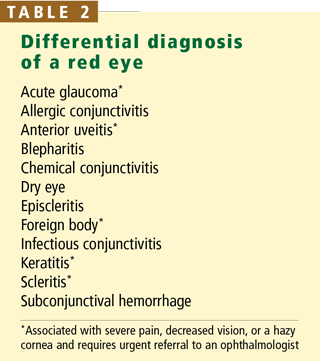
Other causes of a red eye. Many patients with a red eye have conjunctivitis, but other conditions can also present in a similar manner. Whether a patient has a serious vision-threatening condition (eg, acute-angle closure glaucoma, microbial keratitis, or anterior uveitis) can usually be determined with a focused ophthalmologic history and physical examination. Any alarming clinical features such as severe pain, decreased vision, or a hazy cornea in a patient with a red eye should alert the clinician to a more serious condition and prompt a referral to an ophthalmologist for an urgent evaluation (Table 2). A complete review for internists on how to manage a red eye was recently published in this journal.21
TREATMENT
Systemic treatment needed for gonococcal or chlamydial infections
The US Centers for Disease Control and Prevention recommend treating gonococcal conjunctivitis with ceftriaxone (Rocephin) 1 g in a single intramuscular dose plus topical saline lavage of the eye.22,23 Sexual partners of the patient should be referred for evaluation and treatment, as should mothers of affected neonates and the mother’s sexual partners.
Chlamydial conjunctivitis is also treated with systemic antibiotics. In neonates, the treatment is the same as for pneumonia caused by C trachomatis: erythromycin taken orally for 14 days. In adults, it can be treated with a single oral dose of azithromycin (Zithromax) 1 g.
Some authors recommend that H influenzae conjunctivitis also be treated with systemic antibiotics, as it is frequently associated with concurrent otitis media.24
Topical antibiotics hasten cure
Other types of bacterial conjunctivitis usually resolve spontaneously: early placebo-controlled studies found that more than 70% of cases of bacterial conjunctivitis resolve within 8 days.25 However, treatment with antibacterial agents leads to a faster clinical and microbiological cure26 and reduces the chance of rare complications27 and of transmitting the infection.
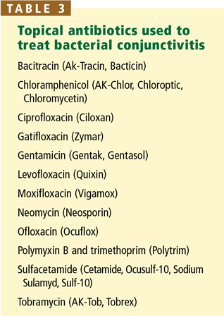
Is culture necessary?
A predictable set of organisms accounts for most cases of bacterial conjunctivitis in outpatients, so many physicians start therapy empirically without culturing the conjunctiva. But in the hospital the organisms and their antibiotic resistance patterns are more varied, so culturing the conjunctiva before starting broad-spectrum therapy may be warranted.15 For an outpatient with possible hyperacute conjunctivitis, it is reasonable to perform a Gram stain in the office if the facilities exist, but it is not essential because urgent referral to an ophthalmologist is warranted regardless of the results to rule out corneal involvement.
Unfortunately, antibiotic resistance is increasing even among outpatients. Susceptibility of the most common ocular pathogens to ophthalmic antimicrobial agents has dropped dramatically: S pneumoniae and S aureus have developed high rates of resistance.30 Recent data also suggest that treatment with topical ophthalmic antibiotics can induce resistance among colonizing bacteria in nonocular locations.31 Widespread systemic treatment with azithromycin or tetracycline for control of endemic trachoma in two villages in Nepal resulted in increased rates of antibiotic resistance among nasopharyngeal isolates of S pneumoniae. S aureus is developing resistance to methicillin and to fluoroquinolones, such as levofloxacin (Levaquin).32,33 But fluoroquinolones are still effective against most bacteria that cause conjunctivitis or keratitis, and because they penetrate the cornea well, they should be used if clinical features suggest corneal involvement. Remember also that most patients recover without treatment even if the organism has appreciable antibiotic resistance.28
Corticosteroids should be avoided
Although corticosteroid drops (either alone or combined with antibiotic drops) may quickly relieve symptoms, some conditions that present as a red eye with watery discharge, such as herpetic keratitis, worsen with corticosteroid use. We recommend that internists avoid prescribing corticosteroid drops.
Remove contact lenses, replace eye drops
Contact lenses should be taken out until an infection is completely resolved. Disposable lenses should be thrown away. Nondisposable lenses should be cleaned thoroughly as recommended by the manufacturer, and a new lens case should be used.
Patients who use prescription eye drops for glaucoma should continue to use them, but the bottles should be replaced in case they have been contaminated by inadvertent contact with the eye.
Over-the-counter lubricating eye drops may be continued if desired, but a fresh bottle or vial should be used.
WHEN TO REFER
Red flags indicating that a patient may have a serious vision-threatening condition that requires urgent referral to an ophthalmologist include severe eye pain or headache, photophobia, decreased vision, or contact lens use. Patients with hyperacute cases should also be referred at once to rule out corneal involvement, although the internist should start treatment for gonorrhea. In addition, patients with apparent bacterial conjunctivitis that does not improve after 24 hours of antibiotic treatment should also be referred to an ophthalmologist.
- Tipple MA, Beem MO, Saxon EM. Clinical characteristics of the afebrile pneumonia associated with Chlamydia trachomatis infection in infants less than 6 months of age. Pediatrics 1979; 63:192–197.
- De Toledo AR, Chandler JW. Conjunctivitis of the newborn. Infect Dis Clin North Am 1992; 6:807–813.
- Buznach N, Dagan R, Greenberg D. Clinical and bacterial characteristics of acute bacterial conjunctivitis in children in the antibiotic resistance era. Pediatr Infect Dis J 2005; 24:823–828.
- Bodor FF. Conjunctivitis-otitis syndrome. Pediatrics 1982; 69:695–698.
- Crum NF, Barrozo CP, Chapman FA, Ryan MA, Russell KL. An outbreak of conjunctivitis due to a novel unencapsulated Streptococcus pneumoniae among military trainees. Clin Infect Dis 2004; 39:1148–1154.
- Gigliotti F, Williams WT, Hayden FG, et al. Etiology of acute conjunctivitis in children. J Pediatr 1981; 98:531–536.
- Weiss A, Brinser JH, Nazar-Stewart V. Acute conjunctivitis in childhood. J Pediatr 1993; 122:10–14.
- Block SL, Hedrick J, Tyler R, et al. Increasing bacterial resistance in pediatric acute conjunctivitis (1997–1998). Antimicrob Agents Chemother 2000; 44:1650–1654.
- Singer TR, Isenberg SJ, Apt L. Conjunctival anaerobic and aerobic bacterial flora in paediatric versus adult subjects. Br J Ophthalmol 1998; 72:448–451.
- Kato T, Hayasaka S. Methicillin-resistant Staphylococcus aureus and methicillin-resistant coagulase-negative staphylococci from conjunctivas of preoperative patients. Jpn J Ophthalmol 1998; 42:461–465.
- Nakata K, Inoue Y, Harada J, et al. A high incidence of Staphylococcus aureus colonization in the external eyes of patients with atopic dermatitis. Ophthalmology 2000; 107:2167–2171.
- Kluytmans J, van Belkum A, Verbrugh H. Nasal carriage of Staphylococcus aureus: epidemiology, underlying mechanisms, and associated risks. Clin Microbiol Rev 1997; 10:505–520.
- Kowalski RP, Karenchak LM, Romanowski EG. Infectious disease: changing antibiotic susceptibility. Ophthalmol Clin North Am 2003; 16:1–9.
- Haas J, Larson E, Ross B, See B, Saiman L. Epidemiology and diagnosis of hospital acquired conjunctivitis among neonatal intensive care unit patients. Pediatr Infect Dis J 2005; 24:586–589.
- Tarabishy AB, Hall GS, Procop GW, Jeng BH. Bacterial culture isolates from hospitalized pediatric patients with conjunctivitis. Am J Ophthalmol 2006; 142:678–680.
- Limberg MB. A review of bacterial keratitis and bacterial conjunctivitis. Am J Ophthalmol 1991; 112:2S–9S.
- Rietveld RP, van Weert HC, ter Riet G, Bindels PJ. Diagnostic impact of signs and symptoms in acute infectious conjunctivitis: systematic literature search. BMJ 2003; 327:789.
- Rietveld RP, ter Riet G, Bindels PJ, Sloos JH, van Weert HC. Predicting bacterial cause in infectious conjunctivitis: cohort study on informativeness of combinations of signs and symptoms. BMJ 2004; 329:206–210.
- Syed NA, Hyndiuk RA. Infectious conjunctivitis. Infect Dis Clin North Am 1992; 6:789–805.
- Azar MJ, Dhaliwal DK, Bower KS, Kowalski RP, Gordon YJ. Possible consequences of shaking hands with your patients with epidemic keratoconjunctivitis. Am J Ophthalmol 1996; 121:711–712.
- Galor A, Jeng BH. The red eye: a primer for the internist. Cleve Clin J Med 2008; 75:137–144.
- Centers for Disease Control and Prevention,Workowski KA, Berman SM. Sexually transmitted diseases treatment guidelines, 2006. MMWR Recomm Rep 2006; 55( RR11):1–94. http://www.cdc.gov/mmwr/preview/mmwrhtml/rr5511a1.htm. Erratum in: MMWR Recomm Rep 2006; 55:997. Accessed 4/30/08.
- Haimovici R, Roussel TJ. Treatment of gonococcal conjunctivitis with single-dose intramuscular ceftriaxone. Am J Ophthalmol 1989; 107:511–514.
- Bodor FF. Systemic antibiotics for treatment of the conjunctivitis-otitis media syndrome. Pediatr Infect Dis J 1989; 8:287–290.
- Gigliotti F, Hendley JO, Morgan J, Michaels R, Dickens M, Lohr J. Efficacy of topical antibiotic therapy in acute conjunctivitis in children. J Pediatr 1984; 104:623–626.
- Sheikh A, Hurwitz B. Topical antibiotics for acute bacterial conjunctivitis: Cochrane systematic review and meta-analysis update. Br J Gen Pract 2005; 55:962–964.
- Aung T, Chan TK. Nosocomial Klebsiella pneumoniae conjunctivitis resulting in infectious keratitis and bilateral corneal perforation. Cornea 1998; 17:558–561.
- Baum J, Barza M. The evolution of antibiotic therapy for bacterial conjunctivitis and keratitis: 1970–2000. Cornea 2000; 19:659–672.
- Schlech BA, Alfonso E. Overview of the potency of moxifloxacin ophthalmic solution 0.5% (VIGAMOX). Surv Ophthalmol 2005; 50:S7–S15.
- Chalita MR, Höfling-Lima AL, Paranhos A, Schor P, Belfort R. Shifting trends in in vitro antibiotic susceptibilities for common ocular isolates during a period of 15 years. Am J Ophthalmol 2004; 137:43–51.
- Gaynor BD, Chidambaram JD, Cevallos V, et al. Topical ocular antibiotics induce bacterial resistance at extraocular sites. Br J Ophthalmol 2005; 89:1097–1099.
- Marangon FB, Miller D, Muallem MS, Romano AC, Alfonso EC. Ciprofloxacin and levofloxacin resistance among methicillin-sensitive Staphylococcus aureus isolates from keratitis and conjunctivitis. Am J Ophthalmol 2004; 137:453–458.
- Goldstein MH, Kowalski RP, Gordon YJ. Emerging fluoroquinolone resistance in bacterial keratitis: a 5-year review. Ophthalmology 1999; 106:1313–1318.
Bacterial conjunctivitis is common in children and adults presenting with a red eye. Although most cases are self-limited, appropriate antimicrobial treatment accelerates resolution and reduces complications. It is critical to differentiate bacterial conjunctivitis from other types of conjunctivitis and more serious vision-threatening conditions so that patients can be appropriately treated and, if necessary, referred to an ophthalmologist.
This paper is an overview of how to diagnose and manage bacterial conjunctivitis for the office-based internist.
CAUSES VARY BY AGE
In neonates, conjunctivitis is predominantly bacterial, and the most common organism is Chlamydia trachomatis. Chlamydial conjuctivitis typically presents with purulent unilateral or bilateral discharge about a week after birth in children born to mothers who have cervical chlamydial infection. Many infants with chlamydial conjunctivitis develop chlamydial pneumonitis: approximately 50% of infants with chlamydial pneumonitis have concurrent conjunctivitis or a recent history of conjunctivitis.1
Neisseria gonorrhoeae is a rare cause of neonatal conjunctivitis. The onset is somewhat earlier than in chlamydial conjunctivitis, ie, in the first week of life, and this organism classically causes severe “hyperacute” conjunctivitis with profuse discharge and may result in corneal involvement and perforation. Routine antibiotic prophylaxis at birth has markedly reduced its incidence and complications.
Other bacteria that can cause neonatal conjunctivitis include Staphylococcus aureus, Haemophilus influenzae, and Streptococcus pneumoniae.2
In children, bacterial conjunctivitis is most often caused by H influenzae or S pneumoniae, which accounted for 29% and 20% of cases, respectively, in a prospective study in Israel.3 Whether patients had been vaccinated against H influenzae in this study is unclear.
H influenzae conjunctivitis spreads easily in schools and households. It is associated with concurrent upper respiratory tract infections and otitis media (conjunctivitis-otitis syndrome): 45% to 73% of patients with purulent conjunctivitis also have ipsilateral otitis media.4
S pneumoniae, the second most common cause of bacterial conjunctivitis in children, is a common cause in epidemic outbreaks among young adults. Newly described unencapsulated pneumococcal strains caused outbreaks that affected 92 recruits at a military training facility and 100 students at Dartmouth University.5S pneumoniae is also associated with conjunctivitis-otitis syndrome, accounting for approximately 23% of culture-proven cases.4
Moraxella species, S aureus, and coagulase-negative staphylococci are less common causes of bacterial conjunctivitis in children.6–8
In adults, the most common causes of bacterial conjunctivitis are S aureus and H influenzae. Conjunctivitis caused by S aureus is often recurrent and associated with chronic ble-pharoconjunctivitis (inflammation of the eyelid and conjunctiva). The conjunctivae are colonized by S aureus in 3.8% to 6.3% of healthy adults.9–11 In addition, about 20% of people normally harbor S aureus continually in the nasal passages, and another 60% harbor it intermittently; in both cases, the bacteria may be a reservoir for recurrent ocular infection.12
Other organisms that commonly cause conjunctivitis in adults are S pneumoniae, coagulase-negative staphylococci, and Moraxella and Acinetobacter species.13
HOSPITAL-ACQUIRED CONJUNCTIVITIS
Little has been published about hospital-acquired conjunctivitis. In a neonatal intensive care unit, the most common organisms isolated in patients with conjunctivitis were coagulase-negative staphylococci, S aureus, and Klebsiella species.14 We found that about 30% of children who developed bacterial conjunctivitis after 2 days of hospitalization at Cleveland Clinic harbored gram-negative organisms. In addition, in patients who were found to have conjunctivitis caused by Staphylococcus species, the rate of methicillin resistance was higher in those hospitalized for more than 2 days than those with Staphylococcus species who were hospitalized for less than 2 days. This suggests that the bacterial pathogens encountered in hospitalized children with conjunctivitis differ from those found in the outpatient setting.15
EYE DISORDERS PREDISPOSE TO INFECTION
The conjunctiva is a transparent membrane that covers the sclera and lines the inside of the eyelid. It is a protective barrier against invading pathogens and lubricates the ocular surface by secreting components of the tear film (although the lacrimal glands contribute more to the tear film).
Several unique anatomic and functional features of the ocular surface help prevent bacterial infection in the healthy eye. The tear film contains secreted immunoglobulins, lysozyme, complement, and multiple antibacterial enzymes, and it is continuously being flushed and renewed, creating a physically and immunologically adverse environment for bacterial growth.
Disorders involving the eyelids or tear film such as chronic dry eye and lagophthalmos (in which the eye cannot close completely) may predispose the eye to frequent infections. Also, an adjacent focus of infection, such as inflammation of the lacrimal gland (dacryocystitis), can cause recurrent or chronic conjunctivitis.16
CLINICAL FEATURES OF BACTERIAL CONJUNCTIVITIS
Bacterial conjunctivitis is commonly classified according to its clinical presentation: hyperacute, acute, or chronic.
Hyperacute bacterial conjunctivitis presents with the rapid onset of conjunctival injection, eyelid edema, severe, continuous, and copious purulent discharge, chemosis, and discomfort or pain.
N gonorrhoeae is a frequent cause of hyperacute conjunctivitis in sexually active patients; the patient usually also has N gonorrhoeae genital infection, which is often asymptomatic. N gonorrhoeae conjunctivitis also occurs in neonates, as noted above. The cornea is frequently involved, and untreated cases can progress within days to corneal perforation. Unlike most other types of conjunctivitis, gonococcal conjunctivitis should be treated as a systemic disease, with both systemic and topical antibacterial therapy.2
Acute bacterial conjunctivitis typically presents abruptly with red eye and purulent drainage without significant eye pain, discomfort, or photophobia. Visual acuity does not typically decrease unless large amounts of discharge intermittently obscure vision.
Chronic bacterial conjunctivitis, ie, red eye with purulent discharge persisting for longer than a few weeks, is generally caused by Chlamydia trachomatis or is associated with a nidus for infection such as in dacryocystitis.
BACTERIAL CONJUNCTIVITIS VS OTHER CAUSES OF A RED EYE
Clinical signs and symptoms of infection with certain organisms have been extensively described, but a meta-analysis17 found no evidence that these textbook features help to distinguish between bacterial and viral causes of conjunctivitis. Instead, whether a bacterial cause was likely was best determined from just three features: having both eyes glued shut in the morning had an odds ratio of 15:1 in predicting a positive bacterial culture, and either itching or previous conjunctivitis made a bacterial cause less likely.18
In general, however, viral conjunctivitis typically presents as an itchy red eye with mild watery discharge. Many patients have signs and symptoms of a viral upper respiratory tract infection (eg, cough, runny nose, congestion) and have been in contact with a sick person. Ipsilateral preauricular lymphadenopathy is common in viral conjunctivitis and strongly suggests this diagnosis.19
Viral conjunctivitis is often epidemic and is easily contagious. Several epidemics have been traced to eyecare facilities. Adenovirus conjunctivitis is extremely contagious and can be transmitted both between people and via inanimate objects; it has been reported to be spread by workers in health care facilities.20
Allergic conjunctivitis is also common. Patients typically report itching and redness of both eyes in response to an allergen exposure. Other allergic symptoms may be present, such as allergic rhinosinusitis, asthma, or atopic dermatitis in response to seasonal or perennial environmental allergens.
Other causes of a red eye. Many patients with a red eye have conjunctivitis, but other conditions can also present in a similar manner. Whether a patient has a serious vision-threatening condition (eg, acute-angle closure glaucoma, microbial keratitis, or anterior uveitis) can usually be determined with a focused ophthalmologic history and physical examination. Any alarming clinical features such as severe pain, decreased vision, or a hazy cornea in a patient with a red eye should alert the clinician to a more serious condition and prompt a referral to an ophthalmologist for an urgent evaluation (Table 2). A complete review for internists on how to manage a red eye was recently published in this journal.21
TREATMENT
Systemic treatment needed for gonococcal or chlamydial infections
The US Centers for Disease Control and Prevention recommend treating gonococcal conjunctivitis with ceftriaxone (Rocephin) 1 g in a single intramuscular dose plus topical saline lavage of the eye.22,23 Sexual partners of the patient should be referred for evaluation and treatment, as should mothers of affected neonates and the mother’s sexual partners.
Chlamydial conjunctivitis is also treated with systemic antibiotics. In neonates, the treatment is the same as for pneumonia caused by C trachomatis: erythromycin taken orally for 14 days. In adults, it can be treated with a single oral dose of azithromycin (Zithromax) 1 g.
Some authors recommend that H influenzae conjunctivitis also be treated with systemic antibiotics, as it is frequently associated with concurrent otitis media.24
Topical antibiotics hasten cure
Other types of bacterial conjunctivitis usually resolve spontaneously: early placebo-controlled studies found that more than 70% of cases of bacterial conjunctivitis resolve within 8 days.25 However, treatment with antibacterial agents leads to a faster clinical and microbiological cure26 and reduces the chance of rare complications27 and of transmitting the infection.
Is culture necessary?
A predictable set of organisms accounts for most cases of bacterial conjunctivitis in outpatients, so many physicians start therapy empirically without culturing the conjunctiva. But in the hospital the organisms and their antibiotic resistance patterns are more varied, so culturing the conjunctiva before starting broad-spectrum therapy may be warranted.15 For an outpatient with possible hyperacute conjunctivitis, it is reasonable to perform a Gram stain in the office if the facilities exist, but it is not essential because urgent referral to an ophthalmologist is warranted regardless of the results to rule out corneal involvement.
Unfortunately, antibiotic resistance is increasing even among outpatients. Susceptibility of the most common ocular pathogens to ophthalmic antimicrobial agents has dropped dramatically: S pneumoniae and S aureus have developed high rates of resistance.30 Recent data also suggest that treatment with topical ophthalmic antibiotics can induce resistance among colonizing bacteria in nonocular locations.31 Widespread systemic treatment with azithromycin or tetracycline for control of endemic trachoma in two villages in Nepal resulted in increased rates of antibiotic resistance among nasopharyngeal isolates of S pneumoniae. S aureus is developing resistance to methicillin and to fluoroquinolones, such as levofloxacin (Levaquin).32,33 But fluoroquinolones are still effective against most bacteria that cause conjunctivitis or keratitis, and because they penetrate the cornea well, they should be used if clinical features suggest corneal involvement. Remember also that most patients recover without treatment even if the organism has appreciable antibiotic resistance.28
Corticosteroids should be avoided
Although corticosteroid drops (either alone or combined with antibiotic drops) may quickly relieve symptoms, some conditions that present as a red eye with watery discharge, such as herpetic keratitis, worsen with corticosteroid use. We recommend that internists avoid prescribing corticosteroid drops.
Remove contact lenses, replace eye drops
Contact lenses should be taken out until an infection is completely resolved. Disposable lenses should be thrown away. Nondisposable lenses should be cleaned thoroughly as recommended by the manufacturer, and a new lens case should be used.
Patients who use prescription eye drops for glaucoma should continue to use them, but the bottles should be replaced in case they have been contaminated by inadvertent contact with the eye.
Over-the-counter lubricating eye drops may be continued if desired, but a fresh bottle or vial should be used.
WHEN TO REFER
Red flags indicating that a patient may have a serious vision-threatening condition that requires urgent referral to an ophthalmologist include severe eye pain or headache, photophobia, decreased vision, or contact lens use. Patients with hyperacute cases should also be referred at once to rule out corneal involvement, although the internist should start treatment for gonorrhea. In addition, patients with apparent bacterial conjunctivitis that does not improve after 24 hours of antibiotic treatment should also be referred to an ophthalmologist.
- Tipple MA, Beem MO, Saxon EM. Clinical characteristics of the afebrile pneumonia associated with Chlamydia trachomatis infection in infants less than 6 months of age. Pediatrics 1979; 63:192–197.
- De Toledo AR, Chandler JW. Conjunctivitis of the newborn. Infect Dis Clin North Am 1992; 6:807–813.
- Buznach N, Dagan R, Greenberg D. Clinical and bacterial characteristics of acute bacterial conjunctivitis in children in the antibiotic resistance era. Pediatr Infect Dis J 2005; 24:823–828.
- Bodor FF. Conjunctivitis-otitis syndrome. Pediatrics 1982; 69:695–698.
- Crum NF, Barrozo CP, Chapman FA, Ryan MA, Russell KL. An outbreak of conjunctivitis due to a novel unencapsulated Streptococcus pneumoniae among military trainees. Clin Infect Dis 2004; 39:1148–1154.
- Gigliotti F, Williams WT, Hayden FG, et al. Etiology of acute conjunctivitis in children. J Pediatr 1981; 98:531–536.
- Weiss A, Brinser JH, Nazar-Stewart V. Acute conjunctivitis in childhood. J Pediatr 1993; 122:10–14.
- Block SL, Hedrick J, Tyler R, et al. Increasing bacterial resistance in pediatric acute conjunctivitis (1997–1998). Antimicrob Agents Chemother 2000; 44:1650–1654.
- Singer TR, Isenberg SJ, Apt L. Conjunctival anaerobic and aerobic bacterial flora in paediatric versus adult subjects. Br J Ophthalmol 1998; 72:448–451.
- Kato T, Hayasaka S. Methicillin-resistant Staphylococcus aureus and methicillin-resistant coagulase-negative staphylococci from conjunctivas of preoperative patients. Jpn J Ophthalmol 1998; 42:461–465.
- Nakata K, Inoue Y, Harada J, et al. A high incidence of Staphylococcus aureus colonization in the external eyes of patients with atopic dermatitis. Ophthalmology 2000; 107:2167–2171.
- Kluytmans J, van Belkum A, Verbrugh H. Nasal carriage of Staphylococcus aureus: epidemiology, underlying mechanisms, and associated risks. Clin Microbiol Rev 1997; 10:505–520.
- Kowalski RP, Karenchak LM, Romanowski EG. Infectious disease: changing antibiotic susceptibility. Ophthalmol Clin North Am 2003; 16:1–9.
- Haas J, Larson E, Ross B, See B, Saiman L. Epidemiology and diagnosis of hospital acquired conjunctivitis among neonatal intensive care unit patients. Pediatr Infect Dis J 2005; 24:586–589.
- Tarabishy AB, Hall GS, Procop GW, Jeng BH. Bacterial culture isolates from hospitalized pediatric patients with conjunctivitis. Am J Ophthalmol 2006; 142:678–680.
- Limberg MB. A review of bacterial keratitis and bacterial conjunctivitis. Am J Ophthalmol 1991; 112:2S–9S.
- Rietveld RP, van Weert HC, ter Riet G, Bindels PJ. Diagnostic impact of signs and symptoms in acute infectious conjunctivitis: systematic literature search. BMJ 2003; 327:789.
- Rietveld RP, ter Riet G, Bindels PJ, Sloos JH, van Weert HC. Predicting bacterial cause in infectious conjunctivitis: cohort study on informativeness of combinations of signs and symptoms. BMJ 2004; 329:206–210.
- Syed NA, Hyndiuk RA. Infectious conjunctivitis. Infect Dis Clin North Am 1992; 6:789–805.
- Azar MJ, Dhaliwal DK, Bower KS, Kowalski RP, Gordon YJ. Possible consequences of shaking hands with your patients with epidemic keratoconjunctivitis. Am J Ophthalmol 1996; 121:711–712.
- Galor A, Jeng BH. The red eye: a primer for the internist. Cleve Clin J Med 2008; 75:137–144.
- Centers for Disease Control and Prevention,Workowski KA, Berman SM. Sexually transmitted diseases treatment guidelines, 2006. MMWR Recomm Rep 2006; 55( RR11):1–94. http://www.cdc.gov/mmwr/preview/mmwrhtml/rr5511a1.htm. Erratum in: MMWR Recomm Rep 2006; 55:997. Accessed 4/30/08.
- Haimovici R, Roussel TJ. Treatment of gonococcal conjunctivitis with single-dose intramuscular ceftriaxone. Am J Ophthalmol 1989; 107:511–514.
- Bodor FF. Systemic antibiotics for treatment of the conjunctivitis-otitis media syndrome. Pediatr Infect Dis J 1989; 8:287–290.
- Gigliotti F, Hendley JO, Morgan J, Michaels R, Dickens M, Lohr J. Efficacy of topical antibiotic therapy in acute conjunctivitis in children. J Pediatr 1984; 104:623–626.
- Sheikh A, Hurwitz B. Topical antibiotics for acute bacterial conjunctivitis: Cochrane systematic review and meta-analysis update. Br J Gen Pract 2005; 55:962–964.
- Aung T, Chan TK. Nosocomial Klebsiella pneumoniae conjunctivitis resulting in infectious keratitis and bilateral corneal perforation. Cornea 1998; 17:558–561.
- Baum J, Barza M. The evolution of antibiotic therapy for bacterial conjunctivitis and keratitis: 1970–2000. Cornea 2000; 19:659–672.
- Schlech BA, Alfonso E. Overview of the potency of moxifloxacin ophthalmic solution 0.5% (VIGAMOX). Surv Ophthalmol 2005; 50:S7–S15.
- Chalita MR, Höfling-Lima AL, Paranhos A, Schor P, Belfort R. Shifting trends in in vitro antibiotic susceptibilities for common ocular isolates during a period of 15 years. Am J Ophthalmol 2004; 137:43–51.
- Gaynor BD, Chidambaram JD, Cevallos V, et al. Topical ocular antibiotics induce bacterial resistance at extraocular sites. Br J Ophthalmol 2005; 89:1097–1099.
- Marangon FB, Miller D, Muallem MS, Romano AC, Alfonso EC. Ciprofloxacin and levofloxacin resistance among methicillin-sensitive Staphylococcus aureus isolates from keratitis and conjunctivitis. Am J Ophthalmol 2004; 137:453–458.
- Goldstein MH, Kowalski RP, Gordon YJ. Emerging fluoroquinolone resistance in bacterial keratitis: a 5-year review. Ophthalmology 1999; 106:1313–1318.
KEY POINTS
- Viral conjunctivitis typically presents as an itchy red eye with mild watery discharge. Many patients have signs and symptoms of a viral upper respiratory tract infection (eg, cough, runny nose, congestion) and have been in contact with a sick person.
- Having both eyes glued shut in the morning had an odds ratio of 15:1 in predicting a positive bacterial culture, whereas either itching or previous conjunctivitis made a bacterial cause less likely.
- In adults, Neisseria gonorrhoeae causes hyperacute conjunctivitis and is associated with concurrent, often asymptomatic genital infection. Gonococcal conjunctivitis should be treated with a single dose of ceftriaxone (Rocephin) 1 g intramuscularly plus saline eye-washing.
- Corticosteroid drops should not be prescribed for a red eye before consultation with an ophthalmologist because these drops may worsen some conditions.
Generalized pruritus after a beach vacation
A 25-year-old man presents with a 2-month history of generalized itching that began 5 weeks after returning from a trip to a beach in Brazil, during which he had sexual relations without protection. He has already been treated with topical steroids and antihistamines, with no improvement.

Q: What is the most likely diagnosis?
- Herpes simplex type 2
- Acute trypanosomiasis
- Syphilis
- Scabies
- Papular urticaria
A: The most likely diagnosis is scabies, an intensely pruritic skin infestation caused by the host-specific mite Sarcoptes scabiei var hominis.1 Herpes simplex type 2 infection presents with clustered vesicles, not nodules, on an erythematous base in the genital area, without generalized pruritus. Acute trypanosomiasis involves malaise, fever, vomiting, diarrhea, anorexia, rash, tachycardia, and even generalized lymphadenopathy and meningeal irritation, but generalized itching is not typical. The location and morphology of the patient’s lesions are not consistent with syphilis. Topical steroids and oral antihistamines should have improved the pruritus of papular urticaria.
A GLOBAL PUBLIC HEALTH PROBLEM
Scabies is a worldwide public health problem, affecting people of all ages, races, and socioeconomic groups.1 Overcrowding, delayed diagnosis and treatment, and poor public education contribute to the prevalence of scabies in both industrialized and nonindustrialized nations.2 Prevalence rates are higher in children and people who are sexually active.3
Sexual transmission is by close skin-to-skin contact. Poor sensory perception in conditions such as leprosy and compromised immunity due to organ transplantation, human immunodeficiency virus infection, or old age increase the risk for the crusted variant of scabies.2 Patients with the crusted variant tend to present with clinically atypical lesions, and because of this they are often misdiag-nosed, thus delaying treatment and elevating the risk of local epidemics.
CLINICAL ASPECTS
Scabies can mimic a broad range of skin diseases. Patients present with intense itching that is worse at night. The face and neck are rarely affected. The pathognomonic signs of scabies are burrows, erythematous papules, and generalized pruritus (also on non-infested skin) with nocturnal predominance.3 Reddish to brownish extremely pruritic nodules of 2 to 20 mm in diameter may be also present on the genitalia (more commonly in males than in females), buttocks, groin, and axillary regions. Patients usually have secondary papules, pustules, vesicles, and excoriations.
DIAGNOSIS
Every patient with intense pruritus should be suspected of having scabies, but especially if a family member reports similar symptoms.3 A diagnosis can be made clinically if a burrow is detected at a typical predilection site and if the lesion itches severely. In this case, even a single burrow is pathognomonic.2 The diagnosis is confirmed by light-microscopic identification of mites, larvae, ova, or scybala (fecal pellets) in skin scrapings.1
TREATMENT
Treatment includes a scabicidal agent, an antipruritic agent such as a sedating antihistamine, and an appropriate antimicrobial agent in cases of secondary infection. Permethrin (Acticin), a 5% synthetic pyrethroid cream, is an excellent scabicide and is the preferred treatment.1 All family members and close contacts must be evaluated and treated, even if they do not have symptoms.1
- Chosidow O. Clinical practices. Scabies. N Engl J Med 2006; 354:1718–1727.
- Heukelbach J, Feldmeier H. Scabies. Lancet 2006; 367:1767–1774.
- Hengge UR, Currie BJ, Jäger G, Lupi O, Schwartz RA. Scabies: a ubiquitous neglected skin disease. Lancet Infect Dis 2006; 6:769–779.
A 25-year-old man presents with a 2-month history of generalized itching that began 5 weeks after returning from a trip to a beach in Brazil, during which he had sexual relations without protection. He has already been treated with topical steroids and antihistamines, with no improvement.
Q: What is the most likely diagnosis?
- Herpes simplex type 2
- Acute trypanosomiasis
- Syphilis
- Scabies
- Papular urticaria
A: The most likely diagnosis is scabies, an intensely pruritic skin infestation caused by the host-specific mite Sarcoptes scabiei var hominis.1 Herpes simplex type 2 infection presents with clustered vesicles, not nodules, on an erythematous base in the genital area, without generalized pruritus. Acute trypanosomiasis involves malaise, fever, vomiting, diarrhea, anorexia, rash, tachycardia, and even generalized lymphadenopathy and meningeal irritation, but generalized itching is not typical. The location and morphology of the patient’s lesions are not consistent with syphilis. Topical steroids and oral antihistamines should have improved the pruritus of papular urticaria.
A GLOBAL PUBLIC HEALTH PROBLEM
Scabies is a worldwide public health problem, affecting people of all ages, races, and socioeconomic groups.1 Overcrowding, delayed diagnosis and treatment, and poor public education contribute to the prevalence of scabies in both industrialized and nonindustrialized nations.2 Prevalence rates are higher in children and people who are sexually active.3
Sexual transmission is by close skin-to-skin contact. Poor sensory perception in conditions such as leprosy and compromised immunity due to organ transplantation, human immunodeficiency virus infection, or old age increase the risk for the crusted variant of scabies.2 Patients with the crusted variant tend to present with clinically atypical lesions, and because of this they are often misdiag-nosed, thus delaying treatment and elevating the risk of local epidemics.
CLINICAL ASPECTS
Scabies can mimic a broad range of skin diseases. Patients present with intense itching that is worse at night. The face and neck are rarely affected. The pathognomonic signs of scabies are burrows, erythematous papules, and generalized pruritus (also on non-infested skin) with nocturnal predominance.3 Reddish to brownish extremely pruritic nodules of 2 to 20 mm in diameter may be also present on the genitalia (more commonly in males than in females), buttocks, groin, and axillary regions. Patients usually have secondary papules, pustules, vesicles, and excoriations.
DIAGNOSIS
Every patient with intense pruritus should be suspected of having scabies, but especially if a family member reports similar symptoms.3 A diagnosis can be made clinically if a burrow is detected at a typical predilection site and if the lesion itches severely. In this case, even a single burrow is pathognomonic.2 The diagnosis is confirmed by light-microscopic identification of mites, larvae, ova, or scybala (fecal pellets) in skin scrapings.1
TREATMENT
Treatment includes a scabicidal agent, an antipruritic agent such as a sedating antihistamine, and an appropriate antimicrobial agent in cases of secondary infection. Permethrin (Acticin), a 5% synthetic pyrethroid cream, is an excellent scabicide and is the preferred treatment.1 All family members and close contacts must be evaluated and treated, even if they do not have symptoms.1
- Chosidow O. Clinical practices. Scabies. N Engl J Med 2006; 354:1718–1727.
- Heukelbach J, Feldmeier H. Scabies. Lancet 2006; 367:1767–1774.
- Hengge UR, Currie BJ, Jäger G, Lupi O, Schwartz RA. Scabies: a ubiquitous neglected skin disease. Lancet Infect Dis 2006; 6:769–779.
- Chosidow O. Clinical practices. Scabies. N Engl J Med 2006; 354:1718–1727.
- Heukelbach J, Feldmeier H. Scabies. Lancet 2006; 367:1767–1774.
- Hengge UR, Currie BJ, Jäger G, Lupi O, Schwartz RA. Scabies: a ubiquitous neglected skin disease. Lancet Infect Dis 2006; 6:769–779.