User login
CCJM delivers practical clinical articles relevant to internists, cardiologists, endocrinologists, and other specialists, all written by known experts.
Copyright © 2019 Cleveland Clinic. All rights reserved. The information provided is for educational purposes only. Use of this website is subject to the disclaimer and privacy policy.
gambling
compulsive behaviors
ammunition
assault rifle
black jack
Boko Haram
bondage
child abuse
cocaine
Daech
drug paraphernalia
explosion
gun
human trafficking
ISIL
ISIS
Islamic caliphate
Islamic state
mixed martial arts
MMA
molestation
national rifle association
NRA
nsfw
pedophile
pedophilia
poker
porn
pornography
psychedelic drug
recreational drug
sex slave rings
slot machine
terrorism
terrorist
Texas hold 'em
UFC
substance abuse
abuseed
abuseer
abusees
abuseing
abusely
abuses
aeolus
aeolused
aeoluser
aeoluses
aeolusing
aeolusly
aeoluss
ahole
aholeed
aholeer
aholees
aholeing
aholely
aholes
alcohol
alcoholed
alcoholer
alcoholes
alcoholing
alcoholly
alcohols
allman
allmaned
allmaner
allmanes
allmaning
allmanly
allmans
alted
altes
alting
altly
alts
analed
analer
anales
analing
anally
analprobe
analprobeed
analprobeer
analprobees
analprobeing
analprobely
analprobes
anals
anilingus
anilingused
anilinguser
anilinguses
anilingusing
anilingusly
anilinguss
anus
anused
anuser
anuses
anusing
anusly
anuss
areola
areolaed
areolaer
areolaes
areolaing
areolaly
areolas
areole
areoleed
areoleer
areolees
areoleing
areolely
areoles
arian
arianed
arianer
arianes
arianing
arianly
arians
aryan
aryaned
aryaner
aryanes
aryaning
aryanly
aryans
asiaed
asiaer
asiaes
asiaing
asialy
asias
ass
ass hole
ass lick
ass licked
ass licker
ass lickes
ass licking
ass lickly
ass licks
assbang
assbanged
assbangeded
assbangeder
assbangedes
assbangeding
assbangedly
assbangeds
assbanger
assbanges
assbanging
assbangly
assbangs
assbangsed
assbangser
assbangses
assbangsing
assbangsly
assbangss
assed
asser
asses
assesed
asseser
asseses
assesing
assesly
assess
assfuck
assfucked
assfucker
assfuckered
assfuckerer
assfuckeres
assfuckering
assfuckerly
assfuckers
assfuckes
assfucking
assfuckly
assfucks
asshat
asshated
asshater
asshates
asshating
asshatly
asshats
assholeed
assholeer
assholees
assholeing
assholely
assholes
assholesed
assholeser
assholeses
assholesing
assholesly
assholess
assing
assly
assmaster
assmastered
assmasterer
assmasteres
assmastering
assmasterly
assmasters
assmunch
assmunched
assmuncher
assmunches
assmunching
assmunchly
assmunchs
asss
asswipe
asswipeed
asswipeer
asswipees
asswipeing
asswipely
asswipes
asswipesed
asswipeser
asswipeses
asswipesing
asswipesly
asswipess
azz
azzed
azzer
azzes
azzing
azzly
azzs
babeed
babeer
babees
babeing
babely
babes
babesed
babeser
babeses
babesing
babesly
babess
ballsac
ballsaced
ballsacer
ballsaces
ballsacing
ballsack
ballsacked
ballsacker
ballsackes
ballsacking
ballsackly
ballsacks
ballsacly
ballsacs
ballsed
ballser
ballses
ballsing
ballsly
ballss
barf
barfed
barfer
barfes
barfing
barfly
barfs
bastard
bastarded
bastarder
bastardes
bastarding
bastardly
bastards
bastardsed
bastardser
bastardses
bastardsing
bastardsly
bastardss
bawdy
bawdyed
bawdyer
bawdyes
bawdying
bawdyly
bawdys
beaner
beanered
beanerer
beaneres
beanering
beanerly
beaners
beardedclam
beardedclamed
beardedclamer
beardedclames
beardedclaming
beardedclamly
beardedclams
beastiality
beastialityed
beastialityer
beastialityes
beastialitying
beastialityly
beastialitys
beatch
beatched
beatcher
beatches
beatching
beatchly
beatchs
beater
beatered
beaterer
beateres
beatering
beaterly
beaters
beered
beerer
beeres
beering
beerly
beeyotch
beeyotched
beeyotcher
beeyotches
beeyotching
beeyotchly
beeyotchs
beotch
beotched
beotcher
beotches
beotching
beotchly
beotchs
biatch
biatched
biatcher
biatches
biatching
biatchly
biatchs
big tits
big titsed
big titser
big titses
big titsing
big titsly
big titss
bigtits
bigtitsed
bigtitser
bigtitses
bigtitsing
bigtitsly
bigtitss
bimbo
bimboed
bimboer
bimboes
bimboing
bimboly
bimbos
bisexualed
bisexualer
bisexuales
bisexualing
bisexually
bisexuals
bitch
bitched
bitcheded
bitcheder
bitchedes
bitcheding
bitchedly
bitcheds
bitcher
bitches
bitchesed
bitcheser
bitcheses
bitchesing
bitchesly
bitchess
bitching
bitchly
bitchs
bitchy
bitchyed
bitchyer
bitchyes
bitchying
bitchyly
bitchys
bleached
bleacher
bleaches
bleaching
bleachly
bleachs
blow job
blow jobed
blow jober
blow jobes
blow jobing
blow jobly
blow jobs
blowed
blower
blowes
blowing
blowjob
blowjobed
blowjober
blowjobes
blowjobing
blowjobly
blowjobs
blowjobsed
blowjobser
blowjobses
blowjobsing
blowjobsly
blowjobss
blowly
blows
boink
boinked
boinker
boinkes
boinking
boinkly
boinks
bollock
bollocked
bollocker
bollockes
bollocking
bollockly
bollocks
bollocksed
bollockser
bollockses
bollocksing
bollocksly
bollockss
bollok
bolloked
bolloker
bollokes
bolloking
bollokly
bolloks
boner
bonered
bonerer
boneres
bonering
bonerly
boners
bonersed
bonerser
bonerses
bonersing
bonersly
bonerss
bong
bonged
bonger
bonges
bonging
bongly
bongs
boob
boobed
boober
boobes
boobies
boobiesed
boobieser
boobieses
boobiesing
boobiesly
boobiess
boobing
boobly
boobs
boobsed
boobser
boobses
boobsing
boobsly
boobss
booby
boobyed
boobyer
boobyes
boobying
boobyly
boobys
booger
boogered
boogerer
boogeres
boogering
boogerly
boogers
bookie
bookieed
bookieer
bookiees
bookieing
bookiely
bookies
bootee
booteeed
booteeer
booteees
booteeing
booteely
bootees
bootie
bootieed
bootieer
bootiees
bootieing
bootiely
booties
booty
bootyed
bootyer
bootyes
bootying
bootyly
bootys
boozeed
boozeer
boozees
boozeing
boozely
boozer
boozered
boozerer
boozeres
boozering
boozerly
boozers
boozes
boozy
boozyed
boozyer
boozyes
boozying
boozyly
boozys
bosomed
bosomer
bosomes
bosoming
bosomly
bosoms
bosomy
bosomyed
bosomyer
bosomyes
bosomying
bosomyly
bosomys
bugger
buggered
buggerer
buggeres
buggering
buggerly
buggers
bukkake
bukkakeed
bukkakeer
bukkakees
bukkakeing
bukkakely
bukkakes
bull shit
bull shited
bull shiter
bull shites
bull shiting
bull shitly
bull shits
bullshit
bullshited
bullshiter
bullshites
bullshiting
bullshitly
bullshits
bullshitsed
bullshitser
bullshitses
bullshitsing
bullshitsly
bullshitss
bullshitted
bullshitteded
bullshitteder
bullshittedes
bullshitteding
bullshittedly
bullshitteds
bullturds
bullturdsed
bullturdser
bullturdses
bullturdsing
bullturdsly
bullturdss
bung
bunged
bunger
bunges
bunging
bungly
bungs
busty
bustyed
bustyer
bustyes
bustying
bustyly
bustys
butt
butt fuck
butt fucked
butt fucker
butt fuckes
butt fucking
butt fuckly
butt fucks
butted
buttes
buttfuck
buttfucked
buttfucker
buttfuckered
buttfuckerer
buttfuckeres
buttfuckering
buttfuckerly
buttfuckers
buttfuckes
buttfucking
buttfuckly
buttfucks
butting
buttly
buttplug
buttpluged
buttpluger
buttpluges
buttpluging
buttplugly
buttplugs
butts
caca
cacaed
cacaer
cacaes
cacaing
cacaly
cacas
cahone
cahoneed
cahoneer
cahonees
cahoneing
cahonely
cahones
cameltoe
cameltoeed
cameltoeer
cameltoees
cameltoeing
cameltoely
cameltoes
carpetmuncher
carpetmunchered
carpetmuncherer
carpetmuncheres
carpetmunchering
carpetmuncherly
carpetmunchers
cawk
cawked
cawker
cawkes
cawking
cawkly
cawks
chinc
chinced
chincer
chinces
chincing
chincly
chincs
chincsed
chincser
chincses
chincsing
chincsly
chincss
chink
chinked
chinker
chinkes
chinking
chinkly
chinks
chode
chodeed
chodeer
chodees
chodeing
chodely
chodes
chodesed
chodeser
chodeses
chodesing
chodesly
chodess
clit
clited
cliter
clites
cliting
clitly
clitoris
clitorised
clitoriser
clitorises
clitorising
clitorisly
clitoriss
clitorus
clitorused
clitoruser
clitoruses
clitorusing
clitorusly
clitoruss
clits
clitsed
clitser
clitses
clitsing
clitsly
clitss
clitty
clittyed
clittyer
clittyes
clittying
clittyly
clittys
cocain
cocaine
cocained
cocaineed
cocaineer
cocainees
cocaineing
cocainely
cocainer
cocaines
cocaining
cocainly
cocains
cock
cock sucker
cock suckered
cock suckerer
cock suckeres
cock suckering
cock suckerly
cock suckers
cockblock
cockblocked
cockblocker
cockblockes
cockblocking
cockblockly
cockblocks
cocked
cocker
cockes
cockholster
cockholstered
cockholsterer
cockholsteres
cockholstering
cockholsterly
cockholsters
cocking
cockknocker
cockknockered
cockknockerer
cockknockeres
cockknockering
cockknockerly
cockknockers
cockly
cocks
cocksed
cockser
cockses
cocksing
cocksly
cocksmoker
cocksmokered
cocksmokerer
cocksmokeres
cocksmokering
cocksmokerly
cocksmokers
cockss
cocksucker
cocksuckered
cocksuckerer
cocksuckeres
cocksuckering
cocksuckerly
cocksuckers
coital
coitaled
coitaler
coitales
coitaling
coitally
coitals
commie
commieed
commieer
commiees
commieing
commiely
commies
condomed
condomer
condomes
condoming
condomly
condoms
coon
cooned
cooner
coones
cooning
coonly
coons
coonsed
coonser
coonses
coonsing
coonsly
coonss
corksucker
corksuckered
corksuckerer
corksuckeres
corksuckering
corksuckerly
corksuckers
cracked
crackwhore
crackwhoreed
crackwhoreer
crackwhorees
crackwhoreing
crackwhorely
crackwhores
crap
craped
craper
crapes
craping
craply
crappy
crappyed
crappyer
crappyes
crappying
crappyly
crappys
cum
cumed
cumer
cumes
cuming
cumly
cummin
cummined
cumminer
cummines
cumming
cumminged
cumminger
cumminges
cumminging
cummingly
cummings
cummining
cumminly
cummins
cums
cumshot
cumshoted
cumshoter
cumshotes
cumshoting
cumshotly
cumshots
cumshotsed
cumshotser
cumshotses
cumshotsing
cumshotsly
cumshotss
cumslut
cumsluted
cumsluter
cumslutes
cumsluting
cumslutly
cumsluts
cumstain
cumstained
cumstainer
cumstaines
cumstaining
cumstainly
cumstains
cunilingus
cunilingused
cunilinguser
cunilinguses
cunilingusing
cunilingusly
cunilinguss
cunnilingus
cunnilingused
cunnilinguser
cunnilinguses
cunnilingusing
cunnilingusly
cunnilinguss
cunny
cunnyed
cunnyer
cunnyes
cunnying
cunnyly
cunnys
cunt
cunted
cunter
cuntes
cuntface
cuntfaceed
cuntfaceer
cuntfacees
cuntfaceing
cuntfacely
cuntfaces
cunthunter
cunthuntered
cunthunterer
cunthunteres
cunthuntering
cunthunterly
cunthunters
cunting
cuntlick
cuntlicked
cuntlicker
cuntlickered
cuntlickerer
cuntlickeres
cuntlickering
cuntlickerly
cuntlickers
cuntlickes
cuntlicking
cuntlickly
cuntlicks
cuntly
cunts
cuntsed
cuntser
cuntses
cuntsing
cuntsly
cuntss
dago
dagoed
dagoer
dagoes
dagoing
dagoly
dagos
dagosed
dagoser
dagoses
dagosing
dagosly
dagoss
dammit
dammited
dammiter
dammites
dammiting
dammitly
dammits
damn
damned
damneded
damneder
damnedes
damneding
damnedly
damneds
damner
damnes
damning
damnit
damnited
damniter
damnites
damniting
damnitly
damnits
damnly
damns
dick
dickbag
dickbaged
dickbager
dickbages
dickbaging
dickbagly
dickbags
dickdipper
dickdippered
dickdipperer
dickdipperes
dickdippering
dickdipperly
dickdippers
dicked
dicker
dickes
dickface
dickfaceed
dickfaceer
dickfacees
dickfaceing
dickfacely
dickfaces
dickflipper
dickflippered
dickflipperer
dickflipperes
dickflippering
dickflipperly
dickflippers
dickhead
dickheaded
dickheader
dickheades
dickheading
dickheadly
dickheads
dickheadsed
dickheadser
dickheadses
dickheadsing
dickheadsly
dickheadss
dicking
dickish
dickished
dickisher
dickishes
dickishing
dickishly
dickishs
dickly
dickripper
dickrippered
dickripperer
dickripperes
dickrippering
dickripperly
dickrippers
dicks
dicksipper
dicksippered
dicksipperer
dicksipperes
dicksippering
dicksipperly
dicksippers
dickweed
dickweeded
dickweeder
dickweedes
dickweeding
dickweedly
dickweeds
dickwhipper
dickwhippered
dickwhipperer
dickwhipperes
dickwhippering
dickwhipperly
dickwhippers
dickzipper
dickzippered
dickzipperer
dickzipperes
dickzippering
dickzipperly
dickzippers
diddle
diddleed
diddleer
diddlees
diddleing
diddlely
diddles
dike
dikeed
dikeer
dikees
dikeing
dikely
dikes
dildo
dildoed
dildoer
dildoes
dildoing
dildoly
dildos
dildosed
dildoser
dildoses
dildosing
dildosly
dildoss
diligaf
diligafed
diligafer
diligafes
diligafing
diligafly
diligafs
dillweed
dillweeded
dillweeder
dillweedes
dillweeding
dillweedly
dillweeds
dimwit
dimwited
dimwiter
dimwites
dimwiting
dimwitly
dimwits
dingle
dingleed
dingleer
dinglees
dingleing
dinglely
dingles
dipship
dipshiped
dipshiper
dipshipes
dipshiping
dipshiply
dipships
dizzyed
dizzyer
dizzyes
dizzying
dizzyly
dizzys
doggiestyleed
doggiestyleer
doggiestylees
doggiestyleing
doggiestylely
doggiestyles
doggystyleed
doggystyleer
doggystylees
doggystyleing
doggystylely
doggystyles
dong
donged
donger
donges
donging
dongly
dongs
doofus
doofused
doofuser
doofuses
doofusing
doofusly
doofuss
doosh
dooshed
doosher
dooshes
dooshing
dooshly
dooshs
dopeyed
dopeyer
dopeyes
dopeying
dopeyly
dopeys
douchebag
douchebaged
douchebager
douchebages
douchebaging
douchebagly
douchebags
douchebagsed
douchebagser
douchebagses
douchebagsing
douchebagsly
douchebagss
doucheed
doucheer
douchees
doucheing
douchely
douches
douchey
doucheyed
doucheyer
doucheyes
doucheying
doucheyly
doucheys
drunk
drunked
drunker
drunkes
drunking
drunkly
drunks
dumass
dumassed
dumasser
dumasses
dumassing
dumassly
dumasss
dumbass
dumbassed
dumbasser
dumbasses
dumbassesed
dumbasseser
dumbasseses
dumbassesing
dumbassesly
dumbassess
dumbassing
dumbassly
dumbasss
dummy
dummyed
dummyer
dummyes
dummying
dummyly
dummys
dyke
dykeed
dykeer
dykees
dykeing
dykely
dykes
dykesed
dykeser
dykeses
dykesing
dykesly
dykess
erotic
eroticed
eroticer
erotices
eroticing
eroticly
erotics
extacy
extacyed
extacyer
extacyes
extacying
extacyly
extacys
extasy
extasyed
extasyer
extasyes
extasying
extasyly
extasys
fack
facked
facker
fackes
facking
fackly
facks
fag
faged
fager
fages
fagg
fagged
faggeded
faggeder
faggedes
faggeding
faggedly
faggeds
fagger
fagges
fagging
faggit
faggited
faggiter
faggites
faggiting
faggitly
faggits
faggly
faggot
faggoted
faggoter
faggotes
faggoting
faggotly
faggots
faggs
faging
fagly
fagot
fagoted
fagoter
fagotes
fagoting
fagotly
fagots
fags
fagsed
fagser
fagses
fagsing
fagsly
fagss
faig
faiged
faiger
faiges
faiging
faigly
faigs
faigt
faigted
faigter
faigtes
faigting
faigtly
faigts
fannybandit
fannybandited
fannybanditer
fannybandites
fannybanditing
fannybanditly
fannybandits
farted
farter
fartes
farting
fartknocker
fartknockered
fartknockerer
fartknockeres
fartknockering
fartknockerly
fartknockers
fartly
farts
felch
felched
felcher
felchered
felcherer
felcheres
felchering
felcherly
felchers
felches
felching
felchinged
felchinger
felchinges
felchinging
felchingly
felchings
felchly
felchs
fellate
fellateed
fellateer
fellatees
fellateing
fellately
fellates
fellatio
fellatioed
fellatioer
fellatioes
fellatioing
fellatioly
fellatios
feltch
feltched
feltcher
feltchered
feltcherer
feltcheres
feltchering
feltcherly
feltchers
feltches
feltching
feltchly
feltchs
feom
feomed
feomer
feomes
feoming
feomly
feoms
fisted
fisteded
fisteder
fistedes
fisteding
fistedly
fisteds
fisting
fistinged
fistinger
fistinges
fistinging
fistingly
fistings
fisty
fistyed
fistyer
fistyes
fistying
fistyly
fistys
floozy
floozyed
floozyer
floozyes
floozying
floozyly
floozys
foad
foaded
foader
foades
foading
foadly
foads
fondleed
fondleer
fondlees
fondleing
fondlely
fondles
foobar
foobared
foobarer
foobares
foobaring
foobarly
foobars
freex
freexed
freexer
freexes
freexing
freexly
freexs
frigg
frigga
friggaed
friggaer
friggaes
friggaing
friggaly
friggas
frigged
frigger
frigges
frigging
friggly
friggs
fubar
fubared
fubarer
fubares
fubaring
fubarly
fubars
fuck
fuckass
fuckassed
fuckasser
fuckasses
fuckassing
fuckassly
fuckasss
fucked
fuckeded
fuckeder
fuckedes
fuckeding
fuckedly
fuckeds
fucker
fuckered
fuckerer
fuckeres
fuckering
fuckerly
fuckers
fuckes
fuckface
fuckfaceed
fuckfaceer
fuckfacees
fuckfaceing
fuckfacely
fuckfaces
fuckin
fuckined
fuckiner
fuckines
fucking
fuckinged
fuckinger
fuckinges
fuckinging
fuckingly
fuckings
fuckining
fuckinly
fuckins
fuckly
fucknugget
fucknuggeted
fucknuggeter
fucknuggetes
fucknuggeting
fucknuggetly
fucknuggets
fucknut
fucknuted
fucknuter
fucknutes
fucknuting
fucknutly
fucknuts
fuckoff
fuckoffed
fuckoffer
fuckoffes
fuckoffing
fuckoffly
fuckoffs
fucks
fucksed
fuckser
fuckses
fucksing
fucksly
fuckss
fucktard
fucktarded
fucktarder
fucktardes
fucktarding
fucktardly
fucktards
fuckup
fuckuped
fuckuper
fuckupes
fuckuping
fuckuply
fuckups
fuckwad
fuckwaded
fuckwader
fuckwades
fuckwading
fuckwadly
fuckwads
fuckwit
fuckwited
fuckwiter
fuckwites
fuckwiting
fuckwitly
fuckwits
fudgepacker
fudgepackered
fudgepackerer
fudgepackeres
fudgepackering
fudgepackerly
fudgepackers
fuk
fuked
fuker
fukes
fuking
fukly
fuks
fvck
fvcked
fvcker
fvckes
fvcking
fvckly
fvcks
fxck
fxcked
fxcker
fxckes
fxcking
fxckly
fxcks
gae
gaeed
gaeer
gaees
gaeing
gaely
gaes
gai
gaied
gaier
gaies
gaiing
gaily
gais
ganja
ganjaed
ganjaer
ganjaes
ganjaing
ganjaly
ganjas
gayed
gayer
gayes
gaying
gayly
gays
gaysed
gayser
gayses
gaysing
gaysly
gayss
gey
geyed
geyer
geyes
geying
geyly
geys
gfc
gfced
gfcer
gfces
gfcing
gfcly
gfcs
gfy
gfyed
gfyer
gfyes
gfying
gfyly
gfys
ghay
ghayed
ghayer
ghayes
ghaying
ghayly
ghays
ghey
gheyed
gheyer
gheyes
gheying
gheyly
gheys
gigolo
gigoloed
gigoloer
gigoloes
gigoloing
gigololy
gigolos
goatse
goatseed
goatseer
goatsees
goatseing
goatsely
goatses
godamn
godamned
godamner
godamnes
godamning
godamnit
godamnited
godamniter
godamnites
godamniting
godamnitly
godamnits
godamnly
godamns
goddam
goddamed
goddamer
goddames
goddaming
goddamly
goddammit
goddammited
goddammiter
goddammites
goddammiting
goddammitly
goddammits
goddamn
goddamned
goddamner
goddamnes
goddamning
goddamnly
goddamns
goddams
goldenshower
goldenshowered
goldenshowerer
goldenshoweres
goldenshowering
goldenshowerly
goldenshowers
gonad
gonaded
gonader
gonades
gonading
gonadly
gonads
gonadsed
gonadser
gonadses
gonadsing
gonadsly
gonadss
gook
gooked
gooker
gookes
gooking
gookly
gooks
gooksed
gookser
gookses
gooksing
gooksly
gookss
gringo
gringoed
gringoer
gringoes
gringoing
gringoly
gringos
gspot
gspoted
gspoter
gspotes
gspoting
gspotly
gspots
gtfo
gtfoed
gtfoer
gtfoes
gtfoing
gtfoly
gtfos
guido
guidoed
guidoer
guidoes
guidoing
guidoly
guidos
handjob
handjobed
handjober
handjobes
handjobing
handjobly
handjobs
hard on
hard oned
hard oner
hard ones
hard oning
hard only
hard ons
hardknight
hardknighted
hardknighter
hardknightes
hardknighting
hardknightly
hardknights
hebe
hebeed
hebeer
hebees
hebeing
hebely
hebes
heeb
heebed
heeber
heebes
heebing
heebly
heebs
hell
helled
heller
helles
helling
hellly
hells
hemp
hemped
hemper
hempes
hemping
hemply
hemps
heroined
heroiner
heroines
heroining
heroinly
heroins
herp
herped
herper
herpes
herpesed
herpeser
herpeses
herpesing
herpesly
herpess
herping
herply
herps
herpy
herpyed
herpyer
herpyes
herpying
herpyly
herpys
hitler
hitlered
hitlerer
hitleres
hitlering
hitlerly
hitlers
hived
hiver
hives
hiving
hivly
hivs
hobag
hobaged
hobager
hobages
hobaging
hobagly
hobags
homey
homeyed
homeyer
homeyes
homeying
homeyly
homeys
homo
homoed
homoer
homoes
homoey
homoeyed
homoeyer
homoeyes
homoeying
homoeyly
homoeys
homoing
homoly
homos
honky
honkyed
honkyer
honkyes
honkying
honkyly
honkys
hooch
hooched
hoocher
hooches
hooching
hoochly
hoochs
hookah
hookahed
hookaher
hookahes
hookahing
hookahly
hookahs
hooker
hookered
hookerer
hookeres
hookering
hookerly
hookers
hoor
hoored
hoorer
hoores
hooring
hoorly
hoors
hootch
hootched
hootcher
hootches
hootching
hootchly
hootchs
hooter
hootered
hooterer
hooteres
hootering
hooterly
hooters
hootersed
hooterser
hooterses
hootersing
hootersly
hooterss
horny
hornyed
hornyer
hornyes
hornying
hornyly
hornys
houstoned
houstoner
houstones
houstoning
houstonly
houstons
hump
humped
humpeded
humpeder
humpedes
humpeding
humpedly
humpeds
humper
humpes
humping
humpinged
humpinger
humpinges
humpinging
humpingly
humpings
humply
humps
husbanded
husbander
husbandes
husbanding
husbandly
husbands
hussy
hussyed
hussyer
hussyes
hussying
hussyly
hussys
hymened
hymener
hymenes
hymening
hymenly
hymens
inbred
inbreded
inbreder
inbredes
inbreding
inbredly
inbreds
incest
incested
incester
incestes
incesting
incestly
incests
injun
injuned
injuner
injunes
injuning
injunly
injuns
jackass
jackassed
jackasser
jackasses
jackassing
jackassly
jackasss
jackhole
jackholeed
jackholeer
jackholees
jackholeing
jackholely
jackholes
jackoff
jackoffed
jackoffer
jackoffes
jackoffing
jackoffly
jackoffs
jap
japed
japer
japes
japing
japly
japs
japsed
japser
japses
japsing
japsly
japss
jerkoff
jerkoffed
jerkoffer
jerkoffes
jerkoffing
jerkoffly
jerkoffs
jerks
jism
jismed
jismer
jismes
jisming
jismly
jisms
jiz
jized
jizer
jizes
jizing
jizly
jizm
jizmed
jizmer
jizmes
jizming
jizmly
jizms
jizs
jizz
jizzed
jizzeded
jizzeder
jizzedes
jizzeding
jizzedly
jizzeds
jizzer
jizzes
jizzing
jizzly
jizzs
junkie
junkieed
junkieer
junkiees
junkieing
junkiely
junkies
junky
junkyed
junkyer
junkyes
junkying
junkyly
junkys
kike
kikeed
kikeer
kikees
kikeing
kikely
kikes
kikesed
kikeser
kikeses
kikesing
kikesly
kikess
killed
killer
killes
killing
killly
kills
kinky
kinkyed
kinkyer
kinkyes
kinkying
kinkyly
kinkys
kkk
kkked
kkker
kkkes
kkking
kkkly
kkks
klan
klaned
klaner
klanes
klaning
klanly
klans
knobend
knobended
knobender
knobendes
knobending
knobendly
knobends
kooch
kooched
koocher
kooches
koochesed
koocheser
koocheses
koochesing
koochesly
koochess
kooching
koochly
koochs
kootch
kootched
kootcher
kootches
kootching
kootchly
kootchs
kraut
krauted
krauter
krautes
krauting
krautly
krauts
kyke
kykeed
kykeer
kykees
kykeing
kykely
kykes
lech
leched
lecher
leches
leching
lechly
lechs
leper
lepered
leperer
leperes
lepering
leperly
lepers
lesbiansed
lesbianser
lesbianses
lesbiansing
lesbiansly
lesbianss
lesbo
lesboed
lesboer
lesboes
lesboing
lesboly
lesbos
lesbosed
lesboser
lesboses
lesbosing
lesbosly
lesboss
lez
lezbianed
lezbianer
lezbianes
lezbianing
lezbianly
lezbians
lezbiansed
lezbianser
lezbianses
lezbiansing
lezbiansly
lezbianss
lezbo
lezboed
lezboer
lezboes
lezboing
lezboly
lezbos
lezbosed
lezboser
lezboses
lezbosing
lezbosly
lezboss
lezed
lezer
lezes
lezing
lezly
lezs
lezzie
lezzieed
lezzieer
lezziees
lezzieing
lezziely
lezzies
lezziesed
lezzieser
lezzieses
lezziesing
lezziesly
lezziess
lezzy
lezzyed
lezzyer
lezzyes
lezzying
lezzyly
lezzys
lmaoed
lmaoer
lmaoes
lmaoing
lmaoly
lmaos
lmfao
lmfaoed
lmfaoer
lmfaoes
lmfaoing
lmfaoly
lmfaos
loined
loiner
loines
loining
loinly
loins
loinsed
loinser
loinses
loinsing
loinsly
loinss
lubeed
lubeer
lubees
lubeing
lubely
lubes
lusty
lustyed
lustyer
lustyes
lustying
lustyly
lustys
massa
massaed
massaer
massaes
massaing
massaly
massas
masterbate
masterbateed
masterbateer
masterbatees
masterbateing
masterbately
masterbates
masterbating
masterbatinged
masterbatinger
masterbatinges
masterbatinging
masterbatingly
masterbatings
masterbation
masterbationed
masterbationer
masterbationes
masterbationing
masterbationly
masterbations
masturbate
masturbateed
masturbateer
masturbatees
masturbateing
masturbately
masturbates
masturbating
masturbatinged
masturbatinger
masturbatinges
masturbatinging
masturbatingly
masturbatings
masturbation
masturbationed
masturbationer
masturbationes
masturbationing
masturbationly
masturbations
methed
mether
methes
mething
methly
meths
militaryed
militaryer
militaryes
militarying
militaryly
militarys
mofo
mofoed
mofoer
mofoes
mofoing
mofoly
mofos
molest
molested
molester
molestes
molesting
molestly
molests
moolie
moolieed
moolieer
mooliees
moolieing
mooliely
moolies
moron
moroned
moroner
morones
moroning
moronly
morons
motherfucka
motherfuckaed
motherfuckaer
motherfuckaes
motherfuckaing
motherfuckaly
motherfuckas
motherfucker
motherfuckered
motherfuckerer
motherfuckeres
motherfuckering
motherfuckerly
motherfuckers
motherfucking
motherfuckinged
motherfuckinger
motherfuckinges
motherfuckinging
motherfuckingly
motherfuckings
mtherfucker
mtherfuckered
mtherfuckerer
mtherfuckeres
mtherfuckering
mtherfuckerly
mtherfuckers
mthrfucker
mthrfuckered
mthrfuckerer
mthrfuckeres
mthrfuckering
mthrfuckerly
mthrfuckers
mthrfucking
mthrfuckinged
mthrfuckinger
mthrfuckinges
mthrfuckinging
mthrfuckingly
mthrfuckings
muff
muffdiver
muffdivered
muffdiverer
muffdiveres
muffdivering
muffdiverly
muffdivers
muffed
muffer
muffes
muffing
muffly
muffs
murdered
murderer
murderes
murdering
murderly
murders
muthafuckaz
muthafuckazed
muthafuckazer
muthafuckazes
muthafuckazing
muthafuckazly
muthafuckazs
muthafucker
muthafuckered
muthafuckerer
muthafuckeres
muthafuckering
muthafuckerly
muthafuckers
mutherfucker
mutherfuckered
mutherfuckerer
mutherfuckeres
mutherfuckering
mutherfuckerly
mutherfuckers
mutherfucking
mutherfuckinged
mutherfuckinger
mutherfuckinges
mutherfuckinging
mutherfuckingly
mutherfuckings
muthrfucking
muthrfuckinged
muthrfuckinger
muthrfuckinges
muthrfuckinging
muthrfuckingly
muthrfuckings
nad
naded
nader
nades
nading
nadly
nads
nadsed
nadser
nadses
nadsing
nadsly
nadss
nakeded
nakeder
nakedes
nakeding
nakedly
nakeds
napalm
napalmed
napalmer
napalmes
napalming
napalmly
napalms
nappy
nappyed
nappyer
nappyes
nappying
nappyly
nappys
nazi
nazied
nazier
nazies
naziing
nazily
nazis
nazism
nazismed
nazismer
nazismes
nazisming
nazismly
nazisms
negro
negroed
negroer
negroes
negroing
negroly
negros
nigga
niggaed
niggaer
niggaes
niggah
niggahed
niggaher
niggahes
niggahing
niggahly
niggahs
niggaing
niggaly
niggas
niggased
niggaser
niggases
niggasing
niggasly
niggass
niggaz
niggazed
niggazer
niggazes
niggazing
niggazly
niggazs
nigger
niggered
niggerer
niggeres
niggering
niggerly
niggers
niggersed
niggerser
niggerses
niggersing
niggersly
niggerss
niggle
niggleed
niggleer
nigglees
niggleing
nigglely
niggles
niglet
nigleted
nigleter
nigletes
nigleting
nigletly
niglets
nimrod
nimroded
nimroder
nimrodes
nimroding
nimrodly
nimrods
ninny
ninnyed
ninnyer
ninnyes
ninnying
ninnyly
ninnys
nooky
nookyed
nookyer
nookyes
nookying
nookyly
nookys
nuccitelli
nuccitellied
nuccitellier
nuccitellies
nuccitelliing
nuccitellily
nuccitellis
nympho
nymphoed
nymphoer
nymphoes
nymphoing
nympholy
nymphos
opium
opiumed
opiumer
opiumes
opiuming
opiumly
opiums
orgies
orgiesed
orgieser
orgieses
orgiesing
orgiesly
orgiess
orgy
orgyed
orgyer
orgyes
orgying
orgyly
orgys
paddy
paddyed
paddyer
paddyes
paddying
paddyly
paddys
paki
pakied
pakier
pakies
pakiing
pakily
pakis
pantie
pantieed
pantieer
pantiees
pantieing
pantiely
panties
pantiesed
pantieser
pantieses
pantiesing
pantiesly
pantiess
panty
pantyed
pantyer
pantyes
pantying
pantyly
pantys
pastie
pastieed
pastieer
pastiees
pastieing
pastiely
pasties
pasty
pastyed
pastyer
pastyes
pastying
pastyly
pastys
pecker
peckered
peckerer
peckeres
peckering
peckerly
peckers
pedo
pedoed
pedoer
pedoes
pedoing
pedoly
pedophile
pedophileed
pedophileer
pedophilees
pedophileing
pedophilely
pedophiles
pedophilia
pedophiliac
pedophiliaced
pedophiliacer
pedophiliaces
pedophiliacing
pedophiliacly
pedophiliacs
pedophiliaed
pedophiliaer
pedophiliaes
pedophiliaing
pedophilialy
pedophilias
pedos
penial
penialed
penialer
peniales
penialing
penially
penials
penile
penileed
penileer
penilees
penileing
penilely
peniles
penis
penised
peniser
penises
penising
penisly
peniss
perversion
perversioned
perversioner
perversiones
perversioning
perversionly
perversions
peyote
peyoteed
peyoteer
peyotees
peyoteing
peyotely
peyotes
phuck
phucked
phucker
phuckes
phucking
phuckly
phucks
pillowbiter
pillowbitered
pillowbiterer
pillowbiteres
pillowbitering
pillowbiterly
pillowbiters
pimp
pimped
pimper
pimpes
pimping
pimply
pimps
pinko
pinkoed
pinkoer
pinkoes
pinkoing
pinkoly
pinkos
pissed
pisseded
pisseder
pissedes
pisseding
pissedly
pisseds
pisser
pisses
pissing
pissly
pissoff
pissoffed
pissoffer
pissoffes
pissoffing
pissoffly
pissoffs
pisss
polack
polacked
polacker
polackes
polacking
polackly
polacks
pollock
pollocked
pollocker
pollockes
pollocking
pollockly
pollocks
poon
pooned
pooner
poones
pooning
poonly
poons
poontang
poontanged
poontanger
poontanges
poontanging
poontangly
poontangs
porn
porned
porner
pornes
porning
pornly
porno
pornoed
pornoer
pornoes
pornography
pornographyed
pornographyer
pornographyes
pornographying
pornographyly
pornographys
pornoing
pornoly
pornos
porns
prick
pricked
pricker
prickes
pricking
prickly
pricks
prig
priged
priger
priges
priging
prigly
prigs
prostitute
prostituteed
prostituteer
prostitutees
prostituteing
prostitutely
prostitutes
prude
prudeed
prudeer
prudees
prudeing
prudely
prudes
punkass
punkassed
punkasser
punkasses
punkassing
punkassly
punkasss
punky
punkyed
punkyer
punkyes
punkying
punkyly
punkys
puss
pussed
pusser
pusses
pussies
pussiesed
pussieser
pussieses
pussiesing
pussiesly
pussiess
pussing
pussly
pusss
pussy
pussyed
pussyer
pussyes
pussying
pussyly
pussypounder
pussypoundered
pussypounderer
pussypounderes
pussypoundering
pussypounderly
pussypounders
pussys
puto
putoed
putoer
putoes
putoing
putoly
putos
queaf
queafed
queafer
queafes
queafing
queafly
queafs
queef
queefed
queefer
queefes
queefing
queefly
queefs
queer
queered
queerer
queeres
queering
queerly
queero
queeroed
queeroer
queeroes
queeroing
queeroly
queeros
queers
queersed
queerser
queerses
queersing
queersly
queerss
quicky
quickyed
quickyer
quickyes
quickying
quickyly
quickys
quim
quimed
quimer
quimes
quiming
quimly
quims
racy
racyed
racyer
racyes
racying
racyly
racys
rape
raped
rapeded
rapeder
rapedes
rapeding
rapedly
rapeds
rapeed
rapeer
rapees
rapeing
rapely
raper
rapered
raperer
raperes
rapering
raperly
rapers
rapes
rapist
rapisted
rapister
rapistes
rapisting
rapistly
rapists
raunch
raunched
rauncher
raunches
raunching
raunchly
raunchs
rectus
rectused
rectuser
rectuses
rectusing
rectusly
rectuss
reefer
reefered
reeferer
reeferes
reefering
reeferly
reefers
reetard
reetarded
reetarder
reetardes
reetarding
reetardly
reetards
reich
reiched
reicher
reiches
reiching
reichly
reichs
retard
retarded
retardeded
retardeder
retardedes
retardeding
retardedly
retardeds
retarder
retardes
retarding
retardly
retards
rimjob
rimjobed
rimjober
rimjobes
rimjobing
rimjobly
rimjobs
ritard
ritarded
ritarder
ritardes
ritarding
ritardly
ritards
rtard
rtarded
rtarder
rtardes
rtarding
rtardly
rtards
rum
rumed
rumer
rumes
ruming
rumly
rump
rumped
rumper
rumpes
rumping
rumply
rumprammer
rumprammered
rumprammerer
rumprammeres
rumprammering
rumprammerly
rumprammers
rumps
rums
ruski
ruskied
ruskier
ruskies
ruskiing
ruskily
ruskis
sadism
sadismed
sadismer
sadismes
sadisming
sadismly
sadisms
sadist
sadisted
sadister
sadistes
sadisting
sadistly
sadists
scag
scaged
scager
scages
scaging
scagly
scags
scantily
scantilyed
scantilyer
scantilyes
scantilying
scantilyly
scantilys
schlong
schlonged
schlonger
schlonges
schlonging
schlongly
schlongs
scrog
scroged
scroger
scroges
scroging
scrogly
scrogs
scrot
scrote
scroted
scroteed
scroteer
scrotees
scroteing
scrotely
scroter
scrotes
scroting
scrotly
scrots
scrotum
scrotumed
scrotumer
scrotumes
scrotuming
scrotumly
scrotums
scrud
scruded
scruder
scrudes
scruding
scrudly
scruds
scum
scumed
scumer
scumes
scuming
scumly
scums
seaman
seamaned
seamaner
seamanes
seamaning
seamanly
seamans
seamen
seamened
seamener
seamenes
seamening
seamenly
seamens
seduceed
seduceer
seducees
seduceing
seducely
seduces
semen
semened
semener
semenes
semening
semenly
semens
shamedame
shamedameed
shamedameer
shamedamees
shamedameing
shamedamely
shamedames
shit
shite
shiteater
shiteatered
shiteaterer
shiteateres
shiteatering
shiteaterly
shiteaters
shited
shiteed
shiteer
shitees
shiteing
shitely
shiter
shites
shitface
shitfaceed
shitfaceer
shitfacees
shitfaceing
shitfacely
shitfaces
shithead
shitheaded
shitheader
shitheades
shitheading
shitheadly
shitheads
shithole
shitholeed
shitholeer
shitholees
shitholeing
shitholely
shitholes
shithouse
shithouseed
shithouseer
shithousees
shithouseing
shithousely
shithouses
shiting
shitly
shits
shitsed
shitser
shitses
shitsing
shitsly
shitss
shitt
shitted
shitteded
shitteder
shittedes
shitteding
shittedly
shitteds
shitter
shittered
shitterer
shitteres
shittering
shitterly
shitters
shittes
shitting
shittly
shitts
shitty
shittyed
shittyer
shittyes
shittying
shittyly
shittys
shiz
shized
shizer
shizes
shizing
shizly
shizs
shooted
shooter
shootes
shooting
shootly
shoots
sissy
sissyed
sissyer
sissyes
sissying
sissyly
sissys
skag
skaged
skager
skages
skaging
skagly
skags
skank
skanked
skanker
skankes
skanking
skankly
skanks
slave
slaveed
slaveer
slavees
slaveing
slavely
slaves
sleaze
sleazeed
sleazeer
sleazees
sleazeing
sleazely
sleazes
sleazy
sleazyed
sleazyer
sleazyes
sleazying
sleazyly
sleazys
slut
slutdumper
slutdumpered
slutdumperer
slutdumperes
slutdumpering
slutdumperly
slutdumpers
sluted
sluter
slutes
sluting
slutkiss
slutkissed
slutkisser
slutkisses
slutkissing
slutkissly
slutkisss
slutly
sluts
slutsed
slutser
slutses
slutsing
slutsly
slutss
smegma
smegmaed
smegmaer
smegmaes
smegmaing
smegmaly
smegmas
smut
smuted
smuter
smutes
smuting
smutly
smuts
smutty
smuttyed
smuttyer
smuttyes
smuttying
smuttyly
smuttys
snatch
snatched
snatcher
snatches
snatching
snatchly
snatchs
sniper
snipered
sniperer
sniperes
snipering
sniperly
snipers
snort
snorted
snorter
snortes
snorting
snortly
snorts
snuff
snuffed
snuffer
snuffes
snuffing
snuffly
snuffs
sodom
sodomed
sodomer
sodomes
sodoming
sodomly
sodoms
spic
spiced
spicer
spices
spicing
spick
spicked
spicker
spickes
spicking
spickly
spicks
spicly
spics
spik
spoof
spoofed
spoofer
spoofes
spoofing
spoofly
spoofs
spooge
spoogeed
spoogeer
spoogees
spoogeing
spoogely
spooges
spunk
spunked
spunker
spunkes
spunking
spunkly
spunks
steamyed
steamyer
steamyes
steamying
steamyly
steamys
stfu
stfued
stfuer
stfues
stfuing
stfuly
stfus
stiffy
stiffyed
stiffyer
stiffyes
stiffying
stiffyly
stiffys
stoneded
stoneder
stonedes
stoneding
stonedly
stoneds
stupided
stupider
stupides
stupiding
stupidly
stupids
suckeded
suckeder
suckedes
suckeding
suckedly
suckeds
sucker
suckes
sucking
suckinged
suckinger
suckinges
suckinging
suckingly
suckings
suckly
sucks
sumofabiatch
sumofabiatched
sumofabiatcher
sumofabiatches
sumofabiatching
sumofabiatchly
sumofabiatchs
tard
tarded
tarder
tardes
tarding
tardly
tards
tawdry
tawdryed
tawdryer
tawdryes
tawdrying
tawdryly
tawdrys
teabagging
teabagginged
teabagginger
teabagginges
teabagginging
teabaggingly
teabaggings
terd
terded
terder
terdes
terding
terdly
terds
teste
testee
testeed
testeeed
testeeer
testeees
testeeing
testeely
testeer
testees
testeing
testely
testes
testesed
testeser
testeses
testesing
testesly
testess
testicle
testicleed
testicleer
testiclees
testicleing
testiclely
testicles
testis
testised
testiser
testises
testising
testisly
testiss
thrusted
thruster
thrustes
thrusting
thrustly
thrusts
thug
thuged
thuger
thuges
thuging
thugly
thugs
tinkle
tinkleed
tinkleer
tinklees
tinkleing
tinklely
tinkles
tit
tited
titer
tites
titfuck
titfucked
titfucker
titfuckes
titfucking
titfuckly
titfucks
titi
titied
titier
tities
titiing
titily
titing
titis
titly
tits
titsed
titser
titses
titsing
titsly
titss
tittiefucker
tittiefuckered
tittiefuckerer
tittiefuckeres
tittiefuckering
tittiefuckerly
tittiefuckers
titties
tittiesed
tittieser
tittieses
tittiesing
tittiesly
tittiess
titty
tittyed
tittyer
tittyes
tittyfuck
tittyfucked
tittyfucker
tittyfuckered
tittyfuckerer
tittyfuckeres
tittyfuckering
tittyfuckerly
tittyfuckers
tittyfuckes
tittyfucking
tittyfuckly
tittyfucks
tittying
tittyly
tittys
toke
tokeed
tokeer
tokees
tokeing
tokely
tokes
toots
tootsed
tootser
tootses
tootsing
tootsly
tootss
tramp
tramped
tramper
trampes
tramping
tramply
tramps
transsexualed
transsexualer
transsexuales
transsexualing
transsexually
transsexuals
trashy
trashyed
trashyer
trashyes
trashying
trashyly
trashys
tubgirl
tubgirled
tubgirler
tubgirles
tubgirling
tubgirlly
tubgirls
turd
turded
turder
turdes
turding
turdly
turds
tush
tushed
tusher
tushes
tushing
tushly
tushs
twat
twated
twater
twates
twating
twatly
twats
twatsed
twatser
twatses
twatsing
twatsly
twatss
undies
undiesed
undieser
undieses
undiesing
undiesly
undiess
unweded
unweder
unwedes
unweding
unwedly
unweds
uzi
uzied
uzier
uzies
uziing
uzily
uzis
vag
vaged
vager
vages
vaging
vagly
vags
valium
valiumed
valiumer
valiumes
valiuming
valiumly
valiums
venous
virgined
virginer
virgines
virgining
virginly
virgins
vixen
vixened
vixener
vixenes
vixening
vixenly
vixens
vodkaed
vodkaer
vodkaes
vodkaing
vodkaly
vodkas
voyeur
voyeured
voyeurer
voyeures
voyeuring
voyeurly
voyeurs
vulgar
vulgared
vulgarer
vulgares
vulgaring
vulgarly
vulgars
wang
wanged
wanger
wanges
wanging
wangly
wangs
wank
wanked
wanker
wankered
wankerer
wankeres
wankering
wankerly
wankers
wankes
wanking
wankly
wanks
wazoo
wazooed
wazooer
wazooes
wazooing
wazooly
wazoos
wedgie
wedgieed
wedgieer
wedgiees
wedgieing
wedgiely
wedgies
weeded
weeder
weedes
weeding
weedly
weeds
weenie
weenieed
weenieer
weeniees
weenieing
weeniely
weenies
weewee
weeweeed
weeweeer
weeweees
weeweeing
weeweely
weewees
weiner
weinered
weinerer
weineres
weinering
weinerly
weiners
weirdo
weirdoed
weirdoer
weirdoes
weirdoing
weirdoly
weirdos
wench
wenched
wencher
wenches
wenching
wenchly
wenchs
wetback
wetbacked
wetbacker
wetbackes
wetbacking
wetbackly
wetbacks
whitey
whiteyed
whiteyer
whiteyes
whiteying
whiteyly
whiteys
whiz
whized
whizer
whizes
whizing
whizly
whizs
whoralicious
whoralicioused
whoraliciouser
whoraliciouses
whoraliciousing
whoraliciously
whoraliciouss
whore
whorealicious
whorealicioused
whorealiciouser
whorealiciouses
whorealiciousing
whorealiciously
whorealiciouss
whored
whoreded
whoreder
whoredes
whoreding
whoredly
whoreds
whoreed
whoreer
whorees
whoreface
whorefaceed
whorefaceer
whorefacees
whorefaceing
whorefacely
whorefaces
whorehopper
whorehoppered
whorehopperer
whorehopperes
whorehoppering
whorehopperly
whorehoppers
whorehouse
whorehouseed
whorehouseer
whorehousees
whorehouseing
whorehousely
whorehouses
whoreing
whorely
whores
whoresed
whoreser
whoreses
whoresing
whoresly
whoress
whoring
whoringed
whoringer
whoringes
whoringing
whoringly
whorings
wigger
wiggered
wiggerer
wiggeres
wiggering
wiggerly
wiggers
woody
woodyed
woodyer
woodyes
woodying
woodyly
woodys
wop
woped
woper
wopes
woping
woply
wops
wtf
wtfed
wtfer
wtfes
wtfing
wtfly
wtfs
xxx
xxxed
xxxer
xxxes
xxxing
xxxly
xxxs
yeasty
yeastyed
yeastyer
yeastyes
yeastying
yeastyly
yeastys
yobbo
yobboed
yobboer
yobboes
yobboing
yobboly
yobbos
zoophile
zoophileed
zoophileer
zoophilees
zoophileing
zoophilely
zoophiles
anal
ass
ass lick
balls
ballsac
bisexual
bleach
causas
cheap
cost of miracles
cunt
display network stats
fart
fda and death
fda AND warn
fda AND warning
fda AND warns
feom
fuck
gfc
humira AND expensive
illegal
madvocate
masturbation
nuccitelli
overdose
porn
shit
snort
texarkana
direct\-acting antivirals
assistance
ombitasvir
support path
harvoni
abbvie
direct-acting antivirals
paritaprevir
advocacy
ledipasvir
vpak
ritonavir with dasabuvir
program
gilead
greedy
financial
needy
fake-ovir
viekira pak
v pak
sofosbuvir
support
oasis
discount
dasabuvir
protest
ritonavir
section[contains(@class, 'nav-hidden')]
footer[@id='footer']
div[contains(@class, 'pane-pub-article-cleveland-clinic')]
div[contains(@class, 'pane-pub-home-cleveland-clinic')]
div[contains(@class, 'pane-pub-topic-cleveland-clinic')]
div[contains(@class, 'panel-panel-inner')]
div[contains(@class, 'pane-node-field-article-topics')]
section[contains(@class, 'footer-nav-section-wrapper')]
Overview of breast cancer staging and surgical treatment options
In the late 19th century, breast cancer was considered a fatal disease. That began to change in the 1880s when W.S. Halsted described the radical mastectomy as the way to treat patients with breast cancer.1 This aggressive surgical treatment—in which the breast, axillary lymph nodes, and chest muscles are all removed—remained the standard of care throughout much of the 20th century; as late as the early 1970s, nearly half (48%) of breast cancer patients were treated with radical mastectomy. During the 1970s, however, the Halsted radical mastectomy was largely abandoned for a less-disfiguring muscle-sparing technique called the modified radical mastectomy; by 1981, only 3% of patients underwent the Halsted mastectomy.2
The 1980s heralded even more minimally invasive techniques with the advent of breast conservation therapy, in which an incision is made over the tumor and the tumor is completely removed with negative margins, leaving behind normal breast tissue. (This procedure has been referred to by many different names, including definitive excision, lumpectomy, quadrantectomy, and partial mastectomy; since they all mean the same thing, for clarity and consistency this article will use “breast conservation therapy” throughout.) During the 1990s, surgical invasiveness was further minimized with the emergence of sentinel lymph node excision.
An important contributor to this evolution in the standard of breast cancer therapy since the 1970s has been the National Surgical Adjuvant Breast and Bowel Project (NSABP), a National Cancer Institute–funded clinical trials cooperative group. NSABP studies have been the driving force to show that the extent of surgery could be reduced without compromising outcome.3 These studies, along with several other trials, have resulted in a marked reduction in surgical aggressiveness and a multitude of adjuvant therapies for women with breast cancer. This article will briefly explore where this evolution has brought us in terms of the surgical options available for treatment of breast cancer today. We also discuss other key components in the management of women with newly diagnosed breast cancer, including cancer staging, patient counseling, and assessment of axillary lymph nodes.
BREAST CANCER CLASSIFICATION AND STAGING
Pathologic classification

Cancer staging

A simpler method relies on the National Cancer Institute’s SEER (Surveillance, Epidemiology, and End Results) summary staging system.5 This system classifies tumors as “localized” (contained in the breast, either in situ or invasive), “regional” (identified in regional lymph nodes), or “metastatic” (spread to distant organ systems).
Of course, patients cannot be told their stage until after surgery, when a final pathologic report detailing tumor size and nodal status is available. Some patients will never be definitively staged—for instance, those who undergo neoadjuvant chemotherapy for locally advanced disease prior to lymph node dissection, or those who do not have a metastatic work-up. The metastatic work-up involves ordering of additional tests to assess for metastasis, but only when prompted by specific patient symptoms. Thus, if the patient has shortness of breath, a chest radiograph or a chest computed tomograph (CT) needs to be ordered; for elevated liver enzymes, CTs of the abdomen and pelvis are ordered; for central nervous system symptoms, brain magnetic resonance imaging (MRI) is ordered; and for back pain or bone pain, a bone scan is ordered to rule out metastatic disease to bone.
INITIAL PATIENT ASSESSMENT AND COUNSELING
Relationship-building is fundamental
Following an initial diagnosis of breast cancer, the patient must undergo an assessment for local and systemic disease. The surgeon, as a member of a multi-disciplinary breast cancer treatment team, often spearheads this initial assessment. This first visit must go beyond mere clinical evaluation, however, and include thorough discussion and relationship-building with the patient, as this early meeting establishes a relationship with the patient that will carry through her entire process of cancer care. For a true understanding between patient and surgeon to occur, it is critical for patients to be comfortable in sharing their fears, expectations, and lifestyle needs. Following a diagnosis of breast cancer, the initial reactions women go through include both fear and realization of one’s own mortality. Although these responses may no longer be justified by the reality of patient outcomes in most cases, they are normal and fully understandable reactions. For this reason, clinicians must be sensitive to these reactions while being supportive about the efficacy of the treatment options available.
History, breast exam, and review of imaging studies
In addition to the establishment of communication and understanding, the vital components of this first meeting include a detailed medical history, a clinical breast examination, a review of imaging studies, and a discussion of treatment options.
The history should include all aspects of the patient’s reproductive history, her family history of breast cancer, and any comorbidities and medications being taken.
The clinical breast examination should give special attention to the shape (asymmetry), appearance (eg, dimpling, erythema, nipple inversion), and overall feel of the breasts. A palpable mass must be recorded in terms of its location in relation to the skin, the nipple-areola complex, and the chest wall, as well as the quadrant of the breast in which it lies. The regional lymph node basins need to be examined closely, including the axilla and supraclavicular nodes.
Imaging studies also need to be reviewed closely. Patients today frequently present with multiple types of imaging studies, including mammography, ultrasonography, and MRI. Occasionally patients also may present with nuclear medicine exam results, CTs, thermographic images, positron emission tomography studies, and bone scans. All radiology studies need to be reviewed closely and examined in the context of what they were ordered for and what utility they potentially provide.
Treatment options: Surgery is first step in most cases
Once the above components are addressed, the patient should be engaged in a discussion of treatment options. Most women with breast cancer will undergo some type of surgery in conjunction with radiation therapy, chemotherapy, or both. Generally, surgery takes place as the first part of a multiple-component therapy plan. The main goal of surgery is to remove the cancer and accurately define the stage of the disease.
Consider plastic surgery consultation
When indicated and available, consultation with a plastic surgery team may be appropriate at this stage to provide support and comfort to the patient so that she better understands her options for breast reconstruction along with those for breast cancer surgery. Recent data show that most general surgeons do not discuss reconstruction with their breast cancer patients before surgical breast cancer therapy, but that when such discussions do occur, they significantly influence patients’ treatment choices.6 Giving patients the chance to learn about reconstructive options through discussion with a plastic surgeon represents a good opportunity to provide complete patient care in a multidisciplinary way.
OVERVIEW OF SURGICAL OPTIONS
Two general approaches, no difference in survival
The two mainstays of surgical treatment today are (1) breast conservation therapy, generally followed by total or partial breast irradiation, and (2) mastectomy.
The prospective randomized trial data obtained from the NSABP trials have demonstrated no survival differences between patients with early-stage breast cancer based on whether they were treated with breast conservation therapy or mastectomy.2 Beyond this fundamental issue of survival, there are a number of nuances, many of them logistical, related to the success of either operation that the clinician must keep in mind when presenting these surgical choices to patients. These considerations are reviewed below.
Breast conservation therapy
For breast conservation therapy, the ratio of tumor size to breast size must be small enough to ensure complete tumor removal with an acceptable cosmetic outcome. In general, it is estimated that up to 25% of the breast can be removed while still ensuring a “good” cosmetic outcome. Advances in closure techniques allowing for more tissue to be removed with even better cosmetic outcomes are known as oncoplastic closure. These techniques are mostly performed by breast oncologic surgeons, often in consultation or conjunction with plastic surgeons. (Reconstructive options following breast conservation therapy are reviewed in a subsequent article in this supplement.) Additionally, the patient must agree and be deemed a candidate for postoperative radiation therapy. The patient must be able to be followed clinically to enable early detection of a potential local recurrence.
.")
Mastectomy
A second surgical option for patients is mastectomy. Today “mastectomy” can refer to any of several subtypes of surgical procedures, which are outlined below and should be considered on a patient-by-patient basis. Mastectomy is appropriate when breast conservation therapy is not possible (due to a large or multicentric tumor) or would result in poor cosmetic outcome, or when the patient specifically chooses a mastectomy.

Simple mastectomy involves removal of the breast only, without removal of lymph nodes. Either of the incisions depicted in the left and center panels of Figure 3 can be used. Both modified radical mastectomy and simple mastectomy involve removal of the nipple and areola (nipple-areola complex).
Skin-sparing mastectomy (Figure 3, center) is performed when a patient is undergoing immediate breast reconstruction (using either a silicone or saline implant or autologous tissue). The goal is to remove all breast tissue, along with the nipple-areola complex, while preserving as much viable skin as possible to optimize the cosmetic outcome.7,8
Nipple-areola–sparing mastectomy. There is increasing experience with attempts to preserve the nipple-areola complex. These procedures attempt to preserve either the whole complex, termed nipple-areola–sparing mastectomy (sometimes called simply nipple-sparing mastectomy) (Figure 3, right), or just the areola, with removal of the nipple (areola-sparing mastectomy). These procedures are also performed in a skin-sparing fashion.
There is some controversy surrounding these techniques to spare the nipple and/or areola, including debate over which technique.nipple-areola–sparing mastectomy or areola-sparing mastectomy.may be more oncologically safe. Currently the literature shows that both are probably safe oncologic alternatives for remote tumors that do not have an extensive intraductal component. Generally, frozen sections are performed intraoperatively on the retroareola tissue to document that there is no evidence of tumor.9
 and after (right) bilateral mastectomy and breast reconstruction using silicone implants.")
SURGICAL COMPLICATIONS
Breast procedures are fairly safe operations, but every operation has a risk of complications. Reported complications of breast surgery include the following:
- Bleeding
- Infection (including both cellulitis and abscess)
- Seroma
- Arm morbidity (including lymphedema)
- Phantom breast syndrome
- Injury to the motor nerves.
Seromas often occur in patients after mastectomy or lymph node surgery. Prolonged lymphatic drainage is usually exacerbated by extensive axillary node involvement and obesity.
Arm morbidity can present in different ways. Lymphedema is the most common manifestation, with reported incidences of approximately 15% to 20% when axillary lymph node dissection is performed versus 7% when sentinel lymph node biopsy is done.10 The risk of lymphedema can be reduced by avoiding blood pressure measurements, venipunctures, and intravenous insertions in the arm on the side of the operation, as well as by wearing a compression sleeve on the affected arm during airplane flights.
Phantom breast syndrome is rare but may manifest as pain that may also involve itching, nipple sensation, erotic sensations, or premenstrual-type soreness.
Many surgeons have historically removed the intercostobrachial nerves but are now trying to preserve these nerves, which when removed cause loss of sensation in the upper inner arm. Although rare, nerve injury during an axillary procedure has been reported. It may involve the long thoracic nerve (denervating the serratus anterior muscle and causing a winged scapula) or the thoracodorsal bundle (denervating the latissimus dorsi muscle and causing difficulty with arm/shoulder adduction).
LOCAL CANCER RECURRENCE
Among women undergoing mastectomy for breast cancer, 10% to 15% will have a recurrence of cancer in the chest wall or axillary lymph nodes within 10 years.11 Similarly, among women undergoing breast conservation therapy plus radiation therapy, 10% to 15% will have in-breast cancer recurrence or recurrence in axillary lymph nodes within 10 years, although women who undergo breast conservation therapy without radiation have a much higher recurrence rate.11Considerations for screening the surgically altered breast are discussed in the previous article in this supplement.
ASSESSMENT OF AXILLARY LYMPH NODES FOR METASTASIS
Even when patients have a known histologic diagnosis of breast cancer and have made a firm decision regarding the surgical option for removal of their cancer, the status of their axillary lymph nodes remains a great unanswered question until after the surgical procedure is completed. Lymph node status—ie, determining whether the cancer has spread to the axillary lymph nodes—still serves as the critical determinant for guiding adjuvant treatment, predicting survival, and assessing the risk of recurrence.
Axillary lymph node dissection
The standard approach for evaluating lymph node status has been a complete dissection of the axillary space, or axillary lymph node dissection. As briefly noted above, the axillary lymph nodes are anatomically classified into three levels as defined by their location relative to the pectoralis minor muscle. The extent of a nodal dissection can be defined by the number of nodes removed.
Sentinel node biopsy: A less-invasive alternative
Axillary lymph node dissection has been called into question over the last 15 years due to its invasiveness and the potential morbidity associated with it (including lymphedema and paresthesias). As a result, sentinel lymph node biopsy, a minimally invasive technique for identifying axillary metastasis, was developed to avoid the need for (and risk of complications from) axillary lymph node dissection in patients who have a low probability of axillary metastasis.


Overall, however, it is now accepted that intraoperative lymph node mapping with sentinel lymphadenectomy is an effective and minimally invasive alternative to axillary lymph node dissection for identifying nodes containing metastases.
CONCLUSIONS
Decisions surrounding the choice of breast surgery procedure must be individualized to the patient and her desires and based on comprehensive patient evaluation and thorough patient counseling. Optimal results for the patient—oncologically, psychologically, and in terms of cosmetic outcomes—require consultation and collaboration among general surgeons, medical oncologists, genetic counselors, radiation oncologists, radiologists, and plastic surgeons to clarify the risks and benefits of various intervention options. Striving for this multidisciplinary collaboration will promote optimal patient management and the most favorable clinical outcomes.
- Bland CS. The Halsted mastectomy: present illness and past history. West J Med 1981; 134:549–555.
- Frykberg ER, Bland KI. Evolution of surgical principles and techniques for the management of breast cancer. In: Bland KI, Copeland EM III, eds. The Breast: Comprehensive Management of Benign and Malignant Disorders. 3rd ed. St. Louis, MO: Saunders; 2004:759–785.
- Newman LA, Mamounas EP. Review of breast cancer clinical trials conducted by the National Surgical Adjuvant Breast Project. Surg Clin N Am 2007; 87:279–305.
- Greene FL, Page DL, Fleming ID, et al, eds. Breast. In: AJCC Cancer Staging Manual. 6th ed. New York, NY: Springer; 2002:223–240.
- Young JJ, Roffers S, Gloeckler Ries L, et al. SEER Summary Staging Manual 2000: Codes and Coding Instructions. NIH Publication No. 01-4969. Bethesda, MD: National Institutes of Health; 2000.
- Alderman AK, Hawley ST, Waljee J, Mujahid M, Morrow M, Katz SJ. Understanding the impact of breast reconstruction on the surgical decision-making process for breast cancer. Cancer 2007; 112:489–494.
- Toth BA, Lappert P. Modified skin incisions for mastectomy: the need for plastic surgical input in preoperative planning. Plast Reconstr Surg 1991; 87:1048–1053.
- Cunnick GH, Mokbel K. Skin-sparing mastectomy. Am J Surg 2004; 188:78–84.
- Crowe JP Jr, Kim JA, Yetman R, et al. Nipple-sparing mastectomy: technique and results of 54 procedures. Arch Surg 2004; 139:148–150.
- Mansel RE, Fallowfield L, Kissin M, et al. Randomized multicenter trial of sentinel node biopsy versus standard axillary treatment in operable breast cancer: the ALMANAC Trial. J Natl Cancer Inst 2006; 98:599–609.
- Jacobson JA, Danforth DN, Cowan KH, et al. Ten-year results of a comparison of conservation with mastectomy in the treatment of stage I and II breast cancer. N Engl J Med 1995; 332:907–911.
- Tanis PJ, Nieweg OE, Valdés Olmos RA, et al. History of sentinel node and validation of the technique. Breast Cancer Res 2001; 3:109–112.
- Chagpar AB, Martin RC, Scoggins CR, et al. Factors predicting failure to identify a sentinel lymph node in breast cancer. Surgery 2005; 138:56–63.
- McMasters KM, Wong SL, Chao C, et al. Defining the optimal surgeon experience for breast cancer sentinel lymph node biopsy: a model for implementation of new surgical techniques. Ann Surg 2001; 234:292–300.
In the late 19th century, breast cancer was considered a fatal disease. That began to change in the 1880s when W.S. Halsted described the radical mastectomy as the way to treat patients with breast cancer.1 This aggressive surgical treatment—in which the breast, axillary lymph nodes, and chest muscles are all removed—remained the standard of care throughout much of the 20th century; as late as the early 1970s, nearly half (48%) of breast cancer patients were treated with radical mastectomy. During the 1970s, however, the Halsted radical mastectomy was largely abandoned for a less-disfiguring muscle-sparing technique called the modified radical mastectomy; by 1981, only 3% of patients underwent the Halsted mastectomy.2
The 1980s heralded even more minimally invasive techniques with the advent of breast conservation therapy, in which an incision is made over the tumor and the tumor is completely removed with negative margins, leaving behind normal breast tissue. (This procedure has been referred to by many different names, including definitive excision, lumpectomy, quadrantectomy, and partial mastectomy; since they all mean the same thing, for clarity and consistency this article will use “breast conservation therapy” throughout.) During the 1990s, surgical invasiveness was further minimized with the emergence of sentinel lymph node excision.
An important contributor to this evolution in the standard of breast cancer therapy since the 1970s has been the National Surgical Adjuvant Breast and Bowel Project (NSABP), a National Cancer Institute–funded clinical trials cooperative group. NSABP studies have been the driving force to show that the extent of surgery could be reduced without compromising outcome.3 These studies, along with several other trials, have resulted in a marked reduction in surgical aggressiveness and a multitude of adjuvant therapies for women with breast cancer. This article will briefly explore where this evolution has brought us in terms of the surgical options available for treatment of breast cancer today. We also discuss other key components in the management of women with newly diagnosed breast cancer, including cancer staging, patient counseling, and assessment of axillary lymph nodes.
BREAST CANCER CLASSIFICATION AND STAGING
Pathologic classification
Cancer staging
A simpler method relies on the National Cancer Institute’s SEER (Surveillance, Epidemiology, and End Results) summary staging system.5 This system classifies tumors as “localized” (contained in the breast, either in situ or invasive), “regional” (identified in regional lymph nodes), or “metastatic” (spread to distant organ systems).
Of course, patients cannot be told their stage until after surgery, when a final pathologic report detailing tumor size and nodal status is available. Some patients will never be definitively staged—for instance, those who undergo neoadjuvant chemotherapy for locally advanced disease prior to lymph node dissection, or those who do not have a metastatic work-up. The metastatic work-up involves ordering of additional tests to assess for metastasis, but only when prompted by specific patient symptoms. Thus, if the patient has shortness of breath, a chest radiograph or a chest computed tomograph (CT) needs to be ordered; for elevated liver enzymes, CTs of the abdomen and pelvis are ordered; for central nervous system symptoms, brain magnetic resonance imaging (MRI) is ordered; and for back pain or bone pain, a bone scan is ordered to rule out metastatic disease to bone.
INITIAL PATIENT ASSESSMENT AND COUNSELING
Relationship-building is fundamental
Following an initial diagnosis of breast cancer, the patient must undergo an assessment for local and systemic disease. The surgeon, as a member of a multi-disciplinary breast cancer treatment team, often spearheads this initial assessment. This first visit must go beyond mere clinical evaluation, however, and include thorough discussion and relationship-building with the patient, as this early meeting establishes a relationship with the patient that will carry through her entire process of cancer care. For a true understanding between patient and surgeon to occur, it is critical for patients to be comfortable in sharing their fears, expectations, and lifestyle needs. Following a diagnosis of breast cancer, the initial reactions women go through include both fear and realization of one’s own mortality. Although these responses may no longer be justified by the reality of patient outcomes in most cases, they are normal and fully understandable reactions. For this reason, clinicians must be sensitive to these reactions while being supportive about the efficacy of the treatment options available.
History, breast exam, and review of imaging studies
In addition to the establishment of communication and understanding, the vital components of this first meeting include a detailed medical history, a clinical breast examination, a review of imaging studies, and a discussion of treatment options.
The history should include all aspects of the patient’s reproductive history, her family history of breast cancer, and any comorbidities and medications being taken.
The clinical breast examination should give special attention to the shape (asymmetry), appearance (eg, dimpling, erythema, nipple inversion), and overall feel of the breasts. A palpable mass must be recorded in terms of its location in relation to the skin, the nipple-areola complex, and the chest wall, as well as the quadrant of the breast in which it lies. The regional lymph node basins need to be examined closely, including the axilla and supraclavicular nodes.
Imaging studies also need to be reviewed closely. Patients today frequently present with multiple types of imaging studies, including mammography, ultrasonography, and MRI. Occasionally patients also may present with nuclear medicine exam results, CTs, thermographic images, positron emission tomography studies, and bone scans. All radiology studies need to be reviewed closely and examined in the context of what they were ordered for and what utility they potentially provide.
Treatment options: Surgery is first step in most cases
Once the above components are addressed, the patient should be engaged in a discussion of treatment options. Most women with breast cancer will undergo some type of surgery in conjunction with radiation therapy, chemotherapy, or both. Generally, surgery takes place as the first part of a multiple-component therapy plan. The main goal of surgery is to remove the cancer and accurately define the stage of the disease.
Consider plastic surgery consultation
When indicated and available, consultation with a plastic surgery team may be appropriate at this stage to provide support and comfort to the patient so that she better understands her options for breast reconstruction along with those for breast cancer surgery. Recent data show that most general surgeons do not discuss reconstruction with their breast cancer patients before surgical breast cancer therapy, but that when such discussions do occur, they significantly influence patients’ treatment choices.6 Giving patients the chance to learn about reconstructive options through discussion with a plastic surgeon represents a good opportunity to provide complete patient care in a multidisciplinary way.
OVERVIEW OF SURGICAL OPTIONS
Two general approaches, no difference in survival
The two mainstays of surgical treatment today are (1) breast conservation therapy, generally followed by total or partial breast irradiation, and (2) mastectomy.
The prospective randomized trial data obtained from the NSABP trials have demonstrated no survival differences between patients with early-stage breast cancer based on whether they were treated with breast conservation therapy or mastectomy.2 Beyond this fundamental issue of survival, there are a number of nuances, many of them logistical, related to the success of either operation that the clinician must keep in mind when presenting these surgical choices to patients. These considerations are reviewed below.
Breast conservation therapy
For breast conservation therapy, the ratio of tumor size to breast size must be small enough to ensure complete tumor removal with an acceptable cosmetic outcome. In general, it is estimated that up to 25% of the breast can be removed while still ensuring a “good” cosmetic outcome. Advances in closure techniques allowing for more tissue to be removed with even better cosmetic outcomes are known as oncoplastic closure. These techniques are mostly performed by breast oncologic surgeons, often in consultation or conjunction with plastic surgeons. (Reconstructive options following breast conservation therapy are reviewed in a subsequent article in this supplement.) Additionally, the patient must agree and be deemed a candidate for postoperative radiation therapy. The patient must be able to be followed clinically to enable early detection of a potential local recurrence.
Mastectomy
A second surgical option for patients is mastectomy. Today “mastectomy” can refer to any of several subtypes of surgical procedures, which are outlined below and should be considered on a patient-by-patient basis. Mastectomy is appropriate when breast conservation therapy is not possible (due to a large or multicentric tumor) or would result in poor cosmetic outcome, or when the patient specifically chooses a mastectomy.
Simple mastectomy involves removal of the breast only, without removal of lymph nodes. Either of the incisions depicted in the left and center panels of Figure 3 can be used. Both modified radical mastectomy and simple mastectomy involve removal of the nipple and areola (nipple-areola complex).
Skin-sparing mastectomy (Figure 3, center) is performed when a patient is undergoing immediate breast reconstruction (using either a silicone or saline implant or autologous tissue). The goal is to remove all breast tissue, along with the nipple-areola complex, while preserving as much viable skin as possible to optimize the cosmetic outcome.7,8
Nipple-areola–sparing mastectomy. There is increasing experience with attempts to preserve the nipple-areola complex. These procedures attempt to preserve either the whole complex, termed nipple-areola–sparing mastectomy (sometimes called simply nipple-sparing mastectomy) (Figure 3, right), or just the areola, with removal of the nipple (areola-sparing mastectomy). These procedures are also performed in a skin-sparing fashion.
There is some controversy surrounding these techniques to spare the nipple and/or areola, including debate over which technique.nipple-areola–sparing mastectomy or areola-sparing mastectomy.may be more oncologically safe. Currently the literature shows that both are probably safe oncologic alternatives for remote tumors that do not have an extensive intraductal component. Generally, frozen sections are performed intraoperatively on the retroareola tissue to document that there is no evidence of tumor.9
SURGICAL COMPLICATIONS
Breast procedures are fairly safe operations, but every operation has a risk of complications. Reported complications of breast surgery include the following:
- Bleeding
- Infection (including both cellulitis and abscess)
- Seroma
- Arm morbidity (including lymphedema)
- Phantom breast syndrome
- Injury to the motor nerves.
Seromas often occur in patients after mastectomy or lymph node surgery. Prolonged lymphatic drainage is usually exacerbated by extensive axillary node involvement and obesity.
Arm morbidity can present in different ways. Lymphedema is the most common manifestation, with reported incidences of approximately 15% to 20% when axillary lymph node dissection is performed versus 7% when sentinel lymph node biopsy is done.10 The risk of lymphedema can be reduced by avoiding blood pressure measurements, venipunctures, and intravenous insertions in the arm on the side of the operation, as well as by wearing a compression sleeve on the affected arm during airplane flights.
Phantom breast syndrome is rare but may manifest as pain that may also involve itching, nipple sensation, erotic sensations, or premenstrual-type soreness.
Many surgeons have historically removed the intercostobrachial nerves but are now trying to preserve these nerves, which when removed cause loss of sensation in the upper inner arm. Although rare, nerve injury during an axillary procedure has been reported. It may involve the long thoracic nerve (denervating the serratus anterior muscle and causing a winged scapula) or the thoracodorsal bundle (denervating the latissimus dorsi muscle and causing difficulty with arm/shoulder adduction).
LOCAL CANCER RECURRENCE
Among women undergoing mastectomy for breast cancer, 10% to 15% will have a recurrence of cancer in the chest wall or axillary lymph nodes within 10 years.11 Similarly, among women undergoing breast conservation therapy plus radiation therapy, 10% to 15% will have in-breast cancer recurrence or recurrence in axillary lymph nodes within 10 years, although women who undergo breast conservation therapy without radiation have a much higher recurrence rate.11Considerations for screening the surgically altered breast are discussed in the previous article in this supplement.
ASSESSMENT OF AXILLARY LYMPH NODES FOR METASTASIS
Even when patients have a known histologic diagnosis of breast cancer and have made a firm decision regarding the surgical option for removal of their cancer, the status of their axillary lymph nodes remains a great unanswered question until after the surgical procedure is completed. Lymph node status—ie, determining whether the cancer has spread to the axillary lymph nodes—still serves as the critical determinant for guiding adjuvant treatment, predicting survival, and assessing the risk of recurrence.
Axillary lymph node dissection
The standard approach for evaluating lymph node status has been a complete dissection of the axillary space, or axillary lymph node dissection. As briefly noted above, the axillary lymph nodes are anatomically classified into three levels as defined by their location relative to the pectoralis minor muscle. The extent of a nodal dissection can be defined by the number of nodes removed.
Sentinel node biopsy: A less-invasive alternative
Axillary lymph node dissection has been called into question over the last 15 years due to its invasiveness and the potential morbidity associated with it (including lymphedema and paresthesias). As a result, sentinel lymph node biopsy, a minimally invasive technique for identifying axillary metastasis, was developed to avoid the need for (and risk of complications from) axillary lymph node dissection in patients who have a low probability of axillary metastasis.
Overall, however, it is now accepted that intraoperative lymph node mapping with sentinel lymphadenectomy is an effective and minimally invasive alternative to axillary lymph node dissection for identifying nodes containing metastases.
CONCLUSIONS
Decisions surrounding the choice of breast surgery procedure must be individualized to the patient and her desires and based on comprehensive patient evaluation and thorough patient counseling. Optimal results for the patient—oncologically, psychologically, and in terms of cosmetic outcomes—require consultation and collaboration among general surgeons, medical oncologists, genetic counselors, radiation oncologists, radiologists, and plastic surgeons to clarify the risks and benefits of various intervention options. Striving for this multidisciplinary collaboration will promote optimal patient management and the most favorable clinical outcomes.
- Bland CS. The Halsted mastectomy: present illness and past history. West J Med 1981; 134:549–555.
- Frykberg ER, Bland KI. Evolution of surgical principles and techniques for the management of breast cancer. In: Bland KI, Copeland EM III, eds. The Breast: Comprehensive Management of Benign and Malignant Disorders. 3rd ed. St. Louis, MO: Saunders; 2004:759–785.
- Newman LA, Mamounas EP. Review of breast cancer clinical trials conducted by the National Surgical Adjuvant Breast Project. Surg Clin N Am 2007; 87:279–305.
- Greene FL, Page DL, Fleming ID, et al, eds. Breast. In: AJCC Cancer Staging Manual. 6th ed. New York, NY: Springer; 2002:223–240.
- Young JJ, Roffers S, Gloeckler Ries L, et al. SEER Summary Staging Manual 2000: Codes and Coding Instructions. NIH Publication No. 01-4969. Bethesda, MD: National Institutes of Health; 2000.
- Alderman AK, Hawley ST, Waljee J, Mujahid M, Morrow M, Katz SJ. Understanding the impact of breast reconstruction on the surgical decision-making process for breast cancer. Cancer 2007; 112:489–494.
- Toth BA, Lappert P. Modified skin incisions for mastectomy: the need for plastic surgical input in preoperative planning. Plast Reconstr Surg 1991; 87:1048–1053.
- Cunnick GH, Mokbel K. Skin-sparing mastectomy. Am J Surg 2004; 188:78–84.
- Crowe JP Jr, Kim JA, Yetman R, et al. Nipple-sparing mastectomy: technique and results of 54 procedures. Arch Surg 2004; 139:148–150.
- Mansel RE, Fallowfield L, Kissin M, et al. Randomized multicenter trial of sentinel node biopsy versus standard axillary treatment in operable breast cancer: the ALMANAC Trial. J Natl Cancer Inst 2006; 98:599–609.
- Jacobson JA, Danforth DN, Cowan KH, et al. Ten-year results of a comparison of conservation with mastectomy in the treatment of stage I and II breast cancer. N Engl J Med 1995; 332:907–911.
- Tanis PJ, Nieweg OE, Valdés Olmos RA, et al. History of sentinel node and validation of the technique. Breast Cancer Res 2001; 3:109–112.
- Chagpar AB, Martin RC, Scoggins CR, et al. Factors predicting failure to identify a sentinel lymph node in breast cancer. Surgery 2005; 138:56–63.
- McMasters KM, Wong SL, Chao C, et al. Defining the optimal surgeon experience for breast cancer sentinel lymph node biopsy: a model for implementation of new surgical techniques. Ann Surg 2001; 234:292–300.
Breast reconstruction options following mastectomy
Patients recently diagnosed with breast cancer are distraught with concerns not only about surviving their disease but also about how its treatment will affect their body image and self-image. Although the risk of breast cancer increases with age, it is not a disease limited to the elderly. With advances in screening and awareness, breast cancers are now detected at earlier stages and in younger women. Approximately 5% of breast cancer patients are age 40 years or younger, which explains the recommendation that women be told about the benefits (and limits) of regular breast self-examinations beginning in their 20s.1 Additionally, breast cancer is the most common cancer in pregnant and postpartum women, occurring in about 1 in 3,000 pregnant women.2 Although breast conservation therapy is an attractive option, for many patients mastectomy is still the recommended surgical treatment. When mastectomy is required, it is understandable that many women are very concerned about losing their breast.
REASONS FOR RECONSTRUCTION
Mastectomies are commonly performed for women with ductal carcinoma in situ or with early or locally advanced invasive breast cancer (infiltrating ductal carcinoma) and sometimes for recurrent disease or for prophylaxis in high-risk women such as those with BRCA gene mutations or lobular carcinoma in situ. As reviewed in the preceding article in this supplement, mastectomy can be performed in various ways, using modified radical, skin-sparing, or nipple-sparing mastectomy techniques.
An emotional ‘double hit’
Following mastectomy, women are often left with what may be regarded as an emotional “double hit.” First, of course, is the anxiety from having a cancer diagnosis. Second, and perhaps equally devastating for some, is the emotional impact of losing a breast and the accompanying perception of disfigurement or loss of femininity and sexuality. These latter feelings often lead women who have undergone or will undergo mastectomy to explore the possibility of breast reconstruction.3–5
Both a medical and an emotional decision
While the reasons that women may seek breast reconstruction are many and varied (eg, to restore their self-esteem and social functioning, to help put their cancer experience behind them), it is important for primary care providers and other referring physicians to recognize that this decision is both a medical and an emotional one. Most women healthy enough to undergo extirpative surgical procedures are, in fact, healthy enough to undergo breast reconstruction if desired. Since choosing a reconstructive strategy is a complex process that takes into account many therapeutic and individual patient factors, plastic surgery consultation plays a major role in the comprehensive treatment of breast cancer.
TIMING AND TYPE OF RECONSTRUCTION
The timing of breast reconstruction can vary. In cases where the patient knows she will want reconstruction and the cancer surgery is performed at a site where a reconstructive surgery team is available, reconstruction can be performed immediately following mastectomy during a single trip to the operating room. When a reconstructive surgeon is not available locally or when systemic or local cancer therapies need to be completed first, reconstruction may need to be delayed.
Immediate reconstruction has the advantage of improved aesthetics while mitigating the sense of loss that can accompany mastectomy. Delayed reconstruction will give the patient more time for her decisions. An additional option, called “delayed-immediate” reconstruction, involves placing a tissue expander at the time of mastectomy (to preserve the breast skin envelope) and awaiting pathology results to determine whether radiation therapy is needed. If radiation is not needed, the patient undergoes reconstruction right away; if radiation is needed, the patient undergoes delayed reconstruction after radiation therapy is completed, with the breast skin envelope preserved for better aesthetic results. (The timing of reconstruction and these various timing options are discussed in detail in the final article in this supplement.) Selecting the correct timing and method of reconstruction requires good communication and coordination between the patient, her oncologist, and her multidisciplinary surgical team comprising both breast and plastic surgery specialists.
The patient and her surgeon will also discuss which reconstructive technique is best for her. Choosing a reconstructive strategy is a highly individualized process that takes into account the patient’s body characteristics, overall health, breast cancer treatment plan, and personal preferences. Consequently, a strategy offered to one patient is not necessarily valid for another. In general, options for reconstruction include use of the patient’s own tissue (autologous tissue), use of implant material (nonautologous), or a combination of an implant and autologous tissue.
IMPLANT-BASED RECONSTRUCTION
What the procedure involves
Nonautologous breast reconstruction usually involves a two-step procedure: placement of a tissue expander followed by later placement of a permanent implant.

Choice of permanent implant
Permanent implants vary by shape, texture of the implant shell, and filler material. They are typically filled with either silicone gel or saline.
Breast implants have been available for many years for use in both reconstructive breast surgery and cosmetic augmentation. A great deal of bad press and misinformation had surrounded the use of silicone gel-filled implants, with the result that they ceased to be marketed in the United States beginning in the early 1990s while the US Food and Drug Administration (FDA) reviewed additional safety information on their use. During this period when the use of silicone implants was limited, saline-filled implants became the preferred choice until the FDA approved the reintroduction of silicone implants to the market in November 2006, after what the agency described as years of rigorous scientific review of multiple clinical studies and other data.6 The FDA concluded that silicone implants are safe and effective for general use in breast reconstruction, correction of congenital breast anomalies, and breast augmentation.6 There is no evidence that silicone implants pose a significant systemic risk to women undergoing breast reconstruction.
 and postoperative (right) photos of a patient who underwent mastectomy of the right breast followed by silicone implant placement and nipple reconstruction")
Potential complications
 and postoperative (right) photos of a patient who underwent reconstruction with silicone implants after bilateral nipple-sparing mastectomy.")
Capsular contracture is another potential and more frequent complication of implant-based reconstruction. In all cases, the body forms a protective coverage, or fibrous capsule, around the implant. This process is called encapsulation. Most of the time, the capsule is relatively thin and pliable. Infrequently, however, the capsule can become thickened, hardened, and contracted, which constitutes capsular con-tracture. Although rare, severe contractures cause deformation of the reconstructed breast as well as pain. Severe contractures often require an operation to replace or remove the implant and treat the excessively thickened capsule. This can be done by exchanging the implant and either opening the capsule (capsulotomy) or removing the capsule (capsulectomy). If the contracture is significant enough or if the contracture recurs, then reconstruction using autologous tissue might be needed.
Advantages of implant reconstruction
Although nonautologous implant-based reconstruction can have some limitations, this procedure attracts many patients as a result of its advantages and good aesthetic results. The mastectomy procedure is prolonged by only about 1 hour, and most patients require only an overnight stay after the procedure. The recovery period is approximately 2 to 3 weeks, at which point tissue expansion is started.
What if radiation therapy is needed?
When treatment of the breast cancer is expected to involve radiation therapy right from the beginning, implant-based reconstruction is not an optimal choice. Radiation can affect the reconstruction in several negative ways. By design, radiation treats cancer by destroying dividing cells. Dividing cells are also required for wound healing and tissue remodeling. Without this remodeling ability, surgical scars are more susceptible to breakdown, which leads to tissue loss. In addition, because the effects of radiation are long-term, over time the thin tissue over the implant might respond poorly to the excessive stress of the implant, raising the possibility that tissue thinning could eventually lead to implant loss.7
Certainly there are instances when radiation therapy is not anticipated prior to the extirpative operation but then becomes necessary to complete the cancer treatment, based on final pathology results. Some patients in these circumstances may have had implants placed prior to the decision to give radiation. This does not doom the implant reconstruction to failure, however. Depending on the effect of the radiation and the patient’s body, there might be only a limited impact on the implant and the overall reconstruction result. We recommended close follow-up in these patients to monitor for any long-term complications such as skin discoloration, implant extrusion, or capsular contracture, which can be addressed as they arise.
AUTOLOGOUS RECONSTRUCTION
Techniques using abdominal tissue
As noted above, autologous breast reconstruction uses the patient’s own tissue. If the patient has adequate abdominal fat, the skin and fatty tissue of the lower abdomen may be used to reconstruct the missing breast. Historically, this type of reconstruction has included a portion of the abdominal muscles.
TRAM flap technique. The transverse rectus abdominis muscle (TRAM) flap technique takes advantage of the blood supply within the rectus abdominis muscle and its overlying skin and soft tissue. The muscle serves as the conduit for the blood supply of the skin and fatty tissue used in this method of reconstruction. The distal insertion of the muscle close to the pubic symphysis is cut, and the tissue receives its blood via the superior epigastric artery, which passes through the rectus muscle. This skin and soft tissue is then brought into the defect on the chest beneath the skin by tunneling it through the undermined skin flap between the abdomen and chest.
While the reconstructive results with the TRAM flap are good, this technique has been associated with increased risk of hernias or bulges in the abdominal wall. In sacrificing the rectus abdominis muscle, one of the major contributors to posture and the dynamic abdominal contour of the ventral abdomen is lost and the abdominal wall is weakened. This risk becomes even more significant when both rectus abdominis muscles are used to reconstruct both breasts.
 and postoperative (right) photos of a patient with left breast cancer who underwent mastectomy and immediate autologous reconstruction with the DIEP free flap technique.")
Limitations of techniques using abdominal tissue. Although autologous reconstruction is most commonly performed using tissue from the lower abdomen, flaps from the lower abdomen can be used only when there is sufficient fatty tissue to provide bulk for reconstructing the breast. In thin patients, using flaps from the abdomen may not be a good option. Contraindications to autologous reconstruction using the abdomen include previous abdominal surgery such as abdominoplasty, liposuction, open cholecystectomy, or other major abdominal operations that would compromise circulation to the skin and tissue over the flap. Other relative contraindications to autologous tissue reconstruction using the abdomen are obesity, smoking, a history of blood clots, and other major systemic medical conditions.
Options when abdominal tissue cannot be used
For patients who have insufficient tissue on the abdomen or have had previous abdominal surgery that compromises perfusion to the abdominal tissue, other options for autologous breast reconstruction are available. The gluteal tissue can be used, based on its superior or inferior blood supply, known as the superior gluteal artery perforator (SGAP) flap or the inferior gluteal artery perforator (IGAP) flap. Like the DIEP free flap technique, reconstruction using these flaps also requires a microsurgical procedure.
Another common option involves using skin and muscle from the back, or the latissimus dorsi myocutaneous flap. This flap does not require microsurgery; however, often the amount of tissue available to reconstruct the breast is inadequate to create a breast mound, requiring that the reconstruction be supplemented with an implant beneath the flap.8
Pros and cons of autologous reconstruction
Unlike implant-based reconstruction, autologous reconstruction obviously eliminates the need for implant replacement in the future. It also generally results in a more natural-feeling and natural-looking breast. Another advantage is that the breast reconstructed with autologous tissue will grow and decrease in size with weight fluctuations, just as a nonreconstructed breast would. Finally, in many cases the patient also essentially undergoes an abdominoplasty, or “tummy tuck” procedure, by virtue of how the tissue is harvested for reconstruction, which is likely to be welcomed by many patients.
 and postoperative (right) photos of a patient with left breast cancer who underwent mastectomy with immediate autologous reconstruction using the DIEP free flap procedure.")
COMPLETING THE RECONSTRUCTION
Nipple reconstruction
Reconstruction of the nipple and areola is important in that many patients feel that the nipple is what makes a breast. With the increased use of nipple-sparing mastectomy and improved reconstructive techniques, the aesthetic outcomes of reconstruction are often regarded as superior to many breast conservation procedures. A recent study by Cocquyt et al suggests that skin-sparing mastectomy with immediate DIEP flap reconstruction or TRAM flap reconstruction appears to yield a better cosmetic outcome than breast conservation therapy.9
Reconstruction of the nipple and areola restores the shape of the nipple, the shape of the areola, and the color of both with tattoos. Closing the autologous flap in a circular manner creates the shape of the areola, and the nipple is formed by local bilobed or trilobed skin flaps wrapped around each other to create a cone. Although nipple reconstruction can be performed at the time of immediate reconstruction, it is usually performed at a later time in the outpatient setting when the shape of the reconstructed breast is more definite after healing has occurred.
Revisional procedures
In many cases reconstructive breast surgery is not able to provide a breast that is shaped or sized exactly as desired or that perfectly matches the contralateral breast. Revisional procedures are sometimes performed to improve breast appearance and symmetry. Most revisional breast surgeries are performed on an outpatient basis and at times can be completed at the time of nipple reconstruction.
Modifying the contralateral breast
Modification of the contralateral breast is often necessary, and either a mastopexy (breast lift), reduction, or augmentation of the contralateral side may be needed for symmetry.
Mastopexy and reduction mammaplasty. Mastopexy, a skin-tightening and nipple-repositioning procedure, is performed to correct soft tissue descent without removing much breast tissue (see Figure 2), while reduction mammaplasty involves removing 400 to 2,000 grams of breast tissue (see Figure 4). A patient who has had a unilateral mastectomy without reconstruction may be a candidate for reduction mammaplasty of the contralateral breast. A unilateral large breast can cause marked neck and back pain due to the asymmetry of the weight on the chest.
Augmentation. Patients with smaller breasts often will undergo a matching augmentation procedure on the contralateral breast following completion of mastectomy and reconstruction on the other side.
Prophylactic mastectomy. For some women with a very high lifetime risk of breast cancer, such as those with BRCA1 or BRCA2 gene mutations, prophylactic mastectomy of the contralateral breast or even bilateral prophylactic mastectomy may be recommended by the oncologic surgeon. In some of these selected patients with sufficient abdominal tissue, bilateral DIEP flaps may be suitable; otherwise, the reconstruction can be completed with tissue expanders and implants.
WHAT ABOUT INSURANCE COVERAGE?
As the result of a federal law enacted 10 years ago, insurance coverage should not be a concern for women who are considering breast reconstruction following mastectomy. The Women’s Health and Cancer Rights Act of 1998 requires all medical insurers that provide coverage for mastectomy to also cover all stages of reconstruction of the affected breast as well as surgery and reconstruction of the contralateral breast to produce a symmetrical appearance.10
CONCLUSION
Although breast cancer remains a significant health risk to women and can result in significant disfigurement, breast reconstruction strategies continue to improve. These strategies offer women who have undergone mastectomy some excellent options for creating a near-normal-appearing breast. Women interested in pursuing reconstruction should meet with a plastic surgeon early in the course of their breast cancer treatment planning in order to better understand the options available and make an informed and individualized choice.
- Smith RA, Saslow D, Sawyer KA, et al. American Cancer Society guidelines for breast cancer screening: update 2003. CA Cancer J Clin 2003; 53:141–169.
- Breast cancer treatment and pregnancy. National Cancer Institute Web site. http://www.cancer.gov/cancertopics/pdq/treatment/breastcancer-and-pregnancy/HealthProfessional. Updated February 8, 2008. Accessed February 11, 2008.
- Reaby LL. Reasons why women who have mastectomy decide to have or not to have breast reconstruction. Plast Reconstr Surg 1998; 101:1810–1818.
- Nold RJ, Beamer RL, Helmer SD, McBoyle MF. Factors influencing a woman’s choice to undergo breast-conserving surgery versus modified radical mastectomy. Am J Surg 2000; 180:413–418.
- Pusic A, Thompson TA, Kerrigan CL, et al. Surgical options for the early-stage breast cancer: factors associated with patient choice and postoperative quality of life. Plast Reconstr Surg 1999; 104:1325–1333.
- FDA approves silicone gel-filled breast implants after in-depth evaluation [news release]. Rockville, MD: U.S. Food and Drug Administration; November 17, 2006. http://www.fda.gov/bbs/topics/NEWS/2006/NEW01512.html. Accessed February 7, 2008.
- Kronowitz SJ, Robb GL. Breast reconstruction with postmastectomy radiation therapy: current issues. Plast Reconstr Surg 2004; 114:950–960.
- Bostwick J III. Abdominal flap reconstruction. In: Plastic and Reconstructive Breast Surgery. 2nd ed. St. Louis, MO: Quality Medical Publishing; 2000:982–1015.
- Cocquyt VF, Blondeel PN, Depypere HT, et al. Better cosmetic results and comparable quality of life after skin-sparing mastectomy and immediate autologous breast reconstruction compared to breast conservative treatment. Br J Plast Surg 2003; 56:462–470.
- Your rights after a mastectomy...Women’s Health & Cancer Rights Act of 1998. U.S. Department of Labor Web site. http://www.dol.gov/ebsa/publications/whcra.html. Accessed February 11, 2008.
ADDITIONAL READING
Hoover SJ, Kenkel JM. Breast cancer, cancer prevention, and breast reconstruction. Selected Readings in Plastic Surgery 2002; 9:1–40.
Hartrampf CR, Scheflan M, Black PW. Breast reconstruction with a transverse abdominal island flap. Plast Reconstr Surg 1982; 69:216–225.
Hartrampf CR Jr, Bennett GK. Autogenous tissue reconstruction in the mastectomy patient: a critical review of 300 patients. Ann Surg 1987; 205:508–519.
Elliott LF, Eskenazi L, Beegle PH Jr, Podres PE, Drazan L. Immediate TRAM flap breast reconstruction: 128 consecutive cases. Plast Reconstr Surg 1993; 92:217–227.
Schusterman MA, Kroll SS, Weldon ME. Immediate breast reconstruction: why the free TRAM over the conventional TRAM flap? Plast Reconstr Surg 1992; 90:255–262.
Patients recently diagnosed with breast cancer are distraught with concerns not only about surviving their disease but also about how its treatment will affect their body image and self-image. Although the risk of breast cancer increases with age, it is not a disease limited to the elderly. With advances in screening and awareness, breast cancers are now detected at earlier stages and in younger women. Approximately 5% of breast cancer patients are age 40 years or younger, which explains the recommendation that women be told about the benefits (and limits) of regular breast self-examinations beginning in their 20s.1 Additionally, breast cancer is the most common cancer in pregnant and postpartum women, occurring in about 1 in 3,000 pregnant women.2 Although breast conservation therapy is an attractive option, for many patients mastectomy is still the recommended surgical treatment. When mastectomy is required, it is understandable that many women are very concerned about losing their breast.
REASONS FOR RECONSTRUCTION
Mastectomies are commonly performed for women with ductal carcinoma in situ or with early or locally advanced invasive breast cancer (infiltrating ductal carcinoma) and sometimes for recurrent disease or for prophylaxis in high-risk women such as those with BRCA gene mutations or lobular carcinoma in situ. As reviewed in the preceding article in this supplement, mastectomy can be performed in various ways, using modified radical, skin-sparing, or nipple-sparing mastectomy techniques.
An emotional ‘double hit’
Following mastectomy, women are often left with what may be regarded as an emotional “double hit.” First, of course, is the anxiety from having a cancer diagnosis. Second, and perhaps equally devastating for some, is the emotional impact of losing a breast and the accompanying perception of disfigurement or loss of femininity and sexuality. These latter feelings often lead women who have undergone or will undergo mastectomy to explore the possibility of breast reconstruction.3–5
Both a medical and an emotional decision
While the reasons that women may seek breast reconstruction are many and varied (eg, to restore their self-esteem and social functioning, to help put their cancer experience behind them), it is important for primary care providers and other referring physicians to recognize that this decision is both a medical and an emotional one. Most women healthy enough to undergo extirpative surgical procedures are, in fact, healthy enough to undergo breast reconstruction if desired. Since choosing a reconstructive strategy is a complex process that takes into account many therapeutic and individual patient factors, plastic surgery consultation plays a major role in the comprehensive treatment of breast cancer.
TIMING AND TYPE OF RECONSTRUCTION
The timing of breast reconstruction can vary. In cases where the patient knows she will want reconstruction and the cancer surgery is performed at a site where a reconstructive surgery team is available, reconstruction can be performed immediately following mastectomy during a single trip to the operating room. When a reconstructive surgeon is not available locally or when systemic or local cancer therapies need to be completed first, reconstruction may need to be delayed.
Immediate reconstruction has the advantage of improved aesthetics while mitigating the sense of loss that can accompany mastectomy. Delayed reconstruction will give the patient more time for her decisions. An additional option, called “delayed-immediate” reconstruction, involves placing a tissue expander at the time of mastectomy (to preserve the breast skin envelope) and awaiting pathology results to determine whether radiation therapy is needed. If radiation is not needed, the patient undergoes reconstruction right away; if radiation is needed, the patient undergoes delayed reconstruction after radiation therapy is completed, with the breast skin envelope preserved for better aesthetic results. (The timing of reconstruction and these various timing options are discussed in detail in the final article in this supplement.) Selecting the correct timing and method of reconstruction requires good communication and coordination between the patient, her oncologist, and her multidisciplinary surgical team comprising both breast and plastic surgery specialists.
The patient and her surgeon will also discuss which reconstructive technique is best for her. Choosing a reconstructive strategy is a highly individualized process that takes into account the patient’s body characteristics, overall health, breast cancer treatment plan, and personal preferences. Consequently, a strategy offered to one patient is not necessarily valid for another. In general, options for reconstruction include use of the patient’s own tissue (autologous tissue), use of implant material (nonautologous), or a combination of an implant and autologous tissue.
IMPLANT-BASED RECONSTRUCTION
What the procedure involves
Nonautologous breast reconstruction usually involves a two-step procedure: placement of a tissue expander followed by later placement of a permanent implant.
Choice of permanent implant
Permanent implants vary by shape, texture of the implant shell, and filler material. They are typically filled with either silicone gel or saline.
Breast implants have been available for many years for use in both reconstructive breast surgery and cosmetic augmentation. A great deal of bad press and misinformation had surrounded the use of silicone gel-filled implants, with the result that they ceased to be marketed in the United States beginning in the early 1990s while the US Food and Drug Administration (FDA) reviewed additional safety information on their use. During this period when the use of silicone implants was limited, saline-filled implants became the preferred choice until the FDA approved the reintroduction of silicone implants to the market in November 2006, after what the agency described as years of rigorous scientific review of multiple clinical studies and other data.6 The FDA concluded that silicone implants are safe and effective for general use in breast reconstruction, correction of congenital breast anomalies, and breast augmentation.6 There is no evidence that silicone implants pose a significant systemic risk to women undergoing breast reconstruction.
Potential complications
Capsular contracture is another potential and more frequent complication of implant-based reconstruction. In all cases, the body forms a protective coverage, or fibrous capsule, around the implant. This process is called encapsulation. Most of the time, the capsule is relatively thin and pliable. Infrequently, however, the capsule can become thickened, hardened, and contracted, which constitutes capsular con-tracture. Although rare, severe contractures cause deformation of the reconstructed breast as well as pain. Severe contractures often require an operation to replace or remove the implant and treat the excessively thickened capsule. This can be done by exchanging the implant and either opening the capsule (capsulotomy) or removing the capsule (capsulectomy). If the contracture is significant enough or if the contracture recurs, then reconstruction using autologous tissue might be needed.
Advantages of implant reconstruction
Although nonautologous implant-based reconstruction can have some limitations, this procedure attracts many patients as a result of its advantages and good aesthetic results. The mastectomy procedure is prolonged by only about 1 hour, and most patients require only an overnight stay after the procedure. The recovery period is approximately 2 to 3 weeks, at which point tissue expansion is started.
What if radiation therapy is needed?
When treatment of the breast cancer is expected to involve radiation therapy right from the beginning, implant-based reconstruction is not an optimal choice. Radiation can affect the reconstruction in several negative ways. By design, radiation treats cancer by destroying dividing cells. Dividing cells are also required for wound healing and tissue remodeling. Without this remodeling ability, surgical scars are more susceptible to breakdown, which leads to tissue loss. In addition, because the effects of radiation are long-term, over time the thin tissue over the implant might respond poorly to the excessive stress of the implant, raising the possibility that tissue thinning could eventually lead to implant loss.7
Certainly there are instances when radiation therapy is not anticipated prior to the extirpative operation but then becomes necessary to complete the cancer treatment, based on final pathology results. Some patients in these circumstances may have had implants placed prior to the decision to give radiation. This does not doom the implant reconstruction to failure, however. Depending on the effect of the radiation and the patient’s body, there might be only a limited impact on the implant and the overall reconstruction result. We recommended close follow-up in these patients to monitor for any long-term complications such as skin discoloration, implant extrusion, or capsular contracture, which can be addressed as they arise.
AUTOLOGOUS RECONSTRUCTION
Techniques using abdominal tissue
As noted above, autologous breast reconstruction uses the patient’s own tissue. If the patient has adequate abdominal fat, the skin and fatty tissue of the lower abdomen may be used to reconstruct the missing breast. Historically, this type of reconstruction has included a portion of the abdominal muscles.
TRAM flap technique. The transverse rectus abdominis muscle (TRAM) flap technique takes advantage of the blood supply within the rectus abdominis muscle and its overlying skin and soft tissue. The muscle serves as the conduit for the blood supply of the skin and fatty tissue used in this method of reconstruction. The distal insertion of the muscle close to the pubic symphysis is cut, and the tissue receives its blood via the superior epigastric artery, which passes through the rectus muscle. This skin and soft tissue is then brought into the defect on the chest beneath the skin by tunneling it through the undermined skin flap between the abdomen and chest.
While the reconstructive results with the TRAM flap are good, this technique has been associated with increased risk of hernias or bulges in the abdominal wall. In sacrificing the rectus abdominis muscle, one of the major contributors to posture and the dynamic abdominal contour of the ventral abdomen is lost and the abdominal wall is weakened. This risk becomes even more significant when both rectus abdominis muscles are used to reconstruct both breasts.
Limitations of techniques using abdominal tissue. Although autologous reconstruction is most commonly performed using tissue from the lower abdomen, flaps from the lower abdomen can be used only when there is sufficient fatty tissue to provide bulk for reconstructing the breast. In thin patients, using flaps from the abdomen may not be a good option. Contraindications to autologous reconstruction using the abdomen include previous abdominal surgery such as abdominoplasty, liposuction, open cholecystectomy, or other major abdominal operations that would compromise circulation to the skin and tissue over the flap. Other relative contraindications to autologous tissue reconstruction using the abdomen are obesity, smoking, a history of blood clots, and other major systemic medical conditions.
Options when abdominal tissue cannot be used
For patients who have insufficient tissue on the abdomen or have had previous abdominal surgery that compromises perfusion to the abdominal tissue, other options for autologous breast reconstruction are available. The gluteal tissue can be used, based on its superior or inferior blood supply, known as the superior gluteal artery perforator (SGAP) flap or the inferior gluteal artery perforator (IGAP) flap. Like the DIEP free flap technique, reconstruction using these flaps also requires a microsurgical procedure.
Another common option involves using skin and muscle from the back, or the latissimus dorsi myocutaneous flap. This flap does not require microsurgery; however, often the amount of tissue available to reconstruct the breast is inadequate to create a breast mound, requiring that the reconstruction be supplemented with an implant beneath the flap.8
Pros and cons of autologous reconstruction
Unlike implant-based reconstruction, autologous reconstruction obviously eliminates the need for implant replacement in the future. It also generally results in a more natural-feeling and natural-looking breast. Another advantage is that the breast reconstructed with autologous tissue will grow and decrease in size with weight fluctuations, just as a nonreconstructed breast would. Finally, in many cases the patient also essentially undergoes an abdominoplasty, or “tummy tuck” procedure, by virtue of how the tissue is harvested for reconstruction, which is likely to be welcomed by many patients.
COMPLETING THE RECONSTRUCTION
Nipple reconstruction
Reconstruction of the nipple and areola is important in that many patients feel that the nipple is what makes a breast. With the increased use of nipple-sparing mastectomy and improved reconstructive techniques, the aesthetic outcomes of reconstruction are often regarded as superior to many breast conservation procedures. A recent study by Cocquyt et al suggests that skin-sparing mastectomy with immediate DIEP flap reconstruction or TRAM flap reconstruction appears to yield a better cosmetic outcome than breast conservation therapy.9
Reconstruction of the nipple and areola restores the shape of the nipple, the shape of the areola, and the color of both with tattoos. Closing the autologous flap in a circular manner creates the shape of the areola, and the nipple is formed by local bilobed or trilobed skin flaps wrapped around each other to create a cone. Although nipple reconstruction can be performed at the time of immediate reconstruction, it is usually performed at a later time in the outpatient setting when the shape of the reconstructed breast is more definite after healing has occurred.
Revisional procedures
In many cases reconstructive breast surgery is not able to provide a breast that is shaped or sized exactly as desired or that perfectly matches the contralateral breast. Revisional procedures are sometimes performed to improve breast appearance and symmetry. Most revisional breast surgeries are performed on an outpatient basis and at times can be completed at the time of nipple reconstruction.
Modifying the contralateral breast
Modification of the contralateral breast is often necessary, and either a mastopexy (breast lift), reduction, or augmentation of the contralateral side may be needed for symmetry.
Mastopexy and reduction mammaplasty. Mastopexy, a skin-tightening and nipple-repositioning procedure, is performed to correct soft tissue descent without removing much breast tissue (see Figure 2), while reduction mammaplasty involves removing 400 to 2,000 grams of breast tissue (see Figure 4). A patient who has had a unilateral mastectomy without reconstruction may be a candidate for reduction mammaplasty of the contralateral breast. A unilateral large breast can cause marked neck and back pain due to the asymmetry of the weight on the chest.
Augmentation. Patients with smaller breasts often will undergo a matching augmentation procedure on the contralateral breast following completion of mastectomy and reconstruction on the other side.
Prophylactic mastectomy. For some women with a very high lifetime risk of breast cancer, such as those with BRCA1 or BRCA2 gene mutations, prophylactic mastectomy of the contralateral breast or even bilateral prophylactic mastectomy may be recommended by the oncologic surgeon. In some of these selected patients with sufficient abdominal tissue, bilateral DIEP flaps may be suitable; otherwise, the reconstruction can be completed with tissue expanders and implants.
WHAT ABOUT INSURANCE COVERAGE?
As the result of a federal law enacted 10 years ago, insurance coverage should not be a concern for women who are considering breast reconstruction following mastectomy. The Women’s Health and Cancer Rights Act of 1998 requires all medical insurers that provide coverage for mastectomy to also cover all stages of reconstruction of the affected breast as well as surgery and reconstruction of the contralateral breast to produce a symmetrical appearance.10
CONCLUSION
Although breast cancer remains a significant health risk to women and can result in significant disfigurement, breast reconstruction strategies continue to improve. These strategies offer women who have undergone mastectomy some excellent options for creating a near-normal-appearing breast. Women interested in pursuing reconstruction should meet with a plastic surgeon early in the course of their breast cancer treatment planning in order to better understand the options available and make an informed and individualized choice.
- Smith RA, Saslow D, Sawyer KA, et al. American Cancer Society guidelines for breast cancer screening: update 2003. CA Cancer J Clin 2003; 53:141–169.
- Breast cancer treatment and pregnancy. National Cancer Institute Web site. http://www.cancer.gov/cancertopics/pdq/treatment/breastcancer-and-pregnancy/HealthProfessional. Updated February 8, 2008. Accessed February 11, 2008.
- Reaby LL. Reasons why women who have mastectomy decide to have or not to have breast reconstruction. Plast Reconstr Surg 1998; 101:1810–1818.
- Nold RJ, Beamer RL, Helmer SD, McBoyle MF. Factors influencing a woman’s choice to undergo breast-conserving surgery versus modified radical mastectomy. Am J Surg 2000; 180:413–418.
- Pusic A, Thompson TA, Kerrigan CL, et al. Surgical options for the early-stage breast cancer: factors associated with patient choice and postoperative quality of life. Plast Reconstr Surg 1999; 104:1325–1333.
- FDA approves silicone gel-filled breast implants after in-depth evaluation [news release]. Rockville, MD: U.S. Food and Drug Administration; November 17, 2006. http://www.fda.gov/bbs/topics/NEWS/2006/NEW01512.html. Accessed February 7, 2008.
- Kronowitz SJ, Robb GL. Breast reconstruction with postmastectomy radiation therapy: current issues. Plast Reconstr Surg 2004; 114:950–960.
- Bostwick J III. Abdominal flap reconstruction. In: Plastic and Reconstructive Breast Surgery. 2nd ed. St. Louis, MO: Quality Medical Publishing; 2000:982–1015.
- Cocquyt VF, Blondeel PN, Depypere HT, et al. Better cosmetic results and comparable quality of life after skin-sparing mastectomy and immediate autologous breast reconstruction compared to breast conservative treatment. Br J Plast Surg 2003; 56:462–470.
- Your rights after a mastectomy...Women’s Health & Cancer Rights Act of 1998. U.S. Department of Labor Web site. http://www.dol.gov/ebsa/publications/whcra.html. Accessed February 11, 2008.
ADDITIONAL READING
Hoover SJ, Kenkel JM. Breast cancer, cancer prevention, and breast reconstruction. Selected Readings in Plastic Surgery 2002; 9:1–40.
Hartrampf CR, Scheflan M, Black PW. Breast reconstruction with a transverse abdominal island flap. Plast Reconstr Surg 1982; 69:216–225.
Hartrampf CR Jr, Bennett GK. Autogenous tissue reconstruction in the mastectomy patient: a critical review of 300 patients. Ann Surg 1987; 205:508–519.
Elliott LF, Eskenazi L, Beegle PH Jr, Podres PE, Drazan L. Immediate TRAM flap breast reconstruction: 128 consecutive cases. Plast Reconstr Surg 1993; 92:217–227.
Schusterman MA, Kroll SS, Weldon ME. Immediate breast reconstruction: why the free TRAM over the conventional TRAM flap? Plast Reconstr Surg 1992; 90:255–262.
Reconstruction options following breast conservation therapy
Oncoplastic surgery refers to immediate or delayed breast reconstruction following partial mastectomy, also known as breast conservation therapy. The term was coined by Audretsch et al in 19981 and is now often referred to as oncoplasty. It involves four integral components:2
- Oncologically sound techniques of tumor removal
- Partial reconstruction of the breast to correct small defects
- Immediate reconstruction for larger defects using various principles of plastic surgery
- Creation of symmetry with the contralateral breast.
This article provides a brief overview of various procedures used for reconstruction following breast conservation therapy and the factors that guide selection among these procedures for individual patients. It concludes with a discussion of complications of oncoplastic procedures, patient counseling, and other general considerations in patient management.
THE RATIONALE FOR RECONSTRUCTION
 followed by radiation therapy without oncoplastic reconstruction.")
Effects of radiation argue for immediate reconstruction
Although radiation therapy is integral to the comprehensive treatment of breast cancer after breast conservation therapy, radiation-induced changes to the breast are one of the greatest obstacles faced when delayed reconstruction is performed. Radiation results in deformation of the parenchyma, leading to retraction, fibrosis, vasculitis, and skin breakdown. The effects of radiation on breast tissue may possibly be a larger problem when reconstruction is delayed, as wound healing is inhibited and vascular supply is impaired. Therefore, immediate reconstruction should be undertaken whenever possible.7 (The timing of reconstruction is discussed in greater detail in the final article in this supplement, although mainly in the context of mastectomy.)
OPTIONS FOR RECONSTRUCTION
Various techniques of partial breast reconstruction can be used to achieve an aesthetically acceptable result. They can be thought of as volume-displacement procedures, such as local tissue rearrangement and reduction mammaplasty, or as volume-replacement procedures using flap reconstruction.8 Additionally, simple wound closure (primary closure) may be performed if small amounts of tissue can be removed without creating a noticeable defect, but simple closure is an option only for large breasts. The decision among techniques depends on a variety of factors, as delineated below.
Local tissue rearrangement
Local tissue rearrangement is defined as the use of local tissue (skin and subcutaneous and/or breast tissue) from either the breast or the axilla. This technique involves the transfer of adjacent breast parenchyma and skin to the area of the defect. It is dependent on a random blood supply and does not involve creating a parenchymal tissue pedicle.4,5 It does rely, however, on a balance between the amount of tissue resected and the available residual breast size and volume. This procedure is not suitable for patients who require large-volume resection with a small breast or limited breast tissue.
When local tissue rearrangement is to be performed, the surgical incision needs to be planned by both the oncologic surgeon and the plastic surgeon to ensure an appropriate cosmetic outcome and prevent displacement or distortion of the nipple-areola complex. If such planning is not done, the cosmetic outcome may be compromised, thereby undermining one of the reasons for breast conservation in the first place. When full-thickness excisions of tissue are removed from a certain area of the breast—termed “no man’s land” by Grisotti and Calabrese7—the nipple-areola complex shifts to an unnatural position. Therefore, resections in this area, located superiomedial to the nipple, should include little or no skin.
Other techniques of tissue transposition include circumareolar incisions for tumors located adjacent to the nipple-areola complex, radially designed resections for lateral tumors, and donut-shaped resections for superior or lateral tumors.8
 and postoperative (right) photos of a 58-year-old woman who underwent bilateral breast conservation therapy and reconstruction with local tissue rearrangement.")
Reduction mammaplasty
 and postoperative (right) photos of a 64-year-old woman who underwent right partial mastectomy and moderate reduction mammaplasty/mastopexy for symmetry.")
Standard breast reduction techniques are used on the contralateral (uninvolved) breast. This matching procedure can be performed at the same time as the initial cancer operation or as a delayed procedure. The matching procedure is usually performed at a later date for those who need to undergo radiation therapy, allowing time for healing and for final breast volume and shape to be achieved. Reduction of the contralateral breast does not increase its risk for cancer; in fact, reduction may improve body image and make breast self-examinations and follow-up mammography easier.
Therapeutic reduction mammaplasty is highly versatile and gives a better aesthetic result in the immediate setting when compared with flap reconstruction. However, it is usually limited to patients with a brassiere cup size of D or larger.4
An advantage of reduction mammaplasty is that reducing the size of the affected breast facilitates postoperative radiation therapy. Some radiation oncologists are reluctant to administer radiation to a large breast because of increased toxicity to the skin and the likelihood of a poor aesthetic outcome. With reduction mammaplasty, lower radiation doses are required and the delivery of radiation is more uniform.4
Reduction mammaplasty is ideal for women with moderate-sized or large breasts with ptosis (sagging), for whom a reduction in size would be considered a positive outcome.10 Patients with symptomatic macromastia likewise benefit from reduction in breast volume. An additional advantage is that the reduction procedure on the contralateral breast affords the opportunity for tissue sampling from this presumedly uninvolved breast; occult carcinomas in the contralateral breast have been identified in a small percentage of patients.11
At the same time, the exposure of the contralateral breast to surgery also constitutes the main disadvantage of this procedure, as both breasts are placed at risk for wound or nipple complications and the discomfort of surgery.9 Moreover, surgery time is also increased. Lastly, reduction mammaplasty can be offered only to patients who possess enough breast tissue to undergo reduction.12
Flap reconstruction
Flap reconstruction is indicated in patients who have significant breast volume deficit after resection and have insufficient adjacent tissue for local tissue recruitment and rearrangement. This method of reconstruction is based on an axial blood supply, which means that a specific vascular pedicle is responsible for a given distribution of tissue. For this purpose, flaps can be either myocutaneous (muscle-skin flaps), fasciocutaneous (fascia, subcutaneous tissue, and skin) or adipocutaneous (containing fat and skin). Examples include the latissimus dorsi myocutaneous flap, the transverse thoracoepigastric skin flap, and the lateral thoracic adipocutaneous flap.4–6
The latissimus dorsi myocutaneous flap is used most often, especially when more than 25% of the breast volume has been resected. Since a large volume of tissue is removed, either the tumor and a margin can be resected or a nipple-sparing subcutaneous mastectomy may be performed10 (nipple-sparing mastectomy would not be breast-conserving and has been discussed earlier in this supplement). This myocutaneous flap is based on the thoracodorsal vessels and was first described for volume replacement after breast-conserving surgery by Noguchi et al.13 A benefit of this flap is that most patients do not need reduction of the contralateral breast for symmetry, as the flap usually provides adequate tissue volume.4 This is beneficial for the patient, as she is not exposed to the potential complications of an operation on the contralateral breast.
The lateral thoracic adipocutaneous flap is another option. This flap has the benefit of sparing the muscle while using skin and fat from the axillary region. It can be based on one of three vascular pedicles that have been shown to be reliable as a sole blood supply. The most common pedicle for this technique is the thoracodorsal artery, as the main blood supply for the thoracodorsal artery perforator flap. This flap provides a potentially large amount of tissue for use and affords patients the chance to have a redundant roll of axillary tissue removed. This tissue can be used alone for reconstruction or in conjunction with a breast implant.6
One drawback of the latissimus dorsi flap is the potential for mismatch of skin color and texture when there is a need to address a significant skin deficit on the breast. Replacing a whole aesthetic unit, as opposed to only a small skin paddle, can minimize this potential; thus, using a larger amount of skin may provide a better aesthetic result. Rarely, if there is no skin defect, the muscle alone can be used, with no skin component.5 The lateral thoracic flap, on the other hand, may be more similar in skin color and texture to the native breast and may allow the scar to be better hidden in the axilla than is the case with the latissimus dorsi flap.6 Any type of flap presents potential donor site problems as well as breast complications (discussed below).
Flap reconstruction broadens the application of breast conservation therapy to women who would not otherwise be candidates because of the large volume of tissue they need to have removed.2 Oncoplasty reconstruction also allows the oncologic surgeon to be more aggressive with tissue removal without concerns about compromising the aesthetic outcome. Patients with small to moderate breasts are therefore candidates for flap reconstruction, as even modest resections in such patients result in a large volume of tissue loss and the need for additional tissue to reconstruct the breast.14 Any of the aforementioned flaps are advantageous, as they are in close proximity to the breast and can readily be used for reconstruction.6
CHOICE OF TECHNIQUE
Many factors contribute to the choice among reconstructive methods for a particular patient after breast conservation therapy.
Tumor location plays a significant role. Kronowitz et al described using breast reduction as their primary reconstructive modality, particularly for tumors of the upper inner, upper outer, and lower inner quadrants of the breast.4 They used flap reconstruction only for outer-quadrant tumors, and they found that tumors of the lower outer quadrant were the largest and lent themselves to local tissue rearrangement, often with axillary tissue.4 Centrally located tumors usually require removal of the nipple-areola complex and can be challenging to reconstruct. The techniques include either (1) direct closure with some degree of local tissue remodeling, or (2) reduction mammaplasty. The majority of patients with centrally located tumors will need contralateral breast reduction for symmetry14 and nipple-areola reconstruction at a later date.
The size of the defect created by the tumor resection also significantly affects the choice of technique, as does the patient’s preoperative brassiere size. In the analysis by Kronowitz et al, defects smaller than 20% of the overall breast size were found to be amenable to breast reduction, whereas larger defects were reconstructed with flaps or local tissue rearrangement.4 Also, women with a brassiere cup size of D or larger tended to undergo breast reduction, whereas those with a size smaller than D underwent local tissue rearrangement or flap reconstruction.4

COMPLICATIONS
Complications of breast surgery include seromas (of the breast as well as the donor site when a flap is used), nipple necrosis, wound dehiscence, infection, hematoma, fat necrosis, and mastectomy flap necrosis. Postoperative hematomas and superficial wound infections tend to occur in the immediate postoperative period (usually within the first few days), whereas the other complications mentioned may take 1 to 2 weeks to develop. These complications are common to all breast operations and are not specific to reconstruction after breast conservation therapy.
Postoperative complications vary in frequency but are more common when reconstruction is delayed.4,7 They also vary depending on the reconstructive technique. Donor-site seromas and fat necrosis are most common with immediate reconstruction using a flap; wound dehiscence is most common with delayed local tissue rearrangement; and breast seroma is most common with delayed reduction mammaplasty.4
Other issues to consider include the possible delay in adjuvant therapy in patients who experience wound healing problems, especially in those who are obese, who smoke, or who undergo therapeutic mammaplasty.15,16 Moreover, operative time is increased with oncoplasty as compared with simple wide local excision, which increases patients’ exposure to anesthesia and thereby raises the risk of complications, particularly in older patients with comorbidities.16
Risk factors for complications
Certain patient characteristics carry an increased risk for postoperative complications. These include tobacco smoking, previous breast surgeries, comorbidities that impair wound healing, and obesity.4,15–17
The vasoconstrictive, thrombotic, and hypoxic effects of tobacco place patients who smoke at an increased risk for necrosis of the nipple-areola complex, as well as for pulmonary complications, when breast reduction is performed. The standard recommendation is cessation of smoking for 6 to 8 weeks preoperatively to reduce pulmonary risks, although rigorous scientific validation is lacking.17
Breasts that have been previously operated on have scarring of the skin and subcutaneous tissues, which may affect the surgical incision and technique. Additionally, vascular compromise of the underlying breast tissue and nipple-areola complex is a possibility in patients who have had previous breast operations.4 For these reasons, it is of utmost importance to obtain a full history of any previous breast procedures a patient has had.
Obesity is a risk factor for impaired wound healing, as delayed wound healing has been correlated with increased body mass index in patients undergoing breast reduction.15
What about positive margins?
Addressing positive margins can be problematic after breast conservation therapy with immediate reconstruction, as it is difficult to locate the resection margin after the breast tissue has been rearranged.4,5,12,14 Patients who have positive margins will usually need to undergo completion mastectomy and opt for immediate reconstruction with a transverse rectus abdominis myocutaneous (TRAM) flap or a latissimus dorsi flap with an implant. Therefore, use of a TRAM flap for initial reconstruction after breast conservation therapy is discouraged.4,14 If a TRAM flap is needed to restore the shape and contour of the breast after breast conservation, it is usually better to perform a mastectomy, as it provides a superior aesthetic result and reduces the risk of a subsequent malignancy since the breast tissue is removed.5
PATIENT COUNSELING, PREOPERATIVE PLANNING
The diagnosis of breast cancer is devastating for most women and is compounded by mental anguish associated with the anticipated changes in their appearance.10 There is a psychological advantage to having reconstruction performed during the same operation as resection because the breast’s preoperative form is immediately restored and little to no asymmetry is seen postoperatively.12 One study showed that breast cancer patients who underwent reconstructive surgery had better body images and felt they had more control over their treatment compared with patients who simply had breast conservation therapy or mastectomy without reconstruction; these perceptions also conferred a psychological benefit among the patients who had reconstructive procedures.18
At the same time, all patients need to be counseled about the potential drawbacks of reconstruction, including the possibility of reoperation for positive margins, wound complications, or a disappointing or unacceptable aesthetic outcome.
Oncoplastic surgery is a multispecialty collaboration. Good communication and preoperative planning is imperative and must include the general surgeon, plastic surgeon, oncologist, and, most importantly, the patient. Considerations in how to approach diagnostic biopsies, lymph node sampling, timing of contralateral breast symmetrizing procedures, and the possibility of positive margins all need to be discussed preoperatively.8,10
ADDITIONAL CONSIDERATIONS
Timing of reconstruction
Immediate reconstruction is preferred for many reasons, including a reduced incidence of wound healing problems, facility in administering postoperative radiation therapy, and better aesthetic results.3,4,11 A one-stage procedure also facilitates breast remodeling, as there is no scar tissue to deal with. Patients’ psychological trauma of coping with a deformity is also reduced because better symmetry is achieved with immediate reconstruction.10
Additionally, some authors have reported lower rates of local recurrence in breast conservation therapy patients who received immediate reconstruction, likely owing to the larger amount of tissue resected and subsequent lower incidence of positive margins.4,11,14 Local recurrence in patients undergoing breast conservation therapy and oncoplasty is between 2% and 9%, depending on the study.11,12
Postoperative surveillance
Postoperative surveillance can still be performed effectively despite the tissue transposition involved in any of the oncoplastic reconstruction techniques. A new baseline mammogram is obtained, to which future imaging studies are compared. Fat necrosis may appear to be new calcifications. Titanium clips may also be placed within the defect cavity so that it can be tracked to its new location. These clips also aid in localizing postoperative radiation therapy.11
Patient satisfaction
Several studies have assessed patient satisfaction with breast conservation therapy without and with reconstruction. Following breast conservation therapy without reconstruction, cosmetic results are rated as poor by 15% to 20% of patients.10 Patients notice breast asymmetry and are generally dissatisfied to some degree after breast conservation with radiation therapy and no further reconstruction.3 In contrast, a survey in a series of patients who had oncoplasty found that 95% reported good aesthetic results at short-term follow-up.10 Another series found that 88% of patients undergoing oncoplastic techniques reported fair to excellent outcomes at 2 years, and 82% did so at 5 years.12 When these patients were further analyzed, assessments of cosmetic outcomes were worse in those who received preoperative rather than postoperative radiation therapy.12
SUMMARY
Oncoplastic surgical approaches can be applied to the full spectrum of patients undergoing breast conservation therapy. They are particularly useful when a large defect is anticipated, when a symmetrizing procedure is desired for the contralateral breast, and when the tumor-to-breast volume ratio is unfavorable for simple closure.14 Immediate reconstruction is clearly preferred over delayed reconstruction, as it is associated with fewer complications, easier administration of postoperative radiation therapy, better aesthetic results, and possibly lower rates of local recurrence. Patients are more satisfied with the cosmetic outcome of oncoplastic procedures compared with breast conservation therapy alone. Successful oncoplasty requires thorough patient counseling and comprehensive preoperative planning among patient, oncologist, and general and plastic surgeons.
- Audretsch W, Rezai M, Kolotas C, et al. Tumor-specific immediate reconstruction in breast cancer patients. Perspect Plast Surg 1998; 11:71–100.
- Baildam AD. Oncoplastic surgery of the breast. Br J Surg 2002; 89:532–533.
- Bajaj AK, Kon PS, Oberg KC, Miles DA. Aesthetic outcomes in patients undergoing breast conservation therapy for the treatment of localized breast cancer. Plast Reconstr Surg 2004; 114:1442–1449.
- Kronowitz SJ, Feledy JA, Hunt KK, et al. Determining the optimal approach to breast reconstruction after partial mastectomy. Plast Reconstr Surg 2006; 117:1–11.
- Clough KB, Kroll SS, Audretsch W. An approach to the repair of partial mastectomy defects. Plast Reconstr Surg 1999; 104:409–420.
- Levine JL, Soucid NE, Allen RJ. Algorithm for autologous breast reconstruction for partial mastectomy defects. Plast Reconstr Surg 2005; 116:762–767.
- Grisotti A, Calabrese C. Conservative treatment of breast cancer: reconstructive issues. In: Spears S, ed. Surgery of the Breast: Principles and Art. 2nd ed. Philadelphia: Lippincott, Williams & Wilkins; 2006:147–178.
- Anderson BO, Masetti R, Silverstein MJ. Oncoplastic approaches to partial mastectomy: an overview of volume-displacement techniques. Lancet Oncol 2005; 6:145–157.
- McCulley SJ, Durani P, Macmillan RD. Therapeutic mammaplasty for centrally located breast tumors. Plast Reconstr Surg 2006; 117:366–373.
- Papp C, Wechselberger G, Schoeller T. Autologous breast reconstruction after breast-conserving cancer surgery. Plast Reconstr Surg 1998; 102:1932–1936.
- Losken A, Styblo TM, Carlson GW, et al. Management algorithm and outcome evaluation of partial mastectomy defects treated using reduction or mastopexy techniques. Ann Plast Surg 2007; 59:235–242.
- Clough KB, Lewis JS, Couturaud B, Fitoussi A, Nos C, Falcou MC. Oncoplastic techniques allow extensive resections for breast-conserving therapy of breast carcinomas. Ann Surg 2003; 237:26–34.
- Noguchi M, Taniya T, Miyazaki I, Saito Y. Immediate transposition of a latissimus dorsi muscle for correcting a postquadrantectomy breast deformity in Japanese patients. Int Surg 1990; 75:166–170.
- Huemer GM, Schrenk P, Moser F, Wagner E, Wayand W. Oncoplastic techniques allow breast-conserving treatment in centrally located breast cancers. Plast Reconstr Surg 2007; 120:390–398.
- Platt AJ, Mohan D, Baguley P. The effect of body mass index and wound irrigation on outcome after bilateral breast reduction. Ann Plast Surg 2003; 51:552–555.
- Iwuagwu OC. Additional considerations in the application of oncoplastic approaches [letter]. Lancet Oncol 2005; 6:356.
- Rohrich RJ, Coberly DM, Krueger JK, Brown SA. Planning elective operations on patients who smoke: survey of North American plastic surgeons. Plast Reconstr Surg 2002; 109:350–355.
- Nicholson RM, Leinster S, Sassoon EM. A comparison of the cosmetic and psychological outcome of breast reconstruction, breast conserving surgery and mastectomy without reconstruction. Breast 2007; 16:396–410.
Oncoplastic surgery refers to immediate or delayed breast reconstruction following partial mastectomy, also known as breast conservation therapy. The term was coined by Audretsch et al in 19981 and is now often referred to as oncoplasty. It involves four integral components:2
- Oncologically sound techniques of tumor removal
- Partial reconstruction of the breast to correct small defects
- Immediate reconstruction for larger defects using various principles of plastic surgery
- Creation of symmetry with the contralateral breast.
This article provides a brief overview of various procedures used for reconstruction following breast conservation therapy and the factors that guide selection among these procedures for individual patients. It concludes with a discussion of complications of oncoplastic procedures, patient counseling, and other general considerations in patient management.
THE RATIONALE FOR RECONSTRUCTION
Effects of radiation argue for immediate reconstruction
Although radiation therapy is integral to the comprehensive treatment of breast cancer after breast conservation therapy, radiation-induced changes to the breast are one of the greatest obstacles faced when delayed reconstruction is performed. Radiation results in deformation of the parenchyma, leading to retraction, fibrosis, vasculitis, and skin breakdown. The effects of radiation on breast tissue may possibly be a larger problem when reconstruction is delayed, as wound healing is inhibited and vascular supply is impaired. Therefore, immediate reconstruction should be undertaken whenever possible.7 (The timing of reconstruction is discussed in greater detail in the final article in this supplement, although mainly in the context of mastectomy.)
OPTIONS FOR RECONSTRUCTION
Various techniques of partial breast reconstruction can be used to achieve an aesthetically acceptable result. They can be thought of as volume-displacement procedures, such as local tissue rearrangement and reduction mammaplasty, or as volume-replacement procedures using flap reconstruction.8 Additionally, simple wound closure (primary closure) may be performed if small amounts of tissue can be removed without creating a noticeable defect, but simple closure is an option only for large breasts. The decision among techniques depends on a variety of factors, as delineated below.
Local tissue rearrangement
Local tissue rearrangement is defined as the use of local tissue (skin and subcutaneous and/or breast tissue) from either the breast or the axilla. This technique involves the transfer of adjacent breast parenchyma and skin to the area of the defect. It is dependent on a random blood supply and does not involve creating a parenchymal tissue pedicle.4,5 It does rely, however, on a balance between the amount of tissue resected and the available residual breast size and volume. This procedure is not suitable for patients who require large-volume resection with a small breast or limited breast tissue.
When local tissue rearrangement is to be performed, the surgical incision needs to be planned by both the oncologic surgeon and the plastic surgeon to ensure an appropriate cosmetic outcome and prevent displacement or distortion of the nipple-areola complex. If such planning is not done, the cosmetic outcome may be compromised, thereby undermining one of the reasons for breast conservation in the first place. When full-thickness excisions of tissue are removed from a certain area of the breast—termed “no man’s land” by Grisotti and Calabrese7—the nipple-areola complex shifts to an unnatural position. Therefore, resections in this area, located superiomedial to the nipple, should include little or no skin.
Other techniques of tissue transposition include circumareolar incisions for tumors located adjacent to the nipple-areola complex, radially designed resections for lateral tumors, and donut-shaped resections for superior or lateral tumors.8
Reduction mammaplasty
Standard breast reduction techniques are used on the contralateral (uninvolved) breast. This matching procedure can be performed at the same time as the initial cancer operation or as a delayed procedure. The matching procedure is usually performed at a later date for those who need to undergo radiation therapy, allowing time for healing and for final breast volume and shape to be achieved. Reduction of the contralateral breast does not increase its risk for cancer; in fact, reduction may improve body image and make breast self-examinations and follow-up mammography easier.
Therapeutic reduction mammaplasty is highly versatile and gives a better aesthetic result in the immediate setting when compared with flap reconstruction. However, it is usually limited to patients with a brassiere cup size of D or larger.4
An advantage of reduction mammaplasty is that reducing the size of the affected breast facilitates postoperative radiation therapy. Some radiation oncologists are reluctant to administer radiation to a large breast because of increased toxicity to the skin and the likelihood of a poor aesthetic outcome. With reduction mammaplasty, lower radiation doses are required and the delivery of radiation is more uniform.4
Reduction mammaplasty is ideal for women with moderate-sized or large breasts with ptosis (sagging), for whom a reduction in size would be considered a positive outcome.10 Patients with symptomatic macromastia likewise benefit from reduction in breast volume. An additional advantage is that the reduction procedure on the contralateral breast affords the opportunity for tissue sampling from this presumedly uninvolved breast; occult carcinomas in the contralateral breast have been identified in a small percentage of patients.11
At the same time, the exposure of the contralateral breast to surgery also constitutes the main disadvantage of this procedure, as both breasts are placed at risk for wound or nipple complications and the discomfort of surgery.9 Moreover, surgery time is also increased. Lastly, reduction mammaplasty can be offered only to patients who possess enough breast tissue to undergo reduction.12
Flap reconstruction
Flap reconstruction is indicated in patients who have significant breast volume deficit after resection and have insufficient adjacent tissue for local tissue recruitment and rearrangement. This method of reconstruction is based on an axial blood supply, which means that a specific vascular pedicle is responsible for a given distribution of tissue. For this purpose, flaps can be either myocutaneous (muscle-skin flaps), fasciocutaneous (fascia, subcutaneous tissue, and skin) or adipocutaneous (containing fat and skin). Examples include the latissimus dorsi myocutaneous flap, the transverse thoracoepigastric skin flap, and the lateral thoracic adipocutaneous flap.4–6
The latissimus dorsi myocutaneous flap is used most often, especially when more than 25% of the breast volume has been resected. Since a large volume of tissue is removed, either the tumor and a margin can be resected or a nipple-sparing subcutaneous mastectomy may be performed10 (nipple-sparing mastectomy would not be breast-conserving and has been discussed earlier in this supplement). This myocutaneous flap is based on the thoracodorsal vessels and was first described for volume replacement after breast-conserving surgery by Noguchi et al.13 A benefit of this flap is that most patients do not need reduction of the contralateral breast for symmetry, as the flap usually provides adequate tissue volume.4 This is beneficial for the patient, as she is not exposed to the potential complications of an operation on the contralateral breast.
The lateral thoracic adipocutaneous flap is another option. This flap has the benefit of sparing the muscle while using skin and fat from the axillary region. It can be based on one of three vascular pedicles that have been shown to be reliable as a sole blood supply. The most common pedicle for this technique is the thoracodorsal artery, as the main blood supply for the thoracodorsal artery perforator flap. This flap provides a potentially large amount of tissue for use and affords patients the chance to have a redundant roll of axillary tissue removed. This tissue can be used alone for reconstruction or in conjunction with a breast implant.6
One drawback of the latissimus dorsi flap is the potential for mismatch of skin color and texture when there is a need to address a significant skin deficit on the breast. Replacing a whole aesthetic unit, as opposed to only a small skin paddle, can minimize this potential; thus, using a larger amount of skin may provide a better aesthetic result. Rarely, if there is no skin defect, the muscle alone can be used, with no skin component.5 The lateral thoracic flap, on the other hand, may be more similar in skin color and texture to the native breast and may allow the scar to be better hidden in the axilla than is the case with the latissimus dorsi flap.6 Any type of flap presents potential donor site problems as well as breast complications (discussed below).
Flap reconstruction broadens the application of breast conservation therapy to women who would not otherwise be candidates because of the large volume of tissue they need to have removed.2 Oncoplasty reconstruction also allows the oncologic surgeon to be more aggressive with tissue removal without concerns about compromising the aesthetic outcome. Patients with small to moderate breasts are therefore candidates for flap reconstruction, as even modest resections in such patients result in a large volume of tissue loss and the need for additional tissue to reconstruct the breast.14 Any of the aforementioned flaps are advantageous, as they are in close proximity to the breast and can readily be used for reconstruction.6
CHOICE OF TECHNIQUE
Many factors contribute to the choice among reconstructive methods for a particular patient after breast conservation therapy.
Tumor location plays a significant role. Kronowitz et al described using breast reduction as their primary reconstructive modality, particularly for tumors of the upper inner, upper outer, and lower inner quadrants of the breast.4 They used flap reconstruction only for outer-quadrant tumors, and they found that tumors of the lower outer quadrant were the largest and lent themselves to local tissue rearrangement, often with axillary tissue.4 Centrally located tumors usually require removal of the nipple-areola complex and can be challenging to reconstruct. The techniques include either (1) direct closure with some degree of local tissue remodeling, or (2) reduction mammaplasty. The majority of patients with centrally located tumors will need contralateral breast reduction for symmetry14 and nipple-areola reconstruction at a later date.
The size of the defect created by the tumor resection also significantly affects the choice of technique, as does the patient’s preoperative brassiere size. In the analysis by Kronowitz et al, defects smaller than 20% of the overall breast size were found to be amenable to breast reduction, whereas larger defects were reconstructed with flaps or local tissue rearrangement.4 Also, women with a brassiere cup size of D or larger tended to undergo breast reduction, whereas those with a size smaller than D underwent local tissue rearrangement or flap reconstruction.4
COMPLICATIONS
Complications of breast surgery include seromas (of the breast as well as the donor site when a flap is used), nipple necrosis, wound dehiscence, infection, hematoma, fat necrosis, and mastectomy flap necrosis. Postoperative hematomas and superficial wound infections tend to occur in the immediate postoperative period (usually within the first few days), whereas the other complications mentioned may take 1 to 2 weeks to develop. These complications are common to all breast operations and are not specific to reconstruction after breast conservation therapy.
Postoperative complications vary in frequency but are more common when reconstruction is delayed.4,7 They also vary depending on the reconstructive technique. Donor-site seromas and fat necrosis are most common with immediate reconstruction using a flap; wound dehiscence is most common with delayed local tissue rearrangement; and breast seroma is most common with delayed reduction mammaplasty.4
Other issues to consider include the possible delay in adjuvant therapy in patients who experience wound healing problems, especially in those who are obese, who smoke, or who undergo therapeutic mammaplasty.15,16 Moreover, operative time is increased with oncoplasty as compared with simple wide local excision, which increases patients’ exposure to anesthesia and thereby raises the risk of complications, particularly in older patients with comorbidities.16
Risk factors for complications
Certain patient characteristics carry an increased risk for postoperative complications. These include tobacco smoking, previous breast surgeries, comorbidities that impair wound healing, and obesity.4,15–17
The vasoconstrictive, thrombotic, and hypoxic effects of tobacco place patients who smoke at an increased risk for necrosis of the nipple-areola complex, as well as for pulmonary complications, when breast reduction is performed. The standard recommendation is cessation of smoking for 6 to 8 weeks preoperatively to reduce pulmonary risks, although rigorous scientific validation is lacking.17
Breasts that have been previously operated on have scarring of the skin and subcutaneous tissues, which may affect the surgical incision and technique. Additionally, vascular compromise of the underlying breast tissue and nipple-areola complex is a possibility in patients who have had previous breast operations.4 For these reasons, it is of utmost importance to obtain a full history of any previous breast procedures a patient has had.
Obesity is a risk factor for impaired wound healing, as delayed wound healing has been correlated with increased body mass index in patients undergoing breast reduction.15
What about positive margins?
Addressing positive margins can be problematic after breast conservation therapy with immediate reconstruction, as it is difficult to locate the resection margin after the breast tissue has been rearranged.4,5,12,14 Patients who have positive margins will usually need to undergo completion mastectomy and opt for immediate reconstruction with a transverse rectus abdominis myocutaneous (TRAM) flap or a latissimus dorsi flap with an implant. Therefore, use of a TRAM flap for initial reconstruction after breast conservation therapy is discouraged.4,14 If a TRAM flap is needed to restore the shape and contour of the breast after breast conservation, it is usually better to perform a mastectomy, as it provides a superior aesthetic result and reduces the risk of a subsequent malignancy since the breast tissue is removed.5
PATIENT COUNSELING, PREOPERATIVE PLANNING
The diagnosis of breast cancer is devastating for most women and is compounded by mental anguish associated with the anticipated changes in their appearance.10 There is a psychological advantage to having reconstruction performed during the same operation as resection because the breast’s preoperative form is immediately restored and little to no asymmetry is seen postoperatively.12 One study showed that breast cancer patients who underwent reconstructive surgery had better body images and felt they had more control over their treatment compared with patients who simply had breast conservation therapy or mastectomy without reconstruction; these perceptions also conferred a psychological benefit among the patients who had reconstructive procedures.18
At the same time, all patients need to be counseled about the potential drawbacks of reconstruction, including the possibility of reoperation for positive margins, wound complications, or a disappointing or unacceptable aesthetic outcome.
Oncoplastic surgery is a multispecialty collaboration. Good communication and preoperative planning is imperative and must include the general surgeon, plastic surgeon, oncologist, and, most importantly, the patient. Considerations in how to approach diagnostic biopsies, lymph node sampling, timing of contralateral breast symmetrizing procedures, and the possibility of positive margins all need to be discussed preoperatively.8,10
ADDITIONAL CONSIDERATIONS
Timing of reconstruction
Immediate reconstruction is preferred for many reasons, including a reduced incidence of wound healing problems, facility in administering postoperative radiation therapy, and better aesthetic results.3,4,11 A one-stage procedure also facilitates breast remodeling, as there is no scar tissue to deal with. Patients’ psychological trauma of coping with a deformity is also reduced because better symmetry is achieved with immediate reconstruction.10
Additionally, some authors have reported lower rates of local recurrence in breast conservation therapy patients who received immediate reconstruction, likely owing to the larger amount of tissue resected and subsequent lower incidence of positive margins.4,11,14 Local recurrence in patients undergoing breast conservation therapy and oncoplasty is between 2% and 9%, depending on the study.11,12
Postoperative surveillance
Postoperative surveillance can still be performed effectively despite the tissue transposition involved in any of the oncoplastic reconstruction techniques. A new baseline mammogram is obtained, to which future imaging studies are compared. Fat necrosis may appear to be new calcifications. Titanium clips may also be placed within the defect cavity so that it can be tracked to its new location. These clips also aid in localizing postoperative radiation therapy.11
Patient satisfaction
Several studies have assessed patient satisfaction with breast conservation therapy without and with reconstruction. Following breast conservation therapy without reconstruction, cosmetic results are rated as poor by 15% to 20% of patients.10 Patients notice breast asymmetry and are generally dissatisfied to some degree after breast conservation with radiation therapy and no further reconstruction.3 In contrast, a survey in a series of patients who had oncoplasty found that 95% reported good aesthetic results at short-term follow-up.10 Another series found that 88% of patients undergoing oncoplastic techniques reported fair to excellent outcomes at 2 years, and 82% did so at 5 years.12 When these patients were further analyzed, assessments of cosmetic outcomes were worse in those who received preoperative rather than postoperative radiation therapy.12
SUMMARY
Oncoplastic surgical approaches can be applied to the full spectrum of patients undergoing breast conservation therapy. They are particularly useful when a large defect is anticipated, when a symmetrizing procedure is desired for the contralateral breast, and when the tumor-to-breast volume ratio is unfavorable for simple closure.14 Immediate reconstruction is clearly preferred over delayed reconstruction, as it is associated with fewer complications, easier administration of postoperative radiation therapy, better aesthetic results, and possibly lower rates of local recurrence. Patients are more satisfied with the cosmetic outcome of oncoplastic procedures compared with breast conservation therapy alone. Successful oncoplasty requires thorough patient counseling and comprehensive preoperative planning among patient, oncologist, and general and plastic surgeons.
- Audretsch W, Rezai M, Kolotas C, et al. Tumor-specific immediate reconstruction in breast cancer patients. Perspect Plast Surg 1998; 11:71–100.
- Baildam AD. Oncoplastic surgery of the breast. Br J Surg 2002; 89:532–533.
- Bajaj AK, Kon PS, Oberg KC, Miles DA. Aesthetic outcomes in patients undergoing breast conservation therapy for the treatment of localized breast cancer. Plast Reconstr Surg 2004; 114:1442–1449.
- Kronowitz SJ, Feledy JA, Hunt KK, et al. Determining the optimal approach to breast reconstruction after partial mastectomy. Plast Reconstr Surg 2006; 117:1–11.
- Clough KB, Kroll SS, Audretsch W. An approach to the repair of partial mastectomy defects. Plast Reconstr Surg 1999; 104:409–420.
- Levine JL, Soucid NE, Allen RJ. Algorithm for autologous breast reconstruction for partial mastectomy defects. Plast Reconstr Surg 2005; 116:762–767.
- Grisotti A, Calabrese C. Conservative treatment of breast cancer: reconstructive issues. In: Spears S, ed. Surgery of the Breast: Principles and Art. 2nd ed. Philadelphia: Lippincott, Williams & Wilkins; 2006:147–178.
- Anderson BO, Masetti R, Silverstein MJ. Oncoplastic approaches to partial mastectomy: an overview of volume-displacement techniques. Lancet Oncol 2005; 6:145–157.
- McCulley SJ, Durani P, Macmillan RD. Therapeutic mammaplasty for centrally located breast tumors. Plast Reconstr Surg 2006; 117:366–373.
- Papp C, Wechselberger G, Schoeller T. Autologous breast reconstruction after breast-conserving cancer surgery. Plast Reconstr Surg 1998; 102:1932–1936.
- Losken A, Styblo TM, Carlson GW, et al. Management algorithm and outcome evaluation of partial mastectomy defects treated using reduction or mastopexy techniques. Ann Plast Surg 2007; 59:235–242.
- Clough KB, Lewis JS, Couturaud B, Fitoussi A, Nos C, Falcou MC. Oncoplastic techniques allow extensive resections for breast-conserving therapy of breast carcinomas. Ann Surg 2003; 237:26–34.
- Noguchi M, Taniya T, Miyazaki I, Saito Y. Immediate transposition of a latissimus dorsi muscle for correcting a postquadrantectomy breast deformity in Japanese patients. Int Surg 1990; 75:166–170.
- Huemer GM, Schrenk P, Moser F, Wagner E, Wayand W. Oncoplastic techniques allow breast-conserving treatment in centrally located breast cancers. Plast Reconstr Surg 2007; 120:390–398.
- Platt AJ, Mohan D, Baguley P. The effect of body mass index and wound irrigation on outcome after bilateral breast reduction. Ann Plast Surg 2003; 51:552–555.
- Iwuagwu OC. Additional considerations in the application of oncoplastic approaches [letter]. Lancet Oncol 2005; 6:356.
- Rohrich RJ, Coberly DM, Krueger JK, Brown SA. Planning elective operations on patients who smoke: survey of North American plastic surgeons. Plast Reconstr Surg 2002; 109:350–355.
- Nicholson RM, Leinster S, Sassoon EM. A comparison of the cosmetic and psychological outcome of breast reconstruction, breast conserving surgery and mastectomy without reconstruction. Breast 2007; 16:396–410.
Options and considerations in the timing of breast reconstruction after mastectomy
Timing of breast reconstruction after mastectomy involves many factors that are important in choosing between three options—immediate, delayed, or “delayed-immediate” reconstruction.
Immediate reconstruction is performed at the time of initial breast cancer surgery and allows for joint planning of incisions between the oncologic and plastic surgery teams. This produces the optimal aesthetic result since it allows for preservation of the breast skin envelope and sometimes for nipple preservation, and is oncologically safe for patients treated for cure of their cancers.
Delayed reconstruction involves initially performing a mastectomy and then determining the need for postmastectomy radiation, which cannot be assessed until review of permanent sections on pathology. Reconstruction is then performed after chemotherapy, radiation therapy, or both (if needed) are completed.
Delayed-immediate reconstruction involves placing a tissue expander at the time of skin-sparing mastectomy to preserve the breast skin envelope. After the final pathology is reviewed following mastectomy, immediate reconstruction is performed if the patient does not require postmastectomy radiation therapy. If radiation therapy is required, then the patient undergoes standard delayed reconstruction after the radiation therapy is completed. This allows for skin conservation, thereby improving aesthetic outcome, while still allowing final reconstructive decisions to be made after it is determined whether radiation will be required.
IMMEDIATE RECONSTRUCTION: WHEN INDICATED, THE OPTION WITH THE BEST AESTHETIC RESULTS
Currently, the majority of breast reconstructions are performed as immediate reconstructions at the time of mastectomy. Immediate reconstruction is a routine consideration for patients suspected to have stage 0, I, or IIA breast cancers (see table in the article on staging and surgical treatment by Hammer et al). These patients with early-stage cancer represent more than 70% of women who undergo mastectomy. Less-extensive resection of the breast skin by oncologic surgeons and the development of reconstructive options by plastic surgeons have improved quality of life for breast cancer patients.1 Nipple-sparing mastectomy in selected patients is associated with high levels of patient satisfaction, improved aesthetic outcomes, and oncologic safety in the setting of early-stage tumors with no skin involvement.2
Oncologic safety is established
Numerous factors affect patient decision-making regarding reconstruction. The primary reason patients elect not to undergo immediate reconstruction is fear that reconstruction will hamper the ability to detect a cancer recurrence. In addition, patients as well as many physicians may have the unfounded fear that cancer cells can remain viable in the mastectomy bed and therefore that immediate reconstruction is ill-advised.
Multiple studies have shown that immediate reconstruction is oncologically safe after mastectomy, even in patients with locally advanced breast cancer.
In a study of 540 patients who underwent immediate reconstruction following mastectomy, Newman et al identified 50 patients with locally advanced breast cancer; all of these patients received postoperative chemotherapy, and 40% received postoperative radiation therapy as determined by tumor characteristics.3 At median follow-up of 58.5 months, there were no differences in either local or distant recurrence between these 50 patients and 72 matched patients with locally advanced breast cancer who did not undergo immediate reconstruction but received standard chemotherapy and radiation therapy for locally advanced disease.3
Similarly, a study by Langstein et al demonstrated that immediate reconstruction does not delay detection of cancer recurrence in the chest wall, in that the time to diagnosis of recurrence was similar whether patients underwent immediate reconstruction or not.4 No differences in local recurrence rates were noted based on the type of reconstruction performed (autologous flaps or implants). In addition, most cases of chest wall recurrence were associated with distant metastatic disease.4
Importance of physician input, other factors
Physician input is of vital importance to the patient considering mastectomy with immediate reconstruction. Traditionally, many patients have been advised by their health care providers to wait until mastectomy and chemotherapy or radiation therapy are complete before considering reconstruction. After undergoing such physically and emotionally exhausting treatments, however, patients are often spent and have no interest in undergoing another surgical procedure. Proper counseling by physicians—including the explanation that immediate reconstruction is associated with no difference in recurrence or survival outcomes compared with delayed reconstruction or no reconstruction at all—is essential to allay the fear of recurrence or death that often guides patients’ decision-making.
Indeed, a recent questionnaire-based study of factors influencing mastectomy patients’ choices regarding reconstruction found that patients regarded their surgeon’s advice as the most important factor.1 Moreover, women in the study who chose to undergo reconstruction were more likely than women who chose mastectomy alone to identify their surgeon’s advice as the most important influencing factor. These women who chose reconstruction also were more likely than those not choosing reconstruction to have discussed their decision with their partner and to express interest in meeting other women who had undergone mastectomy. The study’s quality-of-life assessment demonstrated that women who chose reconstruction were in better physical health, placed more importance on body image and sexuality, and were less afraid of surgery compared with those not choosing reconstruction.1
The type of cancerous lesion also contributes to patient decision-making regarding immediate reconstruction. Patients with ductal carcinoma in situ are twice as likely to choose immediate reconstruction as those with invasive cancer.5 Age plays an important role as well. Younger patients are more likely to elect to undergo reconstruction, with patients younger than age 50 having a 4.3-fold greater likelihood of choosing reconstruction than their older counterparts.5
Accounting for adjuvant medical therapy
Preoperative evaluation and postoperative histologic lymph node status determine the potential need for adjuvant therapy and facilitate optimal surgical decision-making. Chemotherapy usually begins within 30 to 40 days after surgery but can be delayed up to 12 weeks. Thus, a reconstruction that will be healed within this time frame is ideal. Reconstruction choices that involve well-vascularized tissue will optimize healing prior to chemotherapy. Chemotherapy cannot be started in the presence of seroma, infection, or necrotic tissue. In cases of breast conservation surgery and radiation therapy only, radiation can be delayed up to 8 weeks for complete healing prior to its commencement.
In a patient who will require radiation, autologous reconstruction (using the patient’s own tissue) is preferable to tissue expander and implant reconstruction. Indications for radiation after mastectomy include tumor invasion of the chest wall, invasive cancers larger than 5 centimeters, and, in some cases, positive lymph nodes. Patients who undergo radiation of an autologous flap often have some shrinkage of the flap volume. Dense scar formation, capsular contraction, and implant extrusion may occur with radiation of implants, leading to a poor cosmetic outcome. Implant reconstructions that fail for these reasons are best corrected by autologous means.
Another consideration that should be addressed between the oncologic surgeon and the plastic surgeon is the possibility of an axillary lymph node dissection after reconstruction in the event of a positive sentinel node biopsy. If the oncologic surgeon must return to the axilla for removal of nodes after reconstruction, cooperation is needed between the two teams for incision planning and dissection. This is especially true in cases of microvascular free-tissue transfer reconstruction, in which vessels in the axilla are used for anastomosis. Recent data suggest that most microsurgery practitioners prefer to use the internal mammary vessels to avoid the need to return for another operation involving the axilla, which can jeopardize flap viability.6
DELAYED RECONSTRUCTION: A VIABLE OPTION REQUIRING REALISTIC EXPECTATIONS
Although reconstruction at the time of mastectomy is the preferred approach at present, delayed reconstruction in a patient who previously had a mastectomy is also a viable option. Since surgical therapy for breast cancer has been practiced long before reconstructive procedures were in widespread use, many patients were not offered any reconstructive options at the time of mastectomy. Other patients chose to delay reconstruction until after radiation therapy and/or chemotherapy were completed.
Why patients may choose to delay
Delayed reconstruction may be preferable for patients who are not ready to make a decision at the time of initial surgery as a result of the overwhelming news of their cancer diagnosis and the many treatment options they have to consider. These patients may benefit from first focusing on treatment of their cancer and reserving consideration of reconstruction for later. In other cases, patients with multiple medical comorbidities may benefit from a staged procedure to minimize the length of surgery. It should be recognized, however, that if reconstruction is not performed at the time of initial mastectomy, the likelihood that it ultimately will be performed may be significantly reduced.
What prompts the decision to eventually seek reconstruction?
The goals of patients seeking delayed reconstruction are numerous. Some express a desire to put the “cancer phase” of their life behind them, while others hope to escape the stigma of being different. Generally these women wish to think, feel, and carry on their lives as they did before their mastectomy. In addition, patients may desire a tangible, lasting result to symbolize that their treatment is finished. In the late phase of the recovery process, breast reconstruction may be viewed as a healthy route of return to the patient’s “normal” life before cancer.
It is important for mastectomy patients to know that they are still candidates for breast reconstruction as a delayed procedure, even if their mastectomy was performed in the distant past.
Expectations must be tempered

DELAYED-IMMEDIATE RECONSTRUCTION
The goal of delayed-immediate reconstruction is to optimize reconstruction in patients who are at risk of needing postmastectomy radiation therapy, since it is not known until after review of permanent sections, several days following mastectomy, whether these patients will require radiation.
The rationale
If immediate reconstruction is performed and the patient is found to have pathologic lymph node involvement, postoperative radiation therapy may compromise aesthetic results. Additionally, the reconstructed breast may pose technical difficulties in terms of delivery of radiation to the internal mammary nodes. At the same time, if breast reconstruction is delayed and final pathology review shows that radiation is not indicated, the mastectomy skin and shape of the breast skin envelope will be lost (and the aesthetic outcome compromised) unless measures are taken to preserve them.7
The protocol at a glance
Those measures to preserve the breast skin envelope consist of placement of a tissue expander at the time of mastectomy, pending final pathology results. If no radiation therapy is needed, the optimal reconstructive procedure can be chosen and performed within the next 1 to 2 weeks. If radiation is necessary, the expander can be deflated in the clinic before initiation of radiation therapy, to optimize radiation delivery to the internal mammary nodes. The expander can then be serially expanded after radiation, and delayed reconstruction with an autologous flap can be performed at a later date. Delayed-immediate reconstruction also offers the opportunity to revise the inframammary crease and debride any nonviable mastectomy skin.
Insurance coverage is federally mandated
Patients should be aware that the Women’s Health and Cancer Rights Act of 1998 (see article by Djohan et al earlier in this supplement) applies to delayed and delayed-immediate reconstruction as well as to immediate reconstruction, requiring that medical insurers that cover mastectomy cover these procedures as well.
CONCLUSIONS
The timing of breast reconstruction is determined primarily by patient factors and the necessity for postmastectomy radiation therapy. If the risk of needing postmastectomy radiation is low, then immediate reconstruction produces the optimal aesthetic outcome. The main advantage of immediate reconstruction is the availability of relatively supple nonscarred tissue that can be recruited for reconstruction. If the risk of needing postmastectomy radiation is high, then delayed reconstruction is preferable to optimize both radiation delivery and aesthetic outcome. Delayed reconstruction is somewhat more challenging, as it involves well-healed scar tissue that is already retracted and adherent to the chest. Nevertheless, reconstruction remains possible at this point and options depend on tissue quality and the plastic surgeon’s expertise. For patients with an increased risk of needing postmastectomy radiation, delayed-immediate reconstruction represents a viable approach that optimizes oncologic as well as aesthetic outcomes regardless of whether the patient ultimately does or does not need radiation therapy.
- Ananian P, Houvenaeghel G, Protière C, et al. Determinants of patients’ choice of reconstruction with mastectomy for primary breast cancer. Ann Surg Oncol 2004; 11:762–771.
- Patani N, Devalia H, Anderson A, Mokbel K. Oncologic safety and patient satisfaction with skin-sparing mastectomy and immediate breast reconstruction. Surg Oncol [published online ahead of print December 17, 2007].
- Newman LA, Kuerer HM, Hunt KK, et al. Feasibility of immediate breast reconstruction for locally advanced breast cancer. Ann Surg Oncol 1999; 6:671–675.
- Langstein HN, Cheng MH, Singletary SE, et al. Breast cancer recurrence after immediate reconstruction: patterns and significance. Plast Reconstr Surg 2003; 111:712–722.
- Morrow M, Scott SK, Menck HR, Mustoe TA, Winchester DP. Factors influencing the use of breast reconstruction postmastectomy: a National Cancer Database study. J Am Coll Surg 2001; 192:1–8.
- Saint-Cyr M, Youssef A, Bae HW, Robb GL, Chang DW. Changing trends in recipient vessel selection for microvascular autologous breast reconstruction: an analysis of 1483 consecutive cases. Plast Reconstr Surg 2007; 119:1993–2000.
- Kronowitz SJ, Hunt KK, Kuerer HM, et al. Delayed-immediate breast reconstruction. Plast Reconstr Surg 2004; 113:1617–1628.
Timing of breast reconstruction after mastectomy involves many factors that are important in choosing between three options—immediate, delayed, or “delayed-immediate” reconstruction.
Immediate reconstruction is performed at the time of initial breast cancer surgery and allows for joint planning of incisions between the oncologic and plastic surgery teams. This produces the optimal aesthetic result since it allows for preservation of the breast skin envelope and sometimes for nipple preservation, and is oncologically safe for patients treated for cure of their cancers.
Delayed reconstruction involves initially performing a mastectomy and then determining the need for postmastectomy radiation, which cannot be assessed until review of permanent sections on pathology. Reconstruction is then performed after chemotherapy, radiation therapy, or both (if needed) are completed.
Delayed-immediate reconstruction involves placing a tissue expander at the time of skin-sparing mastectomy to preserve the breast skin envelope. After the final pathology is reviewed following mastectomy, immediate reconstruction is performed if the patient does not require postmastectomy radiation therapy. If radiation therapy is required, then the patient undergoes standard delayed reconstruction after the radiation therapy is completed. This allows for skin conservation, thereby improving aesthetic outcome, while still allowing final reconstructive decisions to be made after it is determined whether radiation will be required.
IMMEDIATE RECONSTRUCTION: WHEN INDICATED, THE OPTION WITH THE BEST AESTHETIC RESULTS
Currently, the majority of breast reconstructions are performed as immediate reconstructions at the time of mastectomy. Immediate reconstruction is a routine consideration for patients suspected to have stage 0, I, or IIA breast cancers (see table in the article on staging and surgical treatment by Hammer et al). These patients with early-stage cancer represent more than 70% of women who undergo mastectomy. Less-extensive resection of the breast skin by oncologic surgeons and the development of reconstructive options by plastic surgeons have improved quality of life for breast cancer patients.1 Nipple-sparing mastectomy in selected patients is associated with high levels of patient satisfaction, improved aesthetic outcomes, and oncologic safety in the setting of early-stage tumors with no skin involvement.2
Oncologic safety is established
Numerous factors affect patient decision-making regarding reconstruction. The primary reason patients elect not to undergo immediate reconstruction is fear that reconstruction will hamper the ability to detect a cancer recurrence. In addition, patients as well as many physicians may have the unfounded fear that cancer cells can remain viable in the mastectomy bed and therefore that immediate reconstruction is ill-advised.
Multiple studies have shown that immediate reconstruction is oncologically safe after mastectomy, even in patients with locally advanced breast cancer.
In a study of 540 patients who underwent immediate reconstruction following mastectomy, Newman et al identified 50 patients with locally advanced breast cancer; all of these patients received postoperative chemotherapy, and 40% received postoperative radiation therapy as determined by tumor characteristics.3 At median follow-up of 58.5 months, there were no differences in either local or distant recurrence between these 50 patients and 72 matched patients with locally advanced breast cancer who did not undergo immediate reconstruction but received standard chemotherapy and radiation therapy for locally advanced disease.3
Similarly, a study by Langstein et al demonstrated that immediate reconstruction does not delay detection of cancer recurrence in the chest wall, in that the time to diagnosis of recurrence was similar whether patients underwent immediate reconstruction or not.4 No differences in local recurrence rates were noted based on the type of reconstruction performed (autologous flaps or implants). In addition, most cases of chest wall recurrence were associated with distant metastatic disease.4
Importance of physician input, other factors
Physician input is of vital importance to the patient considering mastectomy with immediate reconstruction. Traditionally, many patients have been advised by their health care providers to wait until mastectomy and chemotherapy or radiation therapy are complete before considering reconstruction. After undergoing such physically and emotionally exhausting treatments, however, patients are often spent and have no interest in undergoing another surgical procedure. Proper counseling by physicians—including the explanation that immediate reconstruction is associated with no difference in recurrence or survival outcomes compared with delayed reconstruction or no reconstruction at all—is essential to allay the fear of recurrence or death that often guides patients’ decision-making.
Indeed, a recent questionnaire-based study of factors influencing mastectomy patients’ choices regarding reconstruction found that patients regarded their surgeon’s advice as the most important factor.1 Moreover, women in the study who chose to undergo reconstruction were more likely than women who chose mastectomy alone to identify their surgeon’s advice as the most important influencing factor. These women who chose reconstruction also were more likely than those not choosing reconstruction to have discussed their decision with their partner and to express interest in meeting other women who had undergone mastectomy. The study’s quality-of-life assessment demonstrated that women who chose reconstruction were in better physical health, placed more importance on body image and sexuality, and were less afraid of surgery compared with those not choosing reconstruction.1
The type of cancerous lesion also contributes to patient decision-making regarding immediate reconstruction. Patients with ductal carcinoma in situ are twice as likely to choose immediate reconstruction as those with invasive cancer.5 Age plays an important role as well. Younger patients are more likely to elect to undergo reconstruction, with patients younger than age 50 having a 4.3-fold greater likelihood of choosing reconstruction than their older counterparts.5
Accounting for adjuvant medical therapy
Preoperative evaluation and postoperative histologic lymph node status determine the potential need for adjuvant therapy and facilitate optimal surgical decision-making. Chemotherapy usually begins within 30 to 40 days after surgery but can be delayed up to 12 weeks. Thus, a reconstruction that will be healed within this time frame is ideal. Reconstruction choices that involve well-vascularized tissue will optimize healing prior to chemotherapy. Chemotherapy cannot be started in the presence of seroma, infection, or necrotic tissue. In cases of breast conservation surgery and radiation therapy only, radiation can be delayed up to 8 weeks for complete healing prior to its commencement.
In a patient who will require radiation, autologous reconstruction (using the patient’s own tissue) is preferable to tissue expander and implant reconstruction. Indications for radiation after mastectomy include tumor invasion of the chest wall, invasive cancers larger than 5 centimeters, and, in some cases, positive lymph nodes. Patients who undergo radiation of an autologous flap often have some shrinkage of the flap volume. Dense scar formation, capsular contraction, and implant extrusion may occur with radiation of implants, leading to a poor cosmetic outcome. Implant reconstructions that fail for these reasons are best corrected by autologous means.
Another consideration that should be addressed between the oncologic surgeon and the plastic surgeon is the possibility of an axillary lymph node dissection after reconstruction in the event of a positive sentinel node biopsy. If the oncologic surgeon must return to the axilla for removal of nodes after reconstruction, cooperation is needed between the two teams for incision planning and dissection. This is especially true in cases of microvascular free-tissue transfer reconstruction, in which vessels in the axilla are used for anastomosis. Recent data suggest that most microsurgery practitioners prefer to use the internal mammary vessels to avoid the need to return for another operation involving the axilla, which can jeopardize flap viability.6
DELAYED RECONSTRUCTION: A VIABLE OPTION REQUIRING REALISTIC EXPECTATIONS
Although reconstruction at the time of mastectomy is the preferred approach at present, delayed reconstruction in a patient who previously had a mastectomy is also a viable option. Since surgical therapy for breast cancer has been practiced long before reconstructive procedures were in widespread use, many patients were not offered any reconstructive options at the time of mastectomy. Other patients chose to delay reconstruction until after radiation therapy and/or chemotherapy were completed.
Why patients may choose to delay
Delayed reconstruction may be preferable for patients who are not ready to make a decision at the time of initial surgery as a result of the overwhelming news of their cancer diagnosis and the many treatment options they have to consider. These patients may benefit from first focusing on treatment of their cancer and reserving consideration of reconstruction for later. In other cases, patients with multiple medical comorbidities may benefit from a staged procedure to minimize the length of surgery. It should be recognized, however, that if reconstruction is not performed at the time of initial mastectomy, the likelihood that it ultimately will be performed may be significantly reduced.
What prompts the decision to eventually seek reconstruction?
The goals of patients seeking delayed reconstruction are numerous. Some express a desire to put the “cancer phase” of their life behind them, while others hope to escape the stigma of being different. Generally these women wish to think, feel, and carry on their lives as they did before their mastectomy. In addition, patients may desire a tangible, lasting result to symbolize that their treatment is finished. In the late phase of the recovery process, breast reconstruction may be viewed as a healthy route of return to the patient’s “normal” life before cancer.
It is important for mastectomy patients to know that they are still candidates for breast reconstruction as a delayed procedure, even if their mastectomy was performed in the distant past.
Expectations must be tempered
DELAYED-IMMEDIATE RECONSTRUCTION
The goal of delayed-immediate reconstruction is to optimize reconstruction in patients who are at risk of needing postmastectomy radiation therapy, since it is not known until after review of permanent sections, several days following mastectomy, whether these patients will require radiation.
The rationale
If immediate reconstruction is performed and the patient is found to have pathologic lymph node involvement, postoperative radiation therapy may compromise aesthetic results. Additionally, the reconstructed breast may pose technical difficulties in terms of delivery of radiation to the internal mammary nodes. At the same time, if breast reconstruction is delayed and final pathology review shows that radiation is not indicated, the mastectomy skin and shape of the breast skin envelope will be lost (and the aesthetic outcome compromised) unless measures are taken to preserve them.7
The protocol at a glance
Those measures to preserve the breast skin envelope consist of placement of a tissue expander at the time of mastectomy, pending final pathology results. If no radiation therapy is needed, the optimal reconstructive procedure can be chosen and performed within the next 1 to 2 weeks. If radiation is necessary, the expander can be deflated in the clinic before initiation of radiation therapy, to optimize radiation delivery to the internal mammary nodes. The expander can then be serially expanded after radiation, and delayed reconstruction with an autologous flap can be performed at a later date. Delayed-immediate reconstruction also offers the opportunity to revise the inframammary crease and debride any nonviable mastectomy skin.
Insurance coverage is federally mandated
Patients should be aware that the Women’s Health and Cancer Rights Act of 1998 (see article by Djohan et al earlier in this supplement) applies to delayed and delayed-immediate reconstruction as well as to immediate reconstruction, requiring that medical insurers that cover mastectomy cover these procedures as well.
CONCLUSIONS
The timing of breast reconstruction is determined primarily by patient factors and the necessity for postmastectomy radiation therapy. If the risk of needing postmastectomy radiation is low, then immediate reconstruction produces the optimal aesthetic outcome. The main advantage of immediate reconstruction is the availability of relatively supple nonscarred tissue that can be recruited for reconstruction. If the risk of needing postmastectomy radiation is high, then delayed reconstruction is preferable to optimize both radiation delivery and aesthetic outcome. Delayed reconstruction is somewhat more challenging, as it involves well-healed scar tissue that is already retracted and adherent to the chest. Nevertheless, reconstruction remains possible at this point and options depend on tissue quality and the plastic surgeon’s expertise. For patients with an increased risk of needing postmastectomy radiation, delayed-immediate reconstruction represents a viable approach that optimizes oncologic as well as aesthetic outcomes regardless of whether the patient ultimately does or does not need radiation therapy.
- Ananian P, Houvenaeghel G, Protière C, et al. Determinants of patients’ choice of reconstruction with mastectomy for primary breast cancer. Ann Surg Oncol 2004; 11:762–771.
- Patani N, Devalia H, Anderson A, Mokbel K. Oncologic safety and patient satisfaction with skin-sparing mastectomy and immediate breast reconstruction. Surg Oncol [published online ahead of print December 17, 2007].
- Newman LA, Kuerer HM, Hunt KK, et al. Feasibility of immediate breast reconstruction for locally advanced breast cancer. Ann Surg Oncol 1999; 6:671–675.
- Langstein HN, Cheng MH, Singletary SE, et al. Breast cancer recurrence after immediate reconstruction: patterns and significance. Plast Reconstr Surg 2003; 111:712–722.
- Morrow M, Scott SK, Menck HR, Mustoe TA, Winchester DP. Factors influencing the use of breast reconstruction postmastectomy: a National Cancer Database study. J Am Coll Surg 2001; 192:1–8.
- Saint-Cyr M, Youssef A, Bae HW, Robb GL, Chang DW. Changing trends in recipient vessel selection for microvascular autologous breast reconstruction: an analysis of 1483 consecutive cases. Plast Reconstr Surg 2007; 119:1993–2000.
- Kronowitz SJ, Hunt KK, Kuerer HM, et al. Delayed-immediate breast reconstruction. Plast Reconstr Surg 2004; 113:1617–1628.
Preventing a first episode of esophageal variceal hemorrhage
Variceal hemorrhage is a medical emergency in which up to 20% of patients die.1 Even if the patient survives an initial episode of variceal bleeding, the probability of another episode is high: the rebleeding rate without treatment is 70% within 1 year. The mortality rate with rebleeding is 33%.
With such overwhelming consequences, the best strategy in any patient with cirrhosis and known varices is to try to prevent the first episode of bleeding.
WHO IS AT RISK?
Esophageal varices are present in 30% of patients with compensated cirrhosis and in up to 60% of those with decompensated cirrhosis (ie, with evidence of ascites or encephalopathy).2
The risk of variceal hemorrhage is related to three factors:
- The size of the varices. Varices 5 mm in diameter or smaller have a 7% risk of bleeding in 2 years, while those larger than 5 mm have a 30% risk of bleeding within 2 years.3
- The appearance of the varices. Morphologic features of varices, including red wale signs (red streaks of the mucosa overlying the varix), have been correlated with an increased risk of hemorrhage.
- The severity of liver dysfunction, as assessed by the Child-Pugh classification—an index of liver dysfunction based on serum albumin concentration, bilirubin level, prothrombin time, and the presence of ascites and encephalopathy. A high Child-Pugh score (ie, class B or C), representing decompensated cirrhosis, is associated with an increased risk of bleeding.
HOW VARICES DEVELOP: PORTAL HYPERTENSION
Esophageal varices form as a result of increased portal pressure, the product of increased portal venous inflow and resistance to outflow from the portal venous system. Portal hypertension is a major complication of chronic liver disease. In cirrhosis, architectural distortion of the liver causes an increase in the intrahepatic vascular resistance.
Portal venous inflow depends on mesenteric arteriolar tone, increasing when tone decreases. In cirrhotic patients, the increase in portal pressure results from a combination of increased portal blood flow secondary to splanchnic arteriolar vasodilation and elevated resistance to outflow through distorted hepatic sinusoids.
The potent vasodilator nitric oxide (NO) plays an important role in portal hypertension. In patients with cirrhosis, NO bioavailability is decreased in the intrahepatic circulation due to defects in the posttranslational regulation of endothelial NO synthase.4 This deficiency of NO, along with mechanical factors in the sinusoids, contributes to the increase in intrahepatic resistance. In the systemic and splanchnic circulation, NO bioavailability is increased due to upregulation and posttranslational regulation of endothelial NO synthase, thereby increasing splanchnic vasodilatation and leading to increased portal venous inflow.5 This results in a marked increase in cardiac output and so-called hyperdynamic circulation.
Portal hypertension results in the development of collateral circulation, including venous channels in the esophagus and stomach, by the dilation of preexisting vessels and active angiogenesis. Esophagogastric varices increase in size with the severity of portal hypertension and can rupture when the tension in their walls exceeds a maximal point.
HEPATIC VEIN PRESSURE GRADIENT: A PROXY FOR PORTAL PRESSURE
Ideally, the portal venous pressure should be directly measured. However, since direct measurement is invasive and impractical, the hepatic vein pressure gradient (HVPG) can be measured instead and correlates well with the portal pressure.6
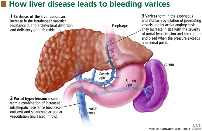
The normal HVPG is 5 mm Hg or less; anything above this value denotes portal hypertension. However, studies have shown that varices may develop but do not bleed if the HVPG is less than 12 mm Hg.8
TWO WAYS TO PREVENT BLEEDING
Bleeding can be prevented either by reducing the portal venous pressure or by obliterating the varices. Portal pressure can be reduced by placing a portosystemic shunt either surgically or percutaneously with radiographic guidance or by giving drugs such as nonselective beta-blockers, nitrates, or a combination of these drugs. Variceal obliteration is typically done by endoscopic methods with either injection of a sclerosant or band ligation.
NONSELECTIVE BETA-BLOCKERS: THE MAINSTAY OF TREATMENT
Nonselective beta-blockers, the most commonly used drugs for preventing first esophageal variceal bleeding, decrease portal pressure by blocking both beta-1 and beta-2 adrenergic receptors.9 Beta-1 blockade decreases portal flow by decreasing the heart rate and cardiac output, while blockade of beta-2 receptors results in unopposed alpha-adrenergic-mediated vasoconstriction.
Selective beta-blockers do not appear to be as useful for primary prophylaxis. More than 2 decades ago, metoprolol (Toprol, Lopressor), a beta-1 selective antagonist, was compared with propranolol (Inderal), a nons-elective agent, in patients with cirrhosis and portal hypertension.10 Although both drugs significantly reduced the heart rate and cardiac output, only those taking propranolol showed a marked fall in portal pressure (mean decrease of 6.8 mm Hg vs 3.8 mm Hg with metoprolol) and a significant reduction in hepatic blood flow. The differences were thought to be related to beta-2 blockade of vasodilator receptors in the splanchnic circulation, which occurs only with nonselective beta-blockers such as propranolol.
The two nonselective beta-blockers most often used to prevent variceal bleeding are nadolol (Corgard) and propranolol. Both have been extensively studied in preventing a first variceal hemorrhage.
Effectiveness of beta-blockers
D’Amico et al11 performed a meta-analysis in 1995, examining nine trials (996 patients total) of the effectiveness of beta-blockers in preventing a first variceal hemorrhage. Seven trials found that bleeding risk was reduced with beta-blockers (significantly in four), one trial found that risk was unchanged, and one trial found that risk was increased—an outlier due to a small sample size. The meta-analysis showed a significant bleeding reduction with the use of a beta-blocker, either including the outlier trial (pooled odds ratio 0.54; 95% confidence interval 0.39–0.74) or excluding it (pooled odds ratio 0.48; 95% confidence interval 0.35–0.66).
Mortality rates were also reduced in seven trials, but the reduction was statistically significant in only one. However, in the pooled estimate, the mortality risk reduction approached statistical significance (pooled odds ratio 0.75; 95% confidence interval 0.57–1.06).
Ideo et al12 gave either nadolol or placebo to 79 patients with cirrhosis and large esophageal varices that had never bled. Nadolol was found to protect against a first variceal hemorrhage: at 2-year follow-up, only 1 of the 30 patients allocated to nadolol had had bleeding, vs 11 of the 49 patients in the placebo group.
Merkel et al13 found that the risk of variceal bleeding was lower in patients who started treatment with beta-blockers when their varices were small (12% at 5 years) than in those who started treatment after a diagnosis of large esophageal varices (22% at 5 years). They concluded that nadolol helps prevent small varices from growing into larger ones.
Response to beta-blockers is not uniform
Although beta-blockers decrease the portal pressure in many cirrhotic patients, the response is not uniform. In a study of 60 cirrhotic patients,14 40% showed no reduction or even a slight increase in HVPG with propranolol. Most patients showed a significant reduction in heart rate (17.5% ± 10%) after receiving 40 mg of propranolol. In the patients whose HVPG did not decrease by at least 10% with 40 mg of propranolol, increasing the dose caused a decrease in HVPG without a further decrease in heart rate. This suggests that 40 mg of propranolol successfully produced beta-1 blockade but that a higher dose was required for effective beta-2 blockade.
Failure to respond in certain patients may be due to a concurrent rise in collateral or hepatic sinusoidal resistance, or both. This was confirmed in a study in portal-hypertensive rats treated with propranolol.15 The reduction in portal blood flow expected was accompanied by a disproportionately small reduction in portal pressure, which was thought to be due to a rise in portal and collateral vascular resistance.
How to tell if beta-blocker treatment is ‘working’
An HVPG ≤ 12 mm Hg? Studies have shown that the most important predictor of efficacy of prophylaxis for variceal bleeding is a decrease in the HVPG to 12 mm Hg or less or a decrease in the initial HVPG of more than 20%.9 Although measuring the HVPG is invasive, expensive, and not routinely done in clinical practice, several studies have investigated the role of measuring hemodynamic response to medication.
Merkel et al16 measured the HVPG in 49 cirrhotic patients with previously nonbleeding varices before starting therapy with beta-blockers with or without nitrates and after 1 to 3 months of treatment. They followed the patients for up to 5 years. The mean HVPG value at baseline was 18.8 mm Hg. At 3 years of follow-up, 7% of those who had responded well to therapy (defined as achieving an HVPG less than 13 mm Hg or a decrease of more than 20%) had experienced a bleeding episode, which was significantly less than the rate (41%) in those who did not meet those hemodynamic end points. No patient reaching an HVPG of 12 mm Hg or less during treatment had variceal bleeding during follow-up.
Groszmann et al17 also prospectively measured the HVPG in patients with cirrhosis and varices, but their patients received either propranolol or placebo. Variceal hemorrhage occurred in 13 patients (11 of 51 in the placebo group and 2 of 51 in the propranolol group), all of whom had an HVPG greater than 12 mm Hg. Again, none of the patients whose HVPG was decreased to 12 mm Hg or less bled from esophageal varices.
Unfortunately, routine HVPG measurement to guide primary prophylaxis is an expensive strategy. Data suggest that measuring the HVPG is cost-effective only when the cost of measuring the HVPG is very low, the risk of variceal bleeding is very high, or the patient is expected to survive at least 3 to 5 years.18
A heart rate of 55 to 60? An alternative to HVPG measurement to monitor the effectiveness of beta-blocker therapy is to follow the heart rate. A 25% reduction from baseline or a heart rate of 55 to 60 beats per minute is the standard goal19,20; yet, at least 40% of patients treated with enough propranolol to decrease the heart rate by 25% do not respond with significant HVPG reductions.14,21
So, although beta-blockade is effective peripherally, it may not reduce HVPG to less than 12 mm Hg or 20% from baseline, and direct HVPG measurement is still the gold standard.
Treatment should be lifelong
Once a patient is started on a beta-blocker to prevent variceal hemorrhage, the treatment should be lifelong.
In 2001, a group of patients (most of them in Child-Pugh class A or B) completing a prospective randomized controlled trial of propranolol for primary prevention of variceal hemorrhage were tapered off propranolol or placebo.22 Of the 49 patients, 9 experienced variceal hemorrhage (6 of 25 former propranolol recipients and 3 of 24 former placebo recipients), and 17 patients died (12 former propranolol and 5 former placebo recipients), suggesting that treatment should be maintained for life.
Therefore, when beta-blocker therapy is discontinued, the risk of variceal hemorrhage returns to what would be expected in an untreated population.
Beta-blockers may not prevent varices
Although many trials have shown that beta-blockers are effective as prophylaxis against a first variceal hemorrhage, there is no evidence that these drugs prevent varices from forming in cirrhotic patients.
Groszmann et al23 treated more than 200 patients who had biopsy-proven cirrhosis and portal hypertension (HVPG > 6 mm Hg) but no varices with timolol (Blocadren), a nonselective beta-blocker, or placebo. At a median follow-up of about 55 months, the groups did not differ significantly in the incidence of primary events (development of varices or variceal hemorrhage) or treatment failures (transplantation or death). Varices developed less frequently among patients with a baseline HVPG of less than 10 mm Hg and among those whose HVPG had decreased by more than 10% at 1 year. In patients whose HVPG increased by more than 10%, varices developed more frequently.
Contraindications, side effects
The major drawbacks to therapy with beta-blockers are their contraindications and side effects.
Contraindications include chronic obstructive lung disease, psychosis, atrioventricular heart blocks, and aortic-valve disease.
Side effects are reported in 15% of patients but severe events are rare.24 Still, an estimated 10% to 20% of patients discontinue treatment because they cannot tolerate it.25 The more common complaints include fatigue, shortness of breath, sexual dysfunction, and sleep disorders.
Dosage
No specific starting dose of beta-blockers is agreed upon, but nadolol 20 to 40 mg once daily or long-acting propranolol 60 mg once daily can be used as initial therapy.25 Once-daily dosing increases the likelihood of compliance.
Since portal pressure progressively declines from 12 noon to 7 PM and then increases throughout the night and back to baseline by 9 AM,26 we recommend that the medication be taken in the evening to counteract increases in portal pressure that occur in the middle of the night.
ENDOSCOPIC VARICEAL LIGATION
Endoscopic variceal ligation has been investigated extensively for use as prophylaxis against first variceal hemorrhage. The procedure involves placing a rubber band around a varix aspirated into a cylinder on the tip of an endoscope.
Effectiveness of ligation
Lay et al,27 in a prospective, randomized trial in 126 cirrhotic patients endoscopically judged to be at high risk of hemorrhage, found that ligation significantly reduced the 2-year cumulative bleeding rate (19% with ligation vs 60% in an untreated control group) and the overall mortality rate (28% vs 58%). The lower risk of bleeding in the ligation group was attributed to a rapid reduction of variceal size; 60% of those in the ligation group had complete eradication of varices and 38% had varices reduced in size.
Imperiale and Chalasani28 performed a meta-analysis in 2001 that included 601 patients in five trials comparing prophylactic ligation with untreated controls and 283 patients in four trials comparing ligation with beta-blocker therapy. Compared with no treatment, ligation reduced the risk of first variceal hemorrhage, bleeding-related death, and death from any cause. Compared with propranolol, ligation reduced the risk of first-time bleeding but had no effect on the death rate.
Schepke et al,29 in a randomized controlled multicenter trial in 152 cirrhotic patients with two or more esophageal varices, found that neither bleeding incidence nor death rate differed significantly between ligation and propranolol.
Lui et al30 followed 172 cirrhotic patients with grade II or III esophageal varices for 6 years and found that ligation was equivalent to propranolol. However, many patients reported side effects with propranolol, and 30% of patients withdrew from propranolol treatment, making ligation a more attractive option.
Khuroo et al31 performed a meta-analysis of eight randomized controlled trials including 596 patients and found that ligation significantly reduced the rates of first gastrointestinal hemorrhage by 31% and of first variceal hemorrhage by 43%. In subgroup analysis, ligation had a significant advantage compared with beta-blockers in trials with patients with a high bleeding risk, ie, trials in which more than 30% of patients were in Child-Pugh class C and more than 50% of the patients had large varices.
Jutabha et al32 performed a multicenter, prospective trial (published in 2005) in 62 patients with high-risk esophageal varices randomized to propranolol or banding. The trial was ended early after an interim analysis showed that the failure rate of propranolol was significantly higher than that of banding (6/31 vs 0/31, P = .0098). Esophageal variceal hemorrhage occurred in 4 (12.9%) of the patients in the propranolol group compared with 0 in the ligation group. Similarly, 4 patients in the propranolol group died, compared with 0 in the ligation group. All the patients in this trial were liver transplant candidates and therefore all had severe liver disease.
In another trial favoring variceal banding over beta-blockers, Psilopoulos et al33 in 2005 followed 60 patients with cirrhosis and esophageal varices with no history of bleeding. Thirty percent of the patients in the propranolol group developed variceal bleeding compared with 6.7% in the ligation group (P = .043).
Lay et al34 followed 100 cirrhotic patients for 2 years and found comparable cumulative bleeding rates with ligation vs propranolol (18% vs 16%, respectively) and also comparable rates of death (28% vs 24%, respectively).
Sarin et al35 investigated the role of propranolol in addition to ligation in the prevention of first hemorrhage in 144 patients. Adding propranolol did not further decrease the incidence of initial bleeding (7% in the combination group vs 11% in the ligation-only group). Survival rates were similar at 20 months: 92% in the combination group vs 85% in the ligation-only group. However, the rate of variceal recurrence was lower with combination therapy: 6% in the combination group vs 15% with ligation alone.
Does esophageal variceal ligation increase gastric varices?
A less researched topic is whether variceal ligation results in gastric hemodynamic changes that increase the size of fundal varices and worsen portal hypertensive gastropathy.
Yuksel et al36 found that 37 of 85 patients had fundal varices before they underwent ligation of esophageal varices, increasing to 46 after the procedure, a statistically significant increment. The severity of portal hypertensive gastropathy also increased.
Further research is required regarding the long-term consequences of these findings.
ANGIOTENSIN II RECEPTOR ANTAGONISTS: ROLE UNKNOWN
Angiotensin II increases portal pressure, and angiotensin II levels are elevated in patients with cirrhosis, suggesting that this hormone plays a role in the pathogenesis of portal hypertension.
Losartan (Cozaar), an angiotensin II receptor antagonist, was found to decrease the HVPG significantly in patients with severe and moderate portal hypertension in a pilot study37 in 1999. However, in two subsequent studies,38,39 losartan only moderately reduced the HVPG and caused hypotension and a reduction in the glomerular filtration rate. The role of angiotensin II receptor blockers in primary prevention of variceal bleeding is still unknown.
SURGICAL PORTAL DECOMPRESSION HAS BEEN ABANDONED
The first method investigated to prevent variceal bleeding was surgical portal decompression.
A meta-analysis of four randomized controlled trials in 302 patients with varices of all sizes compared portocaval shunt surgery and medical therapy.11 Although shunt surgery was very effective in preventing variceal bleeding, the risk of chronic or recurrent encephalopathy was significantly increased (odds ratio 2.0), as was the risk of death (odds ratio 1.6).
These poor results, combined with advances in endoscopic procedures, led to the abandonment of surgical shunting for primary prophylaxis.
TIPS PROCEDURE: NO ROLE AT PRESENT
The transjugular intrahepatic portosystemic shunt (TIPS) procedure is used to treat the main consequences of portal hypertension, including ascites and variceal hemorrhage. The procedure entails accessing the hepatic vein via the right jugular vein and placing a stent to the portal vein, forming a low-resistance channel and allowing blood to return to the systemic circulation.
TIPS placement increases the risk of encephalopathy; liver failure is a rare complication, and procedural complications (ie, shunt dysfunction) also occur. Trials comparing the TIPS procedure with other forms of therapy to prevent first variceal hemorrhages have not been performed.40 Research to improve the outcome of the TIPS procedure is ongoing, but currently this procedure has no role in primary prevention of variceal bleeding.
ENDOSCOPIC SCLEROTHERAPY MAY INCREASE THE RISK OF DEATH
Numerous clinical trials evaluated sclerotherapy as prophylaxis against a first esophageal variceal hemorrhage. The procedure involves injecting a sclerosant in and around varices.
In a large Veterans Administration study,41 sclerotherapy was compared with sham treatment in 281 men with alcoholic liver disease who had documented varices but no history of bleeding. The trial was terminated after 22.5 months because the rate of all-cause mortality was significantly higher in the sclerotherapy group (32.5%) than in the sham therapy group (17.4%). The higher death rate did not persist after the treatment was discontinued, and it was speculated that, although sclerotherapy had reduced new episodes of variceal hemorrhage, the procedure might have caused bleeding from esophageal ulcers, leading to an increased mortality rate in that group.
The PROVA Study Group from Norway and Denmark found similar results when 286 cirrhotic patients were randomized to receive sclerotherapy, propranolol, combination sclerotherapy and propranolol, or no treatment to prevent a first variceal hemorrhage.42 The incidence of variceal bleeding was almost identical in the four groups, but the mortality rate with variceal bleeding was 2.75 times higher in the sclerotherapy groups than in the other groups (P = .002). It was speculated that repeated sclerotherapy sessions might be poorly tolerated by patients in Child-Pugh classes B and C and might have contributed to the precipitation of liver failure and other common complications of cirrhosis.
A meta-analysis by D’Amico et al11 evaluated 19 trials (1,630 patients) comparing sclerotherapy with nonactive treatment. Sclerotherapy tended to be favorable in trials with a high bleeding rate in the control patients and unfavorable in trials with a low bleeding rate. The benefit seen in patients at high risk is consistent with the efficacy of sclerotherapy for preventing rebleeding, whereas the harmful effect in the low-risk patients points towards side effects and complications exceeding the potential benefits.
In general, currently available evidence suggests that the benefits of prophylactic sclerotherapy are marginal, and therefore sclerotherapy is not recommended as primary prophylaxis for variceal hemorrhage.
NITRATES: NO LONGER USED AS MONOTHERAPY
Unlike vasoconstrictors, which decrease portal pressure by decreasing blood flow, vasodilators reduce hepatic pressure by decreasing intrahepatic and portocollateral vascular resistance.43 In addition, larger doses directly affect the arterial circulation, lowering systemic and therefore splanchnic perfusion pressure.44 Unfortunately, the systemic vasodilatory effects of nitrates exacerbate the hyperdynamic state that is characteristic of cirrhosis, thereby limiting their use and tolerability in many patients.
A trial comparing propranolol vs isosorbide mononitrate initially found that the groups did not differ significantly with regard to bleeding rates and 2-year survival rates,45 but a 6-year follow-up found the likelihood of death greater in patients older than 50 years in the nitrate group.46 In an additional study comparing isosorbide mononitrate vs placebo in patients with contraindications to or intolerance of beta-blockers, no difference in the relative risk of first variceal hemorrhage was found between the two groups.47 Therefore, nitrates are no longer used as monotherapy to prevent variceal bleeding.
Combination therapy with beta-blockers plus nitrates is controversial. In a trial in 1996, Merkel et al48 found the cumulative risk of variceal bleeding was 18% at 40 months with nadolol alone vs 7.5% with nadolol plus isosorbide mononitrate. However, in a later trial, Garcia-Pagán et al49 found no significant advantage to combination therapy. The incidence of variceal bleeding at 1 year was 8.3% in the group receiving propranolol plus placebo and 5% in the group receiving propranolol plus isosorbide mononitrate; at 2 years, the rates were 10.6% vs 12.5%.
RECOMMENDATIONS FOR SCREENING AND PROPHYLAXIS
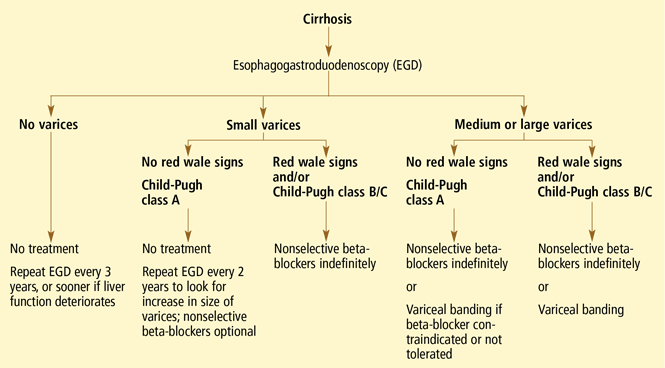
- All patients with cirrhosis should be screened for varices at the time of diagnosis.
- The size of the varices, including small (≤ 5 mm) and large (> 5 mm), and the presence of red wale marks on the varices should be recorded.
- Patients who have no varices on screening endoscopy should be rescreened every 3 years if their liver function is stable or every year if their liver function deteriorates. (Varices grow at a rate proportional to the severity of the liver disease.)
- Patients with portal hypertension but without varices do not need treatment with nonselective beta-blockers. Endoscopy should be performed at the intervals suggested above.
- Those who are found to have small varices on screening endoscopy but who have well-compensated liver disease (Child-Pugh class A) and no red wale marks should be rescreened every other year because the development of large varices is greater in patients with small varices on initial endoscopy than in patients with no varices. Emerging data support the use of beta-blockers to prevent varices from increasing in size.
- Patients who have small varices with red wale signs or who are in Child-Pugh class B or C have an increased risk of bleeding and should be treated with beta-blockers. If beta-blockers are not used, endoscopy should be done every year to look for an increase in variceal size.
- Patients who have large varices without red wale signs or who are in Child-Pugh class B or C should be treated with nonselective beta-blockers. The dose should be adjusted to achieve maximal tolerable decrease in heart rate to a minimum of 55 beats per minute, and treatment should be continued indefinitely.
- Endoscopic variceal ligation is an acceptable alternative to beta-blocker treatment as first-line therapy in those who cannot tolerate beta-blockers or who have contraindications to their use, or in those who have red wale marking or who are in Child-Pugh class B or C.
- D’Amico G, De Franchis R Cooperative Study Group. Upper digestive bleeding in cirrhosis. Post-therapeutic outcome and prognostic indicators. Hepatology 2003; 38:599–612.
- D’Amico G, Luca A. Natural history. Clinicalhaemodynamic correlations. Prediction of the risk of bleeding. Baillieres Clin Gastroenterol 1997; 11:243–256.
- Kamath PS. Esophageal variceal bleeding: primary prophylaxis. Clin Gastroenterol Hepatol 2005; 3:90–93.
- Shah V, Toruner M, Haddad F, et al. Impaired endothelial nitric oxide synthase activity associated with enhanced caveolin binding in experimental cirrhosis in the rat. Gastroenterology 1999; 117:1222–1228.
- Morales-Ruiz M, Jimenez W, Perez-Sala D, et al. Increased nitric oxide synthase expression in arterial vessels of cirrhotic rats with ascites. Hepatology 1996; 24:1481–1486.
- De Franchis R, Dell’Era A, Iannuzzi F. Diagnosis and treatment of portal hypertension. Dig Liver Dis 2004; 36:787–798.
- Groszmann RJ, Wongcharatrawee S. The hepatic venous pressure gradient: anything worth doing should be done right. Hepatology 2004; 39:280–282.
- Garcia-Tsao G, Groszmann RJ, Fisher RL, Conn HO, Atterbury CE, Glickman M. Portal pressure, presence of gastroesophageal varices and variceal bleeding. Hepatology 1985; 5:419–424.
- Bosch J, Masti R, Kravetz D, et al. Effects of propranolol on azygos venous blood flow and hepatic and systemic hemodynamics in cirrhosis. Hepatology 1984; 4:1200–1205.
- Westaby D, Bihari DJ, Gimson AE, Crossley IR, Williams R. Selective and non-selective beta receptor blockade in the reduction of portal pressure in patients with cirrhosis and portal hypertension. Gut 1984; 25:121–124.
- D’Amico G, Pagliaro L, Bosch J. The treatment of portal hypertension: a meta-analytic review. Hepatology 1995; 22:332–354.
- Ideo G, Bellati G, Fesce E, Grimoldi D. Nadolol can prevent the first gastrointestinal bleeding in cirrhotics: a prospective, randomized study. Hepatology 1988; 8:6–9.
- Merkel C, Marin R, Angeli P, et al. Gruppo Triveneto per l’Ipertensione Portale. A placebo-controlled clinical trial of nadolol in the prophylaxis of growth of small esophageal varices in cirrhosis. Gastroenterology 2004; 127:476–484.
- Garcia-Tsao G, Grace ND, Groszmann RJ, et al. Short-term effects of propranolol on portal venous pressure. Hepatology 1986; 6:101–106.
- Kroeger RJ, Groszmann RJ. Increased portal venous resistance hinders portal pressure reduction during the administration of beta-adrenergic blocking agents in a portal hypertensive model. Hepatology 1985; 5:97–101.
- Merkel C, Bolognesi M, Sacerdoti D, et al. The hemodynamic response to medical treatment of portal hypertension as a predictor of clinical effectiveness in the primary prophylaxis of variceal bleeding in cirrhosis. Hepatology 2000; 32:930–934.
- Groszmann RJ, Bosch J, Grace ND, et al. Hemodynamic events in a prospective randomized trial of propranolol versus placebo in the prevention of a first variceal hemorrhage. Gastroenterology 1990; 99:1401–1407.
- Hicken BL, Sharara AI, Abrams GA, Eloubeidi M, Fallon MB, Arguedas MR. Hepatic venous pressure gradient measurements to assess response to primary prophylaxis in patients with cirrhosis: a decision analytical study. Aliment Pharmacol Ther 2003; 17:145–153.
- Grace ND. Diagnosis and treatment of gastrointestinal bleeding secondary to portal hypertension. American College of Gastroenterology Practice Parameters Committee. Am J Gastroenterol 1997; 92:1081–1091.
- Grace ND, Groszmann RJ, Garcia-Tsao G, et al. Portal hypertension and variceal bleeding: an AASLD single topic symposium. Hepatology 1998; 28:868–880.
- Feu F, Garcia-Pagán JC, Bosch J, et al. Relation between portal pressure response to pharmacotherapy and risk of recurrent variceal haemorrhage in patients with cirrhosis. Lancet 1995; 346:1056–1059.
- Abraczinskas DR, Ookubo R, Grace ND, et al. Propranolol for the prevention of first esophageal variceal hemorrhage: a lifetime commitment? Hepatology 2001; 34:1096–1102.
- Groszmann RJ, Garcia-Tsao G, Bosch J, et al Portal Hypertension Collaborative Group. Beta-blockers to prevent gastroesophageal varices in patients with cirrhosis. N Engl J Med 2005; 353:2254–2261.
- Garcia-Pagán JC, Bosch J. Pharmacological prevention of variceal bleeding. New developments. Baillier Clin Gastroenterol 1997; 11:271–287.
- Talwalkar JA, Kamath PS. An evidence-based medicine approach to beta-blocker therapy in patients with cirrhosis. Am J Med 2004; 116:759–766.
- Garcia-Pagan JC, Feu F, Castells A, et al. Circadian variations of portal pressure and variceal hemorrhage in patients with cirrhosis. Hepatology 1994; 19:595–601.
- Lay CS, Tsai YT, Teg CY, et al. Endoscopic variceal ligation in prophylaxis of first variceal bleeding in cirrhotic patients with high-risk esophageal varices. Hepatology 1997; 25:1346–1350.
- Imperiale TF, Chalasani N. A meta-analysis of endoscopic variceal ligation for primary prophylaxis of esophageal variceal bleeding. Hepatology 2001; 33:802–807.
- Schepke M, Kleber G, Nurnberg D, et al. Ligation versus propranolol for the primary prophylaxis of variceal bleeding in cirrhosis. Hepatology 2004; 40:65–72.
- Lui HF, Stanley AJ, Forrest EH, et al. Primary prophylaxis of variceal hemorrhage: a randomized controlled trial comparing band ligation, propranolol, and isosorbide mononitrate. Gastroenterology 2002; 123:735–744.
- Khuroo MS, Khuroo NS, Farahat KL, Khuroo YS, Sofi AA, Dahab ST. Meta-analysis: endoscopic variceal ligation for primary prophylaxis of oesophageal variceal bleeding. Aliment Pharmacol Ther 2005; 21:347–361.
- Jutabha R, Jensen DM, Martin P, Savides T, Han SH, Gornbein J. Randomized study comparing banding and propranolol to prevent initial variceal hemorrhage in cirrhotics with high-risk esophageal varices. Gastroenterology 2005; 128:870–881.
- Psilopoulos D, Galanis P, Goulas S, et al. Endoscopic variceal ligation vs. propranolol for prevention of first variceal bleeding: a randomized controlled trial. Eur J Gastroenterol Hepatol 2005; 17:1111–1117.
- Lay CS, Tsai YT, Lee FY, et al. Endoscopic variceal ligation versus propranolol in prophylaxis of first variceal bleeding in patients with cirrhosis. J Gastroenterol Hepatol 2006; 21:413–419.
- Sarin SK, Wadhawan M, Agarwal SR, Tyagi P, Sharma BC. Endoscopic variceal ligation plus propranolol versus endoscopic variceal ligation alone in primary prophylaxis of variceal bleeding. Am J Gastroenterol 2005; 100:797–804.
- Yuksel O, Koklu S, Arhan M, Yolcu OF, et al. Effects of esophageal variceal eradication on portal hypertensive gastropathy and fundal varices: a retrospective and comparative study. Dig Dis Sci 2006; 51:27–30.
- Schneider AW, Kalk JF, Klein CP. Effect of losartan, an angiotensin II receptor antagonist, on portal pressure in cirrhosis. Hepatology 1999; 29:334–339.
- Schepke M, Werner E, Biecker E, et al. Hemodynamic effects of the angiotensin II receptor antagonist irbesartan in patients with cirrhosis and portal hypertension. Gastroenterology 2001; 121:389–395.
- Gonzalez-Abraldes J, Albillos A, Banares R, et al. Randomized comparison of long-term losartan versus propranolol in lowering portal pressure in cirrhosis. Gastroenterology 2001; 121:382–388.
- Boyer TD, Haskal ZJ American Association for the Study of Liver Diseases. The role of transjugular intrahepatic portosystemic shunt in the management of portal hypertension. Hepatology 2005; 41:386–400.
- Prophylactic sclerotherapy for esophageal varices in men with alcoholic liver disease. A randomized, single-blind, multicenter clinical trial. The Veterans Affairs Cooperative Variceal Sclerotherapy Group. N Engl J Med 1991; 324:1779–1784.
- The PROVA Study Group. Prophylaxis of first hemorrhage from esophageal varices by sclerotherapy, propranolol or both in cirrhotic patients: a randomized multicenter trial. Hepatology 1991; 14:1016–1024.
- Escorsell A, Feu F, Bordas JM, et al. Effects of isosorbide-5-mononitrate on variceal pressure and systemic and splanchnic haemodynamics in patients with cirrhosis. J Hepatol 1996; 24:423–429.
- Hayes PC, Westaby D, Williams R. Effect and mechanism of action of isosorbide-5-mononitrate. Gut 1988; 29:752–755.
- Angelico M, Carli L, Piat C, et al. Isosorbide-5-mononitrate versus propranolol in the prevention of first bleeding in cirrhosis. Gastroenterology 1993; 104:1460–1465.
- Angelico M, Carli L, Piat C, Gentile S, Capocaccia L. Effects of isosorbide-5-mononitrate compared with propranolol on first bleeding and long-term survival in cirrhosis. Gastroenterology 1997; 113:1632–1639.
- Garcia-Pagan JC, Villanueva C, Vila MC, et al. MOVE Group. Mononitrato Varices Esofagicas. Isosorbide mononitrate in the prevention of first variceal bleed in patients who cannot receive beta-blockers. Gastroenterology 2001; 121:908–914.
- Merkel C, Marin R, Enzo E, et al. Randomised trial of nadolol alone or with isosorbide mononitrate for primary prophylaxis of variceal bleeding in cirrhosis. Gruppo-Triveneto per L’ipertensione portale (GTIP). Lancet 1996; 348:1677–1681.
- Garcia-Pagán JC, Morillas R, Banares R, et al Spanish Variceal Bleeding Study Group. Propranolol plus placebo versus propranolol plus isosorbide-5-mononitrate in the prevention of a first variceal bleed: a double-blind RCT. Hepatology 2003; 37:1260–1266.
- Garcia-Tsao G, Sanyal A, Grace N, et al. Prevention and management of gastroesophageal varices and variceal hemorrhage in cirrhosis. Hepatology 2007; 46:922–938.
Variceal hemorrhage is a medical emergency in which up to 20% of patients die.1 Even if the patient survives an initial episode of variceal bleeding, the probability of another episode is high: the rebleeding rate without treatment is 70% within 1 year. The mortality rate with rebleeding is 33%.
With such overwhelming consequences, the best strategy in any patient with cirrhosis and known varices is to try to prevent the first episode of bleeding.
WHO IS AT RISK?
Esophageal varices are present in 30% of patients with compensated cirrhosis and in up to 60% of those with decompensated cirrhosis (ie, with evidence of ascites or encephalopathy).2
The risk of variceal hemorrhage is related to three factors:
- The size of the varices. Varices 5 mm in diameter or smaller have a 7% risk of bleeding in 2 years, while those larger than 5 mm have a 30% risk of bleeding within 2 years.3
- The appearance of the varices. Morphologic features of varices, including red wale signs (red streaks of the mucosa overlying the varix), have been correlated with an increased risk of hemorrhage.
- The severity of liver dysfunction, as assessed by the Child-Pugh classification—an index of liver dysfunction based on serum albumin concentration, bilirubin level, prothrombin time, and the presence of ascites and encephalopathy. A high Child-Pugh score (ie, class B or C), representing decompensated cirrhosis, is associated with an increased risk of bleeding.
HOW VARICES DEVELOP: PORTAL HYPERTENSION
Esophageal varices form as a result of increased portal pressure, the product of increased portal venous inflow and resistance to outflow from the portal venous system. Portal hypertension is a major complication of chronic liver disease. In cirrhosis, architectural distortion of the liver causes an increase in the intrahepatic vascular resistance.
Portal venous inflow depends on mesenteric arteriolar tone, increasing when tone decreases. In cirrhotic patients, the increase in portal pressure results from a combination of increased portal blood flow secondary to splanchnic arteriolar vasodilation and elevated resistance to outflow through distorted hepatic sinusoids.
The potent vasodilator nitric oxide (NO) plays an important role in portal hypertension. In patients with cirrhosis, NO bioavailability is decreased in the intrahepatic circulation due to defects in the posttranslational regulation of endothelial NO synthase.4 This deficiency of NO, along with mechanical factors in the sinusoids, contributes to the increase in intrahepatic resistance. In the systemic and splanchnic circulation, NO bioavailability is increased due to upregulation and posttranslational regulation of endothelial NO synthase, thereby increasing splanchnic vasodilatation and leading to increased portal venous inflow.5 This results in a marked increase in cardiac output and so-called hyperdynamic circulation.
Portal hypertension results in the development of collateral circulation, including venous channels in the esophagus and stomach, by the dilation of preexisting vessels and active angiogenesis. Esophagogastric varices increase in size with the severity of portal hypertension and can rupture when the tension in their walls exceeds a maximal point.
HEPATIC VEIN PRESSURE GRADIENT: A PROXY FOR PORTAL PRESSURE
Ideally, the portal venous pressure should be directly measured. However, since direct measurement is invasive and impractical, the hepatic vein pressure gradient (HVPG) can be measured instead and correlates well with the portal pressure.6
The normal HVPG is 5 mm Hg or less; anything above this value denotes portal hypertension. However, studies have shown that varices may develop but do not bleed if the HVPG is less than 12 mm Hg.8
TWO WAYS TO PREVENT BLEEDING
Bleeding can be prevented either by reducing the portal venous pressure or by obliterating the varices. Portal pressure can be reduced by placing a portosystemic shunt either surgically or percutaneously with radiographic guidance or by giving drugs such as nonselective beta-blockers, nitrates, or a combination of these drugs. Variceal obliteration is typically done by endoscopic methods with either injection of a sclerosant or band ligation.
NONSELECTIVE BETA-BLOCKERS: THE MAINSTAY OF TREATMENT
Nonselective beta-blockers, the most commonly used drugs for preventing first esophageal variceal bleeding, decrease portal pressure by blocking both beta-1 and beta-2 adrenergic receptors.9 Beta-1 blockade decreases portal flow by decreasing the heart rate and cardiac output, while blockade of beta-2 receptors results in unopposed alpha-adrenergic-mediated vasoconstriction.
Selective beta-blockers do not appear to be as useful for primary prophylaxis. More than 2 decades ago, metoprolol (Toprol, Lopressor), a beta-1 selective antagonist, was compared with propranolol (Inderal), a nons-elective agent, in patients with cirrhosis and portal hypertension.10 Although both drugs significantly reduced the heart rate and cardiac output, only those taking propranolol showed a marked fall in portal pressure (mean decrease of 6.8 mm Hg vs 3.8 mm Hg with metoprolol) and a significant reduction in hepatic blood flow. The differences were thought to be related to beta-2 blockade of vasodilator receptors in the splanchnic circulation, which occurs only with nonselective beta-blockers such as propranolol.
The two nonselective beta-blockers most often used to prevent variceal bleeding are nadolol (Corgard) and propranolol. Both have been extensively studied in preventing a first variceal hemorrhage.
Effectiveness of beta-blockers
D’Amico et al11 performed a meta-analysis in 1995, examining nine trials (996 patients total) of the effectiveness of beta-blockers in preventing a first variceal hemorrhage. Seven trials found that bleeding risk was reduced with beta-blockers (significantly in four), one trial found that risk was unchanged, and one trial found that risk was increased—an outlier due to a small sample size. The meta-analysis showed a significant bleeding reduction with the use of a beta-blocker, either including the outlier trial (pooled odds ratio 0.54; 95% confidence interval 0.39–0.74) or excluding it (pooled odds ratio 0.48; 95% confidence interval 0.35–0.66).
Mortality rates were also reduced in seven trials, but the reduction was statistically significant in only one. However, in the pooled estimate, the mortality risk reduction approached statistical significance (pooled odds ratio 0.75; 95% confidence interval 0.57–1.06).
Ideo et al12 gave either nadolol or placebo to 79 patients with cirrhosis and large esophageal varices that had never bled. Nadolol was found to protect against a first variceal hemorrhage: at 2-year follow-up, only 1 of the 30 patients allocated to nadolol had had bleeding, vs 11 of the 49 patients in the placebo group.
Merkel et al13 found that the risk of variceal bleeding was lower in patients who started treatment with beta-blockers when their varices were small (12% at 5 years) than in those who started treatment after a diagnosis of large esophageal varices (22% at 5 years). They concluded that nadolol helps prevent small varices from growing into larger ones.
Response to beta-blockers is not uniform
Although beta-blockers decrease the portal pressure in many cirrhotic patients, the response is not uniform. In a study of 60 cirrhotic patients,14 40% showed no reduction or even a slight increase in HVPG with propranolol. Most patients showed a significant reduction in heart rate (17.5% ± 10%) after receiving 40 mg of propranolol. In the patients whose HVPG did not decrease by at least 10% with 40 mg of propranolol, increasing the dose caused a decrease in HVPG without a further decrease in heart rate. This suggests that 40 mg of propranolol successfully produced beta-1 blockade but that a higher dose was required for effective beta-2 blockade.
Failure to respond in certain patients may be due to a concurrent rise in collateral or hepatic sinusoidal resistance, or both. This was confirmed in a study in portal-hypertensive rats treated with propranolol.15 The reduction in portal blood flow expected was accompanied by a disproportionately small reduction in portal pressure, which was thought to be due to a rise in portal and collateral vascular resistance.
How to tell if beta-blocker treatment is ‘working’
An HVPG ≤ 12 mm Hg? Studies have shown that the most important predictor of efficacy of prophylaxis for variceal bleeding is a decrease in the HVPG to 12 mm Hg or less or a decrease in the initial HVPG of more than 20%.9 Although measuring the HVPG is invasive, expensive, and not routinely done in clinical practice, several studies have investigated the role of measuring hemodynamic response to medication.
Merkel et al16 measured the HVPG in 49 cirrhotic patients with previously nonbleeding varices before starting therapy with beta-blockers with or without nitrates and after 1 to 3 months of treatment. They followed the patients for up to 5 years. The mean HVPG value at baseline was 18.8 mm Hg. At 3 years of follow-up, 7% of those who had responded well to therapy (defined as achieving an HVPG less than 13 mm Hg or a decrease of more than 20%) had experienced a bleeding episode, which was significantly less than the rate (41%) in those who did not meet those hemodynamic end points. No patient reaching an HVPG of 12 mm Hg or less during treatment had variceal bleeding during follow-up.
Groszmann et al17 also prospectively measured the HVPG in patients with cirrhosis and varices, but their patients received either propranolol or placebo. Variceal hemorrhage occurred in 13 patients (11 of 51 in the placebo group and 2 of 51 in the propranolol group), all of whom had an HVPG greater than 12 mm Hg. Again, none of the patients whose HVPG was decreased to 12 mm Hg or less bled from esophageal varices.
Unfortunately, routine HVPG measurement to guide primary prophylaxis is an expensive strategy. Data suggest that measuring the HVPG is cost-effective only when the cost of measuring the HVPG is very low, the risk of variceal bleeding is very high, or the patient is expected to survive at least 3 to 5 years.18
A heart rate of 55 to 60? An alternative to HVPG measurement to monitor the effectiveness of beta-blocker therapy is to follow the heart rate. A 25% reduction from baseline or a heart rate of 55 to 60 beats per minute is the standard goal19,20; yet, at least 40% of patients treated with enough propranolol to decrease the heart rate by 25% do not respond with significant HVPG reductions.14,21
So, although beta-blockade is effective peripherally, it may not reduce HVPG to less than 12 mm Hg or 20% from baseline, and direct HVPG measurement is still the gold standard.
Treatment should be lifelong
Once a patient is started on a beta-blocker to prevent variceal hemorrhage, the treatment should be lifelong.
In 2001, a group of patients (most of them in Child-Pugh class A or B) completing a prospective randomized controlled trial of propranolol for primary prevention of variceal hemorrhage were tapered off propranolol or placebo.22 Of the 49 patients, 9 experienced variceal hemorrhage (6 of 25 former propranolol recipients and 3 of 24 former placebo recipients), and 17 patients died (12 former propranolol and 5 former placebo recipients), suggesting that treatment should be maintained for life.
Therefore, when beta-blocker therapy is discontinued, the risk of variceal hemorrhage returns to what would be expected in an untreated population.
Beta-blockers may not prevent varices
Although many trials have shown that beta-blockers are effective as prophylaxis against a first variceal hemorrhage, there is no evidence that these drugs prevent varices from forming in cirrhotic patients.
Groszmann et al23 treated more than 200 patients who had biopsy-proven cirrhosis and portal hypertension (HVPG > 6 mm Hg) but no varices with timolol (Blocadren), a nonselective beta-blocker, or placebo. At a median follow-up of about 55 months, the groups did not differ significantly in the incidence of primary events (development of varices or variceal hemorrhage) or treatment failures (transplantation or death). Varices developed less frequently among patients with a baseline HVPG of less than 10 mm Hg and among those whose HVPG had decreased by more than 10% at 1 year. In patients whose HVPG increased by more than 10%, varices developed more frequently.
Contraindications, side effects
The major drawbacks to therapy with beta-blockers are their contraindications and side effects.
Contraindications include chronic obstructive lung disease, psychosis, atrioventricular heart blocks, and aortic-valve disease.
Side effects are reported in 15% of patients but severe events are rare.24 Still, an estimated 10% to 20% of patients discontinue treatment because they cannot tolerate it.25 The more common complaints include fatigue, shortness of breath, sexual dysfunction, and sleep disorders.
Dosage
No specific starting dose of beta-blockers is agreed upon, but nadolol 20 to 40 mg once daily or long-acting propranolol 60 mg once daily can be used as initial therapy.25 Once-daily dosing increases the likelihood of compliance.
Since portal pressure progressively declines from 12 noon to 7 PM and then increases throughout the night and back to baseline by 9 AM,26 we recommend that the medication be taken in the evening to counteract increases in portal pressure that occur in the middle of the night.
ENDOSCOPIC VARICEAL LIGATION
Endoscopic variceal ligation has been investigated extensively for use as prophylaxis against first variceal hemorrhage. The procedure involves placing a rubber band around a varix aspirated into a cylinder on the tip of an endoscope.
Effectiveness of ligation
Lay et al,27 in a prospective, randomized trial in 126 cirrhotic patients endoscopically judged to be at high risk of hemorrhage, found that ligation significantly reduced the 2-year cumulative bleeding rate (19% with ligation vs 60% in an untreated control group) and the overall mortality rate (28% vs 58%). The lower risk of bleeding in the ligation group was attributed to a rapid reduction of variceal size; 60% of those in the ligation group had complete eradication of varices and 38% had varices reduced in size.
Imperiale and Chalasani28 performed a meta-analysis in 2001 that included 601 patients in five trials comparing prophylactic ligation with untreated controls and 283 patients in four trials comparing ligation with beta-blocker therapy. Compared with no treatment, ligation reduced the risk of first variceal hemorrhage, bleeding-related death, and death from any cause. Compared with propranolol, ligation reduced the risk of first-time bleeding but had no effect on the death rate.
Schepke et al,29 in a randomized controlled multicenter trial in 152 cirrhotic patients with two or more esophageal varices, found that neither bleeding incidence nor death rate differed significantly between ligation and propranolol.
Lui et al30 followed 172 cirrhotic patients with grade II or III esophageal varices for 6 years and found that ligation was equivalent to propranolol. However, many patients reported side effects with propranolol, and 30% of patients withdrew from propranolol treatment, making ligation a more attractive option.
Khuroo et al31 performed a meta-analysis of eight randomized controlled trials including 596 patients and found that ligation significantly reduced the rates of first gastrointestinal hemorrhage by 31% and of first variceal hemorrhage by 43%. In subgroup analysis, ligation had a significant advantage compared with beta-blockers in trials with patients with a high bleeding risk, ie, trials in which more than 30% of patients were in Child-Pugh class C and more than 50% of the patients had large varices.
Jutabha et al32 performed a multicenter, prospective trial (published in 2005) in 62 patients with high-risk esophageal varices randomized to propranolol or banding. The trial was ended early after an interim analysis showed that the failure rate of propranolol was significantly higher than that of banding (6/31 vs 0/31, P = .0098). Esophageal variceal hemorrhage occurred in 4 (12.9%) of the patients in the propranolol group compared with 0 in the ligation group. Similarly, 4 patients in the propranolol group died, compared with 0 in the ligation group. All the patients in this trial were liver transplant candidates and therefore all had severe liver disease.
In another trial favoring variceal banding over beta-blockers, Psilopoulos et al33 in 2005 followed 60 patients with cirrhosis and esophageal varices with no history of bleeding. Thirty percent of the patients in the propranolol group developed variceal bleeding compared with 6.7% in the ligation group (P = .043).
Lay et al34 followed 100 cirrhotic patients for 2 years and found comparable cumulative bleeding rates with ligation vs propranolol (18% vs 16%, respectively) and also comparable rates of death (28% vs 24%, respectively).
Sarin et al35 investigated the role of propranolol in addition to ligation in the prevention of first hemorrhage in 144 patients. Adding propranolol did not further decrease the incidence of initial bleeding (7% in the combination group vs 11% in the ligation-only group). Survival rates were similar at 20 months: 92% in the combination group vs 85% in the ligation-only group. However, the rate of variceal recurrence was lower with combination therapy: 6% in the combination group vs 15% with ligation alone.
Does esophageal variceal ligation increase gastric varices?
A less researched topic is whether variceal ligation results in gastric hemodynamic changes that increase the size of fundal varices and worsen portal hypertensive gastropathy.
Yuksel et al36 found that 37 of 85 patients had fundal varices before they underwent ligation of esophageal varices, increasing to 46 after the procedure, a statistically significant increment. The severity of portal hypertensive gastropathy also increased.
Further research is required regarding the long-term consequences of these findings.
ANGIOTENSIN II RECEPTOR ANTAGONISTS: ROLE UNKNOWN
Angiotensin II increases portal pressure, and angiotensin II levels are elevated in patients with cirrhosis, suggesting that this hormone plays a role in the pathogenesis of portal hypertension.
Losartan (Cozaar), an angiotensin II receptor antagonist, was found to decrease the HVPG significantly in patients with severe and moderate portal hypertension in a pilot study37 in 1999. However, in two subsequent studies,38,39 losartan only moderately reduced the HVPG and caused hypotension and a reduction in the glomerular filtration rate. The role of angiotensin II receptor blockers in primary prevention of variceal bleeding is still unknown.
SURGICAL PORTAL DECOMPRESSION HAS BEEN ABANDONED
The first method investigated to prevent variceal bleeding was surgical portal decompression.
A meta-analysis of four randomized controlled trials in 302 patients with varices of all sizes compared portocaval shunt surgery and medical therapy.11 Although shunt surgery was very effective in preventing variceal bleeding, the risk of chronic or recurrent encephalopathy was significantly increased (odds ratio 2.0), as was the risk of death (odds ratio 1.6).
These poor results, combined with advances in endoscopic procedures, led to the abandonment of surgical shunting for primary prophylaxis.
TIPS PROCEDURE: NO ROLE AT PRESENT
The transjugular intrahepatic portosystemic shunt (TIPS) procedure is used to treat the main consequences of portal hypertension, including ascites and variceal hemorrhage. The procedure entails accessing the hepatic vein via the right jugular vein and placing a stent to the portal vein, forming a low-resistance channel and allowing blood to return to the systemic circulation.
TIPS placement increases the risk of encephalopathy; liver failure is a rare complication, and procedural complications (ie, shunt dysfunction) also occur. Trials comparing the TIPS procedure with other forms of therapy to prevent first variceal hemorrhages have not been performed.40 Research to improve the outcome of the TIPS procedure is ongoing, but currently this procedure has no role in primary prevention of variceal bleeding.
ENDOSCOPIC SCLEROTHERAPY MAY INCREASE THE RISK OF DEATH
Numerous clinical trials evaluated sclerotherapy as prophylaxis against a first esophageal variceal hemorrhage. The procedure involves injecting a sclerosant in and around varices.
In a large Veterans Administration study,41 sclerotherapy was compared with sham treatment in 281 men with alcoholic liver disease who had documented varices but no history of bleeding. The trial was terminated after 22.5 months because the rate of all-cause mortality was significantly higher in the sclerotherapy group (32.5%) than in the sham therapy group (17.4%). The higher death rate did not persist after the treatment was discontinued, and it was speculated that, although sclerotherapy had reduced new episodes of variceal hemorrhage, the procedure might have caused bleeding from esophageal ulcers, leading to an increased mortality rate in that group.
The PROVA Study Group from Norway and Denmark found similar results when 286 cirrhotic patients were randomized to receive sclerotherapy, propranolol, combination sclerotherapy and propranolol, or no treatment to prevent a first variceal hemorrhage.42 The incidence of variceal bleeding was almost identical in the four groups, but the mortality rate with variceal bleeding was 2.75 times higher in the sclerotherapy groups than in the other groups (P = .002). It was speculated that repeated sclerotherapy sessions might be poorly tolerated by patients in Child-Pugh classes B and C and might have contributed to the precipitation of liver failure and other common complications of cirrhosis.
A meta-analysis by D’Amico et al11 evaluated 19 trials (1,630 patients) comparing sclerotherapy with nonactive treatment. Sclerotherapy tended to be favorable in trials with a high bleeding rate in the control patients and unfavorable in trials with a low bleeding rate. The benefit seen in patients at high risk is consistent with the efficacy of sclerotherapy for preventing rebleeding, whereas the harmful effect in the low-risk patients points towards side effects and complications exceeding the potential benefits.
In general, currently available evidence suggests that the benefits of prophylactic sclerotherapy are marginal, and therefore sclerotherapy is not recommended as primary prophylaxis for variceal hemorrhage.
NITRATES: NO LONGER USED AS MONOTHERAPY
Unlike vasoconstrictors, which decrease portal pressure by decreasing blood flow, vasodilators reduce hepatic pressure by decreasing intrahepatic and portocollateral vascular resistance.43 In addition, larger doses directly affect the arterial circulation, lowering systemic and therefore splanchnic perfusion pressure.44 Unfortunately, the systemic vasodilatory effects of nitrates exacerbate the hyperdynamic state that is characteristic of cirrhosis, thereby limiting their use and tolerability in many patients.
A trial comparing propranolol vs isosorbide mononitrate initially found that the groups did not differ significantly with regard to bleeding rates and 2-year survival rates,45 but a 6-year follow-up found the likelihood of death greater in patients older than 50 years in the nitrate group.46 In an additional study comparing isosorbide mononitrate vs placebo in patients with contraindications to or intolerance of beta-blockers, no difference in the relative risk of first variceal hemorrhage was found between the two groups.47 Therefore, nitrates are no longer used as monotherapy to prevent variceal bleeding.
Combination therapy with beta-blockers plus nitrates is controversial. In a trial in 1996, Merkel et al48 found the cumulative risk of variceal bleeding was 18% at 40 months with nadolol alone vs 7.5% with nadolol plus isosorbide mononitrate. However, in a later trial, Garcia-Pagán et al49 found no significant advantage to combination therapy. The incidence of variceal bleeding at 1 year was 8.3% in the group receiving propranolol plus placebo and 5% in the group receiving propranolol plus isosorbide mononitrate; at 2 years, the rates were 10.6% vs 12.5%.
RECOMMENDATIONS FOR SCREENING AND PROPHYLAXIS
- All patients with cirrhosis should be screened for varices at the time of diagnosis.
- The size of the varices, including small (≤ 5 mm) and large (> 5 mm), and the presence of red wale marks on the varices should be recorded.
- Patients who have no varices on screening endoscopy should be rescreened every 3 years if their liver function is stable or every year if their liver function deteriorates. (Varices grow at a rate proportional to the severity of the liver disease.)
- Patients with portal hypertension but without varices do not need treatment with nonselective beta-blockers. Endoscopy should be performed at the intervals suggested above.
- Those who are found to have small varices on screening endoscopy but who have well-compensated liver disease (Child-Pugh class A) and no red wale marks should be rescreened every other year because the development of large varices is greater in patients with small varices on initial endoscopy than in patients with no varices. Emerging data support the use of beta-blockers to prevent varices from increasing in size.
- Patients who have small varices with red wale signs or who are in Child-Pugh class B or C have an increased risk of bleeding and should be treated with beta-blockers. If beta-blockers are not used, endoscopy should be done every year to look for an increase in variceal size.
- Patients who have large varices without red wale signs or who are in Child-Pugh class B or C should be treated with nonselective beta-blockers. The dose should be adjusted to achieve maximal tolerable decrease in heart rate to a minimum of 55 beats per minute, and treatment should be continued indefinitely.
- Endoscopic variceal ligation is an acceptable alternative to beta-blocker treatment as first-line therapy in those who cannot tolerate beta-blockers or who have contraindications to their use, or in those who have red wale marking or who are in Child-Pugh class B or C.
- D’Amico G, De Franchis R Cooperative Study Group. Upper digestive bleeding in cirrhosis. Post-therapeutic outcome and prognostic indicators. Hepatology 2003; 38:599–612.
- D’Amico G, Luca A. Natural history. Clinicalhaemodynamic correlations. Prediction of the risk of bleeding. Baillieres Clin Gastroenterol 1997; 11:243–256.
- Kamath PS. Esophageal variceal bleeding: primary prophylaxis. Clin Gastroenterol Hepatol 2005; 3:90–93.
- Shah V, Toruner M, Haddad F, et al. Impaired endothelial nitric oxide synthase activity associated with enhanced caveolin binding in experimental cirrhosis in the rat. Gastroenterology 1999; 117:1222–1228.
- Morales-Ruiz M, Jimenez W, Perez-Sala D, et al. Increased nitric oxide synthase expression in arterial vessels of cirrhotic rats with ascites. Hepatology 1996; 24:1481–1486.
- De Franchis R, Dell’Era A, Iannuzzi F. Diagnosis and treatment of portal hypertension. Dig Liver Dis 2004; 36:787–798.
- Groszmann RJ, Wongcharatrawee S. The hepatic venous pressure gradient: anything worth doing should be done right. Hepatology 2004; 39:280–282.
- Garcia-Tsao G, Groszmann RJ, Fisher RL, Conn HO, Atterbury CE, Glickman M. Portal pressure, presence of gastroesophageal varices and variceal bleeding. Hepatology 1985; 5:419–424.
- Bosch J, Masti R, Kravetz D, et al. Effects of propranolol on azygos venous blood flow and hepatic and systemic hemodynamics in cirrhosis. Hepatology 1984; 4:1200–1205.
- Westaby D, Bihari DJ, Gimson AE, Crossley IR, Williams R. Selective and non-selective beta receptor blockade in the reduction of portal pressure in patients with cirrhosis and portal hypertension. Gut 1984; 25:121–124.
- D’Amico G, Pagliaro L, Bosch J. The treatment of portal hypertension: a meta-analytic review. Hepatology 1995; 22:332–354.
- Ideo G, Bellati G, Fesce E, Grimoldi D. Nadolol can prevent the first gastrointestinal bleeding in cirrhotics: a prospective, randomized study. Hepatology 1988; 8:6–9.
- Merkel C, Marin R, Angeli P, et al. Gruppo Triveneto per l’Ipertensione Portale. A placebo-controlled clinical trial of nadolol in the prophylaxis of growth of small esophageal varices in cirrhosis. Gastroenterology 2004; 127:476–484.
- Garcia-Tsao G, Grace ND, Groszmann RJ, et al. Short-term effects of propranolol on portal venous pressure. Hepatology 1986; 6:101–106.
- Kroeger RJ, Groszmann RJ. Increased portal venous resistance hinders portal pressure reduction during the administration of beta-adrenergic blocking agents in a portal hypertensive model. Hepatology 1985; 5:97–101.
- Merkel C, Bolognesi M, Sacerdoti D, et al. The hemodynamic response to medical treatment of portal hypertension as a predictor of clinical effectiveness in the primary prophylaxis of variceal bleeding in cirrhosis. Hepatology 2000; 32:930–934.
- Groszmann RJ, Bosch J, Grace ND, et al. Hemodynamic events in a prospective randomized trial of propranolol versus placebo in the prevention of a first variceal hemorrhage. Gastroenterology 1990; 99:1401–1407.
- Hicken BL, Sharara AI, Abrams GA, Eloubeidi M, Fallon MB, Arguedas MR. Hepatic venous pressure gradient measurements to assess response to primary prophylaxis in patients with cirrhosis: a decision analytical study. Aliment Pharmacol Ther 2003; 17:145–153.
- Grace ND. Diagnosis and treatment of gastrointestinal bleeding secondary to portal hypertension. American College of Gastroenterology Practice Parameters Committee. Am J Gastroenterol 1997; 92:1081–1091.
- Grace ND, Groszmann RJ, Garcia-Tsao G, et al. Portal hypertension and variceal bleeding: an AASLD single topic symposium. Hepatology 1998; 28:868–880.
- Feu F, Garcia-Pagán JC, Bosch J, et al. Relation between portal pressure response to pharmacotherapy and risk of recurrent variceal haemorrhage in patients with cirrhosis. Lancet 1995; 346:1056–1059.
- Abraczinskas DR, Ookubo R, Grace ND, et al. Propranolol for the prevention of first esophageal variceal hemorrhage: a lifetime commitment? Hepatology 2001; 34:1096–1102.
- Groszmann RJ, Garcia-Tsao G, Bosch J, et al Portal Hypertension Collaborative Group. Beta-blockers to prevent gastroesophageal varices in patients with cirrhosis. N Engl J Med 2005; 353:2254–2261.
- Garcia-Pagán JC, Bosch J. Pharmacological prevention of variceal bleeding. New developments. Baillier Clin Gastroenterol 1997; 11:271–287.
- Talwalkar JA, Kamath PS. An evidence-based medicine approach to beta-blocker therapy in patients with cirrhosis. Am J Med 2004; 116:759–766.
- Garcia-Pagan JC, Feu F, Castells A, et al. Circadian variations of portal pressure and variceal hemorrhage in patients with cirrhosis. Hepatology 1994; 19:595–601.
- Lay CS, Tsai YT, Teg CY, et al. Endoscopic variceal ligation in prophylaxis of first variceal bleeding in cirrhotic patients with high-risk esophageal varices. Hepatology 1997; 25:1346–1350.
- Imperiale TF, Chalasani N. A meta-analysis of endoscopic variceal ligation for primary prophylaxis of esophageal variceal bleeding. Hepatology 2001; 33:802–807.
- Schepke M, Kleber G, Nurnberg D, et al. Ligation versus propranolol for the primary prophylaxis of variceal bleeding in cirrhosis. Hepatology 2004; 40:65–72.
- Lui HF, Stanley AJ, Forrest EH, et al. Primary prophylaxis of variceal hemorrhage: a randomized controlled trial comparing band ligation, propranolol, and isosorbide mononitrate. Gastroenterology 2002; 123:735–744.
- Khuroo MS, Khuroo NS, Farahat KL, Khuroo YS, Sofi AA, Dahab ST. Meta-analysis: endoscopic variceal ligation for primary prophylaxis of oesophageal variceal bleeding. Aliment Pharmacol Ther 2005; 21:347–361.
- Jutabha R, Jensen DM, Martin P, Savides T, Han SH, Gornbein J. Randomized study comparing banding and propranolol to prevent initial variceal hemorrhage in cirrhotics with high-risk esophageal varices. Gastroenterology 2005; 128:870–881.
- Psilopoulos D, Galanis P, Goulas S, et al. Endoscopic variceal ligation vs. propranolol for prevention of first variceal bleeding: a randomized controlled trial. Eur J Gastroenterol Hepatol 2005; 17:1111–1117.
- Lay CS, Tsai YT, Lee FY, et al. Endoscopic variceal ligation versus propranolol in prophylaxis of first variceal bleeding in patients with cirrhosis. J Gastroenterol Hepatol 2006; 21:413–419.
- Sarin SK, Wadhawan M, Agarwal SR, Tyagi P, Sharma BC. Endoscopic variceal ligation plus propranolol versus endoscopic variceal ligation alone in primary prophylaxis of variceal bleeding. Am J Gastroenterol 2005; 100:797–804.
- Yuksel O, Koklu S, Arhan M, Yolcu OF, et al. Effects of esophageal variceal eradication on portal hypertensive gastropathy and fundal varices: a retrospective and comparative study. Dig Dis Sci 2006; 51:27–30.
- Schneider AW, Kalk JF, Klein CP. Effect of losartan, an angiotensin II receptor antagonist, on portal pressure in cirrhosis. Hepatology 1999; 29:334–339.
- Schepke M, Werner E, Biecker E, et al. Hemodynamic effects of the angiotensin II receptor antagonist irbesartan in patients with cirrhosis and portal hypertension. Gastroenterology 2001; 121:389–395.
- Gonzalez-Abraldes J, Albillos A, Banares R, et al. Randomized comparison of long-term losartan versus propranolol in lowering portal pressure in cirrhosis. Gastroenterology 2001; 121:382–388.
- Boyer TD, Haskal ZJ American Association for the Study of Liver Diseases. The role of transjugular intrahepatic portosystemic shunt in the management of portal hypertension. Hepatology 2005; 41:386–400.
- Prophylactic sclerotherapy for esophageal varices in men with alcoholic liver disease. A randomized, single-blind, multicenter clinical trial. The Veterans Affairs Cooperative Variceal Sclerotherapy Group. N Engl J Med 1991; 324:1779–1784.
- The PROVA Study Group. Prophylaxis of first hemorrhage from esophageal varices by sclerotherapy, propranolol or both in cirrhotic patients: a randomized multicenter trial. Hepatology 1991; 14:1016–1024.
- Escorsell A, Feu F, Bordas JM, et al. Effects of isosorbide-5-mononitrate on variceal pressure and systemic and splanchnic haemodynamics in patients with cirrhosis. J Hepatol 1996; 24:423–429.
- Hayes PC, Westaby D, Williams R. Effect and mechanism of action of isosorbide-5-mononitrate. Gut 1988; 29:752–755.
- Angelico M, Carli L, Piat C, et al. Isosorbide-5-mononitrate versus propranolol in the prevention of first bleeding in cirrhosis. Gastroenterology 1993; 104:1460–1465.
- Angelico M, Carli L, Piat C, Gentile S, Capocaccia L. Effects of isosorbide-5-mononitrate compared with propranolol on first bleeding and long-term survival in cirrhosis. Gastroenterology 1997; 113:1632–1639.
- Garcia-Pagan JC, Villanueva C, Vila MC, et al. MOVE Group. Mononitrato Varices Esofagicas. Isosorbide mononitrate in the prevention of first variceal bleed in patients who cannot receive beta-blockers. Gastroenterology 2001; 121:908–914.
- Merkel C, Marin R, Enzo E, et al. Randomised trial of nadolol alone or with isosorbide mononitrate for primary prophylaxis of variceal bleeding in cirrhosis. Gruppo-Triveneto per L’ipertensione portale (GTIP). Lancet 1996; 348:1677–1681.
- Garcia-Pagán JC, Morillas R, Banares R, et al Spanish Variceal Bleeding Study Group. Propranolol plus placebo versus propranolol plus isosorbide-5-mononitrate in the prevention of a first variceal bleed: a double-blind RCT. Hepatology 2003; 37:1260–1266.
- Garcia-Tsao G, Sanyal A, Grace N, et al. Prevention and management of gastroesophageal varices and variceal hemorrhage in cirrhosis. Hepatology 2007; 46:922–938.
KEY POINTS
- The hepatic vein pressure gradient (HVPG) correlates well with the portal pressure and is easier to measure. However, whether it is cost-effective to measure the HVPG in clinical practice is controversial.
- Nonselective beta-blockers are the mainstay of treatment; selective beta-blockers do not reduce portal pressure to the same degree and are not recommended for preventing variceal bleeding.
- Endoscopic variceal ligation is an acceptable alternative to beta-blocker therapy for patients who cannot tolerate these drugs and for patients with varices at high risk of bleeding.
- Nitrates are no longer used as monotherapy for preventing variceal hemorrhage, and their use in combination with beta-blockers is controversial. Surgical portal decompression, transjugular intrahepatic portosystemic shunting, and endoscopic sclerotherapy are not recommended.
How to evaluate ‘dipstick hematuria’: What to do before you refer
Many people have some amount of blood in their urine, but relatively few have a serious problem.
In a population-based study in Rochester, Minnesota, red blood cells were found in the urine of 13% of symptom-free adults.1 In other studies, the figure ranged from 9% to 18%.2
Hematuria is sometimes detected during investigation of symptoms such as dysuria, urinary frequency, or flank pain. However, many referrals to urologists are made purely on the basis of the results of a dipstick urinalysis screening test in a patient without symptoms.
The United States Preventive Task Force3 discourages routine testing for hematuria to screen for bladder cancer in patients without symptoms, and the Canadian Task Force on the Periodic Health Examination4 and the American Urological Association (AUA)2,5 do not recommend it either. Nevertheless, approximately 40% of primary care physicians believe in it,6 although the number seems to be declining.7 A reason that dipstick testing is so popular is that it is an inexpensive and simple way to detect glucosuria and medical renal disease.8–11
The risk of significant disease in a patient with microhematuria but without symptoms is low, and the evaluation for hematuria can be costly and invasive. For an individual patient with a hemoglobin-positive dipstick test, the finding should not be ignored, but the patient does not necessarily need a complete evaluation. It is important to determine which patients require urologic studies and consultation, nephrologic evaluation, or no intervention at all.
This review addresses issues related to hematuria for the primary care physician, clarifies some of the important definitions, builds on these terms to delineate which patients should be referred to a urologist, and provides simple recommendations about ancillary studies and their potential role before urologic consultation. Understanding this information will ultimately assure appropriate management of patients without symptoms who have positive dipstick screening tests, leading to decreased use of costly and invasive tests and more appropriate long-term follow-up.
BASIC DEFINITIONS
Gross (macroscopic) hematuria is blood in the urine that is visible without microscopy. This condition almost always warrants urologic evaluation.2,5
Microscopic hematuria, or microhematuria, is the finding of red blood cells in the urine on microscopy. (In contrast, in dipstick hematuria—see below—blood cells may or may not be present in the urine.)
“Dipstick hematuria” and “dipstick microhematuria” are potential misnomers. The dip-stick test for hematuria is a nondiagnostic screening test. A positive result is simply a color change due to oxidation of a test-strip reagent; it does not confirm that blood cells are present. Factors that can cause a false-positive result on a dipstick test include hemoglobinuria, myoglobinuria, concentrated urine, menstrual blood in the urine sample, and rigorous exercise.12 Thus, the diagnosis of hematuria cannot be made by dipstick alone. Unless red blood cells are seen microscopically, the term microhematuria is inappropriate.
Of note, if the specific gravity of the urine is very low (< 1.007), microscopy can fail to detect urinary red blood cells, owing to cell lysis.2 This limitation can be overcome by restricting the patient’s fluid intake and then repeating the urinalysis.
Many patients with a positive dipstick oxidation reaction are labeled as having dip-stick hematuria although microscopic analysis would show that red blood cells are absent. Perhaps the term dipstick pseudohematuria would be more accurate. These patients will not benefit from a costly and invasive urologic workup, so it is crucial to distinguish them from patients with true microhematuria.
SIGNIFICANT HEMATURIA: ≥ 3 RBCs/HPF
Microhematuria is often intermittent, and many healthy patients occasionally have a few red blood cells in the urine.13 However, no cutoff point for the amount of hematuria can be used to rule out the possibility of cancer.14 To account for these complicating factors, the AUA Best Practice Policy Panel on Asymptomatic Hematuria considered the literature and expert opinions to define the amount of microhematuria warranting evaluation in patients with risk factors for significant urologic disease.2,5
The AUA panel defined microhematuria as an average of three or more red blood cells per high-power microscopic field (RBCs/HPF) in two out of three properly collected and prepared specimens. Urine should be collected midstream after wiping the urethral meatus with a disinfectant and voiding the initial portion of urine into the toilet. For proper preparation of the urine sample, 10 mL of urine should be centrifuged at 2,000 rpm for 5 minutes and the supernatant discarded. The sediment should then be resuspended in 0.5 to 1.0 mL of remaining urine, and a drop of this suspension should be examined microscopically. If contamination is suggested (ie, if squamous epithelial cells, bacteria, or both are present), one should consider collecting a specimen through catheterization.2,5
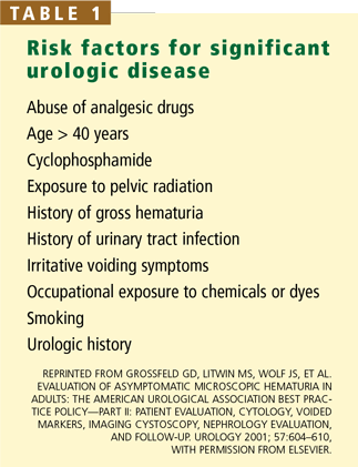
The AUA guidelines do not specify some of the details of management, such as the timing of subsequent microscopic urinalyses, but we recommend that all urinalyses to establish whether significant hematuria is present be done within 3 to 6 months of the screening dip-stick test. If a patient has no risk factors for cancer and has a negative result on the first microscopic urinalysis, the follow-up test can be performed in 1 year. Table 1 shows risk factors for significant urologic disease that warrant evaluation in patients with asymptomatic hematuria.5
Isolated urinary findings that might warrant evaluation by a nephrologist rather than a urologist include proteinuria, red cell casts, and dysmorphic red blood cells, especially if the serum creatinine level is elevated.2,5 Many medical renal conditions (eg, glomerulonephritis) and hemoglobinopathies (eg, sickle cell trait) can cause blood in the urine; when these conditions are accompanied by risk factors for urologic disease, urologic evaluation is indicated as well.
CLINICAL RELEVANCE OF HEMATURIA
Approximately 25% of cases of macroscopic hematuria are due to urologic cancers,14,15 and another 34% are due to other significant urologic diseases14—thus, the recommendation that patients with macroscopic hematuria be evaluated by a urologist. In contrast, in microhematuria, the rates of cancer are much lower, ranging between 1% and 10% in large studies.2,5
The urine dipstick test has been found to be 65% to 99% specific for the presence of blood cells, free hemoglobin, or myoglobin.2,5 If the true specificity is closer to the lower figure and all patients with a positive dipstick test were referred to a urologist, this would mean the urologic workup would be unnecessary in up to 35% of them, because the dipstick result would be falsely positive.
But that is not all. Most causes of hemoglobinuria or myoglobinuria are of limited clinical significance, except for rare conditions that are usually clinically obvious, such as severe burn injury. Further, remember that from 9% to 18% of patients without symptoms have red blood cells in the urine.2,5 In theory, if everyone in the United States had a dipstick test, this would be positive in patients with hematuria as well as in those with hemoglobinuria, myoglobinuria, and other false-positives; if everyone with a positive dipstick result were then referred to a urologist, a substantial portion of the population would receive an unnecessary urologic referral.
Urologic referral and evaluation in these patients not only wastes money: if they undergo imaging studies, they are exposed to radiation and contrast media, with their associated risks, and if they undergo cystoscopy, they face its attendant discomfort and risk of infection.
WHICH PATIENTS WITH HEMATURIA TO REFER TO A UROLOGIST
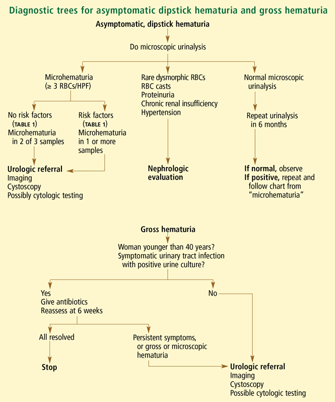
Gross hematuria
Red or tea-colored urine usually indicates gross hematuria. When there is any doubt—such as in the case of a color-blind patient—the presence of red blood cells can be confirmed or ruled out by a microscopic urinalysis.
Nearly all patients with an episode of gross hematuria should be referred to a urologist. The sole exception to this rule can be made when a woman younger than 40 years experiences gross hematuria in the classic setting of a culture-proven, symptomatic urinary tract infection (UTI) and her infection, symptoms, and hematuria all resolve completely with appropriate antibiotics.2,5 However, bleeding from cancer is classically intermittent. Therefore, one should not skip the urine culture and just prescribe antibiotics empirically: the patient might actually have cancer, but the supposed UTI may appear to resolve with antibiotic therapy. For the same reason, resolution of hematuria in any other setting does not obviate the need for referral.
Another scenario usually associated with a benign cause is bleeding after extreme physical activity—also known as “runner’s hematuria” or “march hematuria” (so named because it sometimes occurs in soldiers after a particularly grueling training march). Importantly, even in this situation, one should still be suspicious and probably refer the patient to a urologist: just because the patient has just run a marathon, it does not mean that he or she does not have cancer.
Depending on the character, timing, location, and many other characteristics of the patient’s bleeding, a variety of studies may or may not be necessary. For example, blood-spotting of the underpants might signify urethral bleeding, and imaging and cytologic studies might not be indicated. In view of the variability in presentation and workup, we recommend that the proper workup for these patients be determined by a urologist.
Symptomatic microhematuria
Patients with true microhematuria (three or more RBCs/HPF) accompanied by bothersome or worrisome symptoms should be referred to a urologist. In a study in Scotland, Sultana et al16 found that cancer was present in 6 (5%) of 126 patients with microhematuria without symptoms compared with 13 (10.5%) of 124 patients with microhematuria and irritative voiding symptoms; the difference, however, was not statistically significant.
Microscopic urinalysis should be part of the evaluation for flank pain or certain urinary symptoms such as frequency, urgency, retention, or dysuria; results of this test at the time of symptoms can later help the urologist distinguish the cause of the symptoms or hematuria. In addition, in combination with dipstick analysis, microscopic analysis can help distinguish patients with UTI or medical renal disease. If the evaluation suggests that the hematuria and symptoms are due to a UTI, then the findings on a repeat microscopic analysis, performed after the infection has cleared, should be normal. If hematuria, defined as three or more RBCs/HPF, persists in two of three subsequent urinalyses, then the guidelines mandate diagnostic evaluation even if the urinalysis subsequently becomes negative.
Asymptomatic hematuria
In symptom-free patients with dipstick hematuria found on a screening examination, it is crucial to confirm and document true microhematuria. Per the AUA guidelines, microhematuria worthy of urologic workup is the presence of three or more RBCs/HPF on at least two out of three microscopic urinalyses.2,5 Patients with dipstick pseudohematuria do not meet this criterion and will not benefit from a costly and invasive evaluation. Conversely, patients with higher levels of microhematuria, who have any risk factors for cancer, or who are anxious about the test results might benefit from urologic consultation before a second urinalysis to confirm the first, positive finding.
Many patients younger than age 40 with asymptomatic microhematuria but no other risk factors for urinary tract cancer can be followed conservatively. Khadra et al14 reported that only 1 of 143 patients younger than 40 years with microhematuria had cancer. Similarly, Jones et al17 found, in a prospective study, that no man younger than 40 years with microscopic hematuria had cancer.
Of note: gross or microscopic blood in the urine, even in the setting of anticoagulation, is a marker of urinary tract pathology such as cancer, stones, or infection. Just as patients on anticoagulation therapy who develop gastrointestinal bleeding need a gastrointestinal evaluation, those with hematuria require a urologic evaluation.2,5
STUDIES TO CONSIDER BEFORE CONSULTATION
In symptom-free patients, it is inappropriate to order laboratory or imaging tests on the basis of a dipstick test alone, without confirming that they actually have hematuria. When the blood is confirmed to be present by microscopic examination of centrifuged urine (as described above), benign causes such as UTI should be considered. If a patient does have a UTI with hematuria, urinalysis should be repeated once the infection has cleared up.
Imaging studies
For symptomatic microhematuria. Patients with acute symptoms of renal colic should undergo computed tomography (CT) in a “stone protocol” (without contrast, with 3- to 5-mm cuts of the abdomen and pelvis) to assess for urinary lithiasis. Pregnancy should always be ruled out before radiation exposure; renal ultrasonography is generally the first-choice imaging study for pregnant patients.
For asymptomatic microhematuria. Patients without the classic flank pain of urolithiasis should undergo more extensive studies. For patients at increased risk of cancer, such as heavy smokers, CT urography is the optimal imaging study and is the test least likely to necessitate other follow-up studies.18–20 Other imaging options, including ultrasonography and intravenous pyelography, incompletely assess the upper urinary tracts including both renal parenchyma and urothelial surfaces. CT urography has been shown to find more than 40% of hematuria-causing lesions missed by other studies.18 Because ordering alternative imaging first will often result in redundant studies, CT urography is the preferred initial imaging study in the evaluation of hematuria.
Before exposing a patient to contrast media, one should ascertain that he or she is not allergic to it. In addition, in patients at risk of contrast nephropathy (ie, those older than 60 years, with diabetes, or with preexisting medical renal disease), one should check the serum creatinine concentration. Magnetic resonance urography, a more expensive study, is as accurate as CT for diagnosing many urologic conditions, so it can be performed in lieu of CT urography in patients with renal insufficiency, iodine allergy, or any reason to avoid ionizing radiation. Some clinicians perform plain radiography of the kidneys, ureters, and bladder as well as ultrasonography in this setting, but determination of the appropriate alternative to CT urography, if required, is best left to the urologist.
Other tests
Cytologic testing of the urine can be valuable in patients with gross hematuria and in those with microhematuria who have risk factors for urinary tract cancer. Although its reported median sensitivity for malignancy is only 48%, a positive cytologic test is approximately 94% specific for malignancy.21 Other studies, such as the fluorescence in situ hybridization assay, and the nuclear matrix protein 22 test do not yet have a clear role in the diagnosis of urinary tract disease.22
However, in general, we caution non-urologists not to order special tumor marker or cytologic tests, or to do so only with careful forethought. Although these studies occasionally detect occult cancer in patients at high risk, an “atypical” finding on cytology or a positive tumor marker test can lead to inappropriate referral and unnecessary biopsy or other tests.
WHEN NOT TO REFER A PATIENT WITH HEMATURIA TO A UROLOGIST
Symptom-free patients with a positive dipstick hemoglobin test should not immediately be referred to a urologist: they should have a microscopic urinalysis first to determine whether they actually have microhematuria, unless microscopic laboratory services are unavailable. Only patients with documented true hematuria, as defined by the AUA guidelines, should be referred for urologic evaluation and diagnostic testing. Once a patient is referred for evaluation, the consultant is under clear pressure to perform a complete investigation to fulfill the expectations of the referring physician. Avoiding expensive unnecessary testing and referral in those without hematuria allows appropriate utilization of resources.
Patients with microhematuria associated with a UTI should have a repeat urinalysis after the UTI is successfully treated; if the hematuria clears with the infection, then the patient needs no further evaluation. Patients with dipstick pseudohematuria and significant proteinuria or a predominance of dysmorphic urinary blood cells might benefit from an evaluation by a nephrologist rather than a urologist.2 This is especially true if the patient has an elevated serum creatinine level.
ECONOMIC RELEVANCE
In our tertiary care urology clinic, approximately 75% of patients who are referred to us because of microhematuria have not had a microscopic urinalysis before coming here. On further evaluation, up to 75% of these patients are found to have dipstick pseudohematuria that did not actually require consultation or evaluation.23 It is possible that this occurs even more frequently in the general practice setting.
A Medicare level-4 urologic consultation for hematuria costs $170; the cumulative cost of unwarranted referrals is undoubtedly substantial. Even more money is wasted on CT urography, cytology, and other testing performed before urologic consultation in patients ultimately found not to have true hematuria. The economic and iatrogenic risks of evaluation cannot be justified in patients who do not exhibit findings that can be considered abnormal as defined in this article.
CONCLUSION
It is important to distinguish whether hematuria is microscopic or macroscopic, whether there are associated symptoms, and whether a patient has risk factors for significant urologic disease. While dipstick tests are sensitive, they do not reliably diagnose microhematuria, which is the microscopically proven presence of urinary red blood cells. Positive dipstick tests should always be followed by microscopic urinalysis; failure to do so can result in the unfortunate and unnecessary evaluation of dipstick pseudohematuria, a normal condition.
The AUA defines significant hematuria as three or more RBCs/HPF in two of three properly prepared specimens.2 This should determine whether a symptom-free patient needs urologic referral and evaluation for hematuria.
By following these principles, primary care physicians have a valuable opportunity to direct medical care, increase the efficiency of our health care system, and protect patients from the anxiety, costs, and risks of an unnecessary urologic workup.
- Mohr DN, Offord KP, Owen RA, Melton LJ. Asymptomatic microhematuria and urologic disease. A population-based study. JAMA 1986; 256:224–229.
- Grossfeld GD, Litwin MS, Wolf JS, et al. Evaluation of asymptomatic microscopic hematuria in adults: the American Urological Association best practice policy—part I: definition, detection, prevalence, and etiology. Urology 2001; 57:599–603.
- US Preventive Services Task Force. Screening for Bladder Cancer, updated November 2004. Available at www.ahrq.gov/clinic/uspstf/uspsblad.htm.
- Logsetty S. Screening for bladder cancer. Canadian Task Force on the Periodic Health Examination. Canadian Guide to Clinical Preventive Health Care. 1994: Ottawa: Health Canada,826–836.
- Grossfeld GD, Litwin MS, Wolf JS, et al. Evaluation of asymptomatic microscopic hematuria in adults: The American Urological Association Best Practice Policy—Part II: patient evaluation, cytology, voided markers, imaging cystoscopy, nephrology evaluation, and follow-up. Urology 2001; 57:604–610.
- Prochazka AV, Lundahl K, Pearson W, Oboler SK, Anderson RJ. Support of evidence-based guidelines for the annual physical examination: a survey of primary care providers. Arch Intern Med 2005; 165:1347–1352.
- Chacko KM, Feinberg LE. Laboratory screening at preventive health exams: trend of testing, 1978–2004. Am J Prev Med 2007; 32:59–62.
- Mariani AJ, Luangphinith S, Loo S, Scottolini A, Hodges CV. Dipstick chemical urinalysis: an accurate cost-effective screening test. J Urol 1984; 132:64–66.
- Murphy TE. The urinalysis—inexpensive and informative. J Insur Med 2004; 36:320–326.
- Woolhandler S, Pels RJ, Bor DH, Himmelstein DU, Lawrence RS. Dipstick urinalysis screening of asymptomatic adults for urinary tract disorders. I. Hematuria and proteinuria. JAMA 1989; 262:1214–1219.
- Pels RJ, Bor DH, Woolhandler S, Himmelstein DU, Lawrence RS. Dipstick urinalysis screening of asymptomatic adults for urinary tract disorders. II. Bacteriuria. JAMA 1989; 262:1221–1224.
- Gerber GS, Brendler CBWein AJ, Kavoussi LR, Novick AC, Partin AW, Peters CA. Evaluation of the urologic patient: history, physical examination, and urinalysis. Campbell-Walsh Urology 2007; 9th ed. Saunders Elsevier: Philadelphia:81–110.
- Cohen RA, Brown RS. Clinical practice. Microscopic hematuria. N Engl J Med 2003; 348:2330–2338.
- Khadra MH, Pickard RS, Charlton M, Powell PH, Neal DE. A prospective analysis of 1,930 patients with hematuria to evaluate current diagnostic practice. J Urol 2000; 163:524–527.
- Alishahi S, Byrne D, Goodman CM, Baxby K. Haematuria investigation based on a standard protocol: emphasis on the diagnosis of urological malignancy. J R Coll Surg Edinb 2002; 47:422–427.
- Sultana SR, Goodman CM, Byrne DJ, Baxby K. Microscopic haematuria: urological investigation using a standard protocol. Br J Urol 1996; 78:691–696.
- Jones DJ, Langstaff RJ, Holt SD, Morgans BT. The value of cystourethroscopy in the investigation of microscopic haematuria in adult males under 40 years. A prospective study of 100 patients. Br J Urol 1988; 62:541–545.
- Lang EK, Thomas R, Davis R, et al. Multiphasic helical computerized tomography for the assessment of microscopic hematuria: a prospective study. J Urol 2004; 171:237–243.
- Gray Sears CL, Ward JF, Sears ST, Puckett MF, Kane CJ, Amling CL. Prospective comparison of computerized tomography and excretory urography in the initial evaluation of asymptomatic microhematuria. J Urol 2002; 168:2457–2460.
- Liu W, Mortelé KJ, Silverman SG. Incidental extraurinary findings at MDCT urography in patients with hematuria: prevalence and impact on imaging costs. AJR Am J Roentgenol 2005; 185:1051–1056.
- van Rhijn BW, van der Poel HG, van der Kwast TH. Urine markers for bladder cancer surveillance: a systematic review. Eur Urol 2005; 47:736–748.
- Black PC, Brown GA, Dinney CP. Molecular markers of urothelial cancer and their use in the monitoring of superficial urothelial cancer. J Clin Oncol 2006; 24:5528–5535.
- Rao PR, Jones JS. Retrospective chart review of consultations for hematuria, 2004–2006. Unpublished data. Manuscript in progress.
Many people have some amount of blood in their urine, but relatively few have a serious problem.
In a population-based study in Rochester, Minnesota, red blood cells were found in the urine of 13% of symptom-free adults.1 In other studies, the figure ranged from 9% to 18%.2
Hematuria is sometimes detected during investigation of symptoms such as dysuria, urinary frequency, or flank pain. However, many referrals to urologists are made purely on the basis of the results of a dipstick urinalysis screening test in a patient without symptoms.
The United States Preventive Task Force3 discourages routine testing for hematuria to screen for bladder cancer in patients without symptoms, and the Canadian Task Force on the Periodic Health Examination4 and the American Urological Association (AUA)2,5 do not recommend it either. Nevertheless, approximately 40% of primary care physicians believe in it,6 although the number seems to be declining.7 A reason that dipstick testing is so popular is that it is an inexpensive and simple way to detect glucosuria and medical renal disease.8–11
The risk of significant disease in a patient with microhematuria but without symptoms is low, and the evaluation for hematuria can be costly and invasive. For an individual patient with a hemoglobin-positive dipstick test, the finding should not be ignored, but the patient does not necessarily need a complete evaluation. It is important to determine which patients require urologic studies and consultation, nephrologic evaluation, or no intervention at all.
This review addresses issues related to hematuria for the primary care physician, clarifies some of the important definitions, builds on these terms to delineate which patients should be referred to a urologist, and provides simple recommendations about ancillary studies and their potential role before urologic consultation. Understanding this information will ultimately assure appropriate management of patients without symptoms who have positive dipstick screening tests, leading to decreased use of costly and invasive tests and more appropriate long-term follow-up.
BASIC DEFINITIONS
Gross (macroscopic) hematuria is blood in the urine that is visible without microscopy. This condition almost always warrants urologic evaluation.2,5
Microscopic hematuria, or microhematuria, is the finding of red blood cells in the urine on microscopy. (In contrast, in dipstick hematuria—see below—blood cells may or may not be present in the urine.)
“Dipstick hematuria” and “dipstick microhematuria” are potential misnomers. The dip-stick test for hematuria is a nondiagnostic screening test. A positive result is simply a color change due to oxidation of a test-strip reagent; it does not confirm that blood cells are present. Factors that can cause a false-positive result on a dipstick test include hemoglobinuria, myoglobinuria, concentrated urine, menstrual blood in the urine sample, and rigorous exercise.12 Thus, the diagnosis of hematuria cannot be made by dipstick alone. Unless red blood cells are seen microscopically, the term microhematuria is inappropriate.
Of note, if the specific gravity of the urine is very low (< 1.007), microscopy can fail to detect urinary red blood cells, owing to cell lysis.2 This limitation can be overcome by restricting the patient’s fluid intake and then repeating the urinalysis.
Many patients with a positive dipstick oxidation reaction are labeled as having dip-stick hematuria although microscopic analysis would show that red blood cells are absent. Perhaps the term dipstick pseudohematuria would be more accurate. These patients will not benefit from a costly and invasive urologic workup, so it is crucial to distinguish them from patients with true microhematuria.
SIGNIFICANT HEMATURIA: ≥ 3 RBCs/HPF
Microhematuria is often intermittent, and many healthy patients occasionally have a few red blood cells in the urine.13 However, no cutoff point for the amount of hematuria can be used to rule out the possibility of cancer.14 To account for these complicating factors, the AUA Best Practice Policy Panel on Asymptomatic Hematuria considered the literature and expert opinions to define the amount of microhematuria warranting evaluation in patients with risk factors for significant urologic disease.2,5
The AUA panel defined microhematuria as an average of three or more red blood cells per high-power microscopic field (RBCs/HPF) in two out of three properly collected and prepared specimens. Urine should be collected midstream after wiping the urethral meatus with a disinfectant and voiding the initial portion of urine into the toilet. For proper preparation of the urine sample, 10 mL of urine should be centrifuged at 2,000 rpm for 5 minutes and the supernatant discarded. The sediment should then be resuspended in 0.5 to 1.0 mL of remaining urine, and a drop of this suspension should be examined microscopically. If contamination is suggested (ie, if squamous epithelial cells, bacteria, or both are present), one should consider collecting a specimen through catheterization.2,5
The AUA guidelines do not specify some of the details of management, such as the timing of subsequent microscopic urinalyses, but we recommend that all urinalyses to establish whether significant hematuria is present be done within 3 to 6 months of the screening dip-stick test. If a patient has no risk factors for cancer and has a negative result on the first microscopic urinalysis, the follow-up test can be performed in 1 year. Table 1 shows risk factors for significant urologic disease that warrant evaluation in patients with asymptomatic hematuria.5
Isolated urinary findings that might warrant evaluation by a nephrologist rather than a urologist include proteinuria, red cell casts, and dysmorphic red blood cells, especially if the serum creatinine level is elevated.2,5 Many medical renal conditions (eg, glomerulonephritis) and hemoglobinopathies (eg, sickle cell trait) can cause blood in the urine; when these conditions are accompanied by risk factors for urologic disease, urologic evaluation is indicated as well.
CLINICAL RELEVANCE OF HEMATURIA
Approximately 25% of cases of macroscopic hematuria are due to urologic cancers,14,15 and another 34% are due to other significant urologic diseases14—thus, the recommendation that patients with macroscopic hematuria be evaluated by a urologist. In contrast, in microhematuria, the rates of cancer are much lower, ranging between 1% and 10% in large studies.2,5
The urine dipstick test has been found to be 65% to 99% specific for the presence of blood cells, free hemoglobin, or myoglobin.2,5 If the true specificity is closer to the lower figure and all patients with a positive dipstick test were referred to a urologist, this would mean the urologic workup would be unnecessary in up to 35% of them, because the dipstick result would be falsely positive.
But that is not all. Most causes of hemoglobinuria or myoglobinuria are of limited clinical significance, except for rare conditions that are usually clinically obvious, such as severe burn injury. Further, remember that from 9% to 18% of patients without symptoms have red blood cells in the urine.2,5 In theory, if everyone in the United States had a dipstick test, this would be positive in patients with hematuria as well as in those with hemoglobinuria, myoglobinuria, and other false-positives; if everyone with a positive dipstick result were then referred to a urologist, a substantial portion of the population would receive an unnecessary urologic referral.
Urologic referral and evaluation in these patients not only wastes money: if they undergo imaging studies, they are exposed to radiation and contrast media, with their associated risks, and if they undergo cystoscopy, they face its attendant discomfort and risk of infection.
WHICH PATIENTS WITH HEMATURIA TO REFER TO A UROLOGIST
Gross hematuria
Red or tea-colored urine usually indicates gross hematuria. When there is any doubt—such as in the case of a color-blind patient—the presence of red blood cells can be confirmed or ruled out by a microscopic urinalysis.
Nearly all patients with an episode of gross hematuria should be referred to a urologist. The sole exception to this rule can be made when a woman younger than 40 years experiences gross hematuria in the classic setting of a culture-proven, symptomatic urinary tract infection (UTI) and her infection, symptoms, and hematuria all resolve completely with appropriate antibiotics.2,5 However, bleeding from cancer is classically intermittent. Therefore, one should not skip the urine culture and just prescribe antibiotics empirically: the patient might actually have cancer, but the supposed UTI may appear to resolve with antibiotic therapy. For the same reason, resolution of hematuria in any other setting does not obviate the need for referral.
Another scenario usually associated with a benign cause is bleeding after extreme physical activity—also known as “runner’s hematuria” or “march hematuria” (so named because it sometimes occurs in soldiers after a particularly grueling training march). Importantly, even in this situation, one should still be suspicious and probably refer the patient to a urologist: just because the patient has just run a marathon, it does not mean that he or she does not have cancer.
Depending on the character, timing, location, and many other characteristics of the patient’s bleeding, a variety of studies may or may not be necessary. For example, blood-spotting of the underpants might signify urethral bleeding, and imaging and cytologic studies might not be indicated. In view of the variability in presentation and workup, we recommend that the proper workup for these patients be determined by a urologist.
Symptomatic microhematuria
Patients with true microhematuria (three or more RBCs/HPF) accompanied by bothersome or worrisome symptoms should be referred to a urologist. In a study in Scotland, Sultana et al16 found that cancer was present in 6 (5%) of 126 patients with microhematuria without symptoms compared with 13 (10.5%) of 124 patients with microhematuria and irritative voiding symptoms; the difference, however, was not statistically significant.
Microscopic urinalysis should be part of the evaluation for flank pain or certain urinary symptoms such as frequency, urgency, retention, or dysuria; results of this test at the time of symptoms can later help the urologist distinguish the cause of the symptoms or hematuria. In addition, in combination with dipstick analysis, microscopic analysis can help distinguish patients with UTI or medical renal disease. If the evaluation suggests that the hematuria and symptoms are due to a UTI, then the findings on a repeat microscopic analysis, performed after the infection has cleared, should be normal. If hematuria, defined as three or more RBCs/HPF, persists in two of three subsequent urinalyses, then the guidelines mandate diagnostic evaluation even if the urinalysis subsequently becomes negative.
Asymptomatic hematuria
In symptom-free patients with dipstick hematuria found on a screening examination, it is crucial to confirm and document true microhematuria. Per the AUA guidelines, microhematuria worthy of urologic workup is the presence of three or more RBCs/HPF on at least two out of three microscopic urinalyses.2,5 Patients with dipstick pseudohematuria do not meet this criterion and will not benefit from a costly and invasive evaluation. Conversely, patients with higher levels of microhematuria, who have any risk factors for cancer, or who are anxious about the test results might benefit from urologic consultation before a second urinalysis to confirm the first, positive finding.
Many patients younger than age 40 with asymptomatic microhematuria but no other risk factors for urinary tract cancer can be followed conservatively. Khadra et al14 reported that only 1 of 143 patients younger than 40 years with microhematuria had cancer. Similarly, Jones et al17 found, in a prospective study, that no man younger than 40 years with microscopic hematuria had cancer.
Of note: gross or microscopic blood in the urine, even in the setting of anticoagulation, is a marker of urinary tract pathology such as cancer, stones, or infection. Just as patients on anticoagulation therapy who develop gastrointestinal bleeding need a gastrointestinal evaluation, those with hematuria require a urologic evaluation.2,5
STUDIES TO CONSIDER BEFORE CONSULTATION
In symptom-free patients, it is inappropriate to order laboratory or imaging tests on the basis of a dipstick test alone, without confirming that they actually have hematuria. When the blood is confirmed to be present by microscopic examination of centrifuged urine (as described above), benign causes such as UTI should be considered. If a patient does have a UTI with hematuria, urinalysis should be repeated once the infection has cleared up.
Imaging studies
For symptomatic microhematuria. Patients with acute symptoms of renal colic should undergo computed tomography (CT) in a “stone protocol” (without contrast, with 3- to 5-mm cuts of the abdomen and pelvis) to assess for urinary lithiasis. Pregnancy should always be ruled out before radiation exposure; renal ultrasonography is generally the first-choice imaging study for pregnant patients.
For asymptomatic microhematuria. Patients without the classic flank pain of urolithiasis should undergo more extensive studies. For patients at increased risk of cancer, such as heavy smokers, CT urography is the optimal imaging study and is the test least likely to necessitate other follow-up studies.18–20 Other imaging options, including ultrasonography and intravenous pyelography, incompletely assess the upper urinary tracts including both renal parenchyma and urothelial surfaces. CT urography has been shown to find more than 40% of hematuria-causing lesions missed by other studies.18 Because ordering alternative imaging first will often result in redundant studies, CT urography is the preferred initial imaging study in the evaluation of hematuria.
Before exposing a patient to contrast media, one should ascertain that he or she is not allergic to it. In addition, in patients at risk of contrast nephropathy (ie, those older than 60 years, with diabetes, or with preexisting medical renal disease), one should check the serum creatinine concentration. Magnetic resonance urography, a more expensive study, is as accurate as CT for diagnosing many urologic conditions, so it can be performed in lieu of CT urography in patients with renal insufficiency, iodine allergy, or any reason to avoid ionizing radiation. Some clinicians perform plain radiography of the kidneys, ureters, and bladder as well as ultrasonography in this setting, but determination of the appropriate alternative to CT urography, if required, is best left to the urologist.
Other tests
Cytologic testing of the urine can be valuable in patients with gross hematuria and in those with microhematuria who have risk factors for urinary tract cancer. Although its reported median sensitivity for malignancy is only 48%, a positive cytologic test is approximately 94% specific for malignancy.21 Other studies, such as the fluorescence in situ hybridization assay, and the nuclear matrix protein 22 test do not yet have a clear role in the diagnosis of urinary tract disease.22
However, in general, we caution non-urologists not to order special tumor marker or cytologic tests, or to do so only with careful forethought. Although these studies occasionally detect occult cancer in patients at high risk, an “atypical” finding on cytology or a positive tumor marker test can lead to inappropriate referral and unnecessary biopsy or other tests.
WHEN NOT TO REFER A PATIENT WITH HEMATURIA TO A UROLOGIST
Symptom-free patients with a positive dipstick hemoglobin test should not immediately be referred to a urologist: they should have a microscopic urinalysis first to determine whether they actually have microhematuria, unless microscopic laboratory services are unavailable. Only patients with documented true hematuria, as defined by the AUA guidelines, should be referred for urologic evaluation and diagnostic testing. Once a patient is referred for evaluation, the consultant is under clear pressure to perform a complete investigation to fulfill the expectations of the referring physician. Avoiding expensive unnecessary testing and referral in those without hematuria allows appropriate utilization of resources.
Patients with microhematuria associated with a UTI should have a repeat urinalysis after the UTI is successfully treated; if the hematuria clears with the infection, then the patient needs no further evaluation. Patients with dipstick pseudohematuria and significant proteinuria or a predominance of dysmorphic urinary blood cells might benefit from an evaluation by a nephrologist rather than a urologist.2 This is especially true if the patient has an elevated serum creatinine level.
ECONOMIC RELEVANCE
In our tertiary care urology clinic, approximately 75% of patients who are referred to us because of microhematuria have not had a microscopic urinalysis before coming here. On further evaluation, up to 75% of these patients are found to have dipstick pseudohematuria that did not actually require consultation or evaluation.23 It is possible that this occurs even more frequently in the general practice setting.
A Medicare level-4 urologic consultation for hematuria costs $170; the cumulative cost of unwarranted referrals is undoubtedly substantial. Even more money is wasted on CT urography, cytology, and other testing performed before urologic consultation in patients ultimately found not to have true hematuria. The economic and iatrogenic risks of evaluation cannot be justified in patients who do not exhibit findings that can be considered abnormal as defined in this article.
CONCLUSION
It is important to distinguish whether hematuria is microscopic or macroscopic, whether there are associated symptoms, and whether a patient has risk factors for significant urologic disease. While dipstick tests are sensitive, they do not reliably diagnose microhematuria, which is the microscopically proven presence of urinary red blood cells. Positive dipstick tests should always be followed by microscopic urinalysis; failure to do so can result in the unfortunate and unnecessary evaluation of dipstick pseudohematuria, a normal condition.
The AUA defines significant hematuria as three or more RBCs/HPF in two of three properly prepared specimens.2 This should determine whether a symptom-free patient needs urologic referral and evaluation for hematuria.
By following these principles, primary care physicians have a valuable opportunity to direct medical care, increase the efficiency of our health care system, and protect patients from the anxiety, costs, and risks of an unnecessary urologic workup.
- Mohr DN, Offord KP, Owen RA, Melton LJ. Asymptomatic microhematuria and urologic disease. A population-based study. JAMA 1986; 256:224–229.
- Grossfeld GD, Litwin MS, Wolf JS, et al. Evaluation of asymptomatic microscopic hematuria in adults: the American Urological Association best practice policy—part I: definition, detection, prevalence, and etiology. Urology 2001; 57:599–603.
- US Preventive Services Task Force. Screening for Bladder Cancer, updated November 2004. Available at www.ahrq.gov/clinic/uspstf/uspsblad.htm.
- Logsetty S. Screening for bladder cancer. Canadian Task Force on the Periodic Health Examination. Canadian Guide to Clinical Preventive Health Care. 1994: Ottawa: Health Canada,826–836.
- Grossfeld GD, Litwin MS, Wolf JS, et al. Evaluation of asymptomatic microscopic hematuria in adults: The American Urological Association Best Practice Policy—Part II: patient evaluation, cytology, voided markers, imaging cystoscopy, nephrology evaluation, and follow-up. Urology 2001; 57:604–610.
- Prochazka AV, Lundahl K, Pearson W, Oboler SK, Anderson RJ. Support of evidence-based guidelines for the annual physical examination: a survey of primary care providers. Arch Intern Med 2005; 165:1347–1352.
- Chacko KM, Feinberg LE. Laboratory screening at preventive health exams: trend of testing, 1978–2004. Am J Prev Med 2007; 32:59–62.
- Mariani AJ, Luangphinith S, Loo S, Scottolini A, Hodges CV. Dipstick chemical urinalysis: an accurate cost-effective screening test. J Urol 1984; 132:64–66.
- Murphy TE. The urinalysis—inexpensive and informative. J Insur Med 2004; 36:320–326.
- Woolhandler S, Pels RJ, Bor DH, Himmelstein DU, Lawrence RS. Dipstick urinalysis screening of asymptomatic adults for urinary tract disorders. I. Hematuria and proteinuria. JAMA 1989; 262:1214–1219.
- Pels RJ, Bor DH, Woolhandler S, Himmelstein DU, Lawrence RS. Dipstick urinalysis screening of asymptomatic adults for urinary tract disorders. II. Bacteriuria. JAMA 1989; 262:1221–1224.
- Gerber GS, Brendler CBWein AJ, Kavoussi LR, Novick AC, Partin AW, Peters CA. Evaluation of the urologic patient: history, physical examination, and urinalysis. Campbell-Walsh Urology 2007; 9th ed. Saunders Elsevier: Philadelphia:81–110.
- Cohen RA, Brown RS. Clinical practice. Microscopic hematuria. N Engl J Med 2003; 348:2330–2338.
- Khadra MH, Pickard RS, Charlton M, Powell PH, Neal DE. A prospective analysis of 1,930 patients with hematuria to evaluate current diagnostic practice. J Urol 2000; 163:524–527.
- Alishahi S, Byrne D, Goodman CM, Baxby K. Haematuria investigation based on a standard protocol: emphasis on the diagnosis of urological malignancy. J R Coll Surg Edinb 2002; 47:422–427.
- Sultana SR, Goodman CM, Byrne DJ, Baxby K. Microscopic haematuria: urological investigation using a standard protocol. Br J Urol 1996; 78:691–696.
- Jones DJ, Langstaff RJ, Holt SD, Morgans BT. The value of cystourethroscopy in the investigation of microscopic haematuria in adult males under 40 years. A prospective study of 100 patients. Br J Urol 1988; 62:541–545.
- Lang EK, Thomas R, Davis R, et al. Multiphasic helical computerized tomography for the assessment of microscopic hematuria: a prospective study. J Urol 2004; 171:237–243.
- Gray Sears CL, Ward JF, Sears ST, Puckett MF, Kane CJ, Amling CL. Prospective comparison of computerized tomography and excretory urography in the initial evaluation of asymptomatic microhematuria. J Urol 2002; 168:2457–2460.
- Liu W, Mortelé KJ, Silverman SG. Incidental extraurinary findings at MDCT urography in patients with hematuria: prevalence and impact on imaging costs. AJR Am J Roentgenol 2005; 185:1051–1056.
- van Rhijn BW, van der Poel HG, van der Kwast TH. Urine markers for bladder cancer surveillance: a systematic review. Eur Urol 2005; 47:736–748.
- Black PC, Brown GA, Dinney CP. Molecular markers of urothelial cancer and their use in the monitoring of superficial urothelial cancer. J Clin Oncol 2006; 24:5528–5535.
- Rao PR, Jones JS. Retrospective chart review of consultations for hematuria, 2004–2006. Unpublished data. Manuscript in progress.
KEY POINTS
- Dipstick tests by themselves do not confirm that hematuria is present; thus, “dipstick hematuria” is a potential misnomer. Patients without symptoms who have a positive dipstick test and negative microscopic urinalysis are better described as having dipstick pseudohematuria, a clinically insignificant finding.
- Significant hematuria is defined as three or more red blood cells per high-power field in a properly collected and centrifuged urine specimen; this is the definition that should dictate which patients require further urologic evaluation.
- Since the evaluation for hematuria usually includes cystoscopy and imaging studies, it is crucial to confirm that hematuria is truly present before initiating an invasive and costly evaluation.
A young woman with fatigue
A 22-year-old woman presents to the clinic for evaluation of fatigue. She has not felt well for the past few years. Her current symptoms include generalized fatigue and diarrhea, characterized as two to three semi-formed, nonbloody bowel movements each day and occasional episodes of watery diarrhea. Her bowel movements are usually precipitated by meals. She consumes a regular diet and has not recognized any intolerance to any particular foods. She denies having any abdominal pain, nausea, vomiting, recent travel, joint pain, rash, or change in the texture of her hair. She has been seen by several internists in her hometown, who have not provided her with a specific diagnosis.
Her medical history is significant for anemia, anxiety, and depression. Menarche occurred at age 16. Her menstrual cycle has been regular, with bleeding noted to be only modest. Her medications include oral contraceptive pills. She has not had previous surgeries.
On examination, she appears well. She is afebrile, weighs 128 lbs, and is 63 inches tall. The physical examination is normal, including a rectal examination and fecal occult blood testing.
Routine laboratory tests are performed. Results:
- White blood cell count 3.88 × 10⁹/L (normal 4.0–11)
- Hemoglobin 10.4 g/dL (normal 12–16)
- Hematocrit 34% (normal 37%–47%)
- Mean corpuscular volume 80.2 fL (normal 80–100)
- Mean corpuscular hemoglobin 24.5 pG (normal 27–34)
- Platelet count 365 × 109/L (normal 150–400)
- Sodium 141 mmol/L (normal 132–148)
- Potassium 4.2 mmol/L (normal 3.5–5.0)
- Chloride 107 mmol/L (normal 98–110)
- Alanine aminotransferase 22 U/L (normal 0–45)
- Glucose 66 mg/dL (normal 65–100)
- Blood urea nitrogen 6 mg/dL (normal 8–25)
- Creatinine 0.6 mg/dL (normal 0.7–1.4)
- Thyroid-stimulating hormone 2.860 mIU/L (normal 0.4–5.5)
- Red blood cell folate 539 ng/mL (normal 257–800)
- Vitamin B12 321 pg/mL (normal 221–700)
- Iron/total iron-binding capacity 21/445 μg/dL (normal 30–140, 210–415)
- Ferritin 5 ng/mL (normal 9–150).
DIFFERENTIAL DIAGNOSIS
1. Which of the following is the most likely cause of her diarrhea?
- Thyroid disease
- Functional bowel disease
- Gluten-sensitive enteropathy (celiac disease)
Given her constellation of symptoms (fatigue, neuropsychiatric changes, iron deficiency anemia, and diarrhea), celiac disease is the most likely diagnosis. Hyperthyroidism can cause diarrhea, but this is unlikely since her thyroid tests are normal. Functional bowel disease is a diagnosis of exclusion and usually has a more chronic, fluctuating course.
CELIAC DISEASE HAS VARIOUS PRESENTATIONS
Celiac disease has various presentations and therefore has been classified into several types1,2:
Classic disease is dominated by symptoms of malabsorption. The diagnosis is established by serologic testing, findings of villous atrophy on biopsy, and improvement of symptoms on a gluten-free diet. However, the presentation of celiac disease has changed, and now atypical presentations are more common in adults (see below). The reason for the change in presentation is not known, but some have hypothesized that it is related to an increase in breast-feeding and the later introduction of cereals into infants’ diets.
Celiac disease with atypical symptoms is characterized by extraintestinal manifestations with few or no gastrointestinal (GI) symptoms. Patients may present with iron-deficiency anemia; osteoporosis or vitamin D deficiency; arthritis; neurologic symptoms such as ataxia, headaches, or depression or anxiety; myocarditis; infertility; or elevated aminotransferase levels. As in classic celiac disease, the diagnosis is established with serologic testing, findings of villous atrophy on biopsy, and improvement of symptoms on a gluten-free diet.
Latent disease includes cases in patients with positive serologic tests but no villous atrophy on biopsy. These patients have no symptoms but may develop symptoms or histologic changes later.
Silent disease refers to cases in patients who have no symptoms but have a positive serologic test and villous atrophy on biopsy. These cases are usually detected via screening of people at high risk, ie, relatives of patients with celiac disease.
It is important that clinicians be aware of the various symptoms and presentations of celiac disease in order to make the diagnosis.
CONFIRMING CELIAC DISEASE
2. Which of the following is used to test for celiac disease?
- Immunoglobulin G (IgG) and immunoglobulin A (IgA) antigliadin antibody testing
- IgA antiendomysial antibody and IgA antitransglutaminase antibody testing
- HLA DQ2/DQ8 testing
The sensitivity of antigliadin antibody testing is only about 70% to 85%, and its specificity is about 70% to 90%. Better serologic tests are those for IgA antiendomysial and antitransglutaminase antibodies, which have sensitivities greater than 90% and specificities greater than 95%.3 HLA DQ2/DQ8 testing has a high sensitivity (> 90%–95%), but because about 30% of the general population also carry these markers, the specificity of this test is not ideal. This test is best used for its negative predictive value—ie, to rule out the diagnosis of celiac disease.
Of note: 1% to 2% of patients with celiac disease have a deficiency of IgA.4 Therefore, if the clinical suspicion for celiac disease is high but the IgA antibody tests are negative or equivocal, IgG antitransglutaminase and IgG antiendomysial antibody tests can help establish the diagnosis. HLA testing in this situation can also help rule out the diagnosis.
CONFIRMING CELIAC DISEASE—CONTINUED
3. What test should be performed next in this patient?
- Upper GI series with small-bowel follow-through
- Esophagogastroduodenoscopy with biopsies
- Small-bowel barium study
- Video capsule endoscopy
Today, the presumptive diagnosis of celiac disease requires positive serologic testing and biopsy results. Esophagogastroduodenoscopy with biopsies should be ordered. Upper GI series and barium studies do not provide a tissue diagnosis. Barium studies and other radiologic tests can be considered if a patient does not have the expected response to a strict gluten-free diet or if one suspects complications of celiac disease, such as GI lymphoma.
Video capsule endoscopy is an emerging tool for diagnosing celiac disease, as suggested in several trials.5 Some findings seen on video capsule endoscopy in patients with celiac disease include mosaicism, nodularity, visible vessels, and loss of mucosal folds. However, the role of this test continues to be investigated, and biopsy is still required to confirm the diagnosis.
WHO SHOULD BE TESTED FOR CELIAC DISEASE?
The reported prevalence of symptomatic celiac disease is about 1 in 1,000 live births in populations of northern European ancestry, ranging from 1 in 250 (in Sweden) to 1 in 4,000 (in Denmark).6 The prevalence appears to be higher in women than in men.7
In a large US study, the prevalence of celiac disease was 1 in 22 in first-degree relatives of celiac patients, 1 in 39 in second-degree relatives, 1 in 56 in patients with either GI symptoms or a condition associated with celiac disease, and 1 in 133 in groups not at risk.8 Another study found that the prevalence of antiendomysial antibodies in US blood donors was as high as 1 in 2,502.
Given that patients with celiac disease may not present with classic symptoms, it has been suggested that the following groups of patients be tested for it1:
- Patients with GI symptoms such as chronic diarrhea, malabsorption, weight loss, or abdominal symptoms
- Patients without diarrhea but with other unexplained signs or symptoms that could be due to celiac disease, such as iron-deficiency anemia, elevated aminotransferase levels, short stature, delayed puberty, or infertility
- Symptomatic patients at high risk for celiac disease. Risk factors include type 1 diabetes or other autoimmune endocrinopathies, first- and second-degree relatives of people with celiac disease, and patients with Turner, Down, or Williams syndromes.
Screening of the general population is not recommended, even in populations at high risk (eg, white people of northern European ancestry).
WHAT CAN CELIAC PATIENTS EAT?
4. Patients with celiac disease should avoid eating which of the following?
- Wheat
- Barley
- Rye
- Oats
Patients with celiac disease should follow a gluten-free diet and should initially eliminate all of these substances.
Some recent studies have suggested that pure oat powder can be tolerated without disease recurrence, although the long-term safety of oat consumption in patients with celiac disease is uncertain.9 It may be reasonable for patients to reintroduce oats when the disease is under control, especially since uncontaminated oats can be obtained from reliable retail or wholesale stores. The definitive diagnosis of celiac disease requires clinical suspicion, serologic tests, biopsy, and documented clinical and histologic improvement after a gluten-free diet is started.
All patients with celiac disease should receive dietary counseling and referral to a nutritionist who is experienced in the treatment of this disease. Because of the significant lifestyle and dietary changes involved in treating this disease, many patients may also benefit from participating in a celiac support group.
COMPLICATIONS OF CELIAC DISEASE
5. What are the complications of untreated celiac disease?
- Anemia
- Osteoporosis
- Intestinal lymphoma
- Infertility
- Neuropsychiatric symptoms
- Rash
All of the above are complications of untreated celiac disease and are often clinical features at presentation. Patients with celiac disease should be tested for anemia and nutritional deficiencies, including iron, folate, calcium, and vitamin D deficiency.
All patients should also undergo dual-energy x-ray absorptiometric scanning. Bone loss is thought to be related to vitamin D deficiency and secondary hyperparathyroidism, and may be partially reversed with a gluten-free diet.
Celiac disease is associated with hyposplenism, so pneumococcal vaccination should be considered. Celiac disease is also frequently associated with the rash of dermatitis herpetiformis, and diagnosis of this rash should prompt an evaluation for celiac disease.
Other associated conditions include Down syndrome, selective IgA deficiency, and other autoimmune diseases such as type 1 diabetes, thyroid disease, and liver disease.
WHAT HAPPENED TO OUR PATIENT?
Our patient tested positive for antiendomysial and antitransglutaminase antibodies and underwent small-bowel biopsy, which confirmed the diagnosis of celiac disease. She was started on a gluten-free diet, and within 2 weeks she noted an improvement in her symptoms of fatigue, GI upset, mood disorders, and difficulty with concentration. She met with a nutritionist who specializes in celiac disease and joined a celiac support group.
However, about 2 months later, her symptoms recurred. She again met with her nutritionist, who confirmed that she was adhering to a gluten-free and lactose-free diet. Even so, when she was tested again for antitransglutaminase antibodies, the titer was elevated. Stool cultures were obtained and were negative. She was started on a course of prednisone, and her symptoms resolved.
WHAT IF PATIENTS DO NOT RESPOND TO TREATMENT?
The most common cause of recurrent symptoms or nonresponse to treatment is noncompliance with the gluten-free diet or inadvertent ingestion of gluten. Patients who do not respond to treatment or who have a period of response but then relapse should be referred back to a nutritionist who specializes in celiac disease.
If a patient continues to have symptoms despite strict adherence to a gluten-free diet, other disorders should be considered, such as concomitant lactose intolerance, small-bowel bacterial overgrowth, pancreatic insufficiency, or irritable bowel syndrome. If these conditions are ruled out, patients can be considered for treatment with prednisone or other immunosuppressive agents. Patients with refractory symptoms are at higher risk of more severe complications of celiac disease, such as intestinal lymphoma, intestinal strictures, and collagenous colitis.
TAKE-HOME POINTS
- Celiac disease classically presents with symptoms of malabsorption, but nonclassic presentations are much more common.
- Celiac disease should be tested for in patients with or without symptoms of mal-absorption and other associated signs or symptoms including unexplained iron-deficiency anemia, infertility, short stature, delayed puberty, or elevated transaminases. Testing should be considered for symptomatic patients with type 1 diabetes or other autoimmune endocrinopathies, first- and second-degree relatives of patients with known disease, and those with certain chromosomal abnormalities.
- Heightened physician awareness is important in the diagnosis of celiac disease.
- Diagnosis depends on serologic testing, biopsy, and clinical improvement on a gluten-free diet.
- Treatment should consist of education about the disease, consultation with a nutritionist experienced in celiac disease, and lifelong adherence to a gluten-free diet. Referral to a celiac support group should be considered.
- Long-term follow-up should include heightened vigilance and awareness of the complications of celiac disease such as osteoporosis, vitamin D deficiency and other nutritional deficiencies, increased risk of malignancy, association with low birth-weight infants and preterm labor, and occurrence of autoimmune disorders.
Acknowledgments: I would like to extend a special thank you to Dr. Walter Henricks, Director, Center for Pathology Informatics, Pathology and Laboratory Medicine, Cleveland Clinic, for providing biopsy slides and interpretation. I would also like to extend thanks to Dr. Derek Abbott, Department of Pathology, Case Western University Hospitals, for his helpful criticisms.
- National Institutes of Health. NIH Consensus Development Conference on Celiac Disease, 2004 Accessed 1/29/2008. http://consensus.nih.gov/2004/2004CeliacDisease118html.htm.
- Rostom A, Murray JA, Kagnoff MF. American Gastroenterological Association (AGA) Institute technical review on the diagnosis and management of celiac disease. Gastroenterology 2006; 131:1981–2002.
- Hellekson K. AHRQ releases practice guidelines for celiac disease screening. Am Fam Phys 2005; 71:1–3.
- Cataldo F, Marino V, Bottaro G, Greco P, Ventura A. Celiac disease and selective immunoglobulin A deficiency. J Pediatr 1997; 131:306–308.
- Kesari A, Bobba RK, Arsura EL. Video capsule endoscopy and celiac disease. Gastrointest Endosc 2005; 62:796–797.
- Branski D, Fasano A, Troncone R. Latest developments in the pathogenesis and treatment of celiac disease. J Pediatr 2006; 149:295–300.
- Rampertab SD, Pooran N, Brar P, Singh P, Green PH. Trends in the presentation of celiac disease. Am J Med 2006; 119 4:355.e9–e14.
- Fasano A, Berti I, Gerarduzzi T, et al. Prevalence of celiac disease in at-risk and not-at-risk groups in the United States: a large multicenter study. Arch Intern Med 2003; 163:286–292.
- Janatuinen EK, Pikkarainen PH, Kemppainen TA, et al. A comparison of diets with and without oats in adults with celiac disease. N Engl J Med 1995; 333:1033–1037.
A 22-year-old woman presents to the clinic for evaluation of fatigue. She has not felt well for the past few years. Her current symptoms include generalized fatigue and diarrhea, characterized as two to three semi-formed, nonbloody bowel movements each day and occasional episodes of watery diarrhea. Her bowel movements are usually precipitated by meals. She consumes a regular diet and has not recognized any intolerance to any particular foods. She denies having any abdominal pain, nausea, vomiting, recent travel, joint pain, rash, or change in the texture of her hair. She has been seen by several internists in her hometown, who have not provided her with a specific diagnosis.
Her medical history is significant for anemia, anxiety, and depression. Menarche occurred at age 16. Her menstrual cycle has been regular, with bleeding noted to be only modest. Her medications include oral contraceptive pills. She has not had previous surgeries.
On examination, she appears well. She is afebrile, weighs 128 lbs, and is 63 inches tall. The physical examination is normal, including a rectal examination and fecal occult blood testing.
Routine laboratory tests are performed. Results:
- White blood cell count 3.88 × 10⁹/L (normal 4.0–11)
- Hemoglobin 10.4 g/dL (normal 12–16)
- Hematocrit 34% (normal 37%–47%)
- Mean corpuscular volume 80.2 fL (normal 80–100)
- Mean corpuscular hemoglobin 24.5 pG (normal 27–34)
- Platelet count 365 × 109/L (normal 150–400)
- Sodium 141 mmol/L (normal 132–148)
- Potassium 4.2 mmol/L (normal 3.5–5.0)
- Chloride 107 mmol/L (normal 98–110)
- Alanine aminotransferase 22 U/L (normal 0–45)
- Glucose 66 mg/dL (normal 65–100)
- Blood urea nitrogen 6 mg/dL (normal 8–25)
- Creatinine 0.6 mg/dL (normal 0.7–1.4)
- Thyroid-stimulating hormone 2.860 mIU/L (normal 0.4–5.5)
- Red blood cell folate 539 ng/mL (normal 257–800)
- Vitamin B12 321 pg/mL (normal 221–700)
- Iron/total iron-binding capacity 21/445 μg/dL (normal 30–140, 210–415)
- Ferritin 5 ng/mL (normal 9–150).
DIFFERENTIAL DIAGNOSIS
1. Which of the following is the most likely cause of her diarrhea?
- Thyroid disease
- Functional bowel disease
- Gluten-sensitive enteropathy (celiac disease)
Given her constellation of symptoms (fatigue, neuropsychiatric changes, iron deficiency anemia, and diarrhea), celiac disease is the most likely diagnosis. Hyperthyroidism can cause diarrhea, but this is unlikely since her thyroid tests are normal. Functional bowel disease is a diagnosis of exclusion and usually has a more chronic, fluctuating course.
CELIAC DISEASE HAS VARIOUS PRESENTATIONS
Celiac disease has various presentations and therefore has been classified into several types1,2:
Classic disease is dominated by symptoms of malabsorption. The diagnosis is established by serologic testing, findings of villous atrophy on biopsy, and improvement of symptoms on a gluten-free diet. However, the presentation of celiac disease has changed, and now atypical presentations are more common in adults (see below). The reason for the change in presentation is not known, but some have hypothesized that it is related to an increase in breast-feeding and the later introduction of cereals into infants’ diets.
Celiac disease with atypical symptoms is characterized by extraintestinal manifestations with few or no gastrointestinal (GI) symptoms. Patients may present with iron-deficiency anemia; osteoporosis or vitamin D deficiency; arthritis; neurologic symptoms such as ataxia, headaches, or depression or anxiety; myocarditis; infertility; or elevated aminotransferase levels. As in classic celiac disease, the diagnosis is established with serologic testing, findings of villous atrophy on biopsy, and improvement of symptoms on a gluten-free diet.
Latent disease includes cases in patients with positive serologic tests but no villous atrophy on biopsy. These patients have no symptoms but may develop symptoms or histologic changes later.
Silent disease refers to cases in patients who have no symptoms but have a positive serologic test and villous atrophy on biopsy. These cases are usually detected via screening of people at high risk, ie, relatives of patients with celiac disease.
It is important that clinicians be aware of the various symptoms and presentations of celiac disease in order to make the diagnosis.
CONFIRMING CELIAC DISEASE
2. Which of the following is used to test for celiac disease?
- Immunoglobulin G (IgG) and immunoglobulin A (IgA) antigliadin antibody testing
- IgA antiendomysial antibody and IgA antitransglutaminase antibody testing
- HLA DQ2/DQ8 testing
The sensitivity of antigliadin antibody testing is only about 70% to 85%, and its specificity is about 70% to 90%. Better serologic tests are those for IgA antiendomysial and antitransglutaminase antibodies, which have sensitivities greater than 90% and specificities greater than 95%.3 HLA DQ2/DQ8 testing has a high sensitivity (> 90%–95%), but because about 30% of the general population also carry these markers, the specificity of this test is not ideal. This test is best used for its negative predictive value—ie, to rule out the diagnosis of celiac disease.
Of note: 1% to 2% of patients with celiac disease have a deficiency of IgA.4 Therefore, if the clinical suspicion for celiac disease is high but the IgA antibody tests are negative or equivocal, IgG antitransglutaminase and IgG antiendomysial antibody tests can help establish the diagnosis. HLA testing in this situation can also help rule out the diagnosis.
CONFIRMING CELIAC DISEASE—CONTINUED
3. What test should be performed next in this patient?
- Upper GI series with small-bowel follow-through
- Esophagogastroduodenoscopy with biopsies
- Small-bowel barium study
- Video capsule endoscopy
Today, the presumptive diagnosis of celiac disease requires positive serologic testing and biopsy results. Esophagogastroduodenoscopy with biopsies should be ordered. Upper GI series and barium studies do not provide a tissue diagnosis. Barium studies and other radiologic tests can be considered if a patient does not have the expected response to a strict gluten-free diet or if one suspects complications of celiac disease, such as GI lymphoma.
Video capsule endoscopy is an emerging tool for diagnosing celiac disease, as suggested in several trials.5 Some findings seen on video capsule endoscopy in patients with celiac disease include mosaicism, nodularity, visible vessels, and loss of mucosal folds. However, the role of this test continues to be investigated, and biopsy is still required to confirm the diagnosis.
WHO SHOULD BE TESTED FOR CELIAC DISEASE?
The reported prevalence of symptomatic celiac disease is about 1 in 1,000 live births in populations of northern European ancestry, ranging from 1 in 250 (in Sweden) to 1 in 4,000 (in Denmark).6 The prevalence appears to be higher in women than in men.7
In a large US study, the prevalence of celiac disease was 1 in 22 in first-degree relatives of celiac patients, 1 in 39 in second-degree relatives, 1 in 56 in patients with either GI symptoms or a condition associated with celiac disease, and 1 in 133 in groups not at risk.8 Another study found that the prevalence of antiendomysial antibodies in US blood donors was as high as 1 in 2,502.
Given that patients with celiac disease may not present with classic symptoms, it has been suggested that the following groups of patients be tested for it1:
- Patients with GI symptoms such as chronic diarrhea, malabsorption, weight loss, or abdominal symptoms
- Patients without diarrhea but with other unexplained signs or symptoms that could be due to celiac disease, such as iron-deficiency anemia, elevated aminotransferase levels, short stature, delayed puberty, or infertility
- Symptomatic patients at high risk for celiac disease. Risk factors include type 1 diabetes or other autoimmune endocrinopathies, first- and second-degree relatives of people with celiac disease, and patients with Turner, Down, or Williams syndromes.
Screening of the general population is not recommended, even in populations at high risk (eg, white people of northern European ancestry).
WHAT CAN CELIAC PATIENTS EAT?
4. Patients with celiac disease should avoid eating which of the following?
- Wheat
- Barley
- Rye
- Oats
Patients with celiac disease should follow a gluten-free diet and should initially eliminate all of these substances.
Some recent studies have suggested that pure oat powder can be tolerated without disease recurrence, although the long-term safety of oat consumption in patients with celiac disease is uncertain.9 It may be reasonable for patients to reintroduce oats when the disease is under control, especially since uncontaminated oats can be obtained from reliable retail or wholesale stores. The definitive diagnosis of celiac disease requires clinical suspicion, serologic tests, biopsy, and documented clinical and histologic improvement after a gluten-free diet is started.
All patients with celiac disease should receive dietary counseling and referral to a nutritionist who is experienced in the treatment of this disease. Because of the significant lifestyle and dietary changes involved in treating this disease, many patients may also benefit from participating in a celiac support group.
COMPLICATIONS OF CELIAC DISEASE
5. What are the complications of untreated celiac disease?
- Anemia
- Osteoporosis
- Intestinal lymphoma
- Infertility
- Neuropsychiatric symptoms
- Rash
All of the above are complications of untreated celiac disease and are often clinical features at presentation. Patients with celiac disease should be tested for anemia and nutritional deficiencies, including iron, folate, calcium, and vitamin D deficiency.
All patients should also undergo dual-energy x-ray absorptiometric scanning. Bone loss is thought to be related to vitamin D deficiency and secondary hyperparathyroidism, and may be partially reversed with a gluten-free diet.
Celiac disease is associated with hyposplenism, so pneumococcal vaccination should be considered. Celiac disease is also frequently associated with the rash of dermatitis herpetiformis, and diagnosis of this rash should prompt an evaluation for celiac disease.
Other associated conditions include Down syndrome, selective IgA deficiency, and other autoimmune diseases such as type 1 diabetes, thyroid disease, and liver disease.
WHAT HAPPENED TO OUR PATIENT?
Our patient tested positive for antiendomysial and antitransglutaminase antibodies and underwent small-bowel biopsy, which confirmed the diagnosis of celiac disease. She was started on a gluten-free diet, and within 2 weeks she noted an improvement in her symptoms of fatigue, GI upset, mood disorders, and difficulty with concentration. She met with a nutritionist who specializes in celiac disease and joined a celiac support group.
However, about 2 months later, her symptoms recurred. She again met with her nutritionist, who confirmed that she was adhering to a gluten-free and lactose-free diet. Even so, when she was tested again for antitransglutaminase antibodies, the titer was elevated. Stool cultures were obtained and were negative. She was started on a course of prednisone, and her symptoms resolved.
WHAT IF PATIENTS DO NOT RESPOND TO TREATMENT?
The most common cause of recurrent symptoms or nonresponse to treatment is noncompliance with the gluten-free diet or inadvertent ingestion of gluten. Patients who do not respond to treatment or who have a period of response but then relapse should be referred back to a nutritionist who specializes in celiac disease.
If a patient continues to have symptoms despite strict adherence to a gluten-free diet, other disorders should be considered, such as concomitant lactose intolerance, small-bowel bacterial overgrowth, pancreatic insufficiency, or irritable bowel syndrome. If these conditions are ruled out, patients can be considered for treatment with prednisone or other immunosuppressive agents. Patients with refractory symptoms are at higher risk of more severe complications of celiac disease, such as intestinal lymphoma, intestinal strictures, and collagenous colitis.
TAKE-HOME POINTS
- Celiac disease classically presents with symptoms of malabsorption, but nonclassic presentations are much more common.
- Celiac disease should be tested for in patients with or without symptoms of mal-absorption and other associated signs or symptoms including unexplained iron-deficiency anemia, infertility, short stature, delayed puberty, or elevated transaminases. Testing should be considered for symptomatic patients with type 1 diabetes or other autoimmune endocrinopathies, first- and second-degree relatives of patients with known disease, and those with certain chromosomal abnormalities.
- Heightened physician awareness is important in the diagnosis of celiac disease.
- Diagnosis depends on serologic testing, biopsy, and clinical improvement on a gluten-free diet.
- Treatment should consist of education about the disease, consultation with a nutritionist experienced in celiac disease, and lifelong adherence to a gluten-free diet. Referral to a celiac support group should be considered.
- Long-term follow-up should include heightened vigilance and awareness of the complications of celiac disease such as osteoporosis, vitamin D deficiency and other nutritional deficiencies, increased risk of malignancy, association with low birth-weight infants and preterm labor, and occurrence of autoimmune disorders.
Acknowledgments: I would like to extend a special thank you to Dr. Walter Henricks, Director, Center for Pathology Informatics, Pathology and Laboratory Medicine, Cleveland Clinic, for providing biopsy slides and interpretation. I would also like to extend thanks to Dr. Derek Abbott, Department of Pathology, Case Western University Hospitals, for his helpful criticisms.
- National Institutes of Health. NIH Consensus Development Conference on Celiac Disease, 2004 Accessed 1/29/2008. http://consensus.nih.gov/2004/2004CeliacDisease118html.htm.
- Rostom A, Murray JA, Kagnoff MF. American Gastroenterological Association (AGA) Institute technical review on the diagnosis and management of celiac disease. Gastroenterology 2006; 131:1981–2002.
- Hellekson K. AHRQ releases practice guidelines for celiac disease screening. Am Fam Phys 2005; 71:1–3.
- Cataldo F, Marino V, Bottaro G, Greco P, Ventura A. Celiac disease and selective immunoglobulin A deficiency. J Pediatr 1997; 131:306–308.
- Kesari A, Bobba RK, Arsura EL. Video capsule endoscopy and celiac disease. Gastrointest Endosc 2005; 62:796–797.
- Branski D, Fasano A, Troncone R. Latest developments in the pathogenesis and treatment of celiac disease. J Pediatr 2006; 149:295–300.
- Rampertab SD, Pooran N, Brar P, Singh P, Green PH. Trends in the presentation of celiac disease. Am J Med 2006; 119 4:355.e9–e14.
- Fasano A, Berti I, Gerarduzzi T, et al. Prevalence of celiac disease in at-risk and not-at-risk groups in the United States: a large multicenter study. Arch Intern Med 2003; 163:286–292.
- Janatuinen EK, Pikkarainen PH, Kemppainen TA, et al. A comparison of diets with and without oats in adults with celiac disease. N Engl J Med 1995; 333:1033–1037.
Peer-reviewers for 2007
We thank those who reviewed manuscripts submitted to the Cleveland Clinic Journal of Medicine for the year ending December 31, 2007. Reviewing papers for scientific journals is an arduous task and involves considerable time and effort. We are grateful to these reviewers for contributing their expertise this past year.
—Brian F. Mandell, MD, PhD, Editor in Chief
Should patients on long-term warfarin take aspirin for heart disease?
The literature on this topic is limited, but it suggests that the decision to prescribe aspirin to patients already taking warfarin (Coumadin) should be individualized. On one hand, the cardiovascular benefit of starting or continuing aspirin in patients already on warfarin outweighs the increased risk of bleeding in patients presenting with an acute coronary syndrome or those with mechanical heart valves or coronary stents. However, for patients with stable coronary artery disease or at risk of coronary disease, the benefit of adding aspirin is not substantial, and continuing warfarin alone may be the preferred strategy.
In patients with coronary artery disease, aspirin has been shown to reduce the rate of death due to all causes by about 18% and the rate of vascular events by about 25% to 30%.1,2 Warfarin is at least as effective as aspirin in reducing the rate of future cardiovascular events (especially if the target international normalized ratio [INR] is greater than 2.5), albeit with a higher bleeding risk.3–6
The decision to prescribe or continue aspirin in patients with coronary artery disease who also need long-term anticoagulation with warfarin for an unrelated medical problem, such as pulmonary emboli, requires careful assessment of the individual patient’s bleeding risk and cardiovascular benefit.
ESTIMATING THE BLEEDING RISK FOR PATIENTS ON WARFARIN
In patients taking warfarin, the risk of major bleeding (defined in most studies as hospitalization because of bleeding and requiring transfusion of at least two units of packed red cells, or an intracranial, intraperitoneal, or fatal bleeding episode) is reported to be about 2.0% to 3.8% per person-year.7–11 The risk of major bleeding with aspirin alone is estimated to be 0.13% per person-year,12 but when aspirin is combined with warfarin, the risk increases significantly.13 In a meta-analysis of randomized controlled trials,14 the risk of major bleeding was calculated to be about 1.5 times higher with combination therapy with aspirin and warfarin than with warfarin alone.
The individual’s bleeding risk depends on specific risk factors and the intensity of anticoagulation.15 The outpatient Bleeding Risk Index (BRI) can be used to estimate the bleeding risk for patients on warfarin.16 The BRI includes four risk factors for major bleeding, each scored as 1 point:
- Age 65 or older
- History of gastrointestinal bleeding
- History of stroke
- One or more comorbid conditions—recent myocardial infarction, anemia (hematocrit < 30%), renal impairment (serum creatinine level > 1.5 mg/dL), or diabetes mellitus.
The risk is low if the score is 0, moderate if the score is 1 or 2, and high if the score is 3 or more. In a validation study of the BRI, the rate of major bleeding was found to be 0.8%, 2.5%, and 10.6% per person-year on warfarin in the low, intermediate, and high-risk groups, respectively.17 In addition, compared with patients with a target INR of 2.5, those with a target INR higher than 3.0 have a higher frequency of bleeding episodes.10,15
CONDITIONS IN WHICH ADDING ASPIRIN TO WARFARIN IS FAVORABLE
Acute coronary syndromes
Drugs that inhibit platelet function are the mainstay of medical treatment for acute coronary syndromes. The American College of Cardiology/American Heart Association (ACC/AHA) guidelines recommend that aspirin be started in patients who have an acute myocardial infarction even if they have been receiving warfarin long-term and their INR is in the therapeutic range, especially if a percutaneous coronary intervention is anticipated.4
After percutaneous coronary intervention
In patients who have undergone percutaneous coronary intervention with stent implantation, dual antiplatelet therapy with aspirin and a thienopyridine—ie, clopidogrel (Plavix) or ticlopidine (Ticlid)—is superior to aspirin or warfarin alone in reducing the risk of stent thrombosis and major adverse cardiovascular events such as myocardial infarction or urgent revascularization.18,19 If patients have an indication for long-term anticoagulation, triple therapy with aspirin, warfarin, and clopidogrel or ticlopidine may be considered in order to reduce the likelihood of stent thrombosis.4,20,21 In such patients the INR should be maintained between 2.0 and 3.0 to reduce the risk of bleeding.
The duration of triple therapy is guided by the type of stent used. For bare metal stents, aspirin, clopidogrel or ticlopidine, and warfarin should be given for at least 1 month, after which clopidogrel or ticlopidine may be discontinued. If drug-eluting stents are used, the duration of clopidogrel or ticlopidine therapy should be extended to 1 year or more.4,22
Mechanical heart valves
In patients with mechanical heart valves, the combination of aspirin and warfarin has been shown to decrease the frequency of thromboembolism.23 Guidelines recommend adding aspirin (75 to 100 mg per day) to warfarin in all patients with mechanical valves, especially in patients who have had an embolus while on warfarin therapy or who have a history of cerebrovascular or peripheral vascular disease, a hypercoagulable state, or coronary artery disease.24
CONDITIONS IN WHICH WARFARIN ALONE MAY BE SUFFICIENT
At risk of coronary artery disease
Aspirin therapy is generally recommended as primary prevention for patients whose estimated risk of coronary events is 1.5% per year or higher.25 However, warfarin has also been shown to be effective in the primary prevention of coronary artery disease in men,26 and for patients already taking warfarin, the possible benefit of adding aspirin for primary prevention is outweighed by the increased risk of major bleeding.14 The Medical Research Council directly compared low-intensity warfarin therapy (mean INR 1.47), aspirin, and placebo in a two-by-two factorial study of primary prevention of ischemic heart disease in men.26 Warfarin was more effective than aspirin, and men who received warfarin plus aspirin or warfarin plus placebo had a rate of ischemic heart disease that was 21% lower than those who received aspirin plus placebo or double placebo, and their rate of all-cause mortality was 17% lower. Combining aspirin and warfarin for patients at risk of coronary disease led to a higher rate of major bleeding but no difference in cardiovascular events or all-cause mortality (odds ratio 0.98; 95% confidence interval 0.77–1.25).14
Stable coronary artery disease without mechanical heart valves or stents
Large randomized trials have found warfarin to be effective in secondary prevention of coronary artery disease.4–6 For most patients with stable coronary artery disease (ie, who have had no ischemic events or coronary interventions in the last 6 months) who need anticoagulation because of atrial fibrillation or venous thromboembolism, warfarin alone (target INR 2.0–3.0) should provide satisfactory antithrombotic prophylaxis against both cerebral and myocardial ischemic events.27 The addition of an antiplatelet agent is not required unless a patient has a coronary stent, a mechanical valve, or an excessive thrombotic risk.4,24,27
TAKE-HOME POINTS
For patients receiving warfarin therapy, whether to add or continue aspirin to their treatment is a common clinical question. The risk of bleeding is greater with combination therapy than with warfarin alone. The cardiovascular benefit varies depending on the clinical situation:
- In patients who have had an acute coronary syndrome or who have a coronary stent or mechanical valve, combination therapy is usually recommended because the benefits outweigh the risks.
- In patients with stable coronary artery disease or those without coronary artery disease who are at risk of coronary events, the risks outweigh the benefits. Combination therapy is usually not indicated in these patients.
- Weisman SM, Graham DY. Evaluation of the benefits and risks of low-dose aspirin in the secondary prevention of cardiovascular and cerebrovascular events. Arch Intern Med 2002; 162:2197–2202.
- Antithrombotic Trialists’ Collaboration. Collaborative meta-analysis of randomised trials of antiplatelet therapy for prevention of death, myocardial infarction, and stroke in high risk patients. BMJ 2002; 324:71–86.
- Hurlen M, Abdelnoor M, Smith P, Erikssen J, Arnesen H. Warfarin, aspirin, or both after myocardial infarction. N Engl J Med 2002; 347:969–974.
- Antman EM, Anbe DT, Armstrong PW, et al. ACC/AHA guidelines for the management of patients with ST-elevation myocardial infarction; a report of the American College of Cardiology/American Heart Association Task Force on Practice Guidelines (Committee to Revise the 1999 Guidelines for the Management of patients with acute myocardial infarction). J Am Coll Cardiol 2004; 44:E1–E211.
- Van Es RF, Jonker JJ, Verheugt FW, et al. Antithrombotics in the Secondary Prevention of Events in Coronary Thrombosis-2 (ASPECT-2) Research Group. Aspirin and coumadin after acute coronary syndromes (the ASPECT-2 study): a randomised controlled trial. Lancet 2002; 360:109–113.
- Anand SS, Yusuf S. Oral anticoagulant therapy in patients with coronary artery disease: a meta-analysis. JAMA 1999; 282:2058–2067.
- Schulman S, Granqvist S, Holmstrom M, et al. The duration of oral anticoagulant therapy after a second episode of venous thromboembolism. The Duration of Anticoagulation Trial Study Group. N Engl J Med 1997; 336:393–398.
- Kearon C, Gent M, Hirsh J, et al. A comparison of three months of anticoagulation with extended anticoagulation for a first episode of idiopathic venous thromboembolism. N Engl J Med 1999; 340:901–907.
- Agnelli G, Prandoni P, Santamaria MG, et al. Three months versus one year of oral anticoagulant therapy for idiopathic deep venous thrombosis. Warfarin Optimal Duration Italian Trial Investigators. N Engl J Med 2001; 345:165–169.
- Levine MN, Raskob G, Beyth RJ, Kearon C, Schulman S. Hemorrhagic complications of anticoagulant treatment: the Seventh ACCP Conference on Antithrombotic and Thrombolytic Therapy. Chest 2004; 126 suppl:287S–310S.
- Linkins LA, Choi PT, Douketis JD. Clinical impact of bleeding in patients taking oral anticoagulant therapy for venous thromboembolism: a meta-analysis. Ann Intern Med 2003; 139:893–900.
- McQuaid KR, Laine L. Systematic review and meta-analysis of adverse events of low-dose aspirin and clopidogrel in randomized controlled trials. Am J Med 2006; 119:624–638.
- Rothberg MB, Celestin C, Fiore LD, Lawler E, Cook JR. Warfarin plus aspirin after myocardial infarction or the acute coronary syndrome: meta-analysis with estimates of risk and benefit. Ann Intern Med 2005; 143:241–250.
- Dentali F, Douketis JD, Lim W, Crowther M. Combined aspirin-oral anticoagulant therapy compared with oral anticoagulant therapy alone among patients at risk for cardiovascular disease: a meta-analysis of randomized trials. Arch Intern Med 2007; 167:117–124.
- Hirsh J, Fuster V, Ansell J, Halperin JL. American Heart Association; American College of Cardiology Foundation. American Heart Association/American College of Cardiology Foundation guide to warfarin therapy. Circulation 2003; 107:1692–1711.
- Beyth RJ, Quinn LM, Landefeld CS. Prospective evaluation of an index for predicting the risk of major bleeding in outpatients treated with warfarin. Am J Med 1998; 105:91–99.
- Aspinall SL, DeSanzo BE, Trilli LE, Good CB. Bleeding Risk Index in an anticoagulation clinic. Assessment by indication and implications for care. J Gen Intern Med 2005; 20:1008–1013.
- Mehta SR, Yusuf S, Peters RJ, et al. Clopidogrel in Unstable angina to prevent Recurrent Events trial (CURE) Investigators. Effects of pretreatment with clopidogrel and aspirin followed by long-term therapy in patients undergoing percutaneous coronary intervention: the PCICURE study. Lancet 2001; 358:527–533.
- Bertrand ME, Legrand V, Boland J, et al. Randomized multicenter comparison of conventional anticoagulation versus antiplatelet therapy in unplanned and elective coronary stenting. The Full Anticoagulation versus Aspirin and Ticlopidine (FANTASTIC) study. Circulation 1998; 98:1597–1603.
- Kushner FG, Antman EM. Oral anticoagulation for atrial fibrillation after ST-elevation myocardial infarction: new evidence to guide clinical practice. Circulation 2005; 112:3212–3214.
- Porter A, Konstantino Y, Iakobishvili Z, Shachar L, Battler A, Hasdai D. Short-term triple therapy with aspirin, warfarin, and a thienopyridine among patients undergoing percutaneous coronary intervention. Catheter Cardiovasc Interv 2006; 68:56–61.
- Anderson JL, Adams CD, Antman EM, et al. ACC/AHA 2007 Guidelines for the management of patients with unstable angina/non-ST-elevation myocardial infarction—executive summary. A report of the ACC-AHA Task Force on Practice Guidelines (Writing Committee to Revise the 2002 Guidelines for the Management of Patients With Unstable Angina/Non-ST-Elevation Myocardial Infarction). J Am Coll Cardiol 2007; 50:652–726.
- Turpie AG, Gent M, Laupacis A, et al. A comparison of aspirin with placebo in patients treated with warfarin after heart-valve replacement. N Engl J Med 1993; 329:524–529.
- Bonow RO, Carabello BA, Kanu C, et al. ACC/AHA 2006 guidelines for the management of patients with valvular heart disease: a report of the ACC/AHA Task Force on Practice Guidelines (writing committee to revise the 1998 Guidelines for the Management of Patients With Valvular Heart Disease). Circulation 2006; 114:e84–e231.
- Lauer MS. Clinical practice. Aspirin for primary prevention of coronary events. N Engl J Med 2002; 346:1468–1474.
- The Medical Research Council’s General Practice Research Framework. Thrombosis prevention trial: randomised trial of low-intensity oral anticoagulation with warfarin and low-dose aspirin in the primary prevention of ischaemic heart disease in men at increased risk. Lancet 1998; 351:233–241.
- Fuster V, Ryden LE, Cannom DS, et al. ACC/AHA/ESC guidelines for the management of patients with atrial fibrillation. Circulation 2006; 114:260–335.
The literature on this topic is limited, but it suggests that the decision to prescribe aspirin to patients already taking warfarin (Coumadin) should be individualized. On one hand, the cardiovascular benefit of starting or continuing aspirin in patients already on warfarin outweighs the increased risk of bleeding in patients presenting with an acute coronary syndrome or those with mechanical heart valves or coronary stents. However, for patients with stable coronary artery disease or at risk of coronary disease, the benefit of adding aspirin is not substantial, and continuing warfarin alone may be the preferred strategy.
In patients with coronary artery disease, aspirin has been shown to reduce the rate of death due to all causes by about 18% and the rate of vascular events by about 25% to 30%.1,2 Warfarin is at least as effective as aspirin in reducing the rate of future cardiovascular events (especially if the target international normalized ratio [INR] is greater than 2.5), albeit with a higher bleeding risk.3–6
The decision to prescribe or continue aspirin in patients with coronary artery disease who also need long-term anticoagulation with warfarin for an unrelated medical problem, such as pulmonary emboli, requires careful assessment of the individual patient’s bleeding risk and cardiovascular benefit.
ESTIMATING THE BLEEDING RISK FOR PATIENTS ON WARFARIN
In patients taking warfarin, the risk of major bleeding (defined in most studies as hospitalization because of bleeding and requiring transfusion of at least two units of packed red cells, or an intracranial, intraperitoneal, or fatal bleeding episode) is reported to be about 2.0% to 3.8% per person-year.7–11 The risk of major bleeding with aspirin alone is estimated to be 0.13% per person-year,12 but when aspirin is combined with warfarin, the risk increases significantly.13 In a meta-analysis of randomized controlled trials,14 the risk of major bleeding was calculated to be about 1.5 times higher with combination therapy with aspirin and warfarin than with warfarin alone.
The individual’s bleeding risk depends on specific risk factors and the intensity of anticoagulation.15 The outpatient Bleeding Risk Index (BRI) can be used to estimate the bleeding risk for patients on warfarin.16 The BRI includes four risk factors for major bleeding, each scored as 1 point:
- Age 65 or older
- History of gastrointestinal bleeding
- History of stroke
- One or more comorbid conditions—recent myocardial infarction, anemia (hematocrit < 30%), renal impairment (serum creatinine level > 1.5 mg/dL), or diabetes mellitus.
The risk is low if the score is 0, moderate if the score is 1 or 2, and high if the score is 3 or more. In a validation study of the BRI, the rate of major bleeding was found to be 0.8%, 2.5%, and 10.6% per person-year on warfarin in the low, intermediate, and high-risk groups, respectively.17 In addition, compared with patients with a target INR of 2.5, those with a target INR higher than 3.0 have a higher frequency of bleeding episodes.10,15
CONDITIONS IN WHICH ADDING ASPIRIN TO WARFARIN IS FAVORABLE
Acute coronary syndromes
Drugs that inhibit platelet function are the mainstay of medical treatment for acute coronary syndromes. The American College of Cardiology/American Heart Association (ACC/AHA) guidelines recommend that aspirin be started in patients who have an acute myocardial infarction even if they have been receiving warfarin long-term and their INR is in the therapeutic range, especially if a percutaneous coronary intervention is anticipated.4
After percutaneous coronary intervention
In patients who have undergone percutaneous coronary intervention with stent implantation, dual antiplatelet therapy with aspirin and a thienopyridine—ie, clopidogrel (Plavix) or ticlopidine (Ticlid)—is superior to aspirin or warfarin alone in reducing the risk of stent thrombosis and major adverse cardiovascular events such as myocardial infarction or urgent revascularization.18,19 If patients have an indication for long-term anticoagulation, triple therapy with aspirin, warfarin, and clopidogrel or ticlopidine may be considered in order to reduce the likelihood of stent thrombosis.4,20,21 In such patients the INR should be maintained between 2.0 and 3.0 to reduce the risk of bleeding.
The duration of triple therapy is guided by the type of stent used. For bare metal stents, aspirin, clopidogrel or ticlopidine, and warfarin should be given for at least 1 month, after which clopidogrel or ticlopidine may be discontinued. If drug-eluting stents are used, the duration of clopidogrel or ticlopidine therapy should be extended to 1 year or more.4,22
Mechanical heart valves
In patients with mechanical heart valves, the combination of aspirin and warfarin has been shown to decrease the frequency of thromboembolism.23 Guidelines recommend adding aspirin (75 to 100 mg per day) to warfarin in all patients with mechanical valves, especially in patients who have had an embolus while on warfarin therapy or who have a history of cerebrovascular or peripheral vascular disease, a hypercoagulable state, or coronary artery disease.24
CONDITIONS IN WHICH WARFARIN ALONE MAY BE SUFFICIENT
At risk of coronary artery disease
Aspirin therapy is generally recommended as primary prevention for patients whose estimated risk of coronary events is 1.5% per year or higher.25 However, warfarin has also been shown to be effective in the primary prevention of coronary artery disease in men,26 and for patients already taking warfarin, the possible benefit of adding aspirin for primary prevention is outweighed by the increased risk of major bleeding.14 The Medical Research Council directly compared low-intensity warfarin therapy (mean INR 1.47), aspirin, and placebo in a two-by-two factorial study of primary prevention of ischemic heart disease in men.26 Warfarin was more effective than aspirin, and men who received warfarin plus aspirin or warfarin plus placebo had a rate of ischemic heart disease that was 21% lower than those who received aspirin plus placebo or double placebo, and their rate of all-cause mortality was 17% lower. Combining aspirin and warfarin for patients at risk of coronary disease led to a higher rate of major bleeding but no difference in cardiovascular events or all-cause mortality (odds ratio 0.98; 95% confidence interval 0.77–1.25).14
Stable coronary artery disease without mechanical heart valves or stents
Large randomized trials have found warfarin to be effective in secondary prevention of coronary artery disease.4–6 For most patients with stable coronary artery disease (ie, who have had no ischemic events or coronary interventions in the last 6 months) who need anticoagulation because of atrial fibrillation or venous thromboembolism, warfarin alone (target INR 2.0–3.0) should provide satisfactory antithrombotic prophylaxis against both cerebral and myocardial ischemic events.27 The addition of an antiplatelet agent is not required unless a patient has a coronary stent, a mechanical valve, or an excessive thrombotic risk.4,24,27
TAKE-HOME POINTS
For patients receiving warfarin therapy, whether to add or continue aspirin to their treatment is a common clinical question. The risk of bleeding is greater with combination therapy than with warfarin alone. The cardiovascular benefit varies depending on the clinical situation:
- In patients who have had an acute coronary syndrome or who have a coronary stent or mechanical valve, combination therapy is usually recommended because the benefits outweigh the risks.
- In patients with stable coronary artery disease or those without coronary artery disease who are at risk of coronary events, the risks outweigh the benefits. Combination therapy is usually not indicated in these patients.
- Weisman SM, Graham DY. Evaluation of the benefits and risks of low-dose aspirin in the secondary prevention of cardiovascular and cerebrovascular events. Arch Intern Med 2002; 162:2197–2202.
- Antithrombotic Trialists’ Collaboration. Collaborative meta-analysis of randomised trials of antiplatelet therapy for prevention of death, myocardial infarction, and stroke in high risk patients. BMJ 2002; 324:71–86.
- Hurlen M, Abdelnoor M, Smith P, Erikssen J, Arnesen H. Warfarin, aspirin, or both after myocardial infarction. N Engl J Med 2002; 347:969–974.
- Antman EM, Anbe DT, Armstrong PW, et al. ACC/AHA guidelines for the management of patients with ST-elevation myocardial infarction; a report of the American College of Cardiology/American Heart Association Task Force on Practice Guidelines (Committee to Revise the 1999 Guidelines for the Management of patients with acute myocardial infarction). J Am Coll Cardiol 2004; 44:E1–E211.
- Van Es RF, Jonker JJ, Verheugt FW, et al. Antithrombotics in the Secondary Prevention of Events in Coronary Thrombosis-2 (ASPECT-2) Research Group. Aspirin and coumadin after acute coronary syndromes (the ASPECT-2 study): a randomised controlled trial. Lancet 2002; 360:109–113.
- Anand SS, Yusuf S. Oral anticoagulant therapy in patients with coronary artery disease: a meta-analysis. JAMA 1999; 282:2058–2067.
- Schulman S, Granqvist S, Holmstrom M, et al. The duration of oral anticoagulant therapy after a second episode of venous thromboembolism. The Duration of Anticoagulation Trial Study Group. N Engl J Med 1997; 336:393–398.
- Kearon C, Gent M, Hirsh J, et al. A comparison of three months of anticoagulation with extended anticoagulation for a first episode of idiopathic venous thromboembolism. N Engl J Med 1999; 340:901–907.
- Agnelli G, Prandoni P, Santamaria MG, et al. Three months versus one year of oral anticoagulant therapy for idiopathic deep venous thrombosis. Warfarin Optimal Duration Italian Trial Investigators. N Engl J Med 2001; 345:165–169.
- Levine MN, Raskob G, Beyth RJ, Kearon C, Schulman S. Hemorrhagic complications of anticoagulant treatment: the Seventh ACCP Conference on Antithrombotic and Thrombolytic Therapy. Chest 2004; 126 suppl:287S–310S.
- Linkins LA, Choi PT, Douketis JD. Clinical impact of bleeding in patients taking oral anticoagulant therapy for venous thromboembolism: a meta-analysis. Ann Intern Med 2003; 139:893–900.
- McQuaid KR, Laine L. Systematic review and meta-analysis of adverse events of low-dose aspirin and clopidogrel in randomized controlled trials. Am J Med 2006; 119:624–638.
- Rothberg MB, Celestin C, Fiore LD, Lawler E, Cook JR. Warfarin plus aspirin after myocardial infarction or the acute coronary syndrome: meta-analysis with estimates of risk and benefit. Ann Intern Med 2005; 143:241–250.
- Dentali F, Douketis JD, Lim W, Crowther M. Combined aspirin-oral anticoagulant therapy compared with oral anticoagulant therapy alone among patients at risk for cardiovascular disease: a meta-analysis of randomized trials. Arch Intern Med 2007; 167:117–124.
- Hirsh J, Fuster V, Ansell J, Halperin JL. American Heart Association; American College of Cardiology Foundation. American Heart Association/American College of Cardiology Foundation guide to warfarin therapy. Circulation 2003; 107:1692–1711.
- Beyth RJ, Quinn LM, Landefeld CS. Prospective evaluation of an index for predicting the risk of major bleeding in outpatients treated with warfarin. Am J Med 1998; 105:91–99.
- Aspinall SL, DeSanzo BE, Trilli LE, Good CB. Bleeding Risk Index in an anticoagulation clinic. Assessment by indication and implications for care. J Gen Intern Med 2005; 20:1008–1013.
- Mehta SR, Yusuf S, Peters RJ, et al. Clopidogrel in Unstable angina to prevent Recurrent Events trial (CURE) Investigators. Effects of pretreatment with clopidogrel and aspirin followed by long-term therapy in patients undergoing percutaneous coronary intervention: the PCICURE study. Lancet 2001; 358:527–533.
- Bertrand ME, Legrand V, Boland J, et al. Randomized multicenter comparison of conventional anticoagulation versus antiplatelet therapy in unplanned and elective coronary stenting. The Full Anticoagulation versus Aspirin and Ticlopidine (FANTASTIC) study. Circulation 1998; 98:1597–1603.
- Kushner FG, Antman EM. Oral anticoagulation for atrial fibrillation after ST-elevation myocardial infarction: new evidence to guide clinical practice. Circulation 2005; 112:3212–3214.
- Porter A, Konstantino Y, Iakobishvili Z, Shachar L, Battler A, Hasdai D. Short-term triple therapy with aspirin, warfarin, and a thienopyridine among patients undergoing percutaneous coronary intervention. Catheter Cardiovasc Interv 2006; 68:56–61.
- Anderson JL, Adams CD, Antman EM, et al. ACC/AHA 2007 Guidelines for the management of patients with unstable angina/non-ST-elevation myocardial infarction—executive summary. A report of the ACC-AHA Task Force on Practice Guidelines (Writing Committee to Revise the 2002 Guidelines for the Management of Patients With Unstable Angina/Non-ST-Elevation Myocardial Infarction). J Am Coll Cardiol 2007; 50:652–726.
- Turpie AG, Gent M, Laupacis A, et al. A comparison of aspirin with placebo in patients treated with warfarin after heart-valve replacement. N Engl J Med 1993; 329:524–529.
- Bonow RO, Carabello BA, Kanu C, et al. ACC/AHA 2006 guidelines for the management of patients with valvular heart disease: a report of the ACC/AHA Task Force on Practice Guidelines (writing committee to revise the 1998 Guidelines for the Management of Patients With Valvular Heart Disease). Circulation 2006; 114:e84–e231.
- Lauer MS. Clinical practice. Aspirin for primary prevention of coronary events. N Engl J Med 2002; 346:1468–1474.
- The Medical Research Council’s General Practice Research Framework. Thrombosis prevention trial: randomised trial of low-intensity oral anticoagulation with warfarin and low-dose aspirin in the primary prevention of ischaemic heart disease in men at increased risk. Lancet 1998; 351:233–241.
- Fuster V, Ryden LE, Cannom DS, et al. ACC/AHA/ESC guidelines for the management of patients with atrial fibrillation. Circulation 2006; 114:260–335.
What can patients expect from cataract surgery?
While we know that hereditary, environmental, and lifestyle factors promote clouding or opacification of the lens of the eye, we still do not know how to prevent it. Therefore, the management of cataract still consists of removing the clouded lens and implanting a synthetic one.
Fortunately, this is now a common outpatient procedure requiring minimal anesthesia. Patients referred for cataract surgery can expect a rapid recovery and substantial improvement in visual acuity. We present here a brief overview of cataract and current techniques to manage it.
THE IMPACT OF CATARACT
Cataract is the most common cause of reversible vision loss worldwide,1 and its prevalence is increasing as our population ages.
In the United States, several large population-based studies have examined the prevalence of visually significant cataract. The Framingham Eye Study2 found that age-related cataract caused degradation of vision to 20/30 or worse in 15.5% of the population as a whole and in 45.9% of people over age 75. The Beaver Dam Eye Study3 used a similar definition of vision loss and found the frequency to be 38.8% in men and 45.9% in women over age 74.
Each year, almost 1.5 million people in the United States have cataract surgery, at an estimated cost of $3.4 billion. However, the cost of not treating cataract is much greater if one considers the effects of cataract-related vision loss on the ability to work and to function independently.
WHAT CAUSES CATARACT?
The lens tends to lose its clarity with age, as well as in association with certain genetic factors, developmental abnormalities, metabolic disorders, medications, and trauma. As yet, we cannot prevent or reverse the clouding.
Data from studies of families and twins provide strong evidence that heredity plays a role in age-related cataract formation. Genetic factors likely account for 50% to 70% of cataract cases and are important in the development of both nuclear and cortical opacities (see discussion of types of cataract below).4,5 Other known risk factors are smoking, sunlight exposure, diabetes mellitus, and the use of corticosteroids.6–9 Alcohol, nutritional supplements, and other drugs are also under study as possible risk factors for cataract.10
Public campaigns to encourage smoking cessation and protection from ultraviolet B light may be useful strategies for delaying the onset of cataract.11 However, the population-attributable risk of these factors is quite low. In interventional trials, antioxidants have not shown clear efficacy in preventing cataract.12 Therefore, in lieu of actually preventing cataract from developing, the focus is on preventing visual disability from cataract by detecting it early and treating it surgically.
TYPES OF CATARACT
There is no universal classification system for cataract. Many types of congenital and developmental cataract exist, and most are recognized in childhood or early adulthood. In contrast, most cases of age-related cataract fall into one of three categories: nuclear, cortical, and posterior subcapsular. Each type has a characteristic set of features and appearance, and two or more types may coexist in the same patient.
Nuclear cataract
With aging, the center or nucleus of the lens hardens and becomes yellowed, owing to the addition of lens fibers. This process, called nuclear sclerosis, is normal. Nuclear sclerotic changes progress slowly over the course of years.
When this type of cataract becomes visually significant, it can result in a myopic (nearsighted) shift in refraction. Therefore, many patients with nuclear cataract have a greater loss of distance vision than of near vision. Many retain the ability to read the newspaper but cannot pass the motor vehicle bureau vision test.
Cortical cataract
Cortical cataract occurs when discrete opacities form within the outer fibers of the lens (the cortex). These aging changes typically are not visually significant unless they obscure the visual axis. Cortical cataract often causes glare and light scatter during activities such as driving.
Posterior subcapsular cataract
In posterior subcapsular cataract, granular opacities develop within the posterior cortex of the lens. It often occurs in younger patients and causes greater difficulty with near vision than with distance vision. In addition, many patients describe difficulty with glare. This is the type of cataract associated with diabetes mellitus or with corticosteroid use.
WHEN IS IT TIME FOR SURGERY?
In general, patients with cataracts describe a gradual decline in vision over a period of months to years. Patients who present with a sudden change or decrease in vision should therefore be referred immediately to an ophthalmologist.
In rare cases, cataract surgery is necessary because the cataract causes glaucoma or uveitis. Cataract extraction is also indicated if the patient has a posterior segment condition such as diabetic retinopathy and the lens has become too opaque for the ophthalmologist to see the retina clearly.
In general, though, surgery is only performed when the patient’s visual function has declined significantly. This is assessed by asking the patient whether decreased vision has affected his or her daily activities. Several questionnaires have been developed with the goal of improving the objectivity of this assessment; these include the Visual Function Index (VF-14)13 and the Activities of Daily Vision Scale, but they are not routinely used by practicing ophthalmologists.
Surgery is appropriate when the problems associated with the cataract outweigh the (small) risk of a bad outcome. The situation is obviously different for a patient who works, drives, or is active in sports vs a patient with dementia who is in a nursing home. Years ago, cataracts had to reach a certain consistency or “ripeness” to maximize the chances for success, and the surgeon decided when to operate, but with current technology the procedure can be performed equally well at any point. Hence, the patient usually decides if and when to have the surgery, and the surgeon provides helpful advice.
PREOPERATIVE EXAMINATIONS AND COUNSELING
Eye examination
Before cataract surgery, the patient undergoes a comprehensive ophthalmologic examination, including measurement of refraction, measurement of intraocular pressure, slit lamp examination, and examination of the retinal fundus with the pupils dilated. Other causes of impaired vision must be ruled out, such as glaucoma, age-related macular degeneration, and diabetic retinopathy.
Visual function is assessed by use of a vision chart that displays black letters on a white background. Glare effect can be measured by determining visual acuity under conditions of increased ambient lighting. In patients with coexisting eye problems, such as age-related macular degeneration, special testing and clinical judgment are needed to assess the potential value of cataract surgery.
Medical examination
Most surgical centers require a comprehensive medical assessment and laboratory testing before eye surgery. However, the rate of serious perioperative complications requiring hospital admission and the rate of death are so low when local anesthesia with intravenous sedation is used that studies have not found routine laboratory testing to have any effect on the rates of these bad outcomes.14
In general, patients taking aspirin or other anticoagulant drugs do not need to change their regimen before undergoing cataract extraction. However, measures of control such as the international normalized ratio should be within the therapeutic range.
The surgeon should be aware of other drugs the patient may be taking, such as tamsulosin hydrochloride (Flomax), which can cause iris instability and poor pupillary dilation.
Selecting the type of intraocular lens
After obtaining preoperative informed consent, taking the medical history, and performing a physical examination, the ophthalmologist examines the patient’s eye with the specific goal of choosing the type of synthetic intraocular lens.
Modern intraocular lenses were first used by Ridley15 in 1949. Designs and materials have since been refined, and implants can now improve and even correct a patient’s refractive error. To do this, the axial length and corneal curvature of the patient’s eye must be measured. Axial length is measured using ultrasonography, and corneal power is determined with keratometry. The choice of implant power determines whether the patient will be nearsighted, farsighted, or emmetropic after surgery, and this should be discussed with the patient beforehand.
ANESTHESIA FOR CATARACT SURGERY
Nearly all cataract operations are performed with sedation, not with general anesthesia. Local and topical anesthesia are almost always more time-efficient and cost-efficient.16
Retrobulbar and peribulbar injection: Higher risk of complications
Throughout most of the 20th century, anesthesia of the eye was accomplished by injecting lidocaine (Xylocaine), bupivacaine (Marcaine), or both into the retrobulbar space, ie, behind the eyeball. This procedure demands considerable training and technical skill to perform safely, as one cannot see the tip of the needle and must avoid orbital structures such as the optic nerve and the eye itself. Complications can include brainstem anesthesia, oculocardiac reflex, perforation of the globe, and retrobulbar hemorrhage.16–20 Of these, retrobulbar hemorrhage is the most common; the reported incidence rate has ranged from as low as 0.44% to as high as 3%.19,20
Peribulbar injection, a second type of regional anesthesia, involves injecting lidocaine or bupivacaine outside the muscle cone. This method also provides excellent anesthesia and is thought to have a lower risk of perforating the globe or penetrating the optic nerve.
Topical anesthesia now used most often
Topical anesthesia with eyedrops is now the most frequently used type of anesthesia for cataract surgery, specifically in cooperative patients undergoing phacoemulsification (see below). The advantages: fewer vision-threatening complications occur than with retrobulbar or peribulbar injections.16–21 Most patients need less sedation and therefore have fewer systemic postoperative problems such as nausea and vomiting. Also, topical anesthesia does not affect vision and does not cause akinesia (temporary paralysis) of the eye, so many patients have useful and improved vision immediately after surgery.
However, the patient must be able to cooperate during the procedure. Another disadvantage of topical anesthesia is greater patient awareness of surgical manipulation, because the area of local anesthetic effect is smaller than with retrobulbar block. In addition, it does not provide akinesia, so the surgeon has less control of the operating environment.
Rarely, general anesthesia may be appropriate in patients who cannot cooperate because of advanced age, poor mental status, or severe claustrophobia.
CURRENT SURGICAL TECHNIQUES
The three main techniques for cataract extraction today are extracapsular extraction, phacoemulsification, and intracapsular extraction.
Extracapsular cataract extraction involves removing the opacified lens but leaving the capsule of the lens and its zonular attachments intact. The capsular bag then provides a scaffold for implantation of a synthetic lens.
One method of extracapsular cataract extraction involves removing the entire nucleus through an 11-mm incision at the corneal-scleral junction. This procedure is used more often for dense, more advanced cataracts.
Phacoemulsification is currently the most commonly used procedure for cataract extraction in the United States.22–24 This is a less-invasive version of extracapsular cataract extraction, developed by Kelman22 in 1967, in which the lens nucleus is emulsified within its capsule using an ultrasonic probe inserted through a small (3-mm) incision.
The advantages of phacoemulsification compared with regular extracapsular extraction are that the incision is smaller, the rates of intraoperative complications such as vitreous loss and iris prolapse are lower, the procedure time is shorter, and the time to visual recovery is faster. As with the other extracapsular approach, the capsular bag is maintained, allowing for easy placement of a synthetic lens implant.
Intracapsular cataract extraction is the removal of the entire lens including the capsule, after which the patient must wear special (ie, aphakic) eyeglasses. This procedure is no longer used in developed countries except in rare cases such as a partly dislocated lens, although it is still used in the developing world. It has a high rate of intraoperative and postoperative complications.21
POSTOPERATIVE COMPLICATIONS
The most feared complication of cataract surgery is intraocular infection, or endophthalmitis. Acute endophthalmitis generally develops 2 to 5 days after surgery and can cause severe, permanent vision loss. Fortunately, the frequency of endophthalmitis is low (0.08% to 0.1%). However, any patient who develops pain and decreased vision 2 to 5 days after cataract surgery should be evaluated immediately by an ophthalmologist.25–27
The most common postoperative complication of extracapsular cataract extraction is posterior capsular opacification. This results from proliferation of residual lens epithelial cells within the lens capsule, causing opacification and decreased visual acuity. Posterior capsular opacification occurs after approximately 25% of surgeries within 5 years after surgery. The risk is higher in younger patients (because of greater activity of lens epithelial cells in younger people) and with certain intraocular lens designs.28,29 Treatment consists of laser capsulotomy using a neodymium-yttrium-aluminum-garnet (Nd-YAG) laser, a simple office procedure.
OUTCOMES
Outcomes of cataract surgery are generally very good: 90% of patients achieve a “best-corrected vision” (ie, vision corrected with glasses or contact lenses) of 20/40 or better. This includes patients with diabetes and glaucoma. If patients with these conditions are excluded and only those with otherwise-healthy eyes are analyzed, the percentage of patients gaining 20/40 or better vision increases to 95%.30–33
Cataract surgery in the very elderly
The results of cataract surgery in people over age 85 are not quite as good: only 85% have a significant improvement in vision. This lower rate is probably due to unrecognized comorbidity.
Cataract surgery in diabetic patients
Diabetic patients undergoing cataract extraction require special consideration. Visual acuity after surgery may not be as good in patients with advanced diabetic retinopathy as it is in those with mild retinopathy.31–33 In particular, macular edema is likely to persist after cataract surgery and affect final visual acuity.34
If the cataract does not prevent it, pre-treatment of diabetic eye disease is appropriate. Adjunctive treatment such as intravitreal injection of triamcinolone acetate at the time of surgery may also be useful, as may topical nonsteroidal anti-inflammatory drugs.
Cataract surgery in patients with age-related macular degeneration
Age-related macular degeneration is the most frequent cause of irreversible blindness in the United States for patients older than 65 years.35 Because our population is aging, this condition—and cataract surgery—are likely to become even more common.
Studies have shown that patients with age-related macular degeneration experience significant improvement in vision and quality of life after cataract surgery.
However, some studies suggest that, over the long term, cataract extraction can induce the development of age-related macular degeneration or accelerate its progression.36 This could occur through postoperative inflammatory mechanisms. In addition, photo-oxidative retinal damage is known to play a role in age-related macular degeneration.37–39 Therefore, intraocular lenses that block light within the wavelength range proven to cause phototoxicity (“blue-blocking” lenses) may be beneficial in patients susceptible to age-related macular degeneration (eg, whites, patients with a family history of macular degeneration). A randomized clinical trial is needed to study the ability of blue-blocking lenses to prevent this condition. Well-designed large longitudinal studies of older patients undergoing cataract surgery are also needed to more accurately address the risk of developing age-related macular degeneration after cataract surgery.
Cataract surgery in patients who have undergone refractive surgery
In patients who have previously undergone refractive surgery, selecting the appropriate intraocular lens implant can be challenging, because refractive surgery may change the shape of the cornea so that measurement of its refractive power is less reliable. In these patients, the ophthalmic surgeon performs additional tests and may obtain records of the patient’s corneal power measurements before refractive surgery in order to compare these with the patient’s current refraction.1
INNOVATIONS IN CATARACT SURGERY: MULTIFOCAL LENS IMPLANTS
As people age, their eyes lose the ability to accommodate to view objects at close distances, a condition called presbyopia. For this reason, most older adults need glasses or bifocals for near tasks such as reading.
New multifocal intraocular lenses allow some patients to see clearly both at distance and close up without glasses after cataract surgery. The standard intraocular lens is monofocal, ie, it has a single focal length. Multifocal intraocular lens implants have multiple focal lengths through the use of zones of differing refractive power. So far, three multifocal intraocular lenses have been approved by the US Food and Drug Administration. They provide better near vision than standard monofocal lenses.
However, the improvement in near vision and increased freedom from wearing glasses may come at the price of adverse effects. Some patients with multifocal lenses report reduced contrast sensitivity and more halos around lights compared with patients who receive monofocal implants.40 Patients should be counseled about the benefits and possible risks of multifocal lenses, and the surgeon should take the patient’s preferences into account.
Medicare has approved the requirement of an additional cash payment from patients who undergo implantation of a multifocal lens or the newly available astigmatism-correcting lens. This is intended to offset the increased cost of the lens and the time to evaluate and counsel the patient.
- Rosenfeld SI, Blecher MH, Bobrow JC, Bradford CA, Glasser D, Berestka JS. Lens and cataract: section 11, basic and clinical science course. 2004, San Francisco, American Academy of Ophthalmology.
- Kahn HA, Leibowitz HM, Ganley JP. The Framingham Eye Study: outline and major prevalence findings. Am J Epidemiol 1977; 106:17–32.
- Klein BE, Klein R, Linton KL. Prevalence of age-related lens opacities in a population. Ophthalmology 1992; 99:546–552.
- Hammond CJ, Snieder H, Spector TD, Gilbert CE. Genetic and environmental factors in age-related nuclear cataracts in monozygotic and dizygotic twins. N Engl J Med 2000; 342:1786–1790.
- Hammond CJ, Duncan DD, Snieder H, et al. The heritability of age-related cortical cataract: the twin eye study. Invest Ophthalmol Vis Sci 2001; 42:601–605.
- Kelly SP, Thornton J, Edwards R, Sahu A, Harrison R. Smoking and cataract: review of causal association. J Cataract Refract Surg 2005; 31:2395–2404.
- Robman L, Taylor H. External factors in the development of cataract. Eye 2005; 19:1074–1082.
- Carnahan MC, Goldstein DA. Ocular complications of topical, peri-ocular, and systemic corticosteroids. Curr Opin Ophthalmol 2000; 11:478–483.
- Taylor HR, West HK, Rosenthal FA, et al. Effect of ultraviolet radiation on cataract formation. N Engl J Med 1988; 319:1429–1433.
- West SK, Valmadrid CT. Epidemiology of risk factors for age-related cataracts. Surv Ophthalmol 1995; 39:323–334.
- Congdon NG. Prevention strategies for age related cataract: present limitations and future possibilities. Br J Ophthalmol 2001; 85:516–520.
- Age-related Eye Disease Study research group. A randomized placebo-controlled clinical trial of high-dose supplementation with vitamins C and E and beta carotene for age-related cataract and vision loss. Arch Ophthalmol 2001; 119:1439–1452.
- Steinberg EP, Tielsch JM, Schein OD, et al. The VF-14: an index of function impairment in patients with cataract. Arch Ophthalmol 1994; 112:630–638.
- Schein OD, Katz J, Bass EB, et al. The value of routine preoperative medical testing before cataract surgery: study of medical testing for cataract surgery. N Engl J Med 2000; 342:168–175.
- Ridley H. Intraocular acrylic lenses. Trans Ophthalmol Soc UK 1951; 71:617–621.
- Naor J, Slomovic AR. Anesthesia modalities for cataract surgery. Curr Opin Ophthalmol 2000; 11:7–11.
- Ben-David B. Complications of regional anesthesia: an overview. Anesthesiology Clin North Am 2002; 20:665–667.
- Navaleza JS, Pendse SJ, Blecher MH. Choosing anesthesia for cataract surgery. Ophthalmol Clin North Am 2006; 19:233–237.
- Edge KR, Nicoll JM. Retrobulbar hemorrhage after 12,500 retrobulbar blocks. Anesth Analg 1993; 76:1019–1022.
- Morgan CM, Schatz H, Vine AK, et al. Ocular complications associated with retrobulbar injections. Ophthalmology 1998; 95:660–665.
- Allen D, Vasavada A. Cataract and surgery for cataract. BMJ 2006; 333:128–132.
- Kelman CD. Phaco-emulsification and aspiration: a new technique of cataract removal. Am J Ophthalmol 1967; 64:23–35.
- Linebarger EJ, Hardten DR, Shah GK, Lindstrom RL. Phacoemulsification and modern cataract sugery. Surv Ophthalmol 1999; 44:123–147.
- Minassian DC, Rosen P, Dart JK, et al. Extracapsular cataract extraction compared with small incision surgery by phacoemulsification: a randomized trial. Br J Ophthalmol 2001; 85:822–829.
- Aaberg TM, Flynn HW, Schiffman J, Newton J. Nosocomial acute-onset postoperative endophthalmitis survey. Ophthalmology 1991; 98:227–228.
- Liesgang T. Use of antimicrobials to prevent post-operative infections in patients with cataracts. Curr Opin Ophthalmol 2001; 12:68–74.
- Endophthalmitis Vitrectomy Study Group. Microbiologic factors and visual outcome in the Endophthalmitis Vitrectomy Study. Am J Ophthalmol 1996; 122:830–846.
- Dewey S. Posterior capsule opacification. Curr Opin Ophthalmol 2006; 17:45–53.
- Schaumberg DA, Dana MR, Christen WG, Glynn RJ. A systematic review of the incidence of posterior capsule opacification. Ophthalmology 1998; 105:1213–1221.
- Javitt JC, Brenner MH, Curbow B, Legro MW, Street DA. Outcomes of cataract surgery: improvement in visual acuity and subjective visual function after surgery in the first, second, and both eyes. Arch Ophthalmol 1993; 111:686–691.
- Rubin GS, Adamsons IA, Stark WJ. Comparison of acuity, contrast sensitivity, and disability glare before and after cataract surgery. Arch Ophthalmol 1993; 111:56–61.
- Bernth-Petersen P. Visual functioning in cataract patients: methods of measuring and results. Arch Ophthalmol 1981; 59:198–205.
- Powe NR, Schein OD, Gieser SC, et al. Synthesis of the literature on visual acuity and complications following cataract extraction with intraocular lens implantation. Arch Ophthalmol 1994; 112:239–252.
- Dowler JGF, Sehmi KS, Hykin PG, Hamilton AMP. The natural history of macular edema after cataract surgery in diabetes. Ophthalmology 1999; 106:663–668.
- Klein R, Klein BE, Linton KL. Prevalence of age-related maculopathy. The Beaver Dam Eye Study. Ophthalmology 1992; 99:933–943.
- Cugati S, Mitchell P, Rochtchina E, Tan AG, Smith W, Wang JJ. Cataract surgery and the 10-year incidence of age-related maculopathy. Ophthalmology 2006; 113:2020–2025.
- Klein R, Klein BE, Cruickshanks KJ. The relationship of ocular factors to the incidence and progression of age-related maculopathy. Arch Ophthalmol 1998; 116:506–513.
- Mainster MA. Light and macular degeneration: a biophysical and clinical perspective. Eye 1987; 1:304–310.
- Cruickshanks KJ, Klein R, Klein BE, Nondahl DM. Sunlight and the 5-year incidence of early age-related macular degeneration. Arch Ophthalmol 2001; 119:246–250.
- Steinert RF. Visual outcomes with multifocal intraocular lenses. Curr Opin Ophthalmol 2000; 11:12–21.
While we know that hereditary, environmental, and lifestyle factors promote clouding or opacification of the lens of the eye, we still do not know how to prevent it. Therefore, the management of cataract still consists of removing the clouded lens and implanting a synthetic one.
Fortunately, this is now a common outpatient procedure requiring minimal anesthesia. Patients referred for cataract surgery can expect a rapid recovery and substantial improvement in visual acuity. We present here a brief overview of cataract and current techniques to manage it.
THE IMPACT OF CATARACT
Cataract is the most common cause of reversible vision loss worldwide,1 and its prevalence is increasing as our population ages.
In the United States, several large population-based studies have examined the prevalence of visually significant cataract. The Framingham Eye Study2 found that age-related cataract caused degradation of vision to 20/30 or worse in 15.5% of the population as a whole and in 45.9% of people over age 75. The Beaver Dam Eye Study3 used a similar definition of vision loss and found the frequency to be 38.8% in men and 45.9% in women over age 74.
Each year, almost 1.5 million people in the United States have cataract surgery, at an estimated cost of $3.4 billion. However, the cost of not treating cataract is much greater if one considers the effects of cataract-related vision loss on the ability to work and to function independently.
WHAT CAUSES CATARACT?
The lens tends to lose its clarity with age, as well as in association with certain genetic factors, developmental abnormalities, metabolic disorders, medications, and trauma. As yet, we cannot prevent or reverse the clouding.
Data from studies of families and twins provide strong evidence that heredity plays a role in age-related cataract formation. Genetic factors likely account for 50% to 70% of cataract cases and are important in the development of both nuclear and cortical opacities (see discussion of types of cataract below).4,5 Other known risk factors are smoking, sunlight exposure, diabetes mellitus, and the use of corticosteroids.6–9 Alcohol, nutritional supplements, and other drugs are also under study as possible risk factors for cataract.10
Public campaigns to encourage smoking cessation and protection from ultraviolet B light may be useful strategies for delaying the onset of cataract.11 However, the population-attributable risk of these factors is quite low. In interventional trials, antioxidants have not shown clear efficacy in preventing cataract.12 Therefore, in lieu of actually preventing cataract from developing, the focus is on preventing visual disability from cataract by detecting it early and treating it surgically.
TYPES OF CATARACT
There is no universal classification system for cataract. Many types of congenital and developmental cataract exist, and most are recognized in childhood or early adulthood. In contrast, most cases of age-related cataract fall into one of three categories: nuclear, cortical, and posterior subcapsular. Each type has a characteristic set of features and appearance, and two or more types may coexist in the same patient.
Nuclear cataract
With aging, the center or nucleus of the lens hardens and becomes yellowed, owing to the addition of lens fibers. This process, called nuclear sclerosis, is normal. Nuclear sclerotic changes progress slowly over the course of years.
When this type of cataract becomes visually significant, it can result in a myopic (nearsighted) shift in refraction. Therefore, many patients with nuclear cataract have a greater loss of distance vision than of near vision. Many retain the ability to read the newspaper but cannot pass the motor vehicle bureau vision test.
Cortical cataract
Cortical cataract occurs when discrete opacities form within the outer fibers of the lens (the cortex). These aging changes typically are not visually significant unless they obscure the visual axis. Cortical cataract often causes glare and light scatter during activities such as driving.
Posterior subcapsular cataract
In posterior subcapsular cataract, granular opacities develop within the posterior cortex of the lens. It often occurs in younger patients and causes greater difficulty with near vision than with distance vision. In addition, many patients describe difficulty with glare. This is the type of cataract associated with diabetes mellitus or with corticosteroid use.
WHEN IS IT TIME FOR SURGERY?
In general, patients with cataracts describe a gradual decline in vision over a period of months to years. Patients who present with a sudden change or decrease in vision should therefore be referred immediately to an ophthalmologist.
In rare cases, cataract surgery is necessary because the cataract causes glaucoma or uveitis. Cataract extraction is also indicated if the patient has a posterior segment condition such as diabetic retinopathy and the lens has become too opaque for the ophthalmologist to see the retina clearly.
In general, though, surgery is only performed when the patient’s visual function has declined significantly. This is assessed by asking the patient whether decreased vision has affected his or her daily activities. Several questionnaires have been developed with the goal of improving the objectivity of this assessment; these include the Visual Function Index (VF-14)13 and the Activities of Daily Vision Scale, but they are not routinely used by practicing ophthalmologists.
Surgery is appropriate when the problems associated with the cataract outweigh the (small) risk of a bad outcome. The situation is obviously different for a patient who works, drives, or is active in sports vs a patient with dementia who is in a nursing home. Years ago, cataracts had to reach a certain consistency or “ripeness” to maximize the chances for success, and the surgeon decided when to operate, but with current technology the procedure can be performed equally well at any point. Hence, the patient usually decides if and when to have the surgery, and the surgeon provides helpful advice.
PREOPERATIVE EXAMINATIONS AND COUNSELING
Eye examination
Before cataract surgery, the patient undergoes a comprehensive ophthalmologic examination, including measurement of refraction, measurement of intraocular pressure, slit lamp examination, and examination of the retinal fundus with the pupils dilated. Other causes of impaired vision must be ruled out, such as glaucoma, age-related macular degeneration, and diabetic retinopathy.
Visual function is assessed by use of a vision chart that displays black letters on a white background. Glare effect can be measured by determining visual acuity under conditions of increased ambient lighting. In patients with coexisting eye problems, such as age-related macular degeneration, special testing and clinical judgment are needed to assess the potential value of cataract surgery.
Medical examination
Most surgical centers require a comprehensive medical assessment and laboratory testing before eye surgery. However, the rate of serious perioperative complications requiring hospital admission and the rate of death are so low when local anesthesia with intravenous sedation is used that studies have not found routine laboratory testing to have any effect on the rates of these bad outcomes.14
In general, patients taking aspirin or other anticoagulant drugs do not need to change their regimen before undergoing cataract extraction. However, measures of control such as the international normalized ratio should be within the therapeutic range.
The surgeon should be aware of other drugs the patient may be taking, such as tamsulosin hydrochloride (Flomax), which can cause iris instability and poor pupillary dilation.
Selecting the type of intraocular lens
After obtaining preoperative informed consent, taking the medical history, and performing a physical examination, the ophthalmologist examines the patient’s eye with the specific goal of choosing the type of synthetic intraocular lens.
Modern intraocular lenses were first used by Ridley15 in 1949. Designs and materials have since been refined, and implants can now improve and even correct a patient’s refractive error. To do this, the axial length and corneal curvature of the patient’s eye must be measured. Axial length is measured using ultrasonography, and corneal power is determined with keratometry. The choice of implant power determines whether the patient will be nearsighted, farsighted, or emmetropic after surgery, and this should be discussed with the patient beforehand.
ANESTHESIA FOR CATARACT SURGERY
Nearly all cataract operations are performed with sedation, not with general anesthesia. Local and topical anesthesia are almost always more time-efficient and cost-efficient.16
Retrobulbar and peribulbar injection: Higher risk of complications
Throughout most of the 20th century, anesthesia of the eye was accomplished by injecting lidocaine (Xylocaine), bupivacaine (Marcaine), or both into the retrobulbar space, ie, behind the eyeball. This procedure demands considerable training and technical skill to perform safely, as one cannot see the tip of the needle and must avoid orbital structures such as the optic nerve and the eye itself. Complications can include brainstem anesthesia, oculocardiac reflex, perforation of the globe, and retrobulbar hemorrhage.16–20 Of these, retrobulbar hemorrhage is the most common; the reported incidence rate has ranged from as low as 0.44% to as high as 3%.19,20
Peribulbar injection, a second type of regional anesthesia, involves injecting lidocaine or bupivacaine outside the muscle cone. This method also provides excellent anesthesia and is thought to have a lower risk of perforating the globe or penetrating the optic nerve.
Topical anesthesia now used most often
Topical anesthesia with eyedrops is now the most frequently used type of anesthesia for cataract surgery, specifically in cooperative patients undergoing phacoemulsification (see below). The advantages: fewer vision-threatening complications occur than with retrobulbar or peribulbar injections.16–21 Most patients need less sedation and therefore have fewer systemic postoperative problems such as nausea and vomiting. Also, topical anesthesia does not affect vision and does not cause akinesia (temporary paralysis) of the eye, so many patients have useful and improved vision immediately after surgery.
However, the patient must be able to cooperate during the procedure. Another disadvantage of topical anesthesia is greater patient awareness of surgical manipulation, because the area of local anesthetic effect is smaller than with retrobulbar block. In addition, it does not provide akinesia, so the surgeon has less control of the operating environment.
Rarely, general anesthesia may be appropriate in patients who cannot cooperate because of advanced age, poor mental status, or severe claustrophobia.
CURRENT SURGICAL TECHNIQUES
The three main techniques for cataract extraction today are extracapsular extraction, phacoemulsification, and intracapsular extraction.
Extracapsular cataract extraction involves removing the opacified lens but leaving the capsule of the lens and its zonular attachments intact. The capsular bag then provides a scaffold for implantation of a synthetic lens.
One method of extracapsular cataract extraction involves removing the entire nucleus through an 11-mm incision at the corneal-scleral junction. This procedure is used more often for dense, more advanced cataracts.
Phacoemulsification is currently the most commonly used procedure for cataract extraction in the United States.22–24 This is a less-invasive version of extracapsular cataract extraction, developed by Kelman22 in 1967, in which the lens nucleus is emulsified within its capsule using an ultrasonic probe inserted through a small (3-mm) incision.
The advantages of phacoemulsification compared with regular extracapsular extraction are that the incision is smaller, the rates of intraoperative complications such as vitreous loss and iris prolapse are lower, the procedure time is shorter, and the time to visual recovery is faster. As with the other extracapsular approach, the capsular bag is maintained, allowing for easy placement of a synthetic lens implant.
Intracapsular cataract extraction is the removal of the entire lens including the capsule, after which the patient must wear special (ie, aphakic) eyeglasses. This procedure is no longer used in developed countries except in rare cases such as a partly dislocated lens, although it is still used in the developing world. It has a high rate of intraoperative and postoperative complications.21
POSTOPERATIVE COMPLICATIONS
The most feared complication of cataract surgery is intraocular infection, or endophthalmitis. Acute endophthalmitis generally develops 2 to 5 days after surgery and can cause severe, permanent vision loss. Fortunately, the frequency of endophthalmitis is low (0.08% to 0.1%). However, any patient who develops pain and decreased vision 2 to 5 days after cataract surgery should be evaluated immediately by an ophthalmologist.25–27
The most common postoperative complication of extracapsular cataract extraction is posterior capsular opacification. This results from proliferation of residual lens epithelial cells within the lens capsule, causing opacification and decreased visual acuity. Posterior capsular opacification occurs after approximately 25% of surgeries within 5 years after surgery. The risk is higher in younger patients (because of greater activity of lens epithelial cells in younger people) and with certain intraocular lens designs.28,29 Treatment consists of laser capsulotomy using a neodymium-yttrium-aluminum-garnet (Nd-YAG) laser, a simple office procedure.
OUTCOMES
Outcomes of cataract surgery are generally very good: 90% of patients achieve a “best-corrected vision” (ie, vision corrected with glasses or contact lenses) of 20/40 or better. This includes patients with diabetes and glaucoma. If patients with these conditions are excluded and only those with otherwise-healthy eyes are analyzed, the percentage of patients gaining 20/40 or better vision increases to 95%.30–33
Cataract surgery in the very elderly
The results of cataract surgery in people over age 85 are not quite as good: only 85% have a significant improvement in vision. This lower rate is probably due to unrecognized comorbidity.
Cataract surgery in diabetic patients
Diabetic patients undergoing cataract extraction require special consideration. Visual acuity after surgery may not be as good in patients with advanced diabetic retinopathy as it is in those with mild retinopathy.31–33 In particular, macular edema is likely to persist after cataract surgery and affect final visual acuity.34
If the cataract does not prevent it, pre-treatment of diabetic eye disease is appropriate. Adjunctive treatment such as intravitreal injection of triamcinolone acetate at the time of surgery may also be useful, as may topical nonsteroidal anti-inflammatory drugs.
Cataract surgery in patients with age-related macular degeneration
Age-related macular degeneration is the most frequent cause of irreversible blindness in the United States for patients older than 65 years.35 Because our population is aging, this condition—and cataract surgery—are likely to become even more common.
Studies have shown that patients with age-related macular degeneration experience significant improvement in vision and quality of life after cataract surgery.
However, some studies suggest that, over the long term, cataract extraction can induce the development of age-related macular degeneration or accelerate its progression.36 This could occur through postoperative inflammatory mechanisms. In addition, photo-oxidative retinal damage is known to play a role in age-related macular degeneration.37–39 Therefore, intraocular lenses that block light within the wavelength range proven to cause phototoxicity (“blue-blocking” lenses) may be beneficial in patients susceptible to age-related macular degeneration (eg, whites, patients with a family history of macular degeneration). A randomized clinical trial is needed to study the ability of blue-blocking lenses to prevent this condition. Well-designed large longitudinal studies of older patients undergoing cataract surgery are also needed to more accurately address the risk of developing age-related macular degeneration after cataract surgery.
Cataract surgery in patients who have undergone refractive surgery
In patients who have previously undergone refractive surgery, selecting the appropriate intraocular lens implant can be challenging, because refractive surgery may change the shape of the cornea so that measurement of its refractive power is less reliable. In these patients, the ophthalmic surgeon performs additional tests and may obtain records of the patient’s corneal power measurements before refractive surgery in order to compare these with the patient’s current refraction.1
INNOVATIONS IN CATARACT SURGERY: MULTIFOCAL LENS IMPLANTS
As people age, their eyes lose the ability to accommodate to view objects at close distances, a condition called presbyopia. For this reason, most older adults need glasses or bifocals for near tasks such as reading.
New multifocal intraocular lenses allow some patients to see clearly both at distance and close up without glasses after cataract surgery. The standard intraocular lens is monofocal, ie, it has a single focal length. Multifocal intraocular lens implants have multiple focal lengths through the use of zones of differing refractive power. So far, three multifocal intraocular lenses have been approved by the US Food and Drug Administration. They provide better near vision than standard monofocal lenses.
However, the improvement in near vision and increased freedom from wearing glasses may come at the price of adverse effects. Some patients with multifocal lenses report reduced contrast sensitivity and more halos around lights compared with patients who receive monofocal implants.40 Patients should be counseled about the benefits and possible risks of multifocal lenses, and the surgeon should take the patient’s preferences into account.
Medicare has approved the requirement of an additional cash payment from patients who undergo implantation of a multifocal lens or the newly available astigmatism-correcting lens. This is intended to offset the increased cost of the lens and the time to evaluate and counsel the patient.
- Rosenfeld SI, Blecher MH, Bobrow JC, Bradford CA, Glasser D, Berestka JS. Lens and cataract: section 11, basic and clinical science course. 2004, San Francisco, American Academy of Ophthalmology.
- Kahn HA, Leibowitz HM, Ganley JP. The Framingham Eye Study: outline and major prevalence findings. Am J Epidemiol 1977; 106:17–32.
- Klein BE, Klein R, Linton KL. Prevalence of age-related lens opacities in a population. Ophthalmology 1992; 99:546–552.
- Hammond CJ, Snieder H, Spector TD, Gilbert CE. Genetic and environmental factors in age-related nuclear cataracts in monozygotic and dizygotic twins. N Engl J Med 2000; 342:1786–1790.
- Hammond CJ, Duncan DD, Snieder H, et al. The heritability of age-related cortical cataract: the twin eye study. Invest Ophthalmol Vis Sci 2001; 42:601–605.
- Kelly SP, Thornton J, Edwards R, Sahu A, Harrison R. Smoking and cataract: review of causal association. J Cataract Refract Surg 2005; 31:2395–2404.
- Robman L, Taylor H. External factors in the development of cataract. Eye 2005; 19:1074–1082.
- Carnahan MC, Goldstein DA. Ocular complications of topical, peri-ocular, and systemic corticosteroids. Curr Opin Ophthalmol 2000; 11:478–483.
- Taylor HR, West HK, Rosenthal FA, et al. Effect of ultraviolet radiation on cataract formation. N Engl J Med 1988; 319:1429–1433.
- West SK, Valmadrid CT. Epidemiology of risk factors for age-related cataracts. Surv Ophthalmol 1995; 39:323–334.
- Congdon NG. Prevention strategies for age related cataract: present limitations and future possibilities. Br J Ophthalmol 2001; 85:516–520.
- Age-related Eye Disease Study research group. A randomized placebo-controlled clinical trial of high-dose supplementation with vitamins C and E and beta carotene for age-related cataract and vision loss. Arch Ophthalmol 2001; 119:1439–1452.
- Steinberg EP, Tielsch JM, Schein OD, et al. The VF-14: an index of function impairment in patients with cataract. Arch Ophthalmol 1994; 112:630–638.
- Schein OD, Katz J, Bass EB, et al. The value of routine preoperative medical testing before cataract surgery: study of medical testing for cataract surgery. N Engl J Med 2000; 342:168–175.
- Ridley H. Intraocular acrylic lenses. Trans Ophthalmol Soc UK 1951; 71:617–621.
- Naor J, Slomovic AR. Anesthesia modalities for cataract surgery. Curr Opin Ophthalmol 2000; 11:7–11.
- Ben-David B. Complications of regional anesthesia: an overview. Anesthesiology Clin North Am 2002; 20:665–667.
- Navaleza JS, Pendse SJ, Blecher MH. Choosing anesthesia for cataract surgery. Ophthalmol Clin North Am 2006; 19:233–237.
- Edge KR, Nicoll JM. Retrobulbar hemorrhage after 12,500 retrobulbar blocks. Anesth Analg 1993; 76:1019–1022.
- Morgan CM, Schatz H, Vine AK, et al. Ocular complications associated with retrobulbar injections. Ophthalmology 1998; 95:660–665.
- Allen D, Vasavada A. Cataract and surgery for cataract. BMJ 2006; 333:128–132.
- Kelman CD. Phaco-emulsification and aspiration: a new technique of cataract removal. Am J Ophthalmol 1967; 64:23–35.
- Linebarger EJ, Hardten DR, Shah GK, Lindstrom RL. Phacoemulsification and modern cataract sugery. Surv Ophthalmol 1999; 44:123–147.
- Minassian DC, Rosen P, Dart JK, et al. Extracapsular cataract extraction compared with small incision surgery by phacoemulsification: a randomized trial. Br J Ophthalmol 2001; 85:822–829.
- Aaberg TM, Flynn HW, Schiffman J, Newton J. Nosocomial acute-onset postoperative endophthalmitis survey. Ophthalmology 1991; 98:227–228.
- Liesgang T. Use of antimicrobials to prevent post-operative infections in patients with cataracts. Curr Opin Ophthalmol 2001; 12:68–74.
- Endophthalmitis Vitrectomy Study Group. Microbiologic factors and visual outcome in the Endophthalmitis Vitrectomy Study. Am J Ophthalmol 1996; 122:830–846.
- Dewey S. Posterior capsule opacification. Curr Opin Ophthalmol 2006; 17:45–53.
- Schaumberg DA, Dana MR, Christen WG, Glynn RJ. A systematic review of the incidence of posterior capsule opacification. Ophthalmology 1998; 105:1213–1221.
- Javitt JC, Brenner MH, Curbow B, Legro MW, Street DA. Outcomes of cataract surgery: improvement in visual acuity and subjective visual function after surgery in the first, second, and both eyes. Arch Ophthalmol 1993; 111:686–691.
- Rubin GS, Adamsons IA, Stark WJ. Comparison of acuity, contrast sensitivity, and disability glare before and after cataract surgery. Arch Ophthalmol 1993; 111:56–61.
- Bernth-Petersen P. Visual functioning in cataract patients: methods of measuring and results. Arch Ophthalmol 1981; 59:198–205.
- Powe NR, Schein OD, Gieser SC, et al. Synthesis of the literature on visual acuity and complications following cataract extraction with intraocular lens implantation. Arch Ophthalmol 1994; 112:239–252.
- Dowler JGF, Sehmi KS, Hykin PG, Hamilton AMP. The natural history of macular edema after cataract surgery in diabetes. Ophthalmology 1999; 106:663–668.
- Klein R, Klein BE, Linton KL. Prevalence of age-related maculopathy. The Beaver Dam Eye Study. Ophthalmology 1992; 99:933–943.
- Cugati S, Mitchell P, Rochtchina E, Tan AG, Smith W, Wang JJ. Cataract surgery and the 10-year incidence of age-related maculopathy. Ophthalmology 2006; 113:2020–2025.
- Klein R, Klein BE, Cruickshanks KJ. The relationship of ocular factors to the incidence and progression of age-related maculopathy. Arch Ophthalmol 1998; 116:506–513.
- Mainster MA. Light and macular degeneration: a biophysical and clinical perspective. Eye 1987; 1:304–310.
- Cruickshanks KJ, Klein R, Klein BE, Nondahl DM. Sunlight and the 5-year incidence of early age-related macular degeneration. Arch Ophthalmol 2001; 119:246–250.
- Steinert RF. Visual outcomes with multifocal intraocular lenses. Curr Opin Ophthalmol 2000; 11:12–21.
KEY POINTS
- Known risk factors for cataract include age, family history, smoking, sunlight exposure, diabetes, trauma, and corticosteroid use.
- Patients taking aspirin or other anticoagulant drugs do not need to change their regimen before undergoing cataract extraction. However, measures of control such as the international normalized ratio should be within the therapeutic range.
- Any patient who develops pain and decreased vision 2 to 5 days after surgery requires an immediate evaluation by an ophthalmologist.
- Improvements in cataract surgery include topical anesthesia and phacoemulsification—dissolving or emulsifying the lens through a small incision.
- New multifocal intraocular lenses offer refractive correction and give some patients the ability to see both close up and at a distance without glasses after cataract surgery.