User login
American Academy of Neurology (AAN): Annual Meeting 2017
Valbenazine Provides Long-Term Benefits for Tardive Dyskinesia
LOS ANGELES—Once-daily treatment with valbenazine for 48 weeks provides substantial improvements on clinician- and patient-reported outcomes in adults with tardive dyskinesia, according to data described at the 70th Annual Meeting of the American Academy of Neurology. The results are consistent with those of previous trials, said the researchers. Valbenazine is well-tolerated and does not raise significant safety concerns.
Valbenazine was approved as a treatment for tardive dyskinesia on the basis of several short-term placebo-controlled trials, a blinded extension study, and the long-term KINECT 4 study. Stewart Factor, DO, Professor of Neurology at Emory University School of Medicine in Atlanta, and colleagues conducted a study to evaluate the long-term effects of once-daily valbenazine on tardive dyskinesia.
Eligible participants were adults with de novo tardive dyskinesia and those who had participated in prior trials of valbenazine. All participants received 48 weeks of open-label treatment with valbenazine. The initial dose was 40 mg. If an investigator judged that a patient had inadequate clinical response at week four, the dose was increased to 80 mg, based on tolerability. For patients who could not tolerate the 80-mg dose, the dose was decreased to 40 mg.
Dr. Factor and colleagues used the change from baseline in the Abnormal Involuntary Movement Scale (AIMS) total score to assess changes in tardive dyskinesia. Other efficacy assessments included the Patient Global Impression of Change (PGIC) and Clinical Global Impression of Change-Tardive Dyskinesia (CGI-TD) scales. The investigators applied standard safety methods, including treatment-emergent adverse event reporting.
The safety population included 163 participants. Of this group, 107 participants received and tolerated the 80-mg dose, 45 did not require escalation from the 40-mg dose, and 11 were escalated to 80-mg dose, but later required reduction to the 40-mg dose. The mean change from baseline to week 48 in AIMS total score indicated improvements in tardive dyskinesia in all dose groups. The 80-mg group had a decrease of 11.0 points, the 40-mg group had a decrease of 10.2 points, and the group whose dose was decreased from 80 mg to 40 mg had a decrease of 7.2 points.
At week 48, more than 75% of participants in each study arm had a PGIC score of 2 or lower (ie, much improved or very much improved). This outcome was achieved in 89.2% of the 80-mg group, 90.0% of the 40-mg group, and 77.8% of the group whose dose was decreased from 80 mg to 40 mg. Mean CGI-TD scores at week 48 were 1.6 for the 80-mg group, 1.7 for the 40-mg group, and 2.3 for the group whose dose was reduced from 80 mg to 40 mg. These scores indicated clinically meaningful long-term improvement for all dose groups.
Less than 15% of all participants had a serious treatment-emergent adverse event (12.9%) or treatment-emergent adverse event leading to discontinuation (14.7%).
The study was funded by Neurocrine Biosciences, the manufacturer of valbenazine.
Higher ADHD risk seen in children with migraine
BOSTON – Children aged 5-12 years with signs of migraine headache were as much as seven times as likely as other children to also appear to have attention-deficit/hyperactivity disorder (ADHD), but those with tension-type headaches didn’t face a higher risk of ADHD, a Brazilian study showed.
It’s not clear why the apparent link between migraines and ADHD exists. However, lead study author Marco Antônio Arruda, MD, PhD, a pediatric neurologist in São Paulo (Brazil) University, said in an interview that other research has linked migraine to mental health problems.
The study, presented at the annual meeting of the American Academy of Neurology and published online in the Journal of Attention Disorders (2017. doi: 10.1177/1087054717710767), was launched to better understand the connection between migraine and ADHD, which are thought to each affect as many as 1 in 10 children.
The study analyzes data from mothers and teachers of 5,671 children aged 5-12 years who answered questions as part of a Brazil-wide epidemiologic study called the Attention Brazil Project. Just over half of the participants were boys, and about half were from the Brazilian middle class.
Based on answers from mothers and teachers, the researchers estimated that 9% of the children had episodic migraine and 0.6% had chronic migraine. The level of episodic tension-type headache was estimated at 12.8%, with a lower rate in the two poorest vs. the two richest quintiles (8.6% vs. 14.4%, respectively; relative risk, 0.6; 95% confidence interval, 0.5-0.8).
The researchers estimated that 5.3% of the children overall showed signs of ADHD (7.5% in boys vs. 3.1% in girls; RR, 2.4; 95% CI, 1.9-3.1).
There was no sign that children with tension-type headaches had a significantly higher risk of ADHD than other children. However, compared with controls, those with various types of migraine headaches did have a higher risk of ADHD. Nearly 11% of those with migraine overall showed signs of ADHD, compared with 2.6% of the control group (RR, 4.1; 95% CI, 2.7-6.2). The numbers for episodic migraine were nearly identical to those for migraine overall: 10.2% vs. 2.6% (RR, 3.8; 95% CI, 2.5-5.9).
Just over 19% of kids in a third migraine group – those with chronic migraine, defined as headaches appearing more than 14 days per month for the last 3 months – showed signs of ADHD, compared with just 2.6% of the control group (RR = 7.3; 95% CI, 3.5-15.5).
“A number of risk factors to the association were identified, including male gender, prenatal exposure to tobacco, high headache frequency, and below average school performance,” Dr. Arruda said. “However, we did not find a significant influence of age, race, city density, national region where a child lives, income class, and prenatal exposure to alcohol in the comorbidity of ADHD and migraine in this sample. These results help us to identify risk groups allowing early interventions.”
Regarding an explanation for the links between migraines and ADHD, Dr. Arruda pointed to genetic and epigenetic factors that affect brain neurotransmitters and said he believes that stress and other stimuli could be influencing dopamine and noradrenergic processes.
Dr. Arruda said his next step is to examine whether stimulant drugs – the class used to treat ADHD – may help reduce headaches too.
“My clinical experience strongly indicates that psychostimulants have a highly positive effect on migraine prophylaxis, although headache occurs in the very beginning of the treatment,” Dr. Arruda said. “So, we are looking for financial support to conduct a randomized, double-blind, placebo control study to check this hypothesis.”
The study received no specific funding, and Dr. Arruda reported no relevant financial disclosures.
AT AAN 2017
Key clinical point: , but there’s no excess risk in children with tension-type headaches.
Major finding: Of children with chronic migraine, 19.4% (0.6% of the total) showed signs of ADHD, vs. 2.6% of the control group (RR, 7.3; 95% CI, 3.5-15.5).
Data source: Surveys of mothers and teachers of 5,671 Brazilian children aged 5-12 years.
Disclosures: The study received no specific funding, and Dr. Arruda reported no relevant financial disclosures.
Gene therapy for spinal muscular atrophy shows promise in early study
BOSTON – Promising results were evident in an ongoing phase I study of a gene therapy for spinal muscular atrophy type 1 (SMA1), with children in the trial walking, talking, and moving.
After a single intravenous infusion of the therapy, AVXS-101, children in the industry-funded study achieved unexpected progress in terms of physical achievement ad survival, researchers reported at the annual meeting of the American Academy of Neurology.
Video clips showed children in the trial rolling, sitting unassisted, and showing normal levels of hand and fine motor control. No other children with SMA1 have been reported to reach any major motor milestone.
In one clip, an 18-month-old boy toddles down a hallway and carries an electronic toy to an elevator where he reaches up to press the button. “He’s basically completely back to normal. You see and examine him; it just about takes your breath away,” said the study’s lead investigator, Jerry R. Mendell, MD, a neurologist at Nationwide Children’s Hospital, Columbus, Ohio.
All 15 patients in the study were alive as of the AAN presentation, with six older than aged 2 years. Previous studies have reported various life expectancies for SMA1 patients: A 2010 Korean study of 14 SMA1 patients reported that the average lifespan was 22.8 ± 2.0 months (Korean J Pediatr. 2010 Nov;53[11]:965-70), while a 2007 Hong Kong study (n = 22) found that only 30% survived to aged 4 years and all survivors were venilator-dependent (Pediatrics. 2004 Nov;114[5]:e548-53).
The open label phase I dose-escalating study recruited 15 patients (nine under aged 9 months; six 6 under aged 6 months) with SMA1 as defined by genetic criteria and onset between birth and 6 months. All received a one-time intravenous infusion of AVXS-101 after a 1-mg/1-kg dose of prednisolone the previous day. AVXS-101 is designed to boost levels of the SMN protein via delivery of a functional human SMN gene into motor neuron cells.
The first cohort of three patients received one dose. All survived to greater than aged 30 months, although one did require respiratory assistance at about 30 months, said Dr. Mendell, professor of pediatrics, neurology, pathology, and physiology and cell Biology at Ohio State University, Columbus.
Researchers moved to a larger dose, “the highest amount of virus that’s ever been given in any clinical trial,” Dr. Mendell said. The first patient has passed 30 months of age, and 9 patients have reached at least 20 months, he noted.
In this second cohort, all patients “are able to bring hand to mouth, which is obviously important for feeding. Eleven of the 12 have good head control, and 9 of the patients can roll over. And 11 can sit without assistance,” he said.
In addition, eight can sit more than 30 seconds, and two can crawl, stand, and walk independently. Eight of 12 patients are speaking, and 11 of 12 are feeding orally.
To date, five treatment-related adverse events in four patients have been reported – all asymptomatic increases in liver function enzymes, which resolved.
The study is funded by AveXis, the company developing this gene therapy. Dr. Mendell reported compensation for consulting and research support from AveXis and Sarepta Therapeutics.
AT AAN 2017
TB meningitis cases in U.S. are fewer but more complicated
BOSTON – The number of cases of meningitis caused by tuberculosis has fallen dramatically in the United States in recent decades as TB itself has become less common, according to findings from a study presented at the annual meeting of the American Academy of Neurology.
However, these findings from patient hospitalizations during 1993-2013 in the Nationwide Inpatient Sample database also indicate that neurologic complications from TB meningitis are on the rise.
The findings suggest that neurologists need to become involved whenever a patient with TB shows signs of neurologic problems, said study lead author Alexander E. Merkler, MD, of Cornell University, New York, in an interview. “They’re at high risk, and some complications can be life threatening.”
According to Dr. Merkler, TB meningitis occurs when a patient’s case of TB invades the meninges surrounding the brain. “It can lead to seizures, stroke, hydrocephalus, and death,” he said at the meeting.
TB meningitis can affect anyone with TB, he said, but those who are immunocompromised and those with diabetes are especially vulnerable.
For their current study, Dr. Merkler and his associates used the Nationwide Inpatient Sample database to track patients hospitalized in the United States with TB meningitis from 1993 to 2013. They found 16,196 new cases over the 20-year period and uncovered a dramatic decrease in the rate of hospitalizations: The incidence fell from 6.2 to 1.9 hospitalizations per million people (rate difference, 4.3; 95% confidence interval, 2.1-6.5; P less than .001), and mortality during index hospitalization fell from 17.6% (95% CI, 12.0%-23.2%) to 7.6%, (95% CI, 2.2%-13.0%).
Dr. Merkler said that mortality appears to have declined as TB itself has become less common. According to the Centers for Disease Control and Prevention, the number of reported TB cases nationally was 9,557 in 2015, a rate of 3.0 cases per 100,000 persons. The total number of annual cases fell each year from 1993 to 2014, the CDC reported, although the rate leveled off at around 3.0/100,000 from 2013 to 2015.
“The fewer people have lung TB, the less they’ll have it going into meningitis and the brain,” Dr. Merkler said. “In terms of mortality, it is going down because we have better supportive care. We’re better at keeping these patients alive and giving them antibiotics sooner.”
However, the study found that the rates of the following complications in hospitalized TB meningitis patients rose over the 20-year period:
• Hydrocephalus, from 2.3% (95% confidence interval, 0.5%-4.2%) to 5.4% (95% CI, 2.3%-10.0%).
• Seizure, from 2.9% (95% CI, 0.3%-5.4%) to 14.1% (95% CI, 7.3%-21.0%).
• Stroke, from 2.9% (95% CI, 0.6%-5.3%) to 13.0% (95% CI, 6.3%-19.8%).
• Vision and hearing impairment, from 8.2% (95% CI, 4.8%-11.6%) to 10.9% (95% CI, 4.1%-17.6%), and from 1.1% (95% CI, 0.0%-2.3%) to 3.3% (95% CI, 0.0%-6.9%), respectively.
Dr. Merkler said it’s not clear why these rates are going up, but it may be because patients have more complications as a result of living longer. Another theory is that a form of drug-resistant TB is boosting the level of these complications, Dr. Merkler said, but he’s skeptical of that idea: “I don’t know why drug resistance would lead to more neurological complications.”
The study was funded by the National Institute of Neurological Disorders and Stroke and the Michael Goldberg Stroke Research Fund. Dr. Merkler reported no relevant financial disclosures.
AT AAN 2017
Key clinical point:
Major finding: The rate of TB meningitis hospitalizations fell from 6.2 to 1.9 per million people (rate difference, 4.3; 95% CI, 2.1-6.5; P less than .001).
Data source: The Nationwide Inpatient Sample database, which revealed 16,196 new cases of TB meningitis from 1993 to 2013.
Disclosures: The study was funded by the National Institute of Neurological Disorders and Stroke and the Michael Goldberg Stroke Research Fund. Dr. Merkler reported no relevant financial disclosures.
App allows monitoring of drug effects in Parkinson’s
BOSTON – The developers of a free app that tests mental and physical symptoms in patients with Parkinson’s disease (PD) report that their software allows the monitoring of responses to treatment.
“We can track markers of medication,” Larsson Omberg, PhD, vice president of systems biology at the nonprofit Sage Bionetworks, said in an interview. “We can statistically predict whether someone has taken their medication or not, and we can even subdivide populations into strong responders to l-dopa and individuals who don’t necessarily have strong responses to medication but have strong fluctuations based on the time of day.”
A total of 10,326 participants (86%) served as a control group of people who do not have PD. Their average age was 32 (interquartile range, 23-38). The other 14% of participants (n = 2,373) disclosed that they have PD. Their average age was 60 (IQR, 54-68), and they reported having the disease for an average of 8 years (IQR, 4-10).
Overall, 80% of the control participants were male, as were 64% of the PD participants.
Researchers found that 96% of the PD participants were taking PD medications, and 10% reported having had deep brain stimulation.
Participants were asked to use the app’s surveys and tests to measure things such as tremor, sleep quality, frequency of gait, tapping ability, and voice jitter.
“We ask them to perform these active tasks, things they might do in the clinic,” Dr. Omberg said. “The difference is that we are capturing measurements using the sensors in the phone. It’s relatively easy so we can track changes over a day and from day to day.”
For example, one of the tests measures how quickly users can tap the screen over a 20-second interval. “It is a two-finger tap between two fingers and two buttons on the screen,” Dr. Omberg said. “Finger tapping is associated with the severity of the disease.”
The researchers found that they could track patient variability throughout the day and connect the data to the times when patients took medication. In half of the PD patients, the data showed a correlation between medication use and performance of at least one of five types of activities – gait, balance, voice, memory, and tapping.
The app and the information it has provided have limitations, he said. While one participant has used the app at least three times a day for 2 years, many users tried it out for a short time and became inactive. And while users can access some of their own data, it’s not very helpful yet: “It’s very raw and probably not the most useful to a clinician,” Dr. Omberg said.
Regarding whether the app could be used to reveal early signs of PD, Dr. Omberg cautioned that it wasn’t designed for that purpose. Still, “I’m pretty sure applications like mPower would be technically able to do this at some point in the future,” he said. “But there is a lot of validation work that will have to be done first.”
For now, the app remains available for free for iOS devices, and researchers are working on a new version. Meanwhile, Dr. Omberg said the app is serving as a research tool to collect baseline and trial data in the Safety of Urate Elevation in Parkinson’s Disease (SURE-PD) study, which is examining the use of oral inosine to boost serum urate in PD patients.
The study was funded by the Robert Wood Johnson Foundation.
AT AAN 2017
Promising phase II results for Rytary reformulation for Parkinson’s
BOSTON – The results of a small phase II trial confirm that IPX203, an investigational reformulation of the extended-release carbidopa-levodopa combination called Rytary, holds the potential to greatly extend “on” time in Parkinson’s disease patients.
While it’s early in the research process, “this is a promising new formulation,” said Mark Stacy, MD, who led the trial that pitted IPX203 against immediate-release (IR) carbidopa-levodopa (CD-LD) and Rytary.
Dr. Stacy, professor of neurology and vice dean for clinical research at Duke University, Durham, N.C., and his colleagues released the phase II results at the annual meeting of the American Academy of Neurology.
Parkinson’s disease patients often aren’t adherent to their drugs despite the risk that they’ll see symptoms return. “It’s been demonstrated that they miss medicines as often as in any other disease,” Dr. Stacy said in an interview.
Rytary, which was approved by the Food and Drug Administration in 2015, has improved adherence in Parkinson’s disease patients by allowing them to reduce the number of doses they need to separately take every day to four. “If this new formulation is able to last 8 hours, you could essentially take that at bedtime and have medication in your system when you awaken,” Dr. Stacy said. “Then you could take it twice more during the day.”
The new, open-label, crossover trial randomly assigned 26 patients on stable IR regimens (at least 400 mg LD/day, at least 4 doses a day) to single doses of IR, Rytary, and IPX203. For 10 hours after dose, raters evaluated every 30 minutes whether the subjects were on (with troublesome dyskinesia) or off (without troublesome dyskinesia).
All but one subject finished the trial. Over the first hour, the patients fared about the same in terms of converting to on status. Overall, PX203 significantly decreased average off time (4.5 hours), compared with IR (7.2 hours; P less than .0001) and Rytary (5.4 hours; P less than .05).
Researchers also found that IPX203 had the highest duration of a 4-point improvement in the Unified Parkinson’s Disease Rating Scale Part III score at 6.1 hours, compared with IR (3.7 hours; P less than .0001) and Rytary (5.2 hours; P less than .05). In addition, 13-point improvements were higher for IPX203 at 4.8 hours, compared with IR (2.2 hours; P less than .0001) and Rytary (3.6 hours; P less than .05).
No serious adverse events were reported. Nausea, dizziness, and hypertension occurred in two or more patients in each treatment group and were more common with IR (28.0%) and IPX203 (19.2%) than with Rytary (8.0%).
The study was funded by Impax Laboratories. Dr. Stacy reported several disclosures, including consulting work for numerous drug makers.
AT AAN 2017
Key clinical point: Investigational drug IPX203, a reformulation of Rytary (extended release carbidopa-levodopa), holds promise as an improved extended-release Parkinson’s disease medication.
Major finding: After one dose, PX203 significantly decreased average off time (4.5 hours), compared with immediate-release carbidopa-levodopa (7.2 hours; P less than .0001) and Rytary (5.4 hours; P less than .05).
Data source: Open-label, rater-blinded, randomized, crossover study of 25 Parkinson’s disease patients
Disclosures: The study was funded by Impax Laboratories. The presenter reported several disclosures, including consulting work for numerous drug makers.
Apomorphine pump dramatically decreases ‘off’ time in Parkinson’s
BOSTON – The most rigorous study of its kind suggests that apomorphine subcutaneous infusion (APO) via pump can substantially reduce ‘off’ time when conventional therapy fails to control dyskinesias in patients with Parkinson’s disease.
“When a patient´s off periods start to become refractory to adjustments to their oral medication, consider continuous dopaminergic drug delivery such as subcutaneous apomorphine infusion, which has proven efficacy and is generally well tolerated in this group of patients,” lead author Regina Katzenschlager, MD, head of the department of neurology and the Karl Landsteiner Institute for Neuroimmunological and Neurodegenerative Disorders at Danube Hospital in Vienna, said in an interview.
According to the study authors, previous open-label research has supported the use of apomorphine to reduce dyskinesias, off time, and the need for levodopa.
For the new study, researchers recruited 106 patients aged at least 30 years with a diagnosis of idiopathic Parkinson’s disease and motor fluctuations that were not adequately controlled by medication. They all were taking L-dopa and had an average off time of at least 3 hours a day.
The subjects, who came from 23 centers in seven countries, were randomly assigned to receive apomorphine infusion while they were awake (16 hours; 8 or less mg/hour) or placebo saline infusion.
“Apomorphine is administered via bespoke pumps, which are produced by several manufacturers,” Dr. Katzenschlager said. “The pump used in the trial is a mechanical delivery system and has been in clinical use in Europe for many years for this purpose. The pump is worn outside the body; no surgical procedure is required. It delivers the drug continuously into the subcutaneous fatty tissue, usually abdomen or thighs, via a tube and a thin metal or Teflon needle.”
The pump is about the size of a mobile phone, Dr. Katzenschlager said, and typically worn in a pouch attached to a belt.
The study abstract does not note the dose of apomorphine, and Dr. Katzenschlager declined to say how much of the drug is given to patients. The trial information on clinicaltrials.gov states only that a 5-mg/mL solution of APO-go was to be provided via prefilled syringe. The hourly flow rate of apomorphine and dosages of other Parkinson’s disease medications were adjusted during the first 4 weeks of the trial for efficacy and tolerability purposes.
Researchers report that daily off time declined over the 12-week trial (–0.58 hours for placebo vs. –2.47 hours for apomorphine), accounting for a difference of –1.89 hours (95% CI, –3.16 to –0.62; P = .0025).
“This was highly statistically significant. This degree of improvement has been proven to be clinically relevant to the patients,” Dr. Katzenschlager said. “This was reflected in the patients’ own assessment of the overall treatment effect in the study.”
The study examined Patient Global Impression of Change in apomorphine vs. placebo. The abstract released at the AAN meeting does not report the full results but says that the apomorphine patients had higher scores at the end of the study (P less than .001).
Why might this infusion approach work? “Apomorphine infusion achieves more than injections, which are intended as a rescue therapy for off periods in addition to the patients’ oral medication,” Dr. Katzenschlager said. “The continuous delivery of apomorphine resembles the natural state of dopamine levels in the brain in persons without Parkinson’s disease, where this is mostly stable. The administration via the pump system usually enables oral drugs to be reduced. This may contribute to the improvement in dyskinesias often observed and is a benefit to the patients in itself.”
The study authors noted that apomorphine “was generally well tolerated and no unexpected adverse events were observed.” Dr. Katzenschlager declined to discuss cost.
Britannia Pharmaceuticals, which manufactures APO-go, funded the study. Dr. Katzenschlager reports multiple disclosures, including research support and speaking/consulting fees from Brittania Pharmaceuticals.
AT AAN 2017
Key clinical point:
Major finding: Daily ‘off’ time declined over 12 weeks by –0.58 hours for placebo vs. –2.47 hours for apomorphine, accounting for a difference of –1.89 hours (95% CI, –3.16 to –0.62; P = .0025).
Data source: Double-blind, randomized, placebo-controlled, phase III study of 106 Parkinson’s disease patients.
Disclosures: Britannia Pharmaceuticals, which manufactures APO-go, funded the study.
Vets with TBIs are more likely to develop Parkinson’s
BOSTON – New research finds that military veterans who suffered mild traumatic brain injuries (TBIs) faced more than 1.5 times the risk of developing Parkinson’s disease (PD), compared with other veterans over up to 12 years of follow-up. The risk doubled for those who suffered moderate to severe TBIs.
The findings don’t confirm a link between brain injury and PD, and the number of PD diagnoses remained small even among those who’d suffered the worst TBIs.
Still, the findings suggest that “mild TBI may have long-term consequences, including PD,” said study lead author Raquel C. Gardner, MD, a neurologist whose findings were released at the annual meeting of the American Academy of Neurology. “We need to ramp up efforts to prevent TBI and also make sure we are carefully screening TBI-exposed patients for some of these long-term consequences, for which we may be able to offer therapies to improve quality of life.”
Most recently, a 2016 study found signs of a link between previous TBIs that caused more than an hour of unconsciousness and PD (hazard ratio, 3.56; 95% confidence interval, 1.52-8.28; JAMA Neurol. 2016;73[9]:1062-9).
As for mild TBI, a 2014 systematic review examined five studies and found that only one linked it to PD (OR, 1.5; 95% CI, 1.4-1.7).
For the new study, researchers analyzed records of patients served by the Veterans Health Administration from 2002 to 2014. They age matched 162,935 veterans who had suffered TBIs (half mild, half moderate to severe) to 162,935 veterans who had not (a 2% sample of all veterans served by the VHA).
Mild TBIs are defined as those that caused loss of consciousness of less than 30 minutes. Mild to moderate TBIs caused more than 30 minutes of unconsciousness.
The study participants hadn’t been diagnosed with PD or dementia at baseline or over the following year. Their average age was 48 years.
Compared with those who hadn’t suffered TBIs, those who did were more likely to be male (92% vs. 85%) and to suffer from hypertension (12% vs. 8%), cerebrovascular disease (4% vs. 1%), posttraumatic stress disorder (21% vs. 4%), and depression (24% vs. 9%; P less than .001).
“Prior studies have determined that TBI is a risk factor for depression and PTSD,” Dr. Gardner said. “Thus, higher rates of these outcomes among the patient with TBI in our study may represent sequelae of the TBI.”
Indications of education and income were similar among the two groups (P = .94 and P = .29, respectively). Those who suffered TBIs were more likely to be white than those who didn’t (73% vs. 67%) and less likely to be of other or unknown race (7% vs. 13%; P less than .001).
The percentages of veterans who developed PD were 0.31% (no TBI), 0.58% (any TBI), 0.47% (mild TBI), and 0.75% (moderate/severe TBI).
The unadjusted hazard ratios for PD were 1.81 (1.63-2.01) for any TBI, 1.59 (1.39-1.82) for mild TBI, and 2.01 (1.78-2.26) for moderate/severe TBI (P less than .0001).
Hazard ratios adjusted for demographics and comorbidities were 1.71 (1.53-1.92) for any TBI, 1.56 (1.35-1.80) for mild TBI, and 1.83 (1.61-2.07) for mild/moderate TBI (P less than .0001).
“The vast majority of people in this study did not develop PD,” Dr. Gardner said. “However, those with TBI had about a 50%-60% increased risk of PD that was statistically significant. While the P value is very small, the important numbers are really the confidence intervals around the estimate. According to our confidence intervals, we are very confident that the true estimate is between about 35% and 80% increased risk.”
Researchers also found that TBI sufferers who developed PD were 2 years younger at diagnosis than those who didn’t suffer TBIs (70 vs. 72; P = .003).
To limit the possibility of reverse causation, researchers tried excluding veterans who were diagnosed with PD within 4 years after baseline. The results remained similar.
The study has limitations. It’s not clear when the TBIs occurred. Also, the study doesn’t take the causes of TBIs into account. “In this veteran population, particularly among the younger veterans of Operation Iraqi Freedom and Operation Enduring Freedom, many are likely blast-related TBIs,” Dr. Gardner said.
The study is also limited because of the sample, said Paul K. Crane, MD, of the University of Washington, Seattle, in an interview. He was lead author of a 2016 study into links between TBI and PD and other neurodegenerative conditions (JAMA Neurol. 2016;73[9]:1062-9).
“People treated at the VA are not a representative sample of anyone other than people treated at the VA,” he said. “The ability to generalize beyond the large convenience sample is difficult.”
He added that “many people who do not have a diagnosis of mild TBI in the VA medical system nevertheless have had a mild TBI. Medical records for TBI are very incomplete. Perhaps this is especially true for veterans, who are at extremely high risk of TBI.”
Still, the research “reinforces the idea that TBI, including so-called ‘mild’ TBI – and in this case, that means mild TBI that has resulted in electronic data codes in a health records system – is definitely not innocuous, and, in particular, there is a relationship between TBI exposure and risk for Parkinson’s disease.”
How could this research be useful? Dr. Crane said it shouldn’t change practice. “We should avoid head injuries, but we should have done so before. We should diagnose Parkinson’s disease because it can be treated,” he said. “The individual risk for PD is not tons more among those with a history of head injury as defined in this paper, so I doubt we would find that heightened awareness of PD in that group is warranted.”
However, he added that “this kind of research is useful in helping us to conceptualize the downstream consequences of TBI and reinforce a strong and growing literature that finds links between TBI exposure and PD risk. Much remains to be learned.”
The study was supported by the National Institute of Neurological Disorders and Stroke, the National Institute on Aging, the American Federation for Aging Research, the Weill Institute for Neurosciences, and the U.S. Departments of Defense and Veterans Affairs. Dr. Gardner reported no relevant disclosures. Dr. Lane reported receiving funding from the Alzheimer’s Association, the National Institutes of Health, and the Department of Defense.
AT AAN 2017
Key clinical point:
Major finding: Veterans who’d suffered any TBI were more likely to develop PD (adjusted HR, 1.71; 95% CI, 1.53-1.92; P less than .0001).
Data source: A retrospective cohort study of age-matched veterans (162,935 who had suffered TBIs and 162,935 who had not) who received care from the Veterans Health Administration from 2002 to 2014.
Disclosures: The study was supported by the National Institute of Neurological Disorders and Stroke, the National Institute on Aging, the American Federation for Aging Research, the Weill Institute for Neurosciences, and the U.S. Departments of Defense and Veterans Affairs. Dr. Gardner reports no relevant disclosures. Dr. Lane reported receiving funding from the Alzheimer’s Association, the National Institutes of Health, and the Department of Defense.
VIDEO: Phase III results show promise for erenumab as migraine prevention drug
BOSTON – Two phase III trials of the investigational monoclonal antibody erenumab show promising results in reducing – but not eliminating – days affected by migraines and related disruptions in daily life with limited side effects, representing “an entirely new way forward” in migraine prevention, according to Peter Goadsby, MD.
In May, shortly after the results were released at the annual meeting of the American Academy of Neurology, Amgen filed regulatory documents for erenumab with the Food and Drug Administration.
Erenumab, also known as AMG 334, “is going to be the first mechanism-specific, migraine-targeted preventive treatment approach ever,” Dr. Goadsby, a University of California, San Francisco, neurologist, predicted at the annual meeting of the American Academy of Neurology. Erenumab is a fully human monoclonal antibody that is designed to block the calcitonin gene-related peptide (CGRP) receptor, which is linked to migraine.
Several drug makers are investigating CGRP-modulating treatments for migraine. Results suggest that the medications are “effective for episodic and migraine patients,” said Amaal Starling, MD, of the Mayo Clinic, Scottsdale, Ariz., who spoke about the drugs in a plenary session at the meeting. “They have rapid onset of efficacy, minimal side effects, and infrequent administration. All of these things may improve adherence.”
Dr. Goadsby is the lead author of the study reporting phase III results from the 24-week STRIVE trial, which tested two monthly subcutaneous doses of erenumab (70 mg and 140 mg) against placebo in a 1:1:1 ratio in 955 patients. The patients all had suffered from episodic migraine for at least a year.
“STRIVE has shown that the 70-mg and 140-mg doses are better than placebo at the regulatory endpoint and clinically relevant endpoints,” Dr. Goadsby said, “and there are improvements in function, everyday activities, and physical impairment. The overall frequency of adverse and serious events were comparable, even the same.”
The participants reported an average of 8.3 monthly migraine days (MMDs) at the beginning of the study. At the end, the number declined significantly by an average of 3.2 days (70-mg dose), 3.7 days (140-mg dose), and 1.8 days (placebo; P less than .001).
Half of those in the 140-mg group achieved at least a 50% reduction in MMDs, compared with 43% and 27% for the 70-mg and placebo groups, respectively (P less than .001).
The researchers also examined changes in scores regarding Physical Impairment (PI) and Impact on Everyday Activities (EA) as determined by the Migraine Physical Function Impact Diary. PI scores improved by 4.2, 4.8, and 2.4 points in the 70-mg, 140-mg, and placebo groups, respectively. EA scores improved by 5.5, 5.9, and 3.3 points, respectively (P less than .001).
The study authors reported that tolerability was similar for placebo and the drug. The most common adverse events were nasopharyngitis, upper respiratory tract infection, and sinusitis.
The researchers at the AAN meeting also released the results of a second study known as ARISE, led by David W. Dodick, MD, of the Mayo Clinic, Phoenix, Ariz. This double-blind, 12-week trial randomly assigned 577 adults with episodic migraine to a monthly subcutaneous dose of a placebo or 70 mg of erenumab.
The patients reported an average of 8.3 MMDs at the beginning of the trial. Those who took the medication reported an average 2.9 fewer MMDs while those who took the placebo reported 1.8 fewer MMDs (P less than .001) at 9-12 weeks.
Forty percent of those who took the drug saw a decrease of at least half in MMDs, compared with 30% of those who took placebo (odds ratio, 1.6; P = .010).
The PI levels declined by at least 5 points in 27% of placebo patients and 33% of erenumab patients (P = .13). EA levels declined by at least 5 points in 36% of placebo patients and 40% of erenumab patients (P = .26)
There were similar levels of adverse events in both drug and placebo groups, led by upper respiratory tract infection, injection site pain, and nasopharyngitis.
The Mayo Clinic’s Dr. Starling said anti-CGRP medications may dramatically improve the world of preventive migraine treatments, which are recommended for a third of migraine patients. Only about 3%-13% use them, she said.
In the future, it may be possible to be able to identify and target “super-responders” whose MMDs dip by 75% or more in some cases.
But there are questions, she said. The drugs’ specific mechanism for blocking migraine is not yet clear, and it’s also not clear if the CGRP antagonists could push patients at risk of TIA or cardiac angina to have a stroke instead.
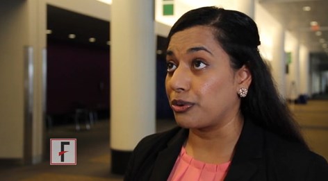
Dr. Starling discussed some of the implications of the CGRP antagonists in development in a video interview.
Both studies were funded by Amgen. Dr. Goadsby reported numerous grants and personal fees from multiple drug makers, including Amgen. Dr. Starling reported support from Amgen, eNeura, and Eli Lilly. Dr. Dodick disclosed many relationships with pharmaceutical companies developing or marketing drugs for headache and migraine, including Amgen.
The video associated with this article is no longer available on this site. Please view all of our videos on the MDedge YouTube channel
AT AAN 2017
ENDEAR Study Demonstrates Efficacy of Nusinersen in Infants With Spinal Muscular Atrophy
BOSTON—Infants with spinal muscular atrophy (SMA) type 1 who were treated with nusinersen demonstrated clinically and statistically significant gains across multiple efficacy end points, according to a report presented at the 69th Annual Meeting of the American Academy of Neurology. Nancy L. Kuntz, MD, an attending physician at the Ann and Robert H. Lurie Children’s Hospital of Chicago, on behalf of the ENDEAR Study Group, reported the final results of the phase III ENDEAR study assessing efficacy and safety of nusinersen in infants with SMA.
SMA is a rare, debilitating, autosomal recessive neuromuscular disorder causing varying degrees of weakness. The disease is caused by insufficient levels of SMN protein. Nusinersen is an antisense oligonucleotide that promotes the production of full-length SMN protein.
The ENDEAR study was a phase III, randomized, double-blind, sham-procedure controlled 13-month study to assess the efficacy and safety of nusinersen in infants with SMA. The ENDEAR study had an interim efficacy analysis in September of 2016. This analysis showed that the primary end point—motor milestone response—was positive in 41% of nusinersen-treated infants, and information was submitted to the FDA. Under priority review, Spinraza (nusinersen) was approved for the treatment of SMA in pediatric and adult patients by the FDA on December 23, 2016.
Study Design
Symptomatic infants diagnosed with SMA (with clinical features consistent with type 1 SMA) were randomized (2:1) to receive intrathecal nusinersen (12-mg scaled equivalent dose) or sham procedure. For both groups, four doses were given over two months, on days 1, 15, 29, and 64. This was followed by a maintenance phase, with dosing every four months.
Key eligibility criteria included 5q SMN1 homozygous gene deletion or mutation, two SMN2 gene copies, onset of SMA symptoms at younger than 6 months, and no hypoxemia at baseline screening at age 7 months or younger. A total of 122 infants were enrolled.
Primary end points included proportion of modified section 2 Hammersmith Infant Neurological Examination (HINE) motor milestone responders (ie, more categories improving [≥ 2-point increase or maximal score in kicking ability, or ≥ 1-point increase in head control, rolling, sitting, crawling, standing, or walking] than worsening) and event-free survival (time to death or permanent ventilation). Secondary end points included percentage of Children’s Hospital of Philadelphia Infant Test of Neuromuscular Disorders (CHOP INTEND) responders (≥ 4-point increase), overall survival, and percentage of peroneal nerve compound muscle action potential (CMAP) responders (amplitude ≥ 1 mV).
The preplanned interim efficacy analysis was triggered when two-thirds of the infants reached day 183 involvement in the study. Because the primary end point—motor milestone response—was positive, the study was ended, and all of the infants were transferred into the open-label extension study, which is called SHINE. Event-free survival and all of the secondary end points were not assessed at the ENDEAR interim analysis. With further analysis now complete, Dr. Kuntz presented the end-of-study data set.
ENDEAR Final Results
At the end of the study, there was a significantly greater proportion of nusinersen-treated motor milestone responders versus sham-control responders (51% vs 0%), demonstrating continued improvement over the previous interim analysis (41% vs 0%). In the nusinersen-treated group, 22% of infants developed full head control, 10% of the infants developed the ability to independently roll from supine to prone positions, 8% developed independent sitting, with half of those being able to sit and pivot, and one infant was able to stand with minimal to moderate support.
Looking at change over time, the improvement in HINE motor milestone scores seen in ENDEAR matches the trajectory seen in a previous open-label trial. Patients in the previous trial have now been followed for another year or so, and they slowly continue to attain their motor milestones. Additionally, infants with presymptomatic SMA who were identified and treated within the first six weeks of life showed improvements in the rate and the range of their motor skills that were much greater than those in the other groups, suggesting that early treatment makes a difference.
Additional analyses included event-free survival, overall survival, CHOP INTEND score, peroneal nerve CMAP response, and need for mechanical ventilation. A significant nusinersen treatment benefit was seen with regard to event-free survival (hazard ratio = 0.530) and overall survival (hazard ratio = 0.372). Dr. Kuntz reported that 61% of the nusinersen-treated infants were alive at the end of the study, compared with 32% of controls. For nusinersen versus sham-control infants, 71% versus 3% were CHOP INTEND responders, and 36% versus 5% were CMAP responders. The risk of permanent ventilation was 34% lower in the nusinersen-treated group. Over the course of the study, 31% of the nusinersen-treated infants required permanent ventilation, defined as at least 16 hours per day, compared with 48% of the control infants.
The ENDEAR study was supported by Ionis Pharmaceuticals and Biogen.
Good News, Bad News
Following Dr. Kuntz’s plenary presentation of the ENDEAR study results, Charlotte J. Sumner, MD, Associate Professor of Neurology at Johns Hopkins University in Baltimore, served as the discussant. While Dr. Sumner praised the study findings and the breakthrough they represent, she did point out the staggering cost of the drug. At about $120,000 per dose, the price “has raised issues about insurance approval and reimbursement and raises concerns about delays
to treatment initiation and institutional risk,” she said. “But I would say that despite these challenges, well over 100 patients have already been dosed commercially at very different ages, and this is very promising that we will be able to deliver this drug in a widespread way.”
—Glenn S. Williams
Suggested Reading
Finkel RS, Chiriboga CA, Vajsar J, et al. Treatment of infantile-onset spinal muscular atrophy with nusinersen: a phase 2, open-label, dose-escalation study. Lancet. 2016;388(10063):3017-3026.


