User login
American College of Chest Physicians (ACCP): Annual International Scientific Assembly (CHEST 2015)
Yoga Performs Like Pulmonary Rehab for COPD Patients
MONTREAL – A structured regimen of regular yoga exercises was as effective as a standard pulmonary rehabilitation program in patients with chronic obstructive pulmonary disease for improving lung function, exercise tolerance, dyspnea severity, and quality of life in a single-center, randomized comparison of the two strategies with 60 patients.
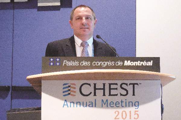
In addition, chronic obstructive pulmonary disease (COPD) patients had a higher level of acceptance of yoga and were more comfortable doing it, compared with standard pulmonary rehabilitation, and it is a cost-effective approach given the minimal equipment required, Dr. Randeep Guleria said at the annual meeting of the American College of Chest Physicians.
“Patients with difficulty walking, osteoarthritis, knee problems, or unable to do exercises like cycling or treadmill found yoga to be much more acceptable,” said Dr. Guleria in an interview. Acceptance of yoga was also higher than standard rehabilitation among patients with more severe COPD, said Dr. Guleria, professor and head of the department of pulmonary medicine and sleep disorders at the All India Institute of Medical Sciences in New Delhi.
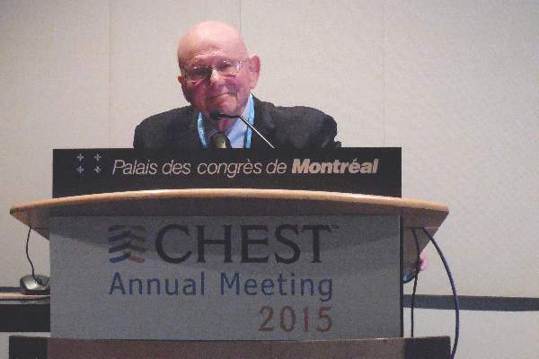
“I think that yoga could be a very valuable adjunct” to pulmonary rehabilitation in COPD patients, commented Dr. Roger S. Goldstein, director of the divisional program in respiratory rehabilitation at the University of Toronto. Dr. Goldstein speculated that even better than comparing yoga against conventional pulmonary rehabilitation would be a study that compared a combined yoga plus rehabilitation program against standard rehabilitation alone. Yoga is “a tremendous opportunity,” he said.
The 12-week study enrolled 60 patients who averaged 56 years old who had been diagnosed with COPD for an average of 8 years. Just under a third of the patients had moderate COPD, 42% had severe COPD, and 28% had very severe COPD.
Dr. Guleria and his associates randomized 30 patients into a yoga program that included 4 weeks of biweekly 1-hour sessions that instructed patients in a series of specially designed yoga exercises. That was followed by 8 weeks during which patients were mostly left to perform their learned exercises on their own, but with a supervised session once every 2 weeks. The other 30 patients participated in a standard pulmonary rehabilitation program for 12 weeks.
The researchers measured several parameters at baseline and after 12 weeks, including two measures of dyspnea severity, 6-minute walk distance, a quality of life assessment, and two serum markers of inflammation, C-reactive protein and interleukin 6.
Both interventions resulted in modest but statistically significant improvements, such as increases in 6-minute walk distance and a reduced modified Borg scale assessment. The Borg scale score fell from an average of 1.5 at baseline to 1.0 after 12 weeks in the yoga patients, and from an average 3.0 at baseline to 0.5 after 12 weeks in the rehabilitation patients.
A score that measured total quality of life improved by an average of 32% in the yoga patients and by an average of 21% in the rehabilitation patients, changes that approached statistical significance in both subgroups.
Comparing the assessment measures after 12 weeks in both arms of the study showed no statistically significant between-group differences, Dr. Guleria reported.
The researchers commissioned a specially designed yoga program from a professional yoga instructor who had been briefed about COPD. The yoga exercises included physical postures, breathing technique, and meditation and relaxation. The pulmonary rehabilitation program included patient education, upper and lower limb exercises, and breathing exercises.
Dr. Guleria said he believed the opportunity exists to further modify the yoga program to better optimize its potential to benefit COPD patients.
Dr. Guleria had no relevant disclosures. Dr. Goldstein had no relevant disclosures.
The video associated with this article is no longer available on this site. Please view all of our videos on the MDedge YouTube channel
On Twitter@mitchelzoler
AT CHEST 2015
Radiation exposure exceeds 50 mSv in 2% of ICU patients
MONTREAL – Some of the sickest patients treated at U.S. hospitals receive high levels of radiation exposure, based on a review of more than 4,000 medical ICU patients treated recently at one U.S. quaternary-care center.
During 2013, 98 patients admitted to the medical ICU at the Cleveland Clinic – 2% of the 4,155 patients who passed through the medical ICU that year – had cumulative radiation exposure of at least 50 mSv while in the ICU, thereby exceeding the U.S. standard for maximum annual workplace exposure, Dr. Sudhir Krishnan said at the annual meeting of the American College of Chest Physicians. The finding raises questions of whether all these exposures are appropriate and whether they reflect overuse of certain imaging modalities.
Dr. Krishnan and his associates ran a retrospective review of case records for the medical ICU–admitted patients at the Cleveland Clinic during 2013 (Chest. 2015 Oct 25. doi: 10.1378/chest.2278486). During their ICU stay, 3,490 patients (84%) received some amount of radiation exposure. Exposure averaged 7 mSv, with a median of 1.5 mSv. The radiation exposure came primarily from imaging and more specifically from CT examinations, which produced more than half of all radiation-exposure episodes. Other sources included x-rays, nuclear scans, and interventional procedures.
Based on typical radiation dosages received during each type of procedure, the researchers calculated an estimated total radiation dosage received by each patient during their ICU stay. Nearly two-thirds of patients had an exposure of less than 3 mSv, the average annual exposure a person receives from ambient radiation. A quarter of the patients had an exposure of 3-14 mSv, 11% had an exposure of 15-49 mSv, and 2% – 98 patients – had exposure during their ICU stay that ran to 50 mSv or greater, exceeding the U.S. workplace annual maximum . Thirteen patients had an exposure level during their ICU stay that reached 100 mSv or higher; the maximum exposure level was in a patient with cumulative exposure of 176 mSv, said Dr. Krishnan, a critical-care medicine specialist at the Cleveland Clinic.
He and his coworkers did a multivariate analysis to identify factors that linked with a higher likelihood of having high radiation exposure. Patients at greatest risk for high exposure levels were sicker patients with higher APACHE 3 scores, longer stays in the ICU, and the presence of cirrhosis, but those most at risk also tended to be younger. Rates of both ICU deaths and deaths during the entire hospitalization were significantly higher among those with radiation exposure that was 50 mSv or greater.
Dr. Krishnan cautioned that he has not run any analysis that assessed the appropriateness of the imaging that the ICU patients received, nor did he have any data documenting the clinical consequences to the patients who had higher radiation exposure. Despite that uncertainty, he suggested that efforts focus on avoiding unnecessary radiation exposure to patients.
On Twitter @mitchelzoler
AT CHEST 2015
Key clinical point: A small but significant percentage of medical ICU patients receive cumulative radiation doses that exceed federal standards for annual workplace exposure.
Major finding: Two percent of medical ICU patients received at least 50 mSv of radiation exposure during their ICU stay.
Data source: Single-center, retrospective study with 4,155 consecutive medical ICU patients during 2013.
Disclosures: Dr. Krishnan had no disclosures.
CHEST: Catheter-directed thrombolysis shows pulmonary embolism efficacy
MONTREAL – Catheter-directed thrombolysis surpassed systemic thrombolysis for minimizing in-hospital mortality of patients with an acute pulmonary embolism in a review of more than 1,500 U.S. patients.
The review also found evidence that U.S. pulmonary embolism (PE) patients increasingly undergo catheter-directed thrombolysis, with usage jumping by more than 50% from 2010 to 2012, although in 2012 U.S. clinicians performed catheter-directed thrombolysis on 160 patients with an acute pulmonary embolism (PE) who were included in a national U.S. registry of hospitalized patients, Dr. Amina Saqib said at the annual meeting of the American College of Chest Physicians.
Catheter-directed thrombolysis resulted in a 9% in-hospital mortality rate and a 10% combined rate of in-hospital mortality plus intracerebral hemorrhages, rates significantly below those tallied in propensity score–matched patients who underwent systemic thrombolysis of their acute PE. The matched group with systemic thrombolysis had a 17% in-hospital mortality rate and a 17% combined mortality plus intracerebral hemorrhage rate, said Dr. Saqib, a researcher at Staten Island (N.Y.) University Hospital.
“To the best of our knowledge, this is the first, large, nationwide, observational study that compared safety and efficacy outcomes between systemic thrombolysis and catheter-directed thrombolysis in acute PE,” Dr. Saqib said.
The U.S. data, collected during 2010-2012, also showed that, after adjustment for clinical and demographic variables, each acute PE treatment by catheter-directed thrombolysis cost an average $9,428 above the cost for systemic thrombolysis, she said.
“We need to more systematically identify the patients with an acute PE who could benefit from catheter-directed thrombolysis, especially patients with a massive PE,” commented Dr. Muthiah P. Muthiah, a critical-care medicine physician at the University of Tennessee Health Science Center in Memphis. “This may be something to offer to patients who have an absolute contraindication for systemic thrombolysis, such as recent surgery, but it is not available everywhere,” Dr. Muthiah said in an interview.
Dr. Saqib and her associates used data collected by the Federal National Inpatient Sample. Among U.S. patients hospitalized during 2010-2012 and entered into this database, they identified 1,169 adult acute PE patients who underwent systemic thrombolysis and 352 patients who received catheter-directed thrombolysis. The patients averaged about 58 years old and just under half were men.
The propensity score–adjusted analysis also showed no statistically significant difference between the two treatment approaches for the incidence of intracerebral hemorrhage, any hemorrhages requiring a transfusion, new-onset acute renal failure, or hospital length of stay. Among the patients treated by catheter-directed thrombolysis, all the intracerebral hemorrhages occurred during 2010; during 2011 and 2012 none of the patients treated this way had an intracerebral hemorrhage, Dr. Saqib noted.
Although the findings were consistent with results from prior analyses, the propensity-score adjustment used in the current study cannot fully account for all unmeasured confounding factors. The best way to compare catheter-directed thrombolysis and systemic thrombolysis for treating acute PE would be in a prospective, randomized study, Dr. Saqib said.
Dr. Saqib and Dr. Muthiah had no disclosures.
On Twitter @mitchelzoler
AT CHEST 2015
Key clinical point: Catheter-directed thrombolysis was linked to reduced mortality, compared with systemic thrombolysis in patients with an acute pulmonary embolism.
Major finding: In-hospital mortality in acute pulmonary embolism patients ran 10% with catheter-directed thrombolysis and 17% with systemic thrombolysis.
Data source: Review of 1,521 U.S. patients treated for acute pulmonary embolism during 2010-2012 in the National Inpatient Sample.
Disclosures: Dr. Saqib and Dr. Muthiah had no disclosures.
CHEST: Less intrusive respiratory support scores CCU success
MONTREAL – A relatively new system for delivering noninvasive respiratory support, high-flow nasal cannula therapy, appeared well suited to ventilation therapy for coronary care unit patients, according to a single-center experience in New Zealand with 497 adult patients.
“The high-flow nasal cannula largely replaced noninvasive ventilation in our CCU patients with no change in escalation rates or mortality. The high-flow nasal cannula should be considered a first-line treatment in this setting,” said Dr. Troy S. Browne at the annual meeting of the American College of Chest Physicians.
He and his associates at Tauranga (New Zealand) Hospital compared their experience using standard noninvasive ventilation on 249 CCU patients who needed respiratory support during May-November 2012 with 248 patients who received ventilation with a high-flow nasal cannula once it became the default strategy in the coronary care unit. The high-flow nasal cannula produced similar outcomes while being “better tolerated and less of a burden for the nursing staff to manage,” said Dr. Browne, an anesthesiologist and critical care medicine specialist at Tauranga Hospital. A key feature of the high-flow nasal cannula system is the warmed and humidified mix of oxygen and air that it pumps.
“If you run a nasal cannula without this, you dry out the patient’s nose at 5 or 6 L/min, and it’s very uncomfortable. But I’ve worn a high-flow nasal cannula with 30 L/min flow and I couldn’t tell I was wearing it,” commented Dr. Thomas Fuhrman, chief of anesthesiology at the Bay Pines (Fla.) VA Healthcare System. “It’s high enough flow to open the lungs but much better tolerated” than standard noninvasive ventilation, Dr. Fuhrman said in an interview. Patients can open their mouths, swallow, and eat while wearing the high-flow nasal cannula, he noted.
“This report is a step forward and will help spur adoption” of the high-flow nasal cannula, Dr. Fuhrman predicted. Many cardiologists are not yet aware of it as it’s usually placed by intensivists. Over time, additional indications for the device will develop, he added.
The specific system used in Tauranga Hospital”s coronary care unit is the Optiflow high-flow nasal cannula along with the Airvo pumping system, both marketed by Fisher & Paykel Healthcare. During the 6 months preceding November 2012, 107 of 249 (43%) CCU patients underwent noninvasive ventilation during the first 40 hours following CCU admission. During the subsequent period ventilation started on 67 of 248 (27%) during the first 40 hours. Of those, 60 (90%) received sufficient treatment with a high-flow nasal cannula, while 7 (10%) required standard noninvasive ventilation. Patient outcomes during the two periods were similar.
A more definitive comparison of noninvasive ventilation and high-flow nasal cannula treatment requires a prospective, randomized study, Dr. Browne said.
On Twitter @mitchelzoler
AT CHEST 2015
Key clinical point: Most CCU patients requiring respiratory support received adequate treatment from a new and less intrusive system.
Major finding: Sixty of 67 (90%) CCU patients received adequate respiratory support from a high-flow nasal cannula.
Data source: Single-center, prospective series of 248 patients and a historical comparison group of 249 patients.
Disclosures: The study received partial funding from Fisher & Paykel Healthcare, the company that markets the respiratory support equipment tested in the study. Dr. Browne had no disclosures. One of the coauthors on the study is a Fisher & Paykel employee.
VIDEO: MMF equals cyclophosphamide’s efficacy in sclerodermal lung disease
MONTREAL – The immunosuppressant mycophenolate mofetil worked as effectively as cyclophosphamide for treating scleroderma-related interstitial lung disease while being better tolerated and causing fewer adverse effects in a multicenter, head-to-head comparison with 142 randomized patients.
“The findings support the increasingly common clinical practice of prescribing MMF [mycophenolate mofetil] for this disease,” said Dr. Donald P. Tashkin at the annual meeting of the American College of Chest Physicians.
Another limitation of cyclophosphamide is that it is usually not used for more than 1 year because of concerns that longer use substantially increases a patient’s risk for developing malignancy. That’s another reason why there is a “strong need for longer and safer immunosuppressive treatment with a drug like MMF,” said Dr. Tashkin, a pulmonologist at the University of California, Los Angeles.
When used in this trial on patients with scleroderma, as defined by the American College of Rheumatology and with a baseline forced vital capacity of no more than 80% of predicted, “MMF was effective at reducing the rate of decline in vital capacity, improving symptoms such as dyspnea – the cardinal symptom of interstitial lung disease, and reducing lung fibrosis seen on CT scans, and MMF was better tolerated” than cyclophosphamide, Dr. Tashkin said in an interview. Cyclophosphamide treatment in this new trial “was associated with more toxicity, especially hematologic toxicity, an was not nearly as well tolerated, with more patients withdrawing because of side effects or a perceived lack of benefit.”
“Cyclophosphamide has a lot of side effects. MMF is just now coming into increased use. I think we’ll see it being used more for first-line treatment because the side effects with cyclophosphamide are so bad,” commented Dr. Thomas Fuhrman, chief of anesthesiology at the Bay Pines (Fla.) VA Healthcare System.
Dr. Tashkin and his associates conceived the Scleroderma Lung Study II (SLSII) as a follow-up to the first SLS run about a decade ago that compared cyclosphosphamide against placebo for controlling progression of interstitial lung disease in scleroderma patients. The results from the first SLS trial established cyclosphosphamide as a treatment that could preserve forced vital capacity percent predicted in patients with scleroderma-induced interstitial lung disease (N Engl J Med. 2006 Jun 22;354[25]:2655-666).
For the new study they enrolled patients who averaged 52 years old, with an average scleroderma duration of almost 3 years. Their average percent predicted forced vital capacity was 67%, and their baseline dyspnea index was 7.1.
Patients received either a target oral MMF dosage of 1.5 g b.i.d. for 2 years, or a target cyclophosphamide dosage of 2 mg/kg/day for up to 1 year, followed by a year of placebo. Cyclophosphamide treatment was capped at 1 year to protect against causing malignancy. Among the 73 patients randomized to the cyclophosphamide arm, 58 had data available after 12 months with 48 patients continuing on cyclophosphamide, and 53 had data available out to 2 years, with 37 patients remaining on their assigned regimen. Among 69 patients randomized to MMF 58 had data available after 12 months with 53 continuing on MMF, and 53 patients had data available through 24 months with 49 remaining on their MMF regimen.
After 24 months, the average percent predicted forced vital capacity, the study’s primary endpoint, had increased by 3.3% among patients on MMF and 3.0% among those in the cyclophosphamide arm in an intention-to-treat analysis, a nonsignificant difference. After 24 months 72% of patients in the MMF arm and 65% in the cyclophosphamide arm had a positive change, compared with baseline, in their percent predicted forced vital capacity, Dr. Tashkin reported.
MMF also showed a superior overall safety profile. Patients on cyclophosphamide had a significantly increased rate of withdrawal from the study medication. Drug discontinuations occurred in 36 of the cyclophosphamide patients and in 20 of those on MMF. Serious adverse events attributable to study medication occurred in eight patients on cyclophosphamide and three patients on MMF. The most frequent protocol-defined adverse event was leukopenia, which occurred in 30 patients on cyclophosphamide and four patients on MMF.
The video associated with this article is no longer available on this site. Please view all of our videos on the MDedge YouTube channel
On Twitter @mitchelzoler
AT CHEST 2015
Key clinical point: Mycophenolate mofetil controlled scleroderma-induced interstitial lung disease as well as cyclosphosphamide did but with reduced toxicity.
Major finding: Average percent predicted forced vital capacity rose 3.0% in patients treated with cyclosphosphamide and 3.3% in those on MMF.
Data source: SLSII, a randomized trial that enrolled 142 scleroderma patients at 13 U.S. centers.
Disclosures: SLSII received partial funding from Hoffman-La Roche, a company that markets a formulation of mycophenolate mofetil (CellCept). Dr. Tashkin had no disclosures. Dr. Fuhrman had no disclosures.
CHEST: Losartan slows emphysema progression in pilot trial
MONTREAL – Daily treatment with the antihypertensive drug losartan for a year appeared to slow progression of emphysema, compared with placebo, in a controlled pilot study involving 46 patients at one U.S. center, Dr. Allison Lambert reported at the annual meeting of the American College of Chest Physicians.
One year of daily, 100-mg oral treatment with the angiotensin receptor–blocking drug losartan in patients with emphysema produced a statistically significant regression of the disease in their right-middle lung lobes, compared with progression of right-middle lung lobe disease among control patients, as assessed by CT lung scans. In this lung segment, emphysema regressed by 0.72% in patients on losartan (Cozaar), compared with an average 3.34% rate of progression in patients on placebo.
The losartan-treated patients also showed a consistent pattern of either substantially slowed or reversed emphysema throughout multiple lung segments, although the between-group differences did not reach statistical significance in any other segment, said Dr. Lambert, a pulmonologist at Johns Hopkins Hospital in Baltimore.
Throughout the entire lung, 12 months of losartan treatment was linked to an average 0.32% reduction in emphysema extent from baseline when measured by CT, compared with a 2.18% rate of emphysema progression in control patients on usual care, which just missed statistical significance (P = .064).
Data from other researchers “suggest the right-middle lobe most commonly progresses in emphysema,” which may explain why that lung segment showed the most dramatic effect from treatment, she suggested. Another finding Dr. Lambert found striking was the consistent trend toward slowed emphysema progression in multiple segments throughout patients’ lungs.
Dr. Lambert called this a phase II, “proof of concept” trial. She and her associates have begun a larger, phase III version that will study the effect of 100-mg daily losartan during 1 year of treatment in 220 patients with emphysema, she said. This trial received funding from the Pulmonary Trials Cooperative of the National Heart Lung and Blood Institute.
“These are some of the most interesting and exciting data I’ve seen,” commented Dr. David P.L. Sachs, a pulmonologist who practices in Stanford, Calif., and cochaired the session in which Dr. Lambert gave her report. “Having an agent that could slow progression of emphysema would be unique,” he said in an interview. One aspect that makes this treatment especially attractive is losartan’s extensive safety record as an antihypertensive drug that is also often used to treat patients with heart failure, Dr. Sachs noted.
To put the 100-mg/day dosage used in the current study in perspective, results from a multicenter randomized trial of more than 3,800 heart failure patients published in 2009 showed that a losartan dosage of 150 mg once daily was safe and effective and produced outcomes superior to those seen with a 50 mg once-daily dosage (Lancet. 2009;374[9704]:1840-8).
Dr. Lambert and her associates designed this pilot study because of previously reported results from other groups showing favorable effects of losartan on animal models of emphysema or other lung disease, as well as suggestions from nonprospective clinical studies of benefits from angiotensin-receptor blockers on lung function and on chronic obstructive pulmonary disease (COPD) in particular.
The current study enrolled patients with mild to severe COPD who were current or former cigarette smokers who had at least a 10 pack-year history and were on stable treatment for their COPD. The researchers excluded patients already taking an angiotensin-receptor blocker or angiotensin-converting enzyme inhibitor.
The study included a total of 106 patients with COPD, including the 46 with emphysema at baseline. Their average age was about 58 years old, the enrolled patients included roughly equal numbers of men and women, and about two-thirds were current smokers. All patients underwent CT lung scans at baseline and after 6 and 12 months, as well as other lung function assessments. The study’s primary endpoint was the amount of additional emphysema in their lungs beyond that seen at baseline using CT imaging.
The entire group of 106 COPD patients showed no significant differences in emphysema progression at either 6 or 12 months between the 54 patients treated with losartan and the 52 controls on placebo, but a second, prespecified analysis that focused only on the 46 enrolled patients who had visible emphysema at baseline showed a significant slowing of progression at the 1-year follow-up, Dr. Lambert said.
The study received partial funding from Merck, which markets losartan (Cozaar). Dr. Lambert had no disclosures. Dr. Sachs had no disclosures.
On Twitter @mitchelzoler
AT CHEST 2015
Key clinical point: A year of daily treatment with losartan produced significant slowing of emphysema progression in a placebo-controlled pilot study with 46 patients.
Major finding: Right middle-lobe emphysema regressed by 0.72% in patients using losartan, compared with 3.34% progression in controls, measured by CT imaging.
Data source: A single-center, prospective, controlled study involving 46 patients with emphysema and 60 additional patients with less severe COPD.
Disclosures: The study received partial funding from Merck, which markets losartan (Cozaar). Dr. Lambert had no disclosures. Dr. Sachs had no disclosures.
CHEST: Yoga performs like pulmonary rehab for COPD patients
MONTREAL – A structured regimen of regular yoga exercises was as effective as a standard pulmonary rehabilitation program in patients with chronic obstructive pulmonary disease for improving lung function, exercise tolerance, dyspnea severity, and quality of life in a single-center, randomized comparison of the two strategies with 60 patients.
In addition, chronic obstructive pulmonary disease (COPD) patients had a higher level of acceptance of yoga and were more comfortable doing it, compared with standard pulmonary rehabilitation, and it is a cost-effective approach given the minimal equipment required, Dr. Randeep Guleria said at the annual meeting of the American College of Chest Physicians.
“Patients with difficulty walking, osteoarthritis, knee problems, or unable to do exercises like cycling or treadmill found yoga to be much more acceptable,” said Dr. Guleria in an interview. Acceptance of yoga was also higher than standard rehabilitation among patients with more severe COPD, said Dr. Guleria, professor and head of the department of pulmonary medicine and sleep disorders at the All India Institute of Medical Sciences in New Delhi.
“I think that yoga could be a very valuable adjunct” to pulmonary rehabilitation in COPD patients, commented Dr. Roger S. Goldstein, director of the divisional program in respiratory rehabilitation at the University of Toronto. Dr. Goldstein speculated that even better than comparing yoga against conventional pulmonary rehabilitation would be a study that compared a combined yoga plus rehabilitation program against standard rehabilitation alone. Yoga is “a tremendous opportunity,” he said.
The 12-week study enrolled 60 patients who averaged 56 years old who had been diagnosed with COPD for an average of 8 years. Just under a third of the patients had moderate COPD, 42% had severe COPD, and 28% had very severe COPD.
Dr. Guleria and his associates randomized 30 patients into a yoga program that included 4 weeks of biweekly 1-hour sessions that instructed patients in a series of specially designed yoga exercises. That was followed by 8 weeks during which patients were mostly left to perform their learned exercises on their own, but with a supervised session once every 2 weeks. The other 30 patients participated in a standard pulmonary rehabilitation program for 12 weeks.
The researchers measured several parameters at baseline and after 12 weeks, including two measures of dyspnea severity, 6-minute walk distance, a quality of life assessment, and two serum markers of inflammation, C-reactive protein and interleukin 6.
Both interventions resulted in modest but statistically significant improvements, such as increases in 6-minute walk distance and a reduced modified Borg scale assessment. The Borg scale score fell from an average of 1.5 at baseline to 1.0 after 12 weeks in the yoga patients, and from an average 3.0 at baseline to 0.5 after 12 weeks in the rehabilitation patients.
A score that measured total quality of life improved by an average of 32% in the yoga patients and by an average of 21% in the rehabilitation patients, changes that approached statistical significance in both subgroups.
Comparing the assessment measures after 12 weeks in both arms of the study showed no statistically significant between-group differences, Dr. Guleria reported.
The researchers commissioned a specially designed yoga program from a professional yoga instructor who had been briefed about COPD. The yoga exercises included physical postures, breathing technique, and meditation and relaxation. The pulmonary rehabilitation program included patient education, upper and lower limb exercises, and breathing exercises.
Dr. Guleria said he believed the opportunity exists to further modify the yoga program to better optimize its potential to benefit COPD patients.
Dr. Guleria had no relevant disclosures. Dr. Goldstein had no relevant disclosures.
The video associated with this article is no longer available on this site. Please view all of our videos on the MDedge YouTube channel
On Twitter@mitchelzoler
AT CHEST 2015
Key clinical point: COPD patients randomized to a 12-week program of yoga exercises had improvements similar to patients randomized to a standard pulmonary rehabilitation program.
Major finding: Average total quality of life scores improved by a relative 32% with yoga and 21% with standard rehabilitation.
Data source: Single-center, prospective, randomized trial with 60 patients.
Disclosures: Dr. Guleria had no relevant disclosures. Dr. Goldstein had no relevant disclosures.
CHEST: Oral solithromycin shows pneumonia pivotal-trial efficacy
MONTREAL – A new, next-generation macrolide, solithromycin, showed safety and efficacy as a once-daily oral agent that was noninferior to the comparator oral antibiotic, the fluoroquinolone moxifloxacin, in a phase III trial.
Macrolide resistance among strains of Streptococcus pneumoniae that cause many U.S. cases of severe community-acquired pneumonia has become common, complicating treatment of this common infection with a macrolide, Dr. Carlos M. Barrera explained at the annual meeting of the American College of Chest Physicians.
The SOLITAIRE-ORAL (Efficacy and Safety Study of Oral Solithromycin [CEM-101] Compared to Oral Moxifloxacin in Treatment of Patients With Community-Acquired Bacterial Pneumonia) trial enrolled 860 patients with moderate to moderately severe community-acquired pneumonia.
About half of the patients enrolled in the trial underwent microbiologic assessment of their infecting pathogen, and about 40% of cases in each treatment arm had infections caused by S. pneumoniae. In this subgroup, the 5-day regimen of solithromycin tested in the study succeeded in clearing the infection in 89% of patients, comparable to the 83% success rate achieved with a 7-day course of moxifloxacin (Avelox), said Dr. Barrera, a pulmonologist who practices in Miami.
The study’s primary endpoint for Food and Drug Administration approval of solithromycin was early clinical response, defined as an improvement in at least two listed symptoms at 72 hours after onset of treatment. That endpoint occurred in 78% of patients enrolled in each of the two arms of the study.
The data make solithromycin look like a promising way to once again have a macrolide available for empiric oral treatment of more severe community-acquired pneumonia, pending full peer review of the data, commented Dr. Muthiah P. Muthiah, a pulmonologist at the University of Tennessee Health Science Center in Memphis.
“A couple of decades ago, you could comfortably treat a patient with severe community-acquired pneumonia with a macrolide, but you can’t do that anymore,” Dr. Muthiah said in an interview.
If the newly reported data on oral solithromycin hold up under further review, it would mean that solithromycin was as effective as a potent quinolone, which remains an effective monotherapy for community-acquired pneumonia in patients who do not require treatment in an intensive care unit, Dr. Muthiah noted.
A companion study, SOLITAIRE-IV, is a phase III pivotal trial assessing the safety and efficacy of solithromycin when begun intravenously for treating community-acquired pneumonia, followed by a switch to oral dosing, in comparison with intravenous followed by oral treatment with moxifloxacin.
Once those data are fully collected and analyzed, the company will submit the information from both trials to the FDA, said Dr. David Oldach, chief medical officer for Cempra.
Results from the intravenous trial, reported in a preliminary way by Cempra Oct. 16 in a press release, showed that the solithromycin treatment regimen tested in SOLITAIRE-IV met its noninferiority targets, compared with moxifloxacin. The safety results, however, showed that solithromycin produced a higher number of patients with a liver-enzyme elevation, compared with patients treated with moxifloxacin.
In SOLITAIRE-IV, Cempra reported that grade 3 increase in levels of alanine transaminase (ALT) occurred in 8% of patients on solithromycin and in 3% of patients on moxifloxacin. Grade 4 increases in ALT occurred in less than 1% of patients in both treatment arms.
In the current, orally administered trial, grade 3 ALT increases occurred in 5% of patients treated with solithromycin and in 2% of patients treated with moxifloxacin, Dr. Barrera reported. Grade 4 ALT increases occurred in 0.5% of patients treated with solithromycin and in 1.2% of those treated with moxifloxacin. No patients in either arm developed an elevation of both ALT and bilirubin, and the ALT increases seen were reversible and asymptomatic, Dr. Barrera said.
By other assessments, the safety profiles of solithromycin and moxifloxacin were similar: 7% of patients on solithromycin and 6% on moxifloxacin had a serious adverse event, and 4% of patients in each study arm discontinued treatment because of an adverse event.
SOLITAIRE-ORAL was sponsored by Cempra, the company developing solithromycin. Dr. Barrera has received research funding from Cempra. Dr. Muthiah had no disclosures.
The video associated with this article is no longer available on this site. Please view all of our videos on the MDedge YouTube channel
On Twitter @mitchelzoler
AT CHEST 2015
Key clinical point: A next-generation, orally administered macrolide, solithromycin, showed efficacy that was noninferior to moxifloxacin against moderate to moderately severe community-acquired pneumonia in a phase III pivotal trial.
Major finding: Both solithromycin and moxifloxacin produced a 78% early clinical response rate, the study’s primary endpoint for Food and Drug Administration approval.
Data source: SOLITAIRE-ORAL, a multicenter, international phase III trial involving 860 patients with community-acquired pneumonia.
Disclosures: SOLITAIRE-ORAL was sponsored by Cempra, the company developing solithromycin. Dr. Barrera has received research funding from Cempra. Dr. Muthiah had no disclosures.

