User login
European Association for the Study of Diabetes (EASD): Annual Meeting
When Stepping up Type 2 Treatment, GLP-1 Agonists Have the Edge
STOCKHOLM – Patients with poorly controlled type 2 diabetes are more likely to hit glycemic targets if their add-on therapy is a GLP-1 receptor agonist.
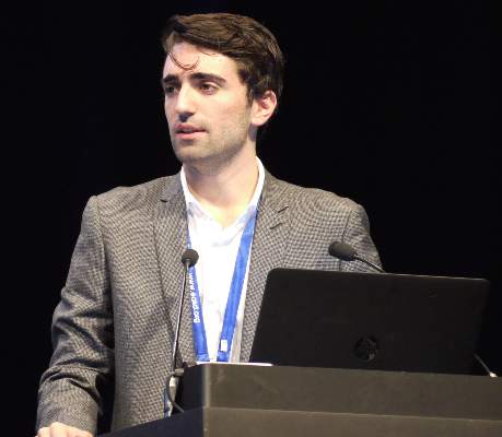
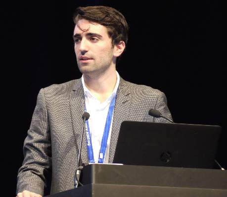
The GLP-1 drugs seemed slightly more effective than adding a bolus of premixed insulin; data from a recent study show that after receiving a GLP-1 receptor agonist as an add-on drug, patients achieved a mean hemoglobin A1c of 7.4% within 6 months of treatment intensification, Dr. Reimar W. Thomsen said at the annual meeting of the European Association for the Study of Diabetes. But his Danish database study wasn’t able to control for socioeconomic factors, which may influence whether patients are able to get that more expensive class of drug in Denmark.
Nevertheless, the study provides a “real-world” look at the management of patients who don’t hit therapeutic goals on basal insulin only, said Dr. Thomsen of Aarhus University, Denmark. In the cohort of 7,000 patients, fewer than half were able to reach an HbA1c of 7.5% or lower on the single treatment.
The patients were drawn from Danish national health care databases, and diagnosed with type 2 diabetes from 2000 to 2012. They were a mean of 64 years old with a mean baseline HbA1c of 9.2%. Although newly diagnosed, they had probably had the disease for a while – 26% had macrovascular complications and 47%, microvascular disease. All were started on basal insulin monotherapy. Within 6 months, the mean HbA1c had dropped to 7.6%.
However, just 45% of the group hit the treatment goal of 7.5% or lower, with 29% attaining the 7% HbA1c target.
A temporal assessment of response showed some improvements over the course of the study, Dr. Thomsen noted. In 2000-2003, about 25% hit the goal on basal insulin only; by 2010-2012, that had increased to 33%. This probably reflects an earlier diagnosis of disease, especially as the mean HbA1c at diagnosis was lower in the later years (9% vs.9.6%), he said.
In another noteworthy temporal association, choices for add-on therapy changed over the years. In the first few years, premixed insulin was the top choice, accounting for 80% of intensification prescriptions. By 2012, that had dropped to about 22%. Bolus insulin use rose from about 17% in 2000 to 35% in 2012. GLP-1 receptor agonists didn’t arrive on the scene until 2008 but gained rapid acceptance. By 2010-2012 they accounted for 30% of intensification prescriptions.
Patients whose glucose control was intensified with GLP-1 drugs (326) were the youngest (55 years) and healthiest, with only 22% having medical comorbidities. They started the add-on therapy at a mean of 27 months after diagnosis. At that time, the mean HbA1c was 8.4%; this dropped by 0.8%, landing at a mean of 7.6%.
Those who added bolus insulin (893) were a mean of 59 years old; 30% had comorbidities. They started the new treatment at a mean of 13 months after diagnosis. Their mean HbA1c at intensification was 8.2%; this dropped 0.4%, landing at 7.8%
Patients who added premixed insulin (1,798) were a mean of 64 years old; 32% had medical comorbidities. They started add-on therapy 11 months after diagnosis. Their HbA1c was a mean of 8.8% at intensification; it dropped 0.9%, landing at a mean of 7.9%.
Fifty-nine patients were intensified with dual therapy; Dr. Thomsen did not specify what combinations were employed. These patients were a mean of 61 years old; 42% of them had medical comorbidities. They had a mean HbA1c of 8.9% at the time of intensification. Add-on therapy dropped that measurement by 1.2%, landing this group at a mean HbA1c of 7.7%.
A multivariate analysis determined the likelihood of attaining the treatment target with the various add-on monotherapies; it adjusted for age, gender, diabetes complications, disease duration, medical comorbidities, and baseline HbA1c. Compared with premixed insulin, the chance of meeting goal was similar with bolus insulin (relative risk, 1.03) and higher with GLP-1 agonists (RR, 1.56 for less than 7%; RR, 1.27 for less than 7.5%).
Dr. Thomsen had no financial disclosures.
AT EASD 2015
When stepping up type 2 treatment, GLP-1 agonists have the edge
STOCKHOLM – Patients with poorly controlled type 2 diabetes are more likely to hit glycemic targets if their add-on therapy is a GLP-1 receptor agonist.
The GLP-1 drugs seemed slightly more effective than adding a bolus of premixed insulin; data from a recent study show that after receiving a GLP-1 receptor agonist as an add-on drug, patients achieved a mean hemoglobin A1c of 7.4% within 6 months of treatment intensification, Dr. Reimar W. Thomsen said at the annual meeting of the European Association for the Study of Diabetes. But his Danish database study wasn’t able to control for socioeconomic factors, which may influence whether patients are able to get that more expensive class of drug in Denmark.
Nevertheless, the study provides a “real-world” look at the management of patients who don’t hit therapeutic goals on basal insulin only, said Dr. Thomsen of Aarhus University, Denmark. In the cohort of 7,000 patients, fewer than half were able to reach an HbA1c of 7.5% or lower on the single treatment.
The patients were drawn from Danish national health care databases, and diagnosed with type 2 diabetes from 2000 to 2012. They were a mean of 64 years old with a mean baseline HbA1c of 9.2%. Although newly diagnosed, they had probably had the disease for a while – 26% had macrovascular complications and 47%, microvascular disease. All were started on basal insulin monotherapy. Within 6 months, the mean HbA1c had dropped to 7.6%.
However, just 45% of the group hit the treatment goal of 7.5% or lower, with 29% attaining the 7% HbA1c target.
A temporal assessment of response showed some improvements over the course of the study, Dr. Thomsen noted. In 2000-2003, about 25% hit the goal on basal insulin only; by 2010-2012, that had increased to 33%. This probably reflects an earlier diagnosis of disease, especially as the mean HbA1c at diagnosis was lower in the later years (9% vs.9.6%), he said.
In another noteworthy temporal association, choices for add-on therapy changed over the years. In the first few years, premixed insulin was the top choice, accounting for 80% of intensification prescriptions. By 2012, that had dropped to about 22%. Bolus insulin use rose from about 17% in 2000 to 35% in 2012. GLP-1 receptor agonists didn’t arrive on the scene until 2008 but gained rapid acceptance. By 2010-2012 they accounted for 30% of intensification prescriptions.
Patients whose glucose control was intensified with GLP-1 drugs (326) were the youngest (55 years) and healthiest, with only 22% having medical comorbidities. They started the add-on therapy at a mean of 27 months after diagnosis. At that time, the mean HbA1c was 8.4%; this dropped by 0.8%, landing at a mean of 7.6%.
Those who added bolus insulin (893) were a mean of 59 years old; 30% had comorbidities. They started the new treatment at a mean of 13 months after diagnosis. Their mean HbA1c at intensification was 8.2%; this dropped 0.4%, landing at 7.8%
Patients who added premixed insulin (1,798) were a mean of 64 years old; 32% had medical comorbidities. They started add-on therapy 11 months after diagnosis. Their HbA1c was a mean of 8.8% at intensification; it dropped 0.9%, landing at a mean of 7.9%.
Fifty-nine patients were intensified with dual therapy; Dr. Thomsen did not specify what combinations were employed. These patients were a mean of 61 years old; 42% of them had medical comorbidities. They had a mean HbA1c of 8.9% at the time of intensification. Add-on therapy dropped that measurement by 1.2%, landing this group at a mean HbA1c of 7.7%.
A multivariate analysis determined the likelihood of attaining the treatment target with the various add-on monotherapies; it adjusted for age, gender, diabetes complications, disease duration, medical comorbidities, and baseline HbA1c. Compared with premixed insulin, the chance of meeting goal was similar with bolus insulin (relative risk, 1.03) and higher with GLP-1 agonists (RR, 1.56 for less than 7%; RR, 1.27 for less than 7.5%).
Dr. Thomsen had no financial disclosures.
AT EASD 2015
Key clinical point: Patients with poorly controlled type 2 diabetes are more likely to hit glycemic targets if their add-on therapy is a GLP-1 receptor agonist.
Major finding: Patients with poorly controlled type 2 diabetes were 26% more likely to hit glycemic goals when their add-on therapy was a GLP-1 receptor agonist compared with another insulin.
Data source: A retrospective cohort study of 7,000 patients.
Disclosures: Dr. Thomsen had no financial disclosures.
Liraglutide/metformin Targets Glycemic Goal in Type 2 Diabetes
STOCKHOLM – Patients with poorly controlled type 2 diabetes who take the combination of liraglutide and metformin are about four times more likely to hit their glycemic target than those who take lixisenatide and metformin.
After 26 weeks of treatment, hemoglobin A1cdropped a mean of 1.8% among those on the liraglutide regimen, compared with 1.2% among those taking lixisenatide. The change brought 74% of the liraglutide group into a treatment target of HbA1c less than 7%; 45% of the lixisenatide group reached that goal (odds ratio, 4.16).
Those taking liraglutide also were almost four times as likely to reach a target of 6.5% or less (55% vs. 26%; OR, 3.6), Dr. Michael A. Nauck said at the annual meeting of the European Association for the Study of Diabetes.
Both of the drugs are glucagon-like peptide-1 receptor agonists, and both are formulated for once-daily injection, but liraglutide’s 13-hour half-life gives it a strong clinical edge.
“Lixisenatide has a 3-hour half-life, so it’s only going to be around for 6 to a maximum of 10 hours after injection,” said Dr. Nauck, head of the Diabeteszentrum in Bad Lauterberg, Germany. “For the most part of the night there’s no exposure to lixisenatide at all, while liraglutide is present 24 hours.”
The study randomized 404 patients with poorly controlled type 2 diabetes to metformin plus either liraglutide 1.8 mg/day or lixisenatide 20 mcg/day, he reported. Patients were a mean of 56 years old, with a mean body mass index of 35 kg/m2. Their mean HbA1c was 8.4%.
At the end of the 26-week study, fasting plasma glucose measurements mirrored the HbA1c improvements. Both drugs significantly reduced glucose within the first 6 weeks, but liraglutide did so to a significantly greater extent (–2.85 mmol/L vs. –1.7 mmol/L). Each reduction held steady through the end of the trial.
Both drugs effected a similar weight loss by the study’s end – about 4 kg. Both also reduced systolic blood pressure similarly, with a mean drop of about 5 mm Hg for liraglutide and 4 mm Hg for lixisenatide. Liraglutide increased the mean pulse rate from 75 beats per minute (bpm) at baseline to 80 bpm by week 6. The mean decreased to 78 by the end of the study. Pulse declined by 1 bpm from baseline among patients taking lixisenatide.
Liraglutide seemed to exert a beneficial effect on beta cells, as measured by the homeostatic model assessment (HOMA-B). At baseline, mean HOMA-B was 60%. By 26 weeks, it had increased to near 100% among those taking the drug; among those taking lixisenatide, HOMA-B had increased to about 77% – a significant difference.
Liraglutide was associated with a significant increase in lipase, Dr. Nauck said. Baseline lipase was 40 U/L. By 12 weeks, both drugs had increased lipase significantly – lixisenatide to about 50 U/L and liraglutide to about 60 U/L. These levels were constant through the study’s end. There were, however, no cases of pancreatitis or pancreatic cancer in either treatment arm, he noted.
Liraglutide use was associated with significantly more adverse events than lixisenatide (72% vs. 64%), but few of those were serious (6% vs. 3.5%). There were no cases of severe hypoglycemia and no deaths. Less than 5% of patients experienced a treatment-emergent or serious adverse event. The most common of these were nausea (22% in each group), diarrhea (12% liraglutide vs. 10% lixisenatide), vomiting (7% vs. 9%), increased lipase (8% vs. 2.5%), nasopharyngitis (6% vs. 10%), decreased appetite (6% vs. 2.5%), and dyspepsia (5.4% vs. 3%).
Although lixisenatide was outperformed in this analysis, it is still a valuable alternative for some patients, Dr. Bo Ahren said in an interview.
“Its primary action is to reduce postprandial glucose,” said Dr. Ahren, who is dean of the Faculty of Medicine at Lund (Sweden) University. “Therefore, there may be a use for this agent in patients who have controlled fasting glucose, for example, with insulin, but still have high postprandial glucose.”
Although the adverse event data were encouraging, more study is necessary, he added. The current data aren’t enough to hone any patient selection criteria.
“The lipase increases need to be examined in more detail, although it’s true there were no cases of pancreatitis. Also, the heart rate was measured only in the clinic, not as a 24-hour measurement. Therefore it’s too early to base any patient selection on this,” Dr. Ahren said.
Both Dr. Nauck and Dr. Ahren disclosed numerous financial relationships with pharmaceutical companies.
AT EASD 2015
Liraglutide/metformin targets glycemic goal in type 2 diabetes
STOCKHOLM – Patients with poorly controlled type 2 diabetes who take the combination of liraglutide and metformin are about four times more likely to hit their glycemic target than those who take lixisenatide and metformin.
After 26 weeks of treatment, hemoglobin A1cdropped a mean of 1.8% among those on the liraglutide regimen, compared with 1.2% among those taking lixisenatide. The change brought 74% of the liraglutide group into a treatment target of HbA1c less than 7%; 45% of the lixisenatide group reached that goal (odds ratio, 4.16).
Those taking liraglutide also were almost four times as likely to reach a target of 6.5% or less (55% vs. 26%; OR, 3.6), Dr. Michael A. Nauck said at the annual meeting of the European Association for the Study of Diabetes.
Both of the drugs are glucagon-like peptide-1 receptor agonists, and both are formulated for once-daily injection, but liraglutide’s 13-hour half-life gives it a strong clinical edge.
“Lixisenatide has a 3-hour half-life, so it’s only going to be around for 6 to a maximum of 10 hours after injection,” said Dr. Nauck, head of the Diabeteszentrum in Bad Lauterberg, Germany. “For the most part of the night there’s no exposure to lixisenatide at all, while liraglutide is present 24 hours.”
The study randomized 404 patients with poorly controlled type 2 diabetes to metformin plus either liraglutide 1.8 mg/day or lixisenatide 20 mcg/day, he reported. Patients were a mean of 56 years old, with a mean body mass index of 35 kg/m2. Their mean HbA1c was 8.4%.
At the end of the 26-week study, fasting plasma glucose measurements mirrored the HbA1c improvements. Both drugs significantly reduced glucose within the first 6 weeks, but liraglutide did so to a significantly greater extent (–2.85 mmol/L vs. –1.7 mmol/L). Each reduction held steady through the end of the trial.
Both drugs effected a similar weight loss by the study’s end – about 4 kg. Both also reduced systolic blood pressure similarly, with a mean drop of about 5 mm Hg for liraglutide and 4 mm Hg for lixisenatide. Liraglutide increased the mean pulse rate from 75 beats per minute (bpm) at baseline to 80 bpm by week 6. The mean decreased to 78 by the end of the study. Pulse declined by 1 bpm from baseline among patients taking lixisenatide.
Liraglutide seemed to exert a beneficial effect on beta cells, as measured by the homeostatic model assessment (HOMA-B). At baseline, mean HOMA-B was 60%. By 26 weeks, it had increased to near 100% among those taking the drug; among those taking lixisenatide, HOMA-B had increased to about 77% – a significant difference.
Liraglutide was associated with a significant increase in lipase, Dr. Nauck said. Baseline lipase was 40 U/L. By 12 weeks, both drugs had increased lipase significantly – lixisenatide to about 50 U/L and liraglutide to about 60 U/L. These levels were constant through the study’s end. There were, however, no cases of pancreatitis or pancreatic cancer in either treatment arm, he noted.
Liraglutide use was associated with significantly more adverse events than lixisenatide (72% vs. 64%), but few of those were serious (6% vs. 3.5%). There were no cases of severe hypoglycemia and no deaths. Less than 5% of patients experienced a treatment-emergent or serious adverse event. The most common of these were nausea (22% in each group), diarrhea (12% liraglutide vs. 10% lixisenatide), vomiting (7% vs. 9%), increased lipase (8% vs. 2.5%), nasopharyngitis (6% vs. 10%), decreased appetite (6% vs. 2.5%), and dyspepsia (5.4% vs. 3%).
Although lixisenatide was outperformed in this analysis, it is still a valuable alternative for some patients, Dr. Bo Ahren said in an interview.
“Its primary action is to reduce postprandial glucose,” said Dr. Ahren, who is dean of the Faculty of Medicine at Lund (Sweden) University. “Therefore, there may be a use for this agent in patients who have controlled fasting glucose, for example, with insulin, but still have high postprandial glucose.”
Although the adverse event data were encouraging, more study is necessary, he added. The current data aren’t enough to hone any patient selection criteria.
“The lipase increases need to be examined in more detail, although it’s true there were no cases of pancreatitis. Also, the heart rate was measured only in the clinic, not as a 24-hour measurement. Therefore it’s too early to base any patient selection on this,” Dr. Ahren said.
Both Dr. Nauck and Dr. Ahren disclosed numerous financial relationships with pharmaceutical companies.
AT EASD 2015
Key clinical point: Liraglutide plus metformin bested lixisenatide plus metformin in reaching HbA1c goals.
Major finding: The liraglutide combination put 74% of patients at their glycemic target, compared with 45% of the lixisenatide group.
Data source: The study randomized 404 patients to metformin plus either liraglutide or lixisenatide.
Disclosures: Both Dr. Nauck and Dr. Ahren disclosed numerous financial relationships with pharmaceutical companies.
Long-acting Basal Insulin Controls A1C as Well as Glargine
STOCKHOLM – In patients with type 1 diabetes, treatment with the long-acting basal insulin peglispro conferred lower A1C with fewer episodes of nocturnal hypoglycemia – and a bonus weight loss to boot.
“To the best of my knowledge, no registry study of any insulin analogue has ever showed that it is not only noninferior, but [also] actually better than the corresponding molecule,” said Dr. Satish K. Garg, chief of the young adult clinics at the Barbara Davis Center for Diabetes, Aurora, Colo.
The significant benefits came with some downsides, though Dr. Garg said they were neither surprising nor serious. PEGylated basal insulin lispro (BIL), which is expected to come up for review with the Food and Drug Administration in 2016, acts preferentially in the liver; patients who took it experienced increases in triglycerides and alanine aminotransferase. Some also showed a slight increase in liver fat content, although the numbers were very close to the high end of normal.
Dr. Garg presented results from Eli Lilly’s open-label IMAGINE 1 study, which randomized 455 patients with type 1 diabetes to either BIL or insulin glargine (GL) alone for 78 weeks. The primary endpoint – noninferiority of BIL as demonstrated by A1C – was collected at 26 weeks. Secondary endpoints included weight loss, total and nocturnal hypoglycemia, lipid measurements, and liver safety.
A subset of these patients, along with some from the double-blind IMAGINE 3 study, underwent liver fat measurements by MRI (182 patients).
The cohort was a typical one. Patients were a mean of 40 years old, with disease duration of about 16 years. Their body mass index was not excessive (25 kg/m2); this reflected a multinational cohort, which included patients in Asia. The baseline A1C was 7.9%; about 20% were below 7%.
They were randomized to bedtime BIL or insulin glargine (GL) with prandial insulin lispro. Most patients had been taking glargine as their baseline insulin (70%). Other baseline treatments were insulin detemir (16%) and NPH insulin (10%); the remainder used a pump.
At 26 weeks, A1C levels were significantly lower in those taking BIL than GL (7% vs. 7.4%). The measurements began to separate around week 13, and the curves remained significantly different throughout the end of the study.
Fasting serum glucose was also significantly lower among those taking BIL at 26 weeks (7.7 mmol/L vs. 8.9 mmol/L). Again, the curves separated significantly by 13 weeks and continued to do so until the study’s end.
Overall, BIL was associated with significantly more hypoglycemic events (16 vs. 12 events per patient/30 days). But those taking BIL reported significantly fewer nocturnal events (3 vs. 2 per patient/30 days).
BIL was also associated with significantly more episodes of severe hypoglycemia in both IMAGINE 1 and 3. At 26 weeks in IMAGINE 1, severe hypoglycemia was almost twice as common in the BIL group as in the GL group (11.3% vs. 5.7%). The proportion was similar at 78 weeks (15% vs. 8.2%). The rate per 100 patient-years in IMAGINE 1 was also significantly higher with BIL (23 vs. 9). In IMAGINE 3, however, the event rate associated with BIL was lower than it was with GL (19 vs. 22 per 100 patient/years; P = .445). Most of the episodes (68%) occurred within 5 hours of talking an insulin bolus, Dr. Garg noted.
Patients taking BIL also lost weight during the first half of the study, while those taking GL gained. By week 26, those taking the study drug had dropped a mean of 1.2 kg; those in the control group had gained about 0.8 kg. That difference was maintained throughput the study, although patients taking BIL did tend to gain a bit of weight in the last 8 weeks (about 0.25 kg).
Low- and high-density lipoproteins remained unchanged in both arms, but triglycerides increased significantly more in the BIL group than in the GL group (a mean of 1.3 vs 1.0 mmol/L ).
Alanine aminotransferase was the only liver enzyme affected. It increased significantly by week 13 and remained elevated the entire study, reaching about 28 U/L by week 78, compared with about 20 U/L for those taking GL.
Liver fat content was higher in the BIL group throughout the study, rising from 3% at baseline to 6% at week 78. The upper limit of normal MRI is 5.56%, Dr. Garg noted. Liver fat was unchanged in patients taking GL.
In both IMAGINE 1 and 3, there were more injection-site reactions in the BIL group (IMAGINE 1, 25% vs. 0%; IMAGINE 3, 13% vs. 0.2%). Lipohypertrophy also was more common in both IMAGINE 1 (15% vs. 0%) and IMAGINE 3 3 (7.7% vs. 0.2%).
Eli Lilly & Co. funded the study. Dr. Garg has received research funding from the company.
AT EASD 2015
Long-acting basal insulin controls HbA1c as well as glargine
STOCKHOLM – In patients with type 1 diabetes, treatment with the long-acting basal insulin peglispro conferred lower hemoglobin A1c with fewer episodes of nocturnal hypoglycemia – and a bonus weight loss to boot.
“To the best of my knowledge, no registry study of any insulin analogue has ever showed that it is not only noninferior, but [also] actually better than the corresponding molecule,” said Dr. Satish K. Garg, chief of the young adult clinics at the Barbara Davis Center for Diabetes, Aurora, Colo.
The significant benefits came with some downsides, though Dr. Garg said they were neither surprising nor serious. PEGylated basal insulin lispro (BIL), which is expected to come up for review with the Food and Drug Administration in 2016, acts preferentially in the liver; patients who took it experienced increases in triglycerides and alanine aminotransferase. Some also showed a slight increase in liver fat content, although the numbers were very close to the high end of normal.
Dr. Garg presented results from Eli Lilly’s open-label IMAGINE 1 study, which randomized 455 patients with type 1 diabetes to either BIL or insulin glargine (GL) alone for 78 weeks. The primary endpoint – noninferiority of BIL as demonstrated by HbA1c – was collected at 26 weeks. Secondary endpoints included weight loss, total and nocturnal hypoglycemia, lipid measurements, and liver safety.
A subset of these patients, along with some from the double-blind IMAGINE 3 study, underwent liver fat measurements by MRI (182 patients).
The cohort was a typical one. Patients were a mean of 40 years old, with disease duration of about 16 years. Their body mass index was not excessive (25 kg/m2); this reflected a multinational cohort, which included patients in Asia. The baseline HbA1c was 7.9%; about 20% were below 7%.
They were randomized to bedtime BIL or insulin glargine (GL) with prandial insulin lispro. Most patients had been taking glargine as their baseline insulin (70%). Other baseline treatments were insulin detemir (16%) and NPH insulin (10%); the remainder used a pump.
At 26 weeks, HbA1c levels were significantly lower in those taking BIL than GL (7% vs. 7.4%). The measurements began to separate around week 13, and the curves remained significantly different throughout the end of the study.
Fasting serum glucose was also significantly lower among those taking BIL at 26 weeks (7.7 mmol/L vs. 8.9 mmol/L). Again, the curves separated significantly by 13 weeks and continued to do so until the study’s end.
Overall, BIL was associated with significantly more hypoglycemic events (16 vs. 12 events per patient/30 days). But those taking BIL reported significantly fewer nocturnal events (3 vs. 2 per patient/30 days).
BIL was also associated with significantly more episodes of severe hypoglycemia in both IMAGINE 1 and 3. At 26 weeks in IMAGINE 1, severe hypoglycemia was almost twice as common in the BIL group as in the GL group (11.3% vs. 5.7%). The proportion was similar at 78 weeks (15% vs. 8.2%). The rate per 100 patient-years in IMAGINE 1 was also significantly higher with BIL (23 vs. 9). In IMAGINE 3, however, the event rate associated with BIL was lower than it was with GL (19 vs. 22 per 100 patient/years; P = .445). Most of the episodes (68%) occurred within 5 hours of talking an insulin bolus, Dr. Garg noted.
Patients taking BIL also lost weight during the first half of the study, while those taking GL gained. By week 26, those taking the study drug had dropped a mean of 1.2 kg; those in the control group had gained about 0.8 kg. That difference was maintained throughput the study, although patients taking BIL did tend to gain a bit of weight in the last 8 weeks (about 0.25 kg).
Low- and high-density lipoproteins remained unchanged in both arms, but triglycerides increased significantly more in the BIL group than in the GL group (a mean of 1.3 vs 1.0 mmol/L ).
Alanine aminotransferase was the only liver enzyme affected. It increased significantly by week 13 and remained elevated the entire study, reaching about 28 U/L by week 78, compared with about 20 U/L for those taking GL.
Liver fat content was higher in the BIL group throughout the study, rising from 3% at baseline to 6% at week 78. The upper limit of normal MRI is 5.56%, Dr. Garg noted. Liver fat was unchanged in patients taking GL.
In both IMAGINE 1 and 3, there were more injection-site reactions in the BIL group (IMAGINE 1, 25% vs. 0%; IMAGINE 3, 13% vs. 0.2%). Lipohypertrophy also was more common in both IMAGINE 1 (15% vs. 0%) and IMAGINE 3 3 (7.7% vs. 0.2%).
Eli Lilly & Co. funded the study. Dr. Garg has received research funding from the company.
AT EASD 2015
Key clinical point: In patients with type 1 diabetes, treatment with the long-acting basal insulin peglispro conferred lower HbA1c with fewer episodes of nocturnal hypoglycemia – and weight loss as well.
Major finding: At 26 weeks, HbA1c was significantly lower in those taking BIL than GL (7% vs. 7.4%).
Data source: The open-label IMAGINE 1 study comprised 455 patients.
Disclosures: Eli Lilly & Co. funded the study. Dr. Garg has received research funding from the company.
EASD: Intranasal glucagon reverses hypoglycemia in children and adolescents
STOCKHOLM – Intranasal administration of a glucagon powder successfully increased blood glucose within 20 minutes of simulated hypoglycemia in a study of children and adolescents with type 1 diabetes.
The primary outcome of a 25 mg/dL or more increase in plasma glucose from nadir was reached within 20 minutes of intranasal glucagon administration in 47 of 48 of treatments. Levels of blood glucose achieved and the time to reach the primary outcome were on par with those seen with intramuscular injection of glucagon, Dr. Jennifer Scherr of Yale University in New Haven, Conn., reported at the annual meeting of the European Association for the Study of Diabetes.
Current emergency treatment for hypoglycemia consists of use of an intramuscular glucagon injection kit, which often is not immediately accessible when needed, particularly in the case of children who might experience hypoglycemia at school, where kits are perhaps locked up or kept by school nurses. The children are under the care of many others, Dr. Scherr observed, noting there might not always be someone experienced on hand to give the injections. She added that, while intramuscular injection of glucagon took several steps, giving the nasal powder was a “single-use, single-step” process that did not involve reconstitution before administration, which makes it simpler for others to give.
Data on the efficacy and safety of intranasal glucagon have been obtained in adults, but this is the first study to look at its use in children and adolescents, where the convenience of intranasal administration may be even greater.
Seven centers participating in the T1D Exchange were involved in the study, recruiting participants between the ages of 4 years to <17 years. Participants were divided into three age cohorts: 4 years to <8 years (n = 18), 8 years to <12 years (n = 18), and 12 years to <17 years (n = 12).
Insulin was given to lower their blood glucose levels to less than 80 mg/dL and then, 5 minutes after stopping insulin, glucagon was given either intranasally or injected and blood glucose measurements made.
The older children were randomized to receive either 1 mg of intramuscular glucagon or the adult dose of 3 mg of intranasal glucagon during one session, then crossed over to the other treatment at a second session. The younger children were randomized to receive either 1 mg of intramuscular glucagon or intranasal glucagon at one of two doses (2 mg or 3 mg) at their first visit, then swapped over to the other intranasal dose.
In the youngest cohort of children, the mean nadir glucose levels reached after the administration of insulin were 68 mg/dL with the 2-mg and 67 mg/dL with the 3-mg intranasal doses and 71 mg/dL with the intramuscular dose. Following glucagon dosing, the respective mean peak glucose levels were 189, 208, and 211 mg/dL respectively, with 11%, 12%, and 6% of patients achieving a rise in glucose of 25 mg/dL or more by 20 minutes. The actual time to achieve the 25 mg/dL or greater risk in glucose was 20, 15, and 10 minutes, respectively, Dr. Scherr reported.
Similar results were seen in the older-age cohorts, with mean nadir glucose levels of 71-75 mg/dL in the 8 to <12-year-old group and 69-73 in the 12 to <17-year-old group. Mean peak blood glucose values after glucagon administration were 201-206 mg/dL and 178-198 mg/dL in these age groups, respectively, and the percentage of children with ≥25 mg/dL rise in glucose was 11% with intranasal delivery and 6% for intramuscular delivery in the younger children and 12% with intranasal and 12% with intramuscular in the older children. The time to achieve this was 20 minutes in most cases.
Nausea with or without vomiting occurred in fewer children treated with the intranasal doses than with intramuscular glucagon, at 39%, 43%, and 67%, respectively, for the 2-mg and 3-mg intranasal and 1-mg intramuscular doses of glucagon. There were more episodes of head and facial discomfort following intranasal than intramuscular delivery (17% and 25% vs. 13%), however, but Dr. Scherr emphasized that these were transient effects.
“Given the efficacy and safety of the intranasal doses, for simplicity a single dose of 3 mg of intranasal can be used across the pediatric population,” she concluded.
But what would happen if a child who had just taken intranasal glucagon blew his or her nose? Dr. Scherr noted that, in the one case in which the primary endpoint was not met with intranasal glucagon, the 6-year-old child blew his nose immediately after intranasal administration of a 2-mg dose and did not absorb the medication. She noted, however, that it was probably unlikely that anyone experiencing a severe hypoglycemic episode warranting treatment would actually be able to blow their nose.
Although it was not addressed in this study, there were data in adults to show that the intranasal route still resulted in good absorption of glucagon even with a stuffed-up nose. Indeed, Dr. Scherr noted that the uptake of intranasal glucagon did not require inhalation, which again might be difficult in a child or adult with hypoglycemia with reduced or lost consciousness.
Dr. Scherr acknowledged the limitation that intranasal glucagon was administered by a trained health care professional in the study and thus may not reflect the experience of the intended nonmedical users. That said, however, other data presented in a poster at the meeting by Dr. Jean-Francois Yale of McGill University in Montreal and associates showed that untrained, nonmedical individuals were able to give intranasal glucagon as successfully as trained caregivers.
Indeed, not only could lay people give the preparation more quickly by an intranasal than intramuscular route in a simulated hypoglycemia rescue situation in adults, their data showed it was associated with a much higher success rate and was much easier to give overall. Importantly, Dr. Yale and his team said, the different route of administration to insulin might actually reduce the risk for confusion and accidental delivery of insulin.
Dr. Scherr noted that intranasal glucagon could be stored at room temperature and had a projected shelf life of 2 years.
The study sponsor was T1D Exchange supported by Locemia Solutions ULC and the Leona M. and Harry B. Helmsley Charitable Trust. Dr. Scherr reported having no financial disclosures relevant to the current study and receiving product support from Medtronic Diabetes for investigator-initiated studies.
The study findings have previously been presented during a poster session at the annual meeting of the American Diabetes Association and were highlighted in an oral session at the EASD meeting on the future of diabetes therapy.
AT EASD 2015
Key clinical point: Nasal administration of a glucagon powder was as effective as intramuscular injection but could offer greater convenience in children and adolescents.
Major finding: The primary outcome of a 25 mg/dL or more increase in plasma glucose from nadir was reached within 20 minutes of 47/48 intranasal glucagon administration.
Data source: Open-label study of intranasal versus intramuscular administration of glucagon in 48 children and adolescents (aged 4 to <17 years) with type 1 diabetes.
Disclosures: The study sponsor was T1D Exchange supported by Locemia Solutions ULC and the Leona M. and Harry B. Helmsley Charitable Trust. Dr. Scherr reported having no financial disclosures relevant to the current study and receiving product support from Medtronic Diabetes for investigator-initiated studies.
EASD: High A1C Linked to Elevated Dementia Risk in Patients With T2DM
STOCKHOLM – Data from the Swedish National Diabetes Registry add to evidence that high blood glucose levels increase the risk for dementia in individuals with type 2 diabetes and imply that better glycemic control might potentially help prevent such cognitive decline.
In the large observational study, A1C levels in excess of 10% increased the rate of dementia by 23%-77% depending on the time-fixed or time-updated statistical analysis performed, with hazard ratios (HRs) of 1.23 and 1.77, respectively, compared with a reference A1C of less than 6%.
“An A1C between 6% and 7% was associated with a lower [HR, 0.82; 95% confidence interval, 0.86-0.91; P <.001] risk of any dementia compared to the reference, but as A1C increased and was above 10% [HR, 1.23; 95% CI, 1.11-1.35; P <.001] the risk was higher for individuals,” Dr. Aidin Rawshani reported at the annual meeting of the European Association for the Study of Diabetes.
For the study, Dr. Rawshani and his colleagues at the Institute of Medicine at the University of Gothenburg (Sweden) identified all individuals with type 2 diabetes (T2DM) who were registered in the Swedish National Diabetes Registry between 2003 and 2012 and who did not have dementia at enrollment. Data on individuals’ socioeconomic status, hospital admissions, outpatient visits, and deaths were then obtained from four Swedish databases and ICD-10 codes to identify those who later developed Alzheimer’s or vascular, unspecified, or any dementia.
Over a mean follow-up of 4.8 years, accounting for 1.7 million person-years, there were 353,214 individuals with T2DM without dementia at enrollment into the registry. Of these, 13,159 were reported to have any dementia, with 3,499 and 3,377 having Alzheimer disease or vascular dementia.
Cox regression analysis was used to assess the association between dementia and glycemic control, as well as other pertinent patient characteristics, risk factors, complications and comorbidities, and medication use. All covariates were modeled as both time-dependent and time-variable predictors.
The study population was divided into six groups according to baseline A1C: <6% (n = 118,433); 6% to <7% (n = 117,397); 7% to <8% (n = 49,049); 8% to <9% (n = 23,143); 9% to <10% (n = 9,096); ≥10% (n = 8,354). Over half (55%-60%) of subjects were men, the mean age was approximately 68 years, and the duration of diabetes ranged from 4 to 10 years.
Dr. Rawshani reported that the crude event rate for any dementia was 7.25 cases per 1,000 person-years for the lowest (<6%) and 8.47 for the highest (≥10%) levels of HbA1c. The crude event rates for the other HbA1c categories were 7.91, 8.75, 9.13, and 8.71 per 1,000 person-years, respectively.
Modeling A1C as a continuous variable showed that there was no dementia risk at an A1C of 6.7% but that it increased substantially thereafter. Dementia risk also was found to increase with increasing age, diastolic blood pressure, high-density-lipoprotein cholesterol, and low-density-lipoprotein cholesterol, he said.
Looking at time-updated covariates, individuals who developed microalbuminuria (HR, 1.22; 95% CI, 1.15-1.29; P <.001) or macroalbuminuria (HR, 1.39; 95% CI, 1.29-1.50; P <.001) had a higher risk for dementia than those without albuminuria.
A higher risk for dementia also was seen in patients who did not perform any daily physical activity versus those who did, with a HR of 2.21 (95% CI, 2.05-2.38; P <.001).
Comorbid stroke increased the risk for dementia by 43% (HR, 1.43; 95% CI, 1.34-1.53; P <.001), with little risk increase for comorbid atrial fibrillation (HR, 1.11; 95% CI, 1.04-1.18; P <.003) or coronary heart disease (HR, 1.02; 95% CI, 1.02-1.16; P <.009).
Conversely, the use of statins (HR, 0.87; 95% CI, 0.82-0.92; P <.001) and antihypertensive medication (HR, 0.69; 95% CI, 0.64-0.73; P <.001) was associated with lower dementia risk.
The respective Cox analysis–predicted survival rates at 10 years for the patients who did and did not develop dementia during follow-up were 40% and 70%.
“Our conclusion is that higher A1C levels are associated with an increased risk of dementia among persons with type 2 diabetes,” Dr. Rawshani said.
“The conclusion is interesting,” Dr. Naveed Sattar, professor of metabolic medicine at the Institute of Cardiovascular and Medical Sciences, University of Glasgow (Scotland), said in an interview. Dr. Sattar, who was not involved in the study, said that, while interesting, there was no mention that the very lowest blood glucose levels also were linked to an increased risk. There was a J-shaped curve, he observed.
“In people who have very low blood glucose levels, below guideline levels, I suspect that there are other reasons that they are sick and their A1C is low because of this and that then leads to dementia.” Dr. Sattar observed. He added that perhaps it was not necessarily the hyperglycemia causing dementia, it was more likely a case of reverse causality.
“The study showed clearly that high BMI, high LDL, and high blood pressure were also associated with more dementia,” Dr. Sattar said. “And, it also showed that being on antihypertensives and cholesterol-lowering tablets gives you a low risk of dementia,” he added. “So the things we are doing in diabetes to keep blood pressure down, smoking down, cholesterol down, are working and, together with keeping hemoglobin in the normal range, should help to lower the risk of dementia. That’s epidemiology, and we now need the trials to prove it, but it’s going to be very, very tough.”
Dr. Rawshani did not report having any disclosures. Dr. Sattar said that he had no disclosures relevant to his comments.
AT EASD 2015
EASD: High HbA1c linked to elevated dementia risk in patients with 2DM
STOCKHOLM – Data from the Swedish National Diabetes Registry add to evidence that high blood glucose levels increase the risk for dementia in individuals with type 2 diabetes and imply that better glycemic control might potentially help prevent such cognitive decline.
In the large observational study, hemoglobin A1c levels in excess of 10% increased the rate of dementia by 23%-77% depending on the time-fixed or time-updated statistical analysis performed, with hazard ratios (HRs) of 1.23 and 1.77, respectively, compared with a reference HbA1c of less than 6%.
“An HbA1c between 6% and 7% was associated with a lower [HR, 0.82; 95% confidence interval, 0.86-0.91; P <.001] risk of any dementia compared to the reference, but as HbA1c increased and was above 10% [HR, 1.23; 95% CI, 1.11-1.35; P <.001] the risk was higher for individuals,” Dr. Aidin Rawshani reported at the annual meeting of the European Association for the Study of Diabetes.
For the study, Dr. Rawshani and his colleagues at the Institute of Medicine at the University of Gothenburg (Sweden) identified all individuals with type 2 diabetes (2DM) who were registered in the Swedish National Diabetes Registry between 2003 and 2012 and who did not have dementia at enrollment. Data on individuals’ socioeconomic status, hospital admissions, outpatient visits, and deaths were then obtained from four Swedish databases and ICD-10 codes to identify those who later developed Alzheimer’s or vascular, unspecified, or any dementia.
Over a mean follow-up of 4.8 years, accounting for 1.7 million person-years, there were 353,214 individuals with 2DM without dementia at enrollment into the registry. Of these, 13,159 were reported to have any dementia, with 3,499 and 3,377 having Alzheimer’s disease or vascular dementia.
Cox regression analysis was used to assess the association between dementia and glycemic control, as well as other pertinent patient characteristics, risk factors, complications and comorbidities, and medication use. All covariates were modeled as both time-dependent and time-variable predictors.
The study population was divided into six groups according to baseline HbA1c: <6% (n = 118,433); 6% to <7% (n = 117,397); 7% to <8% (n = 49,049); 8% to <9% (n = 23,143); 9% to <10% (n = 9,096); ≥10% (n = 8,354). Over half (55%-60%) of subjects were men, the mean age was approximately 68 years, and the duration of diabetes ranged from 4 to 10 years.
Dr. Rawshani reported that the crude event rate for any dementia was 7.25 cases per 1,000 person-years for the lowest (<6%) and 8.47 for the highest (≥10%) levels of HbA1c. The crude event rates for the other HbA1c categories were 7.91, 8.75, 9.13, and 8.71 per 1,000 person-years, respectively.
Modeling HbA1c as a continuous variable showed that there was no dementia risk at an HbA1c of 6.7% but that it increased substantially thereafter. Dementia risk also was found to increase with increasing age, diastolic blood pressure, high-density-lipoprotein cholesterol, and low-density-lipoprotein cholesterol, he said.
Looking at time-updated covariates, individuals who developed microalbuminuria (HR, 1.22; 95% CI, 1.15-1.29; P <.001) or macroalbuminuria (HR, 1.39; 95% CI, 1.29-1.50; P <.001) had a higher risk for dementia than those without albuminuria.
A higher risk for dementia also was seen in patients who did not perform any daily physical activity versus those who did, with a HR of 2.21 (95% CI, 2.05-2.38; P <.001).
Comorbid stroke increased the risk for dementia by 43% (HR, 1.43; 95% CI, 1.34-1.53; P <.001), with little risk increase for comorbid atrial fibrillation (HR, 1.11; 95% CI, 1.04-1.18; P <.003) or coronary heart disease (HR, 1.02; 95% CI, 1.02-1.16; P <.009).
Conversely, the use of statins (HR, 0.87; 95% CI, 0.82-0.92; P <.001) and antihypertensive medication (HR, 0.69; 95% CI, 0.64-0.73; P <.001) was associated with lower dementia risk.
The respective Cox analysis–predicted survival rates at 10 years for the patients who did and did not develop dementia during follow-up were 40% and 70%.
“Our conclusion is that higher HbA1c levels are associated with an increased risk of dementia among persons with type 2 diabetes,” Dr. Rawshani said.
“The conclusion is interesting,” Dr. Naveed Sattar, professor of metabolic medicine at the Institute of Cardiovascular and Medical Sciences, University of Glasgow (Scotland), said in an interview. Dr. Sattar, who was not involved in the study, said that, while interesting, there was no mention that the very lowest blood glucose levels also were linked to an increased risk. There was a J-shaped curve, he observed.
“In people who have very low blood glucose levels, below guideline levels, I suspect that there are other reasons that they are sick and they’re hemoglobin A1c is low because of this and that then leads to dementia.” Dr. Sattar observed. He added that perhaps it was not necessarily the hyperglycemia causing dementia, it was more likely a case of reverse causality.
“The study showed clearly that high BMI, high LDL, and high blood pressure were also associated with more dementia,” Dr. Sattar said. “And, it also showed that being on antihypertensives and cholesterol-lowering tablets gives you a low risk of dementia,” he added. “So the things we are doing in diabetes to keep blood pressure down, smoking down, cholesterol down, are working and, together with keeping hemoglobin in the normal range, should help to lower the risk of dementia. That’s epidemiology, and we now need the trials to prove it, but it’s going to be very, very tough.”
Dr. Rawshani did not report having any disclosures. Dr. Sattar said that he had no disclosures relevant to his comments.
AT EASD 2015
Key clinical point: Lowering HbA1cand good general diabetes risk-factor control may help prevent dementia in patients with type 2 diabetes.
Major finding: HbA1c ≥10% increased the risk for dementia by 23% in time-fixed analysis and by 73% in time-updated analysis.
Data source: More than 353,000 patients with type 2 diabetes followed for a mean of 4.8 years for the development of dementia, death, or end of follow-up in 2012.
Disclosures: Dr. Rawshani did not report having any disclosures. Dr. Sattar said he had no disclosures relevant to his comments.


