User login
Eastern Association for the Surgery of Trauma (EAST): Annual Scientific Meeting
Cloud-based network reduces repeat trauma imaging
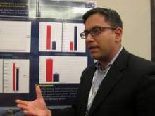
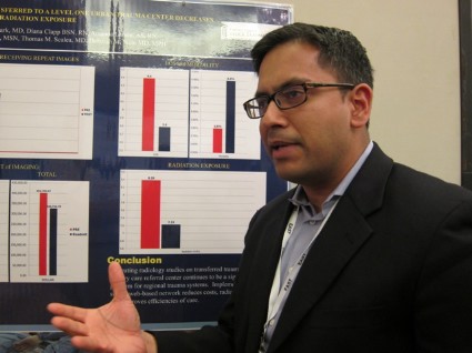
NAPLES, FLA. – Implementing a cloud-based network for sharing radiological images reduced radiation exposure and costs at a Level 1 tertiary care trauma center.
The number of trauma patients who underwent the exact same imaging study decreased significantly from 62% to 47% in the 6 months after the network was available (P less than .01).
The video associated with this article is no longer available on this site. Please view all of our videos on the MDedge YouTube channel
This resulted in a significant 19.2% decline in radiation exposure (mean 8.39 mSv vs. 7.23 mSv; P less than .01), Dr. Mayur Narayan reported at the annual meeting of the Eastern Association for the Surgery of Trauma.
"I think it’s a potential game changer," he said. "... The concept is important. The way we’re doing business currently is not sustainable."
Patients transferred within a regional trauma system to a tertiary care referral center frequently have prior radiology studies, but often undergo repeat imaging because of poor image quality, incompatible imaging software, or misplaced imaging discs. This can delay patient management, bring unwelcome radiation, and raise health care costs, explained Dr. Narayan, with the R Adams Cowley Shock Trauma Center at the University of Maryland Medical Center in Baltimore.
To reverse this trend, the center set up a secure, electronic (lifeIMAGE) network that uses a single platform for all medical image exchanges. Physicians can retrieve outside exams via the cloud or merge any or all picture archiving and communication system (PACS) data into their own PACS.
Ten hospitals are now on board and prospective data have been analyzed for 1,950 patients transferred to the trauma center between Jan. 1 and June 30, 2011, (pre-network) and between Jan. 1 and June 30, 2012, (post-network). About 8,500 patients are transferred to the trauma center annually from across the state. Patients in both time periods had similar demographics and Injury Severity Scores (mean 12).
In the 6 months after the network was implemented, the cost of imaging per patient dropped 18.7% ($413 vs. $333; P less than .01), while total imaging costs declined from $401,765 to $326,756 during the same period, Dr. Narayan said.
The most common repeat study in the analysis was an abdominal pelvic CT scan.
Inexplicably, hospital length of stay also declined from 4.4 days to 3.8 days (P = .07), he said. In-hospital mortality was unchanged (3.8% vs. 4.4%; P = .52).
The network cost Cowley Shock Trauma about $30,000 to set up, but other hospital groups are working to provide similar software for free, Dr. Narayan noted.
"The upfront costs should go away," he said.
During a discussion of the poster, one attendee said they set up a similar cloud-based system for their region, but no one used it. "I think it’s because small hospitals don’t see a lot of trauma. They have one or two patients maybe a month that would require transfer and with different people at the CT scanner every night, there’s no uniformity on how to do it," he said.
Others, however, noted that image sharing between hospitals has been fully integrated into their transfer algorithm, prompting calls between hospital radiology units, so that PAC files and patient record numbers have already been merged before the patient ever arrives in the trauma bay.
The Radiological Society of North America has also harnessed cloud-based computing to allow consumers to share their images and interpretations with any provider, regardless of institutional affiliation. According to 20-month follow-up data reported in late 2013, four out of five patients and 9 out of 10 physicians were satisfied with the RSNA Image Share network. A full 90% of patients were comfortable with the amount of privacy provided by the network, though some criticized the site for being "clunky" to navigate.
Dr. Narayan reported having no financial disclosures.
AT EAST 2014
Major finding: The number of patients who underwent repeat imaging declined from 62% to 47% in the 6 months after the network was implemented (P less than .01).
Data source: Prospective data review of 1,950 trauma patients.
Disclosures: Dr. Narayan reported having no financial disclosures.
Partial flail chest stabilization may suffice
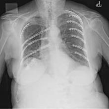
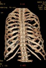
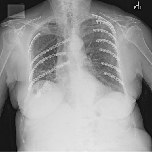
NAPLES, FLA. – Partial repair of flail segment rib fractures may suffice in patients whose anatomy precludes full stabilization, a small study suggests.
After a median follow-up of 189 days, only predicted total lung capacity at 3 months (90% vs. 72%; P = .02) and 6 months (94% vs. 75%; P = .038) significantly differed between patients undergoing complete flail stabilization (CFS) vs. partial flail stabilization (PFS).
No other differences in pulmonary and clinical outcomes were observed between the 43 patients, Dr. Terry P. Nickerson reported at the annual scientific assembly of the Eastern Association for the Surgery of Trauma.
Flail chest is defined by two or more consecutive ribs fractured in two places and a segment of chest wall unable to contribute to respiratory mechanics.
Benefits of surgical stabilization of flail chest reported in the heterogeneous literature include decreased ICU stay, lower rates of pneumonia, increased return to work, improved quality of life, and low surgical morbidity, Dr. Nickerson noted.
The retrospective analysis, included all patients (aged 30-85 years) who underwent surgical rib fracture stabilization for flail chest from August 2009 through February 2013 at the Mayo Clinic Hospital, St. Marys Campus, Rochester, Minn. In all, 23 patients had CFS, defined as all fractures involved in a flail segment undergoing full fixation, and 20 had PFS or at least one flail segment not completely repaired.
The CFS and PFS groups were similar with respect to median age (63 years vs. 58 years), sex (52% male vs. 65% male), operating time (186 minutes vs. 183 minutes), Injury Severity Score (20 vs. 17), hospital length of stay (10 days for both), ICU stay (1 day vs. 2 days), and narcotic use at 1-month follow-up (50% vs. 47%), according to the poster presentation.
At 6 months, there were no significant differences between the CFS and PFS groups in predicted vital capacity (86% vs. 76%), forced vital capacity (85% vs. 77%), forced expiratory volume in 1 second (71% vs. 78%), and FVC/FEV1 ratio (68% vs. 74%).
Rates of pneumonia were also similar in the two groups (21% vs. 20%; P = .39), reported Dr. Nickerson, a general surgeon in Rochester.
No patient or provider observed a significant chest wall deformity in either group during follow-up and no reoperations were required for incomplete repair in the PFS.
"Despite advances in surgical technique, not all flail segment rib fractures are accessible for repair via standard thoracotomy," the authors wrote. "Our data suggest that PFS is acceptable and that extending or creating additional incisions for exposure for CFS is unwarranted."
Additional research assessing late complications, performance/functional outcomes, and perceived benefits of the operation is needed to determine whether partial repair is truly comparable with full stabilization, coauthor Dr. Brian Kim, associate medical director Mayo Clinic Trauma Center, Rochester, said in an interview.
In their experience, 6 months’ postoperative follow-up has been sufficient with respect to bony healing, convalescence from pain, and monitoring of chest wall integrity.
He noted that some surgeons may be dissuaded from pursuing or recommending operative stabilization of a flail segment with fractures deemed outside the boundaries of repair, particularly if the injury is in the elderly or obese patient.
"The results of our study lend support to the practice of repairing the most anatomically disrupted and/or symptomatic fractures within the flail segment," he said. "Age and/or BMI [body mass index] are not absolute contraindications for flail segment stabilization in our practice."
Poster session moderator Dr. Alexander Eastman of University of Texas Southwestern Medical Center in Dallas, said the low patient numbers and short follow-up in the study make it difficult to draw big conclusions, other than the need for further research.
His group uses surgical fixation of flail chest for patients with instability and pain management for those patients who are difficult to wean from the ventilator.
"I think many surgeons still are skeptical of the data supporting fixation of flail chest," he noted in an interview.
Dr. Nickerson and his coauthors reported having no financial disclosures.
AT THE EAST SCIENTIFIC ASSEMBLY
Major finding: Only predicted total lung capacity at 3 months (90% vs. 72%; P = .02) and 6 months (94% vs. 75%; P = .038) significantly differed between CFS and PFS patients.
Data source: A retrospective study of 43 patients undergoing surgical rib fracture stabilization for flail chest.
Disclosures: Dr. Nickerson and his coauthors reported having no financial disclosures.
Regionalized trauma care boosts TBI survival
NAPLES, FLA. – A regional trauma system decreased hospital mortality for traumatic brain injury patients by 21% overall and by 26% for severe brain injuries, according to a partially retrospective study.
"Regionalization represents an additional step in attempting to improve outcomes for trauma patients. It can be defined as a tiered, integrated system that attempts to get the right patient to the right place at the right time," Dr. Michael L. Kelly said at the annual meeting of the Eastern Association for the Surgery of Trauma.
A few American studies and several more outside the United States have shown that regionalization decreases mortality in the general trauma population, but similar studies in traumatic brain injury (TBI) patients are scarce.
The Northern Ohio Trauma System (NOTS) was organized in 2010 and includes a transfer line to the Level I trauma center, a nontrauma hospital transfer protocol, a pilot scene triage protocol for emergency medical services, as well as the creation of a trauma-specific ICU in the level I center, explained Dr. Kelly, a sixth-year neurosurgery resident at the Cleveland Clinic, Ohio. The network includes the Level I MetroHealth Medical Center trauma center, two Level II trauma centers, and 12 nontrauma hospitals.
The three-tiered system mandates that TBI patients with a Glasgow Coma Scale (GCS) score of less than 12 and a traumatic mechanism, any penetrating head injury, or any open/depressed skull fracture, be sent to the Level I trauma center if they can be transferred within 15 minutes.
Patients with a GCS of 12-14 and a penetrating mechanism can be transferred to any trauma center, while those with lesser head injuries can remain at their hospital, unless their condition worsens.
For the study, Dr. Kelly and his coauthors analyzed data from 2008 through 2012 for 11,220 patients more than 14 years old with a TBI in the NOTS database, which was populated prospectively beginning in mid-2010.
Level I admissions increased significantly after NOTS by 10% for all TBIs (36% vs. 46%) and by 15% for severe TBIs with a head Abbreviated Injury Scale (AIS) score of 3 or more, he said. The percentage of patients who underwent transfers between NOTS institutions also increased significantly by 7% (7% vs. 14%) and 11% (10% vs. 21%), respectively.
Hospital mortality declined from 6.2% to 4.9% post-NOTS for all TBI patients (P = .005) and from 19% to 14% for the subset with severe TBI (P less than .0001). Mortality for trauma patients in general in Ohio has consistently hovered at 4% to 5% for the last decade, despite efforts to improve outcomes, including a 2002 law requiring the transfer of trauma patients to a validated trauma center, Dr. Kelly said.
In the post-NOTS period, craniotomies increased significantly for all TBIs (2% vs. 3%; P = .003) and for severe TBIs (6% vs. 8%; P = .02). The use of any neurosurgical procedure and hospital length of stay, however, remained constant for both groups in both time periods.
At baseline, the 6,713 post-NOTS patients were significantly older than the 4,507 pre-NOTS patients (55 years vs. 52 years) and less likely to be male (63% vs. 66%) or black (23% vs. 34%). GCS scores were similar (15 for both), as were Injury Severity Scores (14 for both) and the percentage of patients with a head AIS of 3 or more (34% post- vs. 32% pre-).
Multivariate regression analysis showed that the NOTS time period was an independent predictor of survival for all TBIs, with an odds ratio of 0.76, representing a 24% mortality reduction, and odds ratio of 0.72 for severe TBIs, representing a 28% mortality reduction.
"Of some importance, the multivariate model actually strengthened the effect of NOTS on mortality in our patient population," Dr. Kelly said.
Invited discussant Deborah Stein, medical director of neurotrauma critical care at the R Adams Cowley Shock Trauma Center at the University of Maryland Medical Center in Baltimore, described the study as an important contribution to the growing body of literature demonstrating that regionalization of care is associated with improved outcomes.
Dr. Stein went on to congratulate the NOTS members for participating in what was, at times, likely a contentious and difficult process: to bring a diverse group of hospitals to consensus about the best way to care for injured patients.
"Effecting change is difficult enough in a single division, department, or hospital," she remarked.
Following the formal presentation, audience members questioned whether the change in mortality was accomplished by simply shifting patients to nursing homes to die or whether it reflects a more aggressive surgical approach to TBI or improved critical care.
Dr. Kelly said that the creation of a trauma-specific ICU at MetroHealth, the uptick in transfers to the Level I trauma center, and the increased craniotomy rate all likely affected the outcome. Patient disposition data are still being analyzed, but hospice rates remained similar after NOTS was implemented, he said.
Dr. Kelly and his coauthors reported having no financial disclosures.
For nearly 50 years, data has been accumulating regarding the effects of regionalized trauma systems on postinjury outcomes, and the verdict is clear: Having a system in place that is prepared to rapidly transport the severely injured to a trauma center provides the best opportunity to reduce morbidity and mortality in these patients. While this is true in a general sense, a penetrating injury to the torso is not the same as a TBI, with each requiring different resources and expertise for optimal management.
The group at Cleveland MetroHealth in Ohio sought to answer the question of whether or not regionalization of trauma services would demonstrate a benefit in patients with TBI. Reviewing 4 years' worth of data that coincided at its midpoint with the implementation of the Northern Ohio Trauma System (NOTS), the authors demonstrate a significant reduction in mortality in brain-injured patients, including a 28% reduction in patients with severe TBI on multivariate analysis.

|
| Dr. Robert Winfield |
In the end, the commendation from Dr. Stein regarding the ability of the NOTS group, which comprises three hospitals from two health systems, to coalesce into a highly functioning regional trauma system is prescient and reflective of an impressive achievement for the authors. It is a reminder that in a time of scarce resources and competition for health care dollars, maintaining a focus on patient care through a cooperative and collaborative approach will ultimately yield the best results for all involved.
Dr. Robert Winfield is an ACS Fellow and the chair of the ACS Resident and Associate Society, and assistant professor of trauma and acute and critical care surgery at Washington University, St. Louis.
For nearly 50 years, data has been accumulating regarding the effects of regionalized trauma systems on postinjury outcomes, and the verdict is clear: Having a system in place that is prepared to rapidly transport the severely injured to a trauma center provides the best opportunity to reduce morbidity and mortality in these patients. While this is true in a general sense, a penetrating injury to the torso is not the same as a TBI, with each requiring different resources and expertise for optimal management.
The group at Cleveland MetroHealth in Ohio sought to answer the question of whether or not regionalization of trauma services would demonstrate a benefit in patients with TBI. Reviewing 4 years' worth of data that coincided at its midpoint with the implementation of the Northern Ohio Trauma System (NOTS), the authors demonstrate a significant reduction in mortality in brain-injured patients, including a 28% reduction in patients with severe TBI on multivariate analysis.

|
| Dr. Robert Winfield |
In the end, the commendation from Dr. Stein regarding the ability of the NOTS group, which comprises three hospitals from two health systems, to coalesce into a highly functioning regional trauma system is prescient and reflective of an impressive achievement for the authors. It is a reminder that in a time of scarce resources and competition for health care dollars, maintaining a focus on patient care through a cooperative and collaborative approach will ultimately yield the best results for all involved.
Dr. Robert Winfield is an ACS Fellow and the chair of the ACS Resident and Associate Society, and assistant professor of trauma and acute and critical care surgery at Washington University, St. Louis.
For nearly 50 years, data has been accumulating regarding the effects of regionalized trauma systems on postinjury outcomes, and the verdict is clear: Having a system in place that is prepared to rapidly transport the severely injured to a trauma center provides the best opportunity to reduce morbidity and mortality in these patients. While this is true in a general sense, a penetrating injury to the torso is not the same as a TBI, with each requiring different resources and expertise for optimal management.
The group at Cleveland MetroHealth in Ohio sought to answer the question of whether or not regionalization of trauma services would demonstrate a benefit in patients with TBI. Reviewing 4 years' worth of data that coincided at its midpoint with the implementation of the Northern Ohio Trauma System (NOTS), the authors demonstrate a significant reduction in mortality in brain-injured patients, including a 28% reduction in patients with severe TBI on multivariate analysis.

|
| Dr. Robert Winfield |
In the end, the commendation from Dr. Stein regarding the ability of the NOTS group, which comprises three hospitals from two health systems, to coalesce into a highly functioning regional trauma system is prescient and reflective of an impressive achievement for the authors. It is a reminder that in a time of scarce resources and competition for health care dollars, maintaining a focus on patient care through a cooperative and collaborative approach will ultimately yield the best results for all involved.
Dr. Robert Winfield is an ACS Fellow and the chair of the ACS Resident and Associate Society, and assistant professor of trauma and acute and critical care surgery at Washington University, St. Louis.
NAPLES, FLA. – A regional trauma system decreased hospital mortality for traumatic brain injury patients by 21% overall and by 26% for severe brain injuries, according to a partially retrospective study.
"Regionalization represents an additional step in attempting to improve outcomes for trauma patients. It can be defined as a tiered, integrated system that attempts to get the right patient to the right place at the right time," Dr. Michael L. Kelly said at the annual meeting of the Eastern Association for the Surgery of Trauma.
A few American studies and several more outside the United States have shown that regionalization decreases mortality in the general trauma population, but similar studies in traumatic brain injury (TBI) patients are scarce.
The Northern Ohio Trauma System (NOTS) was organized in 2010 and includes a transfer line to the Level I trauma center, a nontrauma hospital transfer protocol, a pilot scene triage protocol for emergency medical services, as well as the creation of a trauma-specific ICU in the level I center, explained Dr. Kelly, a sixth-year neurosurgery resident at the Cleveland Clinic, Ohio. The network includes the Level I MetroHealth Medical Center trauma center, two Level II trauma centers, and 12 nontrauma hospitals.
The three-tiered system mandates that TBI patients with a Glasgow Coma Scale (GCS) score of less than 12 and a traumatic mechanism, any penetrating head injury, or any open/depressed skull fracture, be sent to the Level I trauma center if they can be transferred within 15 minutes.
Patients with a GCS of 12-14 and a penetrating mechanism can be transferred to any trauma center, while those with lesser head injuries can remain at their hospital, unless their condition worsens.
For the study, Dr. Kelly and his coauthors analyzed data from 2008 through 2012 for 11,220 patients more than 14 years old with a TBI in the NOTS database, which was populated prospectively beginning in mid-2010.
Level I admissions increased significantly after NOTS by 10% for all TBIs (36% vs. 46%) and by 15% for severe TBIs with a head Abbreviated Injury Scale (AIS) score of 3 or more, he said. The percentage of patients who underwent transfers between NOTS institutions also increased significantly by 7% (7% vs. 14%) and 11% (10% vs. 21%), respectively.
Hospital mortality declined from 6.2% to 4.9% post-NOTS for all TBI patients (P = .005) and from 19% to 14% for the subset with severe TBI (P less than .0001). Mortality for trauma patients in general in Ohio has consistently hovered at 4% to 5% for the last decade, despite efforts to improve outcomes, including a 2002 law requiring the transfer of trauma patients to a validated trauma center, Dr. Kelly said.
In the post-NOTS period, craniotomies increased significantly for all TBIs (2% vs. 3%; P = .003) and for severe TBIs (6% vs. 8%; P = .02). The use of any neurosurgical procedure and hospital length of stay, however, remained constant for both groups in both time periods.
At baseline, the 6,713 post-NOTS patients were significantly older than the 4,507 pre-NOTS patients (55 years vs. 52 years) and less likely to be male (63% vs. 66%) or black (23% vs. 34%). GCS scores were similar (15 for both), as were Injury Severity Scores (14 for both) and the percentage of patients with a head AIS of 3 or more (34% post- vs. 32% pre-).
Multivariate regression analysis showed that the NOTS time period was an independent predictor of survival for all TBIs, with an odds ratio of 0.76, representing a 24% mortality reduction, and odds ratio of 0.72 for severe TBIs, representing a 28% mortality reduction.
"Of some importance, the multivariate model actually strengthened the effect of NOTS on mortality in our patient population," Dr. Kelly said.
Invited discussant Deborah Stein, medical director of neurotrauma critical care at the R Adams Cowley Shock Trauma Center at the University of Maryland Medical Center in Baltimore, described the study as an important contribution to the growing body of literature demonstrating that regionalization of care is associated with improved outcomes.
Dr. Stein went on to congratulate the NOTS members for participating in what was, at times, likely a contentious and difficult process: to bring a diverse group of hospitals to consensus about the best way to care for injured patients.
"Effecting change is difficult enough in a single division, department, or hospital," she remarked.
Following the formal presentation, audience members questioned whether the change in mortality was accomplished by simply shifting patients to nursing homes to die or whether it reflects a more aggressive surgical approach to TBI or improved critical care.
Dr. Kelly said that the creation of a trauma-specific ICU at MetroHealth, the uptick in transfers to the Level I trauma center, and the increased craniotomy rate all likely affected the outcome. Patient disposition data are still being analyzed, but hospice rates remained similar after NOTS was implemented, he said.
Dr. Kelly and his coauthors reported having no financial disclosures.
AT THE EAST SCIENTIFIC ASSEMBLY
Major finding: Hospital mortality was 6.2% pre-NOTS vs. 4.9% post-NOTS for all TBIs (P = .005) and 19% vs. 14% for severe TBIs (P less than .001).
Data source: A partially retrospective analysis of 11,220 TBI trauma patients.
Disclosures: Dr. Kelly and his coauthors reported having no financial disclosures.
